Article

New Biocides Based on N4-Alkylcytidines: Effects on Microorganisms and Application for the Protection of Cultural Heritage Objects of Painting

by
Liudmila A. Alexandrova
1,*,
Ivan A. Oskolsky
1,
Dmitry A. Makarov
1,
Maxim V. Jasko
1,
Inna L. Karpenko
1,
Olga V. Efremenkova
2,
Byazilya F. Vasilyeva
2,
Darya A. Avdanina
3,
Anna A. Ermolyuk
3,
Elizaveta E. Benko
3,
Stanislav G. Kalinin
3,
Tat’yana V. Kolganova
3,
Maria Ya. Berzina
4,
Irina D. Konstantinova
4,
Alexander O. Chizhov
5,
Sergey N. Kochetkov
1 and
Alexander A. Zhgun
3,*
1
Engelhardt Institute of Molecular Biology RAS, 32 Vavilov Str., Moscow 119991, Russia
2
Gause Institute of New Antibiotics, 11 Bol’shaya Pirogovskaya, Moscow 119021, Russia
3
Research Center of Biotechnology RAS, 33 Leninsky Ave, Moscow 119071, Russia
4
Shemyakin-Ovchinnikov Institute of Bioorganic Chemistry RAS, 16/10 Miklukho-Maklaya str., Moscow 117997, Russia
5
Zelinsky Institute of Organic Chemistry RAS 47 Leninsky Ave, Moscow 119991, Russia
*
Authors to whom correspondence should be addressed.
Int. J. Mol. Sci. 2024, 25(5), 3053; https://doi.org/10.3390/ijms25053053
Submission received: 15 February 2024 / Revised: 29 February 2024 / Accepted: 1 March 2024 / Published: 6 March 2024
(This article belongs to the Special Issue New Types of Antibacterial Biocides 2.0)

Abstract

:
The rapid increase in the antibiotic resistance of microorganisms, capable of causing diseases in humans as destroying cultural heritage sites, is a great challenge for modern science. In this regard, it is necessary to develop fundamentally novel and highly active compounds. In this study, a series of N4-alkylcytidines, including 5- and 6-methylcytidine derivatives, with extended alkyl substituents, were obtained in order to develop a new generation of antibacterial and antifungal biocides based on nucleoside derivatives. It has been shown that N4-alkyl 5- or 6-methylcytidines effectively inhibit the growth of molds, isolated from the paintings in the halls of the Ancient Russian Paintings of the State Tretyakov Gallery, Russia, Moscow. The novel compounds showed activity similar to antiseptics commonly used to protect works of art, such as benzalkonium chloride, to which a number of microorganisms have acquired resistance. It was also shown that the activity of N4-alkylcytidines is comparable to that of some antibiotics used in medicine to fight Gram-positive bacteria, including resistant strains of Staphylococcus aureus and Mycobacterium smegmatis. N4-dodecyl-5- and 6-methylcytidines turned out to be the best. This compound seems promising for expanding the palette of antiseptics used in painting, since quite often the destruction of painting materials is caused by joint fungi and bacteria infection.

1. Introduction

The discovery of antibiotics was a revolutionary event in the history of mankind, since it became possible to treat a significant number of diseases and save human lives in previously hopeless situations [1,2,3]. However, the widespread introduction of antibiotics into medical practice since the 1950s, at the beginning of the period called the Golden Age of Antibiotics, led to the development of resistance in numerous pathogenic strains against antibiotics of various classes [4,5,6]. This led to the end of the Golden Age of Antibiotics in the early 1970s, and mankind again faced the urgent task of searching for new and improved antimicrobial drugs [7,8,9,10,11,12,13].
Microorganisms can not only be a cause of infectious diseases but also of the destruction of objects of cultural heritage [14,15]. Microorganisms from various systematic groups (especially filamentous fungi) capable of damaging works of art, e.g., tempera painting and oil painting on canvas, have been extensively studied in recent years [16,17,18,19]. There are quite a lot of compounds of various classes used to protect cultural heritage sites, but the number of antiseptics used in painting is extremely limited and has decreased significantly in recent years [20]. Moreover, the compounds used to protect paintings, such as benzalkonium chloride (BAC), do not affect the entire range of microorganisms that destroy painting materials [21]. Moreover, the widespread use of BAC leads to the emergence of resistance in microorganisms against these antiseptics [22].
Therefore, the creation and/or identification of fundamentally new compounds that act on new targets of pathogens and are active against resistant strains of microorganisms is one of the most important problems facing researchers [8,23,24].
Derivatives of natural nucleosides are one of the promising classes of organic compounds, among which prototypes of new drugs are searched. Compounds obtained as a result of the modification of nucleosides are actively studied as drugs to combat viral diseases and bacterial infections, as well as some types of cancer. Currently, nucleoside analogs and derivatives are important elements of antitumor and antiviral therapy and can also be used as antifungal agents [25,26,27,28,29].
Previously, we discovered the antibacterial and antifungal activity of a number of modified pyrimidine N4-alkyl-2′-deoxynucleosides [30,31], containing extended alkyl substituents at the C4 position of the cytosine residue (Figure 1).
These compounds turned out to be active against a number of drug-resistant strains of Gram-positive bacteria, as well as filamentous fungi, while the activity of N4-derivatives of 5-methyl-2′-deoxycytidine (1a,b) noticeably exceeded the activity of C4-modified 2′-deoxycytidines (1c,d) with the same substituent at C4 and was comparable to currently used antibiotics [30]. Ribonucleoside derivatives have not previously been studied in detail, although in the case of decyl derivatives of uridine, we have previously shown that they have better solubility in aqueous media (and, therefore, bioavailability) compared to similar 2′-deoxy analogs and can exhibit pronounced antibacterial activity.
Since the presence of a methyl group fundamentally affects the activity of these compounds, in order to further study the effect of structure on biological activity, we synthesized N4-cytidine derivatives (2ae, 3ac), both containing a methyl group in the fifth or sixth position of the cytosine residue or not. We hypothesized that nucleoside derivatives containing a substituent at the C6 position of the pyrimidine base may exhibit unusual properties and are also promising objects for research, since they exist in the syn-conformation even in an aqueous environment (unlike most pyrimidine nucleosides) [32,33,34,35]. It was previously shown that 6-modified uridine derivatives can exhibit significant anti-tuberculosis activity [36] and also act as potential antimetabolites; for example, 6-thiocarboxamide-UMP, a structural analog of orotidine-5′-phosphate (OMP), is a potent inhibitor of OMP decarboxylase [37].

2. Results

2.1. Chemistry

The classical method of modification at the C4 position of pyrimidine nucleosides is the introduction of a good leaving group followed by its replacement with suitable nucleophiles [38,39]. As a rule, derivatives of pyrimidine nucleosides used for this purpose are contained at C4 thio-, thiomethyl-, chloro-, 1,2,4-triazol-1-yl-, 1,2,3,4-tetrazol-1-yl-, arylsulfonyl groups, and a number of others.
To synthesize the target compounds, we decided to use the convenient one-pot method we had previously used for the preparation of N4-alkyl-2′-deoxynucleosides [30], based on the Diwakar and Reese procedure [40] in its later modification [41], namely, the condensation of nucleosides protected by acetyl groups with 1,2,4-triazole and 2-chlorophenyldichlorophosphate in pyridine followed by a reaction with the corresponding 1-alkylamine and final deblocking.
It was in this way that we synthesized N4-alkylamino-5-methylcytidines (2ac) and N4-alkylaminocytidines (2d,e) in good yields (Scheme 1) starting from 2′,3′,5′-tri-O-acetyluridine (5a) and 2′,3′,5′-tri-O-acetyl-5-methyluridine (5b).
The first stage of the synthesis of 6-methyluridine derivatives was the preparation of 2′,3′,5′-tri-O-acetyl-6-methyluridine (7) by N-glycosylation of silylated 6-methyluracil (6) 1′,2′,3′,5′-tetra-O-acetylribose according to the Vorbruggen method, using trimethylsilyl trifluoromethanesulfonate as a catalyst [36,42] (Scheme 2).
Both dichloroethane and acetonitrile were tried as solvents for these reactions. When N-glycosylation is run in acetonitrile, the yield of compound (8) is higher than when using dichloroethane. Moreover, in dichloroethane, there is a significant formation of the N3-isomer of protected 6-methyluridine in an almost equal ratio with the N1-isomer, while in acetonitrile the formation of the N3-isomer was practically not observed. As a result, 2′,3′,5′-tri-O-acetyl-6-methyluridine (8) was synthesized with a 67% yield.
The second stage of the synthesis was the replacement of oxygen at the C4 position with a good leaving group. First of all, we tried the same approach that we used earlier [30], namely, the replacement of the oxygen atom with a 1,2,4-triazolyl group in the presence of 2-chlorophenyldichlorophosphate in pyridine (Scheme 2), but it was not possible to obtain a triazolyl derivative. Apparently, the 6-methyl group in the meta position with respect to the 4-carbonyl group leads to a reduced reactivity of the latter.
Next, we synthesized 2′,3′,5′-tri-O-acetyl-6-methyl-4-thiouridine (10) using Lawesson’s reagent [43] by boiling compound (8) in dioxane under argon atmosphere for 4 h (Scheme 2) with a yield of 50%; however, attempts to replace the thio group with the corresponding alkylamine led to compounds (3ac) with unsatisfactory yields. N4-Alkyl derivatives of 6-methylcytidine (3ac) were synthesized starting from N4-alkyl-6-methylcytosines (12ac) in accordance with Scheme 3. The first stage of the synthesis was the preparation of 6-methyl-4-thiouracil (11) by replacing the oxygen atom in the C4 position of 6-methyluracil (7) with a sulfur atom using Lawesson’s reagent when heated in a mixture of pyridine and dioxane (Scheme 3) for 4 h at boiling. Next, N4-alkyl-6-methylcytosines (13) were synthesized by analogy with the known procedure for the preparation of 5-aminouracils [44,45] (Scheme 3). Base (11) and the corresponding 1-alkylamine were refluxed in ethylene glycol in the presence of 7-methylquinoline for 1 h. The product was isolated by precipitating it from the reaction mixture with water and then washing the resulting precipitate with ethyl acetate.
The next stage of the synthesis is the N-glycosylation of the resulting N4-alkyl-6-methylcytosines (12ac) with 1′,2′,3′,5′-tetra-O-acetylribose (Scheme 3) according to the Vorbruggen method [42]. The condensation of silylated N4-alkyl-6-methylcytosines (12ac) with 1′,2′,3′,5′-tetra-O-acetylribose in acetonitrile was carried out catalyzed by trimethylsilyl trifluoromethane sulfonate. When heated at a temperature of 82 °C, the reaction took 3.5 h and was stopped after the complete consumption of sugar; however, better yields (65–80%) were achieved when the reaction was carried out at 37 °C for several days. To obtain the target nucleoside derivatives (3ac), protected N4-alkyl-6-methylcytidines (13ac) were deblocked with aqueous 25% ammonia in ethanol (Scheme 3) for 24 h at 20 °C and isolated by column chromatography.
The purity and structure of the target compounds were confirmed by 1H- and 13C-NMR spectroscopy and high-resolution mass spectrometry.

2.2. Enzymology

Due to the complexity of the synthesis of 6-methylcytidine derivatives, it seemed appropriate to us to study the possibility of the enzymatic synthesis of nucleosides using nucleoside phosphorylases (NPs). The use of (NPs) in the enzymatic synthesis of nucleosides is well known and has recently received considerable attention. NP-catalyzed nucleoside transglycosylation uses a readily available nucleoside such as natural uridine or thymidine as a starting compound [46,47].
It was shown previously that a number of uracil derivatives with 6-CH2OH and 6-CH2F substituents are substrates of Escherichia coli pyrimidine nucleoside phosphorylase [33]; however, 6-methyluracil was not a substrate of uridine phosphorylase from E. coli [46]. 6-Methyluridine is slowly converted into 6-methyluracil, and the substrate binding is significantly weaker compared to the 5-methyl derivative [46].
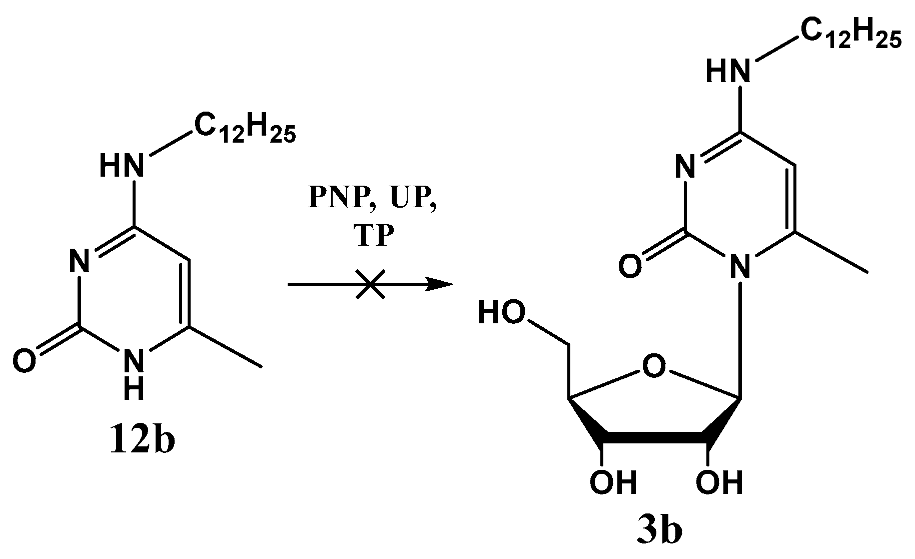
We studied the enzymatic glycosylation of N4-dodecyl-6-methylcytosine (12b), catalyzed by purine nucleoside phosphorylase (PNP), uridine phosphorylase (UP), and thymidine phosphorylase (TP) (Scheme 4).
Unfortunately, N4-dodecyl-6-methylcytosine (12b) was not a substrate for any of the enzymes used. (See Supplementary Materials Figure S1).

2.3. Cytotoxicity

The cytotoxicity of the synthesized compounds (CD50) was estimated by an MTT assay [48] in HeLa and Vero E6 cell lines. The compounds 2ae and 3ac demonstrated cytotoxic activity at concentrations of 25–60 μM.

2.4. In Vitro Study of Antibacterial Activity of the Obtained Compounds

In Vitro Study of Antibacterial Activity of the Obtained Compounds

The antibacterial effect of the obtained compounds was studied by their ability to inhibit in vitro the growth of Gram-positive and Gram-negative bacterial strains, listed in the Methods. Antimicrobial activity was observed only against Gram-positive bacteria, including methicillin-resistant staphylococcus and two drug-resistant strains of mycobacteria (Table S1, Supplementary Materials). A previously developed method was used [49,50].
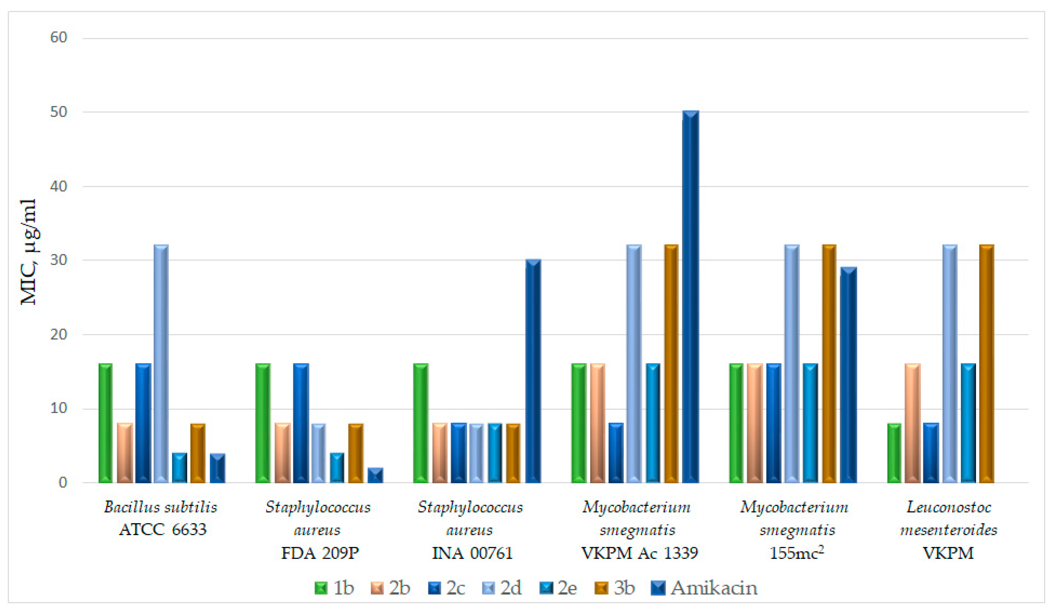
Figure 2 schematically shows the inhibitory effect of the most active compounds and the antibiotic amikacin against a number of Gram-positive bacteria.
Among the N4-alkylcytidine derivatives studied, N4-decylcytidine 2e has the lowest MIC value in the range from 4 to 16 μg/mL. The MIC of both N4-dodecyl derivatives 2b and 3b against five Gram-positive bacteria is 8 μg/mL, with the exception of the MIC of 3b against M. luteus NCTC 8340 (32 μg/mL); the MIC against the two strains of Myc. smegmatis is slightly higher and amounts to 16 and 32 μg/mL, respectively. The 2′-deoxy derivative 1b showed a uniform MIC value against all seven Gram-positive bacteria. The antibacterial effect of compound 2e and N4-dodecyl derivatives 1b, 2b, and 3b is comparable to the effect of a number of antibiotics used in medical practice. Compounds 3a and 3c with decyl and tetradecyl substituents and cytosine derivatives 12ac did not demonstrate antibacterial activity in the studied concentration range.
We have shown that the effects of N4-alkyl derivatives of 5- and 6-methylcytidine are different. While N4-dodecyl-6-methylcytidine (3b) exhibits bactericidal activity with an MIC value of 8 μg/mL against Gram-positive bacteria and an MIC of 32 μg/mL against two strains of mycobacteria, Myc. smegmatis VKPM Ac 1339 and Myc. smegmatis mc2 155, both N4-dodecyl-5-methylcytidine (2b) and its 2′-deoxy analog (1b) also have an MIC of 8–16 μg/mL against all of the bacteria listed above; however, against two strains of mycobacteria, the activity exhibited is bacteriostatic in nature since, despite the absence of mycobacterial growth for 5 days, upon subsequent reseeding of the bacteria on fresh medium without these compounds, growth resumes.

2.5. In Vitro Study of the Antifungal Activity of the Obtained Compounds

Previously, we have shown that 4-modified derivatives of 5-methyl-2′-deoxycytidine with extended alkyl substituents exhibit high inhibitory activity against filamentous fungi, which cause the biodegradation of organic materials used in tempera painting of the 15th–16th centuries [30,31]. The antifungal activity of the obtained nucleoside derivatives, as compared with the previously obtained N4-dodecyl-5-methyl-2′-deoxycytidine 1b [30,31], was studied against 12 filamentous fungi, belonging to the types Ascomycota and Mucoromycota, isolated in the halls of the Ancient Russian Paintings of the State Tretyakov Gallery and capable of the biodegradation of ancient Russian icons [15,20,51].
Previous experiments showed that, among N4-alkyl-5-methyl-2′-deoxycytidines in the C8-C10-C12 alkyl series, the dodecyl derivative has the greatest antifungal activity (1b) [30]. Then we demonstrated that this activity could be somewhat enhanced by replacing the hydroxyl group at the 3′ position of deoxyribose with an amino group or methyl-, ethyl-, or dimethyl-amino groups [31]. However, a number of other modifications of the side radicals of the molecule lead to an almost complete loss of antimycotic activity [52]. In particular, we have identified the dependence of the antifungal activity of alkyl derivatives of pyrimidine nucleosides on the position of the long alkyl substituent in the nitrogenous base residue [53]. It appeared that changing the position from N-4 to C-5 to introduce an extended alkyl substituent, leads to a complete loss of antifungal activity.

2.5.1. The Role of the Hydroxyl Group in the 2′-Position of Pentose

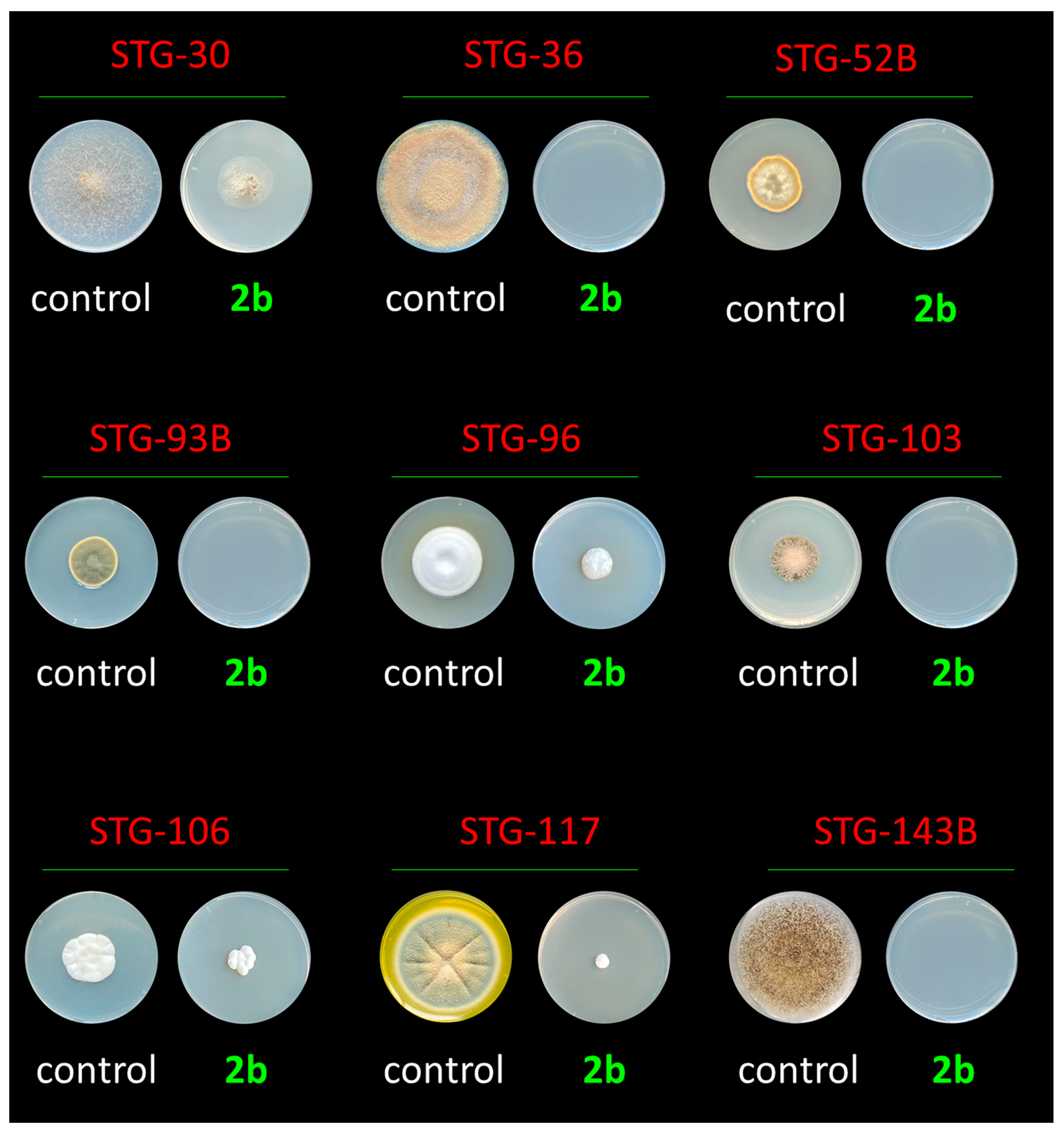
In this work, we showed that the addition of an OH- group to the 2′-position of deoxyribose does not affect the antimycotic activity. Thus, ribo-derivative 2b (N4-dodecyl-5-methyl-cytidine) has similar activity as its 2′-deoxy analog 1b (Figure 3 and Figure 4). It turned out that the addition of 1b and 2b at both tested concentrations, 200 and 1000 μM, led to almost the same inhibition dynamics profile in all tested strains (Figure 4). The exceptions were strain STG-25G, against which 2b was more active after 15 days, and STG-96, against which 1b was more active after the same period of time. However, the differences were insignificant, which indicates a fundamentally similar effect of 1b and 2b on test cultures of fungi-destructors.

2.5.2. The Role of the Methyl Group in the Fifth or Sixth Position of Cytidine Derivatives

The results obtained, i.e., that the replacement of 2′-deoxyribose with ribose in the sugar residue of the nucleoside does not significantly affect the antimycotic activity, allowed us to study the role of the methyl group and the length of the alkyl radical in the cytosine residue also in ribo-derivatives of cytidines. As a result, we demonstrated for the first time that the removal of the methyl group at the fifth position leads to a complete decrease in the activity of compounds of this class. Moreover, a complete loss of activity in the tested concentration range (200–1000 μM) was observed in both 2d (N4-dodecylcytidine) and 2e (N4-tetradecylcytidine) (Figure 4). The replacing of the methyl group from the fifth to the sixth position resulted in a significant loss of activity; compounds 3b (N4-dodecyl-6-methylcytidine) and 3c (N4-tetradecyl-6-methylcytidine) turned out to be 3–8 times less active than 2b (N4-dodecyl-5-methylcytidine).

2.5.3. The Role of the Size of Alkyl Group in the N4 Position of Cytidine Derivatives

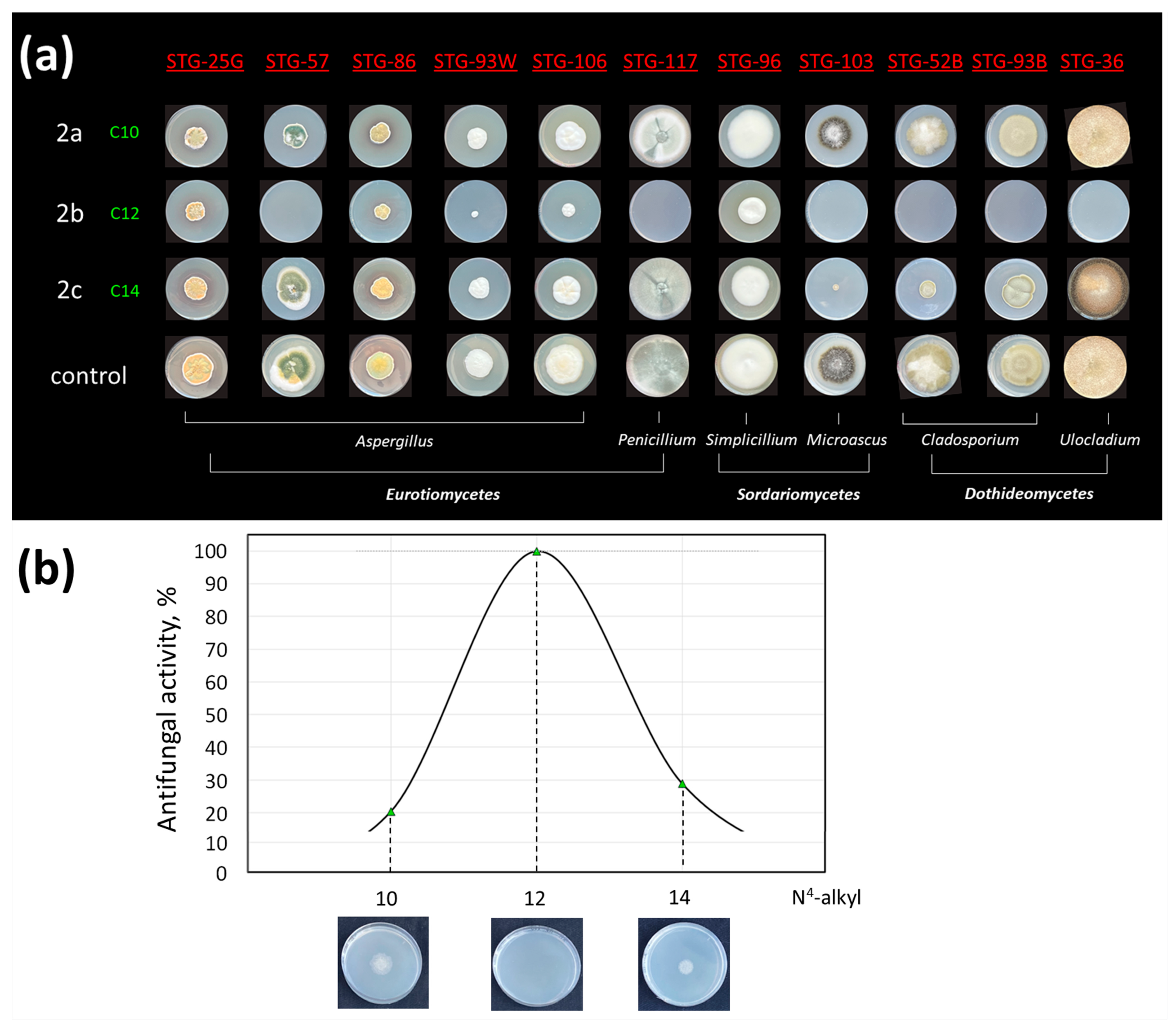
For N4-alkyl-5-methylcytidines in the series C10-C12-C14, a clear bell-shaped dependence of activity on the size of the alkyl substituent at N4 was observed (Figure 5). For all strains, the most active compound was the N4-dodecyl derivative. The activity dropped significantly both when the alkyl radical increased to C14 (about three times) and when it decreased to C10 (about five times), Figure 5.
In the series N4-alkyl-6-methylcytidines C12–C14, cross-activity was observed; for 25% of the studied fungal strains, the C12 derivative 3b was more active, for 42%, the C14 derivative 3c was more active, and in 33% of cases, both compounds showed similar activity. It is possible that this cross-effect is due to differences in resistance mechanisms in different fungal strains, on which C12 and C14 derivatives of 6-methylcytidines have different effects. This cross-activity suggests that the optimal broad-spectrum antiseptic based on 6-methylcytidines derivatives should presumably consist of a cocktail of N4-dodecyl- and N4-tetradecylcytidines. However, the movement of the methyl group to the fifth position of cytidine makes the compound not only more active but also universal in the size of the alkyl radical: only C12 derivatives show the best activity against all test cultures; making a cocktail with C10 or C14 derivatives to expand the spectrum of action does not seem promising.

3. Discussion

For further research into microbial inhibitors, rational methods for the synthesis of N4-alkyl derivatives of cytidines, 5- and 6-methylcytidines, were developed. In contrast to the simple one-pot synthesis of N4-alkyl 5-methylcytidines (2ac) and cytidines (2d,e), N4-alkyl-6-methylcydine (3ac) was obtained only by N-glycosylation of the corresponding N4-alkyl-6-methylcytosines (12ac).
It was shown that N4-tetradecyl-6-methylcytosines (12c) are not substrates of the three nucleoside phosphorylases usually used for the enzymatic synthesis of nucleosides.
The antibacterial effect of the obtained compounds was studied by their ability to inhibit in vitro the growth of a number of microorganisms: seven strains of Gram-positive bacteria, including drug-resistant strains of Myc. smegmatis and S. aureus, and two strains of Gram-negative bacteria. N4-Alkyl-6-methylcytosines (12ac) did not demonstrate inhibitory properties. The synthesized N4-alkyl derivatives of nucleosides (2ae) effectively inhibited the growth of Gram-positive bacteria but did not affect Gram-negative bacteria. The antibacterial effect of N4-decylcytidine 2e and N4-dodecyl derivatives, both 2′-deoxy-5-methylcytidine 1b, and 5- or 6-methylcytidine (2b or 3b) is comparable to the effect of a number of antibiotics used in medical practice. Compounds 3a, 3c with decyl and tetradecyl substituents, and cytosine derivatives 12ac did not demonstrate antibacterial activity in the studied concentration range.
As expected, the effects of N4-alkyl derivatives of 5- and 6-methylcytidine are different. While N4-dodecyl-6-methylcytidine (3b) exhibits bactericidal activity against two strains of mycobacteria, Myc. smegmatis VKPM Ac 1339 and Myc. smegmatis mc2 155, both N4-dodecyl-5-methylcytidine (2b) and its 2′-deoxy analog (1b) are also active against all of the bacteria listed above; however, against two strains of mycobacteria, the activity exhibited is bacteriostatic in nature.
During antifungal experiments, several patterns were established. (i) The presence of a methyl group at the fifth position of N4-alkyl-cytidines is critical for antifungal activity (Figure 4). The removal of the methyl group or its replacement to the sixth position results in either a complete or significant loss of activity (3–8 times), respectively. (ii) The presence of a hydroxyl group in the 2′ position does not significantly affect the activity of compounds of this class. (iii) Among the group of N4-alkyl-5-methylcytidines, which turned out to be the most active compounds against the tested molds, in the C10-C12-C14 series, the N4-dodecyl derivative works best. Reducing or increasing the length of the alkyl substituent leads to a loss of activity by 3–5 times. Less active N4-alkyl-6-methylcytidines have cross-activity, some fungal strains are more sensitive to C12, others to C14, and there are also strains on which both compounds act equally.
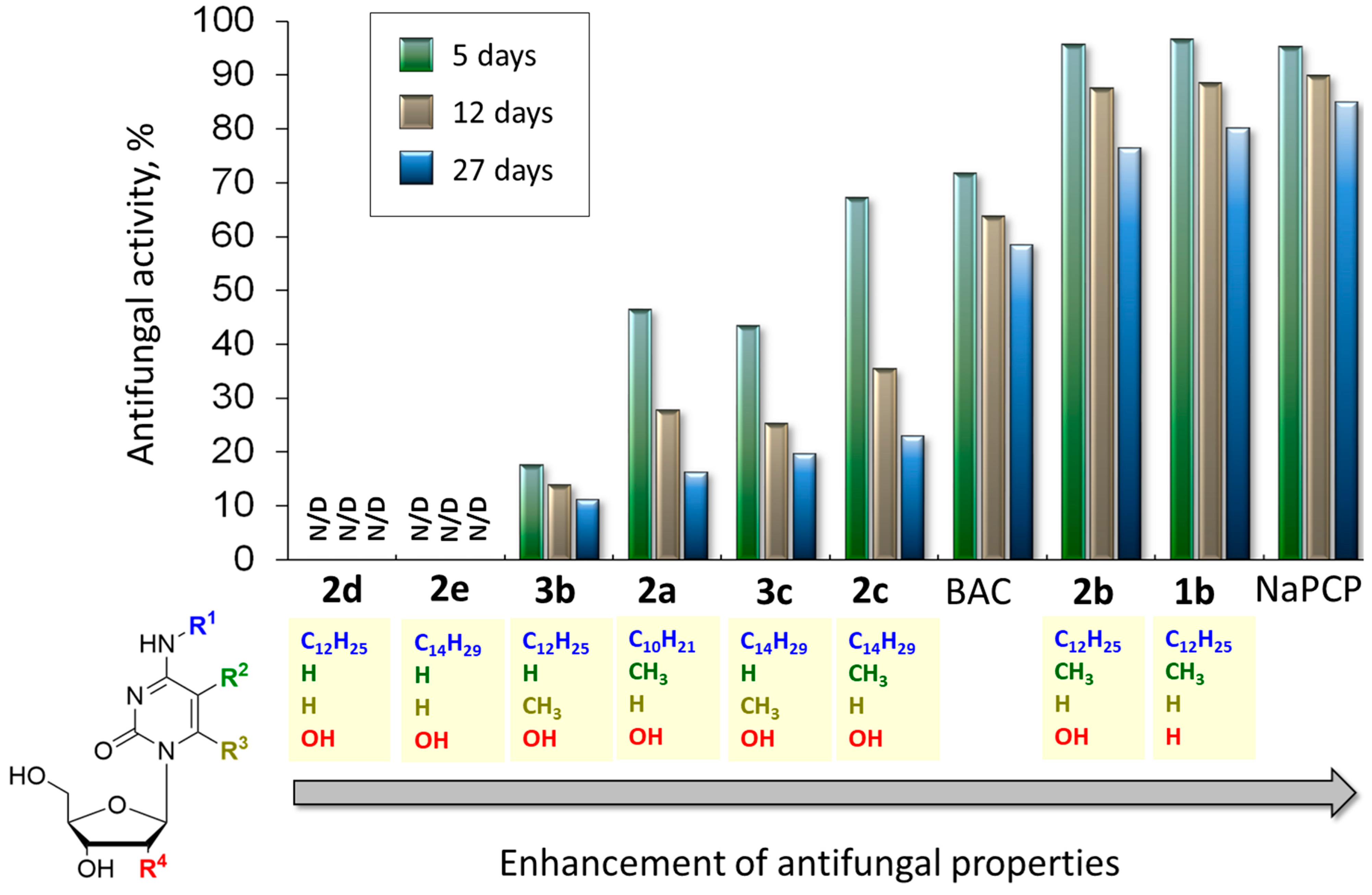
To evaluate the possible application of alkylnucleosides for the protection of paintings, the activity of standard antiseptics used for biodamaged paintings, such as BAC and NaPCP, was also studied. It turned out that NaPCP exhibits the best activity among the studied drugs, but currently, it is in most cases withdrawn from restoration practice due to its high toxicity to humans [54] (Figure 6).
Antiseptic BAC, actively used to protect paintings, works generally worse than 1b and 2b and slightly better than 2a,c and 3b,c. It is also necessary to take into account that BAC is a cocktail of quaternary amines, in which one of the substituents is an alkyl with a variable value from C8 to C18. In this case, a wide spectrum of action is achieved due to the fact that different groups of microorganisms are most sensitive to compounds with different lengths of alkyl radical. Thus, the demonstrated resistance of a number of strains to it, such as STG-25G, STG-30, STG-52B, STG-57, STG-93W, and STG-96, can no longer be controlled by varying the size of this alkyl radical. On the other hand, various modifications in alkyl nucleosides can lead to cross-activity, for example, replacing the hydroxyl group in the 3′-position of a sugar residue with an amino group leads to additional activity against Aspergillus [31]. From this point of view, cocktails based on alkyl nucleosides may potentially have an even wider spectrum of activity than BAC.
Since some of the studied compounds exhibit high activity (at the level of applied antiseptics) against a specific group of microorganisms that destroy paintings, these alkylcytidines can potentially be used as targeted antiseptics. At the next stage of research, it will be necessary to study both the antimicrobial activity of these new compounds in the composition of painting materials and the level of impact on their spectral and surface properties.

4. Materials and Methods

4.1. General Information

Commercial reagents from Fluka (Buchs, Switzerland), Sigma-Aldrich (St. Louis, MO, USA), and Acros Organics (Geel, Belgium) were used in this work. The commercial antiseptics used to protect paintings are as follows: Benzalkonium chloride (BAC, commercial name Katamin AB) from Neochemax (Domodedovo, Russia); sodium pentachlorophenolate (NaPCP) from IndiaMART (Noida, India).
Solvents were purified using standard methods. Column chromatography was performed using Kieselgel 60 (40–63 μm) silica gel (Merck, Darmstadt, Germany). 1H and 13C NMR spectra were recorded with a Bruker (Bremen, Germany) AM300 at ambient temperature in DMSO-d6 and CDCl3 solutions. Chemical shift values are given in δ scale relative to Me4Si. The J values are given in hertz. UV spectra were recorded on a Perkin Elmer lambda 25 spectrophotometer (Perkin Elmer, Shelton, CT, USA) in methanol. HR-ESI-MS were measured on a Bruker Daltonics micrOTOF II instrument (Bruker Daltonik GmbH, Bremen, Germany). All reactions were monitored with thin-layer chromatography (TLC) and carried out with Merck (Darmstadt, Germany) precoated plates DC-AlufolienKieselgel60 F254.
N4-Dodecyl-5-methyl-2′-deoxycytidine 1b was obtained using the method found in [30].

General Method for the Synthesis of Compounds 2ae

2-Chlorophenyl dichlorophosphate (0.255 g, 0.173 mL, 1.05 mmol) was added to a solution of acetyl-protected uridine (5a) or 5-methyluridine (5b) (0.5 mmol) and 1,2,4-triazole (0.2 g, 3 mmol) in anhydrous pyridine, cooled to 0 °C. The mixture was stirred for 20 h at room temperature and then evaporated. The residue was partitioned between chloroform and 0.5 M sodium bicarbonate; the chloroform layer was washed with water, dried over Na2SO4, evaporated, and dissolved in anhydrous dioxane (3 mL). The corresponding 1-alkylamine (0.6 mmol) and diisopropylethylamine (75 mg, 0.1 mL, 0.6 mmol) were added to a solution and cooled to 0 °C. The mixture was stirred for 20 h at room temperature, then 3 mL of conc. aq. ammonia solution was added, and the mixture was stirred for 40 h at room temperature and then evaporated; the compounds were purified on a column of silica gel (2 × 15 cm) in chloroform or ethyl acetate eluted with a gradient of ethanol in chloroform (0–15%) or in ethyl acetate (0–10%), respectively. The target fractions were evaporated in a vacuum to give the expected compound yields as colorless amorphous mass with 60–85% yields.
N4-Decyl-5-methylcytidine (2a). Prepared according to the general procedure from 5b (0.192 mg) and 1-decylamine (0.079 mg). Yield 0.150 g (76%). UV: λmax = 272 nm. 1H NMR (300 MHz, DMSO-d6) δ 7.64 (q, J = 1.2 Hz, 1H, 6-H), 7.12 (t, J = 5.6 Hz, 1H, 4-NH), 5.77 (d, J = 3.8 Hz, 1H, 1′-H), 5.23 (d, J = 4.7 Hz, 1H, 2′-OH), 5.06 (t, J = 5.2 Hz, 1H, 5′-OH), 4.94 (d, J = 5.0 Hz, 1H, 3′-OH), 3.91–4.02 (m, 2H, 2′-H + 3′-H), 3.81 (dt, J = 3.6, 3.5 Hz, 1H, 4′-H), 3.67 (ddd, J = 12.0, 5.0, 3.2 Hz, 1H, 5′-Ha), 3.55 (ddd, J = 12.1, 5.3, 3.4 Hz, 1H, 5′-Hb), 3.33–3.23 (m, 2H, NH-CH2-), 1.84 (d, J = 1.0 Hz, 3H, 5-CH3), 1.46–1.57 (m, 2H, NH-CH2-CH2-), 1.19–1.37 (m, 14H, NH-(CH2)2-(CH2)7-), 0.85 (t, J = 6.9 Hz, 3H, -CH2-CH3). 13C NMR (75 MHz, DMSO-d6) δ 162.66 (4-C), 155.44 (2-C), 137.73 (6-C), 101.54 (5-C), 88.99 (1′-C), 84.08 (4′-C), 73.78 (2′-C), 69.48 (3′-C), 60.72 (5′-C), 40.24 (NH-CH2), 31.29, 28.96, 28.66, 26.49, 22.08, 13.92, 13.06 (NH-CH2-(CH2)8-CH3 + 5-CH3). HRMS (ESI) of C20H35N3O5, m/z: calcd [M + H]+ 398.2649, found: 398.2645.
N4-Dodecyl-5-methylcytidine (2b). Prepared according to the general procedure from 5b (0.192 g) and 1-dodecylamine (0.111 g). Yield 0.165 g (78%). UV: λmax = 272 nm. 1H NMR (300 MHz, DMSO-d6) δ 7.63 (q, J = 1.2 Hz, 1H, 6-H), 7.11 (t, J = 5.6 Hz, 1H, 4-NH), 5.77 (d, J = 3.8 Hz, 1H, 1′-H), 5.21 (d, J = 4.7 Hz, 1H, 2′-OH), 5.05 (t, J = 5.2 Hz, 1H, 5′-OH), 4.93 (d, J = 4.9 Hz, 1H, 3′-OH), 3.90–4.00 (m, 2H, 2′-H + 3′-H), 3.81 (ddd, J = 3.5, 3.4, 3.2 Hz, 1H, 4′-H), 3.66 (ddd, J = 12.0, 5.2, 3.2 Hz, 1H, 5′-Ha), 3.54 (ddd, J = 12.0, 5.4, 3.5 Hz, 1H, 5′-Hb), 3.24–3.32 (m, 2H, NH-CH2-), 1.83 (d, J = 1.0 Hz, 3H, 5-CH3), 1.43–1.57 (m, 2H, NH-CH2-CH2-), 1.20–1.32 (m, 18H, NH-(CH2)2-(CH2)9-), 0.85 (t, J = 6.9 Hz, 3H, -CH2-CH3). 13C NMR (75 MHz, DMSO-d6) δ 162.65 (4-C), 155.39 (2-C), 137.73 (6-C), 101.48 (5-C), 88.96 (1′-C), 84.07 (4′-C), 73.74 (2′-C), 69.48 (3′-C), 60.73 (5′-C), 40.22 (NH-CH2), 31.28, 28.97, 28.65, 26.49, 22.08, 13.92, 13.06 (NH-CH2-(CH2)10-CH3 + 5-CH3). HRMS (ESI) of C22H39N3O5, m/z: calcd [M + H]+ 426.2962, found: 426.2951.
N4-Tetradecyl-5-methylcytidine (2c). Prepared according to the general procedure from 5b (0.192 g) and 1-tetradecylamine (0.128 g). Yield 0.177 g (78%). UV: λmax = 272 nm. 1H NMR (300 MHz, DMSO-d6) δ 7.63 (q, J = 1.2 Hz, 1H, 6-H), 7.11 (t, J = 5.7 Hz, 1H, 4-NH), 5.77 (d, J = 3.8 Hz, 1H, 1′-H), 5.21 (d, J = 5.3 Hz, 1H, 2′-OH), 5.05 (t, J = 5.2 Hz, 1H, 5′-OH), 4.93 (d, J = 5.16 Hz, 1H, 3′-OH), 3.91–4.00 (m, 2H, 2′-H + 3′-H), 3.81 (dt, J = 3.7, 3.5, 3.5 Hz, 1H, 4′-H), 3.67 (ddd, J = 12.0, 5.1, 3.2 Hz, 1H, 5′-Ha), 3.54 (ddd, J = 12.0, 5.3, 3.5 Hz, 1H, 5′-Hb), 3.24–3.32 (m, 2H, NH-CH2-), 1.84 (d, J = 1.0 Hz, 3H, 5-CH3), 1.44–1.59 (m, 2H, NH-CH2-CH2-), 1.20–1.33 (m, 22H, NH-(CH2)2-(CH2)11-), 0.86 (t, J = 6.9 Hz, 3H, -CH2-CH3). 13C NMR (75 MHz, DMSO-d6) δ 162.65 (4-C), 155.38 (2-C), 137.72 (6-C), 101.47 (5-C), 88.97 (1′-C), 84.06 (4′-C), 73.74 (2′-C), 69.46 (3′-C), 60.71 (5′-C), 40.21 (NH-CH2), 31.28, 29.02, 28.65, 26.50, 22.07, 13.90, 13.04 (NH-CH2-(CH2)12-CH3 + 5-CH3). HRMS (ESI) of C24H43N3O5, m/z: calcd [M + H]+ 454.3275, found: 454.3281.
N4-Dodecylcytidine (2d). Prepared according to the general procedure from 5a (0.185 g) and 1-dodecylamine (0.111 g). Yield 0.138 g (67%). UV: λmax = 272 nm. 1H NMR (300 MHz, DMSO-d6) δ 7.63 (q, J = 1.2 Hz, 1H, 6-H), 7.11 (t, J = 5.7 Hz, 1H, 4-NH), 5.77 (d, J = 3.8 Hz, 1H, 1′-H), 5.21 (d, J = 5.3 Hz, 1H, 2′-OH), 5.05 (t, J = 5.2 Hz, 1H, 5′-OH), 4.93 (d, J = 5.2 Hz, 1H, 3′-OH), 3.91–4.00 (m, 2H, 2′-H + 3′-H), 3.81 (dt, J = 3.7, 3.5, 3.5 Hz, 1H, 4′-H), 3.67 (ddd, J = 12.0, 5.1, 3.2 Hz, 1H, 5′-Ha), 3.54 (ddd, J = 12.0, 5.3, 3.5 Hz, 1H, 5′-Hb), 3.24–3.32 (m, 2H, NH-CH2-), 1.84 (d, J = 1.0 Hz, 3H, 5-CH3), 1.44–1.59 (m, 2H, NH-CH2-CH2-), 1.20–1.33 (m, 22H, NH-(CH2)2-(CH2)11-), 0.86 (t, J = 6.9 Hz, 3H, -CH2-CH3). 13C NMR (75 MHz, DMSO-d6) δ 162.65 (4-C), 155.38 (2-C), 137.72 (6-C), 101.47 (5-C), 88.97 (1′-C), 84.06 (4′-C), 73.74 (2′-C), 69.46 (3′-C), 60.71 (5′-C), 40.21 (NH-CH2), 31.28, 29.02, 28.65, 26.50, 22.07, 13.90, 13.04 (NH-CH2-(CH2)12-CH3 + 5-CH3). HRMS (ESI) of C21H37N3O5, m/z: calcd [M + H]+ 412.2806, found: 412.2799.
N4-Tetradecylcytidine (2e). Prepared according to the general procedure from 5a (0.185 g) and 1-tetradecylamine (0.128 g). Yield 0.158 g (72%). UV: λmax = 272 nm. 1H NMR (300 MHz, DMSO-d6) δ 7.77 (d, J = 7.5 Hz, 1H, 6-H), 7.66 (t, J = 5.5 Hz, 1H, 4-NH), 5.76 (d, J = 3.6 Hz, 1H, 1′-H), 5.72 (d, J = 7.5 Hz, 1H, 5-H), 5.26 (d, J = 5.0 Hz, 1H, 2′-OH), 5.02 (t, J = 5.2 Hz, 1H, 5′-OH), 4.95 (d, J = 4.7 Hz, 1H, 3′-OH), 3.88–4.00 (m, 2H, 2′-H + 3′-H), 3.78–3.86 (m, 1H, 4′-H), 3.66 (ddd, J = 12.0, 5.1, 3.1 Hz, 1H, 5′-Ha), 3.54 (ddd, J = 12.0, 5.3, 3.5 Hz, 1H, 5′-Hb), 3.18–3.28 (m, 2H, NH-CH2-), 1.39–1.56 (m, 2H, NH-CH2-CH2-), 1.22–1.31 (m, 22H, NH-(CH2)2-(CH2)11-), 0.86 (t, J = 6.8 Hz, 3H, -CH2-CH3). 13C NMR (75 MHz, DMSO-d6) δ 163.28 (4-C), 155.43 (2-C), 140.12 (6-C), 94.55 (5-C), 89.20 (1′-C), 84.03 (4′-C), 73.98 (2′-C), 69.43 (3′-C), 60.65 (5′-C), 39.79 (NH-CH2), 31.30, 29.05, 28.71, 26.53, 22.09 (NH-CH2-(CH2)8-), 13.92 (-CH2-CH3). HRMS (ESI) of C23H41N3O5, m/z: calcd [M + H]+ 440.3119, found: 440.3115.
2′,3′,5′-Tri-O-acetyl-6-methyluridine (8). 6-Methyluracil (0.788 g, 6.2 mmol) was suspended with stirring in 25 mL of acetonitrile; bis-trimethylsilylacetamide (4 mL, 2.538 g, 3.0 mmol) was added, refluxed for 30 min, evaporated and then re-evaporated with toluene (2 × 5 mL). A solution of 1′,2′,3′,5′-tetra-O-acetylribose (1 g, 3.14 mmol) and trimethylsilyl triflate (0.763 g, 3.43 mmol) in acetonitrile (20 mL) was then added. The reaction mixture was refluxed for 3 h. After cooling, the reaction medium was sequentially treated with 5 mL of 50% aqueous pyridine. The resulting solution was evaporated, re-evaporated with toluene (2 × 5 mL), dissolved in chloroform (20 mL), and extracted with a saturated NaHCO3 aqueous solution, pure water and a saturated sodium chloride aqueous solution (5 mL each). The organic phase was dried with anhydrous sodium sulfate and evaporated. The product was isolated by column chromatography on silica gel using the system chloroform:ethyl acetate:ethanol (42.5:42.5:15). Yield 0.556 g (46%). 1H NMR (300 MHz, DMSO-d6) δ 11.29 (s, 1H, 3-H), 6.18 (d, J = 2.6 Hz, 1H, 1′-H), 5.66 (dd, J = 6.6, 2.6 Hz, 1H, 2′-H), 5.51 (s, 1H, 5-H), 5.49 (dd, J = 7.9, 6.6 Hz, 1H, 3′-H), 4.34 (dd, J = 11.6, 3.2 Hz, 1H, 5′-Ha), 4.14 (ddd, J = 7.9, 6.2, 3.2 Hz, 1H, 4′-H), 4.05 (dd, J = 11.6, 6.2 Hz, 1H, 5′-Hb), 2.00–2.08 (m, 12H, 3(CH3-COO-) + 6-CH3).
2′,3′,5′-Tri-O-acetyl-6-methyl-4-thiouridine (10). Lawesson’s reagent (0.235 g, 0.6 mmol) was added to the solution of 2′,3′,5′-tri-O-acetyl-6-methyluridine (8, 0.140 g, 0.4 mmol) dissolved in 15 mL of dioxane. The reaction mixture was refluxed for 3 h in an argon atmosphere and evaporated. The product was isolated by column chromatography on silica gel using the ethyl acetate: hexane (1:2) system. Yield 0.073 Г (50%). 1H NMR (300 MHz, CDCl3) δ 10.42 (s, 1H, 3-H), 7.50 (br. s, 1H, 5-H), 6.51 (m, 1H, 1′-H), 5.84 (dd, J = 6.7, 2.3 Hz, 1H, 2′-H), 5.60 (dd, J = 8.2, 6.7 Hz, 1H, 3′-H), 4.46 (dd, J = 11.4, 2.9 Hz, 1H, 5′-Ha), 4.28 (ddd, J = 8.2, 6.5, 2.9 Hz, 1H, 4′-H), 4.20 (dd, J = 11.4, 6.5 Hz, 1H, 5′-Hb), 2.06–2.13 (m, 12H, 3 (CH3-COO-) + 6-CH3); 13C NMR (75 MHz, CDCl3) δ 191.50 (4-C), 170.85, 170.08, 169.73 (3(-COO-)), 150.50 (2-C), 145.15 (6-C), 114.73 (5-C), 90.62 (1′-C), 78.78 (4′-C), 73.20 (2′-C), 70.25 (3′-C), 63.64 (5′-C), 20.86, 20.66, 20.51 (3(CH3-COO-)), 18.27 (6-CH3).
6-Methyl-4-thiouracil (10). 6-Methyluracil (0.2 g, 1.8 mmol) and Lawesson’s reagent (0.866 g, 2.1 mmol) were dissolved in a mixture of dioxane and pyridine (20 mL, 1:1). The reaction mixture was refluxed. The extent of the reaction was monitored using TLC, and then the reaction mixture was evaporated and suspended in water (25 mL). The precipitate that formed was filtered off, washed with water (50 mL) and ethyl acetate (200 mL), and then dried in a vacuum desiccator. Yield 0.193 Г (84%). 1H NMR (300 MHz, DMSO-d6) δ 12.20 (s, 1H, 1-H), 11.49 (s, 1H, 3-H), 6.14 (q, J = 1.33 Hz, 1H, 5-H), 2.03 (br. s, 3H, 6-CH3):
The general method for the synthesis of N4-alkyl-6-methylcytosines 11ac. 6-Methyl-4-thiouracil (10, 0.400 g, 2.7 mmol) was dissolved in 10 mL of ethylene glycol and the corresponding alkylamine (5.4 mmol) and 7-methylquinoline (0.550 mL, 0.604 g, 4.05 mmol) were added to the solution. The reaction mixture was refluxed. The extent of the reaction was monitored using the TLC method. After the completion of the reaction, the reaction mixture was evaporated and precipitated with water (25 mL); the precipitate that formed was filtered off and washed with water (50 mL) and ethyl acetate (200 mL) and then dried in a vacuum desiccator.
N4-Decyl-6-methylcytosine (11a). Yield 0.335 g (45%). UV: λmax = 276 nm. 1H NMR (300 MHz, DMSO-d6) δ 10.20 (s, 1H, 1-H), 7.33 (t, J = 5.4 Hz, 1H, 4-NH), 5.38 (s, 1H, s, 1H, 5-H), 3.20 (td, J = 7.0, 5.4 Hz, 2H, -NH-CH2-), 1.97 (s, 3H, 6-CH3), 1.41–1.52 (m, 2H, -NH-CH2-CH2-), 1.23–1.34 (m, 14H, -NH-(CH2)2-(CH2)7-), 0.86 (t, J = 6.6 Hz, 3H, -CH2-CH3). HRMS (ESI) of C15H27N3O, m/z: calcd [M + H]+ 266.2227, found: 266.2234.
N4-Dodecyl-6-methylcytosine (11b). Yield 0.730 g (79%). UV: λmax = 276 nm. 1H NMR (300 MHz, DMSO-d6) δ 10.20 (s, 1H, 1-H), 7.33 (t, J = 5.4 Hz, 1H, 4-NH), 5.38 (s, 1H, 5-H), 3.20 (td, J = 7.0, 5.4 Hz, 2H, -NH-CH2-), 1.97 (s, 3H, 6-CH3), 1.41–1.54 (m, 3H, -NH-CH2-CH2-), 1.21–1.29 (m, 18H, -NH-(CH2)2-(CH2)9-), 0.84 (t, J = 6.6 Hz, 3H, -CH2-CH3). HRMS (ESI) of C17H31N3O, m/z: calcd [M + H]+ 294.2540, found: 294.2539.
N4-Tetradecyl-6-methylcytosine (11c). Yield 1.05 g (82%). UV: λmax = 276 nm. 1H NMR (300 MHz, DMSO-d6) δ 10.28 (s, 1H, 1-H), 7.37 (t, J = 5.5 Hz, 1H, 4-NH), 5.40 (s, 1H, 5-H), 3.21 (td, J = 7.0, 5.5 Hz, 2H, -NH-CH2-), 1.97 (s, 3H, 6-CH3), 1.40–1.51 (m, 2H, -NH-CH2-CH2-), 1.21–1.28 (m, 22H, NH-(CH2)2-(CH2)11-), 0.83 (t, J = 6.6 Hz, 3H, -CH2-CH3). HRMS (ESI) of C19H35N3O, m/z: calcd [M + H]+ 322.2853, found: 322.2854.
Synthesis of 2′,3′,5′-tri-O-acetyl-N4-alkyl-6-methylcytidine (12ac).
N4-alkyl-6-methylcytosine (0.5 mmol) was suspended with stirring in dichloroethane (10 mL) and bis-trimethylsilylacetamide (0.679 mL, 0.432 g, 0.5 mmol) was added, leaving the mixture at room temperature until the precipitate would not dissolve. Then the reaction mixture was evaporated to oil and re-evaporated with toluene; a solution of 1′,2′,3′,5′-tetraacetylribose (0.154 g, 0.5 mmol) and trimethylsilyl triflate (0.119 g, 0.097 mL, 1.0 mmol) was added in dichloroethane (10 mL). The reaction mixture was kept at 37 °C for several days. The extent of the reaction was monitored using the TLC method. The reaction was stopped by adding 50% aqueous pyridine (4 mL). The resulting solution was evaporated and re-evaporated twice with toluene (5 mL). The mixture was dissolved in chloroform (25 mL) and extracted with water (2 × 5 mL) and a saturated sodium chloride solution (5 mL). The organic phase was then dried with anhydrous sodium sulfate. The product was isolated by column chromatography on silica gel using the chloroform–alcohol system (60:1).
2′, 3′, 5′-Tri-O-acetyl-N4-decyl-6-methylcytidine (12a). Yield 0.178 g (68%). 1H NMR (300 MHz, DMSO-d6) δ 7.72 (t, J = 5.6 Hz, 1H, 4-NH), 5.49–5.66 (m, 3H, 1′-H, 2′-H, 5-H), 5.14–5.26 (m, 1H, 3′-H), 4.28–4.38 (m, 1H, 4′-H), 4.00–4.13 (m, 2H, 5′-Ha, 5′-Hb), 3.22 (td, J = 6.9, 5.6 Hz, 2H, -NH-CH2-), 2.17 (s, 3H, 6-CH3), 2.00–2.06 (m, 9H, 3(CH3-COO-)), 1.44–1.50 (m, 2H, -NH-CH2-CH2-), 1.22–1.30 (m, 14H, -NH-(CH2)2-(CH2)7-), 0.83–0.88 (m, 3H, -CH2-CH3).
2′,3′,5′-Tri-O-acetyl-N4-dodecyl-6-methylcytidine (12b). Yield 0.22 g (80%). 1H NMR (300 MHz, DMSO-d6) δ 7.70 (t, J = 5.6 Hz, 1H, 4-NH), 5.50–5.66 (m, 4H, 1′-H, 2′-H, 3′-H, 5-H), 4.26–4.38 (m, 1H, 4′-H), 4.03–4.21 (m, 2H, 5′-Ha, 5′-Hb), 3.22 (td, J = 6.9, 5.6 Hz, 2H, -NH-CH2-), 2.17 (s, 3H, 6-CH3), 1.98–2.10 (m, 6H, 2(CH3-COO-)), 2.01 (s, 3H, CH3-COO-), 1.42–1.52 (m, 2H, -NH-CH2-CH2-), 1.22–1.33 (m, 18H, -NH-(CH2)2-(CH2)9-), 0.81–0.91 (m, 3H, -CH2-CH3).
2′,3′,5′-Tri-O-acetyl-N4-tetradecyl-6-methylcytidine (12c). Yield 0.179 g (62%). 1H NMR (300 MHz, DMSO-d6) δ 7.70 (t, J = 5.6 Hz, 1H, 4-NH), 5.50–5.65 (m, 4H, 1′-H, 2′-H, 3′-H, 5-H), 4.27–4.39 (m, 1H, 4′-H), 4.05–4.19 (m, 2H, 5′-Ha, 5′-Hb), 3.22 (td, J = 6.9, 5.6 Hz, 2H, -NH-CH2-), 2.17 (s, 3H, 6-CH3), 1.98–2.10 (m, 9H, 3(CH3-COO-)), 1.42–1.52 (m, 2H, -NH-CH2-CH2-), 1.22–1.27 (m, 22H, -NH-(CH2)2-(CH2)11-), 0.81–0.91 (m, 3H, -CH2-CH3).
The general method for the synthesis of N4-alkyl-6-methylcytidines 3ac.
The corresponding 2′,3′,5′-tri-O-acetyl-N4-alkyl-6-methylcytidine (12ac) was suspended in 4 mL of a mixture of an aqueous 25% solution of ammonia and ethanol (1:1 v/v). The reaction mixture was kept at 37 °C overnight. The resulting solution was evaporated. The compound was isolated using column chromatography eluting with a 9:1 chloroform–ethanol system.
N4-Decyl-6-methylcytidine (3a) (0.068 g, 0.1 mmol). Yield: 0.047 Г (90%). UV: λmax 272 nm. 1H NMR (300 MHz, DMSO-d6) δ 7.61 (t, J = 5.6 Hz, 4-NH), 5.59 (s, 1H, 5-H), 5.47 (d, J = 4.7 Hz, 1H, 1′-H), 5.03 (d, J = 5.8 Hz, 1H, 2′-OH), 4.77– 4.93 (m, 2H, 5′-OH, 3′-OH), 4.62 (ddd, J = 5.8, 5.5, 4.7 Hz, 1H, 2′-H), 4.14 (ddd, J = 5.8, 5.7, 5.5 Hz, 1H, 3′-H), 3.55– 3.78 (m, 3H, 4′-H, 5′-Ha, 5′-Hb), 3.15–3.28 (m, 2H, -NH-CH2-), 2.23 (s, 3H, 6-CH3), 1.38–1.52 (m, 2H, -NH-CH2-CH2-), 1.21–1.34 (m, 14H, -NH-(CH2)2-(CH2)7-), 0.92–0.81 (m, 3H, -CH2-CH3). 13C NMR (75 MHz, DMSO-d6) δ 162.78 (4-C), 155.98 (2-C) 152.47 (5-C), 95.72 (6-C), 91.67 (1′-C), 85.10 (4′-C), 70.81 (2′-C), 70.13 (3′-C) 62.13 (5′-C), 39.71 (-NH-CH2-), 31.26, 28.99, 28.94, 28.77, 28.66, 26.45, 22.84, 22.06 (-NH-CH2-(CH2)8-), 19.83 (6-CH3), 13.90 (-CH2-CH3). HRMS (ESI) of C20H35N3O5, m/z: calcd [M + H]+ 398.2649, found: 398.2647.
N4-Dodecyl-6-methylcytidine (3b) (0.411 g, 0.7 mmol). Yield: 0.292 g (92%). UV: λmax 272 nm. 1H NMR (300 MHz, DMSO-d6) δ 7.64 (t, J = 5.6 Hz, 1H, 4-NH), 5.59 (s, 1H, 5-H), 5.48 (d, J = 4.6 Hz, 1H, 1′-H), 5.05 (d, J = 5.8 Hz, 1H, 2′-OH), 4.79–4.94 (m, 2H, 5′-OH, 3′-OH), 4.62 (ddd, J = 5.8, 5.5, 4.6 Hz, 1H, 2′-H), 4.14 (ddd, J = 6.2, 5.5, 4.6 Hz, 1H, 3′-H), 3.75 (ddd, J = 4.6, 4.6, 2.9 Hz, 1H, 4′-H), 3.61 (ddd, J = 11.7, 6.9, 2.9 Hz, 1H, 5′-Ha), 3.46 (ddd, J = 11.7, 6.9, 4.6 Hz, 1H, 5′-Hb), 3.18–3.28 (m, 2H, -NH-CH2-), 2.23 (s, 3H, 6-CH3), 1.39–1.51 (m, 2H, -NH-CH2-CH2-), 1.20–1.33 (m, 18H, -NH-(CH2)2-(CH2)9-), 0.81–0.91 (m, 3H, -CH2-CH3). 13C NMR (75 MHz, DMSO-d6) δ 162.76 (4-C), 155.97 (2-C) 152.56 (5-C), 95.74 (6-C), 91.71 (1′-C), 85.10 (4′-C), 70.84 (2′-C), 70.16 (3′-C), 62.20 (5′-C), 39.57 (-NH-CH2-), 31.28, 29.04, 29.01, 28.79, 28.69, 28.65, 26.47, 22.46, 22.08 ((-NH-CH2-(CH2)10-), 19.84 (6-CH3), 13.92 (-CH2-CH3). HRMS (ESI) of C22H39N3O5, m/z: calcd [M + H]+ 426.2962, found: 426.2960.
N4-Tetradecyl-6-methylcytidine (3c) (0.400 g, 0.7 mmol). Yield: 0.304 g (90%). UV: λmax 272 nm. 1H NMR (300 MHz, DMSO-d6) δ 7.62 (t, J = 5.6 Hz, 1H, 4-NH), 5.59 (s, 1H, 5-H), 5.48 (d, J = 4.6 Hz, 1H, 1′-H), 5.03 (d, J = 5.8 Hz, 1H, 2′-OH), 4.77–4.93 (m, J = 12.8, 4.7 Hz, 2H, 5′-OH, 3′-OH), 4.62 (q, J = 5.8, 5.5, 4.7 Hz, 1H, 2′-H), 4.14 (ddd, J = 6.2, 5.5, 4.6 Hz, 1H, 3′-H), 3.75 (ddd, J = 4.6, 4.6, 2.9 Hz, 1H, 4′-H), 3.38–3.53 (m, 2H, 5′-Ha, 5′-Hb), 3.15–3.27 (m, 2H, -NH-CH2-), 2.23 (s, 3H, 6-CH3), 1.41–1.52 (m, 2H, -NH-CH2-CH2-), 1.21–1.34 (m, 22H, -NH-(CH2)2-(CH2)11-), 0.81–0.92 (m, 3H, -CH2-CH3). 13C NMR (75 MHz, DMSO-d6) δ 162.71 (4-C), 152.48 (2-C), 95.70 (5-C), 91.67 (6-C), 85.09 (1′-C), 79.13 (4′-C), 70.80 (2′-C), 70.11 (3′-C), 62.16 (5′-C), 39.57 (-NH-CH2-), 31.25, 29.00, 28.97, 28.77, 28.66, 28.62, 26.45, 22.04 (-NH-(CH2)2-(CH2)12-), 19.82 (6-CH3) 13.88 (-CH2-CH3). HRMS (ESI) of C24H43N3O5, m/z: calcd [M + H]+ 454.3275, found: 454.3276.

4.2. Enzymatic Synthesis of Nucleosides Using Nucleoside Phosphorylases

Enzymes: purine nucleoside phosphorylase (PNP), uridine phosphorylase (UP), thymidine phorphorylase (TP). The following recombinant E. coli enzymes [55] were used in the present study: UP with a specific activity of 100 units per mg of protein, 17 mg per mL; PNP 50 units per mg, 28 mg per mL, TP 80 units per mg, 4 mg per mL. Enzymes remain active in a mixture of DMF—potassium phosphate buffer (7 mM, pH 7.0), 3: 2 (v/v).

Enzymatic Reactions

1. To 0.5 mL of a 1 mM solution of compound 11c in a solution of 60% DMF and 40% phosphate buffer (7 mM, pH 7.0), a 2-deoxyribose donor (2′-deoxyuridine 1.14 mg or 2′-deoxyadenosine 1.35 mg) was added to the concentration at 10 mM and dissolved with vigorous stirring. A total of 1 µL of PNP solution (1400 units of activity per mL, 1.4 units of activity) and 1 µL of UP (1700 units of activity per mL, 1.7 units of activity) were added. The reaction mixture was incubated at 50 °C in a thermostat for 4 days, and the control was carried out by HPLC by a joint injection with the starting compound 11c.
2. To 0.36 mL of a solution of 0.18 mg of compound 12b in DMF, 0.25 mL of a 1.75 mg solution of ribose donor adenosine hydrate in potassium phosphate buffer was added with stirring. The final volume of the reaction mixture was 0.61 mL, 60% DMF, 11c, 1 mM, adenosine 10 mM. A total of 10 μL of PNP (14 activity units), UP (17 activity units), and TP (3.2 activity units) was added. The reaction mixture was incubated at 50 °C in a thermostat for 2 days.

4.3. Biological Evaluation

4.3.1. Antibacterial Effect

Bacterial Strains

The following test strains were used [49,50]: Gram-positive bacteria Bacillus subtilis ATCC 6633, Staphylococcus aureus FDA 209P and INA 00761 (MRSA), and Leuconostoc mesenteroides VKPM B-4177; mycobacteria Mycobacterium smegmatis mc2 155 and M. smegmatis VKPM Ac-1339; Gram-negative bacteria E. coli ATCC 25922 and Pseudomonas aeruginosa ATCC 27853 from the collection of the Gause Institute of New Antibiotics.

In Vitro Study of the Antibacterial Effect

Test strains were incubated in modified Gause’s nutrient medium № 2. The level of infection with test cultures was 106 cells/mL. A compound being tested was dissolved in 30% aq. methanol. Ten volume percent of the tested compound was added to the nutrient medium. Samples without the addition of substances, antibiotics in medical use (amikacin, ciprofloxacin, isoniazid, rifampicin, oxacillin, and vancomycin), and samples of medium supplemented with a mixture of solvents served as controls of the test culture growth. L. mesenteroides was incubated at 28 °C, and all other strains were incubated at 37 °C.

4.3.2. Fungal Growth Inhibition

Fungal Strains

All strains used in the work were isolated from the exhibits and surfaces of the halls of the Ancient Russian Paintings (56, 57, and 61) or in the Storage Fund of the main historical building of the State Tretyakov Gallery (10 Lavrushinsky per., Moscow, Russia) [15]. Twelve strains of filamentous fungi, previously isolated in the halls of the Ancient Russian Paintings (No. 56, 57, and 61) or in the Storage Fund, both located in the State Tretyakov Gallery (10 Lavrushinsky per., Moscow, Russia) [15] were used as test cultures to determine the antimycotic activity of studied compounds. Aspergillus versicolor STG-25G (SRX7729174; MK260015.1), Mucor circinelloides STG-30 (SRX7729212; MK260195.1), and Ulocladium sp. AAZ-2020a STG-36 (MW590700.1; SRX7729176) were isolated from the icon “the Church Militant” (dated 1550s). Cladosporium halotolerans STG-52B (SRX7729178; MK258720.1) was isolated from a bust fragment of the statue “Holy Great Martyr George the Victorious” (1464, Lime Stone, tempera). Aspergillus creber STG-57 (SRX7729151; MK266993.1) was isolated from the icon “Holy Great Martyr Demetrius of Thessaloniki” (dated 16th century). Aspergillus versicolor STG-86 (SRX7729182; MK262781.1), Aspergillus creber STG-93W (SRX7729186; MW575292.1), Cladosporium parahalotolerans STG-93B (SRX7729188; MK262909.1), and Simplicillium lamellicola STG-96 (SRX7729192; MK262921.1) were isolated from the surfaces of hall № 61. Microascus paisii STG-103 (SRX7729190; MW591474.1) was isolated from the hall № 57. Aspergillus protuberus STG-106 (SRX7729192; MK268342.1) was isolated from the hall № 56. Penicillium chrysogenum STG-117 (MW556011.1) was isolated from the surface of the icon ‘‘Prophet Solomon’’ (dated 1731).

Fungal Growth Inhibition

The filamentous fungi were grown on slants of Czapek-Dox agar (CDA) medium (30 g/L sucrose, 2 g/L NaNO3, 1 g/L K2HPO4, 0.5 g/L MgSO4 × 7 H2O, 0.5 g/L KCl, 0.01 g/L FeSO4 × 7 H2O, 20 g/l agar, pH 7.0–7.4). To determine the toxicity effect of 2a2e, 3b, and 3c on the mycelial growth, the drop-dilution method was used as described earlier with some modifications [56,57]. Cells were collected from agar slants and diluted with 0.9% NaCl solution up to OD600 = 0.5 (basic concentration), followed by a tenfold dilution with the same solvent (working concentration). Then, 3 μL of cell suspension from dilution 10-2 was inoculated onto Petri dishes with CDA prepared with or without the addition of alkyl-nucleosides (1b, 2a2e, 3b, and 3c), BAC (for positive control), NaPCP (for positive control) in the concentration 200 µM. The inoculated plates were incubated for 45 days at 26 °C. The inhibitory effects were measured every three days after inoculation and evaluated by the ratio of the colony growth on CDA medium supplemented with the relevant compound to the control growth (CDA medium without any additions). To determine the percent of fungal growth inhibition (FGI), we used the following formula: FGI % = [(Dc − Dt)/Dc] × 100 (1), where Dc indicates the colony diameter in the control set, and Dt indicates the colony diameter in the treatment set, as described earlier [58,59]. The data recorded were measured in triplicate and repeated at least twice. To determine the total antifungal activity against all test cultures (FGIav), we used the following formula: FGIav % = [(Dcav − Dtav)/Dcav] × 100 (2), where Dcav indicates the average diameter of colonies of all strains on a specific measurement day in the control set, and Dtav indicates the average diameter of colonies of all strains on the same day in the treatment set.

5. Conclusions

Our studies have shown that new N4-alkylcytidines are promising prototypes of biocides with a wide spectrum of action against both bacteria and mold fungi that destroy painting materials.
It turned out that the most active compounds, 1b and 2b, act on molds previously isolated in the State Tretyakov Gallery and are capable of damaging paintings, at the level of standard antiseptics used in modern restoration. This is very important for further research in order to expand the palette of antiseptics, which is necessary for the effective protection of cultural heritage works during various types of biodeterioration.

Supplementary Materials

The following supporting information can be downloaded at: https://www.mdpi.com/article/10.3390/ijms25053053/s1.

Author Contributions

Conceptualization, L.A.A., S.N.K. and A.A.Z.; methodology, L.A.A., O.V.E. and A.A.Z.; investigation, L.A.A., I.A.O., D.A.M., M.V.J., I.L.K., O.V.E., B.F.V., D.A.A., A.A.E., E.E.B., S.G.K., T.V.K., M.Y.B., I.D.K., A.O.C. and A.A.Z.; writing—original draft preparation, L.A.A., I.A.O., D.A.M. and A.A.Z.; writing—review and editing, L.A.A., D.A.M., M.V.J., O.V.E., S.N.K. and A.A.Z.; supervision, S.N.K. All authors have read and agreed to the published version of the manuscript.

Funding

The studies were supported by the Russian Science Foundation (Grant number: 23-14-00106).

Institutional Review Board Statement

Not applicable.

Informed Consent Statement

Not applicable.

Data Availability Statement

Data are contained within the article and Supplementary Materials.

Acknowledgments

The authors would like to thank R.A. Novikov (Engelhardt Institute of Molecular Biology RAS) for NMR investigations.

Conflicts of Interest

The authors declare no conflicts of interest.

References

  1. Aminov, R.I. A Brief History of the Antibiotic Era: Lessons Learned and Challenges for the Future. Front. Microbiol. 2010, 1, 134. [Google Scholar] [CrossRef]
  2. Butler, M.S.; Henderson, I.R.; Capon, R.J.; Blaskovich, M.A.T. Antibiotics in the clinical pipeline as of December 2022. J. Antibiot. 2023, 76, 431–473. [Google Scholar] [CrossRef]
  3. Cook, M.A.; Wright, G.D. The past, present, and future of antibiotics. Sci. Transl. Med. 2022, 14, eabo7793. [Google Scholar] [CrossRef]
  4. Zhgun, A.A. Industrial Production of Antibiotics in Fungi: Current State, Deciphering the Molecular Basis of Classical Strain Improvement and Increasing the Production of High-Yielding Strains by the Addition of Low-Molecular Weight Inducers. Fermentation 2023, 9, 1027. [Google Scholar] [CrossRef]
  5. Zhuang, M.; Achmon, Y.; Cao, Y.; Liang, X.; Chen, L.; Wang, H.; Siame, B.A.; Leung, K.Y. Distribution of antibiotic resistance genes in the environment. Environ. Pollut. 2021, 285, 117402. [Google Scholar] [CrossRef]
  6. Hutchings, M.; Truman, A.; Wilkinson, B. Antibiotics: Past, present and future. Curr. Opin. Microbiol. 2019, 51, 72–80. [Google Scholar] [CrossRef]
  7. Gould, K. Antibiotics: From prehistory to the present day. J. Antimicrob. Chemother. 2016, 71, 572–575. [Google Scholar] [CrossRef] [PubMed]
  8. Podolsky, S.H. The evolving response to antibiotic resistance (1945–2018). Palgrave Commun. 2018, 4, 124. [Google Scholar] [CrossRef]
  9. Upmanyu, N.; Malviya, V.N. Antibiotics: Mechanisms of action and modern challenges. In Microorganisms for Sustainable Environment and Health; Chowdhary, P., Raj, A., Verma, D., Akhter, Y., Eds.; Elsevier: Amsterdam, The Netherlands, 2020; Volume 1, pp. 367–382. ISBN 9780128190012. [Google Scholar]
  10. Correia, J.; Borges, A.; Simões, M.; Simões, L.C. Beyond Penicillin: The Potential of Filamentous Fungi for Drug Discovery in the Age of Antibiotic Resistance. Antibiotics 2023, 12, 1250. [Google Scholar] [CrossRef]
  11. Prescott, T.A.K.; Hill, R.; Mas-Claret, E.; Gaya, E.; Burns, E. Fungal Drug Discovery for Chronic Disease: History, New Discoveries and New Approaches. Biomolecules 2023, 13, 986. [Google Scholar] [CrossRef] [PubMed]
  12. Hoffman, P.S. Antibacterial Discovery: 21st Century Challenges. Antibiotics 2020, 9, 213. [Google Scholar] [CrossRef]
  13. World Health Organization. Global Antibiotic Resistance Report 2023; World Health Organization: Geneva, Switzerland, 2023. [Google Scholar]
  14. López-Miras, M.d.M.; Martín-Sánchez, I.; Yebra-Rodríguez, Á.; Romero-Noguera, J.; Bolívar-Galiano, F.; Ettenauer, J.; Sterflinger, K.; Piñar, G. Contribution of the Microbial Communities Detected on an Oil Painting on Canvas to Its Biodeterioration. PLoS ONE 2013, 8, e80198. [Google Scholar] [CrossRef]
  15. Zhgun, A.; Avdanina, D.; Shumikhin, K.; Simonenko, N.; Lyubavskaya, E.; Volkov, I.; Ivanov, V. Detection of potential biodeterioration risks for tempera painting in 16th century exhibits from State Tretyakov Gallery. PLoS ONE 2020, 15, e0230591. [Google Scholar] [CrossRef]
  16. Li, Q.; Zhang, B.; He, Z.; Yang, X. Distribution and Diversity of Bacteria and Fungi Colonization in Stone Monuments Analyzed by High-Throughput Sequencing. PLoS ONE 2016, 11, e0163287. [Google Scholar] [CrossRef]
  17. Gadd, G.M.; Fomina, M.; Pinzari, F. Fungal biodeterioration and preservation of cultural heritage, artwork, and historical artifacts: Extremophily and adaptation. Microbiol. Mol. Biol. Rev. 2024, e00200-22. [Google Scholar] [CrossRef]
  18. Salvador, C.; Sandu, I.C.A.; Sandbakken, E.; Candeias, A.; Caldeira, A.T. Biodeterioration in art: A case study of Munch’s paintings. Eur. Phys. J. Plus 2022, 137, 11. [Google Scholar] [CrossRef]
  19. De Leo, F.; Isola, D. The Role of Fungi in Biodeterioration of Cultural Heritage: New Insights for Their Control. Appl. Sci. 2022, 12, 10490. [Google Scholar] [CrossRef]
  20. Zhgun, A.; Avdanina, D.; Shagdarova, B.; Nuraeva, G.; Shumikhin, K.; Zhuikova, Y.; Il’ina, A.; Troyan, E.; Shitov, M.; Varlamov, V. The Application of Chitosan for Protection of Cultural Heritage Objects of the 15–16th Centuries in the State Tretyakov Gallery. Materials 2022, 15, 7773. [Google Scholar] [CrossRef] [PubMed]
  21. Bastian, F.; Alabouvette, C.; Jurado, V.; Saiz-Jimenez, C. Impact of biocide treatments on the bacterial communities of the Lascaux Cave. Naturwissenschaften 2009, 96, 863–868. [Google Scholar] [CrossRef] [PubMed]
  22. Nagai, K.; Ohta, S.; Zenda, H.; Matsumoto, H.; Makino, M. Biochemical characterization of a Pseudomonas fluorescens strain isolated from a benzalkonium chloride solution. Biol. Pharm. Bull. 1996, 19, 873–875. [Google Scholar] [CrossRef] [PubMed]
  23. Shim, H. Three Innovations of Next-Generation Antibiotics: Evolvability, Specificity, and Non-Immunogenicity. Antibiotics 2023, 12, 204. [Google Scholar] [CrossRef]
  24. Spagnolo, F.; Trujillo, M.; Dennehy, J.J. Why Do Antibiotics Exist? MBio 2021, 12, e0196621. [Google Scholar] [CrossRef]
  25. Alexandrova, L.A.; Khandazhinskaya, A.L.; Matyugina, E.S.; Makarov, D.A.; Kochetkov, S.N. Analogues of Pyrimidine Nucleosides as Mycobacteria Growth Inhibitors. Microorganisms 2022, 10, 1299. [Google Scholar] [CrossRef] [PubMed]
  26. Yssel, A.E.J.; Vanderleyden, J.; Steenackers, H.P. Repurposing of nucleoside- and nucleobase-derivative drugs as antibiotics and biofilm inhibitors. J. Antimicrob. Chemother. 2017, 72, 2326–2333. [Google Scholar] [CrossRef] [PubMed]
  27. Jordheim, L.P.; Durantel, D.; Zoulim, F.; Dumontet, C. Advances in the development of nucleoside and nucleotide analogues for cancer and viral diseases. Nat. Rev. Drug Discov. 2013, 12, 447–464. [Google Scholar] [CrossRef] [PubMed]
  28. Osada, H. Discovery and applications of nucleoside antibiotics beyond polyoxin. J. Antibiot. 2019, 72, 855–864. [Google Scholar] [CrossRef] [PubMed]
  29. Bugg, T.D.H.; Kerr, R.V. Mechanism of action of nucleoside antibacterial natural product antibiotics. J. Antibiot. 2019, 72, 865–876. [Google Scholar] [CrossRef] [PubMed]
  30. Alexandrova, L.A.; Jasko, M.V.; Negrya, S.D.; Solyev, P.N.; Shevchenko, O.V.; Solodinin, A.P.; Kolonitskaya, D.P.; Karpenko, I.L.; Efremenkova, O.V.; Glukhova, A.A.; et al. Discovery of novel N4alkylcytidines as promising antimicrobial agents. Eur. J. Med. Chem. 2021, 215, 113212. [Google Scholar] [CrossRef] [PubMed]
  31. Alexandrova, L.A.; Shevchenko, O.V.; Jasko, M.V.; Solyev, P.N.; Karpenko, I.L.; Negrya, S.D.; Efremenkova, O.V.; Vasilieva, B.F.; Efimenko, T.A.; Avdanina, D.A.; et al. 3′-Amino modifications enhance the antifungal properties of N 4-alkyl-5-methylcytidines for potential biocides. New J. Chem. 2022, 46, 5614–5626. [Google Scholar] [CrossRef]
  32. Suck, D.; Saenger, W. Molecular and crystal structure of 6-methyluridine, a pyrimidine nucleoside in the syn conformation. J. Am. Chem. Soc. 1972, 94, 6520–6526. [Google Scholar] [CrossRef]
  33. Felczak, K.; Drabikowska, A.K.; Vilpo, J.A.; Kulikowski, T.; Shugar, D. 6-Substituted and 5,6-Disubstituted Derivatives of Uridine: Stereoselective Synthesis, Interaction with Uridine Phosphorylase, and in Vitro Antitumor Activity. J. Med. Chem. 1996, 39, 1720–1728. [Google Scholar] [CrossRef]
  34. Sochacka, E.; Czerwinska, G.; Guenther, R.; Cain, R.; Agris, P.F.; Malkiewicz, A. Synthesis and Properties of Uniquely Modified Oligoribonucleotides: Yeast Trna Phe Fragments with 6-Methyluridine and 5,6-Dimethyluridine at Site-Specific Positions. Nucleosides Nucleotides Nucleic Acids 2000, 19, 515–531. [Google Scholar] [CrossRef]
  35. Schweizer, M.P.; Witkowski, J.T.; Robins, R.K. Nuclear magnetic resonance determination of syn and anti conformations in pyrimidine nucleosides. J. Am. Chem. Soc. 1971, 93, 277–279. [Google Scholar] [CrossRef]
  36. Volov, A.N.; Volov, N.A.; Platonova, Y.B. Design and synthesis of novel 5-alkynyl pyrimidine nucleosides derivatives: Influence of C-6-substituent on antituberculosis activity. Bioorg. Med. Chem. Lett. 2021, 48, 128261. [Google Scholar] [CrossRef]
  37. Cody, V.; Kalman, T.I. Conformational Analysis of 6-Substituted Uridine Analogues: Crystal Structures of Uridine-6-Thiocarboxamide and 6-Cyanouridine. Nucleosides Nucleotides 1985, 4, 587–594. [Google Scholar] [CrossRef]
  38. Akula, H.K.; Kokatla, H.; Andrei, G.; Snoeck, R.; Schols, D.; Balzarini, J.; Yang, L.; Lakshman, M.K. Facile functionalization at the C4 position of pyrimidine nucleosides via amide group activation with (benzotriazol-1-yloxy)tris(dimethylamino)phosphonium hexafluorophosphate (BOP) and biological evaluations of the products. Org. Biomol. Chem. 2017, 15, 1130–1139. [Google Scholar] [CrossRef] [PubMed]
  39. Amblard, F.; LeCher, J.C.; De, R.; Goh, S.L.; Li, C.; Kasthuri, M.; Biteau, N.; Zhou, L.; Tber, Z.; Downs-Bowen, J.; et al. Synthesis of Novel N4-Hydrocytidine Analogs as Potential Anti-SARS-CoV-2 Agents. Pharmaceuticals 2022, 15, 1144. [Google Scholar] [CrossRef] [PubMed]
  40. Divakar, K.J.; Reese, C.B. 4-(1,2,4-Triazol-1-yl)-and 4-(3-nitro-1,2,4-triazol-1-yl)-1-(β-D-2,3,5-tri-O-acetylarabinofuranosyl)pyrimidin-2(1H)-ones. Valuable intermediates in the synthesis of derivatives of 1-(β-D-arabinofuranosyl)cytosine (ara-C). J. Chem. Soc. Perkin Trans. 1982, 1, 1171–1176. [Google Scholar] [CrossRef]
  41. Lin, T.S.; Gao, Y.S.; Mancini, W.R. Synthesis and biological activity of various 3′-azido and 3′-amino analogs of 5-substituted pyrimidine deoxyribonucleosides. J. Med. Chem. 1983, 26, 1691–1696. [Google Scholar] [CrossRef]
  42. Niedballa, U.; Vorbrüggen, H. A General Synthesis of Pyrimidine Nucleosides. Angew. Chemie Int. Ed. English 1970, 9, 461–462. [Google Scholar] [CrossRef] [PubMed]
  43. Kaneko, K.; Katayama, H.; Wakabayashi, T.; Kumonaka, T. Pyrimidine Derivatives; 1. The Highly Regioselective 4-Thionation of Pyrimidine-2,4(1 H,3 H)-dione Derivatives with Lawesson Reagent. Synthesis 1988, 1988, 152–154. [Google Scholar] [CrossRef]
  44. Phillips, A.P. Some 5-Substituted Aminouracils. J. Am. Chem. Soc. 1951, 73, 1061–1062. [Google Scholar] [CrossRef]
  45. Kezin, V.A.; Matyugina, E.S.; Novikov, M.S.; Chizhov, A.O.; Snoeck, R.; Andrei, G.; Kochetkov, S.N.; Khandazhinskaya, A.L. New Derivatives of 5-Substituted Uracils: Potential Agents with a Wide Spectrum of Biological Activity. Molecules 2022, 27, 2866. [Google Scholar] [CrossRef]
  46. Alexeev, C.S.; Sivets, G.G.; Safonova, T.N.; Mikhailov, S.N. Substrate specificity of E. coli uridine phosphorylase. Further evidences of high- syn conformation of the substrate in uridine phosphorolysis. Nucleosides Nucleotides Nucleic Acids 2017, 36, 107–121. [Google Scholar] [CrossRef]
  47. Khandazhinskaya, A.; Eletskaya, B.; Fateev, I.; Kharitonova, M.; Konstantinova, I.; Barai, V.; Azhayev, A.; Hyvonen, M.T.; Keinanen, T.A.; Kochetkov, S.; et al. Novel fleximer pyrazole-containing adenosine analogues: Chemical, enzymatic and highly efficient biotechnological synthesis. Org. Biomol. Chem. 2021, 19, 7379–7389. [Google Scholar] [CrossRef] [PubMed]
  48. Niks, M.; Otto, M. Towards an optimized MTT assay. J. Immunol. Methods 1990, 130, 149–151. [Google Scholar] [CrossRef]
  49. Alexandrova, L.A.; Efremenkova, O.V.; Andronova, V.L.; Galegov, G.A.; Solyev, P.N.; Karpenko, I.L.; Kochetkov, S.N. 5-(4-alkyl-1,2,3-triazol-1-yl)methyl derivatives of 2′-deoxyuridine as inhibitors of viral and bacterial growth. Russ. J. Bioorganic Chem. 2016, 42, 677–684. [Google Scholar] [CrossRef]
  50. Negrya, S.D.; Jasko, M.V.; Solyev, P.N.; Karpenko, I.L.; Efremenkova, O.V.; Vasilyeva, B.F.; Sumarukova, I.G.; Kochetkov, S.N.; Alexandrova, L.A. Synthesis of water-soluble prodrugs of 5-modified 2′-deoxyuridines and their antibacterial activity. J. Antibiot. 2020, 73, 236–246. [Google Scholar] [CrossRef]
  51. Zhgun, A.A.; Avdanina, D.A.; Shagdarova, B.T.; Troyan, E.V.; Nuraeva, G.K.; Potapov, M.P.; Il’ina, A.V.; Shitov, M.V.; Varlamov, V.P. Search for Efficient Chitosan-Based Fungicides to Protect the 15th–16th Centuries Tempera Painting in Exhibits from the State Tretyakov Gallery. Microbiology 2020, 89, 750–755. [Google Scholar] [CrossRef]
  52. Alexandrova, L.A.; Karpenko, I.L.; Kochetkov, S.N.; Negrya, S.D.; Solyev, P.N.; Shevchenko, O.V.; Jasko, M.V.; Nuraeva, G.K.; Potapov, M.P.; Avdanina, D.A.; et al. Patent RU 2766333C1. New N4-modified 5-methyl-2′-deoxycytidines exhibiting antimicotic activity. Materials 2022, 15, 7773. [Google Scholar]
  53. Makarov, D.A.; Negrya, S.D.; Jasko, M.V.; Karpenko, I.L.; Solyev, P.N.; Chekhov, V.O.; Kaluzhny, D.N.; Efremenkova, O.V.; Vasilyeva, B.F.; Chizhov, A.O.; et al. 5-Substituted Uridines with Activity against Gram-Positive Bacteria. ChemMedChem 2023, 18, e202300366. [Google Scholar] [CrossRef] [PubMed]
  54. Gordon, D. How dangerous is pentachlorphenol? Med. J. Aust. 1956, 2, 485–488. [Google Scholar] [CrossRef] [PubMed]
  55. Esipov, R.S.; Gurevich, A.I.; Chuvikovsky, D.V.; Chupova, L.A.; Muravyova, T.I.; Miroshnikov, A.I. Overexpression of Escherichia coli genes encoding nucleoside phosphorylases in the pET/Bl21(DE3) system yields active recombinant enzymes. Protein Expr. Purif. 2002, 24, 56–60. [Google Scholar] [CrossRef] [PubMed]
  56. Dumina, M.V.; Zhgun, A.A.; Kerpichnikov, I.V.; Domracheva, A.G.; Novak, M.I.; Valiachmetov, A.Y.; Knorre, D.A.; Severin, F.F.; Eldarov, M.A.; Bartoshevich, Y.E. Functional analysis of MFS protein CefT involved in the transport of beta-lactam antibiotics in Acremonium chrysogenum and Saccharomyces cerevisiae. Appl. Biochem. Microbiol. 2013, 49, 368–377. [Google Scholar] [CrossRef]
  57. Zhgun, A.A.; Nuraeva, G.K.; Dumina, M.V.; Voinova, T.M.; Dzhavakhiya, V.V.; Eldarov, M.A. 1,3-Diaminopropane and Spermidine Upregulate Lovastatin Production and Expression of Lovastatin Biosynthetic Genes in Aspergillus terreus via LaeA Regulation. Appl. Biochem. Microbiol. 2019, 55, 243–254. [Google Scholar] [CrossRef]
  58. Hyvönen, M.T.; Keinänen, T.A.; Nuraeva, G.K.; Yanvarev, D.V.; Khomutov, M.; Khurs, E.N.; Kochetkov, S.N.; Vepsäläinen, J.; Zhgun, A.A.; Khomutov, A.R. Hydroxylamine analogue of agmatine: Magic bullet for arginine decarboxylase. Biomolecules 2020, 10, 406. [Google Scholar] [CrossRef]
  59. Zhgun, A.A.; Nuraeva, G.K.; Volkov, I.A. High-Yielding Lovastatin Producer Aspergillus terreus Shows Increased Resistance to Inhibitors of Polyamine Biosynthesis. Appl. Sci. 2020, 10, 8290. [Google Scholar] [CrossRef]
Figure 1. Synthesized N4-alkylcytidines with antibacterial and antifungal activity.
Ijms 25 03053 g001
Scheme 1. Reagents and conditions: (i) 1,2,4-triazole, 2-chlorophenyl dichlorophosphate, pyridine, RT, overnight, (ii) CnH2n+1NH2, DIPEA, EtOH, RT, overnight, (iii) NH3, H2O, EtOH, RT, overnight.
Ijms 25 03053 sch001
Scheme 2. Reagents and conditions: (i) bis(trimethylsilyl)-acetamide, DCE or AN, Δ, 15 min; (ii) 1′,2′,3′,5′-tetra-O-acetylribose, TMSOTf, DCE or AN, Δ, 3 h; (iii) 1,2,4-triazole, 2-chlorophenyl dichlorophosphate, pyridine, RT, overnight, (iv) Lawesson’s reagent, dioxane, Δ, 3 h; (v) CnH2n+1NH2, DIPEA, dioxane (for 9), EtOH (for 10), RT, overnight; (vi) NH3, H2O, EtOH, RT, overnight.
Ijms 25 03053 sch002
Scheme 3. Reagents and conditions: (i) Lawesson’s reagent, dioxane, pyridine, Δ, 3 h; (ii) CnH2n+1NH2, 7-methylquinoline, ethylene glycol, Δ, 2-3 h; (iii) (1) bis(trimethylsilyl)acetamide, AN, RT, 15 min; (2) 1′,2′,3′,5′-tetra-O-acetylribose, TMSOTf, AN, Δ, 3 h; (iv) NH3, H2O, EtOH, RT, overnight.
Ijms 25 03053 sch003
Scheme 4. Enzymes: purine nucleoside phosphorylase (PNP), uridine phosphorylase (UP), thymidine phosphorylase (TP).
Ijms 25 03053 sch004
Figure 2. The inhibitory effect (MIC, μg/mL) of the most active compounds and the antibiotic amikacin against a number of Gram-positive bacteria.
Ijms 25 03053 g002
Figure 3. The phenotype of fungi strains on CDA medium, supplemented with the 200 µM addition of 2b, or without additives (control). Petri dishes were captured in 5 days (for STG-30 and STG-143B) or in 12 days (all other strains) after inoculation.
Ijms 25 03053 g003
Figure 4. The dynamics of the relative growth inhibition (%) for STG strains on the CDA medium with the synthesized compounds, BAC, and NaPCP (all tested at concentrations of 200 μM and 1000 μM). Data were collected within 3–27 days after inoculation every 3 days.
Ijms 25 03053 g004
Figure 5. The effect of the size of the alkyl group at N4 on the fungicidal activity of compounds: (a) the phenotype of fungi strains on CDA medium, supplemented with the 200 µM addition of 2ac or without additives (control); Petri dishes were captured in 12 days after inoculation; (b) antifungal activity related to the activity of 2b.
Ijms 25 03053 g005
Figure 6. The relative antifungal activity of 200 μM synthetized compounds, BAC, and NaPCP at 5, 12, and 27 days after inoculation. Grown as the ratio of the size of the colonies of all fungal strains on the medium supplemented with compound in relation to the size of the colonies in the control. The compounds are arranged in order of increasing antifungal activity on day 27.
Ijms 25 03053 g006
Disclaimer/Publisher’s Note: The statements, opinions and data contained in all publications are solely those of the individual author(s) and contributor(s) and not of MDPI and/or the editor(s). MDPI and/or the editor(s) disclaim responsibility for any injury to people or property resulting from any ideas, methods, instructions or products referred to in the content.