Review

Impact of α-Globin Gene Expression and α-Globin Modifiers on the Phenotype of β-Thalassemia and Other Hemoglobinopathies: Implications for Patient Management

by
Joanne Traeger-Synodinos
1,*,
Christina Vrettou
1,
Christalena Sofocleous
1,
Matteo Zurlo
2,
Alessia Finotti
2,3 and
Roberto Gambari
3,*,† on behalf of the International Hemoglobinopathy Research Network (INHERENT)
1
Laboratory of Medical Genetics, National and Kapodistrian University of Athens, St. Sophia’s Children’s Hospital, 11527 Athens, Greece
2
Department of Life Sciences and Biotechnology, 40124 Ferrara, Italy
3
Center “Chiara Gemmo and Elio Zago” for the Research on Thalassemia, Department of Life Sciences and Biotechnology, University of Ferrara, 44121 Ferrara, Italy
*
Authors to whom correspondence should be addressed.
Membership of the INHERENT is provided in the Acknowledgments.
Int. J. Mol. Sci. 2024, 25(6), 3400; https://doi.org/10.3390/ijms25063400
Submission received: 12 February 2024 / Revised: 7 March 2024 / Accepted: 11 March 2024 / Published: 17 March 2024

Abstract

:
In this short review, we presented and discussed studies on the expression of globin genes in β-thalassemia, focusing on the impact of α-globin gene expression and α-globin modifiers on the phenotype and clinical severity of β-thalassemia. We first discussed the impact of the excess of free α-globin on the phenotype of β-thalassemia. We then reviewed studies focusing on the expression of α-globin-stabilizing protein (AHSP), as a potential strategy of counteracting the effects of the excess of free α-globin on erythroid cells. Alternative processes controlling α-globin excess were also considered, including the activation of autophagy by β-thalassemia erythroid cells. Altogether, the studies reviewed herein are expected to have a potential impact on the management of patients with β-thalassemia and other hemoglobinopathies for which reduction in α-globin excess is clinically beneficial.

1. Introduction

A major factor influencing the phenotypic expression of human hemoglobinopathies is the degree of globin chain imbalance. Most notably, the excess of α-globin chains tends to be more deleterious to the erythrocyte, compared to excesses of other globin chains. Free α-globin chains cannot form homo-tetramers, as they are highly unstable and tend to aggregate, forming insoluble precipitates in the cell [1]. The reactive oxygen species (ROS) formed as a result, underlie red cell membrane damage, ultimately causing premature cell death, observed as ineffective erythropoiesis. This subsequently triggers apoptosis, causing the release of immature and nucleated erythrocytes into the circulation. In this review, we present and discuss studies on the normal expression of α-globin genes, as well as in individuals affected by hematopoietic diseases (such as β-thalassemia), focusing on the impact of altered α-globin gene expression and α-globin modifiers on phenotype and clinical severity of the diseases. We first discuss the impact of free α-globin excess on the phenotype of β-thalassemia. We then review studies focusing on the expression of α-globin-stabilizing protein as a strategy of erythroid cells to counteract the effects of free α-globin excess. Alternative processes controlling α-globin excess have also been considered, including the activation of autophagy by β-thalassemia erythroid cells.

2. Genetics, Pathophysiology, and Clinical Picture of Beta-Thalassemia

The hereditary β-thalassemia syndromes are primarily caused by mutations of the b-globin gene (HBB) [1,2,3]. Interestingly, about 20 HBB gene variants (of the more than 350 variants reported so far) account for more than 80% of the β-thalassemia variants worldwide [4,5]. These variants cause a large variety of phenotype alterations, leading to low or absent production of the β-globin polypeptide chain and, hence adult hemoglobin (HbA, α2β2) [1,2]. Clinically, TDT (transfusion-dependent thalassemia) and NTDT (non-transfusion-dependent thalassemia) refer to two major different phenotypes, the most severe of them (TDT) requiring regular blood transfusion and chelation therapy to maintain life. A key factor influencing the clinical severity in β-thalassemia patients and the expression of either TDT or NTDT is related to the presence of an excess of free β-globin chains in β-thalassemia erythroid cells [1,2]. The α-globin subunits tend to aggregate and then precipitate in the erythroid cells, causing deleterious effects, overall cytotoxicity and ineffective erythropoiesis. As a result, there is a general consensus on the existence of an interplay between β-globin gene expression, the amount of α-globin production, and the severity of clinical expression in β-thalassemia patients. This is the major focus of this short review.

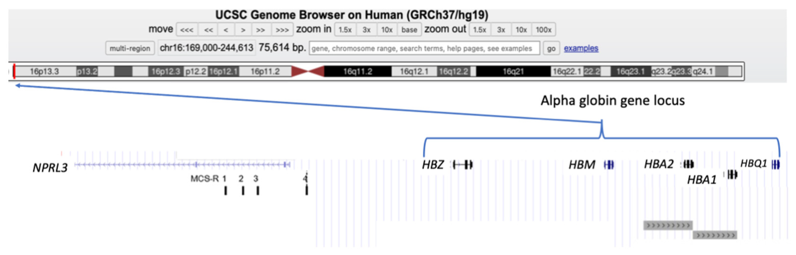
3. Alpha-Globin Gene Cluster and Alpha-Globin Biosynthesis

The level of α-globin genes expression is strongly dependent on variants in the α-globin gene cluster, which may either increase or, more commonly, decrease the synthesis of the α-globin polypeptide chains, with potential consequences on the hematological and clinical phenotype of both β-thalassemia and sickle cell disease.
Alpha-globin is the product of two almost identical α-globin genes, HBA1 and HBA2 (previously known as α1 and α2), located on the short arm of chromosome 16 (Chr16p13.3, GenBank NG_000006). As with all human globin genes, the α-globin genes have three exons and two introns (or intervening sequences, IVSs), which interrupt the coding sequence between codons 31/32 and codons 99/100. The length of HBA1 and HBA2 are 842 and 834 nucleotides (base pairs, bps), respectively, and they are positioned on the forward strand of chromosome 16p, meaning that they are expressed (transcribed) from the telomeric towards centromeric direction. The coding sequences (exons) of HBA1 and HBA2 are identical. However, the two genes differ slightly in their 5′ untranslated regions (UTRs) and their introns, and there are significant differences in their 3′UTRs. Furthermore, the duplicated α-globin genes are both embedded within larger homologous sequences, which play a role in the production of the two most common α+ thalassemia deletions (see Figure 1 and Section 3) [6].
The human α-globin cluster is located about 150 kb from the telomere of the short arm of chromosome 16, in a region of the genome that is GC-rich (around 54%), contains many Alu-repeat sequences and many genes. In fact, the α-globin gene cluster is surrounded by widely expressed genes and, furthermore, the upstream regulatory elements (including HS-40) lie within the introns of the adjacent housekeeping gene, NPRL3 [6,7].
There are conserved sequences motifs within the 5′ promoter regions of the HBA1 and HBA2 genes, including cis-acting sequence motifs, such as a TATA box (around minus 30 bp), a CCAAT box (between minus 70–80 bp), and CCGCCC motifs lying further upstream [2]. The HBA1 and HBA2 genes are located within the so-called α-globin gene cluster, which spans about 30 kb, containing one embryonic ζ globin gene (HBZ), three globin pseudogenes (ψζ1, ψα2, ψα1), and the θ globin gene (HBQ1) of undetermined function. Subsequent studies have shown that the ψα2 gene, in fact, expresses a transcript named μ-globin (mu-globin), and was thus renamed HBM [6]. The genes in the cluster are arranged along the chromosome in the following order: telomere (5′), HBZ, ψζ1, HBM, ψα1, HBA2, HBA1, HBQ1, and centromere (3′) (Figure 1).
There are four highly conserved, non-coding sequences, known as multispecies conserved sequences (MCSs) located between 10–40 kb upstream (5′) to HBZ, named MCS-R1–4, corresponding to previously identified erythroid-specific DNAse1 hypersensitive sites (DHSs) referred to as HS-48, HS-40, HS-33, and HS-10, whereby the number refers to the distance in kilobases from the HBZ gene CAP site. Evidence indicates that only the presence of HS-40 is essential for the expression of HBA1 and HBA2, and the role of the other three MCS elements remain undefined to date (Figure 1) [6].

3.1. Normal Expression of Alpha-Globin Genes

The HBZ gene is expressed until about the 6th week of gestation in humans, and both α- and ζ-globins are observed in the primitive erythroblasts which, until this stage of development, originate from the yolk sac. Studies in mice have shown that, as such cells mature, there is a switch from ζ- to α-globin expression, and it is likely that there is a similar switch in humans. Following the 6th week of gestation, the erythroblasts originating from the liver almost exclusively synthesize α-globin. However, minute quantities of ζ-mRNA are still produced, throughout fetal life, and form Hb Portland (ζ2γ2), detected in the cord bloods of non-thalassemic newborns. Very low levels of θ-globin mRNA transcripts from HBQ1 have been detected in yolk sac, fetal liver, adult blood, and bone marrow and similarly, very low levels of μ-globin mRNA can be detected in cord blood, adult blood, and bone marrow, but neither have yet been observed at the protein level [6,8].
The transcription of the HBA1 and HBA2 genes during hematopoiesis begins at the stage of basophilic erythroblasts, reaching a peak in the intermediate (polychromatophilic) erythroblasts, but subsequently stops as the erythroid cell matures. However, even following the extrusion of the nucleus, sufficient levels of globin mRNA persist in reticulocytes, such that globin polypeptide chains continue to be synthesized for a further 2–3 days, facilitated by the long half-life (>24 h) of globin mRNA. The protein products of the HBA1 and HBA2 genes are identical, although in fetal and adult life there is approximately three times as much α2-mRNA compared to α1-mRNA in erythrocytes from normal individuals. Normal erythroblasts express four (4) α-globin genes [8].

3.2. Genomic Variants in the Alpha-Globin Gene Cluster

Over 380 variants involving the α-globin genes (HBA1, HBA2) and wider clusters have been associated with α-thalassemia to date (see the Ithanet database at https://www.ithanet.eu (accessed on 24 January 2024) and the HbVar database at http://globin.cse.psu.edu (accessed on 24 January 2024)). As for most variants associated with hemoglobinopathies, many are population-specific, although recent genotyping technologies have identified many sporadic novel variants. The majority of individuals with α-thalassemia have genotypes involving deletion variants that remove some, or all of, the α-globin gene cluster. Less commonly, the defects are single or oligonucleotide variants or very small deletions within either of the duplicated HBA1 or HBA2 genes. Variants which partially abolish the synthesis of α-globin chains by the affected chromosome are traditionally termed α+ thalassemia variants, and those that completely abolish the synthesis of α-globin (null variants) are termed α0 thalassemia variants [9,10].
The category of variants most frequently observed in individuals with α-thalassemia are deletions which leave a single functional α globin gene on the chromosome. The two most common α+-thalassemia deletions include one of 3.7 kb or of 4.2 kb, both arising from unequal cross-over during meiosis, due to highly homologous sequences surrounding the HBA1 and HBA2 genes (Figure 1). The reciprocal chromosome causing the deletion of a single α-globin gene is an allele with three α-globin genes, often termed a “triplicated” arrangement (αααanti3.7 and αααanti4.2, respectively). There are over 250 million carriers of α+- thalassemia in the world, with the highest incidence found in the populations of India, Southeast Asia, and Africa, and less commonly in the Mediterranean and Middle East [9].
Most variants causing α0 thalassemia remove at least the two α-globin genes from the affected chromosome, and in some cases the complete ζ–α-globin gene cluster. In addition, several deletions have been characterized that leave the α-globin genes intact but remove conserved regulatory elements, including the critical HS-40, along with variable amounts of flanking DNA. With such deletions, the α-globin genes, although present, are not expressed, giving rise to a phenotype of α-thalassemia. Most α0 thalassemia deletion variants are sporadic. However, four are more common, with so-called --Med and (−α)20.5 found in Mediterranean populations, and --SEA and –FIL in Southeast Asia [9].
More than 250 single nucleotide variants (substitutions, microdeletions, or microduplications) relate to α-thalassemia and are more often observed in HBA2 gene than HBA1. These variants disrupt all the known stages of protein synthesis (including correct RNA processing or translation). Additionally, an interesting group of single nucleotide variants lead to the synthesis of highly unstable α-globin polypeptides, which mimic an α-thalassemia phenotype through reducing the production of normal α-globin chains. These variants are rarely detectable at the protein level (in the form of hemoglobin) but, as they are usually detected through genotyping methods, the amino substitution can be predicted and the variants are often named as hemoglobins, e.g. Hb Quong Sze (α2 cd125 CTG > CCG; Leu > Pro), Hb Agrinio (α2 cd29 CTG > CCG; Leu > Pro), and Hb Taybee (α1 cd38 or 39 delACC; Thr) [9].

4. The Impact of Changes in the Expression of Alpha-Globin Genes on Hematopoietic Pathologies: Genetic Evidence

In populations with a high frequency of α- and β-thalassemic variants, the incidence of complex genotypes, due to the co-inheritance of α- and β-thalassemia, is not rare. These genotypes, with the associated hematological and clinical phenotypes, are useful to illustrate the deleterious effect of the excess of free α-globin chain monomers, the relative level of which is the major factor contributing to the severity of β-thalassemia syndromes [11,12,13]. In β-thalassemia heterozygotes that coinherit less than four α-globin genes (heterozygous α-thalassemia genotypes), diminished levels of excess α-globin chains are expected. Comparison of the hemoglobin levels and red cell indices between the various groups with three or two functional α-genes (-α/αα, --/αα, or -α/-α) with heterozygous β-thalassemia demonstrates a relative increase in hemoglobin levels and red cell indices (MCV and MCH), compared to those of β-thalassemia heterozygotes with the normal number of four functional α-globin genes. The amelioration of these parameters was most apparent in cases with two α-genes deleted, since there is a balanced/almost balanced α/β chain ratio. However, in cases with heterozygous β-thalassemia and only one functional α-globin gene, patients have chronic anemia analogous to that found in patients with hemoglobinopathy H (HbH disease), but without detectable levels of HbH [13].
There is evidence that the co-inheritance of α-thalassemia genotypes which reduce α-globin synthesis may modify the clinical phenotype of homozygous or compound heterozygous β-thalassemia to a milder form, compatible to NTDT. This effect is more obvious in cases with HBB genotypes that allow some expression of β-globin (so-called β+) compared to null variants (so-called β0) [13].
When cases with homozygous or compound heterozygous β-thalassemia alleles co-inherit only a single functional α-globin gene (a genotype typically associated with HbH disease), the rare cases described have a phenotype of non-transfusion-dependent thalassemia (NTDT) rather than transfusion-dependent thalassemia (TDT) [13,14,15]. The male patient described by Kanavakis et al. [13] was first hospitalized at 50 years of age due to fatigue. Examination revealed marked anemia (hematocrit 20%), though he had never been transfused. Following splenectomy, he maintained hemoglobin levels of 85–95 g/dL. DNA analysis identified HBB genotypes equivalent to TDT (β+0), with HBA1/2 alleles involving a single α-globin gene in-trans to complete the deletion of both α-globin genes, equivalent to HbH disease (α+0). The α/non α-globin chain biosynthesis was completely balanced. Hematological parameters demonstrated a well-compensated anemia with ineffective erythropoiesis and oxidative stress, which was ameliorated following splenectomy [13].
In contrast, alleles with additional α-globin genes, most commonly triplicated α-globin gene arrangements, have been observed to co-inherit with heterozygous β-thalassemia genotypes. The majority of the studies evaluating hematological and phenotypic characteristics in β-thalassemia heterozygotes with a single additional functional α-globin gene report that the additional α-globin gene had a marked effect on the hematological phenotype in most cases and often also on the clinical phenotype. Specifically, double heterozygotes present lower hemoglobin levels and red cell indices, and a clinical presentation is often categorized as NTDT with occasional (and exceptionally more regular) transfusion requirements [16,17,18,19,20,21,22]. In contrast, some publications concluded that an additional α-globin gene had a very limited effect on the hematological phenotype of β-thalassemia heterozygotes [23], or even no significant effect [24], with the latter presenting the largest cohort described so far, including 67 cases.
These contradictory results may be attributed to differences in the strategies used to recruit the cases in each study, as well as the criteria for the evaluation of hematological and clinical phenotypes (summarized in Supplementary Table S1). The cohorts that were selected based on a phenotype-driven strategy tended to conclude that the additional α-globin gene exacerbated the phenotype of heterozygous β-thalassemia. In contrast, the studies of cohorts collated based on chance identification of the double heterozygous genotype (e.g., following routine genetic analysis or carrier screening), found no significant consequence. In conclusion, not every double heterozygote has a significantly affected phenotype, indicating the likely contribution of numerous other genetic parameters, as well as environmental and socioeconomic factors.
Despite the differences regarding the effect of five α-globin genes in β-thalassemia heterozygotes, there is an agreement that the interaction of six active α-genes, especially when co-inherited with heterozygous β0-thalassemia, almost always, produces NTDT [16,25,26,27,28,29,30]. These six functional α-globin genes could be the result of homozygosity for the so-called anti 3.7 kb or anti 4.2 kb arrangements or, rarely, of heterozygosity for quadruplicated α-globin gene arrangements [26,30,31], while the presence of more than six functional α-globin genes (i.e., compound heterozygosity ααα/αααα) has been associated with TDT and NTDT, the latter with a relatively more severe presentation [30,32]. An interesting example is reported by Farashi et al. in 2015 [32], regarding a family in which both the father (at the age of 30) and his daughter (at the age of 6 months) required blood transfusions, bimonthly for the father and monthly for the child. Both father and daughter were heterozygous for a β+-thalassemia variant. The mother, a hematologically asymptomatic subject, was found to carry a duplication analogous to anti 3.7 kb, while the father carried an extra complete α-globin gene cluster, resulting in four functional α-globin genes from this allele (total six α-genes), whereby the child was found to be a compound heterozygote with seven α-globin genes [32].
Overall, these studies indicate that the levels of excess α-globin chains in the erythrocytes are a major factor modifying the severity of beta-thalassemia syndromes [33].

5. The Impact of Changes in the Expression of Alpha-Globin Genes on Hematopoietic Pathologies: Evidence in the “Gene Editing Era”

The clinical relevance of the inbalanced expression of α-globin versus β-globin genes is strongly in agreement with data obtained by genome editing approaches, as is summarized in Figure 2.
Genome editing (GE) can be considered among the most promising strategies to correct hereditary alterations in a variety of monogenic diseases, including hematopoietic pathologies [34,35]. Figure 2 depicts how CRISPR-Cas9 gene editing can be applied to β-thalassemia [34,36,37,38,39,40,41,42,43,44]. CRISPR-Cas9 gene editing can be proposed for efficient correction of the NM_000518.5:c.118C>T thalassemia variant (HGVS nomenclature, β039C>T traditional nomenclature) [45]. In addition, CRISPR-Cas9 gene editing has been applied for the reduction of the content of free α-globin chains [46,47,48,49]. With the objective of decreasing α-globin gene expression, Mettananda et al. [49] described the use of CRISPR-Cas9 genome editing to mimic a natural mutation, which deletes the MCS-R2 α-globin enhancer and causes α-thalassemia. When edited CD34+ cells from β-thalassemia patients were differentiated into erythroid cells, they observed the expected reduction in α-globin expression and a correction of the α/β globin chain imbalance, suggesting that this CRISPR-Cas9 based approach might be of clinical relevance [49]. A second study on this very important issue was published by Pavani et al. [48], demonstrating the correction of the pathological phenotype of β-thalassemia by CRISPR-Cas9 editing of the α-globin locus in human hematopoietic stem cells.

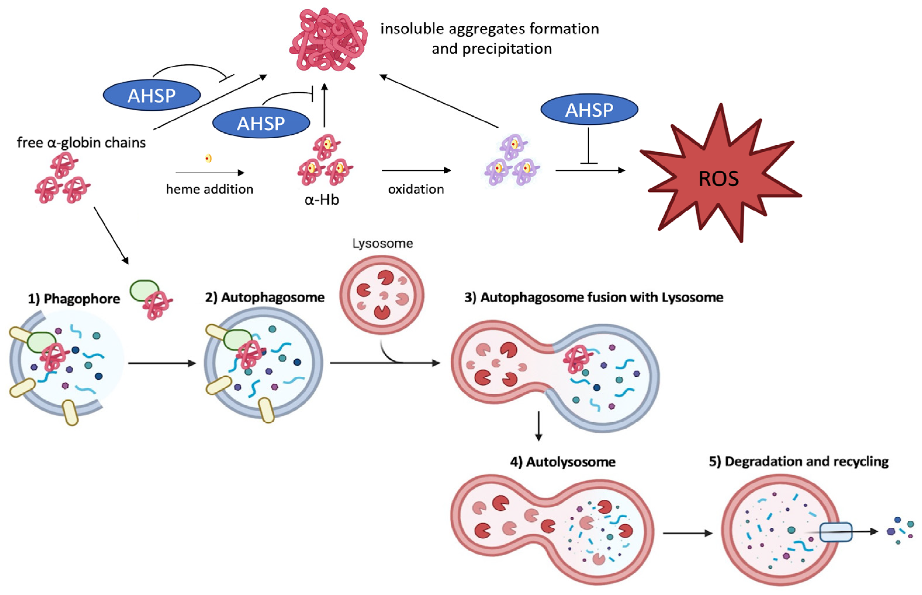
6. Alpha-Hemoglobin-Stabilizing Protein (AHSP): Expression, Function, and Molecular Genetics

Alpha-hemoglobin-stabilizing protein (AHSP) is a molecular chaperon with high cell-type specificity, which reversibly binds to free α-globin polypeptides. This binding supports the stability, folding, and assembly of α-globin chains. Additionally, AHSP can promote the refolding of denatured α-globin polypeptide chains. Overall, AHSP prevents the precipitation of free α-globin chains, and when β-globin polypeptide chains are present, it facilitates the formation of tetrameric HbA molecules. In the absence of AHSP, the α-globin polypeptides generate reactive oxygen species (ROS) and precipitate within the precursor erythrocytes in the bone marrow, prompting apoptosis and ineffective erythropoiesis. The AHSP gene is located on human chromosome 16 (Chr16p11.2). It is approximately 1.8 kb long, has three exons and two introns, and encodes a polypeptide of 102 amino acids. The AHSP polypeptide forms a bundle of three antiparallel α-helices, whereby helices 1 and 2, and the intervening segment, recognize the G and H helices of the α-globin chains (the latter involved in the α1β1 contact in the HbA molecule) to form a simple heterodimer. The AHSP and β-globin polypeptides compete to bind to α-globin polypeptides within the same region of the protein, but the intermolecular contacts between α-globin chains and AHSP are less extensive than those between α-globin and β-globin chains and, thus, the β-globin chains displace AHSP and form HbA molecules. AHSP can bind α-globin chains with heme or without heme (holo-αHb or apo-αHb, respectively) in both reduced (ferrous, FeII) and oxidized (ferric, FeIII) states. The ASHP protein cannot bind to β-globin polypeptide chains or to tetrameric HbA molecules. AHSP is expressed exclusively in hematopoietic tissues at high levels [50,51]. The promoter of the AHSP gene extends from the 5′-flanking region until intron 1 (Base pairs −170 to +269) and includes five consensus GATA-1 binding sites, an Oct-1 consensus site, and an EKLF binding site [52,53]. Therefore, transcription factors GATA-1, Oct-1, and EKLF are expected to coregulate AHSP gene expression during erythropoiesis [50,51].
Several studies have demonstrated a correlation of AHSP and α-globin expression in erythroblasts from normal individuals and also with β0 thalassemia [54], and there may be several potential pathways involved in the upregulation of AHSP expression in the presence of higher levels of free α-globin chains. These mechanisms may not be exclusive and include a feedback mechanism of excess α-globin levels [55], a response to increased ROS induced by excess α-globin chains, as mediated by Nrf2 (nuclear factor erythroid 2-related factor 2) and possibly other components of ROS signaling pathways [56], and a mechanism through iron levels enabled by iron responsive elements and iron regulatory proteins [57].
A few studies have investigated the genetic variation in and around the ASHP gene locus across several populations. Based on single nucleotide polymorphisms (SNPs) and a single nucleotide repeat, 18 different AHSP gene haplotypes have been determined [58,59]. With respect to genomic variation that may potentially alter the expression of the AHSP protein, a common SNP (G>A) at position 12391 within intron 1, was found to disrupt a proposed binding site for transcription factor Oct-1, potentially inhibiting the optimum activation of AHSP gene expression [59].
Missense variants causing altered protein structure are rare in all populations studied (see below), but there are several examples reported. The substitution of Asn75 > Ile (N75I), although it is not located in a region of the AHSP polypeptide important for forming a complex with α-globin, studies found that AHSP with the Asn75 > Ile variant has impaired ability to inhibit reactive oxygen species (ROS) production by α-globin, potentially explaining the deleterious clinical effect of this mutant [59]. Based on hematological parameters in β-thalassemia heterozygotes in Southern China, the rare variants AHSP Asp29 > Val (D29V) and AHSP Val56 > Gly (V56G) found no apparent modifying effect [60], although another study reported that Val56 > Gly (V56G) may be associated with some instability and an increased dissociation rate of the AHSP/α-globin complex [61]. Ray et al. 2019 reported the Ser33 > Phe (S33F) variant was postulated to disrupt the structural formation of AHSP with a negative result on its normal function [62]. Studies which evaluated AHSP mRNA levels in peripheral blood reticulocytes of healthy individuals observed a three-fold variation between individuals that did not correlate with age or sex [62]. This study found that the level of AHSP depended upon the associations of several AHSP haplotypes linking to some clades, possibly associated with sequence variants within the promoter region, including the Oct-1 consensus site [63]. A classical twin heritability study in unselected twins by the same group found that >45% of AHSP expression was influenced by genetic factors, while almost 30% was influenced by environmental factors [64].
In conclusion, although the expression of AHSP has been positively correlated to the levels of excess α-globin in normal and β-thalassemia erythroblasts [54,55], the underlying genetic variability at the ASHP locus which may influence the expression of AHSP has not been completely characterized to date.

7. Alpha-Hemoglobin-Stabilizing Protein as a Modifier of Beta-Thalassemia

Based on the evidence that the level of excess α-globin chains is one of the key factors underlying the severity of β-thalassemia phenotypes and clinical course, ASHP has been proposed as a potential candidate for modulating this severity. Furthermore, an AHSP gene knock-out mouse model presents a similar phenotype to the β-thalassemic mouse model [50,65]. Structural variants in the AHSP gene are uncommon [58,59], and are therefore unlikely to be major modifiers of β-thalassemia in most populations, although the influence of rare AHSP null or missense variants, such as the rare coding AHSP N75I variant, may impair normal AHSP protein function and, thus, the phenotype of β-thalassemia. The first study to implicate a potential influence of AHSP on modulating β-thalassemia phenotypes found reduced erythrocyte AHSP mRNA expression in β-thalassemia heterozygotes with an unusually severe phenotype of NTDT, compared to phenotypically typical β-thalassemia heterozygotes and normal individuals [66]. Of note is that this study did not investigate DNA variants at the AHSP locus or elsewhere in the genome, and that, as mentioned above, mRNA levels were subsequently demonstrated to have significant variability, even between normal (non-thalassemic) individuals [63]. In contrast, another study of Thai patients with HbE/β-thalassemia concluded that there was no correlation of clinical severity with AHSP gene variants [58], and similar conclusions were also made in a study of investigating AHSP gene polymorphisms in β-thalassemia heterozygotes from a Southern Chinese population [60]. Another study which measured AHSP mRNA levels in a cohort of 37 β-thalassemia patients found no significant difference in the levels of AHSP between patients with TDT and NTDT, although the levels were found to be higher in 12 sickle cell patients [67]. A more recent study by Ray et al. performed AHSP gene sequencing in samples from 38 patients with HbE/β-thalassemia [62]. Comparing the AHSP variants observed in 23 TDT and 15 NTDT patients, there was a statistically significant correlation with three of five variants between the two groups. The three variants with statistical significance included two silent nucleotide substitutions and one missense variant (Ser33 > Phe or S33F), with the latter, which was observed in 65% of patients in the TDT group and postulated to potentially disrupt the normal function of the AHSP protein, observed in 65% of patients in the TDT group.
Overall, it should be underlined that it is yet to be clearly established whether (or not) AHSP plays a role in modifying the phenotypic severity in β-thalassemia. In this respect, it is difficult to drive robust conclusions comparing studies that have so far focused on a variety of heterogenous cohorts of β-thalassemia patients, using different biochemical and molecular strategies to evaluate the effect of AHSP variants on AHSP expression, based on heterogenous protocols for measuring AHSP mRNA and protein levels, or correlating phenotypes of patients and/or heterozygotes with genetic variation at the AHSP locus. In conclusion, although the proposed function of the AHSP protein makes it an attractive therapeutic agent in potentially ameliorating the clinical course in β-thalassemia patients [68], further studies are required to clarify a correlation of β-thalassemia phenotypes with AHSP protein expression.
Finally, it is of interest to note that several α-globin chain variants have been described, which are prone to structural instability associated with structural alterations and impaired interaction with AHSP [69,70,71]. This is the case of the recently described α-globin frameshift mutants with stop codon in the last exon at codon 102 (Hb Campania) or at codon 133 (Hb Sciacca) [71]. A 3D model indicated instability of the α-globin chain variants, due to the severe structural alterations causing an impairment of the molecular interactions with AHSP. Similarly, Hb Bronovo [α103(G10)His → Leu, HBA2: c.311A>T] is an α-globin variant that interferes with the AHSP’s binding efficiency. In fact, the histidine residue at position 103 is integral to the AHSP’s hydrogen bond formation and its disruption leads to an increased quantity of cytotoxic free α-globin chains, thereby creating a similar pathophysiology as β-thalassemia [69].

8. Alpha-Hemoglobin-Stabilizing Protein as a Modifier of Sickle Cell Disease

There is little evidence about the influence of AHSP on sickle cell disease (SCD). The expression of the AHSP gene has been reported to be higher in SCD erythrocytes, with respect to unaffected controls. Mahmoud et al. (2015) found that median AHSP expression was significantly higher in patients with SCD, compared to thalassemia patients [67]. Vasseur et al. (2022) were able to demonstrate higher AHSP content in red blood cells of SCD patients with or without hydroxyurea (HU) treatment [72]. The interplay between expression of α-globin genes, the presence of free α-globin, the production of AHSP, and the phenotype of sickle cell disease (SCD) has not been evaluated in as much in depth as for β-thalassemia. However, some studies have been reported, suggesting that this field of investigation deserves consideration. For instance, a limited number of relevant studies demonstrate the impact of genetic variations in α-globin genes on SCD severity.

8.1. Interaction of Alpha-Thalassemia and Homozygous Sickle Cell Disease

The interaction of α-thalassemia and homozygous SCD were reported in different studies [73,74,75] concurrently showing that α-thalassemia reduces the hemolytic rate in homozygous SCD patients [75]. In agreement with these reports, Kirkham et al. have recently reported that α-thalassemia deletions in SCD patients are significantly associated with improvements in clinical and biochemical parameters, such as increased hemoglobin levels and a reduced risk of albuminuria, abnormal transcranial Doppler velocity, and stroke [76]. Interestingly, a single alpha-globin gene deletion, referred to as the alpha thalassemia silent carrier, is present in more than 30 percent of SCD patients of African descent, with an even higher prevalence in some SCD populations in the Middle East and India [77]. A second issue is related to the presence of excess free α-globin chains in SCD. Vasseur et al. reported an elevated soluble α-hemoglobin pool in sickle cell anemia [78]. This finding was confirmed by Domingues-Hamdi et al., who also demonstrated that hydroxycarbamide decreases this free α-hemoglobin pool in red blood cells of adult patients with SCD [79].

8.2. Levels of Alpha-Hemoglobin-Stabilizing Protein in Sickle Cell Disease

The levels of AHSP in red blood cell lysates from patients with SCD were analyzed by different research groups, to evaluate the clinical relevance of AHSP gene expression [67,72]. The first information that Vasseur et al. [72] obtained was that the AHSP concentration was significantly higher in patients with the SS genotype than in the controls. The second observation was that a strong positive correlation was observed between the AHSP concentration and the α-hemoglobin pool. This is of relevance, considering that the free α-Hb pool of patients with SCD is higher than that of controls. Therefore, it is likely to hypothesize that the AHSP concentration increased in these patients to compensate for the relative excess of free α-globin chains [67,72]. In conclusion, even though the real benefits of AHSP expression on SCD phenotype have not been conclusively demonstrated, some interesting reports are available, suggesting that the impact of AHSP on SCD should be still considered in future research efforts.

9. Role of Alpha-Hemoglobin-Stabilizing Protein in Normal and Pathological Erythropoiesis: Updates from Studies Based on Transgenic Mouse Model Systems

Transgenic mice have been extensively used to study the role of AHSP and propose strategies for ameliorating the clinical phenotype of β-thalassemia. In a first study, Kong et al. generated AHSP–/– mice by gene targeting [65], demonstrating an abnormal erythrocyte morphology with hemoglobin precipitates in these animals. Furthermore, they found that the loss of AHSP reduced the lifespan of circulating red-blood cells (RBCs) and increased ROS-dependent apoptosis of erythroid precursors [65]. Importantly, when informative interbreeding of mutant mice was performed, it was found that loss of AHSP exacerbated the severity of β-thalassemia. In agreement, Wang et al. [80] employed a human AHSP vector to generate transgenic human AHSP mice in a model of “βIVS-2-654-thalassemia”. AHSP expression was associated with improvement in the red blood cell parameters. For instance, a dramatic reduction in anisocytosis in the peripheral blood was observed. Splenomegaly with extramedullary hematopoiesis was ameliorated. Serum iron concentration and iron deposition in the liver were decreased in “h-ahsp+/βIVS-2-654+” mice. All these findings suggested amelioration of the anemia phenotype in “h-ahsp+IVS-2-654” mice after the introduction of the AHSP gene. They therefore propose that an AHSP transgene could provide an adjuvant method for gene therapy of β-thalassemia. However, other mice model systems have been described, in which AHSP does not limit α-globin detoxification. In this context, Nasimuzzaman et al. used transgenic mice to investigate the effects of supraphysiologic levels of AHSP on the severity of a NTDT β-thalassemia phenotype [81]. They tested wild-type AHSP and two mutant versions exhibiting 3- or 13-fold higher affinity for α-globin. Erythroid overexpression of these AHSP proteins, up to 11-fold beyond endogenous levels, had no major effects on hematologic parameters in β-thalassemic animals. Therefore, while a consensus does exist on the exacerbation of the β-thalassemia related parameters in the case of low (or absent) AHSP production [65,82], supraphysiologic levels of AHSP might not be associated with clinical improvements [81]. On the other hand, a partial improvement in erythroid parameters has been proposed by Wang et al., associated with AHSP production [80]. This specific issue needs further investigations to associate the endogenous levels of AHSP with the β-thalassemia phenotype in patients with different β-thalassemia genotypes and endogenous levels of AHSP.
In conclusion, AHSP activation could be beneficial, to reduce the physio-pathologic alterations caused by the precipitation of α-globin aggregates in erythroid β-thalassemic cells. Strategies to achieve higher AHSP levels can be designed by taking advantages from studies on the molecular regulation of AHSP gene expression. For instance, the direct binding of STAT3 to the AHSP promoter is involved in the upregulation of the AHSP gene, providing clues to therapeutic strategy for AHSP enhancement [52,53].

10. Inducers of Alpha-Hemoglobin-Stabilizing Protein

Given the recognized importance of AHSP for the lifespan of erythroid cells, AHSP inducers might be of interest, from a therapeutic point of view. In respect to this issue, Liu et al. [83] found that nitidine chloride (NC) induces erythroid differentiation of human leukemic K562 cells, together with increased expression of the genes coding the α-, ε-, and γ-globins, and the erythroid differentiation markers AHSP, CD235a, and CD71. It should be of interest to verify whether NC is able to induce increased expression of AHSP in erythroid precursor cells from patients with β-thalassemia, especially those exhibiting an excess of free α-globin chains. In a more recent study, Han et al. demonstrated that AHSP expression in K562 cells can be stimulated by NFE2-related factor 2 (Nrf2) and its agonist tert-Butylhydroquinone (tBHQ) [56]. Interestingly, the AHSP levels were elevated in α-globin-overexpressing K562 cells and erythroblasts from βIVS-2-654 thalassemic mice. In these experimental model systems, tBHQ treatment partially alleviated, whereas Nrf2 or AHSP knockdown exacerbated, α-globin precipitation and ROS production in fetal liver-derived thalassemic erythroid cells. In this context, it should be mentioned that sulforaphane (SFN), a naturally occurring isothiocyanate found in cruciferous vegetables, has received attention as a natural activator of the Nrf2/Keap1 cytoprotective pathway [84]. Other NRF2 activators have been reported and recently reviewed [85,86]. In the search for AHSP inducers, it is relevant to consider that, in erythroid cells, AHSP gene expression is facilitated by several transcription factors, such as GATA1, EKLF, OCT1, and STAT3 [87,88,89]. In contrast, a reduction in AHSP synthesis in hemin-induced K562 cells leads to alpha-globin precipitation, the impairment of hemoglobin production, and increased cell death [90].

11. The Clearance of Free Alpha-Globin Is Activated by Autophagy in Erythroid Cells from Beta-Thalassemia Patients

Autophagy, a highly conserved process of degradation of cellular components among mammals, plays a crucial role in the recycling of cytosol and organelle-derived macromolecules. This process involves the formation of autophagosomes, which are double membrane vesicles where the macromolecules to be degraded are encapsulated [91]. Subsequently, these autophagosomes fuse with lysosomes and the waste material is degraded, to be recycled by the cells for other purposes [92]. Regarding erythroid maturation, autophagy plays a significant role by facilitating the clearance of redundant organelles, including ribosomes and mitochondria (referred to as mitophagy). Several studies have demonstrated that this process is crucial for ensuring the proper development of mature red blood cells from erythroid precursor cells isolated from healthy donors [93,94,95,96,97,98,99]. Following this consideration, the autophagic process should be particularly relevant in erythroid precursor cells isolated from β-thalassemia patients, since hemolysis and ineffective erythropoiesis stimulate the bone marrow to increase the production of erythroid precursors and their release into the blood stream. Despite this, few scientific studies have evaluated the autophagy process in depth in β-thalassemia patients [100,101,102,103]. Besides contributing to the maturation of the erythroid cells themselves, an additional role for autophagy in β-thalassemia is to promote the clearance of toxic α-globin excess accumulating in erythroid precursors from β-thalassemia patients [96]. Research by several groups indicates that free α-globin is degraded by autophagy in β-thalassemia [101,104,105]. Therefore, autophagy might be considered a mechanism, in addition to that involving AHSP, by which β-thalassemia erythroid cells are able to mitigate the effects of the excess α-globin production. In addition, autophagy pathways have a clear role in the protection against metabolic and proteotoxic stresses [106], such as those exacerbated by the excess of α-globin in β-thalassemia erythroid cells. In a recent study, Lechauve et al. demonstrated that the absence of the autophagy-activating Unc-51–like kinase 1 (ULK1) gene in a β-thalassemia mouse model impairs the autophagic clearance of α-globin within red blood cell precursors, leading to the aggravation of disease symptoms [100]. Treatment with the mTORC1 inhibitor sirolimus (rapamycin) systemically reduces the accumulation of α-globin aggregates and alleviates the pathological manifestations in β-thalassemic mice [100]. Interestingly, K562 cellular clones forced to express α-globin protein at high levels seem to activate the autophagy process as a defense mechanism [107]. In these cellular clones producing high levels of toxic α-globin, ULK1 mRNA was found to be upregulated, and the number of autophagosomes increased proportionally to the accumulation of α-globin protein, supporting the role of autophagy in detoxifying cells from toxic protein accumulation. In this regard, the mTOR inhibitor sirolimus (rapamycin) is a very interesting molecule that can contribute to the reduction of the content of free α-globin chains in erythroid precursor cells of β-thalassemia patients with two mechanisms: the induction of γ-globin chains with subsequent formation of α2γ2 tetramers (HbF) [108,109] and the triggering of the autophagy process to enhance the clearance of toxic free α-globin [100]. Zurlo et al. demonstrated that sirolimus induces autophagy in erythroid cells isolated from β-thalassemia patients [106]. The reduction in soluble α-globin chains found in sirolimus-treated erythroid precursors has, moreover, contributed to the moderate accumulation of insoluble α-globin aggregates, responsible for hemolysis in these patients. The interplay between expression and function of AHSP and autophagy is summarized in Figure 3.
Notably, a reduction in free α-globin and the upregulation of ULK1 gene expression was also found in erythroid cells isolated from rapamycin-treated β-thalassemia patients during the NCT03877809 clinical trial [109,110]. In addition, clinical parameters related to hemolysis and ineffective erythropoiesis (such as soluble transferrin receptor, ferritin, and bilirubin levels) were significantly reduced during sirolimus administration in these patients, suggesting that autophagy and α-globin clearance are important and effective processes to be considered during clinical trials involving thalassemia patients.

12. Conclusions and Future Perspectives

The pathophysiology of β-thalassemia reflects an imbalance between α- and β-globin chains, with an excess of free α-globin chains causing ineffective erythropoiesis and hemolysis [102]. The reduction in free α-globin chains has a clear clinical impact, as suggested from studies demonstrating that, when α-thalassemia is co-inherited with β-thalassemia, excess free α-globin chains are reduced, significantly ameliorating the clinical severity. Robust evidence supports the concept that reduction in the “toxic” excess of free α-globin can be achieved in β-thalassemia erythroid cells either by increasing the expression of the AHSP gene or by activation of the autophagic process. A key player of the induction of autophagy in erythroid cells is ULK1 (the unc-51 like autophagy activating kinase 1). It is presently unknown if the co-activation of AHSP and autophagy occur in β-thalassemia erythroid cells, but this will be presumably considered in future studies by research groups working in this very interesting field. Future perspectives include a possible impact of “microRNA therapeutics” on autophagy. Recent studies linking microRNA modulation with ULK1 expression facilitate further investigation in this area as in the study by Keith et al. demonstrating that loss of miR-144/451 alleviates β-thalassemia by stimulating ULK1-mediated autophagy of free α-globin. This finding supports the potential of miRNA targeting for reducing the excess of free α-globin chains in β-thalassemia and other hemoglobinopathies [111].
In summary, in this short review we focused on the impact of α-globin gene expression and α-globin modifiers on the phenotype and clinical severity of β-thalassemia. It was written on behalf of the INHERENT (International Hemoglobinopthy Research Network) project, concordant with the interests of INHERENT, which has a primary aim to study the role of genetic modifiers in hemoglobinopathies through a large, multi-ethnic genome-wide association study (GWAS) [112]. The evidence to date summarized in this review contributes towards the validation of previously reported genetic modifiers and the potential discovery of new ones, related to the degree of globin chain imbalance in erythrocytes, one of the most important factors influencing the phenotypic expression of human hemoglobinopathies.

Supplementary Materials

The following supporting information can be downloaded at: https://www.mdpi.com/article/10.3390/ijms25063400/s1.

Author Contributions

Conceptualization, J.T.-S. and R.G.; methodology, C.V., C.S., R.G. and M.Z.; formal analysis, R.G. and A.F.; writing—original draft preparation, J.T.-S., R.G., and C.V.; writing—review and editing, J.T.-S., R.G. and C.S.; supervision, A.F. and R.G.; funding acquisition, R.G. All authors have read and agreed to the published version of the manuscript.

Funding

This study was sponsored by the Wellcome Trust (innovator award 208872/Z/17/Z) and AIFA (AIFA-2016-02364887). R.G. also received funding from the UE THALAMOSS Project (Thalassemia Modular Stratification System for Personalized Therapy of Βeta-Thalassemia; no. 306201-FP7-HEALTH-2012-INNOVATION-1). J.T.S. received funding for the publication costs from the University Research Institute for the Study of Genetic & Malignant Disorders in Childhood, National and Kapodistrian University of Athens, Greece.

Institutional Review Board Statement

Not applicable.

Informed Consent Statement

Not applicable.

Data Availability Statement

Not applicable.

Acknowledgments

We thank all the colleagues of the INHERENT Network for their continuous support. The list of the INHERENT members can be found in Kountouris et al. [112].

Conflicts of Interest

The authors declare no conflicts of interest.

References

  1. Weatherall, D.J. Phenotype-genotype relationships in monogenic disease: Lessons from the thalassaemias. Nat. Rev. Genet. 2001, 2, 245–255. [Google Scholar] [CrossRef] [PubMed]
  2. Higgs, D.R.; Engel, J.D.; Stamatoyannopoulos, G. Thalassaemia. Lancet 2012, 379, 373–383. [Google Scholar] [CrossRef] [PubMed]
  3. Thein, S.L. Molecular basis of β thalassemia and potential therapeutic targets. Blood Cells Mol. Dis. 2018, 70, 54–65. [Google Scholar] [CrossRef] [PubMed]
  4. Kumar, R.; Sagar, C.; Sharma, D.; Kishor, P. β-globin genes: Mutation hot-spots in the global thalassemia belt. Hemoglobin 2015, 39, 1–8. [Google Scholar] [CrossRef] [PubMed]
  5. Rao, E.; Kumar Chandraker, S.; Misha Singh, M.; Kumar, R. Global distribution of β-thalassemia mutations: An update. Gene 2023, 896, 148022. [Google Scholar] [CrossRef] [PubMed]
  6. Daniels, R.J.; Peden, J.F.; Lloyd, C.; Horsley, S.W.; Clark, K.; Tufarelli, C.; Kearney, L.; Buckle, V.J.; Doggett, N.A.; Flint, J.; et al. Sequence, structure and pathology of the fully annotated terminal 2 Mb of the short arm of human chromosome 16. Hum. Mol. Genet. 2001, 10, 339–352. [Google Scholar] [CrossRef] [PubMed]
  7. Zhu, D.; Xu, L.; Zhang, Y.; Liang, G.; Wei, X.; Li, L.; Jin, W.; Shang, X. Investigation of the mechanism of copy number variations involving the α-globin gene cluster on chromosome 16: Two case reports and literature review. Mol. Genet. Genom. 2023, 298, 131–141. [Google Scholar] [CrossRef] [PubMed]
  8. Higgs, D.R.; Garrick, D.; Anguita, E.; De Gobbi, M.; Hughes, J.; Muers, M.; Vernimmen, D.; Lower, K.; Law, M.; Argentaro, A.; et al. Understanding alpha-globin gene regulation: Aiming to improve the management of thalassemia. Ann. N. Y. Acad. Sci. 2005, 1054, 92–102. [Google Scholar] [CrossRef]
  9. Harteveld, C.L.; Higgs, D.R. Alpha-thalassaemia. Orphanet J. Rare Dis. 2010, 5, 13. [Google Scholar] [CrossRef]
  10. Higgs, D.R.; Gibbons, R.J. The molecular basis of α-thalassemia: A model for understanding human molecular genetics. Hematol. Oncol. Clin. N. Am. 2010, 24, 1033–1054. [Google Scholar] [CrossRef]
  11. Kanavakis, E.; Wainscoat, J.S.; Wood, W.G.; Weatherall, D.J.; Cao, A.; Furbetta, M.; Galanello, R.; Georgiou, D.; Sophocleous, T. The interaction of alpha thalassaemia with heterozygous beta thalassaemia. Br. J. Haematol. 1982, 52, 465–473. [Google Scholar] [CrossRef]
  12. Wainscoat, J.S.; Thein, S.L.; Weatherall, D.J. Thalassaemia Intermedia. Blood Rev. 1987, 1, 273–279. [Google Scholar] [CrossRef]
  13. Kanavakis, E.; Traeger-Synodinos, J.; Lafioniatis, S.; Lazaropoulou, C.; Liakopoulou, T.; Paleologos, G.; Metaxotou-Mavrommati, A.; Stamoulakatou, A.; Papassotiriou, I. A rare example that coinheritance of a severe form of beta-thalassemia and alpha-thalassemia interact in a ‘‘synergistic’’ manner to balance the phenotype of classic thalassemic syndromes. Blood Cells Mol. Dis. 2004, 32, 319–324. [Google Scholar] [CrossRef]
  14. Loukopoulos, D.; Loutradi, A.; Fessas, P. A unique thalassaemic syndrome: Homozygous alpha-thalassaemia + homozygous beta-thalassaemia. Br. J. Haematol. 1978, 39, 377–389. [Google Scholar] [CrossRef]
  15. Panyasai, S.; Jaiping, K.; Pornprasert, S. Elevated Hb A₂ Levels in a Patient with a Compound Heterozygosity for the (β⁺)-31 (A > G) and (β⁰) Codon 17 (A > T) Mutations Together with a Single α-Globin Gene. Hemoglobin 2015, 39, 292–295. [Google Scholar] [CrossRef]
  16. Traeger-Synodinos, J.; Kanavakis, E.; Vrettou, C.; Maragoudaki, E.; Michael, T.; Metaxotou-Mavromati, A.; Kattamis, C. The triplicated alpha-globin gene locus in beta-thalassaemia heterozygotes: Clinical, haematological, biosynthetic and molecular studies. Br. J. Haematol. 1996, 95, 467–471. [Google Scholar] [CrossRef] [PubMed]
  17. Camaschella, C.; Kattamis, A.C.; Petroni, D.; Roetto, A.; Sivera, P.; Sbaiz, L.; Cohen, A.; Ohene-Frempong, K.; Trifillis, P.; Surrey, S.; et al. Different hematological phenotypes caused by the interaction of triplicated alpha-globin genes and heterozygous beta-thalassemia. Am. J. Hematol. 1997, 55, 83–88. [Google Scholar] [CrossRef]
  18. Ma, S.K.; Au, W.Y.; Chan, A.Y.; Chan, L.C. Clinical phenotype of triplicated alpha-globin genes and heterozygosity for beta0-thalassemia in Chinese subjects. Int. J. Mol. Med. 2001, 8, 171–175. [Google Scholar] [PubMed]
  19. Theodoridou, S.; Balassopoulou, A.; Boutou, E.; Delaki, E.E.; Yfanti, E.; Vyzantiadis, T.A.; Vetsiou, E.; Voskaridou, E.; Vlachaki, E. Coinheritance of Triplicated Alpha-Globin Gene and Beta-Thalassemia Mutations in Adulthood: Ten Years of Referrals in Northern Greece. J. Pediatr. Hematol. Oncol. 2020, 42, e762–e764. [Google Scholar] [CrossRef] [PubMed]
  20. Sundaresan, D.D.; Hira, J.K.; Chhabra, S.; Trehan, A.; Khadwal, A.R.; Malhotra, P.; Sharma, P.; Das, R. Hematological and genetic profiles of persons with co-inherited heterozygous β-thalassemia and supernumerary α-globin genes. Eur. J. Haematol. 2023, 110, 510–517. [Google Scholar] [CrossRef] [PubMed]
  21. Ropero, P.; González Fernández, F.A.; Nieto, J.M.; Torres-Jiménez, W.M.; Benavente, C. β-Thalassemia Intermedia: Interaction of α-Globin Gene Triplication With β-thalassemia Heterozygous in Spain. Front. Med. 2022, 9, 866396. [Google Scholar] [CrossRef]
  22. Gurunathan, A.; Tarango, C.; McGann, P.T.; Niss, O.; Quinn, C.T. Non-transfusion-dependent β-Thalassemia Because of a Single β-Thalassemia Mutation and Coinherited α-Globin Gene Triplication: Need for Increased Awareness to Prevent Incorrect and Delayed Diagnosis. J. Pediatr. Hematol. Oncol. 2020, 42, e494–e496. [Google Scholar] [CrossRef]
  23. Giordano, P.C.; Bakker-Verwij, M.; Harteveld, C.L. Frequency of alpha-globin gene triplications and their interaction with betathalassemia mutations. Hemoglobin 2009, 33, 124–131. [Google Scholar] [CrossRef] [PubMed]
  24. Hamid, M.; Keikhaei, B.; Galehdari, H.; Saberi, A.; Sedaghat, A.; Shariati, G.; Mohammadi-Anaei, M. Alpha-globin gene triplication and its effect in beta-thalassemia carrier, sickle cell trait, and healthy individual. EJHaem 2021, 2, 366–374. [Google Scholar] [CrossRef] [PubMed]
  25. Beris, P.; Darbellay, R.; Hochmann, A.; Pradervand, E.; Pugin, P. Interaction of heterozygous beta (0)-thalassemia and triplicated alpha globin loci in a Swiss-Spanish family. Klin. Wochenschr. 1991, 69, 710–714. [Google Scholar] [CrossRef]
  26. Beris, P.; Solenthaler, M.; Deutsch, S.; Darbellay, R.; Tobler, A.; Bochaton-Pialat, M.L.; Gabbiani, G. Severe inclusion body beta-thalassaemia with haemolysis in a patient double heterozygous for beta(0)-thalassaemia and quadruplicated alpha-globin gene arrangement of the anti-4.2 type. Br. J. Haematol. 1999, 105, 1074–1080. [Google Scholar] [CrossRef] [PubMed]
  27. Galanello, R.; Ruggeri, R.; Paglietti, E.; Addis, M.; Malis, M.A.; Cao, A. A Family with Segregating Triplicated Alpha Globin Loci and Beta Thalassemia. Blood 1983, 62, 1035–1040. [Google Scholar] [CrossRef] [PubMed]
  28. Oron, V.; Filon, D.; Oppenheim, A.; Rund, D. Severe thalassaemia intermedia caused by interaction of homozygosity for alpha-globin gene triplication with heterozygosity for beta zero-thalassaemia. Br. J. Haematol. 1994, 86, 377–379. [Google Scholar] [CrossRef] [PubMed]
  29. Thein, S.L.; Al-Hakim, I.; Hoffbrand, A.V. Thalassaemia intermedia: A new molecular basis. Br. J. Haematol. 1984, 56, 333–337. [Google Scholar] [CrossRef] [PubMed]
  30. Harteveld, C.L.; Refaldi, C.; Cassinerio, E.; Cappellini, M.D.; Giordano, P.C. Segmental duplications involving the alpha-globin gene cluster are causing beta-thalassemia intermedia phenotypes in beta-thalassemia heterozygous patients. Blood Cells Mol. Dis. 2008, 40, 312–316. [Google Scholar] [CrossRef]
  31. Gu, Y.C.; Landman, H.; Huisman, T.H. Two different quadruplicated alpha globin gene arrangements. Br. J. Haematol. 1987, 66, 245–250. [Google Scholar] [CrossRef]
  32. Farashi, S.; Vakili, S.; Faramarzi Garous, N.; Ashki, M.; Imanian, H.; Azarkeivan, A.; Najmabadi, H. Copy number variations of six and seven α-globin genes in a family with intermedia and major thalassemia phenotypes. Expert Rev. Hematol. 2015, 8, 693–698. [Google Scholar] [CrossRef]
  33. Mettananda, S.; Gibbons, R.J.; Higgs, D.R. Understanding alpha-globin gene regulation and implications for the treatment of beta-thalassemia. Ann. N.Y. Acad. Sci. 2016, 1368, 16–24. [Google Scholar] [CrossRef]
  34. Khiabani, A.; Kohansal, M.H.; Keshavarzi, A.; Shahraki, H.; Kooshesh, M.; Karimzade, M.; Gholizadeh Navashenaq, J. CRISPR/Cas9, a promising approach for the treatment of β-thalassemia: A systematic review. Mol. Genet. Genom. 2023, 298, 1–11. [Google Scholar] [CrossRef]
  35. Zakaria, N.A.; Bahar, R.; Abdullah, W.Z.; Mohamed Yusoff, A.A.; Shamsuddin, S.; Abdul Wahab, R.; Johan, M.F. Genetic Manipulation Strategies for β-Thalassemia: A Review. Front. Pediatr. 2022, 10, 901605. [Google Scholar] [CrossRef]
  36. Rahimmanesh, I.; Boshtam, M.; Kouhpayeh, S.; Khanahmad, H.; Dabiri, A.; Ahangarzadeh, S.; Esmaeili, Y.; Bidram, E.; Vaseghi, G.; Haghjooy Javanmard, S.; et al. Gene Editing-Based Technologies for Beta-hemoglobinopathies Treatment. Biology 2022, 11, 862. [Google Scholar] [CrossRef]
  37. Mussolino, C.; Strouboulis, J. Recent Approaches for Manipulating Globin Gene Expression in Treating Hemoglobinopathies. Front. Genome Ed. 2021, 3, 618111. [Google Scholar] [CrossRef]
  38. Koniali, L.; Lederer, C.W.; Kleanthous, M. Therapy Development by Genome Editing of Hematopoietic Stem Cells. Cells 2021, 10, 1492. [Google Scholar] [CrossRef] [PubMed]
  39. Karamperis, K.; Tsoumpeli, M.T.; Kounelis, F.; Koromina, M.; Mitropoulou, C.; Moutinho, C.; Patrinos, G.P. Genome-based therapeutic interventions for β-type hemoglobinopathies. Hum. Genom. 2021, 15, 32. [Google Scholar] [CrossRef] [PubMed]
  40. Finotti, A.; Gambari, R. Combined approaches for increasing fetal hemoglobin (HbF) and de novo production of adult hemoglobin (HbA) in erythroid cells from β-thalassemia patients: Treatment with HbF inducers and CRISPR-Cas9 based genome editing. Front. Genome Ed. 2023, 5, 1204536. [Google Scholar] [CrossRef] [PubMed]
  41. Christakopoulos, G.E.; Telange, R.; Yen, J.; Weiss, M.J. Gene Therapy and Gene Editing for β-Thalassemia. Hematol. Oncol. Clin. N. Am. 2023, 37, 433–447. [Google Scholar] [CrossRef]
  42. Paschoudi, K.; Yannaki, E.; Psatha, N. Precision Editing as a Therapeutic Approach for β-Hemoglobinopathies. Int. J. Mol. Sci. 2023, 24, 9527. [Google Scholar] [CrossRef]
  43. Gabr, H.; El Ghamrawy, M.K.; Almaeen, A.H.; Abdelhafiz, A.S.; Hassan, A.O.S.; El Sissy, M.H. CRISPR-mediated gene modification of hematopoietic stem cells with beta-thalassemia IVS-1-110 mutation. Stem Cell Res. Ther. 2020, 11, 390. [Google Scholar] [CrossRef]
  44. Lu, D.; Gong, X.; Fang, Y.; Guo, X.; Chen, Y.; Yang, F.; Zhao, G.; Ma, Q.; Zeng, Y.; Zeng, F. Correction of RNA splicing defect in β654-thalassemia mice using CRISPR/Cas9 gene-editing technology. Haematologica 2022, 107, 1427–1437. [Google Scholar] [CrossRef]
  45. Cosenza, L.C.; Gasparello, J.; Romanini, N.; Zurlo, M.; Zuccato, C.; Gambari, R.; Finotti, A. Efficient CRISPR-Cas9-based genome editing of β-globin gene on erythroid cells from homozygous β039-thalassemia patients. Mol. Ther. Methods Clin. Dev. 2021, 21, 507–523. [Google Scholar] [CrossRef]
  46. Mettananda, S. Genetic and Epigenetic Therapies for β-Thalassaemia by Altering the Expression of α-Globin Gene. Front. Genome Ed. 2021, 3, 752278. [Google Scholar] [CrossRef]
  47. Cromer, M.K.; Camarena, J.; Martin, R.M.; Lesch, B.J.; Vakulskas, C.A.; Bode, N.M.; Kurgan, G.; Collingwood, M.A.; Rettig, G.R.; Behlke, M.A.; et al. Gene replacement of α-globin with β-globin restores hemoglobin balance in β-thalassemia-derived hematopoietic stem and progenitor cells. Nat. Med. 2021, 27, 677–687. [Google Scholar] [CrossRef] [PubMed]
  48. Pavani, G.; Fabiano, A.; Laurent, M.; Amor, F.; Cantelli, E.; Chalumeau, A.; Maule, G.; Tachtsidi, A.; Concordet, J.P.; Cereseto, A.; et al. Correction of β-thalassemia by CRISPR/Cas9 editing of the α-globin locus in human hematopoietic stem cells. Blood Adv. 2021, 5, 1137–1153. [Google Scholar] [CrossRef]
  49. Mettananda, S.; Fisher, C.A.; Hay, D.; Badat, M.; Quek, L.; Clark, K.; Hublitz, P.; Downes, D.; Kerry, J.; Gosden, M.; et al. Editing an α-globin enhancer in primary human hematopoietic stem cells as a treatment for β-thalassemia. Nat. Commun. 2017, 8, 424. [Google Scholar] [CrossRef] [PubMed]
  50. Kihm, A.J.; Kong, Y.; Hong, W.; Russell, J.E.; Rouda, S.; Adachi, K.; Simon, M.C.; Blobel, G.A.; Weiss, M.J. An abundant erythroid protein that stabilizes free alpha-haemoglobin. Nature 2002, 417, 758–763. [Google Scholar] [CrossRef] [PubMed]
  51. Favero, M.E.; Costa, F.F. Alpha-hemoglobin-stabilizing protein: An erythroid molecular chaperone. Biochem. Res. Int. 2011, 2011, 373859. [Google Scholar] [CrossRef] [PubMed]
  52. Gallagher, P.G.; Liem, R.I.; Wong, E.; Weiss, M.J. GATA-1 and Oct-1 are required for expression of the human alpha-hemoglobin-stabilizing protein gene. J. Biol. Chem. 2005, 280, 39016–39023. [Google Scholar] [CrossRef] [PubMed]
  53. dos Santos, C.O.; Duarte, A.S.; Saad, S.T.; Costa, F.F. Expression of alpha-hemoglobin stabilizing protein gene during human erythropoiesis. Exp. Hematol. 2004, 32, 157–162. [Google Scholar] [CrossRef] [PubMed]
  54. dos Santos, C.O.; Costa, F.F. AHSP and beta-thalassemia: A possible genetic modifier. Hematology 2005, 10, 157–161. [Google Scholar] [CrossRef]
  55. Varricchio, L.; Fabucci, M.E.; Alfani, E.; Godbold, J.; Migliaccio, A.R. Compensated variability in the expression of globin-related genes in erythroblasts generated ex vivo from different donors. Transfusion 2010, 50, 672–684. [Google Scholar] [CrossRef] [PubMed]
  56. Han, G.; Cao, C.; Yang, X.; Zhao, G.W.; Hu, X.J.; Yu, D.L.; Yang, R.F.; Yang, K.; Zhang, Y.Y.; Wang, W.-T.; et al. Nrf2 expands the intracellular pool of the chaperone AHSP in a cellular model of β-thalassemia. Redox Biol. 2022, 50, 102239. [Google Scholar] [CrossRef] [PubMed]
  57. dos Santos, C.O.; Dore, L.C.; Valentine, E.; Shelat, S.G.; Hardison, R.C.; Ghosh, M.; Wang, W.; Eisenstein, R.S.; Costa, F.F.; Weiss, M.J. An iron responsive element-like stem-loop regulates alpha-hemoglobin-stabilizing protein mRNA. J. Biol. Chem. 2008, 283, 26956–26964. [Google Scholar] [CrossRef] [PubMed]
  58. Viprakasit, V.; Tanphaichitr, V.S.; Chinchang, W.; Sangkla, P.; Weiss, M.J.; Higgs, D.R. Evaluation of alpha hemoglobin stabilizing protein (AHSP) as a genetic modifier in patients with beta thalassemia. Blood 2004, 103, 3296–3299. [Google Scholar] [CrossRef]
  59. dos Santos, C.O.; Zhou, S.; Secolin, R.; Wang, X.; Cunha, A.F.; Higgs, D.R.; Kwiatkowski, J.L.; Thein, S.L.; Gallagher, P.G.; Costa, F.F.; et al. Population analysis of the alpha hemoglobin stabilizing protein (AHSP) gene identifies sequence variants that alter expression and function. Am. J. Hematol. 2008, 83, 103–108. [Google Scholar] [CrossRef]
  60. Wang, Z.; Yu, W.; Li, Y.; Shang, X.; Zhang, X.; Xiong, F.; Xu, X. Analysis of alpha-hemoglobin-stabilizing protein (AHSP) gene as a genetic modifier to the phenotype of beta-thalassemia in Southern China. Blood Cells Mol. Dis. 2010, 45, 128–132. [Google Scholar] [CrossRef]
  61. Brillet, T.; Baudin-Creuza, V.; Vasseur, C.; Domingues-Hamdi, E.; Kiger, L.; Wajcman, H.; Pissard, S.; Marden, M.C. α-Hemoglobin stabilizing protein (AHSP), a kinetic scheme of the action of a human mutant, AHSPV56G. J. Biol. Chem. 2010, 285, 17986–17992. [Google Scholar] [CrossRef]
  62. Ray, R.; Kalantri, S.A.; Bhattacharjee, S.; Biswas, A.; Shahab, A.; Biswas, S.; Bhattacharyya, M. Association of alpha hemoglobin-stabilizing protein (AHSP) gene mutation and disease severity among HbE-beta thalassemia patients. Ann. Hematol. 2019, 98, 1827–1834. [Google Scholar] [CrossRef]
  63. Lai, M.I.; Jiang, J.; Silver, N.; Best, S.; Menzel, S.; Mijovic, A.; Colella, S.; Ragoussis, J.; Garner, C.; Weiss, M.J.; et al. Alpha-haemoglobin stabilising protein is a quantitative trait gene that modifies the phenotype of beta-thalassaemia. Br. J. Haematol. 2006, 133, 675–682. [Google Scholar] [CrossRef]
  64. Lai, M.I.; Garner, C.; Jiang, J.; Silver, N.; Best, S.; Menzel, S.; Thein, S.L. A twins heritability study on alpha hemoglobin stabilizing protein (AHSP) expression variability. Twin Res. Hum. Genet. 2010, 13, 567–572. [Google Scholar] [CrossRef]
  65. Kong, Y.; Zhou, S.; Kihm, A.J.; Katein, A.M.; Yu, X.; Gell, D.A.; Mackay, J.P.; Adachi, K.; Foster-Brown, L.; Louden, C.S.; et al. Loss of alpha-hemoglobin-stabilizing protein impairs erythropoiesis and exacerbates beta-thalassemia. J. Clin. Investig. 2004, 114, 1457–1466. [Google Scholar] [CrossRef]
  66. Galanello, R.; Perseu, L.; Giagu, N.; Sole, G. AHSP expression in beta-thalassemia carriers with thalassemia intermedia phenotype. Blood 2003, 102, 1881. [Google Scholar]
  67. Mahmoud, H.M.; Shoeib, A.A.; Abd El Ghany, S.M.; Reda, M.M.; Ragab, I.A. Study of alpha hemoglobin stabilizing protein expression in patients with β thalassemia and sickle cell anemia and its impact on clinical severity. Blood Cells Mol. Dis. 2015, 55, 358–362. [Google Scholar] [CrossRef]
  68. Che Yaacob, N.S.; Islam, M.A.; Alsaleh, H.; Ibrahim, I.K.; Hassan, R. Alpha-hemoglobin-stabilizing protein (AHSP): A modulatory factor in β-thalassemia. Int. J. Hematol. 2020, 111, 352–359. [Google Scholar] [CrossRef] [PubMed]
  69. Mehta, N.; Johnston, J.M.; Hein, M.; Kipp, B.R.; Coon, L.; Savedra, M.E.; Hoyer, J.D.; He, R.; Rangan, A.; Shi, M.; et al. Further Characterization of Hb Bronovo [α103(G10)His→Leu; HBA2: C.311A>T] and First Report of the Homozygous State. Hemoglobin 2020, 44, 174–178. [Google Scholar] [CrossRef] [PubMed]
  70. Cardiero, G.; Musollino, G.; Friscia, M.G.; Testa, R.; Virruso, L.; Di Girgenti, C.; Caldora, M.; Colella Bisogno, R.; Gaudiano, C.; Manco, G.; et al. Effect of Mutations on mRNA and Globin Stability: The Cases of Hb Bernalda/Groene Hart and Hb Southern Italy. Genes 2020, 11, 870. [Google Scholar] [CrossRef] [PubMed]
  71. Cardiero, G.; Musollino, G.; Prezioso, R.; Lacerra, G. mRNA Analysis of Frameshift Mutations with Stop Codon in the Last Exon: The Case of Hemoglobins Campania [α1 cod95 (-C)] and Sciacca [α1 cod109 (-C)]. Biomedicines 2021, 9, 1390. [Google Scholar] [CrossRef] [PubMed]
  72. Vasseur, C.; Domingues-Hamdi, E.; Pakdaman, S.; Galactéros, F.; Baudin-Creuza, V. Alpha haemoglobin-stabilising protein concentration in the red blood cells of patients with sickle cell anaemia with and without hydroxycarbamide treatment. Br. J. Haematol. 2022, 196, 183–192. [Google Scholar] [CrossRef] [PubMed]
  73. Higgs, D.R.; Aldridge, B.E.; Lamb, J.; Clegg, J.B.; Weatherall, D.J.; Hayes, R.J.; Grandison, Y.; Lowrie, Y.; Mason, K.P.; Serjeant, B.E.; et al. The interaction of alpha-thalassemia and homozygous sickle-cell disease. N. Engl. J. Med. 1982, 306, 1441–1446. [Google Scholar] [CrossRef]
  74. Embury, S.H.; Dozy, A.M.; Miller, J.; Davis, J.R., Jr.; Kleman, K.M.; Preisler, H.; Vichinsky, E.; Lande, W.N.; Lubin, B.H.; Kan, Y.W.; et al. Concurrent sickle-cell anemia and alpha-thalassemia: Effect on severity of anemia. N. Engl. J. Med. 1982, 306, 270–274. [Google Scholar] [CrossRef]
  75. de Ceulaer, K.; Higgs, D.R.; Weatherall, D.J.; Hayes, R.J.; Serjeant, B.E.; Serjeant, G.R. alpha-Thalassemia reduces the hemolytic rate in homozygous sickle-cell disease. N. Engl. J. Med. 1983, 309, 189–190. [Google Scholar]
  76. Kirkham, J.K.; Estepp, J.H.; Weiss, M.J.; Rashkin, S.R. Genetic Variation and Sickle Cell Disease Severity: A Systematic Review and Meta-Analysis. JAMA Netw. Open 2023, 6, e2337484. [Google Scholar] [CrossRef] [PubMed]
  77. Vichinsky, E. Overview of Compound Sickle Cell Syndromes. Available online: https://medilib.ir/uptodate/show/7115 (accessed on 24 January 2024).
  78. Vasseur, C.; Domingues-Hamdi, E.; Pakdaman, S.; Barau, C.; Pissard, S.; Le Corvoisier, P.; Pirenne, F.; Galactéros, F.; Baudin-Creuza, V. Elevated soluble α-hemoglobin pool in sickle cell anemia. Am. J. Hematol. 2017, 92, E593–E595. [Google Scholar] [CrossRef]
  79. Domingues-Hamdi, E.; Vasseur, C.; Pakdaman, S.; Moutereau, S.; Habibi, A.; Bartolucci, P.; Galactéros, F.; Baudin-Creuza, V. Hydroxycarbamide decreases the free alpha-hemoglobin pool in red blood cells of adult patients with sickle cell anemia. Am. J. Hematol. 2020, 95, E302–E305. [Google Scholar] [CrossRef]
  80. Wang, B.; Fang, Y.; Guo, X.; Ren, Z.; Zhang, J. Transgenic human alpha-hemoglobin stabilizing protein could partially relieve betaIVS-2-654-thalassemia syndrome in model mice. Hum. Gene Ther. 2010, 21, 149–156. [Google Scholar] [CrossRef]
  81. Nasimuzzaman, M.; Khandros, E.; Wang, X.; Kong, Y.; Zhao, H.; Weiss, D.; Rivella, S.; Weiss, M.J.; Persons, D.A. Analysis of alpha hemoglobin stabilizing protein overexpression in murine β-thalassemia. Am. J. Hematol. 2010, 85, 820–822. [Google Scholar] [CrossRef]
  82. Weiss, M.J.; Zhou, S.; Feng, L.; Gell, D.A.; Mackay, J.P.; Shi, Y.; Gow, A.J. Role of alpha-hemoglobin-stabilizing protein in normal erythropoiesis and beta-thalassemia. Ann. N. Y. Acad. Sci. 2005, 1054, 103–117. [Google Scholar] [CrossRef] [PubMed]
  83. Liu, N.; Li, P.; Zang, S.; Liu, Q.; Ma, D.; Sun, X.; Ji, C. Novel agent nitidine chloride induces erythroid differentiation and apoptosis in CML cells through c-Myc-miRNAs axis. PLoS ONE 2015, 10, e0116880. [Google Scholar] [CrossRef] [PubMed]
  84. Treasure, K.; Harris, J.; Williamson, G. Exploring the anti-inflammatory activity of sulforaphane. Immunol. Cell Biol. 2023, 101, 805–828. [Google Scholar] [CrossRef] [PubMed]
  85. Chauhan, W.; Zennadi, R. Keap1-Nrf2 Heterodimer: A Therapeutic Target to Ameliorate Sickle Cell Disease. Antioxidants 2023, 12, 740. [Google Scholar] [CrossRef]
  86. Maciel, T.T.; Carvalho, C.; Rignault, R.; Andemariam, B. IMR-261, a Novel Oral Nrf2 Activator, Induces Fetal Hemoglobin in Human Erythroblasts, Reduces VOCs, and Ameliorates Ineffective Erythropoiesis in Experimental Mouse Models of Sickle Cell Disease and Beta-Thalassemia. Blood 2021, 138 (Suppl. S1), 853. [Google Scholar] [CrossRef]
  87. Hodge, D.; Coghill, E.; Keys, J.; Maguire, T. A global role for EKLF in definitive and primitive erythropoiesis. Blood 2006, 107, 3359–3370. [Google Scholar] [CrossRef]
  88. Pilon, A.M.; Nilson, D.G.; Zhou, D.; Sangerman, J. Alterations in expression and chromatin configuration of the alpha hemoglobin-stabilizing protein gene in erythroid Kruppel-like factor-deficient mice. Mol. Cell Biol. 2006, 26, 4368–4377. [Google Scholar] [CrossRef]
  89. Cao, C.; Zhao, G.; Yu, W.; Xie, X.; Wang, W.; Yang, R.; Lv, X.; Liu, D. Activation of STAT3 stimulates AHSP expression in K562 cells. Sci. China Life. Sci. 2014, 57, 488–494. [Google Scholar] [CrossRef]
  90. Pinho, F.O.; de Albuquerque, D.M.; Olalla Saad, S.T.; Costa, F.F. Reduction of AHSP synthesis in hemin-induced K562 cells and EPO-induced CD34(+) cells leads to alpha-globin precipitation, impairment of normal hemoglobin production, and increased cell death. Exp. Hematol. 2008, 36, 265–272. [Google Scholar] [CrossRef]
  91. Aman, Y.; Schmauck-Medina, T.; Hansen, M.; Morimoto, R.I.; Simon, A.K.; Bjedov, I.; Palikaras, K.; Simonsen, A.; Johansen, T.; Tavernarakis, N.; et al. Autophagy in healthy aging and disease. Nat. Aging 2021, 1, 634–650. [Google Scholar] [CrossRef]
  92. Klionsky, D.J.; Abdel-Aziz, A.K.; Abdelfatah, S.; Abdellatif, M.; Abdoli, A.; Abel, S.; Abeliovich, H.; Abildgaard, M.H.; Abudu, Y.P.; Acevedo-Arozena, A.; et al. Guidelines for the use and interpretation of assays for monitoring autophagy (4th edition). Autophagy 2021, 17, 1–382. [Google Scholar]
  93. Grosso, R.; Fader, C.M.; Colombo, M.I. Autophagy: A necessary event during erythropoiesis. Blood Rev. 2017, 31, 300–305. [Google Scholar] [CrossRef]
  94. Li, J.; Quan, C.; He, Y.L.; Cao, Y.; Chen, Y.; Wang, Y.F.; Wu, L.Y. Autophagy regulated by the HIF/REDD1/mTORC1 signaling is progressively increased during erythroid differentiation under hypoxia. Front. Cell Dev. Biol. 2022, 10, 896893. [Google Scholar] [CrossRef]
  95. Stolla, M.C.; Reilly, A.; Bergantinos, R.; Stewart, S.; Thom, N.; Clough, C.A.; Wellington, R.C.; Stolitenko, R.; Abkowitz, J.L.; Doulatov, S. ATG4A regulates human erythroid maturation and mitochondrial clearance. Blood Adv. 2022, 6, 3579–3589. [Google Scholar] [CrossRef]
  96. Castillo-Castellanos, F.; Ramírez, L.; Lomelí, H. zmiz1a zebrafish mutants have defective erythropoiesis, altered expression of autophagy genes, and a deficient response to vitamin D. Life Sci. 2021, 284, 119900. [Google Scholar] [CrossRef] [PubMed]
  97. Matte, A.; De Franceschi, L. Oxidation and erythropoiesis. Curr. Opin. Hematol. 2019, 26, 145–151. [Google Scholar] [CrossRef]
  98. Grosso, R.A.; Caldarone, P.V.S.; Sánchez, M.C.; Chiabrando, G.A.; Colombo, M.I.; Fader, C.M. Hemin induces autophagy in a leukemic erythroblast cell line through the LRP1 receptor. Biosci. Rep. 2019, 39, BSR20181156. [Google Scholar] [CrossRef]
  99. Zhang, J.; Wu, K.; Xiao, X.; Liao, J.; Hu, Q.; Chen, H.; Liu, J.; An, X. Autophagy as a regulatory component of erythropoiesis. Int. J. Mol. Sci. 2015, 16, 4083–4094. [Google Scholar] [CrossRef] [PubMed]
  100. Lechauve, C.; Keith, J.; Khandros, E.; Fowler, S.; Mayberry, K.; Freiwan, A.; Thom, C.S.; Delbini, P.; Romero, E.B.; Zhang, J.; et al. The autophagy-activating kinase ULK1 mediates clearance of free α-globin in β-thalassemia. Sci. Transl. Med. 2019, 11, eaav4881. [Google Scholar] [CrossRef] [PubMed]
  101. Chaichompoo, P.; Nithipongvanitch, R.; Kheansaard, W.; Tubsuwan, A.; Srinoun, K.; Vadolas, J.; Fucharoen, S.; Smith, D.R.; Winichagoon, P.; Svasti, S. Increased autophagy leads to decreased apoptosis during β-thalassaemic mouse and patient erythropoiesis. Sci. Rep. 2022, 12, 18628. [Google Scholar] [CrossRef]
  102. Chaichompoo, P.; Svasti, S.; Smith, D.R. The Roles of Mitophagy and Autophagy in Ineffective Erythropoiesis in β-Thalassemia. Int. J. Mol. Sci. 2022, 23, 10811. [Google Scholar] [CrossRef]
  103. Wu, L.; Xu, W.; Xu, L.; Kong, Q.; Fang, J. Mitophagy is increased during erythroid differentiation in β-thalassemia. Int. J. Hematol. 2017, 105, 162–173. [Google Scholar] [CrossRef]
  104. Lithanatudom, P.; Wannatung, T.; Leecharoenkiat, A.; Svasti, S.; Fucharoen, S.; Smith, D.R. Enhanced activation of autophagy in β-thalassemia/Hb E erythroblasts during erythropoiesis. Ann. Hematol. 2011, 90, 747–758. [Google Scholar] [CrossRef]
  105. Khandros, E.; Thom, C.S.; D’Souza, J.; Weiss, M.J. Integrated protein quality-control pathways regulate free α-globin in murine β-thalassemia. Blood 2012, 119, 5265–5275. [Google Scholar] [CrossRef]
  106. Martínez-Borra, J.; López-Larrea, C. Autophagy and self-defense. Adv. Exp. Med. Biol. 2012, 738, 169–184. [Google Scholar] [PubMed]
  107. Zurlo, M.; Gasparello, J.; Cosenza, L.C.; Breveglieri, G.; Papi, C.; Zuccato, C.; Gambari, R.; Finotti, A. Production and Characterization of K562 Cellular Clones Hyper-Expressing the Gene Encoding α-Globin: Preliminary Analysis of Biomarkers Associated with Autophagy. Genes 2023, 14, 556. [Google Scholar] [CrossRef] [PubMed]
  108. Fibach, E.; Bianchi, N.; Borgatti, M.; Zuccato, C.; Finotti, A.; Lampronti, I.; Prus, E.; Mischiati, C.; Gambari, R. Effects of rapamycin on accumulation of alpha-, beta- and gamma-globin mRNAs in erythroid precursor cells from beta-thalassaemia patients. Eur. J. Haematol. 2006, 77, 437–441. [Google Scholar] [CrossRef] [PubMed]
  109. Zuccato, C.; Cosenza, L.C.; Zurlo, M.; Gasparello, J.; Papi, C.; D’Aversa, E.; Breveglieri, G.; Lampronti, I.; Finotti, A.; Borgatti, M.; et al. Expression of γ-globin genes in β-thalassemia patients treated with sirolimus: Results from a pilot clinical trial (Sirthalaclin). Ther. Adv. Hematol. 2022, 13, 20406207221100648. [Google Scholar] [CrossRef] [PubMed]
  110. Zurlo, M.; Zuccato, C.; Cosenza, L.C.; Gasparello, J.; Gamberini, M.R.; Stievano, A.; Fortini, M.; Prosdocimi, M.; Finotti, A.; Gambari, R. Decrease of α-globin and increase of the autophagy-activating kinase ULK1 mRNA in erythroid precursors from β-thalassemia patients treated with sirolimus. Int. J. Mol. Sci. 2023, 24, 15049. [Google Scholar] [CrossRef] [PubMed]
  111. Keith, J.; Christakopoulos, G.E.; Fernandez, A.G.; Yao, Y.; Zhang, J.; Mayberry, K.; Telange, R.; Sweileh, R.B.A.; Dudley, M.; Westbrook, C.; et al. Loss of miR-144/451 alleviates β-thalassemia by stimulating ULK1-mediated autophagy of free α-globin. Blood 2023, 142, 918–932. [Google Scholar] [CrossRef] [PubMed]
  112. Kountouris, P.; Stephanou, C.; Archer, N.; Bonifazi, F.; Giannuzzi, V.; Kuo, K.H.M.; Maggio, A.; Makani, J.; Mañú-Pereira, M.D.M.; Michailidou, K.; et al. The International Hemoglobinopathy Research Network (INHERENT): An international initiative to study the role of genetic modifiers in hemoglobinopathies. Am. J. Hematol. 2021, 96, E416–E420. [Google Scholar] [CrossRef] [PubMed]
Figure 1. The alpha-globin gene cluster. Location of genes in part of the p13.3 region of chromosome 16, including the alpha-globin gene cluster. From telomere towards centromere (5′ to 3′), the genes are as follows: NPLR3, HBZ, HBM, HBA2, HBA1, and HBQ1. The extent of the homologous sequences surrounding HBA1 and HBA2 are depicted as grey boxes at the bottom of the diagram. The locations of the four MCS-R sequences 1–4 are also depicted under NPRL3 (Adapted from the UCSC Genome Browser tool http://genome.ucsc.edu, (accessed on 11 February 2024).
Ijms 25 03400 g001
Figure 2. Applications of CRISPR-Cas9 gene editing to β-thalassemia for correction of β-globin gene variants, reduction in the excess of free α-globin, and increased production of fetal heoglobin (HbF).
Ijms 25 03400 g002
Figure 3. Autophagy and AHSP cooperate to reduce cellular damage induced by toxic α-globin accumulation in β-thalassemia. AHSP prevents precipitation of α-globin chains and the formation of ROS; on the other hand, autophagy recruits free α-globin chains accumulating in erythroid cells through p62 or other cargo protein interaction for transport into the autophagic vesicles. Once autophagosomes with waste material are formed, they undergo fusion with lysosomes to form autolysosomes; finally, the cargo is degraded by acidic hydrolases and resulting nutrients are made available again to the cell.
Ijms 25 03400 g003
Disclaimer/Publisher’s Note: The statements, opinions and data contained in all publications are solely those of the individual author(s) and contributor(s) and not of MDPI and/or the editor(s). MDPI and/or the editor(s) disclaim responsibility for any injury to people or property resulting from any ideas, methods, instructions or products referred to in the content.

