Review

Hydrogels for Cardiac Tissue Regeneration: Current and Future Developments

1
Division of Cell Matrix Biology & Regenerative Medicine, School of Biological Sciences, Faculty of Biology, Medicine and Health, University of Manchester, Manchester M13 9PT, UK
2
Division of Cardiovascular Sciences, School of Medical Sciences, Faculty of Biology, Medicine and Health, University of Manchester, Manchester M13 9NT, UK
*
Authors to whom correspondence should be addressed.
Int. J. Mol. Sci. 2025, 26(5), 2309; https://doi.org/10.3390/ijms26052309
Submission received: 15 January 2025 / Revised: 23 February 2025 / Accepted: 25 February 2025 / Published: 5 March 2025
(This article belongs to the Special Issue Research on Skeletal and Cardiac Muscle Regeneration Mechanisms)

Abstract

:
Myocardial infarction remains a leading cause of death worldwide due to the heart’s limited regenerative capability and the current lack of viable therapeutic solutions. Therefore, there is an urgent need to develop effective treatment options to restore cardiac function after a heart attack. Stem cell-derived cardiac cells have been extensively utilised in cardiac tissue regeneration studies. However, the use of Matrigel as a substrate for the culture and maturation of these cells has been a major limitation for the translation of this research into clinical application. Hydrogels are emerging as a promising system to overcome this problem. They are biocompatible and can provide stem cells with a supportive scaffold that mimics the extracellular matrix, which is essential for repairing damaged tissue in the myocardium after an infarction. Thus, hydrogels provide an alternative and reproducible option in addressing myocardial infarction due to their unique potential therapeutic benefits. This review explores the different types of natural and synthetic polymers used to create hydrogels and their various delivery methods, the most common being via injection and cardiac patches and other applications such as bioprinting. Many challenges remain before hydrogels can be used in a clinical setting, but they hold great promise for the future of cardiac tissue regeneration.

1. Introduction

Cardiovascular disease (CVD) affects the heart and blood vessels [1]. CVD is the leading cause of death worldwide and contributes to an overall decline in the health of the population. It is estimated that, in 2022, 19.8 million deaths were linked to CVD [2]. Comprehensive epidemiological data from the Global Burden of Disease 2021 study [3] indicate that ischemic heart disease (IHD), which falls within the spectrum of CVD, surpassed the COVID-19 pandemic as the direct leading cause of death around the world, and was the second leading cause of disease burden (Figure 1). It is estimated that approximately 250 million people worldwide are living with IHD and that about 9 million deaths are caused each year by this disease, 13% of the global annual mortality [3]. The prevalence of IHD globally is on an upward trend and is forecasted to continue to increase [1,3].
A major cause of the overall increase in CVD is the ageing and growth of the population worldwide [4]. This issue warrants significant attention, as projections by the World Health Organisation indicate that the population over 60 will rise from 1 billion in 2019 to 1.4 billion by 2030, an increase of 40%. This upward trajectory is expected to continue, reaching 2.1 billion by 2050, marking a 50% increase [3,5,6]. As the population ages the prevalence and death toll associated with IHD and CVD are projected to increase. Therefore, it is becoming urgent to develop new therapeutic methods for cardiac tissue regeneration. In this review we discuss the use of hydrogels as substrates for the culture of stem cells and their differentiation into cardiac cells. We summarise the different types of natural and synthetic polymers used to create hydrogels, and discuss the most commonly used applications, injectable hydrogels, and hydrogel-based cardiac patches. Although many challenges still remain before the widespread clinical application of hydrogels, we can conclude that these materials hold great promise for the future of cardiac tissue regeneration.

2. Myocardial Infarction

Myocardial infarction (MI), commonly known as a heart attack, occurs predominantly due to a lack of blood flow to the heart. This typically arises from a blockage in the coronary arteries, which are responsible for supplying blood to the heart’s muscular tissue [7]. The obstruction may stem from a blood clot or the accumulation of fatty deposits known as plaque, leading to a condition referred to as atherosclerosis. When blood circulation to the heart’s muscle is obstructed, it results in the death of cardiac muscle cells [8]. MI can be fatal or lead to lifelong health complications. The heart has limited regenerative capabilities, so when the cardiac muscle cells die, the heart compensates by activating fibroblasts, this leads to fibrosis and the formation of scar tissue and over time weakens the heart’s ability to pump blood efficiently, eventually leading to heart failure [9]. Other MI complications involve arrhythmia, cardiogenic shock, stroke, heart valve problems, and the increased risk of another MI occurring [9,10]. An analysis of Swedish national registries examining 97,254 patients who survived an MI one week after discharge showed that within 365 days, 18.3% of the patients suffered a further cardiovascular event: a stroke, reoccurring MI, or cardiovascular death. Furthermore, of the patients who did not experience a cardiovascular event in this period, 20% would experience one or more within the following 3 years [11], highlighting the significant long-term risk to patients post MI.

Current Treatments for MI

The current treatment options available for MI are devised to restore blood flow to the heart as fast as possible to minimise tissue damage and prevent any future complications. These approaches range from pharmacological options such as tissue plasminogen activator (tPA), which dissolves clots [12], to other medications, such as ACE inhibitors, calcium channel blockers, and beta-blockers, which help manage hypertension and decrease the heart’s need for oxygen. Non-invasive surgical options include percutaneous coronary intervention (PCI), which reduces the narrowing or blockage of the coronary artery [13]. However, in serious cases, a pacemaker or even a heart transplant may be required [12]. Despite advancements in healthcare, MI remains the leading cause of death globally [2]. Regenerative medicine holds a promising approach for the treatment and management of the complications caused by MI. Research efforts have focussed on therapeutic interventions such as biocompatible hydrogels that provide a supportive scaffold to the damaged area and promote tissue repair. Alongside this, stem cells can replace damaged cardiomyocytes with healthy ones [14]. The goal of these approaches is to promote cardiac tissue repair, regeneration, and the restoration of cardiac function following MI.

3. Development of Novel Therapies for Cardiac Regeneration

The heart has very limited regenerative capabilities. Cardiac myocytes (CM) proliferation does occur in the adult mammalian heart but the rate of replacement with new cells is less than 1% annually [15,16], which is inadequate to replace the millions of CMs lost in infarcted heart tissue. Research is striving to find a way to stimulate heart regeneration, to treat the symptoms caused by heart disease and MI, or to prevent them from even occurring [17]. Cell culture is a key approach for researching the biological mechanisms of cells, tissues, and organs [18]. The inner physiological mechanisms of organs are very complex, making it difficult for two-dimensional (2D) cell cultures to accurately represent their properties and biomechanics in vivo [19]. Thus, research has been directed towards developing three-dimensional (3D) cell culture systems, with the goal of replicating the organ’s internal processes, including the complex interactions between the cells and their extracellular environment [20,21]. Specifically, cardiac tissue contains cells with the ability to contract such as CMs and smooth muscle cells, alongside non-contractile cells like fibroblasts, endothelial cells, and leukocytes. These cells interact with each other in a specific manner that is organised to form an intricate 3D structure, crucial for the heart to function [22]. While 2D cultures are still extensively used in the cardiovascular field and possess many benefits for in vitro research, 3D cell culture systems offer a more realistic environment that can produce cardiac-like structures, which better represent the complex cell interactions as seen in vivo [20].

3.1. Cell-Based Therapies

Cell transplantation is one of the most promising fields currently being studied in cardiac tissue regeneration. Stem cell therapy involves generating new CMs following an MI to replenish the lost tissue in the heart [17]. Stem cells are defined as undifferentiated cells with the capacity to endlessly self-renew and can be programmed to differentiate into a variety of different cell types [21] (Table 1). They can be derived from multiple sources such as the placenta, umbilical cord, bone marrow, adipose tissue, and dental pulp [22,23]. By using specific growth factors, these stem cells can be differentiated into different types of cells such as smooth muscle cells, fibroblasts, endothelial cells, or CMs [22]. Adult stem cells, sometimes referred to as somatic stem cells, can be classified via the tissue they came from such as skeletal myoblasts and mesenchymal stem cells [24] (Table 1).

3.1.1. Skeletal Myoblasts

Skeletal myoblasts are progenitor cells that originate in skeletal muscle. They were the first type of cell to be studied for cardiac tissue regeneration because of their ability to regenerate muscle tissue following an injury [25]. These cells have many attractive properties, including easy isolation, quick in vitro expansion, and the capacity to resist ischemia [26]. Given all this, their potential was considered for studying the remuscularization of damaged heart tissue [34] and has been thoroughly researched in vivo [26]. In heart damage because of an MI, the use of skeletal myoblasts successfully prevented remodelling in the left ventricle (LV) while preserving its pressure and ejection fraction [26,34]. Additionally, the contractility of the myocardium was maintained, and the damaged heart saw an improvement in compliance [35]. Furthermore, skeletal myoblasts were the first to be used in a clinical setting. In 2000, a 72-year-old male was the first patient who was given autologous skeletal myoblasts alongside a coronary artery bypass. The patient had been diagnosed with heart failure as a result of a severe MI. A 5-month follow-up showed an increase in left ventricle ejection fraction (LVEF) and an overall improvement in his symptoms of heart failure [36,37]. This study opened the way for multiple isolated cases and further clinical trials to be pursued [35]. While these studies show the promise of using skeletal myocytes as a stem cell therapy, the majority of the clinical trials were performed alongside a coronary artery bypass or a left ventricular assist device which could have skewed the results [35,37]. As such, it is still unclear how effective the use of skeletal myocytes is in cardiac regeneration and further investigation is needed to fully determine the effectiveness of using skeletal myoblasts as a stem cell treatment for cardiac repair.

3.1.2. Mesenchymal Stem Cells

Mesenchymal stem cells (MSCs) are adult stem cells that have gained substantial interest in the field of regenerative medicine [26]. These cells are multipotent and can be derived from a range of tissues, most commonly bone marrow and adipose tissue, making them readily available [27]. MSCs can be differentiated into a variety of different cell types, including cardiomyocytes [26]. However, the results of using MSCs for cardiac regeneration have been mixed. Preclinical studies showed that transplanting MSCs into the heart after damage from an MI resulted in a marked improvement in cardiac function. On the other hand, the findings in multiple clinical trials showed that transplanting MSCs resulted in very limited help for patients with heart injuries [38,39,40]. There is still interest in investigating the potential of adult stem cells as a cell-based therapy. However, this potential has been obstructed by multiple limitations such as low cell retention, poor cell survival, and engraftment [27].

3.1.3. Embryonic Stem Cells

Embryonic stem cells (ESCs) were first discovered in mouse embryonic cells obtained from the inner mass of blastocytes [28]. Subsequently, scientists identified human embryonic stem cells (hESCs) that can differentiate into all cell types and have the capacity for unlimited self-renewal. This discovery sparked great interest in the use of hESCs in tissue repair and organ regeneration [29]. However, their capacity to divide into any cell type is also a limitation as it can lead to the formation of cancerous tumours, making hESCs inappropriate for clinical use [30]. ESCs are also subject to ethical and political controversy, as the method of isolating these cells requires human embryos to be destroyed. People’s moral or religious beliefs question whether it is ethical to use human embryos, as they believe that an embryo represents a fully developed human [37]. However, this point of view is not shared by the entire population, which opens up debates and criticisms.
Focusing on research in cardiac repair, multiple studies have shown that CMs derived from hESCs can provoke arrhythmia after transplantation [41,42]. Nevertheless, there is contrasting evidence from Liu et al., who demonstrated remuscularization in the damaged heart tissue of macaque monkeys with hESC-CM grafts in a 2018 study [43]. These grafts significantly improved cardiac function in an MI model; no teratomas were detected, and only one monkey suffered from minor arrhythmias. This study used a higher than standard dose of embryonic stem cells that fully differentiated into cardiomyocytes, replacing progenitor cells, which may have contributed to the difference in results when compared to the previous studies mentioned. The authors did acknowledge that the arrhythmias observed could be linked to the hESC-CM grafts [43]. Although there was a significant decrease in the number of arrhythmias in this study, it still calls into question the possibility of them arising in human trials using this protocol. Due to the safety and ethical concerns of ESCs, further research is still required to determine their potential in cell-based therapies for treating MI.

3.1.4. Induced Pluripotent Stem Cells

In 2006, induced pluripotent stem cells (iPSCs) were pioneered by Yamanaka’s group through the discovery of four transcription factors: SRY-box 2 (SOX2), Krüppel-like factor (Klf4), V-myc myelocytomatosis viral oncogene homolog (c-Myc), and octamer binding transcription factor 4 (Oct-4) [31]. They demonstrated that the forced expression of these transcription factors in mouse embryonic fibroblasts or adult mouse fibroblasts could revert the cells into a pluripotent state, enabling them to differentiate into all three germ layers: ectoderm, endoderm, and mesoderm [14]. Subsequently, they showed that this method could also be used to generate human induced pluripotent stem cells (hiPSCs) [32]. This discovery led to an alternative source of human stem cells that does not present the ethical complications of ESCs [33]. Additionally, hiPSCs have an even lower risk of immune rejection than other stem cells as they can be directly procured from each patient for individualised therapy [33,44]. One significant drawback of hiPSCs is their lack of maturity, which can cause issues after transplantation. The incorporation of the immature CMs derived from hiPSCs with the native adult CMs can disrupt electrical signalling, leading to an increased risk of arrhythmias [33,45]. In a 2016 study, cynomolgus monkeys were used to investigate the effects of injecting iPSC-CMs into the heart 14 days post MI [46]. The results revealed that the injected iPSC-CMs exhibited a survival period of 12 or more weeks and established electrical connections with the myocardium. Moreover, cardiac function and contractility showed improvement, alongside some remuscularisation of the damaged heart. Non-fatal arrhythmias were initially observed, possibly caused by the immaturity of the iPSC-CMs and their incomplete integration into the heart. However, by day 14, the frequency of these arrhythmias had started to decline, which may be attributed to the iPSC-CMs progressively maturing, improving their electrical integration into the heart [46]. Further research is being directed at developing strategies to facilitate the maturation of iPSC-CMs, among them incorporating biomaterials or fatty acids, using electrical stimulation, and increasing the culture time of the iPSCs [45,47]. Other studies have demonstrated that embedding hiPSC-CMs into an epicardial sheet and then transplanting the sheet onto the heart of an MI Porcine model showed improved left ventricular ejection fraction, contractility, and cardiac function [48,49]. While cardiac differentiation protocols are highly effective in generating hiPSC-CMs, the rate of differentiation may not always be 100% and any remaining undifferentiated iPSCs cells could cause tumour formation [50,51]. This highlights the potential iPSCs have in cardiac tissue regeneration. Nevertheless, there are still many challenges that need to be resolved before they can be effectively used for clinical application, especially those related to the delivery medium.

4. Application of Biomaterials to Enhance Cell-Based Regenerative Therapies

Matrigel is a basement-membrane matrix that contains many proteins found in the extracellular matrix (ECM). The ECM itself is a highly complex structure composed of various proteins, glycoproteins, proteoglycans, and glycosaminoglycans. These components are crucial for maintaining the plasticity and strength of the ECM [52]. The cardiac ECM regulates cellular growth and provides a supporting environment for cardiac cells such as CMs, vascular cells, fibroblasts, and inflammatory cells [53,54]. Replicating the ECM in cell culture is essential to creating a supportive environment to facilitate the differentiation and maturation of cardiac cells [54]. In this regard, Matrigel is widely considered the gold standard for cell culture as it mimics the roles of ECM in promoting cell growth and differentiation [55]. Collagen type IV, perlecan, laminin, and entactin are found in Matrigel alongside other basement membrane constituents, including proteases and growth factors [56], which play a crucial role in its function. However, there are major drawbacks of Matrigel, such as its undefined composition and batch-to-batch variation. Over 14,000 unique peptides and around 2000 proteins have been detected in Matrigel, and proteomic analyses show significant variability between batches [55]. In addition, as it is derived from mice, it can contain xenogenic contamination, further limiting its therapeutic potential [57]. It is also important to consider the ethical implications of its source, as Matrigel is produced from the Engelbreth–Holm–Swarm mouse tumour, an aggressive form of cancer that is induced in mice. This process causes significant suffering and distress to the animals as they endure the growth of the tumour, highlighting the importance of an alternative animal-free cell culture substrate [56]. All these limitations make Matrigel limited for clinical applications in cardiac tissue regenerative medicine. Thus, there is a need for the development of natural and synthetic biomaterials, such as hydrogels, to be used as alternative substrates for cell culture, growth, and differentiation.

4.1. Hydrogels

Hydrogels are three-dimensional networks generated via the crosslinking of hydrophilic polymers, which have the capability of absorbing and retaining large amounts of water causing the hydrogel to swell [58,59,60]. Hydrogels are used as scaffolds to provide support for cell differentiation and maturation by partially mimicking the properties of the ECM [61,62]. There are two broad types of hydrogels: natural and synthetic. Natural hydrogels are derived from naturally occurring polymers such as collagen, fibrin, and gelatin. Synthetic hydrogels are produced from synthetic polymers like polyvinyl alcohol and polyethylene glycol [60].

4.2. Hydrogel Crosslinking

Hydrogel formation may involve different crosslinking mechanisms, developing physical or chemical bonds between polymer chains that create a three-dimensional network [63]. Crosslinking plays a crucial role in the generation of hydrogels as it influences their biological, mechanical and chemical properties [64,65]. The crosslinks, whether physical or chemical, affect the elasticity, degradability, porosity and strength of the hydrogel [66]. The distinction between chemical and physical crosslinking lies in the nature of the bonds that hold the hydrogel together [66,67]. Physical crosslinking involves interactions such as van der Waals forces, hydrogen bonding, or ionic interactions and crystallite formation [68], whereas chemical crosslinking involves covalent bonds formed between the polymer chains, resulting in a stronger, stable and permanent network.

4.2.1. Physical Crosslinking

Physical crosslinking can be reversible, meaning that the hydrogels can respond to environmental changes such as pH levels, temperature, or ionic strength [69]. Physical crosslinking is also biocompatible and less toxic, making it desirable in biomedical applications. Self-assembling hydrogels form spontaneously noncovalent bonds like van der Waals forces, hydrophobic interactions, and hydrogen bonds [70]. Individual hydrogen bonds are relatively weak interactions but when multiple hydrogen bonding occurs it can form the intricate networks characteristic of hydrogels. Natural hydrogel-forming polymers such as collagen, starch, agar, and gelatin can build extensive hydrogen bonding networks [60,69,71]. However, hydrogels that are crosslinked via hydrogen bonding alone have disadvantages such as weak mechanical strength. As such, hydrogen bonding can be combined with other methods of crosslinking to generate mechanically stronger hydrogels [71]. Hydrophobic interactions lead to significant water absorption and swelling, essential for the formation of hydrogel structures. Hydrogels made from chitosan, for example, are typically prepared using this method of hydrophobic interaction [60,72]. Hydrogels can also be formed from ionic and electrostatic interactions, which occur between oppositely charged groups by electrostatic force [71]. Alginate, a polysaccharide containing mannuronic and glucuronic acid, forms crosslinks with divalent cations such as calcium (Ca2+), magnesium (Mg2+), and barium (Ba2+). These cations bind to alginate’s glucuronate blocks, creating gel structures that can be used in biomedical applications such as drug delivery, wound healing, and tissue engineering [73]. While ionic–electrostatic interactions enable the hydrogels to self-repair, allowing them to reform after high stress, this crosslinking method limits the mechanical strength of the hydrogels [65]. A physical crosslinking technique widely used for both natural and synthetic polymers is the freeze–thawing method [74]. As seen in the production of poly vinyl alcohol (PVA) this process leads to the formation of crystallites acting as crosslinking sites in the polymer matrix eventually forming hydrogel structures [75].

4.2.2. Chemical Crosslinking

Chemical crosslinking in hydrogels involves the formation of covalent bonds between polymer chains [76]. These bonds can be generated by incorporating functional monomers into the polymer matrix, or by joining together the carboxyl (-COOH), hydroxyl (-OH), or amino (-NH2) groups of polymer chains via crosslinking agents such as aldehydes [77]. These reactions form stable covalent bonds that define the structure and properties of the hydrogel network. Nevertheless, aldehydes are toxic, affecting the hydrogel’s biocompatibility [76]. Enzymatic crosslinking is an alternative approach in which enzymes such as tyrosinase, peroxidase or lysyl oxidase catalyse the crosslinking within polymer chains [77]. This is more advantageous as it is highly selective and only requires mild catalytic reactions without the need for toxic crosslinking agents [78]. Enzymatic crosslinking has facilitated the production of hydrogels sourced from various proteins, primarily collagen or gelatin. Additionally, it has become increasingly popular for injectable hydrogels due to their biocompatibility and ability to function under physiological conditions [79]. Enzymes typically function under conditions found in living organisms and, unlike many chemicals, are generally considered to be biologically compatible.

4.3. Hydrogels: Natural vs. Synthetic Polymers

There are two broad categories of hydrogels: natural and synthetic (Table 2 and Table 3). Natural hydrogels are derived from naturally occurring polymers such as collagen, fibrin, and gelatin and are highly biocompatible. Synthetic hydrogels are produced from synthetic polymers like polyvinyl alcohol and polyethylene glycol, possessing controllable and strong mechanical properties. These traits make both natural and synthetic hydrogels very attractive for cardiac tissue engineering [80].

4.4. Hybrid Hydrogels

Natural and synthetic polymers both have their own unique advantages. Natural biomaterials are highly biocompatible and have a greater affinity for cells. However, they tend to have a weaker mechanical strength and degrade at a faster rate than synthetic polymers. On the other hand, synthetic polymers offer stronger mechanical properties than natural polymers but may not be as biocompatible [97]. To combine the strengths of both, hybrid hydrogels have been developed [97,100]. Gelatin methacryloyl (GelMA) is a hybrid hydrogel developed by combining methacrylic anhydride with gelatin [101]. It has gained popularity in cardiac tissue engineering due to its biocompatibility, high cell adhesion, and the ability to regulate its biodegradability [102]. In addition, the stiffness and strength of the hydrogel can be increased by adjusting the amount of methacrylic anhydride [103]. Recent and ongoing research into hybrid hydrogels has shifted its focus towards the incorporation of conductive nanomaterials, such as gold nanomaterials, graphene oxide (GO), or carbon nanotubes (CNTs). As the heart relies on electrical signals to function and coordinate heartbeats, the intrinsic electrically conductive properties of these materials can greatly enhance hydrogels for cardiac tissue regeneration [104,105]. For example, Li et al. incorporated polydopamine-reduced graphene oxide (PDA/rGO) into GelMA hydrogels. CMs were then seeded into this modified hydrogel, which showed an increase in cell survival rate and faster maturation compared to GelMA hydrogel alone. Moreover, CMs in the PDA/rGO hydrogel group exhibited an accelerated beating rate, attributed to the increased conductivity resulting from the addition of PDA/rGO [106].

4.5. Self-Assembling Peptide Hydrogels

While hybrid hydrogels are a promising avenue for cardiac tissue regeneration, synthetic peptides have gained significant attention as they show properties of both natural macromolecules and synthetic polymers. To further advance tissue engineering, self-assembling peptide hydrogels (SAPHs) have been developed [107]. SAPHs represent a category of soft materials made from peptides and amino acids that function as fundamental building blocks. These hydrogels can entrap water or fluids within their molecular framework, transforming them into nanoscale hydrogel structures [94]. Utilising specific methods, peptides can be programmed to spontaneously self-assemble in aqueous conditions such as temperature, ionic strength, or pH [107]. SAPHs use various highly specific intermolecular interactions, such as hydrogen bonding, π-π stacking, electrostatic, ionic, hydrophobic, and van der Waals interactions, leading to the formation of different secondary structures such as α-helices and β-sheets [107,108]. These hydrogels are very tuneable and can be easily modified; they also provide ECM-like microenvironments, biocompatibility, and biodegradability, making them ideal for cardiac tissue engineering applications [109]. In a previous study, a SAPH modified with folic acid was created to differentiate iPSCs into cardiomyocytes in the hearts of mice that had suffered from an MI. The results showed that this modified SAPH enhanced the differentiation and survival of the iPSCs, leading to a reduction in fibrosis and increased cardiac function [110]. Another study by Burgess et al. investigated the cell adhesion functionalisation of the self-assembling peptides FEFEFKFK (F-phenylalanine; E: glutamic acid; K: lysine). This peptide can self-assemble into transparent and stable hydrogels [111,112,113]. To enhance its cell adhesion properties, they added the motif RGD (R: arginine; G: glycine; D: aspartic acid). Using the peptide RGDSP-FEFEFKFK (S: serine; P: proline), they developed a hydrogel scaffold that was used to inject rat cardiac progenitor cells (CPCs), a cell type known for its regenerative properties, into rat hearts. This study showed that culturing the CPCs inside the hydrogel for the duration of one week stimulated their differentiation towards adult cardiac phenotypes. The hydrogel injection containing the CPCs significantly reduced the left ventricular dilation and myocardial damage in rats after suffering an MI [111]. Recently, a new SAPH was developed by modifying RADA16, a peptide capable of self-assembling into β-sheets [114], with a cell-adhesive motif (RGDSP) and a BMP-2-binding motif [115]. The functionalized SAPH was tested for its effects on cell adhesion, differentiation, and survival of MSCs. When transplanted into rat myocardial infarction models, the SAPH formed nanofibrous scaffolds that supported cell adhesion and protected MSCs under hypoxic conditions. As the SAPH degraded, it led to sustained BMP-2 release, promoting MSC differentiation into cardiomyocytes. Four weeks after transplantation there was significant improvement in myocardial regeneration, angiogenesis, and cardiac function, along with increased cardiomyocyte differentiation and gap junction formation. This study suggests that SAPHs that enhance BMP-2 release can promote cardiac tissue regeneration [115].

4.6. Cardiac Patches

Cardiac patches are scaffolds engineered to adhere to the heart’s surface following MI damage [116] (Figure 2). They mimic the native ECM and provide mechanical support while promoting cardiac tissue regeneration. To be able to withstand the heart’s systolic pressure cardiac patches must maintain high mechanical properties [117]. Additionally, they must integrate seamlessly into the native cardiomyocyte’s electric conduction to prevent arrhythmias [118]. Patient-specific human cardiac patches have been created using hiPSC-CMs and decellularized rat heart ECM. These patches demonstrated contractility and normal electrical response and improved heart function in an MI rat model [119]. It was also noted that the hiPSC-CMs survived longer than those used in studies where they were injected directly into the heart [119,120]. In an MI porcine model, a combination of hiPSC-CMs, endothelial cells (ECs), and smooth muscle cells (SMCs) was co-injected, resulting in enhanced cell engraftment, improved myocardial wall stress, and contractility [121]. However, before cardiac patches can progress to clinical application, there are a number of obstacles that need to be overcome [122]. In a human heart, a cardiac patch needs to be of a certain size, large enough to cover the typical area of infarcted tissues. To address this, a cardiac patch with the appropriate dimensions of 4 cm × 2 cm × 1.25 mm was developed using hiPSC-CMs, SMCs, and ECs in a scaffold made from fibrin. In an MI porcine model, the patch significantly improved myocardial wall stress, decreased the size of the infarcted tissue area and improved overall cardiac function [123]. This shows the possibility of developing an effective cardiac patch with an increased size, that reflects the size required for a human heart.
Vascularisation is another major obstacle faced by researchers developing cardiac patches. It is essential as it provides the ability for the cardiac patch to exchange oxygen, nutrients, and signals with the host cells [122]. Recently, a partially vascularised cardiac patch was developed by fabricating biomimetic microvessels (BMVs) that contained human umbilical vein endothelial cells (HUVECs) and incorporated them into a fibrin gel that consisted of human cardiac stem cells (CSCs). These fabricated BMVs closely mimicked the architecture and function of natural capillaries. In a rat MI model, the BMV–CSC patch showed a significant improvement in myocardial capillary density and an uptake of cardiomyocyte mitotic activity in the infarcted area [124]. The BMV–CSC patch’s ability to generate vascularized tissue-engineered structures is a promising advancement for cardiac tissue engineering.

4.7. Injectable Hydrogels

Injectable hydrogels are widely used in cardiac tissue engineering due to their strong permeability, biocompatibility, and biodegradability [125] (Figure 2). Once injected into the heart, the hydrogel will change from liquid to a gel via crosslinking [126]. For example, thermosensitive hydrogels are engineered to develop into a gel at body temperature [89]. Similarly, ionic hydrogels transition from a liquid to a gel state in response to pH changes, while photo crosslinked hydrogels will solidify upon exposure to light, usually UV [126]. Research has demonstrated that using hydrogels that can be injected is an effective way to improve cardiac function and CM retention [125]. They can also increase the thickness of the left ventricle wall and offer support to the myocardium, which promotes tissue regeneration [127]. Injectable hydrogels have already been tested in clinical trials. An injectable hydrogel made of acellular alginate was investigated using 27 patients who had previously suffered an MI. Data from an echocardiograph revealed preserved LVEF, indicating that the left ventricle was functioning well. No adverse effects were recorded [128]. However, a larger clinical trial was conducted where they injected alginate-based hydrogels into patients with severe heart failure. The results demonstrated a significant reduction in all the participant’s overall HF symptoms alongside an increase in their exercise performance. Despite this, within a 30-day period after the injection, 8.6% of patients died, whereas in the control group, no patients died [129], emphasising the need to further investigate the safety and biocompatibility of injectable hydrogels.
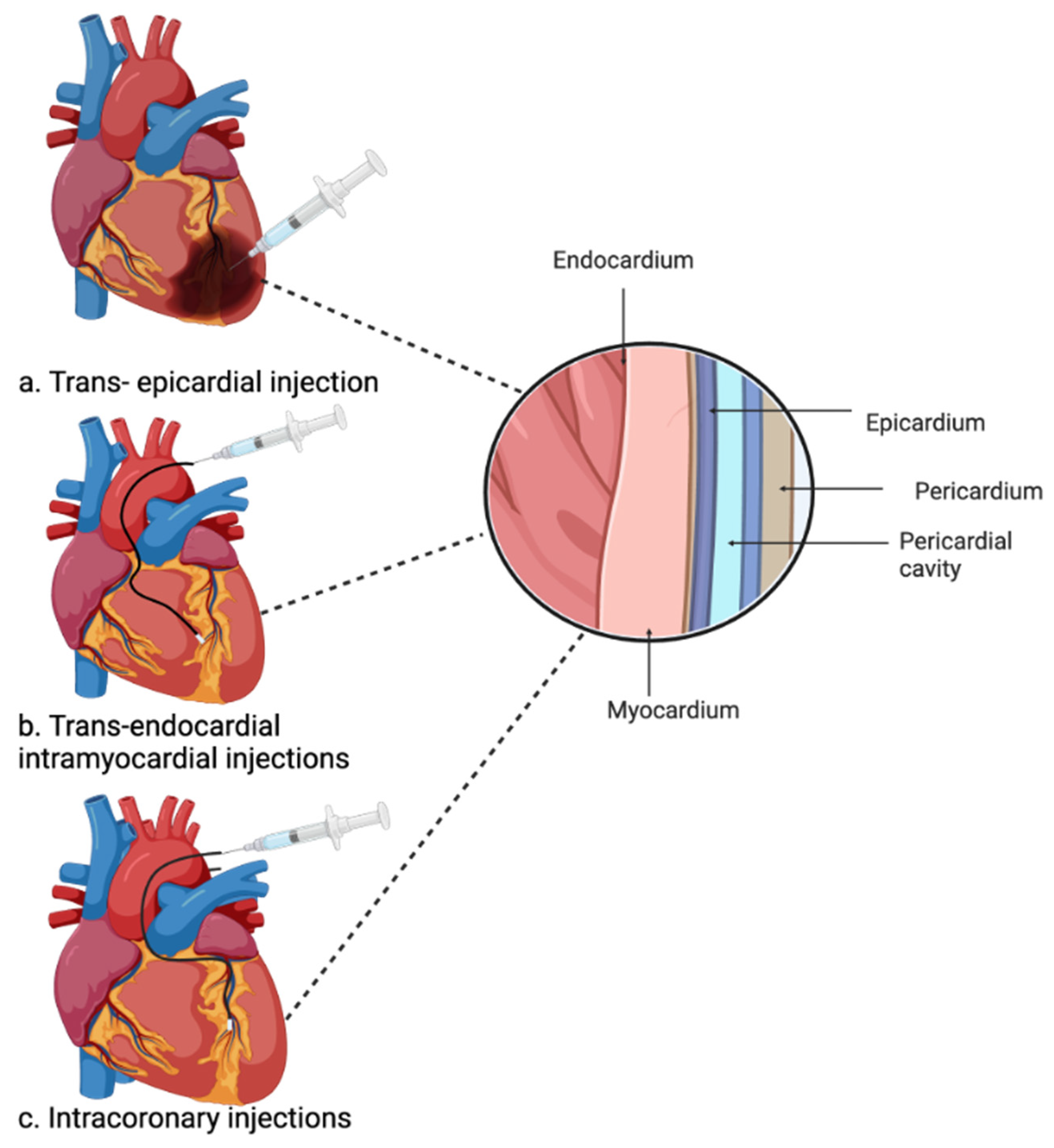
Injectable hydrogels hold significant promise in cardiac regeneration, but several challenges need to be addressed. These hydrogels often exhibit low mechanical strength and carry the potential to trigger an immune reaction upon injection into the heart [126]. Additionally, the application of injectable hydrogels for heart treatment aims to minimise the need for open heart surgery and enhance their clinical significance. However, the implantation methods themselves may pose certain risks. One common method involves directly injecting the hydrogel into the myocardium through the epicardium. This approach, however, necessitates a large injection, making it highly invasive for the patient, alongside a thoracotomy which introduces surgical complications [129,130] (Figure 3). Trans-endocardial intramyocardial injections use a catheter to guide the hydrogel to the heart injury site and are considered safer and more precise in terms of injecting the hydrogel into the myocardium through the endocardium using a catheter and imaging technology. Nevertheless, this method requires hydrogels with very specific mechanical properties, which limits the availability of suitable biomaterials [131,132]. Instead, intracoronary injection is delivered through the main coronary artery, but it carries the risk of coronary artery obstruction or re-embolization of coronary arteries, thus limiting its clinical use [100,128]. Despite the comparative safety and reduced invasiveness of injectable hydrogels as opposed to cardiac patches, a safer method for delivering hydrogels via injection is still needed to advance their clinical applications.

4.8. Current Progress of Organoids and 3D Bioprinting in Cardiac Regeneration

Organoids are scaled-down versions of organs and tissues, usually generated from stem cells, that create 3D structures which replicate to a degree the function and morphology of the original organ [133]. While these organoid constructs are still limited due to the complex nature of organs and tissues, they have proven effective in drug discovery, modelling diseases, and tissue engineering [132]. Scientists have successfully created cardiac organoid models, which have helped in understanding heart disease and provided a platform to study the biological mechanisms of human heart regeneration, bridging the gap between animal studies on MI and heart disease with human studies [134]. Cardiac organoids are still limited, due to their immaturity, lack of vascularisation, poor reproducibility, and a lack of structural features such as valves. As such, these organoids cannot fully replicate the adult human heart. Once these limitations are overcome, a variety of applications will be unlocked [135]. For instance, tissue generated from engineered cardiac organoids could potentially be transplanted into injured hearts post-MI to replace the damaged myocardium and restore cardiac function [136].
In the field of cardiac organoids, 3D bioprinting is a promising innovation that is gaining popularity. It involves the layering of biomaterials to create a 3D structure that is controllable, customisable, and reproducible [137]. A key component of this process is bio-ink, commonly made up of hydrogels derived from natural or synthetic polymers. A bio-ink should meet several important criteria to be suitable for 3D printing applications [138]. It must be biocompatible to support cell viability and integration with living tissues. Additionally, it should possess robust mechanical strength and flexibility to adapt to the diverse mechanical and chemical requirements of different tissue types [138,139]. Recently, advanced bioactive bio-inks have been developed, incorporating cells such as adult stem cells or iPSCs within them prior to printing. This allows for a more precise distribution of cells within the 3D structure [138,139,140].
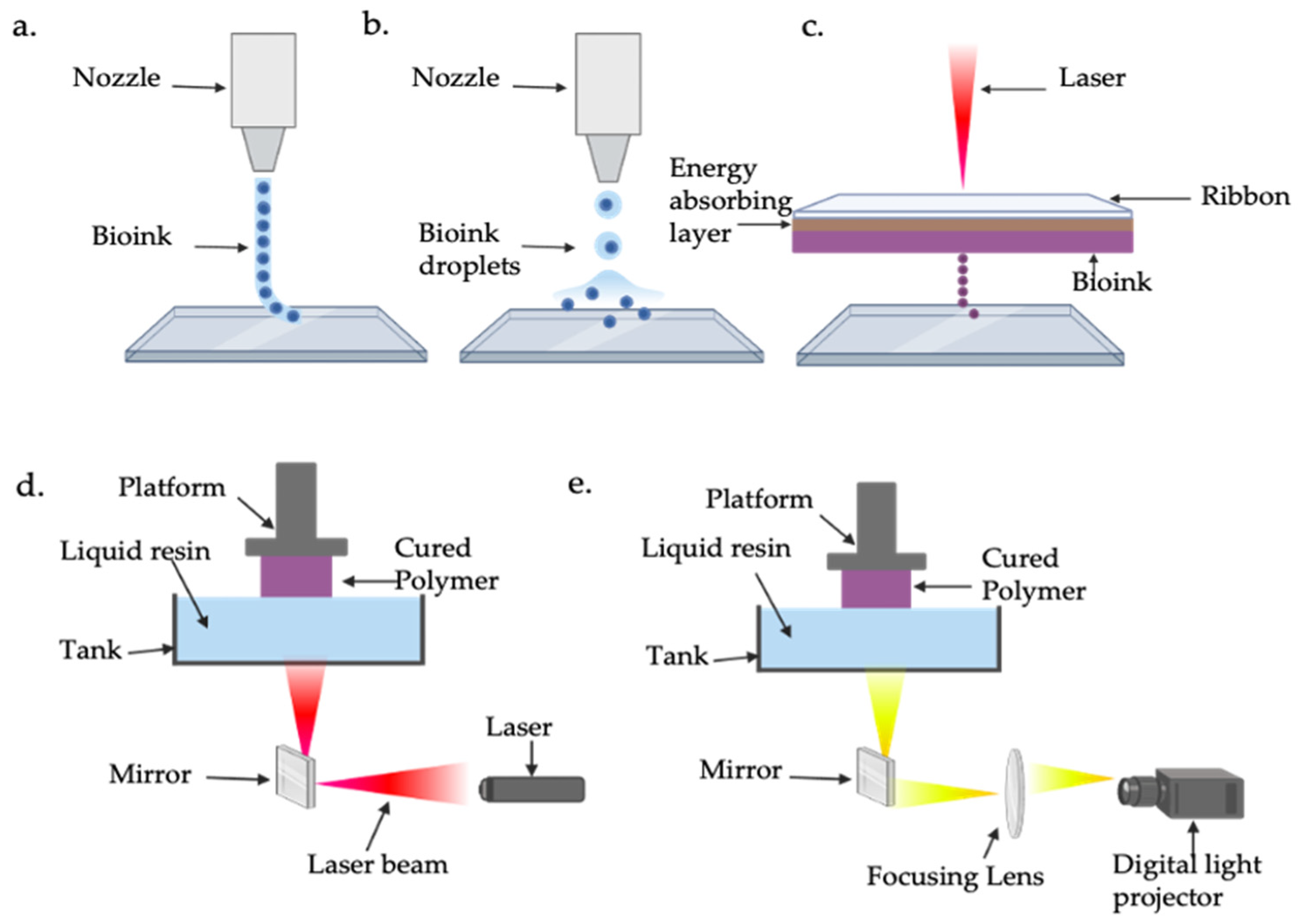
While 3D bioprinting has been effective in tissue engineering for hard tissues such as bone, it is still limited regarding soft materials such as hydrogels and more complete structures like the heart [141]. There are four main bioprinting techniques that vary based on their mechanisms, applications, and resolutions (Table 4) (Figure 4). Droplet-based bioprinting employs bio-printers that continuously deposit biomaterials, bio-inks, or cell-laden droplets to construct 2D or 3D structures. It has high precision, achieving resolutions of up to 50 μm, but struggles with printing highly viscous bio-inks [142,143]. Laser-assisted bioprinting uses a high-intensity laser to propel biomaterial droplets onto a substrate containing cell culture medium. This method requires a ribbon composed of transparent glass, a thin metal layer, and a biomaterial layer, where laser pulses vaporise the metal layer, generating a high-pressure bubble to eject droplets with exceptional precision, achieving resolutions as fine as 5 μm [144,145]. Stereolithography (SLA) and digital light processing (DLP) rely on light-sensitive polymers to fabricate intricate structures. These techniques use either a UV laser beam (SLA) or digital light sources like UV (DLP) to cure liquid photocrosslinkable bio-inks, enabling the creation of highly detailed constructs [146]. Finally, extrusion-based bioprinting stands out as the most commonly used technique due to its simplicity, affordability, and scalability. It employs a computer-controlled system to extrude bio-inks or biomaterials through a nozzle using a pressure system, creating 3D structures layer by layer [147]. While its resolution, at approximately 200 μm, is lower compared to other methods, extrusion-based bioprinting is highly versatile, capable of printing higher cell densities and more viscous bio-inks, and offers better structural integrity through the continuous deposition of filaments. Despite its limitations, extrusion-based bioprinting remains the most popular method due to its practicality in producing larger-scale constructs [148]. In addition, the Freeform Reversible Embedding of Suspended Hydrogels (FRESH) is an emerging extrusion-based bioprinting technique, where hydrogel bio-inks are extruded within a thermo-reversible support medium. This technique aims to address challenges faced when developing soft, complex structures such as the heart [149]. A study introduced an optimised version of the bioprinting technique called FRESH v2.0, which significantly enhanced resolution, enabling the creation of collagen filaments with a resolution of 20 μm in diameter. Using this advanced approach, researchers successfully bioprinted a neonatal-scale human heart using type I collagen bio-ink as a scaffold. To allow for the observation of internal structures, they printed only half of the heart, showcasing intricate features such as trabeculae, atrial and ventricular chambers, and pulmonary and aortic valves [150]. This highlights how bio-inks and advanced 3D bioprinting techniques, such as extrusion-based bioprinting, can be used to generate functional cardiac tissue, offering a promising approach for advancing research in cardiac regeneration and developing effective therapies to repair and regenerate heart tissue damaged by MI.

5. Concluding Remarks and Future Perspectives

5.1. Hydrogel Composition

There is an urgent clinical need for effective novel therapies to treat MI. Various cell types, such as adult stem cells and pluripotent stem cells, show potential for therapeutic application [151]. However, their success depends on the development of suitable scaffolds that can support cell growth and differentiation while also ensuring biological compatibility. Hydrogels offer a promising possibility as a scaffold to support cells in the regeneration of damaged cardiac tissue after an MI [152]. Researchers have faced challenges in developing hydrogels tailored specifically for heart regeneration [55], since they must be meticulously designed to accommodate the mechanical and electrical properties of the heart while also being able to withstand the strong contractions generated by the cardiac muscle [103].
Natural-based hydrogels have proven to be effective in cardiac regeneration due to their ability to mimic the ECM, their biocompatibility, biodegradability, and their very low cytotoxicity, making them suitable for clinical application [153]. Unfortunately, they possess low stability, weak mechanical strength, and rapid degradation in vivo [154]. Conversely, synthetic hydrogels have more controllable features and strong mechanical properties, which provide structural support for the cells, but they have poor biocompatible and biodegradation properties. Some synthetic polymers can be modified through alteration of their functional groups or by combining them with natural polymers to improve their weaker biocompatibility [155], and the development of these hybrid hydrogels along with the incorporation of conductive nanomaterials has led to a significant advancement in cardiac tissue regeneration.

5.2. Hydrogel Delivery Methods

Hydrogels in cardiac tissue regeneration have various delivery methods. Injectable hydrogels are an innovative, minimally invasive approach. They respond to temperature, light, and pH [156], becoming more adaptable after injection. Moreover, incorporation of conductive materials further improves their electrical integration [104]. The advancement of injectable hydrogels has been revolutionary in cardiac tissue regeneration; however, they are affected by poor mechanical force transmission [89] which is being addressed by shear thinning, a technique resulting in increased mechanical strength and stiffness, as well as improved self-healing properties [157]. Nevertheless, to further develop the regenerative capabilities of injectable hydrogels, more novel studies are needed to address the injection application, degradation rate, low survival, and the retention of therapeutic cells.
Hydrogel-based cardiac patches also show promising potential in cardiac tissue repair, especially after an MI. Once integrated with the host, they can deliver therapeutic cells to repair damage in the myocardium. However, limited full electrical integration with the damaged myocardium, immune rejection, and potential surgical complications during transplantation to the heart [116,117,158] limit their therapeutic application. Notably, a novel injectable conductive cardio patch was developed using methacrylated gelatin and elastin, along with CNTs. The remarkable feature of this patch is that it can re-form its original shape post-injection. Tests in rats and minipigs demonstrated significant improvement in cardiac function post-MI [159]. More importantly, this study utilised CNTs to produce cardiac patches that were electrically conductive, a feature not seen in previous research [159]. This breakthrough can lead to the development of an injectable minimally invasive cardiac patch with improved conductive properties. This further highlights the possibilities an injectable cardiac patch could have for post-MI treatment in a clinical setting.

5.3. Current and Future Prospects of Bioprinting in Cardiac Regeneration

Bioprinting has emerged as an innovative technology with the ability to create and develop 3D structures made from various biomaterials. Its potential applications have garnered significant attention, particularly when combined with stem cells [138,160,161]. The ability to fabricate heart tissue through bioprinting holds promise for addressing heart disease and tissue damage, but there are still numerous challenges that must first be addressed and overcome to ensure its success in clinical applications. These obstacles involve the printing techniques, the bio-inks, the mechanical strength of printed 3D structures, cell viability, and vascularisation. In addition, the bio-inks also need improvements in compatibility, stiffnesses, integrin signalling, and cell motility [162]. A promising area of future research in bioprinting is 4D bioprinting, an advanced technique that combines 3D bioprinting technology with smart materials capable of changing their properties or shapes in response to external stimuli. These materials are engineered to react to factors such as pH, light, temperature, or electric and magnetic fields, enabling dynamic and responsive changes in the printed structures over time [163]. In 4D bioprinting, hydrogels play a crucial role as they can be designed to undergo these stimuli-driven changes, making them particularly suitable for biomedical applications. This innovative approach is especially significant in cardiac regeneration, where it enables the creation of scaffolds that can adapt to the heart’s dynamic environment, respond to biomechanical cues, and promote tissue growth and repair [164]. By mimicking the heart’s native behaviour and adapting to its changing conditions, 4D bioprinting offers the potential for advancing research in cardiac regeneration and improving therapies for MI [161].
In summary, synthetic and natural hydrogels possess unique qualities that can provide a supporting environment, enhancing the differentiation and maturation of implanted cells. Hydrogels can be used in various applications such as cardiac patches, injectable hydrogels and 3D bioprinting, which can help promote cardiac tissue regeneration. Although these methods have great potential, some refinement is needed before proceeding to clinical applications.

Author Contributions

S.H., S.M.R., J.B., and C.P. contributed to the literature research, discussion, and interpretation. S.H. drafted the manuscript. All authors have critically read, revised, and approved the final version of the manuscript. All authors have read and agreed to the published version of the manuscript.

Funding

This work has been supported by the Engineering and Physical Sciences Research Council (EPSRC) through a Ph.D. studentship to Sonja Holme; references EP/T517823/1 and EP/W524347/1.

Conflicts of Interest

The authors declare no conflict of interest.

References

  1. Sanchis-Gomar, F.; Perez-Quilis, C.; Leischik, R.; Lucia, A. Epidemiology of coronary heart disease and acute coronary syndrome. Ann. Transl. Med. 2016, 4, 256. [Google Scholar] [CrossRef] [PubMed]
  2. Mensah, G.A.; Fuster, V.; Murray, C.J.L.; Roth, G.A. Global Burden of Cardiovascular Diseases and Risks, 1990–2022. J. Am. Coll. Cardiol. 2023, 82, 2350–2473. [Google Scholar] [CrossRef] [PubMed]
  3. Global Burden of Disease Collaborative Network. Global Burden of Disease Study 2021 (GBD 2021); Institute for Health Metrics and Evaluation (IHME): Seattle, WA, USA, 2024; Available online: http://www.healthdata.org/research-analysis/library/global-burden-disease-2021-findings-gbd-2021-study (accessed on 13 January 2025).
  4. World Health Organization. UN Decade of Healthy Ageing: Plan of Action 2021–2030. Available online: https://cdn.who.int/media/docs/default-source/decade-of-healthy-ageing/decade-proposal-final-apr2020-en.pdf (accessed on 12 December 2023).
  5. Mensah, G.A.; Fuster, V.; Roth, G.A. A Heart-Healthy and Stroke-Free World: Using Data to Inform Global Action. J. Am. Coll. Cardiol. 2023, 82, 2343–2349. [Google Scholar] [CrossRef]
  6. Institute of Medicine Committee on Social Security Cardiovascular Disability Criteria. Cardiovascular Disability: Updating the Social Security Listings; National Academies Press: Washington, DC, USA, 2010. [Google Scholar]
  7. Mendis, S.; Thygesen, K.; Kuulasmaa, K.; Giampaoli, S.; Mähönen, M.; Ngu Blackett, K.; Lisheng, L. World Health Organization definition of myocardial infarction: 2008-09 revision. Int. J. Epidemiol. 2011, 40, 139–146. [Google Scholar] [CrossRef]
  8. Burke, A.P.; Virmani, R. Pathophysiology of Acute Myocardial Infarction. Med. Clin. N. Am. 2007, 91, 553–572. [Google Scholar] [CrossRef]
  9. Lüscher, T.F. Myocardial infarction: Mechanisms, diagnosis, and complications. Eur. Heart J. 2015, 36, 947–949. [Google Scholar] [CrossRef] [PubMed]
  10. Damluji, A.A.; van Diepen, S.; Katz, J.N.; Menon, V.; Tamis-Holland, J.E.; Bakitas, M.; Cohen, M.G.; Balsam, L.B.; Chikwe, J. Mechanical Complications of Acute Myocardial Infarction: A Scientific Statement from the American Heart Association. Circulation 2021, 144, e16–e35. [Google Scholar] [CrossRef]
  11. Jernberg, T.; Hasvold, P.; Henriksson, M.; Hjelm, H.; Thuresson, M.; Janzon, M. Cardiovascular risk in post-myocardial infarction patients: Nationwide real world data demonstrate the importance of a long-term perspective. Eur. Heart J. 2015, 36, 1163–1170. [Google Scholar] [CrossRef]
  12. Lu, L.; Liu, M.; Sun, R.; Zheng, Y.; Zhang, P. Myocardial Infarction: Symptoms and Treatments. Cell Biochem. Biophys. 2015, 72, 865–867. [Google Scholar] [CrossRef]
  13. Ahmad, M.; Mehta, P.; Reddivari, A.K.R.; Mungee, S. Percutaneous Coronary Intervention. In StatPearls; StatPearls Publishing LLC.: Treasure Island, FL, USA, 2024. [Google Scholar]
  14. Mousaei Ghasroldasht, M.; Seok, J.; Park, H.S.; Liakath Ali, F.B.; Al-Hendy, A. Stem Cell Therapy: From Idea to Clinical Practice. Int. J. Mol. Sci. 2022, 23, 2850. [Google Scholar] [CrossRef]
  15. Beltrami, A.P.; Urbanek, K.; Kajstura, J.; Yan, S.M.; Finato, N.; Bussani, R.; Nadal-Ginard, B.; Silvestri, F.; Leri, A.; Beltrami, C.A.; et al. Evidence that human cardiac myocytes divide after myocardial infarction. N. Engl. J. Med. 2001, 344, 1750–1757. [Google Scholar] [CrossRef]
  16. Bergmann, O.; Bhardwaj, R.D.; Bernard, S.; Zdunek, S.; Barnabé-Heider, F.; Walsh, S.; Zupicich, J.; Alkass, K.; Buchholz, B.A.; Druid, H.; et al. Evidence for cardiomyocyte renewal in humans. Science 2009, 324, 98–102. [Google Scholar] [CrossRef] [PubMed]
  17. Beliën, H.; Evens, L.; Hendrikx, M.; Bito, V.; Bronckaers, A. Combining stem cells in myocardial infarction: The road to superior repair? Med. Res. Rev. 2022, 42, 343–373. [Google Scholar] [CrossRef] [PubMed]
  18. Duval, K.; Grover, H.; Han, L.H.; Mou, Y.; Pegoraro, A.F.; Fredberg, J.; Chen, Z. Modeling Physiological Events in 2D vs. 3D Cell Culture. Physiology 2017, 32, 266–277. [Google Scholar] [CrossRef] [PubMed]
  19. Chaicharoenaudomrung, N.; Kunhorm, P.; Noisa, P. Three-dimensional cell culture systems as an in vitro platform for cancer and stem cell modeling. World J. Stem Cells 2019, 11, 1065–1083. [Google Scholar] [CrossRef]
  20. Gisone, I.; Cecchettini, A.; Ceccherini, E.; Persiani, E.; Morales, M.A.; Vozzi, F. Cardiac tissue engineering: Multiple approaches and potential applications. Front. Bioeng. Biotechnol. 2022, 10, 980393. [Google Scholar] [CrossRef]
  21. Oh, S.; Choo, A.B.H. Stem Cells. In Comprehensive Biotechnology, 2nd ed.; Academic Press: Burlington, VT, USA, 2011; pp. 341–365. [Google Scholar] [CrossRef]
  22. Augustine, R.; Dan, P.; Hasan, A.; Khalaf, I.M.; Prasad, P.; Ghosal, K.; Gentile, C.; McClements, L.; Maureira, P. Stem cell-based approaches in cardiac tissue engineering: Controlling the microenvironment for autologous cells. Biomed. Pharmacother. 2021, 138, 111425. [Google Scholar] [CrossRef]
  23. Howard, D.; Buttery, L.D.; Shakesheff, K.M.; Roberts, S.J. Tissue engineering: Strategies, stem cells and scaffolds. J. Anat. 2008, 213, 66–72. [Google Scholar] [CrossRef]
  24. Prochazkova, M.; Chavez, M.G.; Prochazka, J.; Felfy, H.; Mushegyan, V.; Klein, O.D. Chapter 18: Embryonic Versus Adult Stem Cells. In Stem Cell Biology and Tissue Engineering in Dental Sciences; Vishwakarma, A., Sharpe, P., Shi, S., Ramalingam, M., Eds.; Academic Press: Boston, MA, USA, 2015; pp. 249–262. [Google Scholar]
  25. Akter, F.; Hamid, H. Chapter 6-Cardiovascular Tissue Engineering. In Tissue Engineering Made Easy; Akter, F., Ed.; Academic Press: Cambridge, MA, USA, 2016; pp. 55–65. [Google Scholar]
  26. Liew, L.C.; Ho, B.X.; Soh, B.S. Mending a broken heart: Current strategies and limitations of cell-based therapy. Stem Cell Res. Ther. 2020, 11, 138. [Google Scholar] [CrossRef]
  27. Turksen, K. Adult stem cells and cardiac regeneration. Stem Cell Rev. Rep. 2013, 9, 537–540. [Google Scholar] [CrossRef]
  28. Ott, H.C.; Rajab, T.K. Chapter 13-Tissue-Derived Matrices. In In Situ Tissue Regeneration; Lee, S.J., Yoo, J.J., Atala, A., Eds.; Academic Press: Boston, MA, USA, 2016; pp. 229–250. [Google Scholar]
  29. Thomson, J.A.; Itskovitz-Eldor, J.; Shapiro, S.S.; Waknitz, M.A.; Swiergiel, J.J.; Marshall, V.S.; Jones, J.M. Embryonic stem cell lines derived from human blastocysts. Science 1998, 282, 1145–1147. [Google Scholar] [CrossRef] [PubMed]
  30. Ghodsizad, A.; Ruhparwar, A.; Bordel, V.; Mirsaidighazi, E.; Klein, H.M.; Koerner, M.M.; Karck, M.; El-Banayosy, A. Clinical application of adult stem cells for therapy for cardiac disease. Cardiovasc. Ther. 2013, 31, 323–334. [Google Scholar] [CrossRef] [PubMed]
  31. Takahashi, K.; Yamanaka, S. Induction of pluripotent stem cells from mouse embryonic and adult fibroblast cultures by defined factors. Cell 2006, 126, 663–676. [Google Scholar] [CrossRef]
  32. Takahashi, K.; Tanabe, K.; Ohnuki, M.; Narita, M.; Ichisaka, T.; Tomoda, K.; Yamanaka, S. Induction of pluripotent stem cells from adult human fibroblasts by defined factors. Cell 2007, 131, 861–872. [Google Scholar] [CrossRef] [PubMed]
  33. Wang, K.L.; Xue, Q.; Xu, X.H.; Hu, F.; Shao, H. Recent progress in induced pluripotent stem cell-derived 3D cultures for cardiac regeneration. Cell Tissue Res. 2021, 384, 231–240. [Google Scholar] [CrossRef]
  34. Chachques, J.C.; Acar, C.; Herreros, J.; Trainini, J.C.; Prosper, F.; D’Attellis, N.; Fabiani, J.N.; Carpentier, A.F. Cellular cardiomyoplasty: Clinical application. Ann. Thorac. Surg. 2004, 77, 1121–1130. [Google Scholar] [CrossRef]
  35. Baraniak, P.R.; McDevitt, T.C. Stem cell paracrine actions and tissue regeneration. Regen. Med. 2010, 5, 121–143. [Google Scholar] [CrossRef]
  36. Menasché, P.; Hagège, A.A.; Scorsin, M.; Pouzet, B.; Desnos, M.; Duboc, D.; Schwartz, K.; Vilquin, J.T.; Marolleau, J.P. Myoblast transplantation for heart failure. Lancet 2001, 357, 279–280. [Google Scholar] [CrossRef]
  37. Muqresh, M.A.; Harbieh, I.; Haider, K.H. Myogenic Cardiac Regeneration: Clinical Studies Using Skeletal Myoblasts. In Handbook of Stem Cell Applications; Springer: Berlin/Heidelberg, Germany, 2023; pp. 1–37. [Google Scholar]
  38. Hare, J.M.; Fishman, J.E.; Gerstenblith, G.; DiFede Velazquez, D.L.; Zambrano, J.P.; Suncion, V.Y.; Tracy, M.; Ghersin, E.; Johnston, P.V.; Brinker, J.A.; et al. Comparison of allogeneic vs autologous bone marrow–derived mesenchymal stem cells delivered by transendocardial injection in patients with ischemic cardiomyopathy: The POSEIDON randomized trial. JAMA 2012, 308, 2369–2379. [Google Scholar] [CrossRef]
  39. Hashimoto, H.; Olson, E.N.; Bassel-Duby, R. Therapeutic approaches for cardiac regeneration and repair. Nat. Rev. Cardiol. 2018, 15, 585–600. [Google Scholar] [CrossRef]
  40. Mathiasen, A.B.; Qayyum, A.A.; Jørgensen, E.; Helqvist, S.; Fischer-Nielsen, A.; Kofoed, K.F.; Haack-Sørensen, M.; Ekblond, A.; Kastrup, J. Bone marrow-derived mesenchymal stromal cell treatment in patients with severe ischaemic heart failure: A randomized placebo-controlled trial (MSC-HF trial). Eur. Heart J. 2015, 36, 1744–1753. [Google Scholar] [CrossRef]
  41. Chong, J.J.; Yang, X.; Don, C.W.; Minami, E.; Liu, Y.W.; Weyers, J.J.; Mahoney, W.M.; Van Biber, B.; Cook, S.M.; Palpant, N.J.; et al. Human embryonic-stem-cell-derived cardiomyocytes regenerate non-human primate hearts. Nature 2014, 510, 273–277. [Google Scholar] [CrossRef]
  42. Zhu, K.; Wu, Q.; Ni, C.; Zhang, P.; Zhong, Z.; Wu, Y.; Wang, Y.; Xu, Y.; Kong, M.; Cheng, H.; et al. Lack of Remuscularization Following Transplantation of Human Embryonic Stem Cell-Derived Cardiovascular Progenitor Cells in Infarcted Nonhuman Primates. Circ. Res. 2018, 122, 958–969. [Google Scholar] [CrossRef] [PubMed]
  43. Liu, Y.W.; Chen, B.; Yang, X.; Fugate, J.A.; Kalucki, F.A.; Futakuchi-Tsuchida, A.; Couture, L.; Vogel, K.W.; Astley, C.A.; Baldessari, A.; et al. Human embryonic stem cell-derived cardiomyocytes restore function in infarcted hearts of non-human primates. Nat. Biotechnol. 2018, 36, 597–605. [Google Scholar] [CrossRef] [PubMed]
  44. Chi, C.; Song, K. Cellular reprogramming of fibroblasts in heart regeneration. J. Mol. Cell. Cardiol. 2023, 180, 84–93. [Google Scholar] [CrossRef]
  45. Huang, H.; Huang, G.N.; Payumo, A.Y. Two decades of heart regeneration research: Cardiomyocyte proliferation and beyond. WIREs Mech. Dis. 2024, 16, e1629. [Google Scholar] [CrossRef]
  46. Shiba, Y.; Gomibuchi, T.; Seto, T.; Wada, Y.; Ichimura, H.; Tanaka, Y.; Ogasawara, T.; Okada, K.; Shiba, N.; Sakamoto, K.; et al. Allogeneic transplantation of iPS cell-derived cardiomyocytes regenerates primate hearts. Nature 2016, 538, 388–391. [Google Scholar] [CrossRef]
  47. Wu, P.; Sai, X.; Li, Z.; Ye, X.; Jin, L.; Liu, G.; Li, G.; Yang, P.; Zhao, M.; Zhu, S.; et al. Maturation of induced pluripotent stem cell-derived cardiomyocytes and its therapeutic effect on myocardial infarction in mouse. Bioact. Mater. 2023, 20, 286–305. [Google Scholar] [CrossRef] [PubMed]
  48. Shida, M.; Miyagawa, S.; Saito, A.; Fukushima, S.; Harada, A.; Ito, E.; Ohashi, F.; Watabe, T.; Hatazawa, J.; Matsuura, K.; et al. Transplantation of Human-induced Pluripotent Stem Cell-derived Cardiomyocytes Is Superior to Somatic Stem Cell Therapy for Restoring Cardiac Function and Oxygen Consumption in a Porcine Model of Myocardial Infarction. Transplantation 2019, 103, 291–298. [Google Scholar] [CrossRef]
  49. Kawamura, M.; Miyagawa, S.; Miki, K.; Saito, A.; Fukushima, S.; Higuchi, T.; Kawamura, T.; Kuratani, T.; Daimon, T.; Shimizu, T.; et al. Feasibility, safety, and therapeutic efficacy of human induced pluripotent stem cell-derived cardiomyocyte sheets in a porcine ischemic cardiomyopathy model. Circulation 2012, 126, S29–S37. [Google Scholar] [CrossRef]
  50. Kishino, Y.; Tohyama, S.; Morita, Y.; Soma, Y.; Tani, H.; Okada, M.; Kanazawa, H.; Fukuda, K. Cardiac Regenerative Therapy Using Human Pluripotent Stem Cells for Heart Failure: A State-of-the-Art Review. J. Card. Fail. 2023, 29, 503–513. [Google Scholar] [CrossRef]
  51. Zhang, Y.; Wang, D.; Chen, M.; Yang, B.; Zhang, F.; Cao, K. Intramyocardial transplantation of undifferentiated rat induced pluripotent stem cells causes tumorigenesis in the heart. PLoS ONE 2011, 6, e19012. [Google Scholar] [CrossRef]
  52. Rienks, M.; Papageorgiou, A.P.; Frangogiannis, N.G.; Heymans, S. Myocardial extracellular matrix: An ever-changing and diverse entity. Circ. Res. 2014, 114, 872–888. [Google Scholar] [CrossRef]
  53. Kristen, L.; Zhijie, W. Chapter 2: Extracellular Matrix in Cardiac Tissue Mechanics and Physiology: Role of Collagen Accumulation. In Extracellular Matrix; Rama Sashank, M., Joseph Orgel, P.R.O., Zvi, L., Eds.; IntechOpen: Rijeka, Croatia, 2021. [Google Scholar] [CrossRef]
  54. Song, R.; Zhang, L. Cardiac ECM: Its Epigenetic Regulation and Role in Heart Development and Repair. Int. J. Mol. Sci. 2020, 21, 8610. [Google Scholar] [CrossRef]
  55. Aisenbrey, E.A.; Murphy, W.L. Synthetic alternatives to Matrigel. Nat. Rev. Mater. 2020, 5, 539–551. [Google Scholar] [CrossRef]
  56. Funaki, M.; Janmey, P.A. Chapter 23-Technologies to Engineer Cell Substrate Mechanics in Hydrogels. In Biology and Engineering of Stem Cell Niches; Elsevier: Amsterdam, The Netherlands, 2017; pp. 363–373. [Google Scholar]
  57. Peterson, N.C. From bench to cageside: Risk assessment for rodent pathogen contamination of cells and biologics. ILAR J. 2008, 49, 310–315. [Google Scholar] [CrossRef]
  58. Abu Shmeis, R.M. Chapter Four-Nanotechnology in wastewater treatment. In Comprehensive Analytical Chemistry; Turan, N.B., Engin, G.O., Bilgili, M.S., Eds.; Elsevier: Amsterdam, The Netherlands, 2022; Volume 99, pp. 105–134. [Google Scholar]
  59. Spicer, C.D. Hydrogel scaffolds for tissue engineering: The importance of polymer choice. Polym. Chem. 2020, 11, 184–219. [Google Scholar] [CrossRef]
  60. Chaudhary, S.; Chakraborty, E. Hydrogel based tissue engineering and its future applications in personalized disease modeling and regenerative therapy. Beni Suef Univ. J. Basic Appl. Sci. 2022, 11, 3. [Google Scholar] [CrossRef]
  61. Drury, J.L.; Mooney, D.J. Hydrogels for tissue engineering: Scaffold design variables and applications. Biomaterials 2003, 24, 4337–4351. [Google Scholar] [CrossRef]
  62. Gomez-Florit, M.; Pardo, A.; Domingues, R.M.A.; Graça, A.L.; Babo, P.S.; Reis, R.L.; Gomes, M.E. Natural-Based Hydrogels for Tissue Engineering Applications. Molecules 2020, 25, 5858. [Google Scholar] [CrossRef]
  63. Madduma-Bandarage, U.S.K.; Madihally, S.V. Synthetic hydrogels: Synthesis, novel trends, and applications. J. Appl. Polym. Sci. 2021, 138, 50376. [Google Scholar] [CrossRef]
  64. Chamkouri, H.; Chamkouri, M. A review of hydrogels, their properties and applications in medicine. Am. J. Biomed. Sci. Res. 2021, 11, 485–493. [Google Scholar] [CrossRef]
  65. Hu, W.; Wang, Z.; Xiao, Y.; Zhang, S.; Wang, J. Advances in crosslinking strategies of biomedical hydrogels. Biomater. Sci. 2019, 7, 843–855. [Google Scholar] [CrossRef]
  66. Ahmed, E.M. Hydrogel: Preparation, characterization, and applications: A review. J. Adv. Res. 2015, 6, 105–121. [Google Scholar] [CrossRef]
  67. Stojkov, G.; Niyazov, Z.; Picchioni, F.; Bose, R.K. Relationship between Structure and Rheology of Hydrogels for Various Applications. Gels 2021, 7, 255. [Google Scholar] [CrossRef]
  68. Hama, R.; Ulziibayar, A.; Reinhardt, J.W.; Watanabe, T.; Kelly, J.; Shinoka, T. Recent Developments in Biopolymer-Based Hydrogels for Tissue Engineering Applications. Biomolecules 2023, 13, 280. [Google Scholar] [CrossRef] [PubMed]
  69. Chen, T.; Chen, Y.; Rehman, H.U.; Chen, Z.; Yang, Z.; Wang, M.; Li, H.; Liu, H. Ultratough, Self-Healing, and Tissue-Adhesive Hydrogel for Wound Dressing. ACS Appl. Mater. Interfaces 2018, 10, 33523–33531. [Google Scholar] [CrossRef]
  70. Mann, J.L.; Yu, A.C.; Agmon, G.; Appel, E.A. Supramolecular polymeric biomaterials. Biomater. Sci. 2018, 6, 10–37. [Google Scholar] [CrossRef]
  71. Pan, Z.; Ye, H.; Wu, D. Recent advances on polymeric hydrogels as wound dressings. APL Bioeng. 2021, 5, 011504. [Google Scholar] [CrossRef]
  72. Klouda, L.; Mikos, A.G. Thermoresponsive hydrogels in biomedical applications. Eur. J. Pharm. Biopharm. 2008, 68, 34–45. [Google Scholar] [CrossRef]
  73. Gacesa, P. Alginates. Carbohydr. Polym. 1988, 8, 161–182. [Google Scholar] [CrossRef]
  74. Zhang, Y.; Song, M.; Diao, Y.; Li, B.; Shi, L.; Ran, R. Preparation and properties of polyacrylamide/polyvinyl alcohol physical double network hydrogel. RSC Adv. 2016, 6, 112468–112476. [Google Scholar] [CrossRef]
  75. Cascone, M.G.; Maltinti, S.; Barbani, N.; Laus, M. Effect of chitosan and dextran on the properties of poly (vinyl alcohol) hydrogels. J. Mater. Sci. Mater. Med. 1999, 10, 431–435. [Google Scholar] [CrossRef]
  76. Nasution, H.; Harahap, H.; Dalimunthe, N.F.; Ginting, M.H.S.; Jaafar, M.; Tan, O.O.H.; Aruan, H.K.; Herfananda, A.L. Hydrogel and Effects of Crosslinking Agent on Cellulose-Based Hydrogels: A Review. Gels 2022, 8, 568. [Google Scholar] [CrossRef] [PubMed]
  77. Ermis, M.; Calamak, S.; Calibasi Kocal, G.; Guven, S.; Durmus, N.G.; Rizvi, I.; Hasan, T.; Hasirci, N.; Hasirci, V.; Demirci, U. Chapter 15-Hydrogels as a New Platform to Recapitulate the Tumor Microenvironment. In Handbook of Nanomaterials for Cancer Theranostics; Conde, J., Ed.; Elsevier: Amsterdam, The Netherlands, 2018; pp. 463–494. [Google Scholar]
  78. Li, Z.; Lu, F.; Liu, Y. A Review of the Mechanism, Properties, and Applications of Hydrogels Prepared by Enzymatic Cross-linking. J. Agric. Food Chem. 2023, 71, 10238–10249. [Google Scholar] [CrossRef] [PubMed]
  79. Heck, T.; Faccio, G.; Richter, M.; Thöny-Meyer, L. Enzyme-catalyzed protein crosslinking. Appl. Microbiol. Biotechnol. 2013, 97, 461–475. [Google Scholar] [CrossRef] [PubMed]
  80. Gyles, D.A.; Castro, L.D.; Silva, J.O.C.; Ribeiro-Costa, R.M. A review of the designs and prominent biomedical advances of natural and synthetic hydrogel formulations. Eur. Polym. J. 2017, 88, 373–392. [Google Scholar] [CrossRef]
  81. Chattopadhyay, S.; Raines, R.T. Review collagen-based biomaterials for wound healing. Biopolymers 2014, 101, 821–833. [Google Scholar] [CrossRef]
  82. Kassiri, Z.; Khokha, R. Myocardial extra-cellular matrix and its regulation by metalloproteinases and their inhibitors. Thromb. Haemost. 2005, 93, 212–219. [Google Scholar] [CrossRef]
  83. Andreazza, R.; Morales, A.; Pieniz, S.; Labidi, J. Gelatin-Based Hydrogels: Potential Biomaterials for Remediation. Polymers 2023, 15, 1026. [Google Scholar] [CrossRef]
  84. Sun, M.; Sun, X.; Wang, Z.; Guo, S.; Yu, G.; Yang, H. Synthesis and Properties of Gelatin Methacryloyl (GelMA) Hydrogels and Their Recent Applications in Load-Bearing Tissue. Polymers 2018, 10, 1290. [Google Scholar] [CrossRef] [PubMed]
  85. Lierova, A.; Kasparova, J.; Filipova, A.; Cizkova, J.; Pekarova, L.; Korecka, L.; Mannova, N.; Bilkova, Z.; Sinkorova, Z. Hyaluronic Acid: Known for Almost a Century, but Still in Vogue. Pharmaceutics 2022, 14, 838. [Google Scholar] [CrossRef] [PubMed]
  86. Xu, X.; Jha, A.K.; Harrington, D.A.; Farach-Carson, M.C.; Jia, X. Hyaluronic acid-based hydrogels: From a natural polysaccharide to complex networks. Soft Matter 2012, 8, 3280–3294. [Google Scholar] [CrossRef]
  87. Catoira, M.C.; Fusaro, L.; Di Francesco, D.; Ramella, M.; Boccafoschi, F. Overview of natural hydrogels for regenerative medicine applications. J. Mater. Sci. Mater. Med. 2019, 30, 115. [Google Scholar] [CrossRef]
  88. Sionkowska, A. Collagen blended with natural polymers: Recent advances and trends. Prog. Polym. Sci. 2021, 122, 101452. [Google Scholar] [CrossRef]
  89. Hasan, A.; Khattab, A.; Islam, M.A.; Hweij, K.A.; Zeitouny, J.; Waters, R.; Sayegh, M.; Hossain, M.M.; Paul, A. Injectable Hydrogels for Cardiac Tissue Repair after Myocardial Infarction. Adv. Sci. 2015, 2, 1500122. [Google Scholar] [CrossRef]
  90. Christman, K.L.; Vardanian, A.J.; Fang, Q.; Sievers, R.E.; Fok, H.H.; Lee, R.J. Injectable fibrin scaffold improves cell transplant survival, reduces infarct expansion, and induces neovasculature formation in ischemic myocardium. J. Am. Coll. Cardiol. 2004, 44, 654–660. [Google Scholar] [CrossRef]
  91. Alagarsamy, K.N.; Yan, W.; Srivastava, A.; Desiderio, V.; Dhingra, S. Application of injectable hydrogels for cardiac stem cell therapy and tissue engineering. Rev. Cardiovasc. Med. 2019, 20, 221–230. [Google Scholar] [CrossRef]
  92. Khanna, A.; Zamani, M.; Huang, N.F. Extracellular Matrix-Based Biomaterials for Cardiovascular Tissue Engineering. J. Cardiovasc. Dev. Dis. 2021, 8, 137. [Google Scholar] [CrossRef]
  93. Verma, D.; Bhatia, A.; Chopra, S.; Dua, K.; Prasher, P.; Gupta, G.; Tambuwala, M.M.; Chellappan, D.K.; Aljabali, A.A.A.; Sharma, M.; et al. Chapter 28: Advancements on microparticles-based drug delivery systems for cancer therapy. In Advanced Drug Delivery Systems in the Management of Cancer; Dua, K., Mehta, M., de Jesus Andreoli Pinto, T., Pont, L.G., Williams, K.A., Rathbone, M.J., Eds.; Academic Press: Cambridge, MA, USA, 2021; pp. 351–358. [Google Scholar]
  94. Fischer, R.S.; Myers, K.A.; Gardel, M.L.; Waterman, C.M. Stiffness-controlled three-dimensional extracellular matrices for high-resolution imaging of cell behavior. Nat. Protoc. 2012, 7, 2056–2066. [Google Scholar] [CrossRef]
  95. Patel, L.; Worch, J.C.; Dove, A.P.; Gehmlich, K. The Utilisation of Hydrogels for iPSC-Cardiomyocyte Research. Int. J. Mol. Sci. 2023, 24, 9995. [Google Scholar] [CrossRef]
  96. Zhu, J. Bioactive modification of poly (ethylene glycol) hydrogels for tissue engineering. Biomaterials 2010, 31, 4639–4656. [Google Scholar] [CrossRef]
  97. Li, Z.; Guan, J. Hydrogels for Cardiac Tissue Engineering. Polymers 2011, 3, 740–761. [Google Scholar] [CrossRef]
  98. Alexander, A.; Ajazuddin; Khan, J.; Saraf, S.; Saraf, S. Polyethylene glycol (PEG)–Poly (N-isopropylacrylamide) (PNIPAAm) based thermosensitive injectable hydrogels for biomedical applications. Eur. J. Pharm. Biopharm. 2014, 88, 575–585. [Google Scholar] [CrossRef] [PubMed]
  99. Jiang, S.; Liu, S.; Feng, W. PVA hydrogel properties for biomedical application. J. Mech. Behav. Biomed. Mater. 2011, 4, 1228–1233. [Google Scholar] [CrossRef] [PubMed]
  100. Saludas, L.; Pascual-Gil, S.; Prósper, F.; Garbayo, E.; Blanco-Prieto, M. Hydrogel based approaches for cardiac tissue engineering. Int. J. Pharm. 2017, 523, 454–475. [Google Scholar] [CrossRef]
  101. Klotz, B.J.; Gawlitta, D.; Rosenberg, A.J.W.P.; Malda, J.; Melchels, F.P.W. Gelatin-Methacryloyl Hydrogels: Towards Biofabrication-Based Tissue Repair. Trends Biotechnol. 2016, 34, 394–407. [Google Scholar] [CrossRef]
  102. Yue, K.; Trujillo-de Santiago, G.; Alvarez, M.M.; Tamayol, A.; Annabi, N.; Khademhosseini, A. Synthesis, properties, and biomedical applications of gelatin methacryloyl (GelMA) hydrogels. Biomaterials 2015, 73, 254–271. [Google Scholar] [CrossRef]
  103. Peña, B.; Laughter, M.; Jett, S.; Rowland, T.J.; Taylor, M.R.G.; Mestroni, L.; Park, D. Injectable Hydrogels for Cardiac Tissue Engineering. Macromol. Biosci. 2018, 18, 1800079. [Google Scholar] [CrossRef]
  104. Kougkolos, G.; Golzio, M.; Laudebat, L.; Valdez-Nava, Z.; Flahaut, E. Hydrogels with electrically conductive nanomaterials for biomedical applications. J. Mater. Chem. B 2023, 11, 2036–2062. [Google Scholar] [CrossRef]
  105. Ashtari, K.; Nazari, H.; Ko, H.; Tebon, P.; Akhshik, M.; Akbari, M.; Alhosseini, S.N.; Mozafari, M.; Mehravi, B.; Soleimani, M.; et al. Electrically conductive nanomaterials for cardiac tissue engineering. Adv. Drug Deliv. Rev. 2019, 144, 162–179. [Google Scholar] [CrossRef] [PubMed]
  106. Li, X.-P.; Qu, K.-Y.; Zhou, B.; Zhang, F.; Wang, Y.-Y.; Abodunrin, O.D.; Zhu, Z.; Huang, N.-P. Electrical stimulation of neonatal rat cardiomyocytes using conductive polydopamine-reduced graphene oxide-hybrid hydrogels for constructing cardiac microtissues. Colloids Surf. B Biointerfaces 2021, 205, 111844. [Google Scholar] [CrossRef]
  107. Ding, X.; Zhao, H.; Li, Y.; Lee, A.L.; Li, Z.; Fu, M.; Li, C.; Yang, Y.Y.; Yuan, P. Synthetic peptide hydrogels as 3D scaffolds for tissue engineering. Adv. Drug Deliv. Rev. 2020, 160, 78–104. [Google Scholar] [CrossRef] [PubMed]
  108. French, K.M.; Somasuntharam, I.; Davis, M.E. Self-assembling peptide-based delivery of therapeutics for myocardial infarction. Adv. Drug Deliv. Rev. 2016, 96, 40–53. [Google Scholar] [CrossRef]
  109. Chen, J.; Zou, X. Self-assemble peptide biomaterials and their biomedical applications. Bioact. Mater. 2019, 4, 120–131. [Google Scholar] [CrossRef] [PubMed]
  110. Li, H.; Gao, J.; Shang, Y.; Hua, Y.; Ye, M.; Yang, Z.; Ou, C.; Chen, M. Folic Acid Derived Hydrogel Enhances the Survival and Promotes Therapeutic Efficacy of iPS Cells for Acute Myocardial Infarction. ACS Appl. Mater. Interfaces 2018, 10, 24459–24468. [Google Scholar] [CrossRef]
  111. Burgess, K.A.; Frati, C.; Meade, K.; Gao, J.; Castillo Diaz, L.; Madeddu, D.; Graiani, G.; Cavalli, S.; Miller, A.F.; Oceandy, D.; et al. Functionalised peptide hydrogel for the delivery of cardiac progenitor cells. Mater. Sci. Eng. C Mater. Biol. Appl. 2021, 119, 111539. [Google Scholar] [CrossRef] [PubMed]
  112. Caplan, M.R.; Schwartzfarb, E.M.; Zhang, S.; Kamm, R.D.; Lauffenburger, D.A. Control of self-assembling oligopeptide matrix formation through systematic variation of amino acid sequence. Biomaterials 2002, 23, 219–227. [Google Scholar] [CrossRef]
  113. Caplan, M.R.; Schwartzfarb, E.M.; Zhang, S.; Kamm, R.D.; Lauffenburger, D.A. Effects of systematic variation of amino acid sequence on the mechanical properties of a self-assembling, oligopeptide biomaterial. J. Biomater. Sci. Polym. Ed. 2002, 13, 225–236. [Google Scholar] [CrossRef]
  114. Mie, M.; Oomuro, M.; Kobatake, E. Hydrogel scaffolds composed of genetically synthesized self-assembling peptides for three-dimensional cell culture. Polym. J. 2013, 45, 504–508. [Google Scholar] [CrossRef]
  115. Guo, H.-D.; Wu, J.-H.; Wang, H.-J.; Tan, Y.-Z. Delivery of Stem Cells and BMP-2 With Functionalized Self-Assembling Peptide Enhances Regeneration of Infarcted Myocardium. Stem Cell Rev. Rep. 2024, 20, 1540–1554. [Google Scholar] [CrossRef] [PubMed]
  116. Li, M.; Wu, H.; Yuan, Y.; Hu, B.; Gu, N. Recent fabrications and applications of cardiac patch in myocardial infarction treatment. View 2022, 3, 20200153. [Google Scholar] [CrossRef]
  117. Prabhakaran, M.P.; Venugopal, J.; Kai, D.; Ramakrishna, S. Biomimetic material strategies for cardiac tissue engineering. Mater. Sci. Eng. C Mater. Biol. Appl. 2011, 31, 503–513. [Google Scholar] [CrossRef]
  118. Vitali, E.; Colombo, T.; Fratto, P.; Russo, C.; Bruschi, G.; Frigerio, M. Surgical therapy in advanced heart failure. Am. J. Cardiol. 2003, 91, 88F–94F. [Google Scholar] [CrossRef]
  119. Wang, Q.; Yang, H.; Bai, A.; Jiang, W.; Li, X.; Wang, X.; Mao, Y.; Lu, C.; Qian, R.; Guo, F.; et al. Functional engineered human cardiac patches prepared from nature’s platform improve heart function after acute myocardial infarction. Biomaterials 2016, 105, 52–65. [Google Scholar] [CrossRef]
  120. Passier, R.; van Laake, L.W.; Mummery, C.L. Stem-cell-based therapy and lessons from the heart. Nature 2008, 453, 322–329. [Google Scholar] [CrossRef]
  121. Ye, L.; Chang, Y.H.; Xiong, Q.; Zhang, P.; Zhang, L.; Somasundaram, P.; Lepley, M.; Swingen, C.; Su, L.; Wendel, J.S.; et al. Cardiac repair in a porcine model of acute myocardial infarction with human induced pluripotent stem cell-derived cardiovascular cells. Cell Stem Cell 2014, 15, 750–761. [Google Scholar] [CrossRef]
  122. Vasu, S.; Zhou, J.; Chen, J.; Johnston, P.V.; Kim, D.H. Biomaterials-based Approaches for Cardiac Regeneration. Korean Circ. J. 2021, 51, 943–960. [Google Scholar] [CrossRef]
  123. Gao, L.; Gregorich, Z.R.; Zhu, W.; Mattapally, S.; Oduk, Y.; Lou, X.; Kannappan, R.; Borovjagin, A.V.; Walcott, G.P.; Pollard, A.E.; et al. Large Cardiac Muscle Patches Engineered from Human Induced-Pluripotent Stem Cell-Derived Cardiac Cells Improve Recovery from Myocardial Infarction in Swine. Circulation 2018, 137, 1712–1730. [Google Scholar] [CrossRef]
  124. Su, T.; Huang, K.; Daniele, M.A.; Hensley, M.T.; Young, A.T.; Tang, J.; Allen, T.A.; Vandergriff, A.C.; Erb, P.D.; Ligler, F.S.; et al. Cardiac Stem Cell Patch Integrated with Microengineered Blood Vessels Promotes Cardiomyocyte Proliferation and Neovascularization after Acute Myocardial Infarction. ACS Appl. Mater. Interfaces 2018, 10, 33088–33096. [Google Scholar] [CrossRef]
  125. Pupkaite, J.; Sedlakova, V.; Eren Cimenci, C.; Bak, M.; McLaughlin, S.; Ruel, M.; Alarcon, E.I.; Suuronen, E.J. Delivering More of an Injectable Human Recombinant Collagen III Hydrogel Does Not Improve Its Therapeutic Efficacy for Treating Myocardial Infarction. ACS Biomater. Sci. Eng. 2020, 6, 4256–4265. [Google Scholar] [CrossRef] [PubMed]
  126. Vetter, V.C.; Bouten, C.V.C.; van der Pol, A. Hydrogels for Cardiac Restorative Support: Relevance of Gelation Mechanisms for Prospective Clinical Use. Curr. Heart Fail. Rep. 2023, 20, 519–529. [Google Scholar] [CrossRef]
  127. Li, P.; Hu, J.; Wang, J.; Zhang, J.; Wang, L.; Zhang, C. The Role of Hydrogel in Cardiac Repair and Regeneration for Myocardial Infarction: Recent Advances and Future Perspectives. Bioengineering 2023, 10, 165. [Google Scholar] [CrossRef] [PubMed]
  128. Frey, N.; Linke, A.; Süselbeck, T.; Müller-Ehmsen, J.; Vermeersch, P.; Schoors, D.; Rosenberg, M.; Bea, F.; Tuvia, S.; Leor, J. Intracoronary Delivery of Injectable Bioabsorbable Scaffold (IK-5001) to Treat Left Ventricular Remodeling After ST-Elevation Myocardial Infarction. Circ. Cardiovasc. Interv. 2014, 7, 806–812. [Google Scholar] [CrossRef]
  129. Anker, S.D.; Coats, A.J.S.; Cristian, G.; Dragomir, D.; Pusineri, E.; Piredda, M.; Bettari, L.; Dowling, R.; Volterrani, M.; Kirwan, B.-A.; et al. A prospective comparison of alginate-hydrogel with standard medical therapy to determine impact on functional capacity and clinical outcomes in patients with advanced heart failure (AUGMENT-HF trial). Eur. Heart J. 2015, 36, 2297–2309. [Google Scholar] [CrossRef] [PubMed]
  130. He, S.; Zhang, Z.; Luo, R.; Jiang, Q.; Yang, L.; Wang, Y. Advances in Injectable Hydrogel Strategies for Heart Failure Treatment. Adv. Healthc. Mater. 2023, 12, 2300029. [Google Scholar] [CrossRef]
  131. Rodell, C.B.; Lee, M.E.; Wang, H.; Takebayashi, S.; Takayama, T.; Kawamura, T.; Arkles, J.S.; Dusaj, N.N.; Dorsey, S.M.; Witschey, W.R.T.; et al. Injectable Shear-Thinning Hydrogels for Minimally Invasive Delivery to Infarcted Myocardium to Limit Left Ventricular Remodeling. Circ. Cardiovasc. Interv. 2016, 9, e004058. [Google Scholar] [CrossRef]
  132. Silva-Pedrosa, R.; Salgado, A.J.; Ferreira, P.E. Revolutionizing Disease Modeling: The Emergence of Organoids in Cellular Systems. Cells 2023, 12, 930. [Google Scholar] [CrossRef]
  133. Yang, S.; Hu, H.; Kung, H.; Zou, R.; Dai, Y.; Hu, Y.; Wang, T.; Lv, T.; Yu, J.; Li, F. Organoids: The current status and biomedical applications. MedComm 2023, 4, e274. [Google Scholar] [CrossRef]
  134. Xuan, W.; Tipparaju, S.M.; Ashraf, M. Transformational Applications of Human Cardiac Organoids in Cardiovascular Diseases. Front. Cell Dev. Biol. 2022, 10, 936084. [Google Scholar] [CrossRef]
  135. Sahara, M. Recent Advances in Generation of In Vitro Cardiac Organoids. Int. J. Mol. Sci. 2023, 24, 6244. [Google Scholar] [CrossRef] [PubMed]
  136. Tenreiro, M.F.; Louro, A.F.; Alves, P.M.; Serra, M. Next generation of heart regenerative therapies: Progress and promise of cardiac tissue engineering. NPJ Regen. Med. 2021, 6, 30. [Google Scholar] [CrossRef] [PubMed]
  137. Sun, Z.; Zhao, J.; Leung, E.; Flandes-Iparraguirre, M.; Vernon, M.; Silberstein, J.; De-Juan-Pardo, E.M.; Jansen, S. Three-Dimensional Bioprinting in Cardiovascular Disease: Current Status and Future Directions. Biomolecules 2023, 13, 1180. [Google Scholar] [CrossRef] [PubMed]
  138. Gungor-Ozkerim, P.S.; Inci, I.; Zhang, Y.S.; Khademhosseini, A.; Dokmeci, M.R. Bioinks for 3D bioprinting: An overview. Biomater. Sci. 2018, 6, 915–946. [Google Scholar] [CrossRef]
  139. Liu, N.; Ye, X.; Yao, B.; Zhao, M.; Wu, P.; Liu, G.; Zhuang, D.; Jiang, H.; Chen, X.; He, Y.; et al. Advances in 3D bioprinting technology for cardiac tissue engineering and regeneration. Bioact. Mater. 2021, 6, 1388–1401. [Google Scholar] [CrossRef]
  140. Fatimi, A.; Okoro, O.V.; Podstawczyk, D.; Siminska-Stanny, J.; Shavandi, A. Natural Hydrogel-Based Bio-Inks for 3D Bioprinting in Tissue Engineering: A Review. Gels 2022, 8, 179. [Google Scholar] [CrossRef]
  141. Wang, Z.; Wang, L.; Li, T.; Liu, S.; Guo, B.; Huang, W.; Wu, Y. 3D bioprinting in cardiac tissue engineering. Theranostics 2021, 11, 7948–7969. [Google Scholar] [CrossRef]
  142. Kotlarz, M.; Ferreira, A.M.; Gentile, P.; Russell, S.J.; Dalgarno, K. Droplet-based bioprinting enables the fabrication of cell–hydrogel–microfibre composite tissue precursors. Bio-Des. Manuf. 2022, 5, 512–528. [Google Scholar] [CrossRef]
  143. Gudapati, H.; Dey, M.; Ozbolat, I. A comprehensive review on droplet-based bioprinting: Past, present and future. Biomaterials 2016, 102, 20–42. [Google Scholar] [CrossRef]
  144. Ventura, R.D. An Overview of Laser-assisted Bioprinting (LAB) in Tissue Engineering Applications. Med. Lasers 2021, 10, 76–81. [Google Scholar] [CrossRef]
  145. Wu, C.A.; Zhu, Y.; Woo, Y.J. Advances in 3D Bioprinting: Techniques, Applications, and Future Directions for Cardiac Tissue Engineering. Bioengineering 2023, 10, 842. [Google Scholar] [CrossRef]
  146. Li, W.; Wang, M.; Ma, H.; Chapa-Villarreal, F.A.; Lobo, A.O.; Zhang, Y.S. Stereolithography apparatus and digital light processing-based 3D bioprinting for tissue fabrication. iScience 2023, 26, 106039. [Google Scholar] [CrossRef] [PubMed]
  147. Boularaoui, S.; Al Hussein, G.; Khan, K.A.; Christoforou, N.; Stefanini, C. An overview of extrusion-based bioprinting with a focus on induced shear stress and its effect on cell viability. Bioprinting 2020, 20, e00093. [Google Scholar] [CrossRef]
  148. Gusmão, A.; Sanjuan-Alberte, P.; Ferreira, F.C.; Leite, M. Design, fabrication, and testing of a low-cost extrusion based 3D bioprinter for thermo-sensitive and light sensitive hydrogels. Mater. Today Proc. 2022, 70, 148–154. [Google Scholar] [CrossRef]
  149. Wu, C.A.; Zhu, Y.; Venkatesh, A.; Stark, C.J.; Lee, S.H.; Woo, Y.J. Optimization of Freeform Reversible Embedding of Suspended Hydrogel Microspheres for Substantially Improved Three-Dimensional Bioprinting Capabilities. Tissue Eng. Part. C Methods 2023, 29, 85–94. [Google Scholar] [CrossRef]
  150. Lee, A.; Hudson, A.R.; Shiwarski, D.J.; Tashman, J.W.; Hinton, T.J.; Yerneni, S.; Bliley, J.M.; Campbell, P.G.; Feinberg, A.W. 3D bioprinting of collagen to rebuild components of the human heart. Science 2019, 365, 482–487. [Google Scholar] [CrossRef]
  151. Yan, W.; Xia, Y.; Zhao, H.; Xu, X.; Ma, X.; Tao, L. Stem cell-based therapy in cardiac repair after myocardial infarction: Promise, challenges, and future directions. J. Mol. Cell. Cardiol. 2024, 188, 1–14. [Google Scholar] [CrossRef]
  152. Xu, Q.; Xiao, Z.; Yang, Q.; Yu, T.; Deng, X.; Chen, N.; Huang, Y.; Wang, L.; Guo, J.; Wang, J. Hydrogel-based cardiac repair and regeneration function in the treatment of myocardial infarction. Mater. Today Bio 2024, 25, 100978. [Google Scholar] [CrossRef]
  153. Elkhoury, K.; Morsink, M.; Sanchez-Gonzalez, L.; Kahn, C.; Tamayol, A.; Arab-Tehrany, E. Biofabrication of natural hydrogels for cardiac, neural, and bone Tissue engineering Applications. Bioact. Mater. 2021, 6, 3904–3923. [Google Scholar] [CrossRef]
  154. Zhao, L.; Zhou, Y.; Zhang, J.; Liang, H.; Chen, X.; Tan, H. Natural Polymer-Based Hydrogels: From Polymer to Biomedical Applications. Pharmaceutics 2023, 15, 2514. [Google Scholar] [CrossRef]
  155. Ho, T.-C.; Chang, C.-C.; Chan, H.-P.; Chung, T.-W.; Shu, C.-W.; Chuang, K.-P.; Duh, T.-H.; Yang, M.-H.; Tyan, Y.-C. Hydrogels: Properties and Applications in Biomedicine. Molecules 2022, 27, 2902. [Google Scholar] [CrossRef] [PubMed]
  156. Salehi, S.; Naghib, S.M.; Garshasbi, H.R.; Ghorbanzadeh, S.; Zhang, W. Smart stimuli-responsive injectable gels and hydrogels for drug delivery and tissue engineering applications: A review. Front. Bioeng. Biotechnol. 2023, 11, 1104126. [Google Scholar] [CrossRef]
  157. Guvendiren, M.; Lu, H.D.; Burdick, J.A. Shear-thinning hydrogels for biomedical applications. Soft Matter 2012, 8, 260–272. [Google Scholar] [CrossRef]
  158. Cheng, G.; Zhu, D.; Huang, K.; Caranasos, T.G. Minimally invasive delivery of a hydrogel-based exosome patch to prevent heart failure. J. Mol. Cell. Cardiol. 2022, 169, 113–121. [Google Scholar] [CrossRef] [PubMed]
  159. Wang, L.; Liu, Y.; Ye, G.; He, Y.; Li, B.; Guan, Y.; Gong, B.; Mequanint, K.; Xing, M.M.Q.; Qiu, X. Injectable and conductive cardiac patches repair infarcted myocardium in rats and minipigs. Nat. Biomed. Eng. 2021, 5, 1157–1173. [Google Scholar] [CrossRef]
  160. Sung, K.; Patel, N.R.; Ashammakhi, N.; Nguyen, K.-L. 3-Dimensional Bioprinting of Cardiovascular Tissues: Emerging Technology. JACC Basic Transl. Sci. 2021, 6, 467–482. [Google Scholar] [CrossRef] [PubMed]
  161. Gardin, C.; Ferroni, L.; Latremouille, C.; Chachques, J.C.; Mitrečić, D.; Zavan, B. Recent Applications of Three Dimensional Printing in Cardiovascular Medicine. Cells 2020, 9, 742. [Google Scholar] [CrossRef]
  162. Correia Carreira, S.; Begum, R.; Perriman, A.W. 3D Bioprinting: The Emergence of Programmable Biodesign. Adv. Healthc. Mater. 2020, 9, e1900554. [Google Scholar] [CrossRef]
  163. Arif, Z.U.; Khalid, M.Y.; Zolfagharian, A.; Bodaghi, M. 4D bioprinting of smart polymers for biomedical applications: Recent progress, challenges, and future perspectives. React. Funct. Polym. 2022, 179, 105374. [Google Scholar] [CrossRef]
  164. Kabirian, F.; Mela, P.; Heying, R. 4D Printing Applications in the Development of Smart Cardiovascular Implants. Front. Bioeng. Biotechnol. 2022, 10, 873453. [Google Scholar] [CrossRef]
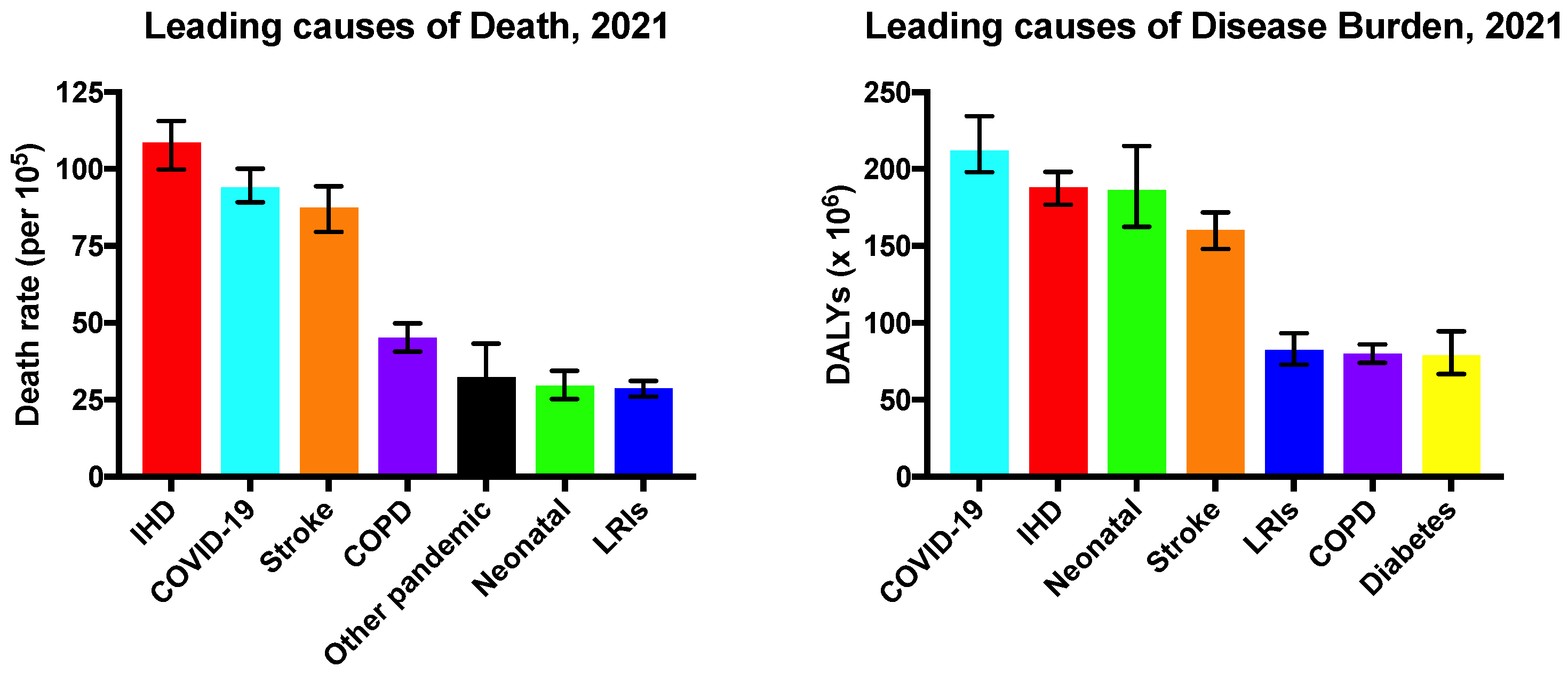
Figure 1. Leading causes of death and disease burden worldwide in 2021. Mortality is shown as the age-standardised death rate per 100,000. Disease burden is measured in the number of disability-adjusted life years (DALYs) in millions. IHD, ischemic heart disease; COPD, chronic obstructive pulmonary disease; LRIs, lower respiratory infections. Other pandemic-related mortality includes excess deaths associated with the COVID-19 pandemic. Data from the Global Burden of Disease 2021 study [3].
Ijms 26 02309 g001
Figure 2. Application of cardiac patches and injectable hydrogels for repairing damaged heart tissue, along with examples of types of cells and hydrogel materials used in their development. Created with BioRender.com.
Ijms 26 02309 g002
Figure 3. The three main administration methods of injectable hydrogels used in preclinical studies. (a) Trans-epicardial injection; (b) trans-endocardial intramyocardial injection; (c) intracoronary injections. Created with BioRender.com.
Ijms 26 02309 g003
Figure 4. Schematic representation of bioprinting techniques. (a) Extrusion based; (b) droplet based; (c) laser assisted; (d) stereolithography; (e) digital light processing. Created with BioRender.com.
Ijms 26 02309 g004
Table 1. Types of cells used in cardiac regenerative research.
Cells TypeOriginsProsConsReferences
Skeletal myoblastsProgenitor cells located within skeletal muscle. Rapidly divide
Resist ischemia
Limited electrical integration
Do not fully differentiate into cardiomyocytes
Increased risk of ischemia
[25,26]
Mesenchymal stem cellsMultipotent cells originate from various tissues/organs, such as bone marrow and adipose tissue.Anti-inflammatory effects
Easy isolation and expansion
Low cell retention
Limited cell engraftment and survival
[26,27]
Embryonic stem cells A pluripotent stem cell that originates from in the inner cell mass of human blastocysts, at early stages of the embryo’s development.Pluripotency
High proliferation
Risk of tumour formation
Ethical concerns
Higher risks of immune rejection
[28,29,30]
Induced pluripotent stem cells A pluripotent stem cell that can be generated directly from a somatic cell using specific growth factors.Pluripotency
Patient specific
High proliferation
No ethical concerns
Immature differentiation and maturation
Risk of tumour formation
High costs
[31,32,33]
Table 2. Natural hydrogel-forming polymers.
PolymersAttributes ProsConsReferences
CollagenProvides structural support to cardiac cells and contributes to the heart’s stiffness and rigidity.
Assists in force transmission ensuring efficient force is generated by the heart to pump blood around the body.
Biocompatible
Biodegradable
Incorporated with other polymers to enhance its physical properties
Unstable
Poor mechanical properties
[19,24,81,82]
GelatinPartially hydrolysed from collagen
Functions as a crosslinking polymer, imparting essential properties like structure and texture to the gel.
Retains the arginine-glycine- aspartic acid (RGD) peptide sequence which promotes cell adhesion, proliferation, and differentiation.
Non-immunogenic
Biocompatible
Biodegradable
Low thermostability
Weak mechanical strength
[27,83,84]
Hyaluronic acid (HA)Natural, linear polysaccharide that contains multiple acid and hydroxyl groups chemically modifiable to improve its mechanical properties.Biocompatible
Biodegradable
Non-immunogenic
Non-thrombogenic
Weak mechanical strength
High degradation rate
[85,86]
ElastinProvides elasticity to various tissues and organs
Tropoelastin is a soluble precursor of elastin that has similar biochemical properties to elastin.
Can be hydrolysed to form smaller chains such as peptides and polypeptides, which are solubleInsoluble nature
Weak mechanical properties
Batch-to-batch variation
[62,87,88]
ChitosanLinear polysaccharide attained by the partial deacetylation of chitin.Low toxicity
Antibacterial properties
Encourages angiogenesis
Weak mechanical properties
Batch-to-batch variation
[62,89]
FibrinPlays a role in the body’s natural tissue repair mechanism.Biologically compatible
Ability to promote repair
Increased survival rate of CMs
Poor mechanical strength
Hydrogel shrinkage
Batch to batch variation
[87,89,90]
Decellularized ECM (dECM)Developed through decellularization, which involves the elimination of cells from the ECM to generate a natural matrix.Supports cell growth, adhesion, and remodelling
Non-immunogenic
Poor mechanical properties
Cytotoxicity
Batch-to-batch variation
[89,91,92]
Table 3. Synthetic hydrogel-forming polymers.
Synthetic PolymersAttributes Advantages Disadvantages References
Polyacrylamide (PAA)Developed via crosslinking of acrylamide monomers.Stable
Non-toxic Hydrophilic
Highly adaptable
Short lifespan, which
limits cell culture studies
[93,94,95]
Polyethylene glycol
(PEG)
FDA-approved water-soluble polymer.
Modifying this hydrogel with an RGD peptides improves cell proliferation and survival.
Biocompatible
Non-immunogenic
Strong mechanical properties
Not soluble
Inadequate cell specific adhesion
[80,96,97]
Poly-N-isopropyl acrylamide (PNIPAAm)Changes from liquid to gel above temperatures over 32 °C.Thermo-responsive behaviourWeak mechanical strength
Low biodegradability
[89,91,98]
Polyvinyl alcohol
(PVA)
Developed via the hydrolysis of polyvinyl acetate.
As a hydrogel, it has high elasticity and can enhance the dispersion of mechanical signals.
Hydrophilic
Biocompatible
Biodegradable
Low adhesion
Low biodegradability
Poor thermo-responsive behaviour
[80,89,99]
Table 4. 3D bioprinting techniques.
Bioprinting TechniqueProsConsReferences
Extrusion-basedNot complex
Affordable
Scalable
Cell damage through sheer stress
Limited available biomaterials
Low resolution
Clogging of the nozzle
[147]
Droplet-basedAffordable
Accessible
High cell viability >90%
High resolution
Limited available biomaterials
Issues with fabricating porous tissue structures
Clogging of the printer’s injector
[143]
Laser-assistedNozzle free
High resolution
Wide range of available biomaterials
High cell viability
Low mechanical stress
High cost
Time consuming
[145]
Stereolithography (SLA) and Digital Light Processing (DLP)High resolution
High speed
High cell viability
Nozzle free
Cytotoxic effects
Limited available bio-inks
[146]
Disclaimer/Publisher’s Note: The statements, opinions and data contained in all publications are solely those of the individual author(s) and contributor(s) and not of MDPI and/or the editor(s). MDPI and/or the editor(s) disclaim responsibility for any injury to people or property resulting from any ideas, methods, instructions or products referred to in the content.