Article

Modulation of the ETV6::RUNX1 Gene Fusion Prevalence in Newborns by Corticosteroid Use During Pregnancy

1
BCNatal|Fetal Medicine Research Center, Hospital Clínic and Hospital Sant Joan de Deu, University of Barcelona, 08950 Barcelona, Spain
2
Institut d’Investigacions Biomèdiques August Pi i Sunyer, 08036 Barcelona, Spain
3
Department of Pediatric Oncology, Hematology and Clinical Immunology, University Children’s Hospital, Medical Faculty, Heinrich Heine University, 40225 Düsseldorf, Germany
4
German Cancer Consortium, Partner Site Essen-Düsseldorf, 40225 Düsseldorf, Germany
5
Deutsches Krebsforschungszentrum (DKFZ), 69120 Heidelberg, Germany
6
Red Española de Terapias Avanzadas—Instituto de Salud Carlos III (ISCII), 28029 Madrid, Spain
7
Centro de Investigación Biomédica en Red Cáncer(CIBER-ONC), Instituto de Salud Carlos III (ISCIII), 28029 Madrid, Spain
8
Institut de Recerca Sant Joan de Déu, 08950 Barcelona, Spain
9
Josep Carreras Leukemia Research Institute, 08916 Barcelona, Spain
10
Department of Biomedicine, School of Medicine, University of Barcelona, 08014 Barcelona, Spain
11
Institució Catalana de Recerca i Estudis Avançats, 08010 Barcelona, Spain
12
Centro de Investigación Biomédica en Red de Fisiopatología de la Obesidad y Nutrición, 28029 Madrid, Spain
13
Institut de Recerca en Nutrició i Seguretat Alimentaria, University of Barcelona, 08014 Barcelona, Spain
14
Department of Internal Medicine, Hospital Clinic, University of Barcelona, 08014 Barcelona, Spain
15
Centro de Investigación Biomédica en Red y Salud Mental, CIBERSAM, Instituto de Salud Carlos III, 28029 Madrid, Spain
16
Department of Psychiatry and Psychology, Hospital Clinic, Neuroscience Institute, IDIBAPS, University of Barcelona, CIBERSAM, 08014 Barcelona, Spain
*
Authors to whom correspondence should be addressed.
Int. J. Mol. Sci. 2025, 26(7), 2971; https://doi.org/10.3390/ijms26072971
Submission received: 18 February 2025 / Revised: 12 March 2025 / Accepted: 14 March 2025 / Published: 25 March 2025

Abstract

:
ETV6::RUNX1-positive pediatric acute lymphoblastic leukemia frequently has a prenatal origin and follows a two-hit model: a first somatic alteration leads to the formation of the oncogenic fusion gene ETV6::RUNX1 and the generation of a preleukemic clone in utero. Secondary hits after birth are necessary to convert the preleukemic clone into clinically overt leukemia. However, prenatal factors triggering the first hit have not yet been determined. Here, we explore the influence of maternal factors during pregnancy on the prevalence of the ETV6::RUNX1 fusion. To this end, we employed a nested interventional cohort study (IMPACT-BCN trial), including 1221 pregnancies (randomized into usual care, a Mediterranean diet, or mindfulness-based stress reduction) and determined the prevalence of the fusion gene in the DNA of cord blood samples at delivery (n = 741) using the state-of-the-art GIPFEL (genomic inverse PCR for exploration of ligated breakpoints) technique. A total of 6.5% (n = 48 of 741) of healthy newborns tested positive for ETV6::RUNX1. Our multiple regression analyses showed a trend toward lower ETV6::RUNX1 prevalence in offspring of the high-adherence intervention groups. Strikingly, corticosteroid use for lung maturation during pregnancy was significantly associated with ETV6::RUNX1 (adjusted OR 3.9, 95% CI 1.6–9.8) in 39 neonates, particularly if applied before 26 weeks of gestation (OR 7.7, 95% CI 1.08–50) or if betamethasone (OR 4.0, 95% CI 1.4–11.3) was used. Prenatal exposure to corticosteroids within a critical time window may therefore increase the risk of developing ETV6::RUNX1+ preleukemic clones and potentially leukemia after birth. Taken together, this study indicates that ETV6::RUNX1 preleukemia prevalence may be modulated and potentially prevented.

Graphical Abstract

1. Introduction

Childhood B-cell acute lymphoblastic leukemia (B-ALL) is the most frequent pediatric cancer and one of the leading causes of childhood mortality in developed countries, with increasing incidence in the past years [1,2,3]. Although advances in molecular risk-stratification, diagnosis, and targeted treatments have significantly improved survival rates, B-ALL remains a health priority that fits the standards for primary prevention [4,5,6].
The most prevalent subtypes of B-ALL frequently have a prenatal origin, following a two-hit model: (1) Somatic acquisition of oncogenic fusion genes (such as ETV6::RUNX1 or TCF3::PBX1) or aberrant chromosome numbers (e.g., high hyperdiploidy) occurs in hematopoietic stem or progenitor cells as a first genetic alteration arising during prenatal life. (2) Secondary oncogenic hits after birth convert the preleukemic clone into clinically overt leukemia [7,8,9]. Retrospective analyses of Guthrie cards from children who subsequently developed leukemia demonstrated the presence of such first-hit molecular lesions at birth [10,11]. Such molecular lesions have subsequently been detected in healthy newborns [12,13,14].
The reported prevalence of the ETV6::RUNX1 fusion was about 100× higher than the actual leukemia incidence, indicating that the fusion gene is only mildly oncogeneic.
In twins with concordant ETV6::RUNX1-positive leukemia, ALL develops at different times, and postnatal latency can be protracted [15]. Most frequently, ETV6::RUNX1+ B-ALL harbors deletions of the second, non-translocated ETV6 allele [16], implying that the initial gene fusion predisposes individuals to leukemia, while subsequent ETV6 deletion acts as a promoting factor. While the postnatal triggers of leukemia have been extensively investigated [8], few studies have focused on the association between prenatal factors and preleukemic lesions. Parental age, ethnicity, maternal diet, folate intake, alcohol consumption, X-ray exposure, pesticides, perinatal infections, and fetal growth may play a role [17].
The investigation of prenatal determinants of childhood leukemia is limited by the technical challenge of evaluating a wide spectrum of preleukemic lesions in large sample sizes [18]. Cord blood (CB) biobanks from well-characterized large cohorts would be the most obvious source for this research, but sample sizes are commonly small and rarely suitable for this type of investigation. First, the reported frequency of cells in healthy newborns with preleukemic lesions at birth is estimated to range from 1 in 103 to 104, making them undetectable by most available laboratory methods. Moreover, the detection of these lesions has been optimized for RNA rather than DNA owing to patients’ unique genomic DNA breakpoints, often occurring in extensive genomic regions not targetable by conventional techniques. Unfortunately, RNA samples are very rarely available in CB biobanks, are very unstable, and are susceptible to contamination, making them non-amenable for these studies.
Fueller et al. recently designed a new laboratory technique called “Genomic inverse PCR for exploration of ligated breakpoints” (GIPFEL) [19], which allows for the sensitive detection of recurrent chromosomal translocations. This technique relies on PCR-based quantification of unique DNA sequences that are created by circular ligation of restricted genomic DNA from translocation-bearing cells. Therefore, prior knowledge of the individual-specific interchromosomal breakpoints is not required. Recently, GIPFEL analysis allowed the assessment of the prevalence of preleukemic lesions in a Danish cohort of 1000 CBs from healthy newborns, and yielded a prevalence of 5% for ETV6::RUNX1 and 0.6% for TCF3::PBX1 [20,21].
Previous studies evaluating prenatal risk factors for childhood leukemia were mostly retrospective case-control studies, and CB samples were rarely available [17]. However, in the IMPACT-BCN (Improving Mothers for a better PrenAtal Care Trial BarCeloNa) randomized trial, CB samples were collected at delivery, and CB-derived mononuclear cells were stored as part of the study design. The study evaluated the impact of lifestyle interventions (Mediterranean diet and stress reduction) in 1221 pregnant women and demonstrated benefits on several perinatal outcomes [22].
In the present study, we evaluated the association between prenatal interventions, maternal and perinatal factors, and the emergence of neonatal preleukemic ETV6::RUNX1 lesions in the well-characterized cohort of CB samples from the IMPACT-BCN trial.

2. Results

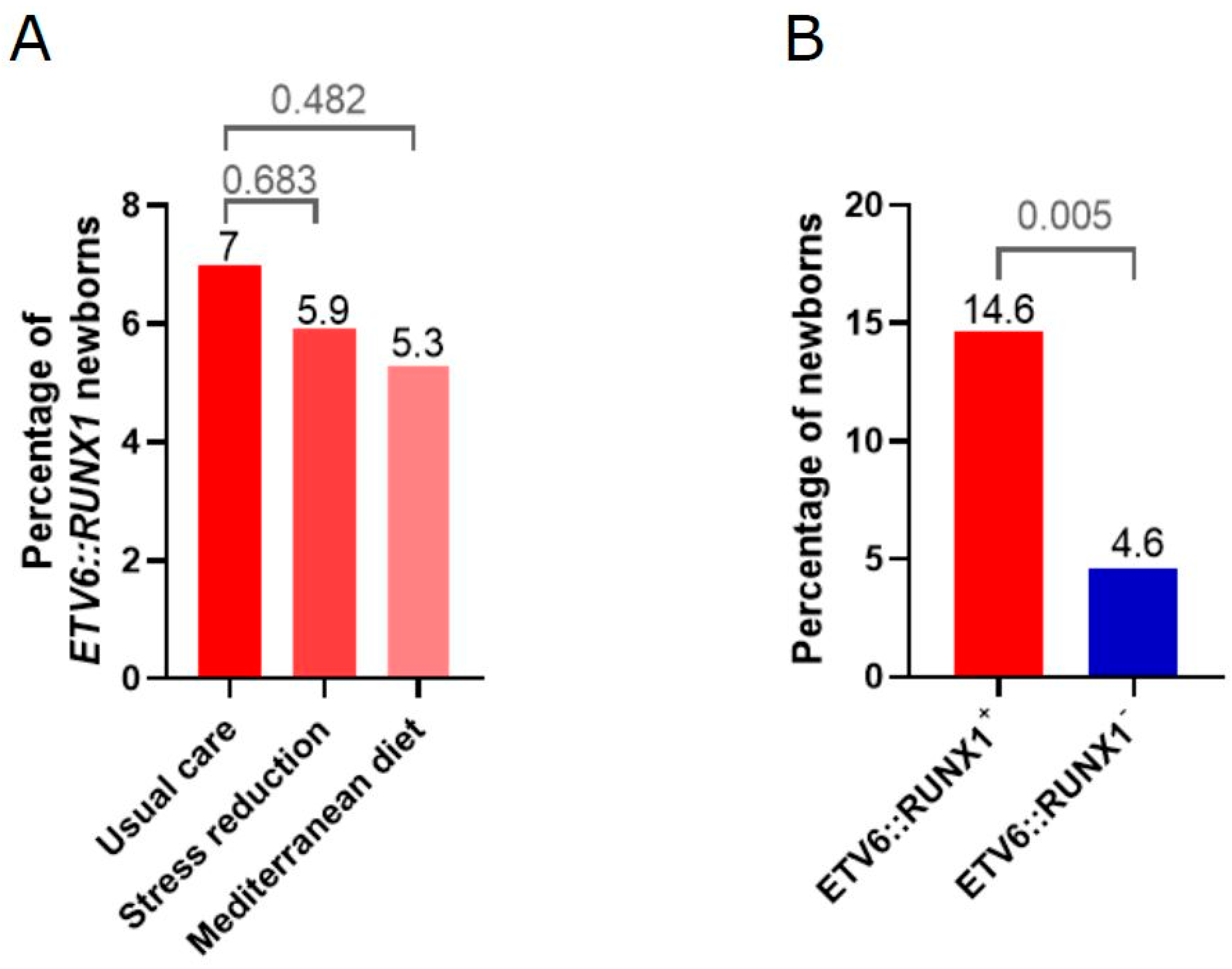
2.1. High Adherence to Mediterranean Diet and Stress Reduction Intervention Is Associated with a Trend Toward Lower Prevalence of the ETV6::RUNX1 Fusion

The ETV6::RUNX1 fusion gene was screened using GIPFEL in n = 741 cord blood samples (Figure 1) in a blinded fashion (n = 245 without intervention, n = 250 Mediterranean diet intervention, and n = 246 stress reduction intervention). The ETV6::RUNX1 fusion was detected in 48 cord blood samples (6.5%) of the study population, as determined by GIPFEL analysis (Table S1).
High adherence to the Mediterranean diet intervention was defined as an improvement of at least 3 points in the final score of the 17-item dietary screener compared with the baseline score. Adherence to the stress reduction intervention was considered high if at least 6 of 9 stress reduction sessions were attended.
Regarding the intervention group, there were no differences in the prevalence of neonatal ETV6::RUNX1 fusion gene among the three groups. However, when only considering participants who demonstrated a high adherence to the intervention, a non-significant trend toward lower neonatal ETV6::RUNX1 prevalence was detected in both interventions (prevalence of 7.0% for usual care versus 5.9% for stress reduction (OR 0.8; 95% CI 0.3–2.0) versus 5.3% for Mediterranean diet (OR 0.7; 95% CI 0.3–1.7) (Figure 2A, Table 1).

2.2. Univariate Analysis Identifies Corticosteroid Application During Pregnancy as Significantly Associated with the Presence of ETV6::RUNX1 Fusions

Maternal and pregnancy characteristics of the study population were evaluated using univariate analyses based on the presence of the neonatal ETV6::RUNX1 fusion gene (Table 2 and Table S2). Considering baseline characteristics, univariate analysis identified Maghreb ethnicity (OR 12.8, p < 0.001) and study class (OR for secondary/technological studies compared to primary/no studies 0.3, p = 0.046; OR for university studies compared to primary/no studies 0.7, p = 0.525) as significantly associated with the appearance of preleukemic lesions. Among perinatal characteristics, the administration of exogenous corticosteroids during pregnancy was significantly associated with the preleukemic status (OR 3.5, p = 0.005) (Figure 2B), while folate supplementation showed a non-significant preventive tendency (OR 0.6, p = 0.056). No specific dietary component or key food was identified as significantly associated with the neonatal ETV6::RUNX1 fusion gene (Tables S3 and S4).

2.3. Multivariate Regression Models Report a Higher Probability of ETV6::RUNX1 Occurrence for Corticosteroid Administration Before 26 Weeks of Gestation and Use of Betamethasone

Multivariate regression analysis confirmed the administration of exogenous corticosteroids as significantly associated with the ETV6::RUNX1 fusion gene (Table 3).
Regarding the use of exogenous corticosteroids, a subanalysis according to gestational age reported a higher estimated probability in those participants receiving corticosteroids before 26 weeks of gestation (estimated probability of neonatal ETV6::RUNX1 appearance of 0.50 (95% CI 0.1–0.9)) compared to those who received the treatment over 26 weeks of pregnancy (estimated probability 0.11 (95% CI 0.01–0.23); OR for administration before 26 weeks 7.7 (95% CI 1.08–50), p = 0.04). Betamethasone (n = 25) was the corticosteroid class most commonly used (with most participants receiving 2 doses for fetal lung maturation purposes), followed by methylprednisone (n = 1), prednisone (n = 11), and topical use (n = 2). GR:MR refers to the glucocorticoid receptor (GR) to mineralocorticoid receptor (MR) activity ratio for each exogenous corticosteroid listed (Table 4 and Table S5).

3. Discussion

This study identified prenatal exposure to corticosteroids as a potential risk factor for prenatal ETV6::RUNX1 fusions and, thus, for leukemia development later in life. In addition, Maghreb ethnicity was associated with ETV6::RUNX1 fusions, although absolute numbers were small. There were no other statistically significant associations between maternal lifestyle interventions and the prevalence of preleukemic ETV6::RUNX1 lesions revealed in our study.
Previous studies have evaluated the association of prenatal risk factors [23,24,25,26,27,28,29,30,31,32,33,34] that suggest maternal age, ethnicity, some dietary components, folate supplementation, alcohol consumption, X-ray exposure, pesticide exposure, perinatal infections, and fetal growth as probable risk factors. Nevertheless, most available evidence is based on retrospective case-control studies that used self-reported questionnaires, which are susceptible to recall and underreporting bias. These studies were also unable to differentiate prenatal from postnatal exposures for most risk factors.
Our current study adds to previous evidence by reporting the analysis of 741 cord blood samples from a well-phenotyped cohort of pregnant women included in the IMPACT-BCN randomized trial. Maternal and prenatal factors were determined prospectively during the trial by the use of medical records by a trained medical doctor, avoiding recall bias related to self-report [35]. Additionally, assessment of maternal diet was prospectively performed by an expert nutritionist in a prospective manner during pregnancy, with proven validity [31]. Likewise, the use of biomarkers to confirm adherence to interventions enhances the reliability of results. The prevalence of ETV6::RUNX1 was large enough to allow statistical comparisons.
The neonatal ETV6::RUNX1 positivity prevalence of 6.5% (48 out of 741) is in line with previously published papers on healthy newborns in the USA, Europe, and Japan, ranging from 0.01 to 7% [14].
There was a significant association between maternal Maghreb ethnicity and neonatal preleukemic fusion genes. Although differences in the incidence of childhood leukemia between ethnicities had been previously described, an association with Maghreb ethnicity has not been reported. Concerning the ETV6::RUNX1 fusion gene, Western countries and Hispanics have consistently described as presenting higher rates [36] compared with Black and Asian ethnicities. Genomic profiles [37] and folate metabolism polymorphisms [4] have been hypothesized as underlying factors. The difference in Maghreb ethnicity could be explained by genetic polymorphisms or by lifestyle or environmental determinants not recorded during the randomized trial. However, the small number of samples hampered the analysis of dietary components in this specific subpopulation.
Regarding corticosteroids, the main indications for their use in our cohort were fetal lung maturation in cases at risk of preterm birth and maternal autoimmune or dermatological conditions. To address the potential confounding effect of the indication for steroid administration, we conducted additional analyses adjusting for clinical conditions that warranted steroid use. These adjustments confirmed that the association between corticosteroid exposure and ETV6::RUNX1 positivity remained significant, independent of the underlying indication for treatment. This suggests that the effect is more likely attributable to the drug itself rather than the condition requiring its administration. Prenatally, corticosteroids improve fetal lung maturation but have strong effects on fetal somatic and brain growth. This study identified exogenous corticosteroids as risk factors for preleukemic ETV6::RUNX1 lesions. To our knowledge, this association has not previously been reported. Most cases had two doses of betamethasone under 26 weeks of gestation. In fact, glucocorticoids represent a key drug in the treatment backbone of B-ALL [38]. However, frequent secondary lesions in ETV6::RUNX1-positive ALL may directly affect the glucocorticoid response (e.g., by mutation of the glucocorticoid receptor NR3C1), the cell death pathway induced via its signaling, or components of the mismatch repair pathways [39]. One speculation on the effect of glucocorticoid treatment on the increased incidence of ETV6::RUNX1 lesions in our study may therefore be an expansion of pre-existing minor ETV6::RUNX1-positive clones, due to secondary mutations that arise under the selective pressure of betamethasone. In this case, children would be expected to have a higher likelihood of developing leukemia later in life, which needs to be evaluated in larger studies with long-term follow-up.
Another probable explanation is the generation of the fusion gene itself as a direct consequence of glucocorticoid treatment. Recently, physiological concentrations of glucocorticoids have been shown to induce DNA double-strand breaks via inhibition of topoisomerase II. The effect of different glucocorticoids depended on their affinity for the glucocorticoid receptors (Figure 3) [40]. Betamethasone was significantly associated with an increased incidence of ETV6::RUNX1 fusions in our study and has a higher affinity for the receptor compared to dexamethasone, prednisolone, or endogenous cortisol [40]. Repair of the DNA double-strand breaks by non-homologous end joining could lead to ETV6::RUNX1 fusion. Interestingly, after glucocorticoid treatment of B-ALL patients, increased numbers of ETV6::RUNX1-positive lymphocytes have been detected [41].
As recently demonstrated by Kim et al., using bisulfite sequencing of DNA extracted from bloodspot “Guthrie” cards, glucocorticoids also induce genome-wide methylation differences following antenatal exposure [43]. Methylation changes have been associated with prenatal environmental exposures and an increased risk of developing childhood leukemia [44], and there is also a correlation of methylation changes and specific genetic subtypes of ALL [45]. Hypomethylated areas are likely transcriptionally active regions and have a more open chromatin that may lend itself to interchromosomal contacts, DNA breaks, and aberrant repair [44]. It is tempting to speculate that the ETV6 and RUNX1 genes that recombine with one another so frequently are spatially localized together in transcriptional or chromatin compartments. The clustering of breakpoints, as seen in ETV6::RUNX1-positive ALL, also likely reflects selection for functional rearrangements during tumorigenesis. Although data from leukemia is lacking, previous studies in prostate cancer [46,47] show a significantly reduced methylation at breakpoints of specific interchromosomal translocations.
Glucocorticoid administration during pregnancy may also lead to permanent changes that impact on the risk of developing leukemia after birth (Figure 4). Glucocorticoid administration during pregnancy leads to higher plasma cortisol levels throughout adult life, indicating persistent reprogramming of the hypothalamus–pituitary–adrenal axis in animal models [48]. In the adrenal hypothesis model proposed by Schmiegelow et al. [49], early childhood infections result in profound changes in the hypothalamus–pituitary–adrenal axis that increase plasma cortisol levels and protect from leukemia by direct elimination of preleukemic cells via immune modulation and promotion of Th1-cytokine response [49]. As a possible explanation, it is conceivable that glucocorticoids may work as a double-edged sword: They may increase the risk of preleukemic fusions arising in utero but may also reduce the risk of leukemic transformation after birth.
Our data indicate that the effects of glucocorticoids in early pregnancy and childhood need to be carefully studied in larger cohorts, and the pros and cons of their application need to be deliberated by informed clinicians. Additionally, our study indicates promising options for potential strategies to prevent leukemia in childhood.
A previous study has reported an association between maternal diet quality and the risk of childhood leukemia [23]. In the present study, no significant associations were found between the Mediterranean diet or specific dietary components and the rate of ETV6::RUNX1. However, there was a trend toward lower rates among pregnancies with a high adherence to the Mediterranean diet, which deserves further investigation in larger sample sizes. Foods containing DNA topoisomerase II inhibitors, including coffee, tea, and other caffeinated beverages, canned food or dried vegetables or legumes, cocoa, red wine, apples, and berries, have been described as potential childhood leukemia triggers [50,51]. Concerning vitamins, deficient maternal folate intake during pregnancy has been extensively described as a risk factor for ALL [4,18,24,27,52,53]. In the present study, there was a non-significant trend toward lower rates of ETV6::RUNX1 in relation to folate intake, although we cannot separate the intrinsic effect of other vitamins that are commonly taken for supplementation during pregnancy. There was no association between smoking, alcohol, or other toxics and ETV6::RUNX1 fusion gene, but the prevalence of these risk factors was too low to allow meaningful comparisons. A lower socioeconomic class has been suggested to have a preventive effect on childhood cancer postnatally [54,55]. In this study, we observed a non-significant trend toward the opposite effect on ETV6::RUNX1 frequency, which could be related to a lower folate intake in this subgroup of women.
Some limitations of our study should be acknowledged. First, the fact that the population of the study was from a single center located in Spain, which, although including several ethnicities, could hamper the external validation of results. Second, given the small sample size of ETV6::RUNX1 positive individuals, our findings should be considered preliminary and require validation in larger studies to confirm their biological and clinical significance. Third, the variability in reported ETV6::RUNX1 prevalence in cord blood across studies may stem from differences in detection methods, sample sizes, and population characteristics; however, the rigorous methodology and quality controls in our study support the credibility of our findings within this context. Fourth, we performed a 4-year follow-up in which none of the ETV6::RUNX1-positive newborns have been diagnosed with acute lymphoblastic leukemia. However, given the relatively short follow-up period, we acknowledge that longer-term monitoring is needed, and child follow-up should be warranted to confirm or discard the development of overt leukemia.

4. Material and Methods

4.1. Study Design and Participant Selection

This study represents a secondary analysis of the IMPACT-BCN trial, a randomized clinical trial conducted at BCNatal (Hospital Clínic and Hospital Sant Joan de Deu, Barcelona, Spain) from 2017 to 2020 [56]. The trial was approved by the Institutional Review Board (HCB-2016-0830). All participants provided written informed consent. Participants were screened for eligibility during routine second-trimester ultrasound scans (19–23.6 weeks of gestation) for being at high risk of developing small-for-gestational-age newborns [57].

4.2. Interventions

Participants were randomly assigned 1:1:1 to one of the three study groups: a Mediterranean diet intervention, a stress reduction program intervention, or usual care without any additional intervention (control group).
The Mediterranean diet intervention was adapted from the PREDIMED trial [58]. Registered dietitians conducted group meetings, as well as monthly face-to-face and additional telephone interviews to ensure adherence to the diet. All participants received olive oil (2 L every month) and 15 g of walnuts per day (450 g every month) at no cost. The participant received dietary training to encourage an increased intake of whole grain cereals (≥5 servings/d); vegetables and dairy products (≥3 servings/d); fresh fruit (≥2 servings/d); and legumes, nuts, fish, and white meat (≥3 servings/week). A 151-item food frequency questionnaire, a 7-day dietary journal, and a 17-item dietary assessment score (range, 0–17) were used to assess baseline nutrients and vitamin intake of all participants and adherence to the Mediterranean diet for the intervention group. A high adherence was defined as an improvement of at least 3 points in the final score of the 17-item dietary screener compared with the baseline score.
Led by experienced, certified instructors, the stress reduction group received an 8-week-long program of structured intervention (based on mindfulness) tested in clinical trials [59]. It included weekly 2.5-h sessions, 1 full-day session, and daily home practice, formal and informal techniques, with the goal of enhancing awareness and reducing anxiety. The sessions included didactic presentations, formal 45-min meditation practices, yoga, body awareness, and group discussions. Adherence to the stress reduction intervention was considered high if at least 6 of 9 stress reduction sessions were attended.
A detailed description of the interventions was previously published [22,56].

4.3. Cord Blood Sample Collection

Cord blood samples were collected at delivery after cord clamping, usually between 30 s to 1 min following international recommendations for delivery, and immediately stored at 4 °C. Ficoll density-gradient centrifugation (GE Healthcare, headquartered in Chicago, IL, USA) was performed in the following hours by an expert technician who was blinded to the intervention group. Cord blood components (serum, plasma, and mononuclear cells) were stored separately at −80 °C. Mononuclear cells were shipped to the University Hospital Düsseldorf, Department of Pediatric Oncology, Hematology, and Clinical Immunology for detection of ETV6::RUNX1 fusions by GIPFEL analysis.

4.4. Genomic Inverse PCR for Exploration of Ligated Breakpoints (GIPFEL)

Cryopreserved cord blood samples were screened using the GIPFEL technique to check the neonatal ETV6::RUNX1 positivity as detailed in the Supplementary Materials and Methods. In brief, genomic DNA was isolated from cord blood samples and digested with the restriction enzyme SacI. After circularization mediated by T4 DNA ligase, ETV6 and RUNX1 joints are ligated if the translocation is present. Circularized/ligated DNA was amplified, and ETV6::RUNX1 was detected by real-time PCR, agarose gel electrophoresis, and confirmed by Sanger sequencing. Researchers and technicians performing the laboratory analysis were blinded to the intervention group.

4.5. Predictive Variables

The main outcome was the presence of the neonatal ETV6::RUNX1 fusion gene in CB samples. All maternal and prenatal variables were considered potential predictive variables and were obtained prospectively at the beginning or during the trial, including maternal age, body mass index, ethnicity, level of education, socioeconomic status, parity, medical conditions, cigarette smoking, alcohol or recreational drug intake during pregnancy, folate supplementation, use of exogenous corticosteroids, occurrence of pregnancy complications (gestational diabetes, preterm birth, preeclampsia, small-for-gestational-age), and gestational age at delivery. Ethnicity was self-reported by participants among White, Latin, Afro-American, Maghreb, or others. Maghreb ethnicity was defined as patients from Western and Central North Africa. Study class was divided into primary/no studies, secondary/technology, and university studies and was self-reported by participants. Socioeconomic status was defined as low if participants reported having never worked or being unemployed for more than two years and having a partner with unskilled work or who was unemployed; and high if they reported university studies regardless of whether they were working; and medium if any other situations. Exogenous use of corticosteroids was determined according to medical records, which specified the dose, class, and duration of use. Gestational diabetes was defined as any degree of glucose intolerance with onset during pregnancy, in our setting diagnosed by the presence of two altered values in a 100-g, 3-h oral glucose tolerance test. Preterm birth was defined as delivery before 37 weeks of gestation. Preeclampsia was defined as the concurrence of pregnancy-onset hypertension and proteinuria. Small-for-gestational-age was defined as birthweight below the 10th centile according to local standards. Steroid use included betamethasone for preterm labor and oral or topical prednisone for maternal autoimmune and skin conditions that were administered during the entire pregnancy. Maternal dietary key food and nutrient intake were obtained by a specialized nutritionist at the beginning and end of the intervention with several questionnaires validated for the present study population [60].

4.6. Statistical Analysis

Maternal and prenatal data are presented as mean (standard deviation, SD) or number (percentage), as appropriate. Statistical analysis for comparison of clinical and prenatal characteristics included the use of the Student’s t-test for continuous variables, the Chi-square test for categorical variables, and logistic univariate and multivariate regression analyses. Data were expressed using odds ratios and their corresponding 95% confidence intervals. Multivariate regression models were adjusted for significant variables in our study and described confounders. Estimated probability (or estimated marginal means) was computed with emmeans library version 1.8.6. Differences were considered significant when the p-value was lower than 0.05. Statistical analyses were conducted using Stata 15.1 (StataCorp LP, Houston, TX, USA) and R version 4.2.1 (Vienna, Austria).

5. Conclusions

Prenatal exposure to corticosteroids within a critical time window may therefore increase the risk of developing ETV6::RUNX1+ preleukemic clones and potentially leukemia after birth. Taken together, this study indicates that the prevalence of ETV6::RUNX1 preleukemia may be modulated and potentially prevented.

Supplementary Materials

The following supporting information can be downloaded at: https://www.mdpi.com/article/10.3390/ijms26072971/s1.

Author Contributions

Conceptualization, L.B., A.B., E.G. and F.C. (Fàtima Crispi); methodology, E.K., F.C. (Francesca Crovetto) and F.C. (Fàtima Crispi); software, E.K. and L.B.; validation, L.B., A.B. and E.G.; formal analysis, E.K. and L.B.; investigation, L.B.; resources, L.B.; data curation, L.B. and R.B.; writing—original draft preparation, L.B., S.C.-B., E.K. and F.C. (Fàtima Crispi); writing—review and editing, A.B., E.G., U.F., E.K., F.C. (Fàtima Crispi), F.C. (Francesca Crovetto), S.C.-B., M.L., L.Y., H.C., C.B., R.C., R.B., E.V., R.E. and P.M.; visualization, L.B.; supervision, E.V., R.E., P.M., A.B. and E.G.; project administration, L.B.; funding acquisition, A.B. All authors have read and agreed to the published version of the manuscript.

Funding

The project was partially funded by a grant from “la Caixa” Foundation (LCF/PR/GN18/10310003); Cerebra Foundation for the Brain Injured Child (Carmarthen, Wales, UK); AGAUR under grant 2017 SGR No. 1531. Dr. Benítez was supported by a research grant from the Instituto de Salud Carlos III (CM21/00058). Dr. Castro-Barquero has received support from the Margarita Salas fellowship, University of Barcelona. Dr. Crovetto has received support from Centro de Investigaciones Biomédicas en Red sobre Enfermedades Raras (CIBERER) and Primary care interventions to prevent maternal and child chronic diseases of perinatal and developmental origin RD21/0012/0003, Instituto de Salud Carlos III, Spain. Dr. Youssef has received support from the Juan de la Cierva grant FJC2021-048123-I, funded by MCIN/AEI/10.13039/501100011033 and by the European Union “NextGenerationEU”/PRTR. Dr. Crispi has received support from the Instituto Carlos III (INT21/00027 and PI20/00246) and Fundació Jesus Serra (Spain). Dr. Fischer was supported by the Deutsche José-Carreras Leukämie-Stiftung (DJCLS 18R/2021), the Deutsche Forschungsgemeinschaft (DFG, German Research Foundation)—grant no. 495318549, the Deutsche Krebshilfe (DKH, German Cancer Aid) within the “Cancer Prevention—Graduate School” (CPGS)—grant no. 70114736, and the Deutsche Kinderkrebsstiftung (DKKS)—grant no. A2023/31. Dr. Fischer and Prof. Borkhardt were supported by the Bundesministerium für Bildung und Forschung (BMBF, the German Ministry for Education and Research)—grant no. 01KD2410A (EDI-4-ALL) and the Bundesamt für Strahlenschutz (BfS)—grant no. 3622S32231. Dr. Casas and Dr. Estruch received a grant from the INSA-Ma María de Maeztu Unit of Excellence (grant CEX2021-001234-M funded by MICIN/AEI/FEDER, UE).

Institutional Review Board Statement

The study was approved by the Hospital Clínic institutional review board (HCB-2016-0830) and the Ethical Commission of the Medical Faculty of Heinrich-Heine University.

Informed Consent Statement

All individuals who agreed to participate provided written informed consent before randomization.

Data Availability Statement

The data presented in this study are available on request from the corresponding author due to local IRB requirements.

Conflicts of Interest

The authors declare no conflict of interest.

Abbreviations

ALLAcute Lymphoblastic Leukemia
B-ALLB-cell Acute Lymphoblastic Leukemia
CBCord Blood
CBGsCord Blood Genomes
CMPCommon Myeloid Progenitor
CRHCorticotropin-Releasing Hormone
DNADeoxyribonucleic Acid
DPDouble-Positive (thymocytes)
ETV6::RUNX1Gene fusion associated with preleukemic clones
GCsGlucocorticoids
GIPFELGenomic Inverse PCR for Exploration of Ligated Breakpoints
GRGlucocorticoid Receptor
HPAHypothalamic–pituitary–adrenal (axis)
HSCHematopoietic Stem Cells
IMPACT-BCNImproving Mothers for a Better Prenatal Care Trial Barcelona
MLLMixed-Lineage Leukemia (gene)
MoMonocyte
NeuNeutrophils
NR3C1Nuclear Receptor Subfamily 3 Group C Member 1 (Glucocorticoid Receptor Gene)
OROdds Ratio
PBX1Pre-B-Cell Leukemia Homeobox 1
PCRPolymerase Chain Reaction
PREDIMEDPrevención con Dieta Mediterránea (Prevention with Mediterranean Diet)
RCTRandomized Controlled Trial
RNARibonucleic Acid
SGASmall for Gestational Age
SPSingle-positive (thymocytes)
TCF3Transcription Factor 3
ThT-helper Cell
Topo IIDNA Topoisomerase II
t-AMLTherapy-Related Acute Myeloid Leukemia

References

  1. Hunger, S.P.; Mullighan, C.G. Acute Lymphoblastic Leukemia in Children. N. Engl. J. Med. 2015, 373, 1541–1552. [Google Scholar] [PubMed]
  2. Steliarova-Foucher, E.; Colombet, M.; Ries, L.A.G.; Moreno, F.; Dolya, A.; Bray, F.; Hesseling, P.; Shin, H.Y.; Stiller, C.A.; Bouzbid, S.; et al. International Incidence of Childhood Cancer, 2001–10: A Population-Based Registry Study. Lancet Oncol. 2017, 18, 719–731. [Google Scholar] [CrossRef]
  3. Siegel, R.L.; Miller, K.D.; Fuchs, H.E.; Jemal, A. Cancer statistics, 2022. CA Cancer J. Clin. 2022, 72, 7–33. [Google Scholar] [PubMed]
  4. Whitehead, T.P.; Metayer, C.; Wiemels, J.L.; Singer, A.W.; Miller, M.D. Childhood Leukemia and Primary Prevention. Curr. Probl. Pediatr. Adolesc. Health Care 2016, 46, 317–352. [Google Scholar] [PubMed]
  5. Greaves, M.; Cazzaniga, V.; Ford, A. Can we prevent childhood Leukaemia? Leukemia 2021, 35, 1258–1264. [Google Scholar] [PubMed]
  6. Hauer, J.; Fischer, U.; Borkhardt, A. Towards prevention of childhood ALL by early-life immune training. Blood 2021, 138, 1412–1428. [Google Scholar]
  7. Knudson, A.G. The genetics of childhood cancer. Bull. Cancer 1988, 75, 135–138. [Google Scholar]
  8. Greaves, M. A causal mechanism for childhood acute lymphoblastic leukaemia. Nat. Rev. Cancer 2018, 18, 471–484. [Google Scholar]
  9. Bueno, C.; Tejedor, J.R.; Bashford-Rogers, R.; Gonzalez-Silva, L.; Valdes-Mas, R.; Agraz-Doblas, A.; Diaz de la Guardia, R.; Ribera, J.; Zamora, L.; Bilhou-Nabera, C.; et al. Natural history and cell of origin of TC F3-ZN F384 and PTPN11 mutations in monozygotic twins with concordant BCP-ALL. Blood 2019, 134, 900–905. [Google Scholar]
  10. Wiemels, J.L.; Cazzaniga, G.; Daniotti, M.; Eden, O.B.; Addison, G.M.; Masera, G.; Saha, V.; Biondi, A.; Greaves, M.F. Prenatal origin of acute lymphoblastic leukaemia in children. Lancet 1999, 354, 1499–1503. [Google Scholar]
  11. Taub, J.W.; Konrad, M.A.; Ge, Y.; Naber, J.M.; Scott, J.S.; Matherly, L.H.; Ravindranath, Y. High frequency of leukemic clones in newborn screening blood samples of children with B-precursor acute lymphoblastic leukemia. Blood 2002, 99, 2992–2996. [Google Scholar] [PubMed]
  12. Eguchi-Ishimae, M.; Eguchi, M.; Ishii, E.; Miyazaki, S.; Ueda, K.; Kamada, N.; Mizutani, S. Breakage and fusion of the TEL (ETV6) gene in immature B lymphocytes induced by apoptogenic signals. Blood 2001, 97, 737–743. [Google Scholar]
  13. Mori, H.; Colman, S.M.; Xiao, Z.; Ford, A.M.; Healy, L.E.; Donaldson, C.; Hows, J.M.; Navarrete, C.; Greaves, M. Chromosome translocations and covert leukemic clones are generated during normal fetal development. Proc. Natl. Acad. Sci. USA 2002, 99, 8242–8247. [Google Scholar] [PubMed]
  14. Hein, D.; Borkhardt, A.; Fischer, U. Insights into the prenatal origin of childhood acute lymphoblastic leukemia. Cancer Metastasis Rev. 2020, 39, 161–171. [Google Scholar]
  15. Wiemels, J.L.; Ford, A.M.; Van Wering, E.R.; Postma, A.; Greaves, M. Protracted and variable latency of acute lymphoblastic leukemia after TEL-AML1 gene fusion in utero. Blood 1999, 94, 1057–1062. [Google Scholar] [PubMed]
  16. Papaemmanuil, E.; Rapado, I.; Li, Y.; Potter, N.E.; Wedge, D.C.; Tubio, J.; Alexandrov, L.B.; Van Loo, P.; Cooke, S.L.; Marshall, J.; et al. RAG-mediated recombination is the predominant driver of oncogenic rearrangement in ETV6-RUNX1 acute lymphoblastic leukemia. Nat. Genet. 2014, 46, 116–125. [Google Scholar] [PubMed]
  17. Benitez, L.; Castro-Barquero, S.; Crispi, F.; Youssef, L.; Crovetto, F.; Fischer, U.; Kameri, E.; Bueno, C.; Camos, M.; Menendez, P.; et al. Maternal Lifestyle and Prenatal Risk Factors for Childhood Leukemia: A Review of the Existing Evidence. Fetal Diagn. Ther. 2024, 51, 395–410. [Google Scholar]
  18. Marcotte, E.L.; Spector, L.G.; Mendes-de-Almeida, D.P.; Nelson, H.H. The Prenatal Origin of Childhood Leukemia: Potential Applications for Epidemiology and Newborn Screening. Front. Pediatr. 2021, 9, 639479. [Google Scholar]
  19. Fueller, E.; Schaefer, D.; Fischer, U.; Krell, P.F.; Stanulla, M.; Borkhardt, A.; Slany, R.K. Genomic inverse PCR for exploration of ligated breakpoints (GIPFEL), a new method to detect translocations in leukemia. PLoS ONE 2014, 9, e104419. [Google Scholar]
  20. Schafer, D.; Olsen, M.; Lahnemann, D.; Stanulla, M.; Slany, R.; Schmiegelow, K.; Borkhardt, A.; Fischer, U. Five percent of healthy newborns have an ETV6-RUNX1 fusion as revealed by DNA-based GIPFEL screening. Blood 2018, 131, 821–826. [Google Scholar]
  21. Hein, D.; Dreisig, K.; Metzler, M.; Izraeli, S.; Schmiegelow, K.; Borkhardt, A.; Fischer, U. The preleukemic TCF3-PBX1 gene fusion can be generated in utero and is present in approximately 0.6% of healthy newborns. Blood 2019, 134, 1355–1358. [Google Scholar] [PubMed]
  22. Crovetto, F.; Crispi, F.; Casas, R.; Martin-Asuero, A.; Borras, R.; Vieta, E.; Estruch, R.; Gratacos, E.; Investigators, I.B.T. Effects of Mediterranean Diet or Mindfulness-Based Stress Reduction on Prevention of Small-for-Gestational Age Birth Weights in Newborns Born to At-Risk Pregnant Individuals: The IMPACT BCN Randomized Clinical Trial. JAMA 2021, 326, 2150–2160. [Google Scholar]
  23. Jensen, C.D.; Block, G.; Buffler, P.; Ma, X.; Selvin, S.; Month, S. Maternal Dietary Risk Factors in Childhood Acute Lymphoblastic Leukemia (United States). Cancer Causes Control 2004, 15, 559–570. [Google Scholar] [CrossRef]
  24. Thompson, J.R.; Gerald, P.F.; Willoughby, M.L.; Armstrong, B.K. Maternal folate supplementation in pregnancy and protection against acute lymphoblastic leukaemia in childhood: A case-control study. Lancet 2001, 358, 1935–1940. [Google Scholar]
  25. Orsi, L.; Rudant, J.; Ajrouche, R.; Leverger, G.; Baruchel, A.; Nelken, B.; Pasquet, M.; Michel, G.; Bertrand, Y.; Ducassou, S.; et al. Parental smoking, maternal alcohol, coffee and tea consumption during pregnancy, and childhood acute leukemia: The ESTELLE study. Cancer Causes Control 2015, 26, 1003–1017. [Google Scholar]
  26. MacArthur, A.C.; McBride, M.L.; Spinelli, J.J.; Tamaro, S.; Gallagher, R.P.; Theriault, G. Risk of childhood leukemia associated with parental smoking and alcohol consumption prior to conception and during pregnancy: The cross-Canada childhood leukemia study. Cancer Causes Control 2008, 19, 283–295. [Google Scholar] [PubMed]
  27. Metayer, C.; Milne, E.; Dockerty, J.D.; Clavel, J.; Pombo-de-Oliveira, M.S.; Wesseling, C.; Spector, L.G.; Schuz, J.; Petridou, E.; Ezzat, S.; et al. Maternal supplementation with folic acid and other vitamins and risk of leukemia in offspring: A Childhood Leukemia International Consortium study. Epidemiology 2014, 25, 811–822. [Google Scholar] [PubMed]
  28. Karalexi, M.A.; Dessypris, N.; Thomopoulos, T.P.; Ntouvelis, E.; Kantzanou, M.; Diamantaras, A.A.; Moschovi, M.; Baka, M.; Hatzipantelis, E.; Kourti, M.; et al. Parental alcohol consumption and risk of leukemia in the offspring: A systematic review and meta-analysis. Eur. J. Cancer Prev. 2017, 26, 433–441. [Google Scholar]
  29. O’Neill, K.A.; Murphy, M.F.; Bunch, K.J.; Puumala, S.E.; Carozza, S.E.; Chow, E.J.; Mueller, B.A.; McLaughlin, C.C.; Reynolds, P.; Vincent, T.J.; et al. Infant birthweight and risk of childhood cancer: International population-based case control studies of 40,000 cases. Int. J. Epidemiol. 2015, 44, 153–168. [Google Scholar] [PubMed]
  30. Klimentopoulou, A.; Antonopoulos, C.N.; Papadopoulou, C.; Kanavidis, P.; Tourvas, A.D.; Polychronopoulou, S.; Baka, M.; Athanasiadou-Piperopoulou, F.; Kalmanti, M.; Sidi, V.; et al. Maternal smoking during pregnancy and risk for childhood leukemia: A nationwide case-control study in Greece and meta-analysis. Pediatr. Blood Cancer 2012, 58, 344–351. [Google Scholar]
  31. Milne, E.; Royle, J.A.; Bennett, L.C.; de Klerk, N.H.; Bailey, H.D.; Bower, C.; Miller, M.; Attia, J.; Scott, R.J.; Kirby, M.; et al. Maternal consumption of coffee and tea during pregnancy and risk of childhood ALL: Results from an Australian case-control study. Cancer Causes Control 2011, 22, 207–218. [Google Scholar] [CrossRef] [PubMed]
  32. Menegaux, F.; Ripert, M.; Hemon, D.; Clavel, J. Maternal alcohol and coffee drinking, parental smoking and childhood leukaemia: A French population-based case-control study. Paediatr. Perinat. Epidemiol. 2007, 21, 293–299. [Google Scholar] [CrossRef]
  33. Petridou, E.; Trichopoulos, D.; Kalapothaki, V.; Pourtsidis, A.; Kogevinas, M.; Kalmanti, M.; Koliouskas, D.; Kosmidis, H.; Panagiotou, J.P.; Piperopoulou, F.; et al. The risk profile of childhood leukaemia in Greece: A nationwide case-control study. Br. J. Cancer 1997, 76, 1241–1247. [Google Scholar] [CrossRef] [PubMed]
  34. McKinney, P.A.; Juszczak, E.; Findlay, E.; Smith, K.; Thomson, C.S. Pre- and perinatal risk factors for childhood leukaemia and other malignancies: A Scottish case control study. Br. J. Cancer 1999, 80, 1844–1851. [Google Scholar] [CrossRef]
  35. Jurek, A.M.; Greenland, S.; Spector, L.G.; Roesler, M.A.; Robison, L.L.; Ross, J.A. Self-report versus medical record—Perinatal factors in a study of infant leukaemia: A study from the Children’s Oncology Group. Paediatr. Perinat. Epidemiol. 2011, 25, 540–548. [Google Scholar] [CrossRef]
  36. Liang, D.-C.; Shih, L.-Y.; Yang, C.-P.; Hung, I.-J.; Liu, H.-C.; Jaing, T.-H.; Yeh, T.-C.; Liang, S.-T.; Chang, C.-L.; Lee, E.-H.; et al. Frequencies of ETV6-RUNX1 Fusion and Hyperdiploidy in Pediatric Acute Lymphoblastic Leukemia Are Lower in Far East than West. Pediatr. Blood Cancer 2010, 55, 430–433. [Google Scholar] [CrossRef] [PubMed]
  37. Hsu, L.-I.; Briggs, F.; Shao, X.; Metayer, C.; Wiemels, J.L.; Chokkalingam, A.P.; Barcellos, L.F. Pathway Analysis of Genome-Wide Association Study in Childhood Leukemia among Hispanics. Cancer Epidemiol. Biomark. Prev. 2016, 25, 815–822. [Google Scholar] [CrossRef] [PubMed]
  38. Gaynon, P.S.; Lustig, R.H. The use of glucocorticoids in acute lymphoblastic leukemia of childhood. Molecular, cellular, and clinical considerations. J. Pediatr. Hematol. Oncol. 1995, 17, 1–12. [Google Scholar] [CrossRef]
  39. Kuster, L.; Grausenburger, R.; Fuka, G.; Kaindl, U.; Krapf, G.; Inthal, A.; Mann, G.; Kauer, M.; Rainer, J.; Kofler, R.; et al. ETV6/RUNX1-positive relapses evolve from an ancestral clone and frequently acquire deletions of genes implicated in glucocorticoid signaling. Blood 2011, 117, 2658–2667. [Google Scholar] [CrossRef]
  40. Akter, S.; Shimba, A.; Ikuta, K.; Mahmud, M.R.A.; Yamada, S.; Sasanuma, H.; Tsuda, M.; Sone, M.; Ago, Y.; Murai, K.; et al. Physiological concentrations of glucocorticoids induce pathological DNA double-strand breaks. Genes Cells 2023, 28, 53–67. [Google Scholar] [CrossRef]
  41. Brassesco, M.S.; Camparoto, M.L.; Tone, L.G.; Sakamoto-Hojo, E.T. Analysis of ETV6/RUNX1 fusions for evaluating the late effects of cancer therapy in ALL (acute lymphoblastic leukemia) cured patients. Cytogenet. Genome Res. 2004, 104, 346–351. [Google Scholar] [PubMed]
  42. Greaves, M.F. Aetiology of acute leukaemia. Lancet 1997, 349, 344–349. [Google Scholar] [PubMed]
  43. Kim, B.; Sasaki, A.; Murphy, K.; Matthews, S.G. DNA methylation signatures in human neonatal blood following maternal antenatal corticosteroid treatment. Transl. Psychiatry 2022, 12, 132. [Google Scholar] [PubMed]
  44. Timms, J.A.; Relton, C.L.; Sharp, G.C.; Rankin, J.; Strathdee, G.; McKay, J.A. Exploring a potential mechanistic role of DNA methylation in the relationship between in utero and post-natal environmental exposures and risk of childhood acute lymphoblastic leukaemia. Int. J. Cancer 2019, 145, 2933–2943. [Google Scholar]
  45. Nordlund, J.; Backlin, C.L.; Zachariadis, V.; Cavelier, L.; Dahlberg, J.; Ofverholm, I.; Barbany, G.; Nordgren, A.; Overnas, E.; Abrahamsson, J.; et al. DNA methylation-based subtype prediction for pediatric acute lymphoblastic leukemia. Clin. Epigenetics 2015, 7, 11. [Google Scholar]
  46. Berger, M.F.; Lawrence, M.S.; Demichelis, F.; Drier, Y.; Cibulskis, K.; Sivachenko, A.Y.; Sboner, A.; Esgueva, R.; Pflueger, D.; Sougnez, C.; et al. The genomic complexity of primary human prostate cancer. Nature 2011, 470, 214–220. [Google Scholar] [CrossRef]
  47. Lin, P.C.; Giannopoulou, E.G.; Park, K.; Mosquera, J.M.; Sboner, A.; Tewari, A.K.; Garraway, L.A.; Beltran, H.; Rubin, M.A.; Elemento, O. Epigenomic alterations in localized and advanced prostate cancer. Neoplasia 2013, 15, 373–383. [Google Scholar]
  48. Seckl Prenatal Glucocorticoids and Long-Term Programming. Eur. J. Endocrinol. 2004, 151, U49–U62. [CrossRef]
  49. Schmiegelow, K.; Vestergaard, T.; Nielsen, S.M.; Hjalgrim, H. Etiology of common childhood acute lymphoblastic leukemia: The adrenal hypothesis. Leukemia 2008, 22, 2137–2141. [Google Scholar]
  50. Ross, J.A.; Potter, J.D.; Reaman, G.H.; Pendergrass, T.W.; Robison, L.L. Maternal exposure to potential inhibitors of DNA topoisomerase II and infant leukemia (United States): A report from the Children’s Cancer Group. Cancer Causes Control 1996, 7, 581–590. [Google Scholar]
  51. Spector, L.G.; Xie, Y.; Robison, L.L.; Heerema, N.A.; Hilden, J.M.; Lange, B.; Felix, C.A.; Davies, S.M.; Slavin, J.; Potter, J.D.; et al. Maternal diet and infant leukemia: The DNA topoisomerase II inhibitor hypothesis: A report from the children’s oncology group. Cancer Epidemiol. Biomark. Prev. 2005, 14, 651–655. [Google Scholar]
  52. Bailey, H.D.; Miller, M.; Langridge, A.; de Klerk, N.H.; van Bockxmeer, F.M.; Attia, J.; Scott, R.J.; Armstrong, B.K.; Milne, E. Maternal dietary intake of folate and vitamins B6 and B12 during pregnancy and the risk of childhood acute lymphoblastic leukemia. Nutr. Cancer 2012, 64, 1122–1130. [Google Scholar]
  53. Chokkalingam, A.P.; Chun, D.S.; Noonan, E.J.; Pfeiffer, C.M.; Zhang, M.; Month, S.R.; Taggart, D.R.; Wiemels, J.L.; Metayer, C.; Buffler, P.A. Blood levels of folate at birth and risk of childhood leukemia. Cancer Epidemiol. Biomark. Prev. 2013, 22, 1088–1094. [Google Scholar]
  54. Poole, C.; Greenland, S.; Luetters, C.; Kelsey, J.L.; Mezei, G. Socioeconomic status and childhood leukaemia: A review. Int. J. Epidemiol. 2006, 35, 370–384. [Google Scholar] [PubMed]
  55. Adam, M.; Rebholz, C.E.; Egger, M.; Zwahlen, M.; Kuehni, C.E. Childhood Leukaemia and Socioeconomic Status: What Is the Evidence? Radiat. Prot. Dosim. 2008, 132, 246–254. [Google Scholar] [CrossRef]
  56. Crovetto, F.; Crispi, F.; Borras, R.; Paules, C.; Casas, R.; Martin-Asuero, A.; Arranz, A.; Vieta, E.; Estruch, R.; Gratacos, E. Mediterranean diet, Mindfulness-Based Stress Reduction and usual care during pregnancy for reducing fetal growth restriction and adverse perinatal outcomes: IMPACT BCN (Improving Mothers for a better PrenAtal Care Trial BarCeloNa): A study protocol for a randomized controlled trial. Trials 2021, 22, 362. [Google Scholar]
  57. Morris, R.K.; Johnstone, E.; Lees, C.; Morton, V.; Smith, G.; Royal College of Obstetricians and Gynaecologists. Investigation and Care of a Small-for-Gestational-Age Fetus and a Growth Restricted Fetus (Green-top Guideline No. 31). BJOG Int. J. Obstet. Gynaecol. 2024, 131, e31–e80. [Google Scholar]
  58. Toledo, E.; Salas-Salvado, J.; Donat-Vargas, C.; Buil-Cosiales, P.; Estruch, R.; Ros, E.; Corella, D.; Fito, M.; Hu, F.B.; Aros, F.; et al. Mediterranean Diet and Invasive Breast Cancer Risk Among Women at High Cardiovascular Risk in the PREDIMED Trial: A Randomized Clinical Trial. JAMA Intern. Med. 2015, 175, 1752–1760. [Google Scholar]
  59. Ludwig, D.S.; Kabat-Zinn, J. Mindfulness in medicine. JAMA 2008, 300, 1350–1352. [Google Scholar]
  60. Juton, C.; Castro-Barquero, S.; Casas, R.; Freitas, T.; Ruiz-Leon, A.M.; Crovetto, F.; Domenech, M.; Crispi, F.; Vieta, E.; Gratacos, E.; et al. Reliability and Concurrent and Construct Validity of a Food Frequency Questionnaire for Pregnant Women at High Risk to Develop Fetal Growth Restriction. Nutrients 2021, 13, 1629. [Google Scholar] [CrossRef]
Figure 1. Study design depicting the study population, the intervention arms, and performed laboratory and statistical analyses. In the nested interventional cohort study (IMPACT-BCN trial).
Ijms 26 02971 g001
Figure 2. High adherence to a Mediterranean diet and stress reduction interventions, as well as exogenous glucocorticoid application, is associated with a lower prevalence of the ETV6::RUNX1 fusion as determined by the GIPFEL method. (A) The bar graphs present the percentage of ETV6::RUNX1 fusions considering only participants with high adherence to the interventions showing a non-significant trend toward lower prevalence (stress reduction (OR = 0.8, p = 0.683) and mediterranean diet (OR = 0.7, p = 0.482)). (B) The bar graph presents the prevalence of ETV6::RUNX1 in newborns whose mothers received exogenous corticosteroids during pregnancy (n = 39 in total; ETV6::RUNX1+ n = 7 of 48; ETV6::RUNX1n = 32 of 693; OR = 3.5, p = 0.005).
Ijms 26 02971 g002
Figure 3. Glucocorticoids can inhibit DNA topoisomerase II (Topo II) activity, potentially by downregulating its expression or modifying its access to DNA through chromatin remodeling. Reduced Topo II activity compromises its role in resolving DNA supercoiling, untangling chromatids, and repairing double-strand breaks (DSBs). One proposed mechanism for leukemia-causing chromosomal translocations entails chromosomal breakage by DNA topoisomerase II and recombination of DNA free ends from different chromosomes through DNA repair. Topo II inhibition increases the risk of chromosomal translocations, such as those involving the MLL gene rearrangements on chromosome 11q23, a hallmark of de novo acute leukemia development. Similarly, dysregulated Topo II activity has been linked to secondary leukemias, particularly therapy-related acute myeloid leukemia (t-AML) [42].
Ijms 26 02971 g003
Figure 4. Potential impact of glucocorticoids on fetal prenatal and early postnatal development, as well as ALL therapy. (A) Endogenous glucocorticoid (cortisol) levels may increase due to prenatal stress perception during the second trimester. However, only a limited amount of endogenous GC crosses the placental barrier as they serve as a good substrate for the placental 11β-HSD2 enzyme, getting inactivated. Hence, exogenous GCs, such as betamethasone, may be administered to promote fetal lung maturation. These GCs, being steroid compounds, can easily cross the placenta and bind to the glucocorticoid receptor (GR) with a higher affinity compared to endogenous ones. Particularly, betamethasone shows a higher affinity for GR than other GCs. Upon binding, GR translocates to the nucleus, where it acts as either a transcriptional activator or repressor of target genes. Fetal exposure to high levels of GCs, whether from elevated maternal stress or medical treatment, may promote myeloid hematopoiesis and bone marrow or bone erythropoiesis by shifting hematopoietic stem cell (HSC) differentiation toward common myeloid progenitors (CMP) rather than common lymphoid progenitors (CLP). Additionally, GCs can influence bone marrow stromal cells, such as osteoblasts, which release soluble factors that regulate HSC differentiation and proliferation. Such changes in hematopoiesis may correlate with compromised humoral (B cell-derived) immune responses and perinatal neutrophil (Neu) function. GC excess in the thymus acts as a potent inducer of immature double-positive (DP) thymocyte apoptosis, as GCs are, in addition, locally produced here toward the end of the pregnancy, subsequently accelerating the maturation of double-negative (DN) thymocytes to occupy the available niche. By inhibiting T-cell receptor signaling (TCR) or autoimmune regulator (AIRE)-mediated autoantigen transcription, glucocorticoids may mitigate the apoptosis event, allowing autoreactive CD4 and CD8 single-positive (SP) T cells to circulate. Prenatal GC exposure also programs CD4 T helper (Th) cells toward a Th2 response. Furthermore, prenatal glucocorticoids may increase postnatal HPA axis activity, resulting in elevated levels of corticotropin-releasing hormone (CRH) and arginine vasopressin (AVP). This hyperactivity can enhance both innate and adaptive immune responses, potentially leading to monocyte (Mo), macrophage, and dendritic cell (DC) tolerance to pathogens or excessive mast cell degranulation. These prenatal adaptations in immune function may increase the risk of infections, asthma, and other immune-related disorders in later life. (B) Exogenous GC, in contrast to endogenous GC, can bypass the placental and fetal 11ẞ-HSD2 enzyme, and promote fetal lung maturation and preterm survival rate. (C) Increased plasma cortisol levels resulting from excess GC-induced perturbations to the hypothalamus–pituitary–adrenal axis may directly eliminate pre-leukemic cells and suppress leukemia-promoting Th1-cytokine responses. (D) Mechanisms of action of glucocorticoid in ALL therapy. Glucocorticoid-mediated apoptosis is thought to be induced via the mitochondrial pathway through caspase activation.
Ijms 26 02971 g004
Table 1. Prevalence of the neonatal ETV6::RUNX1 fusion in the intervention groups, including only participants with high adherence to the intervention.
Intervention GroupNeonatal ETV6::RUNX1+
No. (%)
Neonatal ETV6::RUNX1
No. (%)
OR (95% CI)p-Value
Usual care17 (7.0)227 (93.0)referenceNA
Stress reduction8 (5.9)128 (94.1)0.8 (0.3–2.0)0.683
Mediterranean Diet9 (5.3)162 (94.7)0.7 (0.3–1.7)0.482
Table 2. Univariate analysis of the association between maternal and prenatal characteristics with neonatal ETV6-RUNX1 positivity.
No. (%)Crude Odds Ratio for Neonatal ETV6::RUNX1+
Neonatal ETV6::RUNX1 Positive (n = 48)Neonatal ETV6::RUNX1 Negative (n = 693)OR (95% CI)p-Value
Maternal Baseline Characteristics
Maternal age (years)36.8 (5.0)36.8 (5.1)1.00 (0.95–1.06)0.969
BMI before pregnancy (kg/m2)23.6 (4.0)24.1 (4.8)0.97 (0.9–1.04)0.427
Ethnicity
White34 (70.8)544 (78.5)ReferenceReference
Black0 (0)14 (2.0)NANA
Asian1 (2.1)15 (2.2)1.1 (0.1–8.3)0.951
Indian0 (0)5 (0.7)NANA
Latin American9 (18.8)110 (15.9)1.3 (0.6–2.8)0.489
Maghreb4 (8.3)5 (0.7)12.8 (3.3–49.9)<0.001
Study class
Primary/no studies5 (10.4)43 (6.2)ReferenceReference
Secondary/technological9 (18.7)247 (35.6)0.3 (0.1–0.98)0.046
University34 (70.8)403 (58.2)0.7 (0.3–1.9)0.525
Socioeconomic status ψ
Low5 (10.4)43 (6.2)ReferenceReference
Medium12 (25.0)270 (39.0)0.4 (0.1–1.1)0.084
High31 (64.6)380 (54.8)0.7 (0.3–1.9)0.485
Nulliparous27 (56.3)399 (57.6)0.95 (0.5–1.7)0.857
Pregestational diabetes5 (10.4)38 (5.5)2.0 (0.8–5.4)0.165
Thyroid disorder4 (8.3)90 (13.0)0.6 (0.2–1.7)0.353
Autoimmune disease6 (12.5)112 (16.2)0.7 (0.3–1.8)0.504
Chronic hypertension1 (2.1)29 (4.2)0.5 (0.1–3.7)0.484
Chronic kidney disease1 (2.1)17 (2.5)0.8 (0.1–6.5)0.872
Obesity ф3 (6.3)85 (12.3)0.5 (0.1–1.6)0.223
Pregnancy and prenatal characteristics
Intervention group
Usual care17 (35.4)228 (32.9)ReferenceReference
Stress reduction16 (33.3)230 (33.3)0.9 (0.5–1.9)0.847
Mediterranean Diet15 (31.3)235 (33.9)0.85 (0.4–1.8)0.671
Cigarette smoking2 (4.2)54 (7.8)0.5 (0.1–2.2)0.367
Alcohol intake0 (0)17 (2.5)NANA
Recreational drug consumption 0 (0)3 (0.4)NANA
Folate supplementation 31 (64.6)532 (76.8)0.6 (0.3–1.2)0.059
Exogenous corticosteroids7 (14.6)32 (4.6)3.5 (1.5–8.5)0.005
Gestational diabetes3 (6.3)81 (11.7)0.5 (0.2–1.7)0.259
Preterm birth4 (8.3)37 (5.3)1.6 (0.5–4.7)0.386
Preeclampsia0 (0)57 (8.2)NANA
Small for gestational age Υ7 (14.6)27 (3.9)0.8 (0.3–1.8)0.591
Gestational age at delivery (weeks)39.2 (1.9)39.4 (1.7)0.9 (0.8–1.1)0.404
Neonatal sex 0.367
Female20 (41.7)335 (48.4)Reference
Male28 (58.3)357 (51.6)1.3 (0.7–2.4)
Results are displayed as n (%) or mean (SD). BMI denotes body mass index. ψ Socio economic status defined as low if participants reported having never worked or being unemployed for more than 2 years and having a partner with unqualified work or who was unemployed; high if they reported university studies regardless of whether they were working; and medium if any other situations. ф Obesity is defined as body mass index above 30. Υ Small for gestational age is defined as birthweight below the 10th centile according to local standards.
Table 3. Multivariate regression models for maternal and prenatal risk factors for neonatal ETV6::RUNX1 positivity.
Model 1Model 2Model 3
Crude OR (95%CI)p-ValueAdjusted OR (95%CI)p-ValueAdjusted OR (95%CI)p-Value
Maghreb ethnicity12.8
(3.3–9.9)
<0.00114.8
(3.4–63.9)
<0.00110.5
(2.4–45.4)
0.002
Exogenous corticosteroids3.5
(1.5–8.5)
0.0053.4
(1.4–8.4)
0.0073.9
(1.6–9.8)
0.003
Study class
Primary/no studies
Secondary/technological
University

Reference
0.3
(0.1–0.98)
0.7
(0.3–1.9)
0.0367
Reference
0.5
(0.2–1.9)
1.4
(0.4–4.6)
0.03
Reference
0.5
(0.1–1.9)
1.4
(0.4–4.4)

0.306
Folate supplementation0.6
(0.3–1.2)
0.0590.6
(0.3–1.1)
0.100.6
(0.3–1.1)
0.083
Model 1 is unadjusted. Model 2 is adjusted for ethnicity and corticosteroids, study class, and folate supplementation. Model 3 is adjusted for variables in model 2 plus neonatal sex, pregestational diabetes, and smoking habit.
Table 4. Prevalence of neonatal ETV6::RUNX1 positivity according to the class of exogenous corticosteroid.
Exogenous Corticosteroid ClassNeonatal ETV6::RUNX1+ (n = 7)Neonatal ETV6::RUNX1− (n = 32)OR (95% CI)p-Value
Betamethasone (GR:MR = 25:1)5 204.0 (1.4–11.3)0.008
Metilprednisone (GR:MR = 11:1)1 0NANA
Prednisone (GR:MR = 5:1)1101.6 (0.2–12.9)0.653
Topical corticosteroids (GR:MR = 10–100:1, according to steroid class)02NANA
CI denotes confidence interval; NA, not applicable. GR:MR refers to the glucocorticoid receptor (GR) to mineralocorticoid receptor (MR) activity ratio for each exogenous corticosteroid listed.
Disclaimer/Publisher’s Note: The statements, opinions and data contained in all publications are solely those of the individual author(s) and contributor(s) and not of MDPI and/or the editor(s). MDPI and/or the editor(s) disclaim responsibility for any injury to people or property resulting from any ideas, methods, instructions or products referred to in the content.

