Article

Functions of Tomato (Solanum lycopersicum L.) Signal Transducer and Activator of Transcription (STAT) in Seed Germination and Low-Temperature Stress Response

College of Life Sciences and Medicine, Shandong University of Technology, Zibo 255000, China
*
Author to whom correspondence should be addressed.
Int. J. Mol. Sci. 2025, 26(7), 3338; https://doi.org/10.3390/ijms26073338
Submission received: 18 February 2025 / Revised: 27 March 2025 / Accepted: 31 March 2025 / Published: 3 April 2025
(This article belongs to the Special Issue Plant Responses to Biotic and Abiotic Stresses)

Abstract

:
Tomato (Solanum lycopersicum L.) is one of the major vegetable crops worldwide. Research on the Janus kinase–signal transducer and activator of transcription (JAK–STAT) signaling pathway in tomatoes and other plant systems is extremely limited. In this study, the roles of STAT, a crucial element of the JAK–STAT signaling pathway in tomato seed germination and low-temperature stress responses are examined, employing gene family analysis and genetic transformation. The results indicate that the S. lycopersicum genome contains only one member of the STAT gene family, SlSTAT. Subcellular localization experiments reveal that SlSTAT is found in both the cytoplasm and nucleus, suggesting its potential involvement in biological functions within these cellular compartments. Among the 26 different tomato tissue/organs tested, SlSTAT exhibited higher expression levels in hypocotyl (8 days past germination; 8 DPG), and low expression of SlSTAT significantly reduced the germination rate and impacted biomass at 8 DPG. In addition, the SlSTAT gene was significantly downregulated during low-temperature treatment. Compared with the wild-type (WT) tomatoes, the SlSTAT-overexpressing plants showed more resistance to low-temperature conditions, whereas the downexpressing tomatoes exhibited increased sensitivity. The expressions of low-temperature marker genes (SlCBF1-3) and N6-methyladenosine (m6A)-modification-related genes (m6A writer, reader, and eraser genes) were detected to explore possible molecular mechanisms by which SlSTAT causes changes in tomato low-temperature stress resistance. The expression changes of SlCBF1-3 in transgenic plants do not merely follow a straightforward linear relationship with the changes in SlSTAT expression, suggesting a more complex molecular mechanism and a non-direct interaction between SlSTAT and the promoters of SlCBFs. On the other hand, SlSTAT also changes the expression levels of RNA m6A-modification-related genes, especially SlFIP37 (writer gene), SlYTP8/9 (reader genes), and SlALKBH8 (eraser gene), ultimately leading to changes in the levels of m6A modification. These research findings lay the groundwork for exploring functions of JAK–STAT pathway in tomato development and stress responses, expanding the scope of JAK–STAT signaling studies in plant systems.

1. Introduction

As early as 1957, scientists discovered the existence of the Janus kinase–signal transducer and activator of transcription (JAK–STAT) pathway while studying how cells respond to interferons (IFNs) and gradually elucidated its structure and function [1]. Through continuous exploration, although there are still some unverified and unresolved challenges, the composition and signaling process of the JAK–STAT pathway have been thoroughly studied [2,3]. Typically, the JAK–STAT signaling pathway consists of four components: extracellular signals (cytokines), membrane receptors, intracellular JAKs, and STATs. After extracellular cytokines bind to receptor proteins on the cell membrane, they induce the dimerization or multimerization of the receptors. This promotes the association of JAK proteins with the intracellular domains of receptors, subsequently recruiting and phosphorylating STAT proteins [4]. After phosphorylation, the STAT proteins dissociate from the receptors and are transported from the cytoplasm to the nucleus. Furthermore, phosphorylation induces a conformational change in STAT proteins, which facilitates their binding to specific DNA sequences. Subsequently, STATs regulate gene expression by influencing the transcription process [5]. The seemingly simple JAK–STAT signaling pathway plays a crucial role in many important biological processes, such as cell proliferation, differentiation, migration, apoptosis, and immune system regulation [5,6].
STATs are involved in the final stage of JAK–STAT signaling transduction. The animal STAT family comprises seven members (STAT1, STAT2, STAT3, STAT4, STAT5A, STAT5B, and STAT6) [7]. Phosphorylated STATs translocate from the cytoplasm to the nucleus, where they directly bind to the promoters of target genes to regulate their expression levels [5]. In addition to binding to gene promoters individually, STATs can also interact with transcription regulatory proteins, including transcription factors, to exert their effects collaboratively [8,9]. Recently, Bhattarai et al. reported that METTL3-STAT5B interaction facilitates the co-transcriptional N6-methyladenosine (m6A) of messenger RNA (mRNA) to promote breast tumorigenesis [10]. The RNA m6A modification is one of the most common types of RNA modification in eukaryotes [11]. The cellular RNA m6A modification level is decided by m6A writer (methyltransferase) and eraser (demethylases) proteins and recognized by reader proteins [12]. In addition, an increasing number of researchers have focused on the interactions between STATs/JAK–STAT signaling pathway and RNA m6A modification [13]. However, reports on STAT in the field of plants are extremely limited. The existing literature largely focuses on studies examining the effects of traditional Chinese herbal medicine extracts (TCHMs) on the changes in the JAK–STAT signaling pathway in animal cells [14]. Research on the function of plant STATs is relatively scarce.
Tomato (Solanum lycopersicum L.) is one of the most widely cultivated vegetables globally and is also a frequently used model plant in scientific research. To meet the increasing demands of both domestic and international markets for higher tomato yield and quality, it is essential to start by studying the germination of tomato seeds. This involves identifying key factors that enhance seed germination rates and elucidating the underlying mechanisms to provide a theoretical and experimental foundation for breeding programs aimed at improving tomato yield and quality [15,16,17]. On the other hand, climate change has significantly intensified occurrences of extreme temperatures, thus threatening the sustainability of crop production worldwide [18]. This has led to low temperatures becoming one of the ecological constraints for outdoor and off-season tomato cultivation [19,20]. Exploring novel signaling pathways that differ from the conventional CBFs (C-repeat binding factors)-centred transcriptional regulatory network and analyzing the functions of new genes under low-temperature stress can provide new perspectives and directions for breeding tomatoes with low-temperature tolerance [21].
In this study, Micro-Tom tomatoes (S. lycopersicum cv. Micro-Tom) are used as experimental material to explore the characteristics of the STAT genes during the tomato seed germination period and their functions in response to low-temperature stress. The SlSTAT gene family members are identified in the tomato genome. After generating overexpression (OE) and RNA interference (RNAi) transgenic tomato constructions, phenotypic comparison experiments were conducted to investigate the impact of SlSTAT expression on tomato seed germination and cold resistance. Furthermore, the possible molecular mechanisms underlying phenotypic differences were preliminarily explored by measuring the expression levels of cold marker genes and m6A-related genes. This study can lay the groundwork for exploring the JAK–STAT pathway in plant cold stress responses and identifying its detailed relationship with the conventional cold response network or RNA m6A modification.

2. Results

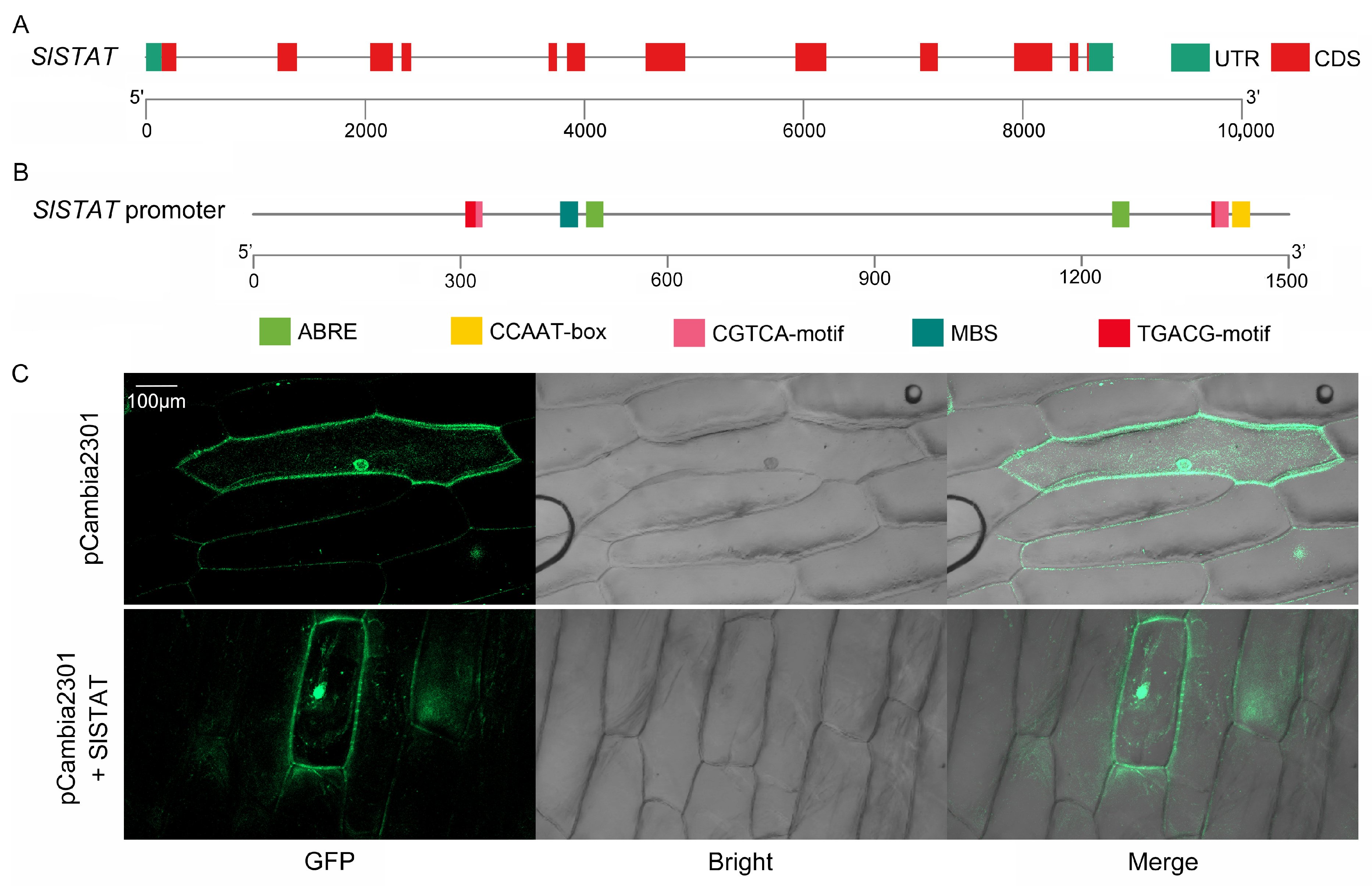
2.1. SlSTAT Family Member Identification and Analysis

The protein sequences of the seven human identified HsSTATs were acquired from the NCBI website (https://www.ncbi.nlm.nih.gov/; accessed on 30 March 2025) and used for detection of AtSTATs and SlSTATs through two iterations of BLASTP. A total of two and one STATs were identified by two round BLASTP in A. thaliana and S. lycopersicum genome, respectively (Table S1). The evolutionary relationships and classification of these STATs have been clarified through the construction of a phylogenetic tree using the protein sequences of 7 HsSTATs, 2 AtSTATs, and 1 SlSTAT. In comparison to animals, the number of STAT family members in Arabidopsis and tomato is relatively limited, and their phylogenetic relationship to human STATs is considerably more distanced (Figure S1).
After gene cloning, the SlSTAT (Solyc07g062050) CDS was obtained, with a length of 2112 bp (Table S2). Through alignment with the SlSTAT gene sequences containing introns in the tomato genome, we identified 12 exons and 11 introns within the SlSTAT genomic genes (Figure 1A). The 1500 bp sequence upstream of the SlSTAT gene was selected as the promoter region. Using genomic DNA as a template, the SlSTAT promoter was cloned, followed by the promoter cis-acting element predictions. The results indicated the presence of various stress-related elements, including abscisic acid responsiveness, MeJA responsiveness, and MYB-binding sites associated with drought inducibility, suggesting the gene’s involvement in stress and plant hormone response mechanisms (Table S3, Figure 1B). To investigate the subcellular localization of SlSTAT proteins, a fusion of SlSTAT with GFP was transiently expressed in onion epidermal cells. The results demonstrated that the SlSTAT protein was localized in both the cytoplasm and the nucleus (Figure 1C).

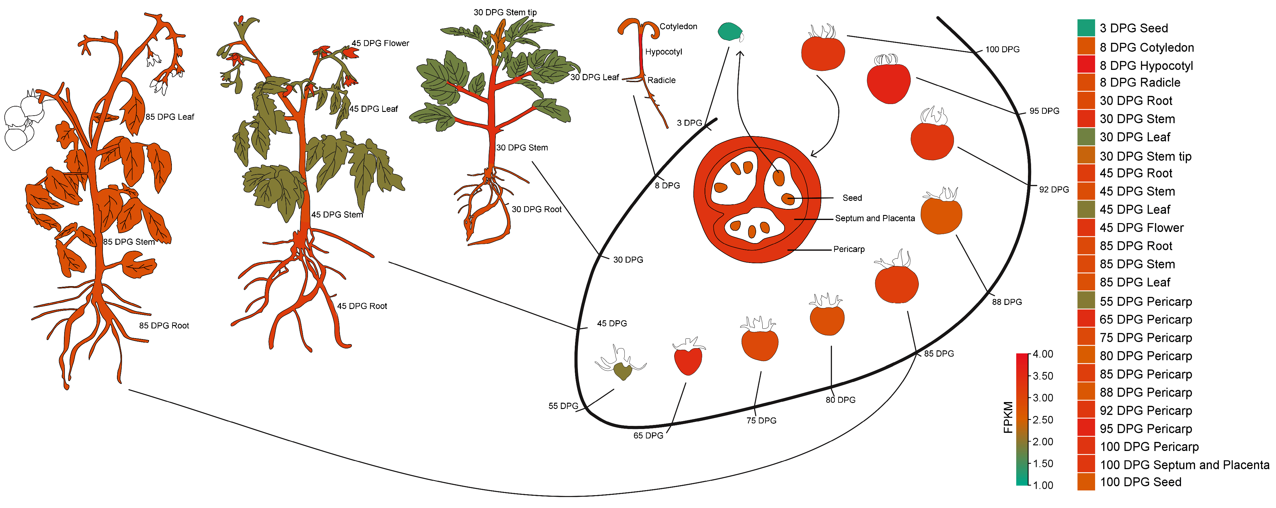
2.2. Expression Levels of SlSTAT in Different Tomato Tissue/Organs

To confirm the tissue-expression of the SlSTAT gene, its expressions in 26 different tomato tissues were obtained from the tomato transcription database of various tissue/organs (Table S4) [22]. Subsequently, its expression patterns are represented using the cartoon heatmap. Notably, the SlSTAT gene exhibits relatively low expression across various tomato tissues and organs, with FPKM values ranging from 1.22 (3 DPG seed) to 3.83 (8 DPG hypocotyl) (Table S4).
As illustrated in Figure 2, SlSTAT displays reduced expression levels in 3 DPG seed (early germination stage), 30 DPG leaf (vegetative growth stage), 45 DPG leaf (flowering stage), and 55 DPG pericarps (immature green fruiting stage). In contrast, SlSTAT exhibits elevated expression levels in 8 DPG hypocotyl (seedling stage). The increased expression from the early germination period to the seedling growth stage underscores the critical role of SlSTAT in seed germination processes.

2.3. Function of SlSTAT During Seed Germination Processes

To examine the impact of SlSTAT gene expression on tomato seed germination and seedling growth, we constructed OE and RNAi vectors for the SlSTAT gene. Subsequently, we conducted genetic transformation in tomato plants and confirmed the presence of transgenic plants at both the DNA and RNA levels (Figure S2 and Figure 3A). Ultimately, we established stable SlSTAT OE and RNAi transgenic tomato lines. For seed germination assays, the WT and transgenic tomato plant seeds were incubated at 25 °C for 8 days. Phenotypic observations were recorded at 0, 3, and 8 d (Figure 3B).
On day 3 of seed germination, only the OE-9 line exhibited a significantly higher germination rate compared to the WT tomatoes (Figure 3C). No significant differences were observed among the other OE lines, RNAi lines, and WT plants (Figure 3C). However, the radicle lengths of all RNAi lines were significantly shorter than those of the WT tomatoes (Figure 3D). In contrast, no significant differences in radicle lengths were found between the OE lines and WT plants (Figure 3D).
On day 8 of germination, only the RNAi-5 line demonstrated a significantly lower germination rate and radicle length compared to the WT plants. The other OE and RNAi lines exhibited no significant differences in either germination rate or radicle length relative to the WT tomatoes (Figure 3C,D). However, the fresh weight of the OE-9 line was significantly higher than that of the WT. In contrast, the fresh weights of the RNAi-1 and RNAi-5 lines were significantly lower than those of the WT (Figure 3E). The experimental findings demonstrate that SlSTAT expression significantly affects the germination of tomato seeds, particularly in those with reduced SlSTAT expression levels (RNAi lines). Early in the germination process, decreased SlSTAT expression notably reduces the radicle lengths and negatively impacts biomass.

2.4. Expression of SlSTAT Change the Chilling Resistance of Tomato

Low temperatures severely affect the yield and quality of tomatoes. The expression of the SlSTAT gene is significantly downregulated during cold treatment, with the lowest expression observed after 48 h at low temperatures. Even after returning to normal growth conditions (R48 h), the expression of SlSTAT does not recover (Figure 4A). To explore the specific role of SlSTAT in the response to low-temperature stress, we analyzed the phenotype and physiological parameters of OE and RNA interference (RNAi) lines in comparison to wild-type (WT) plants following low-temperature treatment.
From a phenotypic perspective, there are no significant differences between the SlSTAT OE plants and the WT tomato plants, whether under 25 °C or 4 °C growth conditions (Figure 4B). Moreover, in accordance with the phenotypic findings, there were no significant differences in the MDA content between the SlSTAT OE lines and WT tomato leaves under both normal temperature and low-temperature treatment conditions (Figure 4C). However, as illustrated in Figure 4D, at 48 h and R48 h, the leaves of SlSTAT OE tomatoes contained lower levels of H2O2 compared to the WT plants, showing a milder degree of cold damage in OE tomatoes. CAT, POD, and SOD are key enzymes in cells that eliminate reactive oxygen species. While no significant difference in CAT activity was observed between OE plants and WT plants at 48 h, the CAT activity was significantly elevated in the OE plants compared to the WT plants at R48 h (Figure 4E). For POD enzyme activities, all SlSTAT OE transgenic lines exhibited significantly higher levels than those in the WT plants at 48 h, and only the OE-9 line showed significantly higher POD enzyme activities at R48 h compared to the WT, which may be related to the higher gene expression of SlSTAT (Figure 3A and Figure 4F). Moreover, SOD enzyme activities in the OE-9 transgenic plant lines also showed higher levels than those in WT tomato plants at both 48 and R48 h (Figure 4G). We did not find significant differences in the H2O2 content and activity of the CAT\POD\SOD between OE and WT tomatoes at 0 h (Figure 4D–G). Thus, overexpression of SlSTAT enhanced the reactive oxygen species scavenging ability of tomato plants under low-temperature stress conditions. In summary, by decreasing peroxide accumulation in the leaves and increasing the activity of enzymes associated with reactive oxygen species scavenging, we obtained overexpression of SlSTAT-enhanced tomato cold stress resistance, which is positively correlated with SlSTAT overexpression levels.
Conversely, SlSTAT RNAi plants exhibited stunted growth compared to the WT plants at the same growth stage, and they demonstrated greater leaf curling in response to chilling stress, demonstrating increased susceptibility to low-temperature stress compared to the WT tomatoes (Figure 4B). In line with the phenotypic observations, no significant variance was observed in the MDA levels between SlSTAT RNAi and WT tomato leaves under 25 °C temperature conditions. However, following low-temperature treatment, the MDA content in the leaves of SlSTAT RNAi plants was significantly elevated compared to WT tomatoes at both 48 h and R48 h (Figure 4C). Similarly, the accumulation of H2O2 in the leaves of RNAi plants was greater than that observed in WT plants, particularly at the R48 h (Figure 4D). While no significant difference in CAT activity was observed between OE plants and WT plants at 48 h, the CAT activity in the RNAi plants was significantly lower than in WT plants at R48 h (Figure 4E). For POD enzyme activity, all SlSTAT RNAi transgenic lines exhibited significantly lower levels than those in the WT plants at R48 h, and only the RNAi-1 and RNAi-17 lines showed significantly lower POD enzyme activities at 48 h compared to the WT plants (Figure 4F). Moreover, SOD enzyme activity in all RNAi transgenic plant lines showed lower levels that those in the WT tomato plants at both 48 and R48 h (Figure 4G). Thus, downexpression of SlSTAT reduced the reactive oxygen species scavenging ability of tomato plants under low-temperature stress conditions. In summary, by increasing peroxide accumulation in the leaves and decreasing the activity of enzymes associated with reactive oxygen species scavenging, the downexpression of SlSTAT enhanced sensitivity to cold stress in tomato.
Meanwhile, the expression levels of the low-temperature response marker genes, SlCBF1-3, were assessed (Figure 4H–J, Table S5). Low temperatures increase the expression of these three genes, with SlCBF2 and SlCBF3 showing a particularly significant rise in expression at R48 h (Figure 4H–J). On one hand, the overexpression or underexpression of SlSTAT affects the expression of SlCBF genes under normal growth conditions. Compared to the WT plants, the expression level of SlCBF1 is up-regulated in OE lines and down-regulated in RNAi transgenic plants (Figure 4H). Interestingly, in contrast to the WT tomatoes, SlCBF2 and SlCBF3 are upregulated in both OE lines and RNAi transgenic plants, although the degree of up-regulation in the OE lines is higher than that in the RNAi transgenic plants (Figure 4I,J). On the other hand, the overexpression or downregulation of SlSTAT also affects the expression of these three genes after low-temperature treatment and during the recovery period of plant growth, particularly at R48 h (Figure 4H–J). At R48 h, the expression levels of SlCBF1 in both the OE and RNAi plants were significantly higher than in the WT plant, with the expression in the OE lines also exceeding that in the RNAi plants (Figure 4H). Conversely, the expression levels of SlCBF2 and SlCBF3 in both the OE and RNAi lines were significantly lower than those in the WT plant, though the expression in the OE lines was still higher than that in the RNAi lines (Figure 4I,J). These results indicate that the expression changes of SlCBF1-3 in transgenic plants do not merely follow a straightforward linear relationship with the changes in SlSTAT expression, suggesting a more complex molecular mechanism and a non-direct interaction between SlSTAT and SlCBFs.

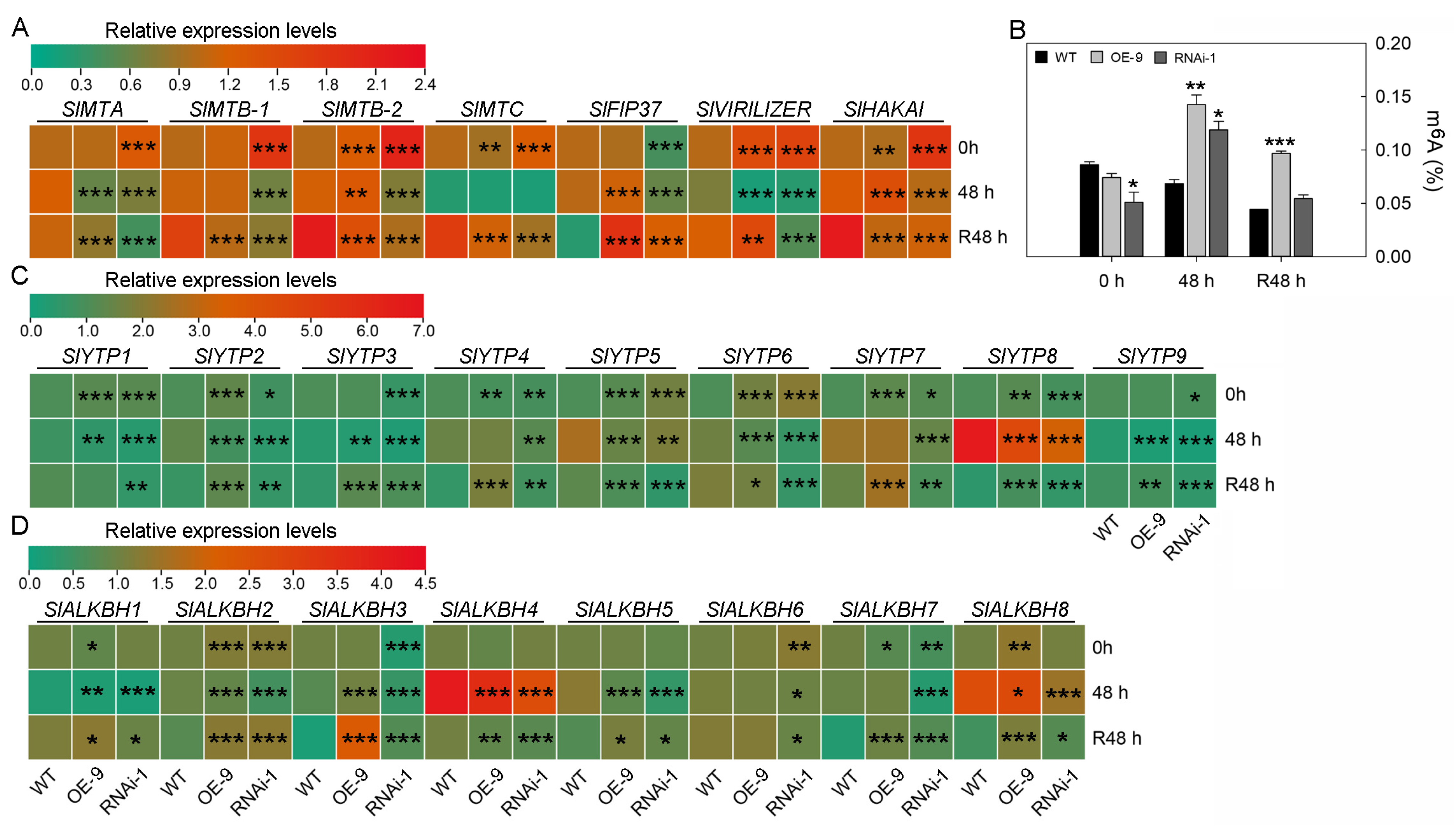
2.5. The Effect of STAT on RNA m6A Modification in Tomato Leaves and Its Relationship with Chilling Resistance

The relationship between SlSTAT and RNA m6A modifications in animals, along with the impact of tomato m6A modifications on their resistance to cold stress, prompts us to explore the effects of SlSTAT expression on the RNA m6A modification levels of tomato leaves and the expression of m6A-related proteins (writers, readers, and erasers) (Table S6).
For m6A writer proteins, under normal growth conditions, both overexpression and downregulation of SlSTAT promote the expression of m6A writer subunit genes, except for SlFIP37, which was not significantly different in expression between the SlSTAT OE plants and the WT plants, and was downregulated in SlSTAT RNAi plants (Figure 5A). After 48 h of cold treatment, the expression level of SlFIP37 in OE plants was higher than in the WT plants, while in the RNAi lines, the expression level was lower than in the WT plants (Figure 5A). At R 48 h, the expression level of SlFIP37 was higher in both OE plants and RNAi lines compared to the wild type, with the expression level in OE plants being higher than that in the RNAi lines (Figure 5A).
As methyltransferases involved in RNA m6A modification, the expression levels of the writer subunit genes reflect, to some extent, the level of m6A modification in cellular RNA. As shown in Figure 5B, under normal temperature conditions, the low expression of STAT significantly reduced the m6A modification levels in tomato leaves, while the m6A modification levels in the OE plant leaves did not show any significant differences compared to WT plants (Figure 5B). After 48 h of low-temperature treatment, the m6A modification levels in the leaves of OE plants and RNAi lines were both higher than those in the wild type, with the OE plants showing higher levels than the RNAi lines (Figure 5B). At R 48 h, the RNA m6A modification levels in the leaves of OE plants were significantly higher than those in the wild type, whereas the RNA m6A modification levels in the leaves of RNAi lines showed no significant difference compared to the wild type (Figure 5B). It is worth noting that, under normal growth condition, the expression level of SlFIP37 and the RNA m6A modification levels in the leaves of both WT and transgenic plants remained consistent (Figure 5A,B). In summary, under normal and low-temperature growth conditions, the expression of SlSTAT affects the expression of RNA m6A writer genes, ultimately influencing the changes in RNA m6A modification levels in tomato leaves.
For m6A reader proteins, SlYTP8 and SlYTP9 are the two genes that respond most significantly to cold stress. Cold temperatures induce the up-regulation of SlYTP8, while SlYTP9 is downregulated (Figure 5C). Compared to the WT plants, SlYTP8 and SlYTP9 exhibited a similar trend in expression level changes in the transgenic plants (Figure 5C). The SlYTP8/9 expression levels in the OE, WT, and RNAi lines decreased sequentially at both 0 h and R48 h of cold treatment, indicating a positive correlation between SlYTP8/9 expression and SlSTAT expression (Figure 5C). However, after 48 h of cold treatment (48 h), the expression of SlYTP8/9 in the SlSTAT OE plants was lower than that in the WT (Figure 5C). Nevertheless, the results above indicate that SlSTAT may regulate the recognition of m6A signals by directly influencing the expression of SlYTP8 and SlYTP9 under low-temperature conditions.
For m6A eraser proteins, the expression changes of SlALKBH1, SlALKBH4, and SlALKBH8 were most pronounced at low temperatures. Low temperatures lead to a downregulation of SlALKBH1, while SlALKBH4 and SlALKBH8 were up-regulated (Figure 5D). As demethylases involved in m6A modification, the expression levels of eraser genes are another important factor influencing the levels of RNA m6A modification within cells. Under normal growth conditions, the overexpression of SlSTAT resulted in an upregulation of the expression levels of most writer genes; however, the RNA m6A modification levels in the OE plants did not show significant differences compared to the WT tomatoes (Figure 5A,B). The reason for the above results may be that the overexpression of SlSTAT also led to an upregulation of m6A eraser genes. Under normal growth conditions, the eraser gene that showed the most significant upregulation in STAT-overexpressing plants compared to the WT plants was SlALKBH8 (Figure 5D). In other words, the relatively higher expression level of SlALKBH8 in the OE plants may explain why, despite the increased expression of m6A writer genes in the OE plants, there was no significant difference in m6A levels compared to the WT plants (Figure 5A,B,D). Regardless of whether in the WT plants, OE lines, or RNAi lines, the expression of the SlALKBH8 gene showed a trend of initially increasing and then decreasing during the low-temperature treatment and subsequent recovery to normal growth temperatures (Figure 5D). In addition, the expression level of SlALKBH8 in the STAT OE lines was higher than in the WT plants at 0 h, 48 h, and R48 h (Figure 5D). Nevertheless, at 0 h, the expression level of SlALKBH8 in the RNAi lines was not significantly different from that in the WT plants. At 48 h, the expression level of SlALKBH8 in the RNAi lines was lower than that in the WT plants. At R48 h, the expression level of SlALKBH8 in the RNAi lines was higher than that in the WT tomatoes (Figure 5D). In summary, SlSTAT may regulate the removal of m6A signals by directly or indirectly influencing the expression of SlALKBH8. Additionally, the differences in expression and interaction of writer and eraser genes in the WT tomatoes, OE lines, or RNAi lines ultimately determine their intracellular RNA m6A modification levels, which involve a very complex molecular mechanism.

3. Discussion

3.1. SlSTAT Family Member Identification and Analysis

The initial STAT protein was discovered in the invertebrate Drosophila, referred to as D-STAT or STAT92E, highlighting its functional significance in invertebrates [23]. Then, Kawata et al. identified a STAT-like protein in Dictyostelium, which is also a DNA-binding protein, and its activation is based on an SH2 domain [24]. Subsequently, the STAT signaling pathway was detected in slime molds, nematodes, fruit flies, and vertebrates, while reports of fungi and plant STATs were lacking [25]. There are seven HsSTATs in the human genome, but there is only one SlSTAT in tomato and two AtSTATs in the Arabidopsis genome (Figure S1). We hypothesize that the significant reduction in the number of STATs in plants may be attributed to the following two possible reasons. Firstly, the tomato SlSTAT protein has a high degree of functional enrichment. Secondly, the presence of other proteins in the tomato genome that took on similar functions resulted in the functional degradation of STATs during evolution and ultimately leading to a decrease in their numbers. For example, the phytohormone cytokinin plays diverse roles in plant development and stress response, and it has a similar extracellular-to-nuclear signaling pathway. However, the protein molecules involved in plant cytokinin signaling differ significantly from those in the animal JAK–STAT pathway [26]. Fleishon et al. have investigated the cross-talk between gibberellin and cytokinin in tomato (S. lycopersicum) [27]. Whether the function of tomato STAT proteins exhibits functional redundancy or can be effectively compensated by other regulatory proteins requires systematic investigation to clarify the molecular mechanisms underlying STAT-mediated signaling transduction.
As Figure 1C shows, the SlSTAT protein is localized in the nucleus and cytoderm. It has been confirmed in animals that the subcellular localization of STAT is related to its phosphorylation [25]. When STAT is unphosphorylated, it resides in the cytoplasm. Upon the binding of cytokines to membrane receptors, JAK is recruited and phosphorylated, which then recruits STAT, causing STAT to aggregate at the receptor. Then, upon phosphorylation, STAT proteins translocate to the cell nucleus, where they undergo conformational changes that facilitate their binding to specific DNA sequences and initiate gene transcription [5,25]. Consequently, STAT proteins can be detectable in both the cytoplasm and the nucleus, corroborating the findings of this study.

3.2. Expression Levels of SlSTAT in Different Tomato Tissues and Its Function in Seed Germination

In animals, the STAT protein family comprises numerous members whose amino acid sequence diversity, coupled with tissue-specific expression patterns, offers substantial variability to account for their various functions in mediating responses to extracellular signaling proteins [3,5,25]. For example, the absence of STAT and N-Myc disrupts normal mammary cell development and elevates the metastatic potential of mammary tumor cells [28]. Despite the presence of a singular STAT gene in the tomato genome, SlSTAT demonstrates distinct expression patterns across different developmental stages and in different tissue/organs (Table S4; Figure 2).
Seed germination is a critical stage in the plant life cycle, and it plays an important role in the efficiency of agricultural production [16]. The increased expression from the early germination period to the seedling growth stage underscores the critical role of SlSTAT in the seed germination process (Table S4; Figure 2). Further, the expression of the SlSTAT gene in tomatoes affects seed germination, particularly at low expression levels. After 3 and 8 days of germination, the low expression of SlSTAT significantly reduced the germination rate and the fresh weight of seedlings, respectively, thereby delaying the germination process (Figure 3). These experimental results highlight the important role of STAT in seed germination. Nevertheless, increasing the expression level of tomato SlSTAT did not have a significant impact on germination rate and biomass (Figure 3). Based on this, it is speculated that normal tomato growth does not require a high level of SlSTAT gene expression, as there is a cascading amplification effect in the signaling transduction process. Additionally, the overall expression level of SlSTAT is relatively low during different tomato growth periods and in various tissue/organs (Table S4; Figure 2), which further support our speculation.

3.3. Expression of SlSTAT Changes the Chilling Resistance of Tomato

In addition to tomato growth and development, roles of SlSTAT in stress responses were examined, alongside initial investigations into its potential molecular mechanisms. SlSTAT responds to low-temperature treatment and shows a downregulation expression trend (Figure 4A). Moreover, the overexpression and downexpression of SlSTAT enhanced tomato cold stress resistance and sensitivity, respectively, by affecting peroxide accumulation in the leaves and the activity of enzymes associated with reactive oxygen species scavenging (Figure 4B–G).
When it comes to their potential molecular mechanisms, STAT proteins could bind to specific DNA sequences in the nuclear and initiate gene transcription [5]. There are various identified DNA-binding proteins (transcription factors, TFs) that modulate the expression of downstream stress-responsive genes either positively or negatively in the cold signaling network of tomato [18]. Among these, the DREB1/CBF TFs are pivotal regulators within the plant’s cold response transcriptional regulation network [29] (Zhao et al., 2015). In Arabidopsis and tomato, these CBF genes are rapidly activated under cold stress, initiating a transcriptional cascade that results in the expression of a wide array of cold-induced genes [30,31,32]. This inevitably leads us to consider whether there are connections between STAT and CBFs. Three CBFs exist in the tomato genome: SlCBF1, SlCBF2, and SlCBF3 [32]. Interestingly, both overexpression and underexpression of SlSTAT not only influence the expression of SlCBF genes under normal growth conditions but also affect the expression of these three genes after low-temperature treatment and during the growth recovery growth, particularly at R48 h (Figure 4H–J). However, the changes in the expression of SlCBF1-3 in transgenic plants do not exhibit a simple linear relationship with the variations in SlSTAT expression, indicating a more complex molecular mechanism and a non-direct interaction between STAT and CBFs. Nevertheless, the experimental results still suggest an interaction between the STAT signaling pathway and DREB1/CBF TF-centered transcriptional regulatory network in tomato cold stress response, which provide directions for further in-depth research in tomatoes and other plants.

3.4. The Effect of SlSTAT on RNA m6A Modification in Tomato Low-Temperature Stress Response

Furthermore, the essential roles of RNA m6A modification in plant low-temperature stress response and reports on the relationship between STAT and m6A modification in animals prompted us to identify the effect of SlSTAT on RNA m6A modification in tomato low-temperature stress response [33,34,35]. RNA m6A modification is one of the most common RNA modification types in eukaryotes, which is catalyzed by m6A writer (methyltransferase), bound and recognized by m6A reader, and removed by m6A eraser (demethylases) proteins [11,12]. The influence of SlSTAT on the RNA m6A modification can be examined from four perspectives: its effects on m6A writer genes, reader genes, eraser genes, and the overall levels of RNA m6A modification.
As shown Figure 5A,B, both the upregulation and downregulation of the SlSTAT gene affect the expression of m6A writer genes, no matter whether under normal or low-temperature growth conditions (Figure 5A). The aforementioned results primarily demonstrate the impact of STAT on the expression of m6A writer genes. In contrast, the literature on animals has reported the influence of m6A modification on the expression of STAT. In porcine bone marrow-derived stem cells (pBMSCs), knockout of the m6A “writer” protein methyltransferase-like 3 (METTL3) resulted in the reduced m6A methylation of JAK mRNAs, which in turn increases JAK protein levels. This enhancement promotes adipogenesis via the JAK1/STAT5/C/EBPβ pathway in pBMSCs adipogenic differentiation, highlighting a coordinated network that connects RNA m6A methylation to the JAK–STAT signaling pathway [36]. In tumor-infiltrating myeloid cells (TIMs), METTL3-mediated m6A modification of Jak1 mRNA enhances JAK1 protein translation efficiency through the m6A-YTHDF1 axis, leading to increased phosphorylation of STAT3 [37]. Moreover, existing literature documents direct interactions between STAT and m6A writer complex in animals. The interaction between METTL3 (m6A writer) and STAT5B promotes co-transcriptional m6A modification of mRNA, thereby enhancing breast tumorigenesis [10]. Therefore, the relationship between STAT and m6A writer gene expressions is not unidirectional.
For m6A reader proteins, SlYTP8 and SlYTP9 are the two genes that responded most significantly to cold stress (Figure 5C) [38,39]. Moreover, the transgenic tomatoes overexpressing the SlYTP8 gene exhibited less resistance to chilling stresses compared to WT plants [38]. The SlYTP8 expression levels in the SlSTAT OE, WT, and SlSTAT RNAi lines decreased sequentially at both 0 h and R48 h of cold treatment, indicating a positive correlation between SlYTP8 and SlSTAT expressions (Figure 5C). It is possible that SlSTAT enhances the expression of SlYTP8, increasing its recognition of m6A signals. Nevertheless, the existing reports only provide evidence that reader proteins influence STAT signaling transduction. YTHDF2 (an m6A reader) facilitates the degradation of STAT1 mRNA by recognizing its m6A modifications, thereby influencing glycolysis and the polarization of M1 macrophages [40]. Additionally, in RNA virus-infected cells, YTHDF3 has a positive role in antiviral JAK–STAT signaling [41].
From the m6A eraser perspective, the expression changes of SlALKBH1, SlALKBH4, and SlALKBH8 are most pronounced at low temperatures (Figure 5D). Shen et al. also reported that SlALKBH8 (Solyc12g096230.2) was upregulated at 24 and 48 h of low-temperature treatment [39]. Moreover, considering the positive correlation between the expression of SlSTAT and SlALKBH8, SlSTAT may regulate the removal of m6A signals by directly or indirectly influencing the expression of SlALKBH8 (Figure 5D). On the other hand, the m6A eraser could also alter the signaling transduction of the JAK–STAT pathway. FTO (m6A eraser) increased the stability of SOCS gene mRNA and thus negatively regulated the JAK–STAT signaling pathway [42]. Conversely, FTO plays a positive role in bladder cancer by maintaining STAT3 mRNA stability through decreasing its m6A modification levels [13].
The differences in expression and interaction between writer and eraser genes in the WT tomatoes, SlSTAT OE lines, or RNAi lines ultimately determine their RNA m6A modification levels, which involve a very complex molecular mechanism (Figure 5B). Although there are no reported studies on the impact of STAT on the overall intracellular RNA m6A modification levels, the research results of Yang et al. and Wang et al. have fully demonstrated the important regulatory role of m6A modification in tomato’s response network to low-temperature stress [35,43]. The aforementioned research provides a foundational basis for exploring how the STAT-m6A axis participates in tomato low-temperature resistance.
In summary, unlike existing literature, which focuses more on the effects of m6A modifications on STAT/JAK–STAT signal transduction, this study preliminarily analyzed the potential molecular mechanisms by which STAT influences m6A modifications in relation to tomato cold stress resistance. This provides a new perspective for exploring the interactions between the JAK–STAT and m6A modification signaling pathways.

4. Materials and Methods

4.1. Identification and Analysis of the SlSTAT Gene Family

Firstly, protein sequences of 7 human (Homo sapiens) STATs (HsSTAT1-4,5A-B,6) were downloaded from the NCBI website (https://www.ncbi.nlm.nih.gov/; accessed on 30 March 2025). Then, BLASTP analysis was conducted via TBtools (version 2.136) using these human STAT sequences to identify STAT family members in Arabidopsis (Arabidopsis thaliana) and tomato (S. lycopersicum) [44]. During this process, Arabidposis genome files were download from TAIR (https://www.arabidopsis.org/; accessed on 30 March 2025), and tomato genomes were obtained from Sol Genomics Network (https://solgenomics.net/; accessed on 30 March 2025). In addition, a phylogenetic tree showing the evolutionary relationships among humans, Arabidopsis, and tomato, was constructed using MEGA7.0 software, with parameters set to 1000 bootstrap replicates [38].

4.2. Gene Clone and Gene Structure Analysis

A total of 30 DPG (30 Days Past Germination) Micro-tom leaves were used as plant materials for total RNA isolation. The FlashPure Plant Total RNA Mini Kit (R019, GeneBetter life science, Beijing, China, http://www.gene-better.cn/; accessed on 30 March 2025) and RevertAid First Strand cDNA Synthesis Kit (K1622, Thermo Scientific, Shanghai, China, https://www.thermofisher.cn/; accessed on 30 March 2025) were used to conduct total RNA extraction and synthesis of first-strand complementary DNA (cDNA) experiments, respectively, according to manufacturer’s guidelines. Table S7 presents the primers for gene cloning, which were designed using Primer Premier 6.0. The coding sequence (CDS) of SlSTAT was obtained through PCR (polymerase chain reaction) amplification using the aforementioned cDNA and gene cloning primers. The genome sequence of SlSTAT was also download form Sol Genomics Network (https://solgenomics.net/; accessed on 30 March 2025). Then, gene structure analysis was conducted using TBtools, comparing CDS and the genome sequence of SlSTAT [44].

4.3. Promoter Clone and Cis-Acting Element Analysis

A total of 30 DPG Micro-tom roots were used as plant materials for the genomic DNA isolation experiments. Then, referring to the instructions of the Hi-Fast Plant Genomic DNA Kit (D200, GeneBetter life science, Beijing, China, http://www.gene-better.cn/; accessed on 30 March 2025), the DNA of these tomato roots was isolated. Promoter clone PCR primers were designed based on about 1500 bp of the gene’s upstream sequence, which was downloaded from the tomato genome available on Sol Genomics Network (https://solgenomics.net/; accessed on 30 March 2025) (Table S7). For promoter cis-acting element predictions, Lescot et al. provides the useful PlantCARE online prediction tool [45].

4.4. Sub-Cellular Localization

pCambia2301, as a plant overexpression vector, contains a green fluorescent protein (GFP) label. SlSTAT CDSs were inserted into this vector after being PCR amplified (see vector construction primers in Table S7). Positive recombinant plasmids, pCambia2301-SlSTAT, were transferred to onion epidermal cells with the help of Agrobacterium tumefaciens (strain GV3101). After infection and growth for 48 h at 22 °C, the onion samples were placed onto glass slides and examined under a fluorescence microscope (Olympus; Tokyo, Japan) equipped with a 20× objective lens.

4.5. Plot Cartoon Heatmap

Cartoon heatmap construction methods were described by Li et al. [22]. Briefly, schematic diagrams of tomato tissues/organs, gene expression values (FPKM; Fragments Per Kilobase of transcript per Million mapped reads), and the SlSTAT gene ID were imported into TBtools, then the cartoon heatmap could be downloaded from the pop-up page [22,44]. Among them, schematic diagrams of tomato tissues/organs and their gene expression values were both provided by the tomato transcription database established by Li et al. [22].

4.6. Plant Growth and Treatments

The seeds of Micro-Tom utilized in this study are those stored in our laboratory (College of Life Sciences and Medicine, Shandong University of Technology, Zibo, Shandong, China). Micro-Tom seedlings were cultivated in plastic pots (7 × 7 cm) containing a mixture of soil, perlite, and vermiculite (4:1:1, v:v:v) as the growth substrate. The light intensity was maintained at 5000 lux, with a day/night cycle of 16 h of light followed by 8 h of darkness. The temperature regimen during the day and night periods was set at 25 °C and 20 °C, respectively.
For the seed germination experiments, seeds of wild-type (WT) and transgenic tomato plants were then put on 1/2 MS medium after surface disinfection with 70% ethanol (v:v) for 1 min, 5% sodium hypochlorite for 10 min, and sterile water for 3–5 times. The light intensity was maintained at 2000 lux, with a day/night cycle of 14 h of light followed by 10 h of darkness. The temperature regimen during the day and night periods was set at 25 °C and 20 °C, respectively. Phenotype analysis was conducted at 0, 3, and 8 d, and at least five biological replicates were used.
For low-temperature (4 °C) treatment experiments, 30 DPG tomato seedlings of WT plants were used as experimental materials. While keeping the light intensity and photoperiod constant, the temperature inside the growth chamber was adjusted to 4 °C. Leaf samples for SlSTAT qRT-PCR (quantitative reverse transcription-PCR) analysis were collected at 0, 1, 3, 6, 12, 24, 48 h, and after a recovery period of 48 h (R48 h) [46].
For the low-temperature (4 °C) comparison experiments, 30 DPG tomato seedlings of both WT and transgenic plants were used as experiment materials, and the treatment methods were the same as those used in the aforementioned low-temperature (4 °C) treatment experiments. For physiological index assessment, leaf samples collected at 0 h, 48 h, and R48 h were utilized. The time point selection for measuring physiological indicators was based on the expression pattern of SlSTAT under low-temperature treatment conditions. Moreover, the leaf samples for SlCBFs and m6A-related gene qRT-PCR analysis were also collected at 0 h, 48 h, and R48 h.

4.7. qRT-PCR

For qRT-PCR, cDNA synthesis was conducted according to the manuscript instruction of the PrimeScript™ RT Reagent Kit with gDNA Eraser (Perfect Real Time) (RR047A, Takara, Beijing, China, https://www.takarabiomed.com.cn/; accessed on 30 March 2025). Then, qRT-PCR was conducted with TB Green® Premix Ex Taq™ II FAST qPCR (CN830A, Takara, Beijing, China, https://www.takarabiomed.com.cn/; accessed on 30 March 2025) via the LightCycler 96/LightCycler 480 System (Roche Diagnostics; Indianapolis, IN, USA). Three technical replicates were used for each cDNA temple. SlACTIN (Solyc11goo5330) was used as the reference gene [19]. qRT-PCR gene-specific primer sequences of SlACTIN and m6A modification-related genes are shown in Table S7 and were download from Shen et al., respectively [39].

4.8. Tomato Transformation and Identification

pCambia2301-SlSTAT obtained at Section 4.4 can also be used as plant OE (overexpression) vectors. For RNAi (RNA interference) plasmid construction, 200-bp specific sequence of SlSTAT CDS was inserted into pK7WWG2D plasmids according to the manuscript instruction of the Gateway™ BP Clonase™ II Enzyme Mix and (11789020, Invitrogen, Thermo Fisher Scientific, Shanghai, China, https://www.thermofisher.cn/; accessed on 30 March 2025) and Gateway™ LR Clonase™ II Enzyme Mix (11791019, Invitrogen, Thermo Fisher Scientific, Shanghai, China, https://www.thermofisher.cn/; accessed on 30 March 2025). The primers used for RNAi plasmid construction are listed in Table S7. The SlSTAT-200 fragment, cloned using the aforementioned primers, was then ligated into the pDONR vector using Gateway™ BP Clonase™ II Enzyme at 25 °C for 1 h. The resulting pDONR-SlYTP9-200 was mixed with the pK7GWIWG2D vector and incubated with Gateway™ LR Clonase™ II Enzyme at 25 °C for 1 h, ultimately yielding pK7WWG2D-SlSTAT-200. The Gateway™ BP Clonase™ II Enzyme and Gateway™ LR Clonase™ II Enzyme are provided in the kits, while the empty pDONR and pK7WWG2D vectors are those maintained in the laboratory (College of Life Sciences and Medicine, Shandong University of Technology, Zibo, Shandong, China). Recombinant plasmids (pCambia2301-SlSTAT and pK7WWG2D-SlSTAT-200) were applied to the tomato transformation process.
The GV3101 strain containing either the pCambia2301-SlSTAT or pK7WWG2D-SlSTAT-200 constructs was employed to infect Micro-Tom tomato cotyledons at Wuhan Biorun Biotechnology Co., Ltd., Wuhan, China. The selection of putative transgenic plants was conducted using Kanamycin at a concentration of 50 mg·L−1, followed by confirmation at the DNA level using GUS gene primers for OE lines and GFP gene primers for RNA RNAi lines (see Table S7). Transgenic lines exhibiting a segregation ratio of 3:1 were selected, and homozygous T3 generation transgenic lines were further verified using qRT-PCR methods.

4.9. Measurement of Stress-Related Physiological Indexes

The concentrations of malondialdehyde (MDA) and hydrogen peroxide (H2O2), as well as the activities of superoxide dismutase (SOD), catalase (CAT), and peroxidase (POD), were assessed according to established protocols using the following test kits: Malondialdehyde Test Kit (MDA-2-Y, Comin, Suzhou, China, http://www.cominbio.com/index.html; accessed on 30 March 2025), Hydrogen Peroxide Test Kit (H2O2-2-Y, Comin), Superoxide Dismutase Test Kit (SOD-2-Y, Comin), Catalase Test Kit (CAT-2-W, Comin), and Peroxidase Test Kit (POD-2-Y, Comin). Detailed experimental procedures are referenced in Zhang et al. [38]. Three technical replicates were used for each sample.

4.10. RNA m6A Modification Level Identification

Following total RNA extraction, the m6A methylation level was quantified utilizing the EpiQuik m6A RNA Methylation Quantification Kit (EPIGENTEK, Farmingdale, NY, USA; https://www.epigentek.com; accessed on 30 March 2025), in accordance with the methodology outlined by Hu et al. [47]. In summary, 200 ng of total RNA was immobilized on strip wells coated with m6A-specific antibodies. Following a wash step, the detection and capture antibody solutions were introduced, and the signal was quantified by measuring absorbance at 450 nm. The m6A levels in the samples were determined using a standard curve. The negative control (NC) comprised RNA devoid of m6A, while the positive control (PC) was an m6A oligonucleotide calibrated to contain 100% m6A, both supplied by the kit. According to the kit instructions, two technical replicates were used for each RNA sample.

4.11. Statistical Analysis

Statistical analysis was conducted using GraphPad Prism (version 8.0.1; GraphPad, La Jolla, CA, USA). Variables between two independent groups were compared using the unpaired t-test (two-tailed). A significance level of p < 0.05 was considered indicative of statistical significance.

5. Conclusions

SlSTAT is the only STAT protein found in the tomato genome, and it is localized in both the nucleus and the cytoplasm. In terms of tomato growth and development, the low expression of SlSTAT significantly reduced seed germination rate and seedling fresh weight, thereby delaying the germination process. Regarding tomato stress responses, overexpression and downexpression of SlSTAT enhanced cold stress resistance and sensitivity, respectively. The changes in cold resistance may be achieved by influencing the gene expression of SlCBFs or through RNA m6A modifications. Although the above findings fill gaps in the research on tomato and plant STAT proteins, the specific functions of SlSTAT in tomato seed germination and cold stress responses, as well as the interactions between the JAK–STAT signaling pathway and m6A modifications, still require further in-depth exploration.

Supplementary Materials

The supporting information can be downloaded at: https://www.mdpi.com/article/10.3390/ijms26073338/s1.

Author Contributions

N.W. and L.J. initiated and designed the experiments. Y.Z., J.Z., Y.L. and J.L. performed the experiments and collected the data. N.W. and Y.L. analyzed the data and wrote the manuscript. N.W. and L.J. revised the manuscript. All authors have read and agreed to the published version of the manuscript.

Funding

This work was financially supported by the National Natural Science Foundation of China (Grant No. 32202518) and Shandong University of Technology PhD Start-up Fund (418097).

Institutional Review Board Statement

Not applicable.

Informed Consent Statement

Not applicable.

Data Availability Statement

The original contributions presented in this study are included in the article/Supplementary Material. Further inquiries can be directed to the corresponding author.

Conflicts of Interest

The authors declare no conflicts of interest.

Abbreviations

Catalase (CAT); coding sequence (CDS); cDNA (complementary DNA); days past germination (DPG); genomic DNA (gDNA); Fragments Per Kilobase of transcript per Million mapped reads (FPKM); green fluorescent protein (GFP); hydrogen peroxide (H2O2); interferons (IFNs); Janus kinase–signal transducer and activator of transcription (JAK-STAT); malondialdehyde (MDA); messenger RNA (mRNA); negative control (NC); no significance (ns); N6-methyladenosine (m6A); overexpression (OE); peroxidase (POD); polymerase chain reaction (PCR); positive control (PC); qRT-PCR (quantitative reverse transcription polymerase chain reaction); RNA interference (RNAi); superoxide dismutase (SOD); Standard Deviation (SD); untranslated regions (UTRs); wild-type (WT).

References

  1. Isaacs, A.; Lindenmann, J. Virus interference. I. The interferon. Proc. R. Soc. Lond. B Biol. Sci. 1957, 147, 258–267. [Google Scholar] [PubMed]
  2. Stark, G.R.; Darnell, J.E., Jr. The JAK-STAT pathway at twenty. Immunity 2012, 36, 503–514. [Google Scholar]
  3. Philips, R.L.; Wang, Y.; Cheon, H.; Kanno, Y.; Gadina, M.; Sartorelli, V.; Horvath, C.M.; Darnell, J.E., Jr.; Stark, G.R.; O’Shea, J.J. The JAK-STAT pathway at 30: Much learned, much more to do. Cell 2022, 185, 3857–3876. [Google Scholar] [PubMed]
  4. Morris, R.; Kershaw, N.J.; Babon, J.J. The molecular details of cytokine signaling via the JAK/STAT pathway. Protein Sci. 2018, 27, 1984–2009. [Google Scholar]
  5. Lv, Y.; Qi, J.; Babon, J.J.; Cao, L.; Fan, G.; Lang, J.; Zhang, J.; Mi, P.; Kobe, B.; Wang, F. The JAK-STAT pathway: From structural biology to cytokine engineering. Signal Transduct. Target. Ther. 2024, 9, 221. [Google Scholar] [PubMed]
  6. Liang, D.; Wang, Q.; Zhang, W.; Tang, H.; Song, C.; Yan, Z.; Liang, Y.; Wang, H. JAK/STAT in leukemia: A clinical update. Mol. Cancer 2024, 23, 25. [Google Scholar]
  7. Li, J.; Rodriguez, J.P.; Niu, F.; Pu, M.; Wang, J.; Hung, L.W.; Shao, Q.; Zhu, Y.; Ding, W.; Liu, Y.; et al. Structural basis for DNA recognition by STAT6. Proc. Natl. Acad. Sci. USA 2016, 113, 13015–13020. [Google Scholar]
  8. Paul, A.; Tang, T.H.; Ng, S.K. Interferon regulatory factor 9 structure and regulation. Front. Immunol. 2018, 9, 1831. [Google Scholar]
  9. Rengachari, S.; Groiss, S.; Devos, J.M.; Caron, E.; Grandvaux, N.; Panne, D. Structural basis of STAT2 recognition by IRF9 reveals molecular insights into ISGF3 function. Proc. Natl. Acad. Sci. USA 2018, 115, E601–E609. [Google Scholar] [CrossRef]
  10. Bhattarai, P.Y.; Kim, G.; Lim, S.C.; Choi, H.S. METTL3-STAT5B interaction facilitates the co-transcriptional m6A modification of mRNA to promote breast tumorigenesis. Cancer Lett. 2024, 603, 217215. [Google Scholar]
  11. Wiener, D.; Schwartz, S. The epitranscriptome beyond m6A. Nat. Rev. Genet. 2021, 22, 119–131. [Google Scholar]
  12. Song, P.; Cai, Z.; Jia, G. Principles, functions, and biological implications of m6A in plants. RNA 2024, 30, 491–499. [Google Scholar]
  13. Sun, Z.; Sun, X.; Qin, G.; Li, Y.; Zhou, G.; Jiang, X. FTO promotes proliferation and migration of bladder cancer via enhancing stability of STAT3 mRNA in an m6A-dependent manner. Epigenetics 2023, 18, 2242688. [Google Scholar] [PubMed]
  14. Wang, Y.; Lai, W.; Zheng, X.; Li, K.; Zhang, Y.; Pang, X.; Gao, J.; Lou, Z. Linderae Radix extract attenuates ulcerative colitis by inhibiting the JAK/STAT signaling pathway. Phytomedicine 2024, 132, 155868. [Google Scholar] [PubMed]
  15. Kazmi, R.H.; Willems, L.A.J.; Joosen, R.V.L.; Khan, N.; Ligterink, W.; Hilhorst, H.W.M. Metabolomic analysis of tomato seed germination. Metabolomics 2017, 13, 145. [Google Scholar] [PubMed]
  16. Song, J.; Shang, L.; Wang, X.; Xing, Y.; Xu, W.; Zhang, Y.; Wang, T.; Li, H.; Zhang, J.; Ye, Z. MAPK11 regulates seed germination and ABA signaling in tomato by phosphorylating SnRKs. J. Exp. Bot. 2021, 72, 1677–1690. [Google Scholar] [CrossRef]
  17. Shohat, H.; Cheriker, H.; Cohen, A.; Weiss, D. Tomato ABA-IMPORTING TRANSPORTER 1.1 inhibits seed germination under high salinity conditions. Plant Physiol. 2023, 191, 1404–1415. [Google Scholar]
  18. Ding, Y.; Shi, Y.; Yang, S. Advances and challenges in uncovering cold tolerance regulatory mechanisms in plants. New Phytol. 2019, 222, 1690–1704. [Google Scholar] [CrossRef]
  19. Lin, R.; Song, J.; Tang, M.; Wang, L.; Yu, J.; Zhou, Y. CALMODULIN6 negatively regulates cold tolerance by attenuating ICE1-dependent stress responses in tomato. Plant Physiol. 2023, 193, 2105–2121. [Google Scholar]
  20. Wang, L.; Chen, H.; Chen, G.; Luo, G.; Shen, X.; Ouyang, B.; Bie, Z. Transcription factor SlWRKY50 enhances cold tolerance in tomato by activating the jasmonic acid signaling. Plant Physiol. 2024, 194, 1075–1090. [Google Scholar]
  21. Guo, M.; Yang, F.; Zhu, L.; Wang, L.; Li, Z.; Qi, Z.; Fotopoulos, V.; Yu, J.; Zhou, J. Loss of cold tolerance is conferred by absence of the WRKY34 promoter fragment during tomato evolution. Nat. Commun. 2024, 15, 6667. [Google Scholar] [PubMed]
  22. Li, J.; Sun, B.; Xu, Q.; Jiang, L.; Wang, N. Transcriptome-level analysis of gene expressions in different tissues of tomato and key gene identifications during seed germination. Sci. Hortic. 2024, 337, 113565. [Google Scholar]
  23. Yan, R.; Luo, H.; Darnell, J.E., Jr.; Dearolf, C.R. A JAK-STAT pathway regulates wing vein formation in Drosophila. Proc. Natl. Acad. Sci. USA 1996, 93, 5842–5847. [Google Scholar]
  24. Kawata, T.; Shevchenko, A.; Fukuzawa, M.; Jermyn, K.A.; Totty, N.F.; Zhukovskaya, N.V.; Sterling, A.E.; Mann, M.; Williams, J.G. SH2 signaling in a lower eukaryote: A STAT protein that regulates stalk cell differentiation in dictyostelium. Cell 1997, 89, 909–916. [Google Scholar]
  25. Aaronson, D.S.; Horvath, C.M. A road map for those who don’t know JAK-STAT. Science 2002, 296, 1653–1655. [Google Scholar] [CrossRef]
  26. Kieber, J.J.; Schaller, G.E. Cytokinin signaling in plant development. Development 2018, 145, dev149344. [Google Scholar] [PubMed]
  27. Fleishon, S.; Shani, E.; Ori, N.; Weiss, D. Negative reciprocal interactions between gibberellin and cytokinin in tomato. New Phytol. 2011, 190, 609–617. [Google Scholar]
  28. Pruitt, H.C.; Metge, B.J.; Weeks, S.E.; Chen, D.; Wei, S.; Kesterson, R.A.; Shevde, L.A.; Samant, R.S. Conditional knockout of N-Myc and STAT interactor disrupts normal mammary development and enhances metastatic ability of mammary tumors. Oncogene 2018, 37, 1610–1623. [Google Scholar]
  29. Zhao, C.; Lang, Z.; Zhu, J.K. Cold responsive gene transcription becomes more complex. Trends Plant Sci. 2015, 20, 466–468. [Google Scholar] [CrossRef]
  30. Yamaguchi-Shinozaki, K.; Shinozaki, K. Transcriptional regulatory networks in cellular responses and tolerance to dehydration and cold stresses. Annu. Rev. Plant Biol. 2006, 57, 781–803. [Google Scholar]
  31. Thomashow, M.F. Molecular basis of plant cold acclimation: Insights gained from studying the CBF cold response pathway. Plant Physiol. 2010, 154, 571–577. [Google Scholar] [CrossRef] [PubMed]
  32. Song, J.; Lin, R.; Tang, M.; Wang, L.; Fan, P.; Xia, X.; Yu, J.; Zhou, Y. SlMPK1- and SlMPK2-mediated SlBBX17 phosphorylation positively regulates CBF-dependent cold tolerance in tomato. New Phytol. 2023, 239, 1887–1902. [Google Scholar]
  33. Li, H.B.; Tong, J.; Zhu, S.; Batista, P.J.; Duffy, E.E.; Zhao, J.; Bailis, W.; Cao, G.; Kroehling, L.; Chen, Y.; et al. m6A mRNA methylation controls T cell homeostasis by targeting the IL-7/STAT5/SOCS pathways. Nature 2017, 548, 338–342. [Google Scholar] [CrossRef] [PubMed]
  34. Chen, J.; Fang, X.; Zhong, P.; Song, Z.; Hu, X. N6-methyladenosine modifications: Interactions with novel RNA-binding proteins and roles in signal transduction. RNA Biol. 2019, 16, 991–1000. [Google Scholar] [CrossRef] [PubMed]
  35. Yang, D.; Xu, H.; Liu, Y.; Li, M.; Ali, M.; Xu, X.; Lu, G. RNA N6-methyladenosine responds to low-temperature stress in tomato anthers. Front. Plant Sci. 2021, 12, 687826. [Google Scholar]
  36. Yao, Y.; Bi, Z.; Wu, R.; Zhao, Y.; Liu, Y.; Liu, Q.; Wang, Y.; Wang, X. METTL3 inhibits BMSC adipogenic differentiation by targeting the JAK1/STAT5/C/EBPβ pathway via an m6A-YTHDF2-dependent manner. FASEB J. 2019, 33, 7529–7544. [Google Scholar] [CrossRef]
  37. Xiong, J.; He, J.; Zhu, J.; Pan, J.; Liao, W.; Ye, H.; Wang, H.; Song, Y.; Du, Y.; Cui, B.; et al. Lactylation-driven METTL3-mediated RNA m6A modification promotes immunosuppression of tumor-infiltrating myeloid cells. Mol. Cell 2022, 82, 1660–1677. [Google Scholar]
  38. Zhang, Y.; Guo, T.; Li, J.; Jiang, L.; Wang, N. Tomato (Solanum lycopersicum L.) YTH domain-containing RNA-binding protein (YTP) family members participate in low-temperature treatment and waterlogging stress responses. Horticulturae 2024, 10, 522. [Google Scholar] [CrossRef]
  39. Shen, H.; Luo, B.; Wang, Y.; Li, J.; Hu, Z.; Xie, Q.; Wu, T.; Chen, G. Genome-wide identification, classification and expression analysis of m6A gene family in Solanum lycopersicum. Int. J. Mol. Sci. 2022, 23, 4522. [Google Scholar] [CrossRef]
  40. Huangfu, N.; Zheng, W.; Xu, Z.; Wang, S.; Wang, Y.; Cheng, J.; Li, Z.; Cheng, K.; Zhang, S.; Chen, X.; et al. RBM4 regulates M1 macrophages polarization through targeting STAT1-mediated glycolysis. Int. Immunopharmacol. 2020, 83, 106432. [Google Scholar] [CrossRef]
  41. Kastan, J.P.; Tremblay, M.W.; Brown, M.C.; Trimarco, J.D.; Dobrikova, E.Y.; Dobrikov, M.I.; Gromeier, M. Enterovirus 2Apro cleavage of the YTHDF m6A readers implicates YTHDF3 as a mediator of type I interferon-driven JAK/STAT signaling. mBio 2021, 12, e00116-21. [Google Scholar] [PubMed]
  42. Kim, S.M.; Oh, S.C.; Lee, S.Y.; Kong, L.Z.; Lee, J.H.; Kim, T.D. FTO negatively regulates the cytotoxic activity of natural killer cells. EMBO Rep. 2023, 24, e55681. [Google Scholar] [PubMed]
  43. Wang, N.; Li, Y.; Guo, T.; Jiang, L. Multi-omics analysis reveals the epitranscriptomic and proteomic regulation network of tomato in low-temperature stress response. Hortic. Plant J. 2025, 11, 758–773. [Google Scholar]
  44. Chen, C.; Wu, Y.; Li, J.; Wang, X.; Zeng, Z.; Xu, J.; Liu, Y.; Feng, J.; Chen, H.; He, Y.; et al. TBtools-II: A “one for all, all for one” bioinformatics platform for biological big-data mining. Mol. Plant 2023, 16, 1733–1742. [Google Scholar]
  45. Lescot, M.; Déhais, P.; Thijs, G.; Marchal, K.; Moreau, Y.; Van de Peer, Y.; Rouzé, P.; Rombauts, S. PlantCARE, a database of plant cis-acting regulatory elements and a portal to tools for in silico analysis of promoter sequences. Nucleic Acids Res. 2002, 30, 325–327. [Google Scholar]
  46. Guo, M.; Li, Z.; Wang, L.; Xu, T.; Huang, H.; Kanwar, M.K.; Yang, P.; Zhou, J. BAG8 positively regulates cold stress tolerance by modulating photosystem, antioxidant system and protein protection in Solanum lycopersicum. Plant Physiol. Biochem. 2024, 206, 108267. [Google Scholar]
  47. Hu, J.; Cai, J.; Park, S.J.; Lee, K.; Li, Y.; Chen, Y.; Yun, J.Y.; Xu, T.; Kang, H. N6-Methyladenosine mRNA methylation is important for salt stress tolerance in Arabidopsis. Plant J. 2021, 106, 1759–1775. [Google Scholar]
Figure 1. Gene structure, promoter analysis, and subcellular localization of SlSTAT. (A) Gene structure of SlSTAT, illustrating the coding sequence (CDS), untranslated regions (UTRs), and introns represented by lines connecting the boxes. (B) Analysis of cis-acting elements in the SlSTAT promoter, with various types of elements indicated by color-coded boxes. (C) Subcellular localization of SlSTAT, shown through confocal microscopy images of onion epidermal cells co-expressing either GFP alone (pCambia2301) or SlSTAT-GFP (pCambia2301 + SlSTAT). From left to right, the columns display GFP fluorescence, bright field, and merged images. The scale bar indicates a measurement of 100 μm.
Ijms 26 03338 g001
Figure 2. Expression patterns of SlSTAT in different tomato tissue/organs. The cartoon heatmap depicts SlSTAT gene expression patterns across various tomato tissues, with average expression values calculated from three independent biological replicates. The scale bar represents FPKM (Fragments Per Kilobase of transcript per Million mapped reads) values.
Ijms 26 03338 g002
Figure 3. Expression of SlSTAT gene affects tomato seed germination and seedling growth. (A) Expression levels of SlSTAT in wild-type (WT), overexpression (OE), and RNA interference (RNAi) transgenic tomato plants. (B) Seed germination phenotype of WT, SlSTAT OE lines, and SlSTAT RNAi lines under 25 °C conditions. Scale bars = 15 mm. (CE) Germination-related physiological indexes measurement, including germination rate (C), radicle length (D), and fresh weight (E). Error bars represent the Standard Deviation (SD). Asterisks denote statistical significance levels: *, **, and *** correspond to p <  0.05, p < 0.01, and p <  0.001, respectively.
Ijms 26 03338 g003
Figure 4. The relationship between SlSTAT expressions and tomato resistance under low-temperatures stress. (A) The relative expression levels of SlSTAT in tomato leaves subjected to low-temperature stress were assessed. The expression levels at other time points were calculated relative to the 0 h. (B) Phenotype of WT tomatoes, SlSTAT OE lines, and SlSTAT RNAi lines under low-temperature conditions. Scale bars = 15 mm. (CG) Stress-related physiological indexes measurement, including malondialdehyde (MDA) concentration (C), hydrogen peroxide (H2O2) concentration (D), catalase (CAT) activity (E), peroxidase (POD) activity (F), and superoxide dismutase (SOD) activity (G). (HJ) Relative expression levels of cold stress marker genes, including SlCBF1 (H), SlCBF2 (I), and SlCBF3 (J), in WT and transgenic tomato leaves under low-temperature stress treatment. The mean expression levels were calculated in comparison to the WT plants at time point 0 h. Significance analyses were separately conducted at 0 h, 48 h, and R48 h, with all significant difference comparisons being made against the WT tomatoes. Asterisks denote statistical significance levels: *, **, and *** indicate p <  0.05, p < 0.01, and p <  0.001, respectively.
Ijms 26 03338 g004
Figure 5. Expressions of SlSTAT gene change RNA N6-methyladenosine (m6A) modification levels and m6A-related gene expressions under low-temperature stress. (A,C,D) Relative expression levels of m6A writer (A), reader (C), and eraser (D) genes in WT and transgenic tomato leaves under low-temperature conditions. The mean expression levels were calculated in comparison to the WT plants at time point 0 h. (B) RNA m6A modification levels of WT and transgenic tomato leaves under low-temperature conditions. Error bars in the figures represent the SD. Significance analyses were separately conducted at 0 h, 48 h, and R48 h, with all comparisons of significant differences being made against the wild type (WT). Asterisks denote statistical significance levels: *, **, and *** indicate p <  0.05, p < 0.01, and p <  0.001, respectively.
Ijms 26 03338 g005
Disclaimer/Publisher’s Note: The statements, opinions and data contained in all publications are solely those of the individual author(s) and contributor(s) and not of MDPI and/or the editor(s). MDPI and/or the editor(s) disclaim responsibility for any injury to people or property resulting from any ideas, methods, instructions or products referred to in the content.

