Article

Sialyllactose Attenuates Inflammation and Injury of Intestinal Epithelial Cells upon Enterotoxigenic Escherichia coli Infection

by
Qiming Duan
1,2,
Bing Yu
1,2,
Zhiqing Huang
1,2,
Yuheng Luo
1,2,
Ping Zheng
1,2,
Xiangbing Mao
1,2,
Jie Yu
1,2,
Junqiu Luo
1,2,
Hui Yan
1,2 and
Jun He
1,2,*
1
Institute of Animal Nutrition, Sichuan Agricultural University, Chengdu 611130, China
2
Key Laboratory for Animal Disease-Resistance Nutrition, Chengdu 611130, China
*
Author to whom correspondence should be addressed.
Int. J. Mol. Sci. 2025, 26(8), 3860; https://doi.org/10.3390/ijms26083860
Submission received: 19 March 2025 / Revised: 11 April 2025 / Accepted: 15 April 2025 / Published: 18 April 2025
(This article belongs to the Section Bioactives and Nutraceuticals)

Abstract

:
Sialyllactose (SL), a bioactive trisaccharide abundant in porcine colostrum, demonstrates multifunctional properties including antimicrobial activity, immune regulation, and apoptosis inhibition. This research uncovers the mechanisms by which SL mitigates enterotoxigenic Escherichia coli (ETEC)-mediated damage to intestinal barrier integrity, employing IPEC-J2 porcine epithelial models. SL pre-treatment effectively blocked pathogen adhesion by competitively binding to cellular receptors, concurrently mitigating inflammation through significant suppression of TNF-α, IL-1β, and IL-6 expression (p < 0.05). Notably, SL exhibited functional parallels to the NF-κB inhibitor BAY11-7082, jointly enhancing tight junction integrity via ZO-1 protein stabilization and inhibiting pro-inflammatory signaling through coordinated suppression of IκB-α/NF-κB phosphorylation cascades. The dual-action mechanism combines molecular interception of microbial attachment with intracellular modulation of the TLR4/MyD88/NF-κB pathway, effectively resolving both pathogenic colonization and inflammatory amplification. These findings position SL as a potential therapeutic application nutraceutical for livestock, with the capacity to address post-weaning porcine enteritis through functional feed formulations that synergistically enhance intestinal barrier resilience while curbing ETEC-mediated inflammatory pathogenesis.

Graphical Abstract

1. Introduction

The intestinal epithelium’s defensive architecture, constituting a pivotal immunological interface, orchestrates a dual biosystemic role: facilitating bidirectional material trafficking through selective permeability for nutrient assimilation, and maintaining evolutionary conserved pathogen exclusion mechanisms against luminal xenobiotic infiltration [1]. Sustaining the architectural integrity and physiological competence of the gastrointestinal mucosal interface constitutes a cornerstone in immune-endocrine crosstalk regulation. ETEC is one of the most common external antigens [2]. It is capable of expressing one or more membrane adhesins that facilitate its attachment to the intestinal mucosa surface and strengthen its high-affinity interaction with glycoprotein receptors localized on the microvillar surface of intestinal epithelial cells. Following pathogenic colonization, ETEC secretes thermolabile (LT) and thermostable (ST) enterotoxins that activate Toll-like receptor-mediated signaling cascades. This immunopathological cascade induces aberrant overexpression of interleukin-driven inflammatory mediators and initiates caspase-dependent programmed cell death in gut epithelial lineages. The cumulative pathophysiological manifestations include compromised mucosal integrity and electrolyte transport dysregulation, clinically presenting as secretory diarrheal syndromes [3,4]. Within commercial pig production systems, neonatal scours associated with ETEC colonization pose a major health challenge during the weaning transition, which can lead to a worsened productive performance and even death in weaning piglets, resulting in significant economic losses [5]. Weaning deprives piglets of the passive immunity provided by sows’ milk, making the piglets more vulnerable to infections from enteric pathogens [6]. Thus, nutritional strategies incorporating breast milk-derived bioactive constituents into feed formulations demonstrate potential efficacy in mitigating ETEC-mediated intestinal barrier compromise.
Breast milk is rich in a variety of structurally distinct oligosaccharides, which are indigestible carbohydrates for humans and mammals, known as milk oligosaccharides (MOs) [7]. Milk oligosaccharides (MOs) consist of five fundamental monosaccharide units: galactose, glucose, fucose, N-acetylglucosamine, and the sialylated derivative N-acetylneuraminic acid. Sialyllactose (SL), the predominant structural variant within MOs, constitutes 70% of the total oligosaccharides in porcine/bovine mammary secretions and 30% in human lactation products [8]. SL is a macromolecule with a molecular weight of 633.55, consisting of an N-acetyl-D-neuraminic acid unit linked to the galactose unit of lactose through α 2,6- and α 2,3-glycosidic bonds (Figure S1) [9,10,11]. Studies have indicated that D-galactose is a central component of the ETEC receptors on the surface of intestinal epithelial cells, playing a significant role in the adhesion of ETEC strains such as K88, K99, 987P, and H10407. D-galactose exhibits the strongest affinity for ETEC K88, and compounds with similar structures, such as oligogalactose and galactosamine, can also inhibit the adhesion of ETEC to intestinal epithelial cells [12,13]. In addition, sialic acid has been proven to be involved in the adhesion processes of various bacteria and viruses [14]. For example, the adhesion of ETEC to piglet intestinal epithelial cells is highly correlated with the degree of sialylation of their brush border glycoproteins [15]. Glycoproteins modified with sialic acid can alleviate ETTC-induced inflammatory responses by inhibiting its adhesion [16]. Sialyllactose, the most abundant oligosaccharide in sow milk, contains both galactose and salic acid residues in its structure, but it is not clear whether it can similarly inhibit ETEC adherence and thereby alleviate intestinal inflammatory injury in piglets.
This investigation employed ETEC K88/LPS-challenged IPEC-J2 monolayers to delineate SL’s modulatory effects on inflammatory signaling cascades. The experimental data robustly validate SL’s capacity to suppress pro-inflammatory mediators while mechanistically elucidating its therapeutic bioactivity through TLR4/MyD88/NF-κB axis regulation.

2. Results

2.1. SL Treatment Dose and Duration

Figure S2A demonstrates the dose-dependent responses of IPEC-J2 cells to SL (0–20 mg/mL), indicating the viability and transcriptional modulation of inflammatory mediators/tight junction biomarkers. A cytocompatibility assessment confirmed non-cytotoxic effects at 80 mg/mL SL (24 h exposure). A 5 mg/mL SL regimen (12 h) exhibited a dual regulatory capacity involving the suppression of IL-1β transcription and the enhancement of Occludin/ZO-1 mRNA synthesis (Figure S2B,C, p < 0.05). This optimized preconditioning protocol (5 mg/mL, 12 h) was consequently implemented to counteract epithelial inflammatory pathogenesis.

2.2. Protective Effect of SL on Intestinal Epithelial Cells (IECs) Challenged by ETEC

As shown in Figure 1, the early-stage and total apoptosis levels in ETEC-exposed cells were markedly attenuated by SL pre-incubation (p < 0.05). Additionally, the qPCR quantification further revealed that Cysteine-dependent aspartate-specific protease 3, 8, and 9 (Caspase 3, 8, and 9) transcripts were markedly downregulated in ETEC-infected IPEC-J2 cells following SL preconditioning (p < 0.05, Figure 1B).

2.3. SL Alleviates Intestinal Inflammation by Preventing the Adhesion of ETEC K88 on IECs

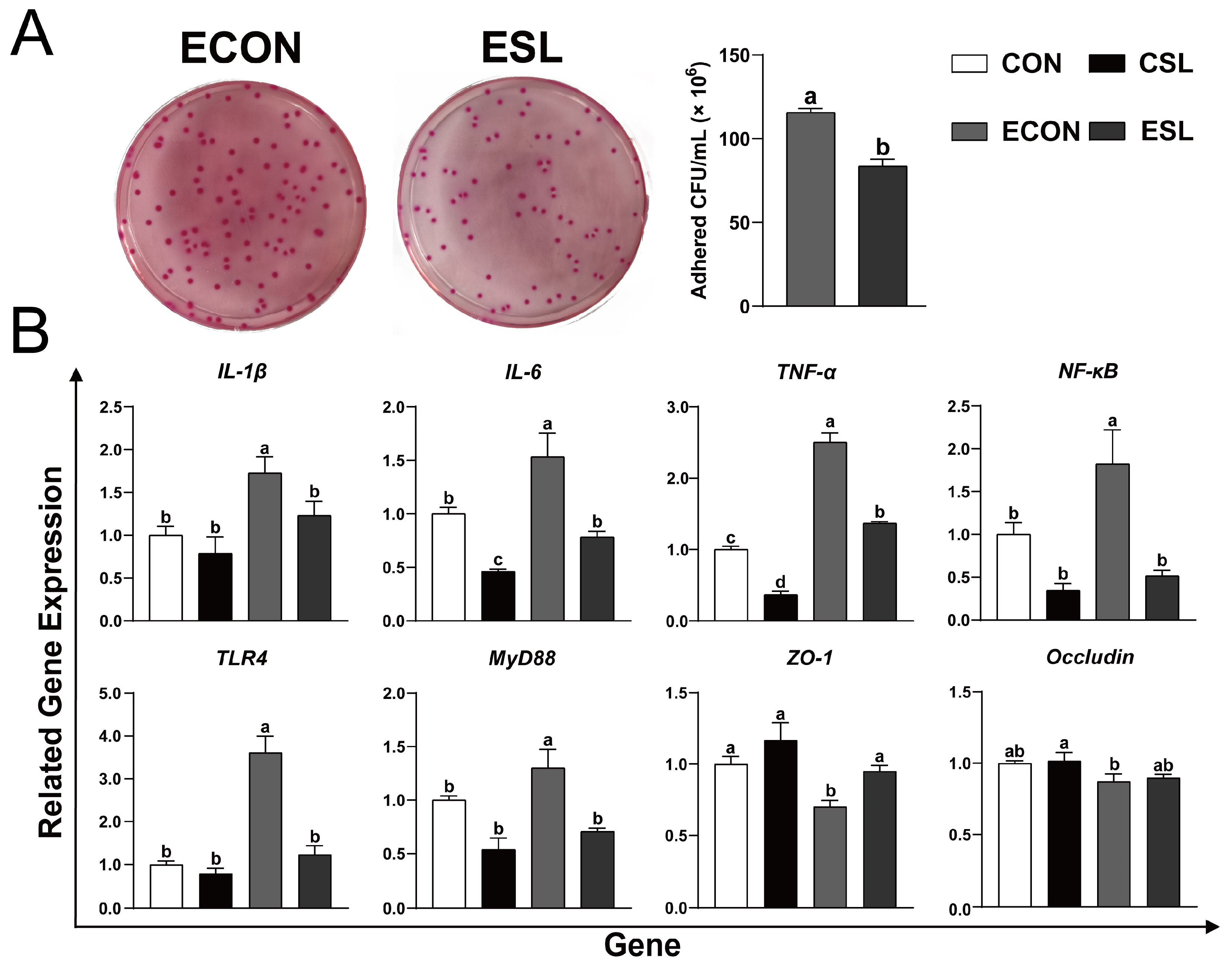
SL preconditioning suppressed ETEC K88 adhesion to IPEC-J2 monolayers and mitigated acute inflammatory damage induced by ETEC infection, as evidenced by downregulated pro-inflammatory mediators including the following: Tumor Necrosis Factor Alpha (TNF-α), Interleukin-1 (IL-1β), and Interleukin-6 (IL-6), as well as up-regulation of the expression of tight-junction protein ZO-1 (p < 0.05, Figure 2). Prophylactic SL administration markedly downregulated transcript levels of key innate immune regulators—MyD88 (myeloid differentiation primary response 88), TLR4 (toll-like receptor 4), and NF-κB (nuclear factor-kappa B)—in ETEC-infected IPEC-J2 cells (Figure 2B, p < 0.05). This transcriptional suppression of the TLR4/MyD88/NF-κB axis implies that SL-mediated anti-inflammatory effects are orchestrated through dampening canonical pathogen recognition signaling, thereby attenuating cytokine storm cascades during enterotoxigenic challenge.

2.4. SL Attenuated Inflammation and Injury of the IECs Challenged by LPS

SL pre-treatment improved the integrity of the LPS-challenged cells, as indicated by the increased distribution and abundance of the tight-junction protein ZO-1 (p < 0.05, Figure 3A). Concomitantly, SL preconditioning attenuated pro-inflammatory mediator release, demonstrating significant reductions in TNF-α, IL-1β, and IL-6 compared to the ETEC-challenged group (p < 0.05, Figure 3B).

2.5. Protective Effect of SL on IECs Challenged by LPS

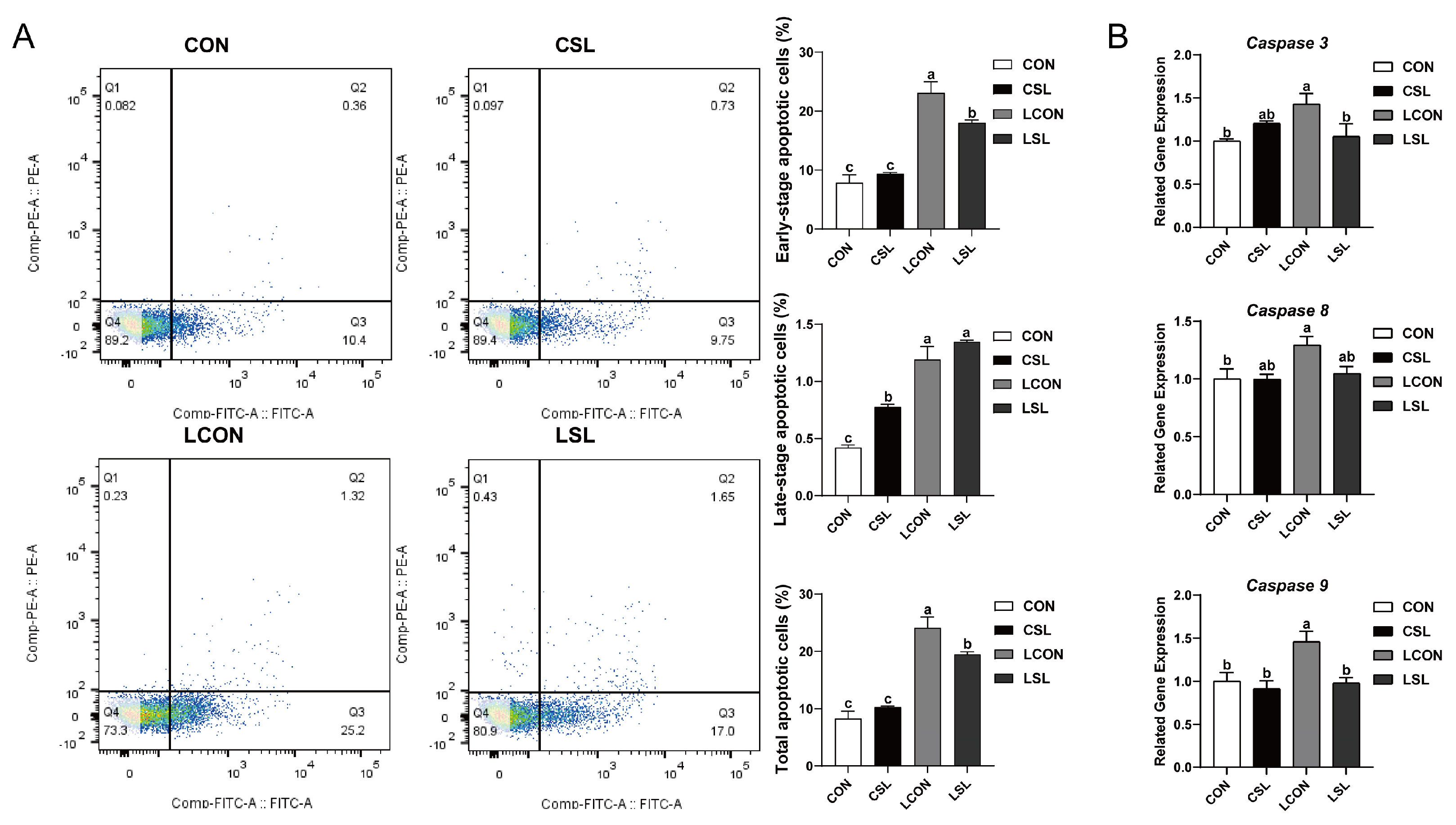
The results of flow cytometry analysis revealed that SL pre-treatment significantly decreased the percentages of early-stage, late-stage, and total apoptotic cells upon LPS challenge (p < 0.05, Figure 4A). Meanwhile, SL pre-treatment significantly decreased the mRNA expression levels of Caspase 3 and 9 in LPS-treated IPEC-J2 cells (p < 0.05, Figure 4B).

2.6. SL Inhibits the Phosphorylation of IκB-α and NF-κB by Preventing the Adhesion of LPS on IECs

LPS mono-stimulation induced robust membrane-localized fluorescence indicative of pathogen-associated molecular pattern binding on IPEC-J2 monolayers, whereas SL coadministration markedly attenuated surface ligand clustering intensity (p < 0.05, Figure 5A). Immunoblotting demonstrated SL-mediated suppression of canonical inflammatory signaling, with dose-dependent attenuation of LPS-triggered phosphorylation events in both IκBα and NF-κB p65 (p < 0.05, Figure 5B).

2.7. SL and BAY 11-7082 Attenuated Inflammation and Injury of the IECs Challenged by LPS

SL-mediated preservation of the tight junction architecture was correlated with ZO-1 transcriptional activation, achieving comparable efficacy to the canonical NF-κB inhibitor BAY 11-7082 in restoring epithelial barrier function (p < 0.05, Figure 6A). Meanwhile, SL and BAY 11-7082 pre-treatment significantly increased the mean optical density of ZO-1 protein LPS-treated IPEC-J2 cells (p < 0.05, Figure 6A). Furthermore, LPS challenge induced robust transcriptional activation of canonical pro-inflammatory mediators (TNF-α, IL-1β, and IL-6) in porcine enterocytes (p < 0.05, Figure 6B). In contrast, pre-treatment with SL or BAY 11-7082 markedly reduced the mRNA expression of these pro-inflammatory cytokines (p < 0.05, Figure 6B).

2.8. SL and BAY 11-7082 Inhibit Apoptosis on IECs Challenged by LPS

Flow cytometry results revealed that SL and BAY 11-7082 pre-treatment significantly decreased the percentages of early-stage and total apoptotic cells in LPS-treated IPEC-J2 cells (p < 0.05, Figure 7A). Concurrently, LPS treatment significantly increased the transcript abundance of Caspase 3, Caspase 8, and Caspase 9, while SL and BAY 11-7082 pre-treatment significantly inhibited Caspase 3, Caspase 8, and Caspase 9 mRNA expression (p < 0.05, Figure 7B).

2.9. SL and BAY 11-7082 Decreased the Phosphorylation of the Critical Inflammation-Associated Proteins IκB-α and NF-κB in LPS-Challenged Cells

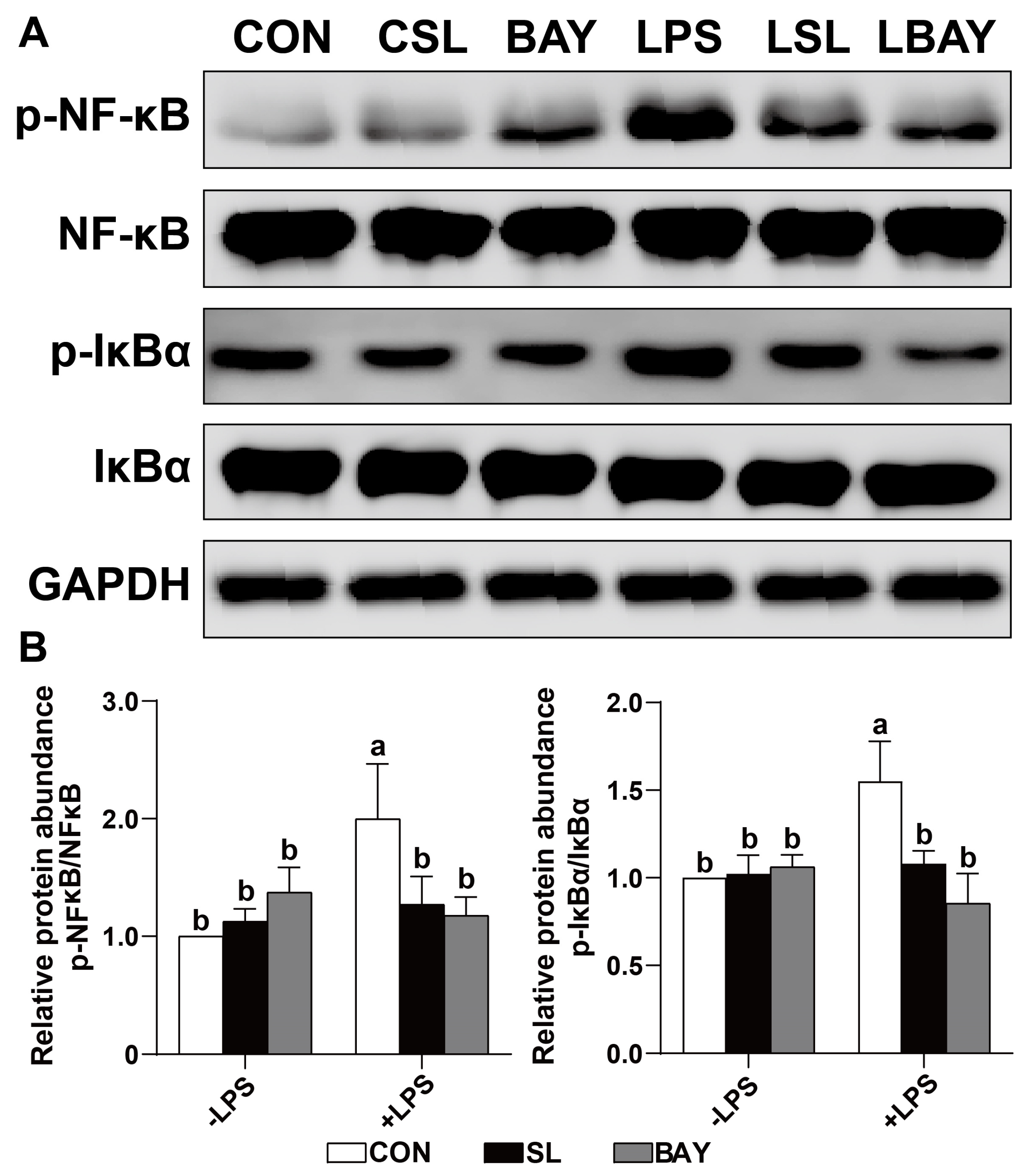
Western blot analysis revealed that SL exerts a similar effect to BAY 11-7082, and both can decrease the abundance of phosphorylation of the NF-κB and IκBα proteins in LPS-treated cells (p < 0.05, Figure 8). These results indicate that SL-mediated suppression of canonical NF-κB nuclear translocation in LPS-stimulated porcine intestinal epithelium can achieve comparable IκBα stabilization efficacy to the pharmacological comparator BAY 11-7082.

3. Discussion

Within porcine production systems, ETEC-associated infections incur substantial economic losses, predominantly through mortality escalation and growth performance reduction. Post-weaning piglets subjected to ETEC colonization typically demonstrate elevated apoptotic indices in IECs [17]. The current experimental data corroborate this pathogenic mechanism, revealing significantly elevated apoptotic indices in ETEC-colonized IPEC-J2 monolayers versus unchallenged controls. Prophylactic SL administration demonstrated substantial anti-apoptotic efficacy in pathogen-exposed models. Molecular profiling further confirmed SL-mediated transcriptional suppression of caspase cascade effectors (caspase-3/-8/-9) in enteropathogen-stimulated epithelial cells. This collective evidence establishes SL’s cytoprotective functionality against ETEC-induced IEC pathophysiology.
ETEC K88 fimbriae exhibit stereoselective binding to sialoglycoconjugates, intestinal mucin-type glycoproteins, and neutral glycosphingolipids via α/β-galactosidic linkages. This microbial adhesion can be competitively inhibited by oligosaccharide structural analogs mimicking host glycan motifs. Such molecular interference effectively blocks microbial adhesion to epithelial membranes, consequently attenuating pathogen virulence potential [18,19]. Sialyllactose contains both galactose and sialic acid moiety (Neu5Ac) [20]. Studies have shown that SL has an antiadhesive antimicrobial ability, which can inhibit the adhesion of Clostridioides difficile, S. enterica subsp, and E. coli O119 to intestinal epithelial cells [21,22]. Consistent with these results, prophylactic SL administration attenuated enteropathogen colonization efficiency, demonstrating a reduction in ETEC K88 fimbrial attachment density on polarized porcine enterocytes compared to untreated challenge controls. TLR4-mediated MyD88-dependent signaling constitutes an essential pathogenic interface wherein ETEC subverts host immunosurveillance, propelling NF-κB-driven pro-inflammatory cascade via FliC flagellin recognition. Functioning as an innate immune sensor, TLR4 identifies microbial signature molecules on ETEC surfaces, initiating the transduction of effector signaling cascades including the MyD88-mediated signaling axis [23]. Ligand engagement induces TLR4-MyD88 heterodimeric assembly, enabling sequential kinase mobilization through IRAK effector recruitment within the interleukin-1 receptor signaling cascade [24]. TLR4 activation initiates kinase signaling cascades, culminating in NF-κB phosphorylation. Nuclear translocation of activated NF-κB induces transcriptional activation of inflammatory mediators—encompassing cytokine/chemokine biosynthesis pathways and cell adhesion machinery—integral to the host’s defense mechanisms against ETEC pathogenesis [23]. In the present study, SL administration mitigated ETEC-induced inflammatory pathogenesis in intestinal epithelia, as evidenced through the transcriptomic suppression of TNF-α/IL-1β/IL-6 cytokine clusters. Concomitant down-modulation of TLR4 receptor expression, MyD88 adaptor protein expression, and NF-κB transcriptional activity mechanistically delineates SL’s barrier-protective effects via blockade of the TLR4-mediated MyD88/NF-κB signaling axis.
Disequilibrium in cytokine homeostasis (anti-/pro-inflammatory) precipitates pathogenic disruption of the integrity of the intestinal barrier—a structure whose molecular foundation resides in intercellular junction complexes comprising occludin, claudin isoforms, and zonula occludens proteins within epithelial monolayers [17,25]. Within junctions with complex architecture, ZO-1 functions as a scaffolding orchestrator, dynamically coordinating epithelial programmed cell death, cellular expansion, and ultrastructural polarization through transmembrane junctional organization [26]. In the present study, immunofluorescence quantification demonstrated that SL preconditioning attenuated ETEC-induced ZO-1 delocalization. These results clearly demonstrate that SL exerts a protective and anti-inflammatory influence on intestinal epithelial cells when challenged with ETEC.
LPS, a structural hallmark of gram-negative bacterial membranes, serves as a standard immunological trigger to activate pro-inflammatory signaling pathways across differentiated epithelial models, notably porcine IPEC-J2 enterocyte lineages [27,28,29]. A recent study revealed that the overproduction of pro-inflammatory cytokines is consistently linked to the disruption of tight junction proteins and the worsening of apoptosis in IPEC-J2 cells [30]. In a consistent manner, SL pre-treatment was found to markedly lower the expression of the pro-inflammatory cytokines TNF-α, IL-1β, and IL-6. Moreover, it enhanced the integrity of LPS-challenged cells, as indicated by an increase in ZO-1 protein abundance, and concurrently decreased the percentages of early-stage and total apoptotic cells, along with the mRNA expression levels of Caspase 3 and 9. Experimental evidence delineates SL’s cytoprotective mechanism in porcine enterocytes, where LPS-triggered inflammatory pathogenesis is ameliorated via dual modulatory effects: (1) the fortification of intercellular junction integrity and (2) the inhibition of programmed cell death through suppressed biosynthesis of IL-6/TNF-α/IL-1β inflammatory mediators.
LPS can bind to the TLR4 receptor on the cell surface and activate the downstream NF-κB-mediated inflammatory cascade [28,31]. Studies have shown that SL can bind directly to the LPS-binding site of the TLR4-MD2 complex, effectively preventing the binding of LPS to TLR4 and inhibiting TLR4-mediated signal transduction [32]. Consistently, immunofluorescence analysis results have revealed that SL coadministration significantly reduces the intensity of surface ligand clustering, thereby effectively inhibiting the adhesion of LPS to the surface of IPEC-J2 cells. Meanwhile, SL pre-exposure has attenuated phospho-activation of NF-κB p65 and IκBα degradation in inflamed epithelia, establishing the molecular basis for SL’s anti-inflammatory efficacy through interference with pathoadhesive interactions.
To delineate SL’s anti-inflammatory properties, we employed a pharmacological intervention with BAY11-7082—a selective suppressor of IκB-α degradation and NF-κB nuclear translocation—to determine its modulatory effects on NF-κB-dependent inflammatory cascades in intestinal epithelia. NF-κB functions as a heterodimeric transcription regulator governing the immunogenetic networks encompassing leukocyte recruitment, apoptosis modulation, and oncogenic transformation through bidirectional control of chemokine/effector protein expression [33,34]. Under basal conditions, NF-κB heterodimers exhibit cytoplasmic quiescence through constitutive binding to IκB regulatory proteins, with IκBα being the predominant isoform. This molecular sequestration effectively precludes nucleocytoplasmic trafficking, thereby maintaining transcriptional dormancy by blocking access to chromatin remodeling complexes [35]. Upon cellular stimulation, IκB-α is phosphorylated and rapidly degraded, which results in the activation of NF-κB. This activation facilitates the translocation of NF-κB into the nucleus, where it triggers the transcription and expression of genes associated with inflammation [36,37]. In addition, SL also exerts a similar effect to BAY (a specific inhibitor of NF-κB) and can both improve the distribution and abundance of ZO-1 in LPS-challenged cells, and suppress apoptosis and the production of inflammatory cytokines in the cells. Additionally, both SL and BAY have notably reduced the phosphorylation of key inflammation-related proteins, including IκB-α and NF-κB, in LPS-stimulated cells. These results provide further evidence supporting the hypothesis that SL could function as an anti-inflammatory agent.

4. Materials and Methods

4.1. Bacterial Strains and Culture

The source and culture of pathogenic Escherichia coli (ETEC) were referenced from previous studies conducted in our laboratory [38].

4.2. Sialylactose

Sialylactose (SL, ≥95%) with a formula of C23H39NO19 and a molecular weight of 633.55 was donated by Glycom A/S and its structure is shown in Figure S1. SL is a trisaccharide produced through microbial fermentation, composed of glucose, galactose, and N-acetylneuraminic acid.

4.3. Cell Culture

The cell culture methods and reagents used were referenced from the study by Wan et al. [17]. The cells were subcultured every 2–3 days at a 1:3 ratio when they reached around 80–90% confluence.

4.4. ETEC Treatment

The experimental protocols and reagents used during ETEC intervention were adapted from the methodology established in Fu et al.’s prior research [31]. The study comprised four experimental conditions:
(1) Baseline control (CON) with neither SL pre-treatment nor ETEC exposure;
(2) SL-only treatment (CSL) involving 12 h pre-incubation with 5 mg/mL SL;
(3) ETEC challenge (ETEC) using 1 × 10⁶ CFU/well bacterial suspension applied for 1 or 2.5 h;
(4) Combined intervention (ESL) combining 5 mg/mL SL pre-treatment followed by dual-phase ETEC exposure.
Following these interventions, both cellular material and conditioned media were collected for subsequent analytical procedures.

4.5. LPS Treatment

The dose and duration of LPS treatment were determined using previous studies carried out in our laboratory [17]. A factorial experimental design was implemented, comprising four distinct cohorts: a control (CON) cohort that received neither SL preconditioning nor LPS stimulation; a CSL cohort that was subjected to 12 h pharmacological preconditioning with 5 mg/mL SL; an LPS cohort that was exposed to 6 h endotoxin challenge (5 μg/mL); and a sequential intervention cohort (LSL) that underwent SL pre-treatment followed by LPS provocation. Post-intervention cellular monolayers and conditioned media were synchronously harvested for downstream multi-omics profiling.

4.6. BAY 11-7082 Treatment

The pharmacological concentrations and exposure periods for LPS and BAY 11-7082 interventions were determined according to established experimental parameters in our institutional research protocol [17]. Eight distinct experimental cohorts were designed:
(1) Negative control (CON) without any pre-treatment or stimuli;
(2) SL monotherapy (CSL) receiving 12 h preconditioning with 5 mg/mL SL;
(3) LPS stimulation (LPS) with 6 h 5 μg/mL exposure;
(4) BAY inhibitor control (BAY) undergoing 2 h 1 μmol/L pre-treatment;
(5) SL-LPS sequential administration (LSL) combining SL preconditioning and subsequent LPS challenge;
(6) BAY-LPS combined regimen (LBAY) pairing inhibitor pre-treatment with LPS stimulation.
Post-interventional specimens including cellular monolayers and corresponding conditioned media were systematically harvested for downstream analytical processing.

4.7. Assay of ETEC K88 Adhesion onto IPEC-J2 Cells

The quantification of ETEC adhesion on IPEC-J2 monolayers was conducted using an optimized protocol derived from Letourneau’s bacterial adherence quantification assay [39]. Post-incubation specimens underwent viable count analysis through serial decimal dilution plating on selective media. Adherent pathogen load was calculated by logarithmic conversion of mean colony counts from diagnostically valid dilution gradients (30–300 colonies/plate threshold), with parallel determination of initial inoculum concentration via control plate enumeration.

4.8. Immunofluorescence Assay

IPEC-J2 monolayers were established on sterile glass coverslips within 6-well plates (2 × 10⁵ cells/well initial seeding density), with culture progression monitored until 80–90% confluency was achieved. Experimental cohorts underwent differential pre-treatment protocols: 12 h incubation with 5 mg/mL SL or 2 h exposure to 1 μM BAY 11-7082 (NF-κB inhibitor), followed by 6 h LPS challenge (5 μg/mL). Subsequent fluorescent immunolocalization of tight junction protein ZO-1 and bacterial endotoxin was conducted according to the standardized immunofluorescence protocol established by Wan et al. [17].

4.9. RNA Extraction and Quantitative Real-Time PCR (qPCR)

Cellular RNA extraction from IPEC-J2 monolayers was performed with RNAiso Plus reagent (Takara Bio, Shiga, Japan), strictly adhering to manufacturer-specified protocols. RNA integrity verification included spectrophotometric assessment (NanoDrop 2000 system; Thermo Fisher, Waltham, MA, USA) through dual-wavelength (260/280 nm) absorbance quantification. Reverse transcription of RNA templates was executed with the PrimeScript RT Master Mix (Takara Bio) with strict adherence to the manufacturer-specified thermocycler protocol, ensuring precise cDNA strand initiation fidelity. Quantitative gene expression profiling followed the qPCR protocol established in Wan et al.’s methodology [17], with 2−ΔΔCt calculations [40] applied for relative quantification against the housekeeping gene β-actin. The oligonucleotide primer sequences utilized in this study are comprehensively delineated in Table 1.

4.10. Detection of Cell Apoptosis

Following enzymatic dissociation of IPEC-J2 monolayers using 0.25% serum-free trypsin solution (EDTA-free formulation), cellular pellets were obtained through centrifugation (350× g, 10 min, 4 °C) under refrigerated conditions. Subsequent washing procedures involved dual cycles of pre-chilled phosphate-buffered saline (PBS, pH 7.4) resuspension. For apoptosis quantification, cell aliquots (100 μL suspension) were sequentially stained with 5 μL PE-conjugated Annexin V and 5 μL 7-AAD viability dye, followed by a 15 min incubation period under light-protected ambient conditions. Reaction termination was achieved by the addition of 400 μL 1× Annexin binding buffer. Fluorescence-based apoptotic profiling was executed within 60 min post-staining using a Beckman Coulter CytoFlex flow cytometry system (Beckman Coulter Life Sciences, Brea, CA, USA) and BD-FACSVerse platform (BD Biosciences, San Jose, CA, USA), with compensation controls applied during acquisition.

4.11. Total Protein Extraction and Western Blot Analysis

Polarized IPEC-J2 monolayers underwent protein fractionation employing ice-cold RIPA buffer (25 mM Tris-HCl pH 7.6, 150 mM NaCl, 1% NP-40, 1% sodium deoxycholate, 0.1% SDS) fortified with protease-phosphatase inhibitor cocktails, with cell debris pelleted via high-speed centrifugation (12,000× g, 15 min, 4 °C) to yield clarified lysate supernatants. An integrated workflow encompassing biomolecular separation, immunodetection parameters, and buffer stoichiometry was implemented in accordance with Wan et al.’s refined proteomic methodology [17]. The target-specific immunodetection reagents with corresponding dilution parameters are systematically cataloged in Table 2.

4.12. Statistics Analysis

Quantitative data processing was executed in IBM SPSS Statistics (v26.0, Armonk, NY, USA), with the experimental outcomes expressed as the arithmetic mean ± SEM from six biological replicates per experimental group. A two-factor factorial ANOVA model with interaction terms was implemented for hypothesis testing under normality and homogeneity of variance assumptions, followed by Fisher’s Least Significant Difference multiple comparison correction. Probabilistic thresholds adhered to the Neyman–Pearson framework, with α = 0.05 defining statistical significance. Data visualization was performed in GraphPad Prism (v8.0.2, La Jolla, CA, USA) following Wilkinson’s grammar of graphics principles for scientific illustration.

5. Conclusions

Collectively, these data establish SL’s therapeutic efficacy against ETEC-mediated pathogenic insults in intestinal mucosa through dual modulation of inflammatory signaling and programmed cell death cascades. Mechanistic interrogation reveals an SL-mediated blockade of LPS’s pathoadhesive interactions with the TLR4/MD2 receptor complex, culminating in the attenuated phosphorylation of NF-κB transcriptional regulators and the subsequent transcriptional quiescence of the IL-6/IL-1β/TNF-α triad biosynthesis. This pharmacological profile highlights SL as a novel nutraceutical agent for intervention in bacterial enteritis pathologies across mammalian systems, particularly in swine model systems exhibiting compromised intestinal barrier function.

Supplementary Materials

The supporting information can be downloaded at: https://www.mdpi.com/article/10.3390/ijms26083860/s1.

Author Contributions

Q.D.: formal analysis, data curation, writing—original draft. B.Y., Z.H., Y.L., P.Z., X.M., J.Y., J.L. and H.Y.: supervision, validation. J.H.: conceptualization, funding acquisition, project administration, methodology, writing—review and editing. All authors have read and agreed to the published version of the manuscript.

Funding

This work was supported by the National Natural Science Foundation of China (32372897) and the Innovation Team of Sichuan Province (SCCXTD-2024-8).

Institutional Review Board Statement

Not applicable.

Informed Consent Statement

Not applicable.

Data Availability Statement

The data are all included in the manuscript.

Acknowledgments

We thank the Danish company Glycom and Vigsnæs Louise for their generous donation, and Huifen Wang, Jiayong Tang, and Hua Li for their help during the experiments.

Conflicts of Interest

Ms. Vigsnæs Louise was employed by Glycom in Denmark. The all authors declare no competing financial interests or personal relationships that could influence the work reported in this paper. The funders had no role in the design of the study; in the collection, analysis, or interpretation of data; in the writing of the manuscript; or in the decision to publish the results.

Abbreviations

The following abbreviations are used in this manuscript:
MOsMilk oligosaccharides
SLSialyllactose
CONControl group
CSLSL group
ESLETEC + SL group
ETECEnterotoxigenic Escherichia coli
IκBαInhibitor of NF-κB Alpha
IL-1βInterleukin-1 beta
IL-6Interleukin-6
IPEC-J2Intestinal Porcine Epithelial Cell Line-J2
LPSLipopolysaccharide
LBAYBAY 11-7082 + LPS group
LSLLPS + SL group
MyD88Myeloid Differentiation Primary Response 88
NF-κBNuclear factor kappa B
TLR4Toll-like receptor 4
TNF-αTumor Necrosis Factor Alpha
ZO-1Zonula Occludens-1 Protein

References

  1. Turner, J.R. Intestinal mucosal barrier function in health and disease. Nat. Rev. Immunol. 2009, 9, 799–809. [Google Scholar] [CrossRef] [PubMed]
  2. Walker, R.I. An assessment of enterotoxigenic Escherichia coli and Shigella vaccine candidates for infants and children. Vaccine 2015, 33, 954–965. [Google Scholar] [CrossRef] [PubMed]
  3. Dubreuil, J.D.; Isaacson, R.E.; Schifferli, D.M. Animal Enterotoxigenic Escherichia coli. EcoSal Plus 2016, 7, 10–1128. [Google Scholar] [CrossRef]
  4. Dubreuil, J.D. Enterotoxigenic Escherichia coli targeting intestinal epithelial tight junctions: An effective way to alter the barrier integrity. Microb. Pathog. 2017, 113, 129–134. [Google Scholar] [CrossRef]
  5. Luppi, A.; Gibellini, M.; Gin, T.; Vangroenweghe, F.; Vandenbroucke, V.; Bauerfeind, R.; Bonilauri, P.; Labarque, G.; Hidalgo, Á. Prevalence of virulence factors in enterotoxigenic Escherichia coli isolated from pigs with post-weaning diarrhoea in Europe. Porc. Health Manag. 2016, 2, 20. [Google Scholar] [CrossRef]
  6. Pohl, C.S.; Medland, J.E.; Mackey, E.; Edwards, L.L.; Bagley, K.D.; DeWilde, M.P.; Williams, K.J.; Moeser, A.J. Early weaning stress induces chronic functional diarrhea, intestinal barrier defects, and increased mast cell activity in a porcine model of early life adversity. Neurogastroenterol. Motil. 2017, 29, e13118. [Google Scholar] [CrossRef]
  7. Dinleyici, M.; Barbieur, J.; Dinleyici, E.C.; Vandenplas, Y. Functional effects of human milk oligosaccharides (HMOs). Gut Microbes 2023, 15, 2186115. [Google Scholar] [CrossRef] [PubMed]
  8. Wiciński, M.; Sawicka, E.; Gębalski, J.; Kubiak, K.; Malinowski, B. Human Milk Oligosaccharides: Health Benefits, Potential Applications in Infant Formulas, and Pharmacology. Nutrients 2020, 12, 266. [Google Scholar] [CrossRef]
  9. Zhang, B.; Li, L.-Q.; Liu, F.; Wu, J.-Y. Human milk oligosaccharides and infant gut microbiota: Molecular structures, utilization strategies and immune function. Carbohydr. Polym. 2022, 276, 118738. [Google Scholar] [CrossRef]
  10. Bode, L. The functional biology of human milk oligosaccharides. Early Hum. Dev. 2015, 91, 619–622. [Google Scholar] [CrossRef]
  11. Zhu, Y.; Zhang, J.; Zhang, W.; Mu, W. Recent progress on health effects and biosynthesis of two key sialylated human milk oligosaccharides, 3’-sialyllactose and 6’-sialyllactose. Biotechnol. Adv. 2023, 62, 108058. [Google Scholar] [CrossRef] [PubMed]
  12. Sarabia-Sainz, H.M.; Armenta-Ruiz, C.; Sarabia-Sainz, J.A.; Guzmán-Partida, A.M.; Ledesma-Osuna, A.I.; Vázquez-Moreno, L.; Ramos-Clamont Montfort, G. Adhesion of enterotoxigenic Escherichia coli strains to neoglycans synthesised with prebiotic galactooligosaccharides. Food Chem. 2013, 141, 2727–2734. [Google Scholar] [CrossRef]
  13. Coppa, G.V.; Facinelli, B.; Magi, G.; Marini, E.; Zampini, L.; Mantovani, V.; Galeazzi, T.; Padella, L.; Marchesiello, R.L.; Santoro, L.; et al. Human milk glycosaminoglycans inhibit in vitro the adhesion of Escherichia coli and Salmonella fyris to human intestinal cells. Pediatr. Res. 2016, 79, 603–607. [Google Scholar] [CrossRef]
  14. Schwegmann, C.; Zimmer, G.; Yoshino, T.; Enss, M.; Herrler, G. Comparison of the sialic acid binding activity of transmissible gastroenteritis coronavirus and E. coli K99. Virus Res. 2001, 75, 69–73. [Google Scholar] [CrossRef] [PubMed]
  15. Seignole, D.; Mouricout, M.; Duval-Iflah, Y.; Quintard, B.; Julien, R. Adhesion of K99 fimbriated Escherichia coli to pig intestinal epithelium: Correlation of adhesive and non-adhesive phenotypes with the sialoglycolipid content. J. Gen. Microbiol. 1991, 137, 1591–1601. [Google Scholar] [CrossRef]
  16. Mouricout, M.; Petit, J.M.; Carias, J.R.; Julien, R. Glycoprotein glycans that inhibit adhesion of Escherichia coli mediated by K99 fimbriae: Treatment of experimental colibacillosis. Infect. Immun. 1990, 58, 98–106. [Google Scholar] [CrossRef] [PubMed]
  17. Wan, J.; Zhang, J.; Xu, Q.; Yin, H.; Chen, D.; Yu, B.; He, J. Alginate oligosaccharide protects against enterotoxigenic Escherichia coli-induced porcine intestinal barrier injury. Carbohydr. Polym. 2021, 270, 118316. [Google Scholar] [CrossRef]
  18. Coppa, G.V.; Zampini, L.; Galeazzi, T.; Facinelli, B.; Ferrante, L.; Capretti, R.; Orazio, G. Human milk oligosaccharides inhibit the adhesion to Caco-2 cells of diarrheal pathogens: Escherichia coli, Vibrio cholerae, and Salmonella fyris. Pediatr. Res. 2006, 59, 377–382. [Google Scholar] [CrossRef]
  19. Sarabia-Sainz, A.; Ramos-Clamont, G.; Del Candia-Plata, M.M.C.; Vázquez-Moreno, L. Biorecognition of Escherichia coli K88 adhesin for glycated porcine albumin. Int. J. Biol. Macromol. 2009, 44, 175–181. [Google Scholar] [CrossRef]
  20. Bode, L. Human milk oligosaccharides: Every baby needs a sugar mama. Glycobiology 2012, 22, 1147–1162. [Google Scholar] [CrossRef]
  21. Piotrowski, M.; Wultańska, D.; Pituch, H. The prebiotic effect of human milk oligosaccharides 3’- and 6’-sialyllactose on adhesion and biofilm formation by Clostridioides difficile—Pilot study. Microbes Infect. 2022, 24, 104929. [Google Scholar] [CrossRef]
  22. Facinelli, B.; Marini, E.; Magi, G.; Zampini, L.; Santoro, L.; Catassi, C.; Monachesi, C.; Gabrielli, O.; Coppa, G.V. Breast milk oligosaccharides: Effects of 2′-fucosyllactose and 6′-sialyllactose on the adhesion of Escherichia coli and Salmonella fyris to Caco-2 cells. J. Matern. Fetal Neonatal Med. 2019, 32, 2950–2952. [Google Scholar] [CrossRef]
  23. Liu, M.; Kang, W.; Hu, Z.; Wang, C.; Zhang, Y. Targeting MyD88: Therapeutic mechanisms and potential applications of the specific inhibitor ST2825. Inflamm. Res. 2023, 72, 2023–2036. [Google Scholar] [CrossRef] [PubMed]
  24. Wei, J.; Zhang, Y.; Li, H.; Wang, F.; Yao, S. Toll-like receptor 4: A potential therapeutic target for multiple human diseases. Biomed. Pharmacother. 2023, 166, 115338. [Google Scholar] [CrossRef] [PubMed]
  25. Anderson, J.M.; Balda, M.S.; Fanning, A.S. The structure and regulation of tight junctions. Curr. Opin. Cell Biol. 1993, 5, 772–778. [Google Scholar] [CrossRef] [PubMed]
  26. Qiao, J.; Sun, Z.; Liang, D.; Li, H. Lactobacillus salivarius alleviates inflammation via NF-κB signaling in ETEC K88-induced IPEC-J2 cells. J. Anim. Sci. Biotechnol. 2020, 11, 76. [Google Scholar] [CrossRef]
  27. Shi, L.; Fang, B.; Yong, Y.; Li, X.; Gong, D.; Li, J.; Yu, T.; Gooneratne, R.; Gao, Z.; Li, S.; et al. Chitosan oligosaccharide-mediated attenuation of LPS-induced inflammation in IPEC-J2 cells is related to the TLR4/NF-κB signaling pathway. Carbohydr. Polym. 2019, 219, 269–279. [Google Scholar] [CrossRef]
  28. Farkas, O.; Palócz, O.; Pászti-Gere, E.; Gálfi, P. Polymethoxyflavone Apigenin-Trimethylether Suppresses LPS-Induced Inflammatory Response in Nontransformed Porcine Intestinal Cell Line IPEC-J2. Oxid. Med. Cell. Longev. 2015, 2015, 673847. [Google Scholar] [CrossRef]
  29. Zhao, L.; Li, M.; Sun, K.; Su, S.; Geng, T.; Sun, H. Hippophae rhamnoides polysaccharides protect IPEC-J2 cells from LPS-induced inflammation, apoptosis and barrier dysfunction in vitro via inhibiting TLR4/NF-κB signaling pathway. Int. J. Biol. Macromol. 2020, 155, 1202–1215. [Google Scholar] [CrossRef] [PubMed]
  30. Takeuchi, O.; Akira, S. Pattern recognition receptors and inflammation. Cell 2010, 140, 805–820. [Google Scholar] [CrossRef]
  31. Fu, Q.; Lin, Q.; Chen, D.; Yu, B.; Luo, Y.; Zheng, P.; Mao, X.; Huang, Z.; Yu, J.; Luo, J.; et al. β-defensin 118 attenuates inflammation and injury of intestinal epithelial cells upon enterotoxigenic Escherichia coli challenge. BMC Vet. Res. 2022, 18, 142. [Google Scholar] [CrossRef] [PubMed]
  32. Sodhi, C.P.; Wipf, P.; Yamaguchi, Y.; Fulton, W.B.; Kovler, M.; Niño, D.F.; Zhou, Q.; Banfield, E.; Werts, A.D.; Ladd, M.R.; et al. The human milk oligosaccharides 2’-fucosyllactose and 6’-sialyllactose protect against the development of necrotizing enterocolitis by inhibiting toll-like receptor 4 signaling. Pediatr. Res. 2021, 89, 91–101. [Google Scholar] [CrossRef] [PubMed]
  33. Won, S.; Sayeed, I.; Peterson, B.L.; Wali, B.; Kahn, J.S.; Stein, D.G. Vitamin D prevents hypoxia/reoxygenation-induced blood-brain barrier disruption via vitamin D receptor-mediated NF-kB signaling pathways. PLoS ONE 2015, 10, e0122821. [Google Scholar] [CrossRef]
  34. Lee, J.-W.; Bae, C.J.; Choi, Y.-J.; Kim, S.-I.; Kim, N.-H.; Lee, H.J.; Kim, S.-S.; Kwon, Y.-S.; Chun, W. 3,4,5-Trihydroxycinnamic Acid Inhibits LPS-Induced iNOS Expression by Suppressing NF-κB Activation in BV2 Microglial Cells. Korean J. Physiol. Pharmacol. 2012, 16, 107–112. [Google Scholar] [CrossRef]
  35. Croy, C.H.; Bergqvist, S.; Huxford, T.; Ghosh, G.; Komives, E.A. Biophysical characterization of the free IkappaBalpha ankyrin repeat domain in solution. Protein Sci. 2004, 13, 1767–1777. [Google Scholar] [CrossRef] [PubMed]
  36. Lawrence, T. The nuclear factor NF-kappaB pathway in inflammation. Cold Spring Harb. Perspect. Biol. 2009, 1, a001651. [Google Scholar] [CrossRef]
  37. Hsuan, C.-F.; Hsu, H.-F.; Tseng, W.-K.; Lee, T.-L.; Wei, Y.-F.; Hsu, K.-L.; Wu, C.-C.; Houng, J.-Y. Glossogyne tenuifolia Extract Inhibits TNF-α-Induced Expression of Adhesion Molecules in Human Umbilical Vein Endothelial Cells via Blocking the NF-kB Signaling Pathway. Molecules 2015, 20, 16908–16923. [Google Scholar] [CrossRef]
  38. Duan, Q.; Chen, D.; Yu, B.; Huang, Z.; Luo, Y.; Zheng, P.; Mao, X.; Yu, J.; Luo, J.; Yan, H.; et al. Effect of sialyllactose on growth performance and intestinal epithelium functions in weaned pigs challenged by enterotoxigenic Escherichia coli. J. Anim. Sci. Biotechnol. 2022, 13, 30. [Google Scholar] [CrossRef]
  39. Letourneau, J.; Levesque, C.; Berthiaume, F.; Jacques, M.; Mourez, M. In Vitro assay of bacterial adhesion onto mammalian epithelial cells. J. Vis. Exp. 2011, 51, e2873. [Google Scholar] [CrossRef]
  40. Fleige, S.; Walf, V.; Huch, S.; Prgomet, C.; Sehm, J.; Pfaffl, M.W. Comparison of relative mRNA quantification models and the impact of RNA integrity in quantitative real-time RT-PCR. Biotechnol. Lett. 2006, 28, 1601–1613. [Google Scholar] [CrossRef]
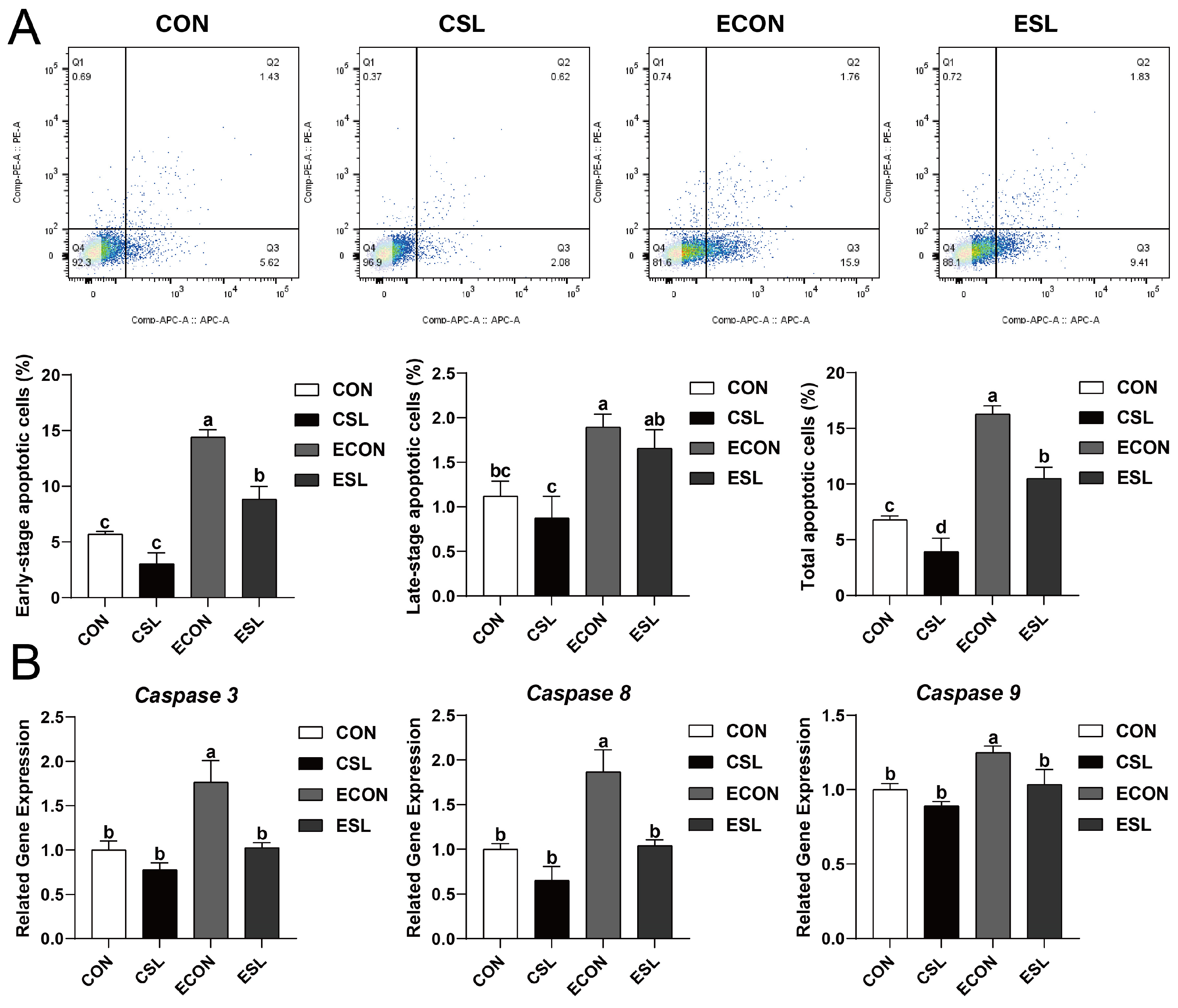
Figure 1. The protective effect of SL on intestinal epithelial cells (IECs) challenged by ETEC. (A) Apoptotic cells detected by flow cytometry. Blue = individual cells; Green = clusters of multiple cells; Orange = dense aggregates of numerous cells. (B) Quantitative PCR analysis using SYBR Green chemistry revealed the regulation of Caspase 3, Caspase 8, and Caspase 9 in IPEC-J2 cells with relative expression normalized to GAPDH via 2−ΔΔCt methodology. CON: control group, CSL: SL group, ECON: ETEC group, ESL: ETEC + SL group. For Caspase 3, 8, and 9, all values are expressed as the mean ± SEM (n = 6). Significant differences (p < 0.05) are denoted by superscript letters (a–d) across treatment groups.
Ijms 26 03860 g001
Figure 2. SL alleviates intestinal inflammation by preventing the adhesion of ETEC K88 on IECs. (A) The number of ETEC K88 cfu adhered to the IPEC-J2 cell surface. (B) Quantitative PCR analysis using SYBR Green chemistry revealed the regulation of IL-1β, IL-6, MyD88, NF-κB, Occludin, TNF-α, TLR4, and ZO-1 in IPEC-J2 cells with relative expression normalized to GAPDH via 2−ΔΔCt methodology. CON: control group, CSL: SL group, ECON: ETEC group, ESL: ETEC + SL group. All values are expressed as the mean ± SEM (n = 6). Significant differences (p < 0.05) are denoted by superscript letters (a–d) across treatment groups.
Ijms 26 03860 g002
Figure 3. SL attenuated inflammation and injury of the IECs challenged by LPS. (A) Representative images of ZO-1 using an immunofluorescence assay (captured using a laser scanning confocal microscope; 400×). Scale bar: 50 μm. (B) Quantitative PCR analysis using SYBR Green chemistry revealed the regulation of IL-1β, IL-6, and TNF-α in IPEC-J2 cells with relative expression normalized to GAPDH via 2−ΔΔCt methodology. CON: control group, CSL: SL group, LCON: LPS group, LSL: LPS + SL group. All values are expressed as the mean ± SEM (n = 6). Significant differences (p < 0.05) are denoted by superscript letters (a–c) across treatment groups.
Ijms 26 03860 g003
Figure 4. The protective effect of SL on intestinal epithelial cells (IECs) challenged by LPS. (A) Apoptotic cells detected by flow cytometry. Blue = individual cells; Green = clusters of multiple cells; Orange = dense aggregates of numerous cells. (B) Quantitative PCR analysis using SYBR Green chemistry revealed the regulation of Caspase 3, Caspase 8, and Caspase 9 mRNA in IPEC-J2 cells with relative expression normalized to GAPDH via 2−ΔΔCt methodology. CON: control group, CSL: SL group, LCON: LPS group, LSL: LPS + SL group. All values are expressed as the mean ± SEM (n = 6). Significant differences (p < 0.05) are denoted by superscript letters (a–c) across treatment groups.
Ijms 26 03860 g004
Figure 5. SL inhibits the phosphorylation of IκB-α and NF-κB by preventing the adhesion of LPS on IECs. (A) LPS ligand-receptor docking efficiency at the porcine intestinal epithelial interface. Scale bar: 50 μm. (B) The protein abundances of IκB-α, p-IκB-α, NF-κB, and p-NF-κB. CON: control group, CSL: SL group, LCON: LPS group, LSL: LPS + SL group. All values are expressed as the mean ± SEM (n = 6). Significant differences (p < 0.05) are denoted by superscript letters (a–c) across treatment groups.
Ijms 26 03860 g005
Figure 6. SL and BAY 11-7082 attenuate inflammation and injury of the IECs challenged by LPS. (A) Representative images of ZO-1 from an immunofluorescence assay (captured using a laser scanning confocal microscope; 400×). Scale bar: 50 μm. (B) Quantitative PCR analysis using SYBR Green chemistry revealed the regulation of IL-1β, IL-6, and TNF-α mRNA in IPEC-J2 cells with relative expression normalized to GAPDH via 2−ΔΔCt methodology. CON: cells without SL or BAY 11-7082 pre-treatment or LPS treatment, SL: cells pre-treated with SL, BAY: cells pre-treated with BAY 11-7082, -LPS: cells without LPS treatment, +LPS: cells pre-treated with LPS., All values are expressed as the mean ± SEM (n = 6). Significant differences (p < 0.05) are denoted by superscript letters (a–d) across treatment groups.
Ijms 26 03860 g006
Figure 7. SL and BAY 11-7082 inhibit apoptosis on the IECs challenged by LPS. (A) Apoptotic cells detected by flow cytometry. Blue = individual cells; Green = clusters of multiple cells; Orange = dense aggregates of numerous cells. (B) Quantitative PCR analysis using SYBR Green chemistry revealed the regulation of Caspase 3, Caspase 8, and Caspase 9 mRNA in IPEC-J2 cells with relative expression normalized to GAPDH via 2−ΔΔCt methodology. CON: cells without SL or BAY 11-7082 pre-treatment or LPS treatment, SL: cells pre-treated with SL, BAY: cells pre-treated with BAY 11-7082, -LPS: cells without LPS treatment, +LPS: cells pre-treated with LPS. All values are expressed as the mean ± SEM (n = 6). Significant differences (p < 0.05) are denoted by superscript letters (a–c) across treatment groups.
Ijms 26 03860 g007
Figure 8. SL and BAY 11-7082 prevent the phosphorylation of the IκB-α and NF-κB of IECs challenged by LPS. (A) The protein abundances of IκB-α and p-IκB-α. CON: cells without SL or BAY 11-7082 pre-treatment or LPS treatment, CSL: cells pre-treated with SL, BAY: cells pre-treated with BAY 11-7082, LPS: cells pre-treated with LPS, LSL: cells pre-treated with SL and subsequently treated with LPS, LBAY: cells pre-treated with BAY 11-7082 and subsequently treated with LPS. (B) The protein abundances of NF-κB and p-NF-κB. CON: cells without SL or BAY 11-7082 pre-treatment or LPS treatment, SL: cells pre-treated with SL, BAY: cells pre-treated with BAY 11-7082, -LPS: cells without LPS treatment, +LPS: cells pre-treated with LPS. All values are expressed as the mean ± SEM (n = 6). Significant differences (p < 0.05) are denoted by superscript letters (a,b) across treatment groups.
Ijms 26 03860 g008
Table 1. Sequences of primers for genes.
Gene *Primer Sequence (5′–3′)Product Size (bp)
β-ActinF: TGGAACGGTGAAGGTGACAGC177
R: GCTTTTGGGAAGGCAGGGACT
Caspase 3F: GGGATTGAGACGGACAGTGG136
R: TGAACCAGGATCCGTCCTTTG
Caspase 8F: TCTGCGGACTGGATGTGATT165
R: TCTGAGGTTGCTGGTCACAC
Caspase 9F: AATGCCGATTTGGCTTACGT195
R: CATTTGCTTGGCAGTCAGGTT
IL-1βF: GTGATGCCAACGTGCAGTCT97
R: AGGTGGAGAGCCTTCAGCAT
IL-6F: TGGCTACTGCCTTCCCTACC153
R: CACACATCTCCTTTCTCATTGC
MyD88F: CCATTCGAGATGACCCCCTG183
R: TAGCAATGGACCAGACGCAG
NF-κBF: GTGTGTAAAGAAGCGGGACCT139
R: CACTGTCACCTGGAAGCAGAG
OccludinF: CTACTCGTCCAACGGGAAAG158
R: ACGCCTCCAAGTTACCACTG
TNF-αF: GCATCGCCGTCTCCTACCAG173
R: GGGCAGGTTGATCTCGGCAC
TLR4F: TTACAGAAGCTGGTTGCCGT152
R: TCCAGGTTGGGCAGGTTAGA
ZO-1F: CAGCCCCCGTACATGGAGA114
R: GCGCAGACGGTGTTCATAGTT
* IL-1β: Interleukin-1 beta, IL-6: Interleukin-6, MyD88: Myeloid Differentiation Primary Response Protein 88, NF-κB: nuclear factor kappa B, TLR4: Toll-like receptor 4, TNF-α: Tumor Necrosis Factor Alpha, ZO-1: Zonula Occludens-1 Protein.
Table 2. Antibody information.
NameSupplier and Catalog NumberDilution Factor
Rabbit anti-ZO-1Abcam plc. (Cambridge, UK)1:150
Mouse anti-E. coli LPSAbcam plc. (Cambridge, UK)1:50
FITC-conjugated goat anti-rabbit IgG antibodyAbcam plc. (Cambridge, UK)1:2500
TRITC-conjugated goat anti-mouse IgGAbcam plc. (Cambridge, UK)1:2500
p-NF-κB p65Cell Signaling Technology 3033S (Danvers, MA, USA)1:1000
NF-κB p65Cell Signaling Technology 6956S (Danvers, MA, USA)1:1000
p-IκBαInvitrogen MA5-15224 (Danvers, MA, USA)1:1000
IκBαCell Signaling Technology4814S (Danvers, MA, USA)1:1000
GAPDHCell Signaling Technology 2118S (Danvers, MA, USA)1:1000
Anti-rabbit IgGCell Signaling Technology 7074S (Danvers, MA, USA)1:2500
Anti-mouse IgGCell Signaling Technology 7076S (Danvers, MA, USA)1:2500
Disclaimer/Publisher’s Note: The statements, opinions and data contained in all publications are solely those of the individual author(s) and contributor(s) and not of MDPI and/or the editor(s). MDPI and/or the editor(s) disclaim responsibility for any injury to people or property resulting from any ideas, methods, instructions or products referred to in the content.