Article

Supplementation of Forskolin and Linoleic Acid During IVC Improved the Developmental and Vitrification Efficiency of Bovine Embryos

1
State Key Laboratory of Sheep Genetic Improvement and Healthy Breeding, Institute of Animal Husbandry and Veterinary Sciences, Xinjiang Academy of Agricultural and Reclamation Sciences, Shihezi 832061, China
2
Tianjin Key Laboratory of Animal Molecular Breeding and Biotechnology, Tianjin Engineering Research Center of Animal Healthy Farming, Institute of Animal Science and Veterinary, Tianjin Academy of Agricultural Sciences, Tianjin 300381, China
3
Institute of Animal Sciences (IAS), Chinese Academy of Agricultural Sciences (CAAS), No. 2 Yuanmingyuan Western Road, Haidian District, Beijing 100193, China
*
Authors to whom correspondence should be addressed.
Int. J. Mol. Sci. 2025, 26(9), 4151; https://doi.org/10.3390/ijms26094151
Submission received: 15 February 2025 / Revised: 21 April 2025 / Accepted: 23 April 2025 / Published: 27 April 2025
(This article belongs to the Section Molecular Biology)

Abstract

:
The success of assisted reproductive technology is contingent upon the growth potential of embryos post-vitrification process. When compared to in vivo embryos, it has been found that the high intracellular lipid accumulation inside the in vitro-derived embryos results in poor survival during vitrification. Based on this finding, the present study assessed the impact of incorporating forskolin and linoleic acid (FL) entering in vitro culture (IVC) on the embryos’ cryo-survival, lipid content, and viability throughout vitrification. Lipid metabolomics and single-cell RNA sequencing (scRNA-seq) techniques were used to determine the underlying mechanism that the therapies were mimicking. It was observed that out of 726 identified lipids, 26 were expressed differentially between the control and FL groups, with 12 lipids upregulated and 14 lipids downregulated. These lipids were classified as Triacylglycerol (TG), Diacylglycerol (DG), Phosphatidylcholine (PC), and so on. A total of 1079 DEGs were detected between the FL and control groups, consisting of 644 upregulated genes and 435 downregulated genes. These DEGs were significantly enhanced in the arachidonic acid metabolism, lipolysis, fatty acid metabolism, cAMP signaling pathway, and other critical developmental pathways. Based on the observation, it was concluded that forskolin and linoleic acid decreased the droplet content of embryos by modulating lipid metabolism, thus enhancing the vitrified bovine embryos’ cryo-survival.

1. Introduction

The cryopreservation of embryos is an effective method to extend the accessibility of valuable germplasm resources, streamline the process of long-distance transportation [1], and mitigate the potential risk associated with venereal disease transmission to a certain degree [2]. This technique allows for storing surplus oocytes and embryos for later use [3], leading to increased pregnancies from a single stimulation and decreased breeding costs and animal manipulation [4]. Gene banks have extensively adopted this technique to protect the native breeds of animal species from extinction and the potential threat posed by disease outbreaks [5,6].
The abundance of lipid droplets in livestock embryos is influenced by the culture environment; thereby, in vitro embryos have higher levels compared to in vivo embryos [7,8]. Moreover, excessive lipid droplet accumulation in embryos intensifies lipid peroxidation during cryopreservation, resulting in oxidative stress, apoptosis, lower blastocyst rate, and compromised cryo-tolerance [9,10]. Therefore, consistent efforts have been made to lower the amount of lipid droplets in developing embryos to improve their cryo-tolerance.
Adenylate cyclase, an enzyme that triggers endogenous lipase via 3’,5’-cyclic adenosine monophosphate (cyclic adenosine monophosphate, cAMP), is activated in cells by forskolin, a diterpene, allowing the release of fatty acids and glycerol [11,12]. It has been indicated that forskolin can induce lipolysis in oocytes and embryos, and improve their survival rate after vitrification [9,13,14]. Studies have demonstrated that forskolin significantly improved the developmental competence of porcine [15], bovine [16], horse [17], and sheep [18] oocytes. In addition, it was shown that when 10 μM forskolin was added to the in vitro culture (IVC) medium, it caused a higher rate of blastocyst development [19]. Meanwhile, the supplementation of forskolin during early embryonic porcine and cattle development in porcine and cattle was enough to reduce the lipid content in blastocysts, thereby improving their low-temperature tolerance [14,20].
Linoleic acid is a long-chain polyunsaturated fatty acid that reduces free fatty acid uptake without increasing lipolysis [21]. Linoleic acid has been found to pass through the lipid bilayer of the plasma membrane in preimplantation embryonic cells and to enter lipid droplets during IVC [22], thereby enhancing the fluidity of embryonic cell membranes and improving the tolerance of embryos to cryopreservation [23]. It has been shown that the addition of linoleic acid alters the intracellular fatty acid content and improves the developmental competence of bovine [22,24] and ovine [25] embryos after vitrification [26].
Consequently, the quality and survival rate of embryos following vitrification may be improved by adding metabolic regulators to the culture medium. This study aimed to investigate the effects of forskolin and linoleic acid on apoptosis, lipid content, ROS levels, and cytoskeletal structure in vitrified bovine oocytes. Through the utilization of lipidomic and transcriptomic techniques, we sought to elucidate the mechanisms by which forskolin and linoleic acid impact lipid metabolism in embryos. Therefore, the study aimed to conceive a strategy to decrease abnormal lipid accumulation in bovine embryos to maintain their development potential following vitrification.

2. Results

2.1. Effect of Forskolin on the Developmental Ability and Survival Rate of Bovine Embryo

Table 1 showed that the rates of cleavage blastocyst formation of the 10 µM forskolin group (91.67 ± 3.71%, 42.42 ± 4.85%) were greater than those of the control group (81.90 ± 2.57%, 36.89 ± 4.47%), 5 µM forskolin group (84.87 ± 4.65%, 35.64 ± 4.70%), and 15 µM forskolin group (64.18 ± 4.32%, 27.91 ± 3.81%). Meanwhile, the survival rate of the 10 µM forskolin group (95.24 ± 7.23%) was greater than the control group (86.67 ± 6.59%), the 5 µM forskolin group (88.89 ± 7.35%), and the 15 µM forskolin group (83.33 ± 7.06%).

2.2. Effect of Linoleic Acid on the Developmental Ability and Survival Rate of Bovine Embryo

Table 2 showed that the cleavage and blastocyst rates of the 100 µM linoleic acid group (88.74 ± 6.73%, 38.81 ± 2.55%) were greater than those of the control group (80.00 ± 6.59%, 33.33 ± 2.41%), 50 µM linoleic acid group (61.17 ± 5.85%, 27.83 ± 1.68%), 500 µM linoleic acid group (60.54 ± 5.68%, 29.21 ± 1.84%). Meanwhile, the survival rate of the 100 µM linoleic acid group (96.15 ± 8.53%) was greater than those of the control group (86.36 ± 7.15%), 50 µM linoleic acid group (81.25 ± 7.46%) and 500 µM linoleic acid group (80.77 ± 7.21%, p < 0.05).

2.3. Effect of Forskolin and Linoleic Acid on the Developmental Ability and Survival Rate of Bovine Embryo

Table 3 revealed the F-L group’s cleavage rate and blastocyst rate (89.54 ± 7.62%, 45.26 ± 5.24%) were greater than those of the control group (75.57 ± 7.46%, 30.30 ± 2.65%), forskolin group (82.39 ± 8.52%, 39.74 ± 4.12%) and linoleic acid group (83.08 ± 8.46%, 37.97 ± 4.03%). Meanwhile, compared to the control group (80.65 ± 7.68%), the forskolin group (90.24 ± 7.31%), and the linoleic acid group (89.47 ± 8.15%, p < 0.05), the F-L group had a greater survival rate (98.18 ± 8.64%, p < 0.05).

2.4. Effect of Forskolin and Linoleic Acid on Lipid Droplet Content in the Bovine Embryo

As illustrated in Figure 1, the fluorescence intensity of the lipid droplets in the F-L group (78.14 ± 6.98) was significantly lower than that of the control group (133.22 ± 9.75), forskolin group (108.05 ± 8.35), and linoleic acid group (106.18 ± 6.98, p < 0.05).

2.5. Metabolomic Analysis of Lipid Metabolites in Bovine Embryos Combined Treatment with Forskolin and Linoleic Acid

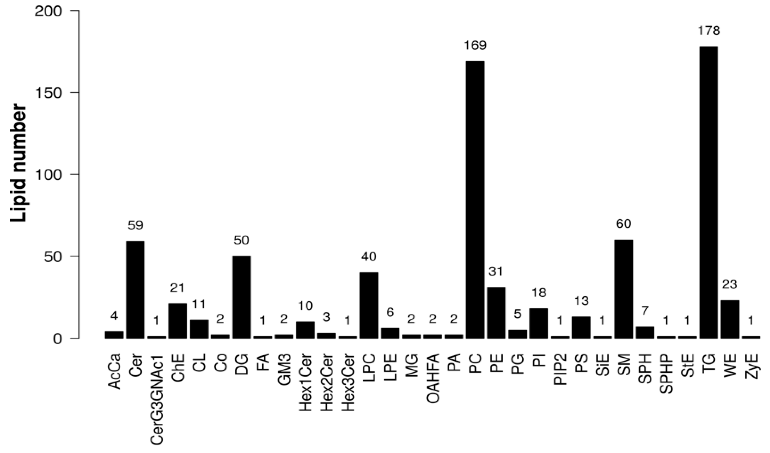
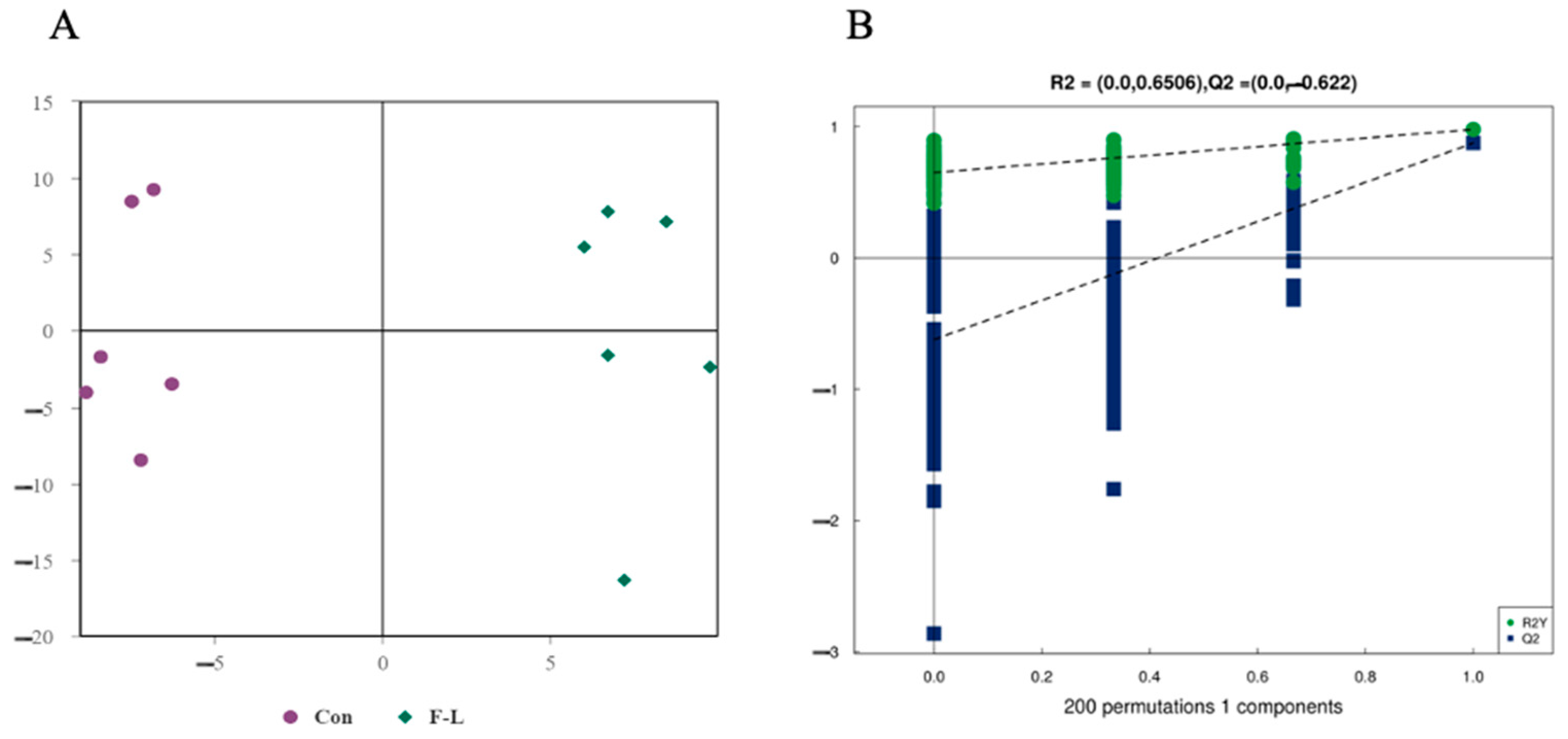
The results of LC-MS/MS analysis showed 31 classes of lipids and 726 lipids, and the proportions of these lipids are shown in Figure 2. The main lipids were Triacylglycerol (TG), Phosphatidylcholine (PC), Sphingomyelin (SM), Ceramide (Cer), and Diacylglycerol (DG). The multivariate statistical analysis of lipid alterations between the F-L and control groups was performed using OPLS-DA. As shown in Figure 3, the samples from the two groups were completely separated along the x-axis, and the model parameters were R2Y = 0.6506 and Q2 = 0.622, indicating that the model was robust and not overfitted. The stability of the model was verified using a permutation test, and the slope of the regression line for the permutation test was positive, demonstrating the excellent degree of fit and strong predictive power of the OPLS-DA model.

2.6. Differential Lipids Between F-L and Control Groups

To further investigate linoleic acid and forskolin effects on embryonic lipid metabolism, the two groups were screened for differential lipid molecules.
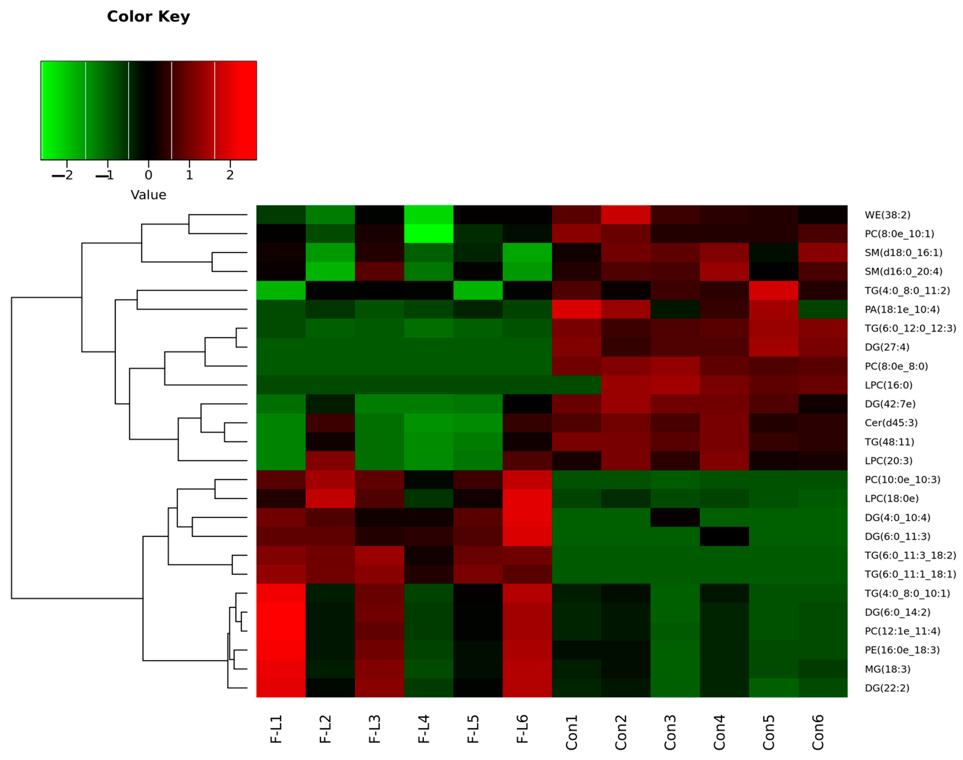
A total of 26 lipid molecules (VIP > 1 and p < 0.05) were found to be significantly unlike between the F-L group and the control group (Table 4). Then, the clustering analysis results of the differential lipids are shown in Figure 4. These included 12 lipids that were seen significantly upregulated and 14 lipids that were seen significantly downregulated in the F-L group. These lipid molecules were mainly attributed to TG, DG, Phosphatidic acid (PA), Lysophosphatidylcholine (LPC), PC, and Phosphatidylethanolamine (PE).

2.7. Differentially Expressed Genes (DEGs) in Forskolin and Linoleic Acid Treated Bovine Embryos

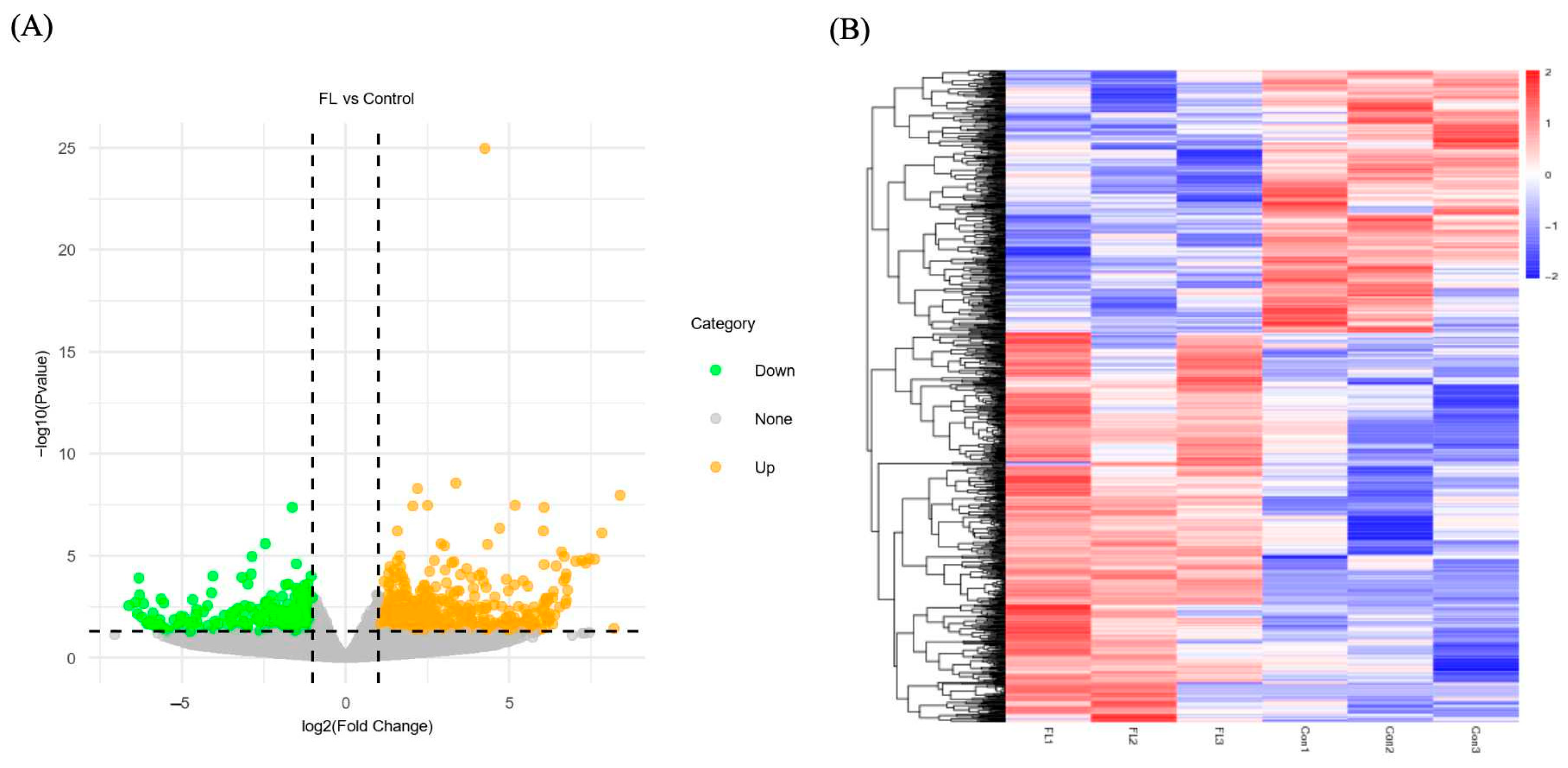
The volcano plots were utilized to clarify the differences in gene expression levels between the FL and control groups (Figure 5A). Subsequently, a clustering analysis was conducted on DEGs based on their expression patterns, and the findings indicated that the samples in the same group were well clustered together (Figure 5B). Comparing the FL group to the control group, 644 genes were upregulated and 435 were downregulated. Some of those DEGs were listed in Table 5, including microtubule-related (TUBA4A), apoptosis genes (CASP7), oxidative stress genes (CAT, GPX7), embryo development (SEC22B, NDRG4, MX2), lipid metabolism-related (ACSL1, DECR1, ACADL, CDKN2C, PPARA).

2.8. GO Enrichment Analysis of the DEGs

Figure 6 shows the GO terms with a significant enrichment of DEGs between the FL and control groups. The GO enrichment analysis revealed that the biological process (BP) mainly consisted of fatty acid metabolic, cellular lipid metabolic, and fatty acid biosynthetic processes. Cell components (CCs) primarily involve the cytoplasm, endoplasmic reticulum, cytoskeleton, and organelles. Molecular function (MF) was primarily associated with monocarboxylic acid binding, fatty acid binding, cytoskeletal protein binding, and protein binding.

2.9. Pathway Enrichment Analysis of DEGs

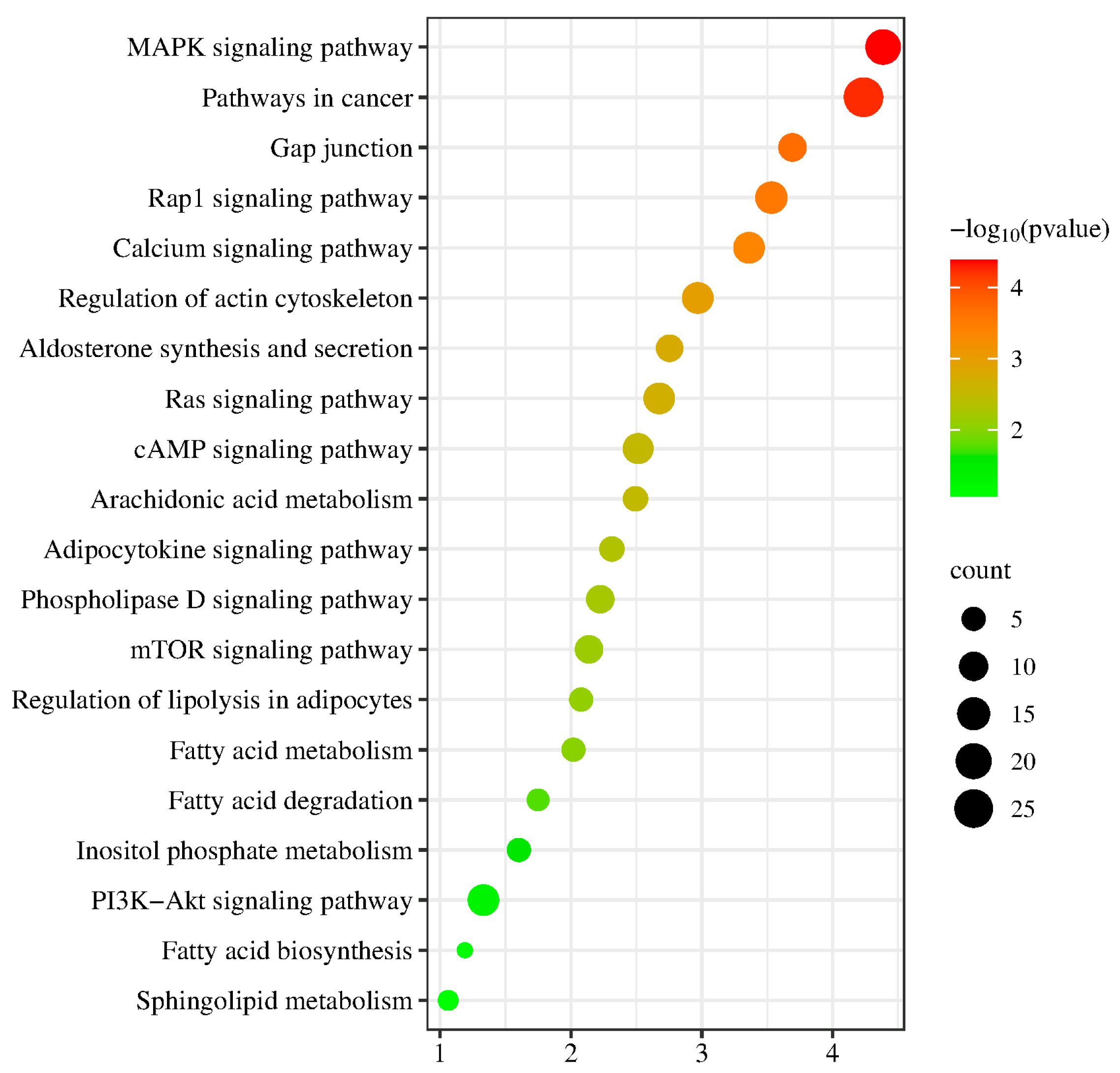
As revealed in Figure 7, DEGs associated genes between the FL and Con groups were mostly enhanced with the MAPK, calcium, and cAMP signaling pathways, as well as the breakdown and metabolism of fatty acids, Inositol phosphate metabolism, and arachidonic acid metabolism.

2.10. Effect of Forskolin and Linoleic Acid on Gene Expression in Bovine Blastocysts

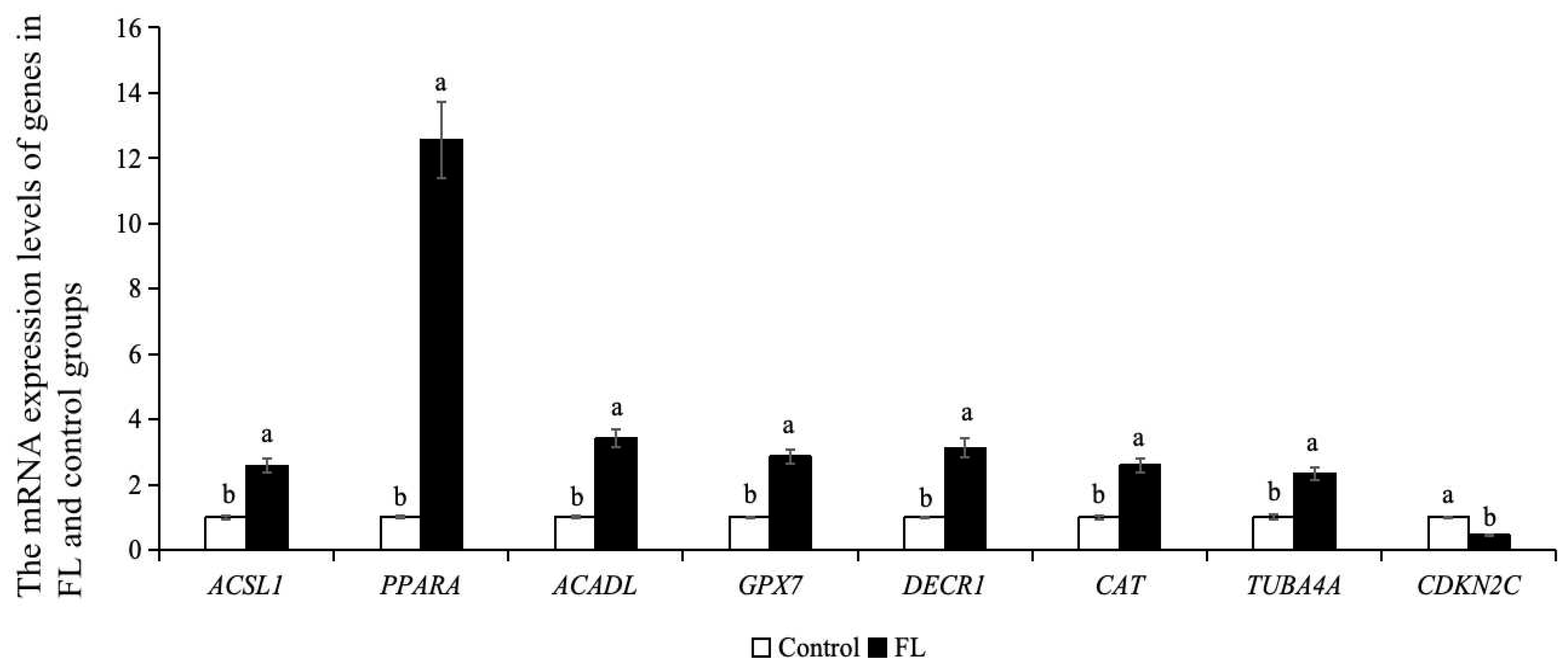
As shown in Figure 8, the mRNA expression levels of lipid metabolism genes (ACSL1, DECR1, ACADL, PPARA), microtubule genes (TUBA4A), and oxidative stress genes (CAT, GPX7) in the blastocysts of the FL group were significantly higher than control group. The mRNA expression level of the CDKN2C in the FL group was lower than that of the control group (p < 0.05).

2.11. Effect of Forskolin and Linoleic Acid on the Apoptosis of Bovine Blastocysts

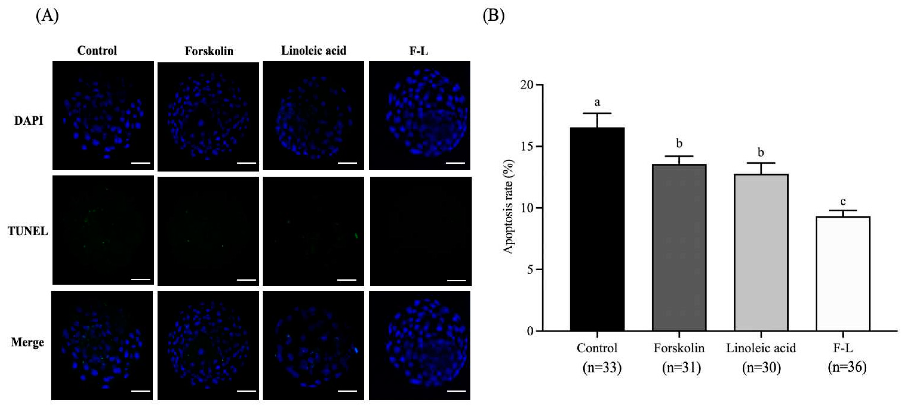
As shown in Figure 9, the apoptosis rate of blastocysts of the F-L group (9.34 ± 0.41%) was significantly lower than the control group (16.53 ± 1.02%), forskolin group (13.58 ± 0.55%), and linoleic acid group (12.76 ± 0.80%; p < 0.05).

2.12. Linoleic Acid and Forskolin’s Effects on the ROS Levels of Bovine Blastocysts

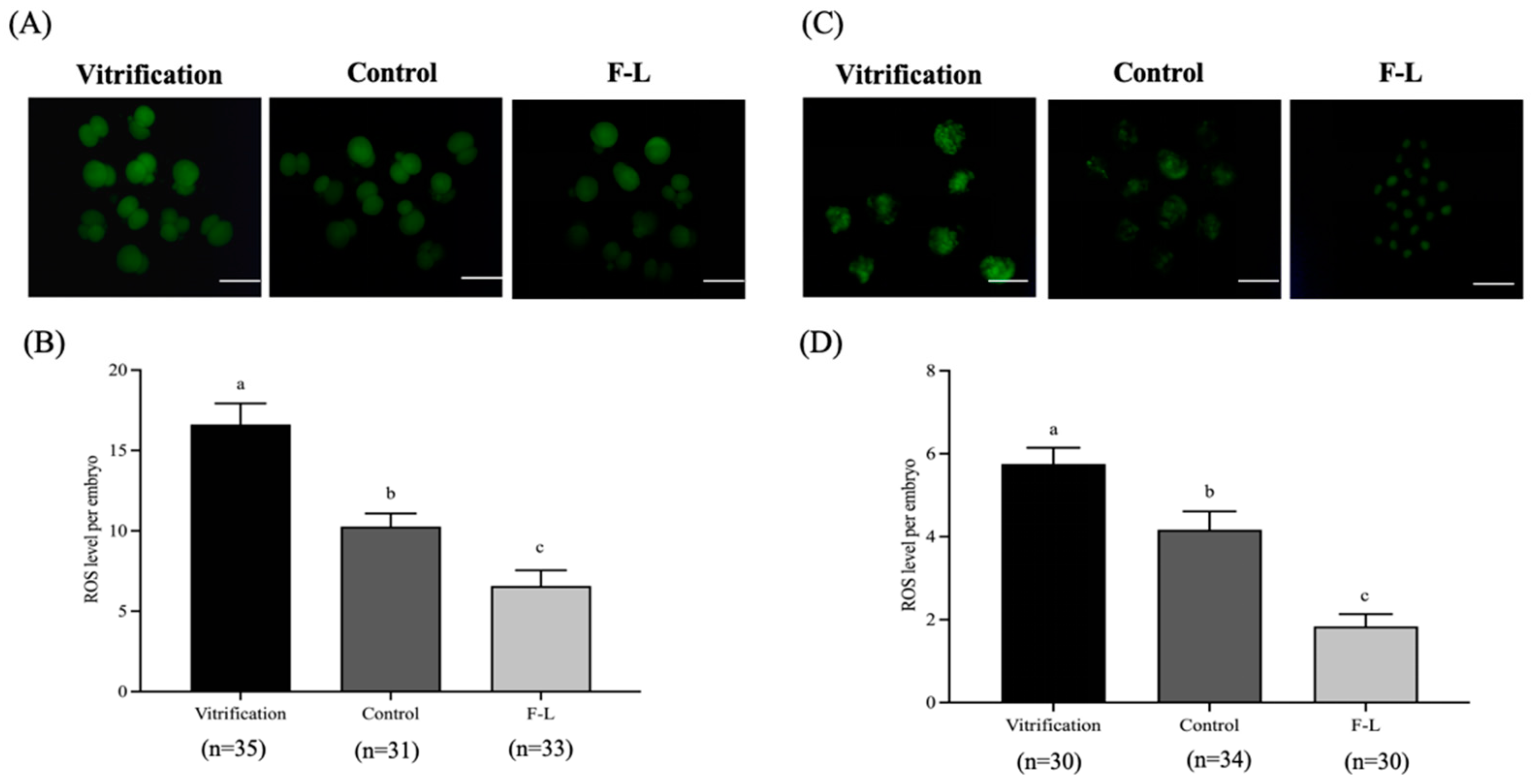
As revealed in Figure 10, the ROS levels of 2-cell and blastocysts of the F-L group (6.58 ± 0.87; 1.84 ± 0.26) were significantly lower than the control (10.27 ± 0.72; 4.17 ± 0.39) and vitrified group (16.62 ± 1.18; 5.75 ± 0.35, p < 0.05).

2.13. Effect of Forskolin and Linoleic Acid on the Cytoskeleton of Bovine Blastocyst

The normal spindle rate of blastocysts in the F-L group (69.68 ± 5.06%), as shown in Figure 11, was substantially greater than that of the vitrified group (41.44 ± 3.12%) and control group (60.32 ± 4.21%, p < 0.05).

3. Discussion

Forskolin is recognized for its lipolytic properties and is commonly utilized as a lipid modulator [27]. It elevates intracellular cAMP levels by stimulating adenylate cyclase (AC) [9], subsequently activating cAMP-dependent protein kinase A (PKA), which phosphorylates intracellular lipase and enhances fatty acid β-oxidation [28]. Forskolin supplementation has demonstrated the ability to improve cryotolerance in oocytes and embryos of various species [29]. The current investigation’s findings showed that the administration of forskolin notably enhanced the rates of cleavage, blastocyst formation, and viability after vitrification cryopreservation in bovine embryos. This observation aligns with similar trends observed in porcine oocytes [30] and bovine embryos [31].
Linoleic acid, a crucial polyunsaturated fatty acid, plays an important part in the embryo’s development and the maturation of mammalian oocytes [32]. Previous research indicates that adding linoleic acid to the medium modifies the lipid composition of oocytes for IVM and enhances the development of sheep [33], porcine [34], and bovine embryos [35]. Coherent with previous reports, the results of the present study showed that linoleic acid treatment increased the cleavage, blastocyst rate, and cryo-tolerance of bovine embryos. Moreover, it was also revealed that the supplementation of forskolin and linoleic acid to IVC could synergistically improve the development of blastocysts and their cryotolerance in bovine embryos.
Forskolin can activate AC to increase intracellular cAMP levels, inducing the activation of intracellular lipase, thus promoting lipolysis with the release of glycerol and fatty acids [20]. Similarly, we found that the forskolin addition to the IVC medium significantly reduced the intracellular lipid content in bovine embryos. Conjugated linoleic acid enhances the fluidity of linoleic acid and facilitates the synthesis of TG and phospholipid, thus improving the lipid profile of adipocytes [36]. Therefore, the supplementation of forskolin and linoleic acid to the IVC medium significantly reduces intracellular lipid accumulation by improving the overall lipid metabolism of developing embryos.
TGs are a class of lipids in mammalian cell cytoplasm stored as droplets, providing energy for early embryonic development [37]. During oocyte maturation, growth factors, hormones, and serum are added, which results in oxidative metabolism, oxidizing fatty acids from TG breakdown in the tricarboxylic acid cycle [38,39]. Therefore, IVC conditions can potentially elicit an abundance of oxidized glycerophospholipids. So, changes in lipid profiles of embryos may be linked to IVC conditions. DG represents a category of structural lipids characterized by the replacement of a fatty acid with a hydroxyl group in TG. It is a crucial intracellular second messenger that plays a role in various cellular reactions. They also promote fatty acid oxidation processes, modulating lipid metabolism and regulating gene expression linked with lipid metabolism [40]. The most prevalent phospholipid in mammalian cells is PC, having a vital role in cell structure and cellular activities, including vesicular transport, signal transduction, and regulation of lipoprotein secretion [41]. Forskolin activates intracellular lipases for lipolysis [20], and linoleic acid enters cells to synthesize triacylglycerols and phospholipids, which enhance the lipid profile of lipocytes [36]. Membrane proteins control the uptake of lipid metabolites and fatty acids from the culture medium. However, release occurs predominantly via passive diffusion [42]. In this case, as shown in our results, the TG and DG content in the F-L group is greater than the control one, and the products of lipolysis can be partially released into the embryonic culture medium to regulate lipid metabolism.
In this study, we compared transcriptomics of the FL and control group embryos using scRNA-seq technology. Several genes related to lipid metabolism (ACSL1, DECR1, ACADL) were noted to be upregulated in the FL group compared to the control group. Fatty acid elongation, oxidative catabolism, phospholipogenesis, and protein acylation are all aspects of lipid metabolism that are regulated by the important enzyme ACSL [43] and oversee fatty acid acylation and activation upon cell entry [44]. It has been evident that upregulation of the ACSL1 gene reduces lipid peroxidation and decreases ROS through lipid reprogramming [45]. Further, KEGG analysis revealed that the gene ACSL1 is enriched with fatty acid degradation and fatty acid metabolism pathways. DECR1 is a mitochondrial enzyme that regulates the balance between saturated phospholipids and unsaturated ones in the auxiliary pathway of β-oxidation, which maintains redox homeostasis in vivo, and the absence of DECR1 impairs lipid metabolism [46]. ACADL is a mitochondrial enzyme that catalyzes fatty acid oxidation [47]. It was found that the gene ACADL is enriched in the fatty acid degradation pathway by KEGG analysis, which explains the reduction in lipid content in the FL group.
The oxidative stress-related gene GPX7 was observed to be significantly upregulated in the FL group. GPX family member GPX7 is an antioxidant enzyme that reduces ROS production and limits their toxicity [48]. GPX7 is a marker for endoplasmic reticulum and oxidative stress, and any change in GPX7 activity indicates how the body reacts to oxidative stress [49]. It is established that GPX7 can ameliorate nonalcoholic steatohepatitis by regulating oxidative stress levels [50]. GPX7 participates in the arachidonic acid metabolic pathway. Through current research work, it was found that GPX7 is enriched in this pathway according to KEGG analysis. An omega-6 polyunsaturated fatty acid named arachidonic acid is a part of the signaling cascade that determines a cell’s destiny in fetal placental development and has an enhanced role in the immunomodulatory effects of angiogenesis in embryonic stem cells and mesenchymal stem cells (MSCs) [51]. Research findings indicate that arachidonic acid plays a significant role in embryonic development, overall health, brain development, and immune response [52]. CDKN2C, a gene associated with lipid metabolism, was shown to be downregulated in the FL group compared to the control group. CDKN2C has been associated with inhibition of the cell cycle during adipocyte differentiation [53] and can prevent G1 by inhibiting cyclin-dependent kinase 4 or 6 (CDK4/6) onset. CDKN2C deficiency decreases lipid stock in adipocytes during the differentiation process and decreases gene expression, which regulates adipocyte function [54].
AC catalyzes the extraction of pyrophosphate from ATP, producing cAMP [55]. It can be deactivated by hydrolysis by phosphodiesterase (PDE) [56]. AC and PDE interact to regulate intracellular cAMP levels and activate β-oxidative regulation in mitochondria [57]. It has been reported that forskolin, as an activator of the cAMP pathway, can increase cAMP levels and maintain oocyte meiotic arrest in sheep [58], bovine [59], and mice [60] oocytes. Moreover, PPAR has been shown to mediate the stimulatory PGI2 effects on embryonic development and blastocyst hatching, as well as to contribute to embryonic cell proliferation [61]. Our results showed that PPARA was upregulated in the FL group, which is intricate in the β-oxidation of fatty acids regulation. Therefore, enhancing lipid metabolism in embryos contributes to the establishment of an optimal cellular environment for embryonic development [62]. At the same time, PPARA is also enriched in the signaling pathway cAMP, recommending that forskolin and linoleic acid may increase the cAMP level by regulating the expression of PPARA and thus reduce the intracellular lipid content.
Apoptosis is a key parameter in embryo quality assessment and has a key role in embryonic development. It has been observed that the rate of apoptosis remains higher in vitrified embryos in contrast to fresh embryos [63,64]. Reducing lipid content before cryopreservation could potentially aid in preserving embryo cytoplasm, boosting lipid metabolism gene expression, and reducing apoptosis in embryos [20,23]. Research indicates that forskolin reduces lipid deposition in porcine and bovine embryos, promoting cryotolerance and diminishing apoptosis [13,20]. It has been shown that linoleic acid modulates NF-κB, which reduces apoptosis in porcine embryos [34], and improves embryo development [65]. Our current experiments demonstrated a significantly reduced rate of apoptosis in the F-L group, compared to forskolin, linoleic acid, and the control. Collectively, these results provide indications that forskolin and linoleic acid act cooperatively to reduce lipid deposition in bovine embryos during the preimplantation period, thereby decreasing apoptosis.
Vitrification induces lipid peroxidation in embryos, leading to the impairment of essential biomolecules, including DNA, proteins, and plasma membrane [66]. It also disrupts the redox state balance, leading to a hike in ROS and poor embryo survival [67]. Forskolin exhibits antioxidant properties and decreases Bax and Caspase-3 expression, thereby significantly lowering cellular ROS levels [68]. Similarly to Cai [47], who demonstrated forskolin efficacy in reducing ROS in vitrified embryos, our findings indicate that forskolin and linoleic acid significantly lower ROS in bovine embryos. This reduction is linked to the upregulation of antioxidant-related genes (CAT and GPX7) (Table 5), which encode critical enzymes in the antioxidant defense mechanism, safeguarding cells from oxidative stress-induced damage.
Vitrification damages the embryo cytoskeleton [69] by microtubule depolymerization [70]. Our transcriptomic analysis revealed upregulation of the microtubule-related gene (TUBA4A) in the FL group, with GO enrichment analysis indicating that differential genes were enriched in the cytoskeleton. Our results suggest that forskolin and linoleic acid enhance embryo spindle morphology and quality. Previous findings have shown that forskolin elevates intracellular cAMP levels, which activate PKA, resulting in Rho inactivation, which aids in preserving cytoskeletal integrity and normal cellular morphology [71].

4. Materials and Methods

Unless otherwise specified, all chemicals used in this study were purchased from Sigma-Aldrich (St. Louis, MO, USA), and plasticware was obtained from Corning Inc. (Corning, NY, USA).

4.1. Oocytes In Vitro Maturation (IVM)

From the slaughterhouse, bovine ovaries were collected and dispatched to the laboratory within 2 h at 37 °C. Cumulus oocyte complexes (COCs) were aspirated from follicles with a diameter of 2–8 mm, and only those with uniform cytoplasm and at least three complete layers of tightly packed cumulus cells were chosen for IVM. 10 μg/mL estradiol, 10% (v/v) fetal bovine serum (FBS; Gibco BRL Division), 10 μg/mL follicle-stimulating hormone (FSH), 10 μg/mL heparin (HP), and 10 μg/mL luteinizing hormone (LH) were added to a HEPES-buffered M199 (Gibco BRL; Grand Island, NY, USA) that served as the IVM solution. For 22–24 h, the COCs were cultivated at a temperature of 38.5 °C with 5% CO2 and high humidity.

4.2. In Vitro Fertilization (IVF)

After oocyte culture, IVF was performed as previously reported by Brackett and Oliphant [72]. Briefly, for 30 s, the frozen sperm underwent thawing at 37 °C. The post-thaw semen was then rinsed twice at 600× g for 5 min in the Brackett and Oliphant (BO) medium, which contained penicillin, heparin, streptomycin, and bovine serum albumin (BSA). A total of 1 × 10⁶ spermatozoa per milliliter was the final concentration following resuspension. A total of 20–30 oocytes were added to each drop of fertilization media, and the gametes were incubated for 8–18 h at 38.5 °C in humidified air containing 5% CO2.

4.3. IVC

After 16–18 h, the surrounding cumulus cells and sperm were taken away from the zygotes through a mouth pipette. After that, forskolin and linoleic acid were added as follows: (1) control group: CR1aa basal culture medium; (2) forskolin group: 10 μM forskolin addition for 24 h in CR1aa medium; (3) linoleic acid group: 100 μM add linoleic acid was added to CR1aa medium; (4) F-L (or FL) group: 10 μM forskolin (24 h) and 100 μM linoleic acid group were added to CR1aa medium. The embryos were cultured at 38.5 °C and 5% CO2 until day 7 after the initiation of fertilization.

4.4. Vitrification and Warming

The thawing and vitrification procedures were slightly modified from Zhao et al. [73]. Briefly, the embryos were initially immersed in vitrification solution with 10% ethylene glycol (EG) and 10% Dimethyl sulfoxide (DMSO) for 30 s. Then, embryos were transferred to EDFSF40 (20% DMSO, 20% EG, 300 g/L Ficoll, 20% FBS, and sucrose concentration 0.5 M) for 25 s. The embryos were immediately plunged into liquid nitrogen after being loaded into open-pulled straw (OPS).
The OPS tubes were taken out from liquid nitrogen for the warming process, and the embryos in the tubes were blown into sucrose solution 0.25 M and incubated for about 5 min. Following that they were shifted to a sucrose solution of 0.25 M and incubated for about 5 min. Afterward, the embryos were cultured at 38.5 °C under 5% CO2.

4.5. Embryo Lipid Droplet Staining

With minor adjustments according to the previously described method [74], embryos were rinsed three times with PBS containing 0.1% polyvinyl alcohol (PVA), fixed with 4% paraformaldehyde fixative for 30 min at room temperature. Following three rounds of washing with 0.1% PVA/PBS, the embryos were incubated in a 10 μg/mL Nile Red staining solution at 37 °C for 2 h before being examined using an epifluorescence inverted microscope (Nikon, Tokyo, Japan). The fluorescent images were statistically analyzed utilizing ImageJ software version 1.8.0 (NIH, Bethesda, MD, USA).

4.6. Assessment of the Apoptotic Index

Following a protocol similar to Khan et al. [75], with slight adjustments, apoptosis was examined utilizing a TUNEL (terminal deoxynucleotidyl transferase biotin-dUTP nick end labeling) test kit (Solarbio, Beijing, China) in compliance with the guidelines provided by the manufacturer. Briefly, the blastocysts were rinsed with 0.1% PVA/PBS, then fixed at room temperature for 30 min using 4% paraformaldehyde, and permeabilized with 0.1% Triton X-100. Following three washes with 0.1% PVA/PBS, embryos were placed in an incubator for 1 h at 37 °C in the dark with TUNEL equilibration and the terminal deoxynucleotidyl transferase enzyme. After that, they received treatment for 5 min at 37 °C with 1 mg/mL DAPI. Following that, a confocal laser microscope (Leica, Wetzlar, Germany) was used to capture the images, and ImageJ software version 1.8.0 (NIH, Bethesda, MD, USA) was used to analyze the images for the intensity of fluorescence.

4.7. Embryo ROS Staining

With some modifications to the method described by García-Martínez et al. [76], intracellular ROS levels in embryos were quantified by labeling embryos that were washed twice in 0.1% PVA/PBS, then incubated in PVA/PBS supplemented with 5 μM 2’,7’-dichlorodihydrofluorescein diacetate (H2DCFDA) for 30 min at 38.5 °C in a humidified 5% CO2 air atmosphere. Then, embryos were observed on the epifluorescence inverted microscope (Nikon, Tokyo, Japan). ImageJ software version 1.8.0 (NIH, Bethesda, MD, USA) was utilized for fluorescence intensity.

4.8. Embryo Cytoskeleton Staining

According to a previous method described by Cai et al. [77], the embryos were fixed with 4% paraformaldehyde at room temperature for 30 min, then washed 3 times with 0.1% TritonX-100 for 5 min each time. The embryos were placed in Tubulin-Tracker Red solution (C1050, Beyotime, Shanghai, China) at room temperature and incubated for 30 min in the dark. After that, they were washed twice in 0.1% PVA/PBS and further incubated in 1 mg/mL DAPI at 37 °C for 5 min. Then, embryos were fixed on a glass slide for examination under the confocal laser microscope (Leica, Wetzlar, Germany). ImageJ software version 1.8.0 (NIH, Bethesda, MD, USA) was utilized for fluorescence intensity.

4.9. Untargeted Lipidomics Analysis

With slight modifications based on the previously described method [78], the embryo samples were first added, and 200 μL of water and 240 μL of methanol were used to homogenize them. After adding 800 μL of MTBE, the mixture was sonicated for 20 min at 4 °C, followed by allowing the mixture to settle for 30 min at room temperature. Then, the solution was centrifuged at 14,000× g for 15 min at 10 °C, supernatant was obtained from the upper layer of the organic phase and dried under nitrogen gas. For mass spectrometry analysis, a solution of 200 μL consisting of 90% isopropanol/acetonitrile was introduced and vigorously vortexed. Further, 90 μL of the solution for 15 min at 10 °C was centrifuged at 14,000× g, followed by collection of the supernatant for analysis.
The Vanquish UHPLC system (Thermo Fisher Scientific) coupled with the Q-Exactive Plus system (Thermofisher Scientific) was used for untargeted lipidomics analysis. Utilizing a CSH C18 column (1.7 μM, 2.1 mm × 100 mm, Waters Corporation, Wexford, Ireland), chromatographic separation was carried out using an LC separation. The auto-sampler temperature was 4 °C. Solvent A (60% acetonitrile, 40% water) had 0.1% formic acid and 0.1 mM ammonium formate, and solvent B (10% acetonitrile + 90% isopropanol) used 0.1% formic acid and 0.1 mM ammonium formate. The elution gradient (0–3.5 min, 40% B; 3.5–13 min, 40–70% B; 13–19 min, 75–99% B; 19–40 min, 40% B). The Q-Exactive Plus system, with the ability to function with both positive and negative modes, was used to gather mass spectrometry data. For every measurement, the ESI parameters were refined and displayed in this fashion: 300 °C was the source temperature, 350 °C was the capillary temperature, 3000 V was the ion spray voltage, 50% was the S-Lens RF Level, and 200–18,000 m/z was the mass range for scanning.

4.10. Sample Preparation, Library Construction, and RNA Sequencing

The zona pellucida of the embryo was digested using a strepase enzyme. Subsequently, each embryo was treated with a lysis solution. The single-cell RNA sequencing (scRNA-seq) library construction and sequencing process adhered to the protocol detailed by Gao et al. [79] with minor adjustments. Individual embryos were lysed to extract RNA, which was later reversely transcribed into first-strand cDNA using a six-nucleotide random primer. The complementary second strand was synthesized in the presence of dNTPs through a DNA polymerase I reaction. The resulting double-stranded cDNA was cleaned using AMPure P magnetic beads, followed by PCR amplification to yield the final cDNA product. The qualified cDNA library was sequenced by the Illumina HiSeq 2500 platform, and the paired-end reads of 150 bp were obtained by the PE 150 bp technique. The original sequences were filtered using Trimmomatic (v0.39) software, and adapter sequences, low-quality bases, and contaminated sequences were removed. Then, HISAT2 (v2.1.0) was used to compare the filtered sequence with the bovine reference genome sequence (ARS-UCD2.0 genome). Then, DEseq2 (v1.20.0) was used to analyze the differences in gene expression between groups. |log2Ratio| ≥ 1 and p < 0.05 were considered as significant differences.

4.11. Functional Enrichment Analysis

In this research, an enrichment analysis was conducted on differentially expressed genes using Gene Ontology (GO), which categorizes genes into 3 terms: biological process, cellular component, and molecular function. The Kyoto encyclopedia of genes and genomes gene ontology (KEGG) database was used to perform functional enrichment analysis of the screened differential genes in order to identify biological pathways with substantial enrichment. Significantly enriched KEGG pathways and GO items are indicated by p < 0.05.

4.12. Quantitative Real-Time Polymerase Chain Reaction (qRT-PCR) of Genes

According to the manufacturer’s protocol, the Single Cell-to-CT quantitative real-time PCR kit (Life Technologies, Carlsbad, CA, USA) was used to measure the gene expression levels of blastocysts. The qRT-PCR was run using an ABI 7500 SDS instrument (Applied Biosystems, Foster City, CA, USA). The reaction took place at 95 °C for 2 min, then 40 cycles for 10 s at 95 °C and 30 s at 60 °C. Using β-actin as the reference gene, the folding changes in gene expression were analyzed by the 2−44CT method. The PCR primers used in the assessment are shown in Table 6.

4.13. Statistical Analysis

All experiments were conducted at least three times. All data were subjected to one-way ANOVA using Duncan’s test with SAS software version 9.2.0 (SAS Institute, Cary, NC, USA).

5. Conclusions

The cocktail of forskolin and linoleic acid in the IVC medium improved bovine embryo development and survival rate after vitrification, reduced the content of lipid droplets, apoptosis, and ROS levels in bovine embryos. Similarly, forskolin and linoleic acid could alter the lipid metabolism, increased PPARA gene expression, and regulate the pathway cAMP signaling to promote β-oxidation of fatty acids, which altered the lipid metabolism process of embryos, decreased the lipid content of embryos to further enhance the freeze tolerance of embryos, and contributed to the enhancement developmental capacity of bovine embryos, and it contributes to the advancement of both the freezing survival rate and developing capacity of bovine embryos.

Author Contributions

Conceptualization, Y.L.; methodology, H.Z. and M.S.; formal analysis, H.A.K.; data curation, H.S.; visualization, X.Z. (Xiaosheng Zhang); writing—original draft preparation, P.Z.; writing—review and editing, P.W.; funding acquisition, X.Z. (Xueming Zhao). All authors have read and agreed to the published version of the manuscript.

Funding

This work was supported by the earmarked fund for the National Key R&D Program of China (2022YFE0100200), the National Natural Science Foundation of China (32161143032), the Specially Appointed Expert Program of Tianchi Talent Program in Xinjiang Province (Xueming Zhao), the earmarked fund for CARS (CARS36-16), the National Germplasm Center of Domestic Animal Resources, and the Agricultural Science and Technology Innovation Program (ASTIP-2016-IAS-06).

Institutional Review Board Statement

All of the animal procedures used in this study were approved by the Animal Care and Use Committee of the Institute of Animal Sciences of Chinese Academy of Agricultural Sciences.

Informed Consent Statement

Not applicable.

Data Availability Statement

The data used to support the findings of this study are included within the article.

Conflicts of Interest

The authors declare no conflicts of interest.

References

  1. Kamoshita, M.; Sugita, H.; Kageyama, A.; Kawata, Y.; Ito, J.; Kashiwazaki, N. Recent advances of oocyte/embryo vitrification in mammals from rodents and large animals. Anim. Sci. J. 2024, 95, e13931. [Google Scholar] [CrossRef] [PubMed]
  2. Vining, L.M.; Zak, L.J.; Harvey, S.C.; Harvey, K.E. The role of apoptosis in cryopreserved animal oocytes and embryos. Theriogenology 2021, 173, 93–101. [Google Scholar] [CrossRef] [PubMed]
  3. Practice Committees of the American Society for Reproductive Medicine and Society of Reproductive Biologists and Technologists. A review of best practices of rapid-cooling vitrification for oocytes and embryos: A committee opinion. Fertil. Steril. 2021, 115, 305–310. [CrossRef]
  4. Kurzella, J.; Miskel, D.; Rings, F.; Tholen, E.; Tesfaye, D.; Schellander, K.; Salilew-Wondim, D.; Held-Hoelker, E.; Groβe-Brinkhaus, C.; Hoelker, M. Mitochondrial bioenergetic profiles of warmed bovine blastocysts are typically altered after cryopreservation by slow freezing and vitrification. Theriogenology 2024, 214, 21–32. [Google Scholar] [CrossRef]
  5. Mathew, E.; Mathew, L. Conservation of Landraces and Indigenous Breeds: An Investment for the Future; Springer: Singapore, 2023. [Google Scholar]
  6. Zullo, G.; Albero, G.; Neglia, C.; De Canditiis, C.; Bifulco, G.; Campanile, G.; Gasparrini, B. L-ergothioneine supplementation during culture improves quality of bovine in vitro-produced embryos. Theriogenology 2016, 85, 688–697. [Google Scholar] [CrossRef]
  7. Abe, H.; Yamashita, S.; Satoh, T.; Hoshi, H. Accumulation of cytoplasmic lipid droplets in bovine embryos and cryotolerance of embryos developed in different culture systems using serum-free or serum-containing media. Mol. Reprod. Dev. 2002, 61, 57–66. [Google Scholar] [CrossRef] [PubMed]
  8. Janati Idrissi, S.; Le Bourhis, D.; Lefevre, A.; Emond, P.; Le Berre, L.; Desnoës, O.; Joly, T.; Buff, S.; Maillard, V.; Schibler, L.; et al. Lipid profile of bovine grade-1 blastocysts produced either in vivo or in vitro before and after slow freezing process. Sci. Rep. 2021, 11, 11618. [Google Scholar] [CrossRef]
  9. Raza, S.H.A.; Abd El-Aziz, A.H.; Abdelnour, S.A.; Easa, A.A.; Alagawany, M.; Farag, M.R.; Al-Mutary, M.G.; Elfadadny, A.; Khan, R.; Quan, G.; et al. The role of forskolin as a lipolytic stimulator during in vitro oocyte maturation and the in vitro embryo production of livestock. Reprod. Domes. Anim. 2021, 56, 1486–1496. [Google Scholar] [CrossRef]
  10. Amstislavsky, S.; Mokrousova, V.; Brusentsev, E.; Okotrub, K.; Comizzoli, P. Influence of Cellular Lipids on Cryopreservation of Mammalian Oocytes and Preimplantation Embryos: A Review. Biopreserv. Biobank. 2019, 17, 76–83. [Google Scholar] [CrossRef]
  11. Bahmanpour, S.; Keshavarz, A.; Zarei Fard, N. Effect of Different Concentrations of Forskolin Along with Mature Granulosa Cell Co-Culturing on Mouse Embryonic Stem Cell Differentiation into Germ-Like Cells. Iran. Biomed. J. 2020, 24, 30–38. [Google Scholar] [CrossRef]
  12. Oliveira, C.S.; Feuchard, V.L.D.S.; Marques, S.C.S.; Saraiva, N.Z. Modulation of lipid metabolism through multiple pathways during oocyte maturation and embryo culture in bovine. Zygote 2022, 30, 258–266. [Google Scholar] [CrossRef]
  13. Panyaboriban, S.; Tharasanit, T.; Chankitisakul, V.; Swangchan-Uthai, T. Treatment with chemical delipidation forskolin prior to cryopreservation improves the survival rates of swamp buffalo (Bubalus bubalis) and bovine (Bos indicus) in vitro produced embryos. Cryobiology 2018, 84, 46–51. [Google Scholar] [CrossRef] [PubMed]
  14. Men, H.; Agca, Y.; Riley, L.K.; Critser, J.K. Improved survival of vitrified porcine embryos after partial delipation through chemically stimulated lipolysis and inhibition of apoptosis. Theriogenology 2006, 66, 2008–2016. [Google Scholar] [CrossRef] [PubMed]
  15. Park, B.; Lee, H.; Lee, Y.; Elahi, F.; Lee, J.; Lee, S.T.; Park, C.K.; Hyun, S.H.; Lee, E. Cilostamide and forskolin treatment during pre-IVM improves preimplantation development of cloned embryos by influencing meiotic progression and gap junction communication in pigs. Theriogenology 2016, 86, 757–765. [Google Scholar] [CrossRef]
  16. Razza, E.M.; Sudano, M.J.; Fontes, P.K.; Franchi, F.F.; Belaz, K.R.A.; Santos, P.H.; Castilho, A.C.S.; Rocha, D.F.O.; Eberlin, M.N.; Machado, M.F.; et al. Treatment with cyclic adenosine monophosphate modulators prior to in vitro maturation alters the lipid composition and transcript profile of bovine cumulus-oocyte complexes and blastocysts. Reprod. Fertil. Dev. 2018, 30, 1314–1328. [Google Scholar] [CrossRef]
  17. Metcalf, E.S.; Masterson, K.R.; Battaglia, D.; Thompson, J.G.; Foss, R.; Bech, R.; Cook, N.L.; O’ Leary, T. Conditions to optimise the developmental competence of immature equine oocytes. Reprod. Fertil. Dev. 2020, 32, 1012–1021. [Google Scholar] [CrossRef]
  18. Medina-Chávez, D.A.; Sánchez-Ajofrín, I.; Peris-Frau, P.; Maside, C.; Montoro, V.; Fernández-Santos, R.; Garde, J.J.; Soler, A.J. cAMP Modulators before In Vitro Maturation Decrease DNA Damage and Boost Developmental Potential of Sheep Oocytes. Animals 2021, 11, 2512. [Google Scholar] [CrossRef]
  19. Paschoal, D.M.; Sudano, M.J.; Guastali, M.D.; Dias Maziero, R.R.; Crocomo, L.F.; Oña Magalhães, L.C.; da Silva Rascado, T.; Martins, A.; da Cruz Landim-Alvarenga, F. Forskolin effect on the cryosurvival of in vitro-produced bovine embryos in the presence or absence of fetal calf serum. Zygote 2014, 22, 146–157. [Google Scholar] [CrossRef]
  20. Paschoal, D.M.; Sudano, M.J.; Schwarz, K.R.L.; Maziero, R.R.D.; Guastali, M.D.; Crocomo, L.F.; Magalhaes, L.C.O.; Martins, A., Jr.; Leal, C.L.V.; Landim-Alvarenga, F.D.C. Cell apoptosis and lipid content of in vitro-produced, vitrified bovine embryos treated with forskolin. Theriogenology 2017, 87, 108–114. [Google Scholar] [CrossRef]
  21. Pereira, R.M.; Baptista, M.C.; Vasques, M.I.; Horta, A.E.; Portugal, P.V.; Bessa, R.J.; Silva, J.C.; Pereira, M.S.; Marques, C.C. Cryosurvival of bovine blastocysts is enhanced by culture with trans-10 cis-12 conjugated linoleic acid (10t,12c CLA). Anim. Reprod. Sci. 2007, 98, 293–301. [Google Scholar] [CrossRef]
  22. Pereira, R.M.; Marques, C.C. Animal oocyte and embryo cryopreservation. Cell Tissue Bank. 2008, 9, 267–277. [Google Scholar] [CrossRef]
  23. Accorsi, M.F.; Leão, B.C.; Rocha-Frigoni, N.A.; Perri, S.H.; Mingoti, G.Z. Reduction in cytoplasmic lipid content in bovine embryos cultured in vitro with linoleic acid in semi-defined medium is correlated with increases in cryotolerance. Zygote 2016, 24, 485–494. [Google Scholar] [CrossRef]
  24. Karaşahin, T. The effect of oleic and linoleic acid addition to the culture media on bovine embryonic development following vitrification. Pol. J. Vet. Sci. 2019, 22, 661–666. [Google Scholar] [CrossRef] [PubMed]
  25. Lapa, M.; Marques, C.C.; Alves, S.P.; Vasques, M.I.; Baptista, M.C.; Carvalhais, I.; Silva Pereira, M.; Horta, A.E.; Bessa, R.J.; Pereira, R.M. Effect of trans-10 cis-12 conjugated linoleic acid on bovine oocyte competence and fatty acid composition. Reprod. Domest. Anim. 2011, 46, 904–910. [Google Scholar] [CrossRef]
  26. Matos, J.E.; Marques, C.C.; Moura, T.F.; Baptista, M.C.; Horta, A.E.; Soveral, G.; Pereira, R.M. Conjugated linoleic acid improves oocyte cryosurvival through modulation of the cryoprotectants influx rate. Reprod. Biol. Endocrinol. 2015, 13, 60. [Google Scholar] [CrossRef] [PubMed]
  27. Rakhmanova, T.; Mokrousova, V.; Okotrub, S.; Kizilova, E.; Brusentsev, E.; Amstislavsky, S. Effects of forskolin on cryopreservation and embryo development in the domestic cat. Theriogenology 2023, 210, 192–198. [Google Scholar] [CrossRef]
  28. Prates, E.G.; Marques, C.C.; Baptista, M.C.; Vasques, M.I.; Carolino, N.; Horta, A.E.; Charneca, R.; Nunes, J.T.; Pereira, R.M. Fat area and lipid droplet morphology of porcine oocytes during in vitro maturation with trans-10, cis-12 conjugated linoleic acid and forskolin. Animal 2013, 7, 602–609. [Google Scholar] [CrossRef] [PubMed]
  29. Leal, G.R.; Oliveira, T.A.; de Paula Guimarães, M.P.; Correia, L.F.L.; Vasconcelos, E.M.; Souza-Fabjan, J.M.G. Lipid modulation during IVM increases the metabolism and improves the cryosurvival of cat oocytes. Theriogenology 2024, 214, 33–42. [Google Scholar] [CrossRef]
  30. Fu, X.W.; Wu, G.Q.; Li, J.J.; Hou, Y.P.; Zhou, G.B.; Suo, L.; Wang, Y.P.; Zhu, S.E. Positive effects of Forskolin (stimulator of lipolysis) treatment on cryosurvival of in vitro matured porcine oocytes. Theriogenology 2011, 75, 268–275. [Google Scholar] [CrossRef]
  31. Sanches, B.V.; Marinho, L.S.; Filho, B.D.; Pontes, J.H.; Basso, A.C.; Meirinhos, M.L.; Silva-Santos, K.C.; Ferreira, C.R.; Seneda, M.M. Cryosurvival and pregnancy rates after exposure of IVF-derived Bos indicus embryos to forskolin before vitrification. Theriogenology 2013, 80, 372–377. [Google Scholar] [CrossRef]
  32. Roura, M.; Catalá, M.G.; Soto-Heras, S.; Hammami, S.; Izquierdo, D.; Fouladi-Nashta, A.; Paramio, M.T. Linoleic (LA) and linolenic (ALA) acid concentrations in follicular fluid of prepubertal goats and their effect on oocyte in vitro maturation and embryo development. Reprod. Fertil. Dev. 2018, 30, 286–296. [Google Scholar] [CrossRef] [PubMed]
  33. Ramos Leal, G.; Santos Monteiro, C.A.; Souza-Fabjan, J.M.G.; de Paula Vasconcelos, C.O.; Garcia Nogueira, L.A.; Reis Ferreira, A.M.; Varella Serapiao, R. Role of cAMP modulator supplementations during oocyte in vitro maturation in domestic animals. Anim. Reprod. Sci. 2018, 199, 1–14. [Google Scholar] [CrossRef] [PubMed]
  34. Lee, D.K.; Choi, K.H.; Oh, J.N.; Kim, S.H.; Lee, M.; Jeong, J.; Choe, G.C.; Lee, C.K. Linoleic acid reduces apoptosis via NF-κB during the in vitro development of induced parthenogenic porcine embryos. Theriogenology 2022, 187, 173–181. [Google Scholar] [CrossRef] [PubMed]
  35. Marei, W.F.; Wathes, D.C.; Fouladi-Nashta, A.A. Impact of linoleic acid on bovine oocyte maturation and embryo development. Reproduction 2010, 139, 979–988. [Google Scholar] [CrossRef]
  36. Kim, J.Y.; Kinoshita, M.; Ohnishi, M.; Fukui, Y. Lipid and fatty acid analysis of fresh and frozen-thawed immature and in vitro matured bovine oocytes. Reproduction 2001, 122, 131–138. [Google Scholar] [CrossRef]
  37. Sturmey, R.G.; Reis, A.; Leese, H.J.; McEvoy, T.G. Role of fatty acids in energy provision during oocyte maturation and early embryo development. Reprod. Domest. Anim. 2009, 44, 50–58. [Google Scholar] [CrossRef]
  38. Rieger, D.; Luciano, A.M.; Modina, S.; Pocar, P.; Lauria, A.; Gandolfi, F. The effects of epidermal growth factor and insulin-like growth factor I on the metabolic activity, nuclear maturation and subsequent development of cattle oocytes in vitro. J. Reprod. Fertil. 1998, 112, 123–130. [Google Scholar] [CrossRef]
  39. Ferguson, E.M.; Leese, H.J. Triglyceride content of bovine oocytes and early embryos. J. Reprod. Fertil. 1999, 116, 373–378. [Google Scholar] [CrossRef]
  40. Li, M.; Li, W.; Wu, J.; Zheng, Y.; Shao, J.; Li, Q.; Kang, S.; Zhang, Z.; Yue, X.; Yang, M. Quantitative lipidomics reveals alterations in donkey milk lipids according to lactation. Food. Chem. 2020, 310, 125866. [Google Scholar] [CrossRef]
  41. Van Der Veen, J.N.; Kennelly, J.P.; Wan, S.; Vance, J.E.; Vance, D.E.; Jacobs, R.L. The critical role of phosphatidylcholine and phosphatidylethanolamine metabolism in health and disease. Biochim. Biophys. Acta Biomembr. 2017, 1859, 1558–1572. [Google Scholar] [CrossRef]
  42. Hamilton, J.A. New insights into the roles of proteins and lipids in membrane transport of fatty acids. Prostaglandins Leukot. Essent. Fatty Acids 2007, 77, 355–361. [Google Scholar] [CrossRef] [PubMed]
  43. Ellis, J.M.; Li, L.O.; Wu, P.C.; Koves, T.R.; Ilkayeva, O.; Stevens, R.D.; Watkins, S.M.; Muoio, D.M.; Coleman, R.A. Adipose acyl-CoA synthetase-1 directs fatty acids toward beta-oxidation and is required for cold thermogenesis. Cell Metab. 2010, 12, 53–64. [Google Scholar] [CrossRef] [PubMed]
  44. Chang, Y.S.; Tsai, C.T.; Huangfu, C.A.; Huang, W.Y.; Lei, H.Y.; Lin, C.F.; Su, I.J.; Chang, W.T.; Wu, P.H.; Chen, Y.T.; et al. ACSL3 and GSK-3β are essential for lipid upregulation induced by endoplasmic reticulum stress in liver cells. J. Cell Biochem. 2011, 112, 881–893. [Google Scholar] [CrossRef] [PubMed]
  45. Zhang, Q.; Li, N.; Deng, L.; Jiang, X.; Zhang, Y.; Lee, L.T.O.; Zhang, H. ACSL1-induced ferroptosis and platinum resistance in ovarian cancer by increasing FSP1 N-myristylation and stability. Cell Death Discov. 2023, 9, 83. [Google Scholar] [CrossRef]
  46. Blomme, A.; Ford, C.A.; Mui, E.; Patel, R.; Ntala, C.; Jamieson, L.E.; Planque, M.; McGregor, G.H.; Peixoto, P.; Hervouet, E.; et al. 2,4-dienoyl-CoA reductase regulates lipid homeostasis in treatment-resistant prostate cancer. Nat. Commun. 2020, 11, 2508. [Google Scholar] [CrossRef]
  47. Cai, J.; Chen, T.; Jiang, Z.; Yan, J.; Ye, Z.; Ruan, Y.; Tao, L.; Shen, Z.; Liang, X.; Wang, Y.; et al. Bulk and single-cell transcriptome profiling reveal extracellular matrix mechanical regulation of lipid metabolism reprograming through YAP/TEAD4/ACADL axis in hepatocellular carcinoma. Int. J. Biol. Sci. 2023, 19, 2114–2131. [Google Scholar] [CrossRef]
  48. Pei, J.; Pan, X.; Wei, G.; Hua, Y. Research progress of glutathione peroxidase family (GPX) in redoxidation. Front. Pharmacol. 2023, 14, 1147414. [Google Scholar] [CrossRef]
  49. Chen, Y.I.; Wei, P.C.; Hsu, J.L.; Su, F.Y.; Lee, W.H. NPGPx (GPx7): A novel oxidative stress sensor/transmitter with multiple roles in redox homeostasis. Am. J. Transl. Res. 2016, 8, 1626–1640. [Google Scholar]
  50. Wei, P.C.; Hsieh, Y.H.; Su, M.I.; Jiang, X.; Hsu, P.H.; Lo, W.T.; Weng, J.Y.; Jeng, Y.M.; Wang, J.M.; Chen, P.L.; et al. Loss of the oxidative stress sensor NPGPx compromises GRP78 chaperone activity and induces systemic disease. Mol. Cell 2012, 48, 747–759. [Google Scholar] [CrossRef]
  51. Nihad, M.; Sen, U.; Chaudhury, D.; Das, U.N.; Shenoy, P.S.; Bose, B. Arachidonic acid modulates the cellular energetics of human pluripotent stem cells and protects the embryoid bodies from embryotoxicity effects in vitro. Reprod. Toxicol. 2023, 120, 108438. [Google Scholar] [CrossRef]
  52. Huang, N.; Liu, X.; Pei, X.; Peng, J.; Wei, H. The Quantitative Profiling of Oxylipins from Arachidonic Acid by LC-MS/MS in Feces at Birth 3 Days and 21 Days of Piglets. Metabolites 2022, 12, 702. [Google Scholar] [CrossRef] [PubMed]
  53. Zhang, J.; Suh, Y.; Choi, Y.M.; Chen, P.R.; Davis, M.E.; Lee, K. Differential Expression of Cell Cycle Regulators During Hyperplastic and Hypertrophic Growth of Broiler Subcutaneous Adipose Tissue. Lipids 2015, 50, 965–976. [Google Scholar] [CrossRef] [PubMed]
  54. Pereira, M.J.; Vranic, M.; Kamble, P.G.; Jernow, H.; Kristófi, R.; Holbikova, E.; Skrtic, S.; Kullberg, J.; Svensson, M.K.; Hetty, S.; et al. CDKN2C expression in adipose tissue is reduced in type II diabetes and central obesity: Impact on adipocyte differentiation and lipid storage? Transl. Res. 2022, 242, 105–121. [Google Scholar] [CrossRef]
  55. Gilchrist, R.B.; Luciano, A.M.; Richani, D.; Zeng, H.T.; Wang, X.; Vos, M.D.; Sugimura, S.; Smitz, J.; Richard, F.J.; Thompson, J.G. Oocyte maturation and quality: Role of cyclic nucleotides. Reproduction 2016, 152, R143–R157. [Google Scholar] [CrossRef]
  56. Appeltant, R.; Somfai, T.; Maes, D.; VAN Soom, A.; Kikuchi, K. Porcine oocyte maturation in vitro: Role of cAMP and oocyte-secreted factors—A practical approach. J. Reprod. Dev. 2016, 62, 439–449. [Google Scholar] [CrossRef]
  57. Richani, D.; Wang, X.; Zeng, H.T.; Smitz, J.; Thompson, J.G.; Gilchrist, R.B. Pre-maturation with cAMP modulators in conjunction with EGF-like peptides during in vitro maturation enhances mouse oocyte developmental competence. Mol. Reprod. Dev. 2014, 81, 422–435. [Google Scholar] [CrossRef]
  58. Buell, M.; Chitwood, J.L.; Ross, P.J. cAMP modulation during sheep in vitro oocyte maturation delays progression of meiosis without affecting oocyte parthenogenetic developmental competence. Anim. Reprod. Sci. 2015, 154, 16–24. [Google Scholar] [CrossRef]
  59. Ezoe, K.; Yabuuchi, A.; Tani, T.; Mori, C.; Miki, T.; Takayama, Y.; Beyhan, Z.; Kato, Y.; Okuno, T.; Kobayashi, T.; et al. Developmental Competence of Vitrified-Warmed Bovine Oocytes at the Germinal-Vesicle Stage is Improved by Cyclic Adenosine Monophosphate Modulators during In Vitro Maturation. PLoS ONE 2015, 10, e0126801. [Google Scholar] [CrossRef]
  60. Albuz, F.K.; Sasseville, M.; Lane, M.; Armstrong, D.T.; Thompson, J.G.; Gilchrist, R.B. Simulated physiological oocyte maturation (SPOM): A novel in vitro maturation system that substantially improves embryo yield and pregnancy outcomes. Hum. Reprod. 2010, 25, 2999–3011. [Google Scholar] [CrossRef]
  61. Huang, J.C.; Wun, W.S.; Goldsby, J.S.; Wun, I.C.; Noorhasan, D.; Wu, K.K. Stimulation of embryo hatching and implantation by prostacyclin and peroxisome proliferator-activated receptor delta activation: Implication in IVF. Hum. Reprod. 2007, 22, 807–814. [Google Scholar] [CrossRef]
  62. Yang, B.; An, Y.; Yang, Y.; Zhao, Y.; Yu, K.; Weng, Y.; Du, C.; Li, H.; Yu, B. The ERβ-cAMP signaling pathway regulates estradiol-induced ovine oocyte meiotic arrest. Theriogenology 2024, 214, 81–88. [Google Scholar] [CrossRef] [PubMed]
  63. Márquez-Alvarado, Y.C.; Galina, C.S.; Castilla, B.; Leon, H.; Moreno-Mendoza, N. Evidence of damage in cryopreserved and fresh bovine embryos using the Tunel technique. Reprod. Domest. Anim. 2004, 39, 141–145. [Google Scholar] [CrossRef] [PubMed]
  64. Park, S.Y.; Kim, E.Y.; Cui, X.S.; Tae, J.C.; Lee, W.D.; Kim, N.H.; Park, S.P.; Lim, J.H. Increase in DNA fragmentation and apoptosis-related gene expression in frozen-thawed bovine blastocysts. Zygote 2006, 14, 125–131. [Google Scholar] [CrossRef] [PubMed]
  65. Hayden, M.S.; Ghosh, S. Shared principles in NF-kappaB signaling. Cell 2008, 132, 344–362. [Google Scholar] [CrossRef]
  66. Su, L.J.; Zhang, J.H.; Gomez, H.; Murugan, R.; Hong, X.; Xu, D.; Jiang, F.; Peng, Z.Y. Reactive Oxygen Species-Induced Lipid Peroxidation in Apoptosis, Autophagy, and Ferroptosis. Oxidative Med. Cell. Longev. 2019, 2019, 5080843. [Google Scholar] [CrossRef]
  67. Castillo-Martín, M.; Bonet, S.; Morató, R.; Yeste, M. Supplementing culture and vitrification-warming media with l-ascorbic acid enhances survival rates and redox status of IVP porcine blastocysts via induction of GPX1 and SOD1 expression. Cryobiology 2014, 68, 451–458. [Google Scholar] [CrossRef]
  68. Guo, X.; Bai, X.; Li, L.; Li, J.; Wang, H. Forskolin protects against cisplatin-induced ototoxicity by inhibiting apoptosis and ROS production. Biomed. Pharmacother. 2018, 99, 530–536. [Google Scholar] [CrossRef]
  69. López, A.; Ducolomb, Y.; Casas, E.; Retana-Marquez, S.; Betancourt, M.; Casillas, F. Effects of Porcine Immature Oocyte Vitrification on Actin Microfilament Distribution and Chromatin Integrity During Early Embryo Development in vitro. Front. Cell Dev. Biol. 2021, 9, 636765. [Google Scholar] [CrossRef]
  70. Morató, R.; Izquierdo, D.; Albarracín, J.L.; Anguita, B.; Palomo, M.J.; Jimenez-Macedo, A.R.; Paramio, M.T.; Mogas, T. Effects of pre-treating in vitro-matured bovine oocytes with the cytoskeleton stabilizing agent taxol prior to vitrification. Mol. Reprod. Dev. 2008, 75, 191–201. [Google Scholar] [CrossRef]
  71. Perez, V.; Bouschet, T.; Fernandez, C.; Bockaert, J.; Journot, L. Dynamic reorganization of the astrocyte actin cytoskeleton elicited by cAMP and PACAP: A role for phosphatidylInositol 3-kinase inhibition. Eur. J. Neurosci. 2005, 21, 26–32. [Google Scholar] [CrossRef]
  72. Brackett, B.G.; Oliphant, G. Capacitation of rabbit spermatozoa in vitro. Biol. Reprod. 1975, 12, 260–274. [Google Scholar] [CrossRef] [PubMed]
  73. Zhao, X.M.; Hao, H.S.; Du, W.H.; Zhao, S.J.; Wang, H.Y.; Wang, N.; Wang, D.; Liu, Y.; Qin, T.; Zhu, H.B. Melatonin inhibits apoptosis and improves the developmental potential of vitrified bovine oocytes. J. Pineal Res. 2016, 60, 132–141. [Google Scholar] [CrossRef] [PubMed]
  74. Buschiazzo, J.; Ríos, G.L.; Canizo, J.R.; Antollini, S.S.; Alberio, R.H. Free cholesterol and cholesterol esters in bovine oocytes: Implications in survival and membrane raft organization after cryopreservation. PLoS ONE 2017, 12, e0180451. [Google Scholar] [CrossRef] [PubMed]
  75. Khan, A.M.; Idrees, M.; Perera, C.D.; Haider, Z.; Joo, M.D.; Kang, J.S.; Lee, S.H.; Kong, I.K. The effects of cycloastragenol on bovine embryo development, implantation potential and telomerase activity. Reprod. Fertil. Dev. 2023, 35, 527–538. [Google Scholar] [CrossRef]
  76. García-Martínez, T.; Vendrell-Flotats, M.; Martínez-Rodero, I.; Ordóñez-León, E.A.; Álvarez-Rodríguez, M.; López-Béjar, M.; Yeste, M.; Mogas, T. Glutathione ethyl Ester protects in vitro-maturing bovine oocytes against oxidative stress induced by subsequent vitrification/warming. Int. J. Mol. Sci. 2020, 21, 7547. [Google Scholar] [CrossRef]
  77. Cai, H.; Xiang, Y.; Zeng, Y.; Li, Z.; Zheng, X.; Luo, Q.; Zhu, H.; Gong, Q.; Gu, Z.; Liu, Y.; et al. Cathepsin B-responsive and gadolinium-labeled branched glycopolymer-PTX conjugate-derived nanotheranostics for cancer treatment. Acta Pharm. Sin. B 2021, 11, 544–559. [Google Scholar] [CrossRef]
  78. Rabotnick, M.H.; Haidari, A.; Dolinoy, D.C.; Meijer, J.L.; Harris, S.M.; Burant, C.F.; Padmanabhan, V.; Goodrich, J.M. Early pregnancy serum PFAS are associated with alterations in the maternal lipidome. Environ. Res. 2024, 263, 120183. [Google Scholar] [CrossRef]
  79. Gao, S.; Yan, L.; Wang, R.; Li, J.; Yong, J.; Zhou, X.; Wei, Y.; Wu, X.; Wang, X.; Fan, X.; et al. Tracing the temporal-spatial transcriptome landscapes of the human fetal digestive tract using single-cell RNA-sequencing. Nat. Cell Biol. 2018, 20, 721–734. [Google Scholar] [CrossRef]
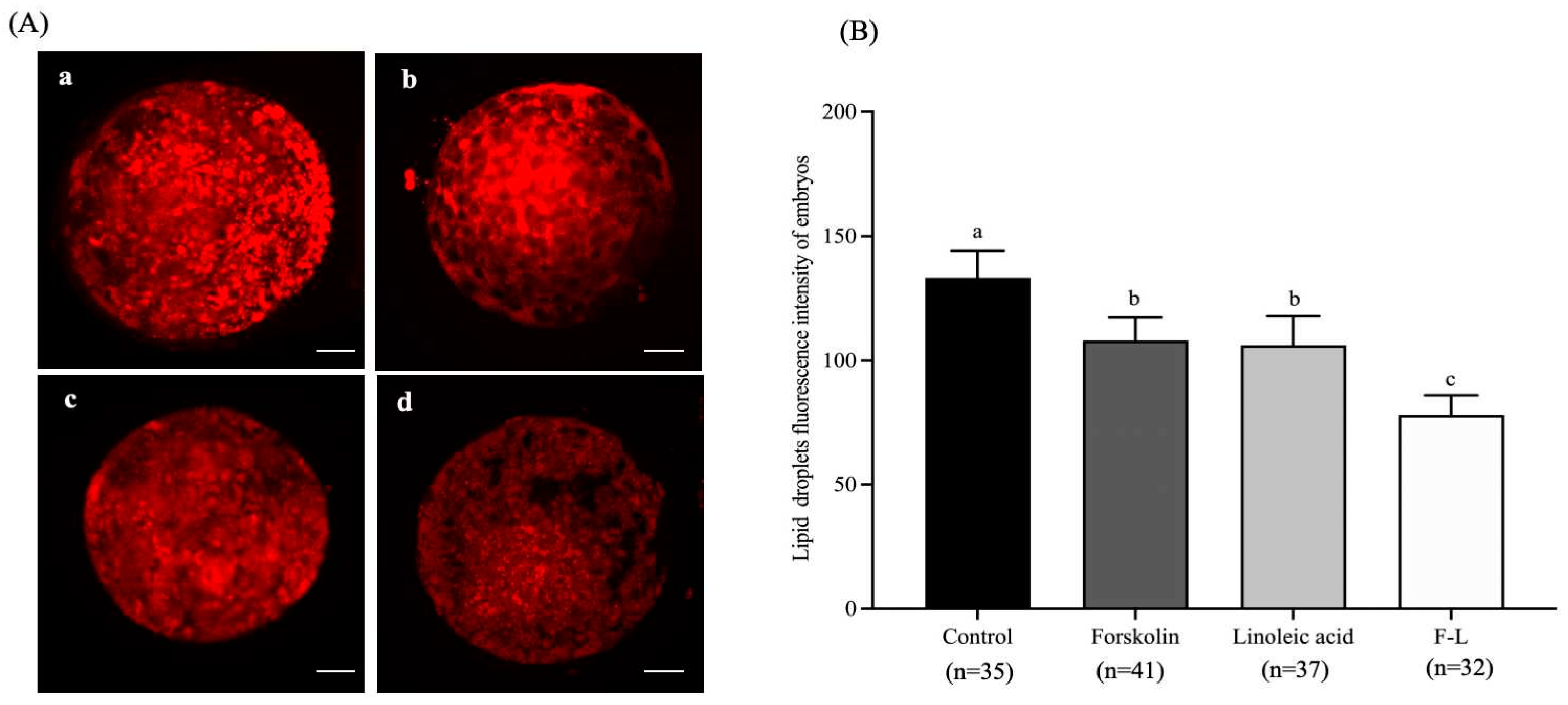
Figure 1. Effect of forskolin and linoleic acid on the lipid droplet content of bovine embryos. (A) Fluorogram of lipid droplet staining of embryos; a: control group, b: forskolin group, c: linoleic acid group, d: F-L group. (B) Effect of forskolin and linoleic acid on the amount of lipid droplet content of bovine embryos. Scale bar = 50 μM. Values with different superscripts indicate differences between groups (p < 0.05).
Ijms 26 04151 g001
Figure 2. Statistical analysis of lipid class and lipid species.
Ijms 26 04151 g002
Figure 3. Score scatter plot and permutation test of the OPLS-DA model. (A) Score plots were derived from the LC-MS/MS data of F-L and the control lipidomic. The x-axis and y-axis represent the first and second principal components, respectively. The purple and green colors indicate biological duplication within their respective groups. (B) The permutation tests based on LC-MS/MS data. In these tests, the x-axis and y-axis represent the correlation coefficient and value of R2Y and Q2, respectively.
Ijms 26 04151 g003
Figure 4. Clustering heatmap of significantly different lipid molecules. Note: Each column represents a group of samples, and each row represents a differential lipid molecule.
Ijms 26 04151 g004
Figure 5. Screening of differential expressions of mRNA. (A) Volcano plot of DEGs in FL and Con groups. (B) Clustering analysis of DEGs. Red indicates high expression, and blue indicates low expression.
Ijms 26 04151 g005
Figure 6. GO enrichment analysis of the DEGs. The spot color represents the FDR, and the spot size represents the enriched gene number.
Ijms 26 04151 g006
Figure 7. KEGG pathway analysis of the DEGs. Results from KEGG analysis; the color of each dot indicates the p value of the two sets of DEGs, and the size of the dot indicates the number of DEGs in each KEGG pathway.
Ijms 26 04151 g007
Figure 8. Effect of forskolin and linoleic acid on gene expression in bovine blastocysts. a, b: Values with different superscripts represent significant differences between groups (p < 0.05).
Ijms 26 04151 g008
Figure 9. Forskolin and linoleic acid on the apoptosis of bovine embryos. Note: (A) Fluorogram of TUNEL-stained embryos. (B) Forskolin and linoleic acid on the apoptosis of bovine embryos. Scale bar = 50 μM. Values with different superscripts indicate differences between groups (p < 0.05).
Ijms 26 04151 g009
Figure 10. Forskolin and linoleic acid on ROS levels in bovine embryos. (A) Representative images of bovine 2-cell ROS staining. (B) Forskolin and linoleic acid on the ROS levels of bovine 2-cell embryos. (C) Representative images of bovine embryo ROS staining; (D) forskolin and linoleic acid on the ROS levels of bovine embryos. Scale bar = 50 μM. Values with different superscripts indicate differences between groups (p < 0.05).
Ijms 26 04151 g010
Figure 11. Forskolin and linoleic acid on the cytoskeleton of bovine embryos. (A) Fluorogram of Tubulin-stained embryos; short arrows: normal spindle chromosome configurations; long arrows: abnormally shaped spindles; DAPI staining showed cell nuclear (blue color) and Tubulin staining showed microtube (red color). (B) Cytoskeletal analysis in vitrification and control and F-L group bovine embryos. Scale bar = 50 μM. Values with different superscripts indicate differences between groups (p < 0.05).
Ijms 26 04151 g011
Table 1. Effect of forskolin on the developmental ability and survival rate of bovine embryos.
GroupsNo. COCsCleavage RateBlastocyst RateSurvival Rate
0 μM10581.90 ± 2.57% (86/105) b34.89 ± 4.47% (30/86) b86.67 ± 6.59% (26/30) b
5 μM11984.87 ± 4.65% (101/119) b35.64 ± 4.70% (36/101) a88.89 ± 7.35% (32/36) b
10 μM10891.67 ± 3.71% (99/108) a42.42 ± 4.85% (42/99) a95.24 ± 7.23% (40/42) a
15 μM13464.18 ± 4.32% (86/134) c27.91 ± 3.81% (24/86) c83.33 ± 7.06% (20/24) c
a, b, c Values with different superscripts represent significant differences between groups (p < 0.05).
Table 2. Effect of linoleic acid on the developmental ability and survival rate of bovine embryos.
GroupsNo. COCsCleavage RateBlastocyst RateSurvival Rate
0 μM16580.00 ± 6.59% (132/165) b33.33 ± 2.41% (44/132) b86.36 ± 7.15% (38/44) b
50 μM18861.17 ± 5.85% (115/188) c27.83 ± 1.68% (32/115) c81.25 ± 7.46% (26/32) c
100 μM15188.74 ± 6.73% (134/151) a38.81 ± 2.55% (52/134) a96.15 ± 8.53% (50/52) a
500 μM14760.54 ± 5.68% (89/147) c29.21 ± 1.84% (26/89) c80.77 ± 7.21% (21/26) c
a, b, c Values with different superscripts represent significant differences between groups (p < 0.05).
Table 3. Effect of forskolin and linoleic acid on the developmental ability and survival rate of bovine embryos.
GroupsNo. COCsCleavage RateBlastocyst RateSurvival Rate
Control26275.57 ± 7.46% (198/262) c30.30 ± 2.65% (60/198) c80.65 ± 7.68% (25/31) b
Forskolin (10 μM)28482.39 ± 8.52% (234/284) b39.74 ± 4.12% (93/234) b90.24 ± 7.31% (37/41) b
Linoleic acid (100 μM)26083.08 ± 8.46% (216/260) b37.97 ± 4.03% (82/216) b89.47 ± 8.15% (34/38) b
F-L30689.54 ± 7.62% (274/306) a45.26 ± 5.24% (124/274) a98.18 ± 8.64% (54/55) a
a, b, c Values with different superscripts represent significant differences between groups (p < 0.05).
Table 4. Data analysis of lipid molecules with a significant difference between the F-L group and the control group.
Numberthe Adduct Ion Form of LipidCal m/z(t/min)VIPFCp-Value
1PA (18:1e_10:4) − 2H283.1694.751.640.47<0.01
2DG (4:0_10:4) + H309.1701.181.709.25<0.01
3DG (6:0_11:3) + Na375.2141.176.9211.21<0.01
4DG (6:0_14:2) + NH4414.3211.882.822.420.03
5DG (22:2) + NH4442.3531.881.802.470.02
6DG (27:4) + H491.3731.463.210.01<0.01
7DG (42:7e) + NH4698.60813.381.730.36<0.01
8LPC (16:0) + H496.3403.569.430.01<0.01
9LPC (18:0e) + H510.3923.641.022.15<0.01
10LPC (20:3) + H546.3553.031.080.770.04
11Car (d45:3) + Na710.64214.892.500.48<0.01
12MG (18:3) + NH4370.2951.931.182.100.04
13PC (8:0e_8:0) + H496.3403.5911.200.01<0.01
14PC (8:0e_10:1) + H522.3552.202.500.830.02
15PC (10:0e_10:3) + H546.3553.621.3512.56<0.01
16PC (12:1e_11:4) + Na606.3531.891.102.740.03
17PE (16:0e_18:3) + Na722.5101.611.872.310.04
18SM (d18:0_16:1) + H703.5759.686.670.72<0.01
19SM (d16:0_20:4) + H725.5599.121.510.800.02
20TG (4:0_8:0_10:1) + NH4458.3481.843.902.440.03
21TG (4:0_8:0_11:2) + NH4470.3481.231.410.470.01
22TG (6:0_12:0_12:3) + H549.4151.612.340.08<0.01
23TG (6:0_11:1_18:1) + NH4638.5356.471.72188.65<0.01
24TG (6:0_11:3_18:2) + H615.4626.442.83237.56<0.01
25TG (48:11) + NH4802.5987.371.030.38<0.01
26WE (38:2) + H561.56116.332.240.860.01
Note: FC indicates fold change, VIP indicates the variable importance for the projection.
Table 5. Statistics of different expressions of mRNA of the FL and Con groups.
GeneSampleUp/Down
FL 1FL 2FL 3Con 1Con 2Con 3
LRPAP124671571188810069961040up
APRT580462498117420127up
NFKBIA603988835315420453up
CRISP225028132812759127up
NDRG418774256676229up
TGM12271319456250342189up
ACSL11688945913664308540up
CASP7479285257171132146up
SEC22B14941150984558716771up
CDC25B5605352341909581up
MX25446336255up
EPHA42881311801058645up
ACADL289135140669944up
SLC38A526159810113up
CLDN118681435647302920602up
TBC1D1691581682381253285up
NSUN3560363339229241193up
CREM429897136up
BMP7107358241610up
IFIT54521702911248591up
PPARA373531207up
IRF812381299420653388189up
NR2F2797110894up
SLC2A41411002938379up
GPX7929499312039up
ADCY82860010up
DECR1256389215171142111up
TGFBR23791422817017371up
IDH1951579246175552531454063up
CAT792383675654361836144907up
DCAF11866680665394480354up
TUBA4A855597787377247500up
ARID1A391324367516516483down
KAT6B7116416398926531606down
ACSF356912293133143down
GLS2112511785835down
CDKN2C279172524down
PLPP318313974280308363down
DDX541533538850152315321475down
PLN2767812549down
TIAM1458531619783874783down
Table 6. Primers used in the present study.
GeneNCBI AccessionPrimer (3′–5′)Size (bp)
ACSL1NM_001076085TTTGTCCACGGAGAGAGCTT108
TTCAAAGGAGCCCACAATGC
DECR1NM_001075423ACAACTTGTCTGTCCAGCCT136
GGTCCCTCACATCACACTGA
ACADLXM_006935526TGATTCCTCACCACGCAGAA85
CCGAGAAGTCCTTGTTTGCC
GPX7NM_001101113CGACAGCAACAAGGAGATCG
TGATTTCCTCCACCGACACA
224
PPARANM_001034036TTCCCTCTTTGTGGCTGCTA112
TAGGTGGAGTTTGAGCACGT
TUBA4AXM_045034511TGGAACATGGGATTCAGCCT152
ATCAATCACAGTGGGCTCCA
CATNM_001035386CTGTGAACTGTCCCTACCGT159
CAGAGAAGTGGGTCCTGTGT
CDKN2CNM_001101054CTGCAATGAATGTGGGGAGG229
TGAGACTGGCAAAGGGAGAG
β-ACTINNM_173979CAAGTACCCCATTGAGCACG159
GTCATCTTCTCACGGTTGGC
Disclaimer/Publisher’s Note: The statements, opinions and data contained in all publications are solely those of the individual author(s) and contributor(s) and not of MDPI and/or the editor(s). MDPI and/or the editor(s) disclaim responsibility for any injury to people or property resulting from any ideas, methods, instructions or products referred to in the content.