


Abstract
Autoimmune diseases arise from the failure of self-tolerance. The recognition of self-antigen peptide–MHC (pMHC) complexes by the T-cell receptor (TCR) is the fundamental event triggering autoimmune pathogenesis. While traditional immunosuppressants provide broad systemic effects, they often compromise global immunity. Emerging molecular strategies aim to selectively disrupt the trimolecular complex—comprising the TCR, the antigenic peptide, and the MHC molecule—to induce antigen-specific tolerance. This review highlights the pMHC–TCR interaction as the primary molecular checkpoint for antigen-specific intervention. We discuss the structural basis of these interactions and their potential to redefine the therapeutic landscape for autoimmune diseases (ADs). We examine the molecular drivers of tolerance breakdown—including genetic susceptibility, molecular mimicry, post-translational modifications (PTMs), and ectopic MHC II expression—that shape the autoreactive T-cell landscape. This review examines current advancements in biological and pharmacological interventions, such as pMHC-decorated nanoparticles and soluble pMHC, to reprogram pathogenic T-cell response. We also explored CAR-T therapy strategies for autoimmune diseases, such as CAR-Treg, designed to precisely modulate pMHC-TCR signaling. Collectively, these precision interventions in immunological synapse assembly during autoimmune response are considered the basis for safer, antigen-specific immunotherapy capable of restoring self-tolerance without global immunosuppression.
1. Introduction
ADs are characterized by a profound loss of self-tolerance, leading to the activation of autoreactive T and B lymphocytes and the subsequent production of autoantibodies that target host tissues [1]. This loss of self-tolerance results in a persistent immune response against endogenous antigens, driving chronic systemic inflammation. Although many autoimmune diseases are considered rare (the number of patients is about 10% of the world’s population [2]), their collective incidence rose substantially between 1985 and 2015, reflecting a broader trend of increasing immune-mediated pathology (by 19.1% per year) [3]. Furthermore, an additional post-COVID-19 surge in autoimmunity was documented between 2019 and 2021, with reports indicating a further 1–2% increase in global incidence [4]. Autoimmune disorders are characterized by extensive comorbidities, with cardiovascular, respiratory, and endocrine diseases contributing to significantly higher mortality rates in these patient populations [5]. It is assumed that this pattern may result not only from systemic inflammation, but also from the use of immunosuppressive therapy.
Currently, systemic immunosuppression remains the mainstay of treatment for autoimmune diseases. The first immunosuppressive drugs (in the 1940s–1950s) were glucocorticosteroids and cytotoxic agents such as cyclophosphamide [6]. Cyclophosphamide exerts its immunosuppressive effect by inhibiting DNA replication, primarily through the formation of crosslinks between N7 atoms of guanine residues [7]. Failure to complete DNA replication due to stable crosslinking prevents S-phase progression, leading to the apoptotic depletion of rapidly dividing immune cells [8,9]. The non-selective cytotoxic profile of cyclophosphamide leads to the inhibition of all rapidly proliferating cells, targeting not only autoreactive lymphocytes but also hematological progenitors in the bone marrow. This mechanism accounts for its significant hematologic toxicity, characterized by a high incidence of neutropenia (73%) and leukopenia (70%) [10]. Similarly, systemic corticosteroids—another class of broad-spectrum immunosuppressants—exert their effects as ligands for the glucocorticoid receptor (GR). Upon activation, the ligand–receptor complex translocates into the nucleus, where it functions as a transcription factor to upregulate the expression of various anti-inflammatory proteins (IL-10, lipocortin-1, and IκBα). Simultaneously, the suppression of NF-κB, STAT, and AP-1 signaling results in a global downregulation of pro-inflammatory cytokines and chemokines, which is central to the therapeutic efficacy of corticosteroids [11,12,13]. However, the binding of glucocorticosteroids to the GR also upregulates the expression of key gluconeogenic enzymes in hepatocytes. This metabolic shift accounts for the high clinical incidence of secondary hyperglycemia (32%) and the development of steroid-induced diabetes mellitus (19%) [14]. Despite the proven efficacy of these drugs, the lack of selectivity for immune cells in general and for autoreactive cells in particular leads to a high risk of severe adverse effects.
The subsequent evolution of autoimmune therapy involved the design of more selective immunosuppressive strategies, specifically focusing on agents that target key components of the adaptive immune system, namely T and B cells. For example, cyclosporine A acts by inhibiting calcineurin, which in turn blocks activation of the transcription factor NFAT and reduces the expression of cytokines required for T-cell activation and proliferation [15]. The increased specificity of this drug stems from the restricted cellular distribution of calcineurin, in contrast to the ubiquitous expression of the GR. Despite its relative selectivity for immune cells, calcineurin is also functionally active in the nervous system [16], which explains the neurotoxic effect of cyclosporine observed in 10–28% of cases [17]. More refined immune-specific therapies are represented by monoclonal antibodies, which selectively target surface receptors or neutralized key pro-inflammatory cytokines. Drugs such as anti-TNF (adalimumab, infliximab, golimumab) and anti-IL17/23 (secukinumab/ustekinumab) bind signaling molecules and thereby block proinflammatory signaling pathways, suppressing inflammation on protein level but not on gene expression level [18]. A prominent example of an agent targeting specific cell surface antigens is rituximab, a chimeric monoclonal antibody directed against CD20. Because CD20 expression is limited to pre-B and mature B cells, the drug’s cytotoxic effect is highly specific, leaving other immune cells unaffected. Upon binding to the CD20 antigen, the antibody specifically induces cellular complement-dependent cytotoxicity [19,20]. Common side effects of rituximab therapy include infusion reactions (25%) caused by cytokine release from lysing B cells [21] and hypogammaglobulinemia (9.5%) [22]. Targeted depletion of specific TCR families, such as the TRBV9+ clones associated with ankylosing spondylitis, is now possible with the humanized antibody Seniprutug. This approach allows for the elimination of disease-driving T cells with minimal impact on overall immune surveillance [23].
The most promising frontier for specific immunosuppression is the targeting of the TCR–autoantigen–MHC complex. Intercepting this initial recognition event provides a pathway to antigen-specific tolerance, representing the next logical step beyond broad B- or T-cell depletion. Precision therapies centered on pMHC represent one of the most promising trends in autoimmunity. By shifting the focus from global suppression to antigen-specific modulation, these agents aim to re-establish tolerance with minimal off-target effects. Selective topological blockade of pMHC complexes, elimination of autoreactive T cells, or switching their activity toward a regulatory phenotype using therapeutic pMHC- based agents may improve the quality and duration of life of patients with autoimmune diseases and also reduce the number and severity of adverse effects [24]. A significant gap remains between the rational design of antigen-specific therapies and their successful translation into late-stage clinical development [25]. Here, we delineate the molecular pathways driving auto reactivity from pMHC assembly to TCR engagement, followed by a comprehensive overview of precise molecular approaches and therapies aimed at restoring self-tolerance.
2. The pMHC–TCR Interaction and Its Role in Autoimmune Pathogenesis
2.1. Antigen Presentation by MHC
The molecular recognition of self-peptides in complex with MHC molecules by T lymphocytes is a pivotal event in the etiology of autoimmune diseases. This process involves the proteolytic processing of endogenous and exogenous proteins, followed by the loading of these fragments onto MHC molecules within endosomal compartments or the endoplasmic reticulum for subsequent display on the cell surface [26]. T-cell activation is initiated by the molecular recognition of the pMHC complex by the TCR, a process stabilized by the recruitment of CD4 or CD8 coreceptors to the invariant domains of MHC class II or I, respectively. To elicit a robust immune response, this primary signal must be integrated with costimulatory signaling, primarily mediated by the CD28-CD80/86 interaction [27]. The combination of these molecular contacts forms a functional immunological synapse that provides precise control over T-cell differentiation [28].
MHC class I molecules facilitate the presentation of endogenous peptides derived from cytosolic proteins and intracellular pathogens to CD8+ T cells. These antigens are primarily processed by the proteasome before being translocated into the endoplasmic reticulum [29]. In contrast, MHC class II molecules specialize in the presentation of exogenous proteins acquired via endocytosis, which are proteolytically cleaved within lysosomal compartments for display to CD4+ T cells [30]. While MHC class I is constitutively present on most nucleated cell types, MHC class II is a hallmark of professional antigen-presenting cells (APCs)—macrophages, B cells, and dendritic cells. However, the inflammatory milieu can trigger the de novo expression of MHC class II on non-professional APCs (such as epithelial cells, endothelial cells, fibroblasts, and others), thereby expanding the repertoire of cells capable of activating CD4+ T-cell responses during chronic autoimmune inflammation [31,32].
Most autoimmune predispositions are linked to specific HLA-DR/DQ alleles, making the MHC-II groove a primary target for blockade.
2.2. Molecular Mechanisms of T-Cell Activation
Under physiological conditions, the introduction of foreign antigens via MHC presentation triggers the adaptive immune cascade. Specifically, for CD8+ T cells, this stimulation initiates a robust cytotoxic response characterized by clonal expansion. During this process, activated cells undergo divergent differentiation into either functional cytotoxic T lymphocytes (CTLs), responsible for immediate effector activity, or long-lived memory T cells for persistent immunosurveillance [33]. Short- lived CTLs identify target cells via specific MHC I–antigen recognition. Upon the assembly of the immunological synapse (trimolecular complex), they execute target cell apoptosis [34]. CD8+ memory T cells persist in the organism even after elimination of infected cells and provide a rapid immune response upon re-encounter with the same antigen [35]. Upon activation, naïve CD4+ T cells undergo lineage-specific differentiation into effector (Th and Tfh cells) and regulatory populations (Tregs). The latter group is further distinguished by the presence of FoxP3+ Tregs and the FoxP3− Tr1 subset (IL-10+ type 1 regulatory T cells) [36]. Effector CD4+ T cells orchestrate immune responses by secreting distinct cytokine profiles [37,38].
Tregs ensure immunological tolerance by modulating autoreactive or excessive responses. One primary mechanism involves the high-affinity inhibitory receptor CTLA-4, which competitively outbinds CD28 for CD80/CD86 ligands on APCs, thereby depriving effector cells of essential co-stimulation. Additionally, Tregs facilitate competitive IL-2 sequestration; by expressing high-affinity CD25 (IL-2Rα), they effectively deplete the local cytokine pool, starving effector populations and limiting their proliferation and survival [39,40]. Beyond metabolic disruption, Tregs exert direct immunosuppression on Th cells, CD8+ T cells, B cells, and DCs via the secretion of IL-10 and TGF-β [41,42]. Given their potent capacity to modulate the immune landscape, Treg-based therapies are currently being explored for both autoimmune disorders and oncological intervention.
The outcome of TCR–pMHC interactions is highly context-dependent; upon encountering self-antigens during maturation, T cells follow a specialized differentiation pathway known as central tolerance. While foreign antigens trigger an effector cascade, the presentation of self-antigens is governed by a fundamentally different developmental paradigm aimed at maintaining tolerance. Within the thymic microenvironment, the initial encounter of a developing T cell with self-pMHC complex facilitates the establishment of central tolerance [43]. This process is dictated by the binding affinity of the TCR for self-antigens. It begins with positive selection, which preserves only those T cells capable of functional interaction with host MHC molecules. This is followed by negative selection, wherein clones exhibiting dangerously high affinity for self-pMHC complexes are eliminated via apoptosis. This stringent thymic filtering serves to purge potentially pathogenic autoreactive clones before they egress into the systemic circulation [44]. Nevertheless, this process is incomplete; evidence suggests that up to 30% of self-reactive T cells bypass thymic deletion and reach the periphery [45]. Consequently, the robust establishment of peripheral tolerance becomes essential for maintaining immunological silence toward host tissues [46].
2.3. Peripheral Tolerance Induction
Peripheral tolerance serves as a secondary check to neutralize mature autoreactive lymphocytes that have bypassed central selection in the thymus or bone marrow. This systemic regulation is achieved through several distinct mechanisms: anergy, a state of functional hypo responsiveness resulting from TCR engagement in the absence of co-stimulatory signals; clonal deletion, the induction of apoptotic pathways in self-reactive cells; and active suppression, primarily orchestrated by Tregs via inhibitory receptors and anti-inflammatory cytokine secretion. In addition, immunological ignorance acts as a passive barrier, keeping autoreactive clones quiescent due to sub-threshold antigen concentrations or their sequestration within immune-privileged sites [46].
CD28-mediated co-stimulation serves as a critical checkpoint for peripheral tolerance (Figure 1). In the absence of robust CD80/CD86 signaling, TCR engagement drives T cells into anergy, characterized by impaired IL-2 production and arrested proliferation [47]. This inhibitory state is further reinforced by CTLA-4, which outcompetes CD28 for ligand binding, thereby actively antagonizing activating signals [48]. Furthermore, the co-stimulatory context dictates the functional polarization of naïve CD4+ T cells into regulatory subsets. Foxp3+ Tregs typically differentiate upon interaction with immature dendritic cells (imDCs), exhibiting low CD80/CD86 density, providing the sub-threshold co-stimulation necessary for induction. This transition is highly dependent on the cytokine microenvironment, where high concentrations of TGF-β and IL-2 are required to stabilize Foxp3 expression. In contrast, Tr1 cell differentiation is favored during interactions with semi-mature DCs (smDCs) or imDCs under moderate co-stimulatory conditions, traditionally associated with IL-10 signaling [49]. However, alternative models suggest that Tr1 induction may rely more heavily on chronic, repetitive TCR stimulation under limited co-stimulation rather than a strict IL-10 requirement [50]. Finally, the total absence of CD28 signaling and IL-2 leads to the passive death of naïve cells, while chronic re-activation—often via the Fas–FasL axis—triggers the deletion of effector populations [51]. This complex mechanism of peripheral tolerance formation (Figure 1) is used to develop therapeutic agents, which are described in detail in the following sections.
Figure 1.
Molecular Mechanisms of Peripheral CD4+ T-Cell Tolerance and Phenotypic Differentiation. Schematic representation of T-cell fate determination based on the pMHC–TCR signaling context. (A) Canonical activation: Robust CD28–CD80/86 co-stimulation (antigen presentation by mature DCs) promotes the differentiation of naive T cells into pro-inflammatory effector populations. (B) Treg induction: Low level of CD80–CD28 co stimulation (antigen presentation by tolerogenic or immature DCs) in the presence of TGF β results in differentiation into FoxP3+ Treg. (C) Tr1 differentiation: Moderate co-stimulatory signaling (antigen presentation by semi-mature DCs) in the presence of IL-10 promotes the development of Type 1 regulatory (Tr1) cells. (D) Functional anergy: The absence of co-stimulation or the competitive engagement of the CTLA-4–CD80/86 inhibitory axis induces a state of functional anergy.
As the important master checkpoint of adaptive immunity, the pMHC–TCR interaction serves as the primary filter between quiescence and activation, where a loss of recognition fidelity triggers the breakdown of tolerance. This fundamental signal is integrated into a regulatory network where the canonical CD28–CTLA-4 axis establishes the response amplitude by either amplifying proximal signaling or competitively aborting autoreactive priming. Further refinement is provided by secondary modulators, for example, PD-1 [52] and Siglec-7/-9 [53], which recalibrate TCR sensitivity via SHP-1/2 recruitment [54,55] or ITIM signaling [56]. Ultimately, while the pMHC–TCR axis remains the dominant control node, the hierarchy of subsequent checkpoints dictates the progression toward a systemic autoimmune phenotype.
2.4. Genomic Drivers of Self-Tolerance Defects
A breakdown in immune homeostasis can be triggered by a genetic predisposition that disrupts mechanisms of self-tolerance [57]. Immune tolerance to self-antigens may be genetically impaired at the stage of negative selection of T lymphocytes in the thymus. For example, in the case of mutations in the AIRE (Autoimmune Regulator) gene, which controls the expression of tissue-specific antigens by thymic epithelial cells [57]. Defects in the apoptotic machinery, caused by mutations in the Fas and FasL genes, can also allow autoreactive T-cell clones to survive [58]. Peripheral tolerance may be compromised by reduced function and numbers of regulatory T cells, which can be associated with mutations in the Foxp3 gene, as well as by dysregulated immune control of effector T cells due to mutations in IL-2 and CTLA-4 [59].
Extensive evidence indicates that genetic susceptibility to numerous autoimmune pathologies is primarily linked to the inheritance of specific MHC alleles. The binding affinity of a given MHC variant for particular antigenic motifs is a critical determinant of whether a self-peptide will be presented for TCR recognition [60]. A classic example is rheumatoid arthritis (RA), where approximately 90% of patients harbor the HLA-DRB1*0401, 0404, or 0101 alleles [61]. This strong clinical association is attributed to a conserved amino acid sequence—the ‘shared epitope’—located at positions 70–74 of the DRβ1 chain. Positioned within the P4 pocket of the antigen-binding groove, this motif facilitates the preferential presentation of arthritogenic peptides, thereby driving the expansion of an autoreactive T-cell repertoire [62].
2.5. Mechanisms of Peripheral Tolerance Breakdown
Exogenous environmental triggers can significantly contribute to the breakdown of self–non-self-discrimination. When a pathogenic protein exhibits structural homology with host proteins, it can lead to cross-reactivity. In this scenario, a single MHC variant can present both foreign and self-peptides, which are subsequently recognized by the same TCR—a phenomenon termed molecular mimicry [63,64].
Following an infectious challenge, the pro-inflammatory cytokine milieu serves as a critical pathogenic driver for the induction of autoreactive responses. A hallmark of this process is the aberrant upregulation of MHC II expression within inflammatory foci. Elevated local concentrations of interferon-gamma (IFN-γ) trigger the activation of the CIITA promoter—the master transcriptional regulator of the MHC class II gene cluster—in non-professional antigen-presenting cells [65]. Consequently, host cells that are typically immunologically ‘invisible’ to CD4+ T helper cells begin to display self-peptides, effectively functioning as ectopic APCs [66]. In Hashimoto’s thyroiditis, for example, thyroid epithelial cells undergo this phenotypic shift under inflammatory stress, transitioning from passive targets to active stimulators of T-lymphocyte infiltration [67].
A critical consequence of localized inflammation is bystander activation, wherein the activation threshold for T cells is significantly lowered. The abundance of pro-inflammatory cytokines (e.g., IL-1, IL-6, and TNF-α) and damage-associated molecular patterns (DAMPs), such as heat-shock proteins (HSPs) or cell-free DNA, drives the robust upregulation of CD80/86 on local APCs. In this hyper-stimulatory milieu, autoreactive T cells possessing only marginal affinity for self-pMHC complexes receive sufficient co-stimulatory signals to bypass normal checkpoints, leading to their clonal expansion and functional activation [68].
Moreover, many autoantigens that are normally sequestered inside cells or in immune-privileged organs with blood–tissue barriers (eyes, brain, testes) and that T cells have never encountered during their “education” become accessible to APCs during inflammation, which causes extensive cell death (necrosis, NETosis) and barrier disruption. This release leads to the systemic accumulation of intracellular material, which can be captured and processed by APCs for MHC II presentation [69,70]. This massive display of self-antigens within a pro-inflammatory milieu frequently triggers epitope spreading. This phenomenon involves a functional shift in the T-cell response, transitioning from a dominant pathogen-specific focus to the recognition of self-epitopes presented by both professional and non-professional APCs. This process facilitates the emergence of neo-epitopes within a single autoantigenic protein, fueling the diversification of the autoreactive T-cell repertoire and the chronic propagation of the immune response [71].
Thus, presentation of self-peptides on MHC II in inflamed tissues converts a local protective process into a self-perpetuating cycle of tissue destruction, preventing spontaneous resolution of inflammation and ultimately leading to the development of full-blown autoimmune disease [72].
The breakdown of self-tolerance is intrinsically linked to antigenic variability. Factors such as PTMs and the formation of protein complexes create neoantigens that the immune system fails to recognize as ‘self,’ thereby serving as primary initiators of autoimmune pathology. Pathogenesis in antiphospholipid syndrome (APS) involves neoepitopes generated through β2GPI interaction with platelet factor 4 (PF4). The resulting immunoreactive complexes trigger platelet-driven thrombosis, highlighting a complex antigenic landscape that exceeds the targeting of native β2GPI alone [73].
A striking example of PTM is citrullination of proteins in rheumatoid arthritis, associated with hyperactivation of peptidyl arginine deiminase (PAD). Conversion of arginine to citrulline has been shown to increase peptide affinity for MHC molecules carrying the shared epitope, which promotes their presentation to CD4+ T cells [74]. Other types of PTMs also occur, such as carbamylation and deamidation. These processes generate “neoepitopes” that were not previously presented to T cells in the thymus during selection. A relevant example is APS, where carbamylation of β2GPI may create additional antigenic determinants beyond native β2GPI [75,76]. Extracellular vesicles may further modulate β2GPI-driven pathogenic pathways in APS by exposing antigenic targets and reinforcing endothelial prothrombotic activation [77].
An autoimmune response can also arise when previously sequestered antigens come “into view” of the immune system. In systemic lupus erythematosus, the immune response targets fragments of double-stranded DNA and chromatin that remain in the extracellular space due to defects in the clearance of apoptotic material [78]. As a result, T cells recognize such modified self-molecules as foreign. At the site of inflammation (for example, a joint in arthritis), MHC class II molecules present precisely these altered peptides.
The fundamental molecular insights into peripheral tolerance breakdown are now being leveraged to engineer antigen-specific therapeutics designed for its restoration. By translating the principles of immune checkpoints and synapse dynamics into programmable platforms, such as pMHC-nanoparticles and Tolerogenic Vaccines, researchers aim to re-induce anergy and active suppression within the autoreactive repertoire. Thus, targeting the individual components of the pMHC II–TCR complex offers a promising avenue for the development of tolerance-inducing biologics. These strategies aim to selectively suppress pathogenic responses while sparing protective immunity, providing a pathway to halt the progression of autoimmune diseases without the harmful effects of broad-spectrum immunosuppressants [79] (Figure 2).
Figure 2.
Molecular determinants of pMHC–TCR synapse dysfunction in autoimmunity: Genetic predisposition through specific MHC (green) polymorphism of risk alleles influences the presentation of autoantigenic peptides (red). The conversion of self-proteins into pathogenic neoantigens via PTMs and molecular mimicry provides high-affinity ligands for autoreactive TCRs (blue). The resulting breakdown in the pMHC–TCR trimolecular complex specificity bypasses peripheral tolerance and initiates tissue destruction. The model of the pMHC–TCR Complex is represented by a Tfh TCR and an aggrecan peptide presented by MHC II. Rendered from PDB entry 7RDV [80].
3. pMHC-Based Strategies for Restoring Peripheral Tolerance
Various therapeutic modalities targeting the pMHC–TCR synapse are currently at distinct stages of preclinical and clinical development, with the primary objective of restoring immunological tolerance to self-antigens. As previously discussed, the TCR recognition of the pMHC complex is a decisive signaling event; the subsequent fate of T cells—from proinflammatory activation to functional anergy or differentiation into regulatory T cells (Treg)—is primarily determined by additional costimulatory signals and the local cytokine environment. The strategies detailed in this section modulate the co-stimulatory context to shift the immune response toward antigen-specific suppression of the autoreactive cascade and the induction of immunological tolerance.
Two fundamentally distinct therapeutic strategies have emerged in this field. The first utilizes APC-independent molecular platforms, employing soluble pMHC complexes, free antigens, or advanced delivery systems such as nanoparticles, liposomes, and other biocompatible carriers. The second approach relies on the presentation of autoantigens by tolerogenic dendritic cells (tolDC), which are characterized by a low expression of CD80/CD86 co-stimulatory molecules [81].
3.1. Soluble Antigen Immunotherapy
The therapeutic efficacy of soluble antigen-mediated immunotherapy is predicated on high-dose administration, which triggers functional T-cell anergy or drives a regulatory immune response [82]. The use of soluble autoantigens has evolved from the administration of whole proteins (insulin, myelin basic protein (MBP)) to the design of short synthetic peptides (12–20 amino acids). Current strategies focus on autoantigenic peptides which, due to their solubility and bioavailability, can efficiently compete for binding within the MHC II peptide-binding groove on the surface of immature antigen-presenting cells, thereby suppressing the autoimmune cascade in individuals carrying specific risk alleles [83] (Table 1). Several peptide-based candidates are undergoing preclinical and clinical evaluation, including fragments of insulin for Type 1 Diabetes [84,85] (Nasal Insulin-Phase III clinical trial, NCT00223613) and MBP-derived fragments for Multiple Sclerosis [86,87] (ATX-MS-1467-Phase II clinical trial, NCT01973491). Numerous studies highlight the HLA-restricted clinical response of these agents, as therapeutic benefits are predominantly observed in patients carrying specific high-risk HLA-II alleles (MBP8298-Phase III clinical trial, NCT00468611) [88,89]. This association confirms that these peptides modulate the autoimmune cascade specifically by targeting the antigen presentation pathway.
Table 1.
Immunotherapeutic Agents Based on soluble antigens.
Extensive research has focused on altered peptide ligands (APLs) to induce immunological tolerance. Being analogs of native antigenic determinants, APLs contain single amino acid substitutions in the region of contact with TCR, which allows modulation of binding affinity and subsequent CD3 phosphorylation pattern [90]. This molecular reprogramming can induce T-cell anergy or drive its phenotype shift from a pro-inflammatory Th1/Th17 profile toward a regulatory Th2 response [91]. Beyond linear peptides, cyclic APLs have emerged as promising candidates for the treatment of multiple sclerosis (cyclo(87–99)(Ala91,Ala96)MBP87–99-preclinical study) [92], (Cyclic MOG35-55-preclinical study) [93].
The most prominent clinical implementation of this strategy is glatiramer acetate (Copaxone—approved NCT00097188), which functions as a universal APL for myelin-reactive T cells in multiple sclerosis, promoting a protective Th2 shift and suppressing neuroinflammation [94]. Another significant development involves insulin B:9–23-based APLs (e.g., NBI-6024-Phase I clinical trial, NCT00873561) for type 1 diabetes, engineered to restore immunological tolerance to pancreatic β-cells [95]. Furthermore, Jusvinza (Phase III clinical trial, RPCEC00000404), a modified peptide derived from heat shock protein 60 (HSP60), has been shown to downregulate pro-inflammatory cytokines and bias the immune response toward a regulatory phenotype in rheumatoid arthritis [96]. Despite their therapeutic promise, the clinical application of APLs necessitates molecular precision to minimize the risks of cross-reactivity or potential disease exacerbation. Thus, the clinical evaluation of APLs in multiple sclerosis unexpectedly triggered severe disease flares; as detailed in an erratum published by the authors in Nature Medicine, this outcome was driven by the cross-activation of encephalitogenic T-cell responses rather than the intended induction of immune tolerance [97].
Peptide-based immunotherapy provides a precise means of inducing antigen-specific tolerance without global immunosuppression [98], yet several hurdles remain. While these strategies effectively promote regulatory T cells or clonal anergy, their clinical translation is hindered by the metabolic instability of peptide-based drugs and their strict HLA restriction. Because therapeutic efficacy is fundamentally contingent upon the patient’s individual HLA haplotype and the presence of specific risk alleles, this genetic diversity results in significant response variability, thereby constraining the broad clinical utility of peptide therapies [26]. Furthermore, a significant translational gap persists; while APLs show promise in experimental models of systemic autoimmunity, human clinical data remain fragmentary and frequently fail to replicate the robust responses observed in preclinical settings [99].
3.2. Soluble pMHC Complex-Based Immunotherapy
The production of soluble pMHC complexes is slightly more demanding than that of soluble antigens, primarily due to the additional biotechnological steps required to generate stable MHC variants. These constructs necessitate either exogenous peptide loading or covalent linkage to the MHC scaffold. To streamline this process, specialized single-chain chimeric molecules, termed recombinant TCR ligands (RTLs), have been engineered; these consist of a single polypeptide chain comprising the antigenic peptide fused to the β chain and a chain domain of the MHC class II molecule [100,101,102]. Furthermore, several studies have demonstrated that pMHC dimers and higher-order multimeric complexes exhibit enhanced therapeutic efficacy compared to their monomeric counterparts [103,104].
The mechanism of action of soluble pMHC complexes is based on the direct and selective delivery of signals to specific T-cell populations, bypassing the requirement for APC-mediated activation and inflammatory co-stimulation. Since these complexes are administered in a soluble monomeric or multimeric form rather than presented on APC cell membrane, the essential CD28–CD80/CD86 co-stimulatory axis is not engaged. This lack of secondary signaling induces functional T-cell anergy or drives differentiation toward a regulatory T-cell (Treg) phenotype. Early proof-of-concept studies at the end of the 20th century demonstrated that soluble pMHC molecules carrying MBP peptide fragments could suppress the development of experimental autoimmune encephalomyelitis (EAE) in murine models [105], while complexes utilizing acetylcholine receptor fragments were effective in experimental myasthenia gravis [106]. In a clinical setting, however, a phase I trial of AG284 (a soluble HLA-DR2:MBP84–102 complex) failed to show significant differences compared to placebo [107]. This lack of efficacy may be attributed to the low intrinsic avidity of the pMHC–TCR interaction or a sub-therapeutic local concentration of the complexes, leading to insufficient T-cell engagement. More recent therapeutic approaches utilize MHC tetramers to enhance binding stability (Table 2).
Table 2.
Immunotherapeutic Agents Based on pMHC complexes and nanoparticle platforms.
Compared to free peptides, pMHC-multimers provide superior therapeutic precision by leveraging high-avidity interactions to engage low-affinity and low frequency autoreactive T cells [111]. While free peptides require uptake and processing by APCs, pMHC-multimers signal T cells directly, enabling the active reprogramming of effectors into Tr1 cells [24]. However, pMHC-multimers face steeper translational hurdles due to extreme HLA-restriction, high production costs, and the technical complexity of maintaining stable recombinant MHC complexes [24,112].
3.3. Antigen-Conjugated Nanoparticle Immunotherapy
Currently, the development of therapeutic agents has shifted toward pMHC complexes conjugated to liposomes or nanoparticles, primarily due to their enhanced biochemical stability. Most nanoparticle- and liposome-based platforms function by inducing the differentiation of effector T cells into IL-10-secreting regulatory T cells. These formulations may consist of nanoparticles coated with multimeric pMHC complexes or, alternatively, with isolated antigen molecules in the absence of the MHC scaffold. When previously activated T cells re-encounter antigens displayed on these nanoparticles in the absence of co-stimulatory signals—a mechanism analogous to MHC tetramer-mediated signaling—they bypass full activation, resulting in functional anergy or regulatory Tr1 differentiation (Figure 3) [113].
Figure 3.
Schematic representation of the antigen-specific reprogramming of autoreactive T cells into Tr1 cells: In the absence of co-stimulatory signals, the engagement of the TCR by pMHC–nanoparticles or pMHC tetramers bypasses canonical effector activation. This suboptimal signaling facilitates a phenotypic shift toward a Treg-mediated suppressive response, effectively inhibiting the autoimmune cascade.
The surface density of pMHC complexes (valence) represents a critical parameter in the molecular design of these therapeutics. Nanoparticles characterized by insufficient pMHC valence typically exhibit sub-optimal biological activity [114]. Conversely, an excessively high pMHC density can trigger aberrant T-cell activation, potentially driving a robust pro-inflammatory effector response [115,116]. Such unintended activation must be strictly avoided, as it counteracts the therapeutic goal of inducing antigen-specific tolerance. Thus, multivalent pMHC display on nanoparticles provides sustained TCR stimulation in the absence of CD28 co-stimulation, biasing the fate of previously activated autoreactive CD4+ T cells toward IL-10+ Tr1 differentiation or the establishment rather than effector reactivation [24].
Peptide–MHC-based nanoparticle medicines represent a sophisticated therapeutic frontier, offering high biochemical stability and precise control over antigen valence to induce antigen-specific regulatory T cells without compromising global immunity. However, their clinical translation is hindered by significant technological and biological challenges: these include the rigorous requirement for accurately defined, clinically relevant autoantigens and the necessity for highly selective delivery to target cells while avoiding off-target accumulation or unintended immune activation. Furthermore, substantial obstacles regarding industrial-scale production, high manufacturing costs, and long-term formulation stability remain to be addressed to ensure their broader clinical viability [117,118].
3.4. Cell-Based Immunotherapy via Tolerogenic Autoantigen Presentation
Immunotherapeutic strategies that exploit the distinct tolDC phenotype in conjunction with antigen-specific presentation constitute a dedicated field of study (Table 3). Tolerogenic DCs are characterized by low expression of the co-stimulatory molecules CD80/CD86 and by a distinctive cytokine profile: IL-10+ TGF-β+ IL-27+. This specialized signaling milieu recalibrates the T-cell response at the antigen-presentation interface, driving either functional anergy or the induction of a regulatory phenotype [119].
TolDC populations can emerge endogenously in humans (e.g., DC-10, smDC) or be generated ex vivo by treating immature iDCs with pharmacological agents such as vitamin D3, rapamycin, or the cytokines IL-10 and TGF-β [120]. Specifically, rapamycin inhibits the mTOR signaling pathway, which subsequently suppresses IL-12 expression while concurrently upregulating IL-10 and TGF-β production [121]. Current tolerogenic modalities often utilize liposomal platforms that co-encapsulate both the target antigen and a tolerizing agent (Figure 4). Alternative strategies involve the adoptive transfer of ex vivo engineered tolDCs loaded with the relevant autoantigen (Table 2). Furthermore, antigen-specific tolerance can be induced via nucleic acid constructs (DNA/RNA) designed to drive the coordinated expression of the autoantigen alongside IL-10 and TGF-β within the dendritic cell. In all the approaches described above, the disease-relevant autoantigen is presented by tolDCs via MHC class II under conditions of minimal CD80/CD86 co-stimulation and an anti-inflammatory cytokine milieu (IL-10/TGF-β/IL-27). This configuration delivers a sub-immunogenic TCR signal to autoreactive CD4+ T cells. Chronic exposure to this tolerogenic pMHC environment triggers anergy or clonal deletion while simultaneously driving the differentiation of FoxP3+ and Tr1 regulatory T cells, thereby actively restoring antigen-specific peripheral tolerance [122,123,124].
Figure 4.
TolDC-Mediated Induction of Regulatory T Cells. Schematic representation of the tolerogenic reprogramming of iDCs. Liposomes containing both a specific autoantigen and a tolerant substance (e.g., rapamycin) are engulfed by phagocytosis. Intracellular rapamycin delivery modulates the DC phenotype, promoting a tolerogenic state characterized by downregulated CD80/CD86 expression and the secretion of IL-10, TGF-β, and IL-27. Subsequent presentation of the autoantigen to naive T cells within this specialized signaling milieu drives their differentiation into Tregs and Tr1 cells.
Cell-based strategies utilizing tolerogenic dendritic cells (tolDCs) offer significant therapeutic potential by enabling antigen-specific modulation and favoring the induction of regulatory T cells over broad immunosuppression. Early-phase clinical trials across multiple autoimmune pathologies—including multiple sclerosis, type 1 diabetes, and rheumatoid arthritis—have already confirmed their favorable safety profile and initial biological activity. Nevertheless, their clinical translation remains hampered by the inherent complexities of patient-specific manufacturing and the high degree of inter-donor variability regarding tolDC functional stability in the context of established autoimmunity. Furthermore, the lack of standardized protocols for antigen loading, optimal dosing, and administration routes remains a critical barrier, significantly escalating the cost and logistical complexity of these personalized interventions [125].
Table 3.
Immunotherapeutic Agents based on autoantigen presentation by tolDC.
4. pMHC-Mediated Depletion of Autoreactive T-Cell Populations
4.1. pMHC Conjugates with Cytotoxic Agents
Beyond the induction of peripheral tolerance via anergy or Treg differentiation, recent therapeutic advancements have focused on the selective depletion of pathogenic clones. This strategy utilizes antigen-specific cytotoxic conjugates—molecular constructs where an autoantigenic peptide, presented within an MHC scaffold, is coupled to a potent effector toxin. By bypassing the need for functional reprogramming of the T-cell phenotype, these agents facilitate the direct elimination of autoreactive TCR-specific populations [136,137] (Table 4).
Table 4.
pMHC-Based therapeutic Agents for Autoreactive T-Cell Elimination.
4.2. pMHC Conjugates with Oxidoreductase Motif
A distinct example is the Imotope platform, which uses antigens engineered with an oxidoreductase CxxC motif capable of oxidizing thiol groups of amino acids to form S–S bonds [141]. The appearance of new disulfide bonds at the T-cell surface shifts CD4, TCR, and integrins into a more clustered, high-avidity state and increases the stability of pMHC–TCR complexes [136]. This results in stronger TCR-mediated signaling and conversion of the T cell to a cytolytic phenotype. Such a cytolytic T cell can induce apoptosis (via Fas–FasL and perforin–granzyme mechanisms) of immune cells involved in the autoimmune response; specifically, this localized cytotoxicity is directed toward both the antigen-presenting cells (APCs) displaying the cognate autoantigen and the pathogenic CD4+ effector T cells physically coupled to the APC via the pMHC–TCR interface (Figure 5, Table 4). Such a dual-targeting approach ensures the disruption of the pro-inflammatory microenvironment at the site of autoantigen recognition.
Figure 5.
Redox-mediated differentiation of cytolytic CD4+ T cells. Antigens containing the CxxC thioreductase motif catalyze the reduction in surface disulfide bonds, promoting clustering of the TCR–CD4–integrin complex. This biochemical rearrangement increases the kinetic stability and robustness of the pMHC–TCR interaction, ensuring the sustained signal intensity necessary for the transition of naive T cells to a cytolytic phenotype.
In summary, pMHC conjugates functionalized with cytotoxic or oxidoreductase domains enable the targeted depletion of pathogenic T-cell and APC populations. By preserving bystander immune function, these agents offer a more precise alternative to broad-spectrum immunosuppressants. This approach may achieve sustained remission through finite treatment courses and facilitate the development of HLA-specific therapies for chronic autoimmune diseases [137].
Compared to free antigens and native pMHC complexes, which are often limited by low efficacy and the risk of unintended immune activation, pMHC–effector conjugates ensure the targeted destruction of both pathogenic T cells and cognate APCs. Nevertheless, the safety profile of pMHC conjugates is more sensitive to epitope mapping precision; whereas tolDCs and free antigens and pMHC complexes operate within natural regulatory boundaries, the use of cytotoxic payloads creates a narrow therapeutic window, necessitating stringent HLA-restriction protocols to avoid collateral damage to healthy tissues, which was reported for CAR- and TCR-based therapies [142]. Consequently, even marginal fluctuations in dosage or systemic exposure can trigger significant off-target cytotoxicity and collateral damage to healthy tissues [137].
5. Therapeutic Strategies for ADs Targeting the TCR
As the primary structural determinant of the trimolecular complex, TCR functions as a molecular switch that triggers the initiation and persistence of autoimmune signaling. This section delineates therapeutic strategies designed to disrupt autoreactive signaling by directly targeting the TCR interface.
5.1. CAR-T Immunotherapy
At present, CAR-T therapy is becoming a highly relevant direction in personalized treatment. It already plays a major role in cancer therapy and is also applicable to autoimmunity. A CAR (chimeric antigen receptor) is an artificially engineered T-cell receptor designed to recognize specific markers or antigens. It consists of an external antigen-binding scFv domain, a hinge region that provides scFv flexibility, a transmembrane domain, anchoring the CAR in the cell membrane, and intracellular signaling domains [143]. In clinical oncology, patient-derived CD8+ T cells are genetically modified via lentiviral transduction to express a chimeric antigen receptor (CAR) [144]. The high-affinity interaction between the scFv domain and its cognate surface antigen (e.g., CD19, BCMA, or CD22) triggers a potent cytotoxic cascade characteristic of effector CD8+ lymphocytes [145,146].
In the context of autoimmunity, several CAR-T-based therapeutic platforms are under active investigation. Currently, the most clinically advanced strategy involves anti-CD19 CAR-T cells designed for the systemic depletion of the B-cell lineage. These engineered CAR-T have demonstrated significant clinical efficacy in patients with systemic lupus erythematosus (NCT06333483) and systemic sclerosis (NCT05085444). A pivotal advantage of CAR-T cells over conventional monoclonal antibodies lies in their superior chemotactic trafficking into secondary lymphoid organs, facilitating the eradication of sequestered pathogenic B-cell populations responsible for autoantibody production.
CAR-T cells targeting other surface markers on B and T cells have also been described, including CD20, CD38, BCMA, CD7, CD70, and IL23R [147,148,149,150,151]. A primary limitation of these systemic depletion strategies is the non-selective elimination of broad lymphocyte subsets, which predisposes patients to opportunistic infections and secondary immune-mediated pathologies. Consequently, the field is pivoting toward the development of precision-engineered T-cell-based platforms with modified synthetic receptors to achieve antigen-specific targeting, thereby preserving global immune surveillance while selectively neutralizing pathogenic clones.
Further therapeutic advancements focus on CAR-T cells directed specifically against the TCR complex. This strategy exploits the mutually exclusive expression of TRBC1 and TRBC2 constant domains, allowing for clonal tumor eradication while preserving a substantial fraction of the healthy T-cell repertoire [152]. Beyond its clinical success in peripheral T-cell lymphoma [153] and γδ TCR-positive malignancies [154], this approach offers a transformative blueprint for autoimmunity. Clinical remission in ankylosing spondylitis via TRBV9+ T-cell depletion [23], alongside foundational work targeting MBP-reactive clones in multiple sclerosis [155], validates the feasibility of clonotype-specific ablation. Given the documented TCR repertoire skews in RA [156], SLE [157], and type 1 diabetes [158], targeting pathogenic clonotypes represents a precise, viable alternative to global immunosuppression.
Another highly specific iteration of CAR-T immunotherapy for autoimmune diseases involves the development of Chimeric Autoantibody Receptor (CAAR) T cells and B-cell Antigen Receptor (BAR) targeted T cells. These platforms extend the principle of precision targeting by shifting the focus toward the humoral arm of the immune response. Unlike pan-B-cell depletion strategies, CAAR and BAR-T cells employ a molecular ‘baiting’ mechanism: the T cells are engineered to express specific autoantigenic epitopes that selectively engage autoreactive B cells via their cognate BCRs. This approach facilitates the elective cytolysis of pathogenic B-cell clones while maintaining the integrity of the broader, non-autoreactive B-cell repertoire, further minimizing the risks associated with systemic immunosuppression [159].
Compared to pMHC multimers and tolerogenic DCs, CAR-T therapies offer the advantage of active, irreversible elimination of pathogenic clones rather than relying on the passive induction of anergy or tolerance. As ‘living drugs,’ they provide long-term immunosurveillance and sustained remission from a single dose. However, these benefits are countered by significant safety risks, including cytokine release syndrome (CRS) and off-target toxicities, which are largely absent in multimer or tolDC strategies. Furthermore, the high manufacturing complexity and costs of CAR-T cells remain major hurdles compared to the more standardized production of pMHC-based biologics.
5.2. CAR-Treg Immunotherapy
By engineering CARs with “TCR-like” scFvs that specifically recognize intracellular autoantigenic peptides presented by MHC molecules, these synthetic receptors effectively substitute for the endogenous TCR. When expressed in regulatory T cells (CAR-Tregs), this approach combines the modular, potent signaling of a CAR with the exquisite specificity of a TCR, creating a programmable platform to bypass natural TCR limitations and enforce antigen-specific tolerance at the site of inflammation. Upon activation through the chimeric receptor, these CAR-Tregs secrete IL-10 and TGF-β and upregulate CTLA-4 and CD25, thereby suppressing activation and survival of effector T cells [160]. These concepts and current examples are summarized in Table 5.
Table 5.
CAR-Treg Therapies for Antigen-Specific Immunosuppression.
5.3. TCR-Treg Immunotherapy
In addition, another high-precision technology is TCR-Treg (TCR-engineered Treg). This strategy utilizes the expression of pathogenic autoreactive T-cell receptors on FoxP3+ regulatory T cells. This approach has demonstrated therapeutic potential in models of systemic lupus erythematosus [164], multiple sclerosis [165], and type 1 diabetes [166], among other autoimmune pathologies. By redirecting the cellular response from a pro-inflammatory to a regulatory profile, these techniques facilitate the restoration of peripheral tolerance through antigen-specific immune modulation.
In contrast to cytolytic CAR-T cells, CAR-Treg and TCR-Treg strategies focus on restoring immune homeostasis through bystander suppression rather than clonal elimination. This allows for broader control of inflammation without depleting the T-cell repertoire. However, Treg-based therapies face steeper clinical hurdles, particularly the risk of lineage instability (conversion into pro-inflammatory Th17-like cells [167]) and the technical difficulty of isolating and expanding sufficient quantities of high-purity cells. While CAR-T cells are easier to manufacture and provide rapid results, the potential for permanent immune suppression makes Tregs a more attractive, albeit more complex and logistically challenging, alternative for chronic autoimmunity.
6. Conclusions
Targeting the pMHC–TCR interface represents the pinnacle of precision immunology, shifting the therapeutic paradigm from broad immunosuppression to antigen-specific modulation. This review has delineated the molecular strategies—from altered peptide ligands to synthetic TCR mimetics like CAR-Tregs—that aim to decouple pathogenic signaling and restore self-tolerance. The successful preclinical and clinical validation of these modalities underscores their potential to induce durable remission by expanding regulatory phenotype cell populations while silencing autoreactive effectors.
Despite this progress, critical challenges remain, including the management of antigen epitope spreading, the high degree of HLA polymorphism, and the requirement for targeted delivery to secondary lymphoid organs. Overcoming these hurdles through the refinement of “inverse” precision therapies will be essential for the next generation of immunotherapeutics. Ultimately, the ability to reprogram the immunological synapse moves the field closer to a definitive cure for autoimmunity, rather than mere symptomatic management.
Author Contributions
Conceptualization, A.M.N. and M.Y.Z.; writing—original draft preparation, A.M.N. and M.Y.Z.; writing—review and editing, A.M.N., A.E.M., L.A.O. and M.Y.Z.; supervision, M.Y.Z.; project administration, M.Y.Z. All authors have read and agreed to the published version of the manuscript.
Funding
This work was supported by the Russian Science Foundation (grant № 25-74-30002).
Data Availability Statement
No new data were created or analyzed in this study. Data sharing is not applicable to this article.
Acknowledgments
During the preparation of this work, the authors used AI-assisted technologies for linguistic refinement and structural optimization. After using this tool, the authors reviewed and edited the content as needed and take full responsibility for the final accuracy and scientific integrity of the publication.
Conflicts of Interest
The authors declare no conflicts of interest.
Abbreviations
The following abbreviations are used in this manuscript:
| ADs | Autoimmune diseases |
| APC | Antigen-presenting cell |
| APS | Antiphospholipid syndrome |
| BAR | B-cell Antigen Receptor |
| BCR | B-cell receptor |
| BCMA | B-cell maturation antigen |
| CAAR | Chimeric autoantibody receptor |
| CAR | Chimeric antigen receptor |
| CAR-T | Chimeric antigen receptor T cell |
| CAR-Treg | Chimeric antigen receptor regulatory T cell |
| CD | Cluster of differentiation |
| CNS | Central nervous system |
| CTL | Cytotoxic T lymphocyte |
| CTLA-4 | Cytotoxic T-lymphocyte-associated protein 4 |
| DAMPs | Damage-associated molecular patterns |
| DC | Dendritic cell |
| imDC | Immature dendritic cell |
| smDC | Semi-mature dendritic cell |
| tolDC | Tolerogenic dendritic cell |
| EAE | Experimental autoimmune encephalomyelitis |
| FoxP3 | Forkhead box P3 |
| GAD65 | Glutamate decarboxylase 65 |
| GLP-1 | Glucagon-like peptide 1 |
| GR | Glucocorticoid receptor |
| HLA | Human leukocyte antigen |
| HSPs | Heat-shock proteins |
| IFN-γ | Interferon gamma |
| IL | Interleukin |
| IL-2R | Interleukin-2 receptor |
| IGRP | Islet-specific glucose-6-phosphatase catalytic subunit-related protein |
| IκBα | Inhibitor of nuclear factor kappa B alpha |
| MBP | Myelin basic protein |
| MHC | Major histocompatibility complex |
| MHC I | Major histocompatibility complex class I |
| MHC II | Major histocompatibility complex class II |
| MMAF | Monomethyl auristatin F |
| MOG | Myelin oligodendrocyte glycoprotein |
| MS | Multiple sclerosis |
| NETosis | Neutrophil extracellular trap-mediated cell death |
| NFAT | Nuclear factor of activated T cells |
| NF-κB | Nuclear factor kappa B |
| NP | Nanoparticle |
| PAD | Peptidyl arginine deiminase |
| PDC-E2 | Pyruvate dehydrogenase complex E2 subunit |
| PLGA | Poly(lactic-co-glycolic acid) |
| PLP | Proteolipid protein |
| PTMs | Post-translational modifications |
| RA | Rheumatoid arthritis |
| RTLs | Recombinant T-cell receptor ligands |
| SLE | Systemic lupus erythematosus |
| STAT | Signal transducer and activator of transcription |
| T1D | Type 1 diabetes |
| TCR | T-cell receptor |
| Tfh | Follicular helper T cell |
| Th1 | Type 1 T helper cell |
| Th2 | Type 2 T helper cell |
| Th9 | Type 9 T helper cell |
| Th17 | Type 17 T helper cell |
| Th22 | Type 22 T helper cell |
| TRBC | TCR β chain constant domain |
| Treg | Regulatory T cell |
| Tr1 | Type 1 regulatory T cell |
| TNF-α | Tumor necrosis factor alpha |
| ZnT8 | Zinc transporter 8 |
References
- Casanova, J.-L.; Peel, J.; Donadieu, J.; Neehus, A.-L.; Puel, A.; Bastard, P. The Ouroboros of Autoimmunity. Nat. Immunol. 2024, 25, 743–754. [Google Scholar] [CrossRef] [Scilit]
- Chen, C.; Yang, F.; Lodder, P.; Liu, X.; Huang, N.; Zhang, M.; Zhang, S.; Guo, J. Global, Regional and National Disparities and Temporal Trends of Common Autoimmune Disease Burdens among Children and Adolescents from 1990 to 2019. BMJ Glob. Health 2025, 10, e017187. [Google Scholar] [CrossRef] [Scilit]
- Lerner, A.; Jeremias, P.; Matthias, T. The World Incidence and Prevalence of Autoimmune Diseases Is Increasing. Int. J. Celiac Dis. 2015, 3, 151–155. [Google Scholar] [CrossRef] [Scilit]
- Zhang, D.; Hua, W.; Sun, F.; Wen, C.; Cheong, L.Y.; Xie, R.; Chan, K.H.; Chan, S.C.W.; Li, X.; Ye, S.; et al. The Changes in Global Burden of Autoimmune Diseases Two Years after the COVID-19 Pandemic: A Trend Analysis Based on the Global Burden of Disease Study 2021. J. Transl. Autoimmun. 2025, 10, 100289. [Google Scholar] [CrossRef] [Scilit]
- Mitratza, M.; Klijs, B.; Hak, A.E.; Kardaun, J.W.P.F.; Kunst, A.E. Systemic Autoimmune Disease as a Cause of Death: Mortality Burden and Comorbidities. Rheumatology 2021, 60, 1321–1330. [Google Scholar] [CrossRef] [Scilit]
- Yasir, M.; Goyal, A.; Sonthalia, S. Corticosteroid Adverse Effects; StatPearls Publishing: Treasure Island, FL, USA, 2026. [Google Scholar]
- Guidolin, V.; Jacobs, F.C.; MacMillan, M.L.; Villalta, P.W.; Balbo, S. Liquid Chromatography-Mass Spectrometry Screening of Cyclophosphamide DNA Damage In Vitro and in Patients Undergoing Chemotherapy Treatment. Chem. Res. Toxicol. 2023, 36, 1278–1289. [Google Scholar] [CrossRef] [Scilit] [PubMed]
- Herr, L.M.; Schaffer, E.D.; Fuchs, K.F.; Datta, A.; Brosh, R.M. Replication Stress as a Driver of Cellular Senescence and Aging. Commun. Biol. 2024, 7, 616. [Google Scholar] [CrossRef] [Scilit] [PubMed]
- de la Hoz-Camacho, R.; Rivera-Lazarín, A.L.; Vázquez-Guillen, J.M.; Caballero-Hernández, D.; Mendoza-Gamboa, E.; Martínez-Torres, A.C.; Rodríguez-Padilla, C. Cyclophosphamide and Epirubicin Induce High Apoptosis in Microglia Cells While Epirubicin Provokes DNA Damage and Microglial Activation at Sub-Lethal Concentrations. EXCLI J. 2022, 21, 197–212. [Google Scholar] [CrossRef] [Scilit] [PubMed]
- Alrefaei, A.; Alzahrani, M.; Alsuhaim, S. Cyclophosphamide Related Toxicity; a Systematic Review. Int. J. Med. Dev. Ctries. 2022, 6, 740–747. [Google Scholar] [CrossRef] [Scilit]
- Rocamora-Reverte, L.; Villunger, A.; Wiegers, G.J. Cell-Specific Immune Regulation by Glucocorticoids in Murine Models of Infection and Inflammation. Cells 2022, 11, 2126. [Google Scholar] [CrossRef] [Scilit]
- Guo, Q.; Jin, Y.; Chen, X.; Ye, X.; Shen, X.; Lin, M.; Zeng, C.; Zhou, T.; Zhang, J. NF-ΚB in Biology and Targeted Therapy: New Insights and Translational Implications. Signal Transduct. Target. Ther. 2024, 9, 53. [Google Scholar] [CrossRef] [Scilit]
- Kalfeist, L.; Galland, L.; Ledys, F.; Ghiringhelli, F.; Limagne, E.; Ladoire, S. Impact of Glucocorticoid Use in Oncology in the Immunotherapy Era. Cells 2022, 11, 770. [Google Scholar] [CrossRef] [Scilit]
- Suh, S.; Park, M.K. Glucocorticoid-Induced Diabetes Mellitus: An Important but Overlooked Problem. Endocrinol. Metab. 2017, 32, 180–189. [Google Scholar] [CrossRef] [Scilit]
- Diker Cohen, T.; Dotan, I.; Calvarysky, B.; Robenshtok, E. Endocrine Effects of Long-Term Calcineurin Inhibitor Use in Solid Organ Transplant Recipients. Eur. J. Endocrinol. 2025, 193, R1–R16. [Google Scholar] [CrossRef] [Scilit] [PubMed]
- Ulengin-Talkish, I.; Cyert, M.S. A Cellular Atlas of Calcineurin Signaling. Biochim. Biophys. Acta (BBA)-Mol. Cell Res. 2023, 1870, 119366. [Google Scholar] [CrossRef] [Scilit] [PubMed]
- Bechstein, W.O. Neurotoxicity of Calcineurin Inhibitors: Impact and Clinical Management. Transpl. Int. 2000, 13, 313–326. [Google Scholar] [CrossRef]
- Liang, J.; Zhou, X.; Jiang, L. Targeting Inflammatory Cytokines in Autoimmune Diseases: Mechanisms and Therapeutic Advances with Antibody-Based Therapies. Health Metab. 2025, 2, 8. [Google Scholar] [CrossRef] [Scilit]
- Popa, L.G.; Dumitras, I.; Giurcaneanu, C.; Berghi, O.; Radaschin, D.S.; Vivisenco, C.I.; Popescu, M.N.; Beiu, C. Mechanisms of Resistance to Rituximab Used for the Treatment of Autoimmune Blistering Diseases. Life 2024, 14, 1223. [Google Scholar] [CrossRef] [Scilit]
- Robinson, W.H.; Fiorentino, D.; Chung, L.; Moreland, L.W.; Deodhar, M.; Harler, M.B.; Saulsbery, C.; Kunder, R. Cutting-Edge Approaches to B-Cell Depletion in Autoimmune Diseases. Front. Immunol. 2024, 15, 1454747. [Google Scholar] [CrossRef] [Scilit] [PubMed]
- Courville, J.; Nastoupil, L.; Kaila, N.; Kelton, J.; Zhang, J.; Alcasid, A.; Nava-Parada, P. Factors Influencing Infusion-Related Reactions Following Dosing of Reference Rituximab and PF-05280586, a Rituximab Biosimilar. BioDrugs 2021, 35, 459–468. [Google Scholar] [CrossRef] [Scilit]
- Hallberg, S.; Evertsson, B.; Lillvall, E.; Boremalm, M.; de Flon, P.; Wang, Y.; Salzer, J.; Lycke, J.; Fink, K.; Frisell, T.; et al. Hypogammaglobulinaemia during Rituximab Treatment in Multiple Sclerosis: A Swedish Cohort Study. Eur. J. Neurol. 2024, 31, e16331. [Google Scholar] [CrossRef] [Scilit]
- Britanova, O.V.; Lupyr, K.R.; Staroverov, D.B.; Shagina, I.A.; Aleksandrov, A.A.; Ustyugov, Y.Y.; Somov, D.V.; Klimenko, A.; Shostak, N.A.; Zvyagin, I.V.; et al. Targeted Depletion of TRBV9+ T Cells as Immunotherapy in a Patient with Ankylosing Spondylitis. Nat. Med. 2023, 29, 2731–2736. [Google Scholar] [CrossRef] [Scilit]
- Serra, P.; Santamaria, P. Peptide-MHC-Based Nanomedicines for the Treatment of Autoimmunity: Engineering, Mechanisms, and Diseases. Front. Immunol. 2021, 11, 621774. [Google Scholar] [CrossRef] [Scilit]
- Carballido, J.M.; Santamaria, P. Taming Autoimmunity: Translating Antigen-Specific Approaches to Induce Immune Tolerance. J. Exp. Med. 2019, 216, 247–250. [Google Scholar] [CrossRef] [Scilit] [PubMed]
- Prinz, J.C. Immunogenic Self-Peptides—The Great Unknowns in Autoimmunity: Identifying T-Cell Epitopes Driving the Autoimmune Response in Autoimmune Diseases. Front. Immunol. 2023, 13, 1097871. [Google Scholar] [CrossRef] [Scilit] [PubMed]
- Yu, L.; Liu, Y.; Lin, X. Transitioning from Native to Synthetic Receptors: Broadening T-Cell Engineering and Beyond. Cell. Mol. Immunol. 2025, 22, 712–729. [Google Scholar] [CrossRef] [Scilit] [PubMed]
- Li, L.; Yang, L.; Jiang, D. Research Progress of CD80 in the Development of Immunotherapy Drugs. Front. Immunol. 2024, 15, 1496992, Correction in Front. Immunol. 2024, 15, 1538349. [Google Scholar] [CrossRef] [Scilit] [PubMed]
- Øynebråten, I. Involvement of Autophagy in MHC Class I Antigen Presentation. Scand. J. Immunol. 2020, 92, e12978. [Google Scholar] [CrossRef] [Scilit]
- Blum, J.S.; Wearsch, P.A.; Cresswell, P. Pathways of Antigen Processing. Annu. Rev. Immunol. 2013, 31, 443–473. [Google Scholar] [CrossRef] [Scilit]
- Neefjes, J.; Jongsma, M.L.M.; Paul, P.; Bakke, O. Towards a Systems Understanding of MHC Class I and MHC Class II Antigen Presentation. Nat. Rev. Immunol. 2011, 11, 823–836. [Google Scholar] [CrossRef] [Scilit]
- Wang, M.-Y.; Qiao, Y.; Wei, S.-J.; Su, Z.-L.; Lu, H.-Y. MHC Class II of Different Non-Professional Antigen-Presenting Cells Mediate Multiple Effects of Crosstalk with CD4+T Cells in Lung Diseases. Front. Med. 2025, 12, 1388814. [Google Scholar] [CrossRef] [Scilit]
- Knörck, A.; Schäfer, G.; Alansary, D.; Richter, J.; Thurner, L.; Hoth, M.; Schwarz, E.C. Cytotoxic Efficiency of Human CD8+ T Cell Memory Subtypes. Front. Immunol. 2022, 13, 838484. [Google Scholar] [CrossRef] [Scilit]
- Lin, Y.; Song, Y.; Zhang, Y.; Li, X.; Kan, L.; Han, S. New Insights on Anti-Tumor Immunity of CD8+ T Cells: Cancer Stem Cells, Tumor Immune Microenvironment and Immunotherapy. J. Transl. Med. 2025, 23, 341. [Google Scholar] [CrossRef] [Scilit]
- Koh, C.-H.; Lee, S.; Kwak, M.; Kim, B.-S.; Chung, Y. CD8 T-Cell Subsets: Heterogeneity, Functions, and Therapeutic Potential. Exp. Mol. Med. 2023, 55, 2287–2299. [Google Scholar] [CrossRef] [Scilit]
- Andreu-Sanz, D.; Kobold, S. Role and Potential of Different T Helper Cell Subsets in Adoptive Cell Therapy. Cancers 2023, 15, 1650. [Google Scholar] [CrossRef] [Scilit] [PubMed]
- de Aguiar, M.P.; Vieira, J.H. Entrance to the Multifaceted World of CD4+ T Cell Subsets. Explor. Immunol. 2024, 4, 152–168. [Google Scholar] [CrossRef] [Scilit]
- Wang, W.; Sung, N.; Gilman-Sachs, A.; Kwak-Kim, J. T Helper (Th) Cell Profiles in Pregnancy and Recurrent Pregnancy Losses: Th1/Th2/Th9/Th17/Th22/Tfh Cells. Front. Immunol. 2020, 11, 2025. [Google Scholar] [CrossRef] [Scilit]
- Golzari-Sorkheh, M.; Zúñiga-Pflücker, J.C. Development and Function of FOXP3+ Regulators of Immune Responses. Clin. Exp. Immunol. 2023, 213, 13–22. [Google Scholar] [CrossRef] [Scilit]
- McCallion, O.; Bilici, M.; Hester, J.; Issa, F. Regulatory T-Cell Therapy Approaches. Clin. Exp. Immunol. 2023, 211, 96–107. [Google Scholar] [CrossRef] [Scilit] [PubMed]
- Cosovanu, C.; Neumann, C. The Many Functions of Foxp3+ Regulatory T Cells in the Intestine. Front. Immunol. 2020, 11, 600973. [Google Scholar] [CrossRef] [Scilit] [PubMed]
- Taylor, A.; Verhagen, J.; Blaser, K.; Akdis, M.; Akdis, C.A. Mechanisms of Immune Suppression by Interleukin-10 and Transforming Growth Factor-β: The Role of T Regulatory Cells. Immunology 2006, 117, 433–442. [Google Scholar] [CrossRef] [Scilit]
- Klein, L.; Petrozziello, E. Antigen Presentation for Central Tolerance Induction. Nat. Rev. Immunol. 2025, 25, 57–72. [Google Scholar] [CrossRef] [Scilit]
- Ishina, I.A.; Zakharova, M.Y.; Kurbatskaia, I.N.; Mamedov, A.E.; Belogurov, A.A.; Gabibov, A.G. MHC Class II Presentation in Autoimmunity. Cells 2023, 12, 314. [Google Scholar] [CrossRef] [Scilit]
- Yu, W.; Jiang, N.; Ebert, P.J.R.; Kidd, B.A.; Müller, S.; Lund, P.J.; Juang, J.; Adachi, K.; Tse, T.; Birnbaum, M.E.; et al. Clonal Deletion Prunes but Does Not Eliminate Self-Specific Αβ CD8+ T Lymphocytes. Immunity 2015, 42, 929–941. [Google Scholar] [CrossRef] [Scilit] [PubMed]
- Kenison, J.E.; Stevens, N.A.; Quintana, F.J. Therapeutic Induction of Antigen-Specific Immune Tolerance. Nat. Rev. Immunol. 2024, 24, 338–357. [Google Scholar] [CrossRef] [Scilit] [PubMed]
- Schwartz, R.H. T Cell Anergy. Annu. Rev. Immunol. 2003, 21, 305–334. [Google Scholar] [CrossRef] [Scilit] [PubMed]
- Wells, A.D.; Walsh, M.C.; Bluestone, J.A.; Turka, L.A. Signaling through CD28 and CTLA-4 Controls Two Distinct Forms of T Cell Anergy. J. Clin. Investig. 2001, 108, 895–904. [Google Scholar] [CrossRef]
- Lutz, M.B.; Schuler, G. Immature, Semi-Mature and Fully Mature Dendritic Cells: Which Signals Induce Tolerance or Immunity? Trends Immunol. 2002, 23, 445–449. [Google Scholar] [CrossRef] [Scilit]
- Gabryšová, L.; Nicolson, K.S.; Streeter, H.B.; Verhagen, J.; Sabatos-Peyton, C.A.; Morgan, D.J.; Wraith, D.C. Negative Feedback Control of the Autoimmune Response through Antigen-Induced Differentiation of IL-10–Secreting Th1 Cells. J. Exp. Med. 2009, 206, 1755–1767. [Google Scholar] [CrossRef] [Scilit]
- Van Parijs, L.; Ibraghimov, A.; Abbas, A.K. The Roles of Costimulation and Fas in T Cell Apoptosis and Peripheral Tolerance. Immunity 1996, 4, 321–328. [Google Scholar] [CrossRef] [Scilit]
- Gao, M.; Shi, J.; Xiao, X.; Yao, Y.; Chen, X.; Wang, B.; Zhang, J. PD-1 Regulation in Immune Homeostasis and Immunotherapy. Cancer Lett. 2024, 588, 216726. [Google Scholar] [CrossRef] [Scilit] [PubMed]
- Ikehara, Y.; Ikehara, S.K.; Paulson, J.C. Negative Regulation of T Cell Receptor Signaling by Siglec-7 (P70/AIRM) and Siglec-9. J. Biol. Chem. 2004, 279, 43117–43125. [Google Scholar] [CrossRef] [Scilit]
- Desimpel, P.-H.; Petit, P.-F.; Van den Eynde, B.J.; Zhu, J. Unlocking the Mystery of the PD-1/PD-L1 Axis: Beyond the Checkpoint Hype. Front. Immunol. 2025, 16, 1708873. [Google Scholar] [CrossRef] [Scilit]
- Xu, X.; Masubuchi, T.; Cai, Q.; Zhao, Y.; Hui, E. Molecular Features Underlying Differential SHP1/SHP2 Binding of Immune Checkpoint Receptors. eLife 2021, 10, e74276. [Google Scholar] [CrossRef] [Scilit] [PubMed]
- Rodrigues Mantuano, N.; Bärenwaldt, A.; Sarcevic, M.; Siqueira, I.V.M.; Jauch, A.; Maja Etter, M.; Matter, M.S.; Hutter, G.; Recher, M.; Läubli, H. Increase of Inhibitory Siglec Receptors on T Cells Mitigates Severe Immune Reactions during Acute Viral Infections. Proc. Natl. Acad. Sci. USA Nexus 2025, 4, pgaf340. [Google Scholar] [CrossRef] [Scilit]
- Atassi, M.Z.; Casali, P. Molecular Mechanisms of Autoimmunity. Autoimmunity 2008, 41, 123–132. [Google Scholar] [CrossRef] [Scilit]
- Volpe, E.; Sambucci, M.; Battistini, L.; Borsellino, G. Fas–Fas Ligand: Checkpoint of T Cell Functions in Multiple Sclerosis. Front. Immunol. 2016, 7, 382. [Google Scholar] [CrossRef] [Scilit]
- Dejaco, C.; Duftner, C.; Grubeck-Loebenstein, B.; Schirmer, M. Imbalance of Regulatory T Cells in Human Autoimmune Diseases. Immunology 2006, 117, 289–300. [Google Scholar] [CrossRef] [Scilit]
- Nepom, G.T.; Erlich, H. MHC Class-II Molecules and Autoimmunity. Annu. Rev. Immunol. 1991, 9, 493–525. [Google Scholar] [CrossRef] [PubMed]
- Jones, E.Y.; Fugger, L.; Strominger, J.L.; Siebold, C. MHC Class II Proteins and Disease: A Structural Perspective. Nat. Rev. Immunol. 2006, 6, 271–282. [Google Scholar] [CrossRef] [Scilit]
- Albani, S.; Roudier, J. Molecular Basis for the Association between HLA DR4 and Rheumatoid Arthritis. From the Shared Epitope Hypothesis to a Peptidic Model of Rheumatoid Arthritis. Clin. Biochem. 1992, 25, 209–212. [Google Scholar] [CrossRef] [Scilit]
- Blank, M.; Barzilai, O.; Shoenfeld, Y. Molecular Mimicry and Auto-Immunity. Clin. Rev. Allergy Immunol. 2007, 32, 111–118. [Google Scholar] [CrossRef] [PubMed]
- Rojas, M.; Restrepo-Jiménez, P.; Monsalve, D.M.; Pacheco, Y.; Acosta-Ampudia, Y.; Ramírez-Santana, C.; Leung, P.S.C.; Ansari, A.A.; Gershwin, M.E.; Anaya, J.-M. Molecular Mimicry and Autoimmunity. J. Autoimmun. 2018, 95, 100–123. [Google Scholar] [CrossRef] [Scilit] [PubMed]
- León Machado, J.A.; Steimle, V. The MHC Class II Transactivator CIITA: Not (Quite) the Odd-One-Out Anymore among NLR Proteins. Int. J. Mol. Sci. 2021, 22, 1074. [Google Scholar] [CrossRef] [Scilit]
- Devaiah, B.N.; Singer, D.S. CIITA and Its Dual Roles in MHC Gene Transcription. Front. Immunol. 2013, 4, 476. [Google Scholar] [CrossRef] [Scilit]
- Davies, T.F.; Piccinini, L.A. Intrathyroidal MHC Class II Antigen Expression and Thyroid Autoimmunity. Endocrinol. Metab. Clin. N. Am. 1987, 16, 247–268. [Google Scholar] [CrossRef] [Scilit]
- Yosri, M.; Dokhan, M.; Aboagye, E.; Al Moussawy, M.; Abdelsamed, H.A. Mechanisms Governing Bystander Activation of T Cells. Front. Immunol. 2024, 15, 1465889. [Google Scholar] [CrossRef] [Scilit] [PubMed]
- Ahmed, A.; Li, S.; Yu, J.J.; Shao, W.-H. Immunopathogenesis of Systemic Lupus Erythematosus: Interplay of Innate and Adaptive Immunity, Microbiome Dysbiosis, and Emerging Therapeutic Targets. Pathophysiology 2025, 32, 61. [Google Scholar] [CrossRef] [Scilit]
- Mölzer, C.; Heissigerova, J.; Wilson, H.M.; Kuffova, L.; Forrester, J.V. Immune Privilege: The Microbiome and Uveitis. Front. Immunol. 2021, 11, 608377. [Google Scholar] [CrossRef] [Scilit]
- Tuohy, V.K.; Kinkel, R.P. Epitope Spreading: A Mechanism for Progression of Autoimmune Disease. Arch. Immunol. Ther. Exp. 2000, 48, 347–351. [Google Scholar]
- Tsai, S.; Santamaria, P. MHC Class II Polymorphisms, Autoreactive T-Cells, and Autoimmunity. Front. Immunol. 2013, 4, 321. [Google Scholar] [CrossRef] [Scilit]
- Villani, F.; Capozzi, A.; Ucci, F.M.; Truglia, S.; De Michele, M.; Mancuso, S.; Rapino, L.; Tripodi, G.; Manganelli, V.; Riitano, G.; et al. Antigenic Assessment for the Β2-Glycoprotein I/Platelet Factor 4 Complex in Thrombotic Patients with Antiphospholipid Syndrome. Front. Immunol. 2026, 16, 1674181. [Google Scholar] [CrossRef] [Scilit]
- Hill, J.A.; Southwood, S.; Sette, A.; Jevnikar, A.M.; Bell, D.A.; Cairns, E. Cutting Edge: The Conversion of Arginine to Citrulline Allows for a High-Affinity Peptide Interaction with the Rheumatoid Arthritis-Associated HLA-DRB1*0401 MHC Class II Molecule. J. Immunol. 2003, 171, 538–541. [Google Scholar] [CrossRef] [Scilit]
- Capozzi, A.; Truglia, S.; Buttari, B.; Recalchi, S.; Riitano, G.; Manganelli, V.; Mancuso, S.; Alessandri, C.; Longo, A.; Mattei, V.; et al. Carbamylation of Β2-Glycoprotein I Generates New Autoantigens for Antiphospholipid Syndrome: A New Tool for Diagnosis of ‘Seronegative’ Patients. Rheumatology 2022, 61, 4187–4197. [Google Scholar] [CrossRef] [Scilit]
- Truglia, S.; Riitano, G.; Mancuso, S.; Recalchi, S.; Rapino, L.; Garufi, C.; Manganelli, V.; Garofalo, T.; Misasi, R.; Alessandri, C.; et al. Antibody Profiles in the Mosaic of ‘Seronegative’ APS Syndrome. Clin. Exp. Immunol. 2024, 218, 275–282. [Google Scholar] [CrossRef] [Scilit] [PubMed]
- Capozzi, A.; Riitano, G.; Recalchi, S.; Longo, A.; Manganelli, V.; Mancuso, S.; Alessandri, C.; Misasi, R.; Garofalo, T.; Sorice, M.; et al. Extracellular Microvesicles from Patients with Antiphospholipid Syndrome Carry Antigenic Targets and Promote Endothelial Cell Activation In Vitro. Haematologica 2026. [Google Scholar] [CrossRef] [Scilit] [PubMed]
- Munoz, L.; van Bavel, C.; Franz, S.; Berden, J.; Herrmann, M.; van der Vlag, J. Apoptosis in the Pathogenesis of Systemic Lupus Erythematosus. Lupus 2008, 17, 371–375. [Google Scholar] [CrossRef] [Scilit] [PubMed]
- Buckner, J.H. Antigen-Specific Immunotherapies for Autoimmune Disease. Nat. Rev. Rheumatol. 2025, 21, 88–97. [Google Scholar] [CrossRef] [Scilit]
- Lim, J.J.; Rossjohn, J.; Reid, H. TFH TCR Bound to MHC Class II IAd Presenting Aggrecan Epitope; Worldwide Protein Data Bank: Piscataway, NJ, USA, 2022. [Google Scholar]
- Zala, A.; Thomas, R. Antigen-Specific Immunotherapy to Restore Antigen-Specific Tolerance in Type 1 Diabetes and Graves’ Disease. Clin. Exp. Immunol. 2023, 211, 164–175. [Google Scholar] [CrossRef] [Scilit]
- Zheng, L.; Li, J.; Lenardo, M. Restimulation-induced Cell Death: New Medical and Research Perspectives. Immunol. Rev. 2017, 277, 44–60. [Google Scholar] [CrossRef] [Scilit]
- Willyard, C. Can Autoimmune Diseases Be Cured? Scientists See Hope at Last. Nature 2024, 625, 646–648. [Google Scholar] [CrossRef] [Scilit]
- Rauchová, H.; Drahota, Z. Activation of the Beef Heart Mitochondrial ATPase by Cadmium Ions. Int. J. Biochem. 1979, 10, 735–738. [Google Scholar] [CrossRef] [Scilit]
- Näntö-Salonen, K.; Kupila, A.; Simell, S.; Siljander, H.; Salonsaari, T.; Hekkala, A.; Korhonen, S.; Erkkola, R.; Sipilä, J.I.; Haavisto, L.; et al. Nasal Insulin to Prevent Type 1 Diabetes in Children with HLA Genotypes and Autoantibodies Conferring Increased Risk of Disease: A Double-Blind, Randomised Controlled Trial. Lancet 2008, 372, 1746–1755. [Google Scholar] [CrossRef] [Scilit]
- Chataway, J.; Martin, K.; Barrell, K.; Sharrack, B.; Stolt, P.; Wraith, D.C.; ATX-MS1467 Study Group. Effects of ATX-MS-1467 Immunotherapy over 16 Weeks in Relapsing Multiple Sclerosis. Neurology 2018, 90, e955–e962. [Google Scholar] [CrossRef] [Scilit]
- Streeter, H.B.; Rigden, R.; Martin, K.F.; Scolding, N.J.; Wraith, D.C. Preclinical Development and First-in-Human Study of ATX-MS-1467 for Immunotherapy of MS. Neurol. Neuroimmunol. Neuroinflamm. 2015, 2, e93. [Google Scholar] [CrossRef] [Scilit]
- Warren, K.G.; Catz, I.; Ferenczi, L.Z.; Krantz, M.J. Intravenous Synthetic Peptide MBP8298 Delayed Disease Progression in an HLA Class II-defined Cohort of Patients with Progressive Multiple Sclerosis: Results of a 24-month Double-blind Placebo-controlled Clinical Trial and 5 Years of Follow-up Treatment. Eur. J. Neurol. 2006, 13, 887–895. [Google Scholar] [CrossRef] [Scilit]
- Freedman, M.S.; Bar-Or, A.; Oger, J.; Traboulsee, A.; Patry, D.; Young, C.; Olsson, T.; Li, D.; Hartung, H.-P.; Krantz, M.; et al. A Phase III Study Evaluating the Efficacy and Safety of MBP8298 in Secondary Progressive MS. Neurology 2011, 77, 1551–1560. [Google Scholar] [CrossRef] [Scilit] [PubMed]
- Swanborg, R.H. The Effect of Selective Modification of Tryptophan, Lysine and Arginine Residues of Basic Brain Protein on Encephalitogenic Activity. J. Immunol. 1970, 105, 865–871. [Google Scholar] [CrossRef] [Scilit]
- Katsara, M.; Minigo, G.; Plebanski, M.; Apostolopoulos, V. The Good, the Bad and the Ugly: How Altered Peptide Ligands Modulate Immunity. Expert Opin. Biol. Ther. 2008, 8, 1873–1884. [Google Scholar] [CrossRef] [Scilit] [PubMed]
- Deraos, G.; Rodi, M.; Kalbacher, H.; Chatzantoni, K.; Karagiannis, F.; Synodinos, L.; Plotas, P.; Papalois, A.; Dimisianos, N.; Papathanasopoulos, P.; et al. Properties of Myelin Altered Peptide Ligand Cyclo(87–99)(Ala91,Ala96)MBP87–99 Render It a Promising Drug Lead for Immunotherapy of Multiple Sclerosis. Eur. J. Med. Chem. 2015, 101, 13–23. [Google Scholar] [CrossRef] [Scilit] [PubMed]
- Lourbopoulos, A.; Deraos, G.; Matsoukas, M.-T.; Touloumi, O.; Giannakopoulou, A.; Kalbacher, H.; Grigoriadis, N.; Apostolopoulos, V.; Matsoukas, J. Cyclic MOG35–55 Ameliorates Clinical and Neuropathological Features of Experimental Autoimmune Encephalomyelitis. Bioorganic Med. Chem. 2017, 25, 4163–4174. [Google Scholar] [CrossRef] [Scilit]
- Aharoni, R.; Milo, R.; Arnon, R. Glatiramer Acetate for the Treatment of Multiple Sclerosis: From First-Generation Therapy to Elucidation of Immunomodulation and Repair. Pharmacol. Rev. 2024, 76, 1133–1158. [Google Scholar] [CrossRef] [Scilit]
- Alleva, D.G.; Gaur, A.; Jin, L.; Wegmann, D.; Gottlieb, P.A.; Pahuja, A.; Johnson, E.B.; Motheral, T.; Putnam, A.; Crowe, P.D.; et al. Immunological Characterization and Therapeutic Activity of an Altered-Peptide Ligand, NBI-6024, Based on the Immunodominant Type 1 Diabetes Autoantigen Insulin B-Chain (9–23) Peptide. Diabetes 2002, 51, 2126–2134. [Google Scholar] [CrossRef] [Scilit]
- Domínguez-Horta, M.d.C.; Serrano-Díaz, A.; Hernández-Cedeño, M.; Martínez-Donato, G.; Guillén-Nieto, G. A Peptide Derived from HSP60 Reduces Proinflammatory Cytokines and Soluble Mediators: A Therapeutic Approach to Inflammation. Front. Immunol. 2023, 14, 1162739. [Google Scholar] [CrossRef] [Scilit]
- Bielekova, B.; Goodwin, B.; Richert, N.; Cortese, I.; Kondo, T.; Afshar, G.; Gran, B.; Eaton, J.; Antel, J.; Frank, J.A.; et al. Encephalitogenic Potential of the Myelin Basic Protein Peptide (Amino Acids 83–99) in Multiple Sclerosis: Results of a Phase II Clinical Trial with an Altered Peptide Ligand. Nat. Med. 2000, 6, 1167–1175, Erratum in Nat. Med. 2000, 6, 1412. https://doi.org/10.1038/82242. [Google Scholar] [CrossRef] [Scilit]
- Wang, B.; Chen, S.; Zheng, Q.; Liu, Y.; Shi, G. Peptide-Based Vaccination Therapy for Rheumatic Diseases. J. Immunol. Res. 2020, 2020, 8060375. [Google Scholar] [CrossRef] [Scilit] [PubMed]
- Candia, M.; Kratzer, B.; Pickl, W.F. On Peptides and Altered Peptide Ligands: From Origin, Mode of Action and Design to Clinical Application (Immunotherapy). Int. Arch. Allergy Immunol. 2016, 170, 211–233. [Google Scholar] [CrossRef] [Scilit]
- Casares, S.; Bona, C.A.; Brumeanu, T.-D. Modulation of CD4 T Cell Function by Soluble MHC II-Peptide Chimeras. Int. Rev. Immunol. 2001, 20, 547–573. [Google Scholar] [CrossRef] [Scilit] [PubMed]
- Burrows, G.G.; Bebo, B.F.; Adlard, K.L.; Vandenbark, A.A.; Offner, H. Two-Domain MHC Class II Molecules Form Stable Complexes with Myelin Basic Protein 69–89 Peptide That Detect and Inhibit Rat Encephalitogenic T Cells and Treat Experimental Autoimmune Encephalomyelitis. J. Immunol. 1998, 161, 5987–5996. [Google Scholar] [CrossRef] [Scilit] [PubMed]
- Vandenbark, A.A.; Rich, C.; Mooney, J.; Zamora, A.; Wang, C.; Huan, J.; Fugger, L.; Offner, H.; Jones, R.; Burrows, G.G. Recombinant TCR Ligand Induces Tolerance to Myelin Oligodendrocyte Glycoprotein 35–55 Peptide and Reverses Clinical and Histological Signs of Chronic Experimental Autoimmune Encephalomyelitis in HLA-DR2 Transgenic Mice. J. Immunol. 2003, 171, 127–133. [Google Scholar] [CrossRef] [Scilit]
- Li, L.; Yi, Z.; Wang, B.; Tisch, R. Suppression of Ongoing T Cell-Mediated Autoimmunity by Peptide-MHC Class II Dimer Vaccination. J. Immunol. 2009, 183, 4809–4816. [Google Scholar] [CrossRef] [Scilit]
- Masteller, E.L.; Warner, M.R.; Ferlin, W.; Judkowski, V.; Wilson, D.; Glaichenhaus, N.; Bluestone, J.A. Peptide-MHC Class II Dimers as Therapeutics to Modulate Antigen-Specific T Cell Responses in Autoimmune Diabetes. J. Immunol. 2003, 171, 5587–5595. [Google Scholar] [CrossRef] [Scilit]
- Sharma, S.D.; Nag, B.; Su, X.M.; Green, D.; Spack, E.; Clark, B.R.; Sriram, S. Antigen-Specific Therapy of Experimental Allergic Encephalomyelitis by Soluble Class II Major Histocompatibility Complex-Peptide Complexes. Proc. Natl. Acad. Sci. USA 1991, 88, 11465–11469. [Google Scholar] [CrossRef] [Scilit]
- Nicolle, M.W.; Nag, B.; Sharma, S.D.; Willcox, N.; Vincent, A.; Ferguson, D.J.; Newsom-Davis, J. Specific Tolerance to an Acetylcholine Receptor Epitope Induced In Vitro in Myasthenia Gravis CD4+ Lymphocytes by Soluble Major Histocompatibility Complex Class II-Peptide Complexes. J. Clin. Investig. 1994, 93, 1361–1369. [Google Scholar] [CrossRef] [Scilit]
- Goodkin, D.E.; Shulman, M.; Winkelhake, J.; Waubant, E.; Andersson, P.-B.; Stewart, T.; Nelson, S.; Fischbein, N.; Coyle, P.K.; Frohman, E.; et al. A Phase I Trial of Solubilized DR2:MBP84–102 (AG284) in Multiple Sclerosis. Neurology 2000, 54, 1414–1420. [Google Scholar] [CrossRef] [Scilit] [PubMed]
- Umeshappa, C.S.; Singha, S.; Blanco, J.; Shao, K.; Nanjundappa, R.H.; Yamanouchi, J.; Parés, A.; Serra, P.; Yang, Y.; Santamaria, P. Suppression of a Broad Spectrum of Liver Autoimmune Pathologies by Single Peptide-MHC-Based Nanomedicines. Nat. Commun. 2019, 10, 2150. [Google Scholar] [CrossRef] [Scilit] [PubMed]
- Clemente-Casares, X.; Blanco, J.; Ambalavanan, P.; Yamanouchi, J.; Singha, S.; Fandos, C.; Tsai, S.; Wang, J.; Garabatos, N.; Izquierdo, C.; et al. Expanding Antigen-Specific Regulatory Networks to Treat Autoimmunity. Nature 2016, 530, 434–440. [Google Scholar] [CrossRef] [Scilit] [PubMed]
- Izquierdo, C.; Ortiz, A.Z.; Presa, M.; Malo, S.; Montoya, A.; Garabatos, N.; Mora, C.; Verdaguer, J.; Stratmann, T. Treatment of T1D via Optimized Expansion of Antigen-Specific Tregs Induced by IL-2/Anti-IL-2 Monoclonal Antibody Complexes and Peptide/MHC Tetramers. Sci. Rep. 2018, 8, 8106. [Google Scholar] [CrossRef] [Scilit]
- Gojanovich, G.S.; Hess, P.R. Making the Most of Major Histocompatibility Complex Molecule Multimers: Applications in Type 1 Diabetes. J. Immunol. Res. 2012, 2012, 380289. [Google Scholar] [CrossRef] [Scilit]
- Serra, P.; Garabatos, N.; Singha, S.; Fandos, C.; Garnica, J.; Solé, P.; Parras, D.; Yamanouchi, J.; Blanco, J.; Tort, M.; et al. Increased Yields and Biological Potency of Knob-into-Hole-Based Soluble MHC Class II Molecules. Nat. Commun. 2019, 10, 4917. [Google Scholar] [CrossRef] [Scilit]
- Solé, P.; Santamaria, P. Re-Programming Autoreactive T Cells Into T-Regulatory Type 1 Cells for the Treatment of Autoimmunity. Front. Immunol. 2021, 12, 684240. [Google Scholar] [CrossRef] [Scilit]
- Singha, S.; Shao, K.; Yang, Y.; Clemente-Casares, X.; Solé, P.; Clemente, A.; Blanco, J.; Dai, Q.; Song, F.; Liu, S.W.; et al. Peptide–MHC-Based Nanomedicines for Autoimmunity Function as T-Cell Receptor Microclustering Devices. Nat. Nanotechnol. 2017, 12, 701–710. [Google Scholar] [CrossRef] [Scilit]
- Thorp, E.B.; Boada, C.; Jarbath, C.; Luo, X. Nanoparticle Platforms for Antigen-Specific Immune Tolerance. Front. Immunol. 2020, 11, 945. [Google Scholar] [CrossRef] [Scilit]
- Ben-Akiva, E.; Witte, S.E.; Meyer, R.A.; Rhodes, K.R.; Green, J.J. Polymeric Micro- and Nanoparticles for Immune Modulation. Biomater. Sci. 2019, 7, 14–30. [Google Scholar] [CrossRef] [Scilit]
- Nachod, B.E.; Thatte, A.S.; Palanki, R.; Mitchell, M.J. Nanoparticle-Based Tolerogenic Vaccines: Next-Generation Strategies for Autoimmune and Allergic Disease Therapies. Angew. Chem. Int. Ed. 2026, 65, e24097. [Google Scholar] [CrossRef] [Scilit] [PubMed]
- LaMothe, R.A.; Kolte, P.N.; Vo, T.; Ferrari, J.D.; Gelsinger, T.C.; Wong, J.; Chan, V.T.; Ahmed, S.; Srinivasan, A.; Deitemeyer, P.; et al. Tolerogenic Nanoparticles Induce Antigen-Specific Regulatory T Cells and Provide Therapeutic Efficacy and Transferrable Tolerance against Experimental Autoimmune Encephalomyelitis. Front. Immunol. 2018, 9, 281. [Google Scholar] [CrossRef] [Scilit] [PubMed]
- Morante-Palacios, O.; Fondelli, F.; Ballestar, E.; Martínez-Cáceres, E.M. Tolerogenic Dendritic Cells in Autoimmunity and Inflammatory Diseases. Trends Immunol. 2021, 42, 59–75. [Google Scholar] [CrossRef] [Scilit]
- Švajger, U.; Rožman, P.J. Recent Discoveries in Dendritic Cell Tolerance-Inducing Pharmacological Molecules. Int. Immunopharmacol. 2020, 81, 106275. [Google Scholar] [CrossRef] [Scilit] [PubMed]
- Fischer, R.; Turnquist, H.R.; Taner, T.; Thomson, A.W. Use of Rapamycin in the Induction of Tolerogenic Dendritic Cells. In Dendritic Cells; Springer: Berlin/Heidelberg, Germany, 2009; pp. 215–232. [Google Scholar]
- Domogalla, M.P.; Rostan, P.V.; Raker, V.K.; Steinbrink, K. Tolerance through Education: How Tolerogenic Dendritic Cells Shape Immunity. Front. Immunol. 2017, 8, 1764. [Google Scholar] [CrossRef] [Scilit]
- Boks, M.A.; Kager-Groenland, J.R.; Haasjes, M.S.P.; Zwaginga, J.J.; van Ham, S.M.; ten Brinke, A. IL-10-Generated Tolerogenic Dendritic Cells Are Optimal for Functional Regulatory T Cell Induction—A Comparative Study of Human Clinical-Applicable DC. Clin. Immunol. 2012, 142, 332–342. [Google Scholar] [CrossRef] [Scilit]
- Raker, V.K.; Domogalla, M.P.; Steinbrink, K. Tolerogenic Dendritic Cells for Regulatory T Cell Induction in Man. Front. Immunol. 2015, 6, 569. [Google Scholar] [CrossRef] [Scilit]
- Mansilla, M.J.; Hilkens, C.M.U.; Martínez-Cáceres, E.M. Challenges in Tolerogenic Dendritic Cell Therapy for Autoimmune Diseases: The Route of Administration. Immunother. Adv. 2023, 3, ltad012. [Google Scholar] [CrossRef] [Scilit] [PubMed]
- Bergot, A.-S.; Buckle, I.; Cikaluru, S.; Naranjo, J.L.; Wright, C.M.; Zheng, G.; Talekar, M.; Hamilton-Williams, E.E.; Thomas, R. Regulatory T Cells Induced by Single-Peptide Liposome Immunotherapy Suppress Islet-Specific T Cell Responses to Multiple Antigens and Protect from Autoimmune Diabetes. J. Immunol. 2020, 204, 1787–1797. [Google Scholar] [CrossRef] [Scilit]
- Sands, E.; Kivitz, A.; DeHaan, W.; Leung, S.S.; Johnston, L.; Kishimoto, T.K. Tolerogenic Nanoparticles Mitigate the Formation of Anti-Drug Antibodies against Pegylated Uricase in Patients with Hyperuricemia. Nat. Commun. 2022, 13, 272. [Google Scholar] [CrossRef] [Scilit]
- Kishimoto, T.K. Development of ImmTOR Tolerogenic Nanoparticles for the Mitigation of Anti-Drug Antibodies. Front. Immunol. 2020, 11, 969. [Google Scholar] [CrossRef] [Scilit]
- Podojil, J.R.; Genardi, S.; Chiang, M.-Y.; Kakade, S.; Neef, T.; Murthy, T.; Boyne, M.T.; Elhofy, A.; Miller, S.D. Tolerogenic Immune-Modifying Nanoparticles Encapsulating Multiple Recombinant Pancreatic β Cell Proteins Prevent Onset and Progression of Type 1 Diabetes in Nonobese Diabetic Mice. J. Immunol. 2022, 209, 465–475. [Google Scholar] [CrossRef] [Scilit]
- Prasad, S.; Neef, T.; Xu, D.; Podojil, J.R.; Getts, D.R.; Shea, L.D.; Miller, S.D. Tolerogenic Ag-PLG Nanoparticles Induce Tregs to Suppress Activated Diabetogenic CD4 and CD8 T Cells. J. Autoimmun. 2018, 89, 112–124. [Google Scholar] [CrossRef] [Scilit] [PubMed]
- Smarr, C.B.; Yap, W.T.; Neef, T.P.; Pearson, R.M.; Hunter, Z.N.; Ifergan, I.; Getts, D.R.; Bryce, P.J.; Shea, L.D.; Miller, S.D. Biodegradable Antigen-Associated PLG Nanoparticles Tolerize Th2-Mediated Allergic Airway Inflammation Pre- and Postsensitization. Proc. Natl. Acad. Sci. USA 2016, 113, 5059–5064. [Google Scholar] [CrossRef] [Scilit]
- Willekens, B.; Presas-Rodríguez, S.; Mansilla, M.; Derdelinckx, J.; Lee, W.-P.; Nijs, G.; De Laere, M.; Wens, I.; Cras, P.; Parizel, P.; et al. Tolerogenic Dendritic Cell-Based Treatment for Multiple Sclerosis (MS): A Harmonised Study Protocol for Two Phase I Clinical Trials Comparing Intradermal and Intranodal Cell Administration. BMJ Open 2019, 9, e030309. [Google Scholar] [CrossRef] [Scilit] [PubMed]
- Kivitz, A.; DeHaan, W.; Azeem, R.; Park, J.; Rhodes, S.; Inshaw, J.; Leung, S.S.; Nicolaou, S.; Johnston, L.; Kishimoto, T.K.; et al. Phase 2 Dose-Finding Study in Patients with Gout Using SEL-212, a Novel PEGylated Uricase (SEL-037) Combined with Tolerogenic Nanoparticles (SEL-110). Rheumatol. Ther. 2023, 10, 825–847. [Google Scholar] [CrossRef] [Scilit]
- Liu, X.; Zhang, S.; Li, X.; Zheng, P.; Hu, F.; Zhou, Z. Vaccination with a Co-Expression DNA Plasmid Containing GAD65 Fragment Gene and IL-10 Gene Induces Regulatory CD4+ T Cells That Prevent Experimental Autoimmune Diabetes. Diabetes/Metab. Res. Rev. 2016, 32, 522–533. [Google Scholar] [CrossRef] [Scilit]
- Villalba, A.; Rodriguez-Fernandez, S.; Perna-Barrull, D.; Ampudia, R.-M.; Gomez-Muñoz, L.; Pujol-Autonell, I.; Aguilera, E.; Risueño, R.M.; Cano-Sarabia, M.; Maspoch, D.; et al. Antigen-Specific Immunotherapy Combined with a Regenerative Drug in the Treatment of Experimental Type 1 Diabetes. Sci. Rep. 2020, 10, 18927. [Google Scholar] [CrossRef] [Scilit]
- Carlier, V.A.; VanderElst, L.; Janssens, W.; Jacquemin, M.G.; Saint-Remy, J.-M.R. Increased Synapse Formation Obtained by T Cell Epitopes Containing a CxxC Motif in Flanking Residues Convert CD4+ T Cells into Cytolytic Effectors. PLoS ONE 2012, 7, e45366. [Google Scholar] [CrossRef] [Scilit]
- Goldberg, S.D.; Felix, N.; McCauley, M.; Eberwine, R.; Casta, L.; Haskell, K.; Lin, T.; Palovick, E.; Klein, D.; Getts, L.; et al. A Strategy for Selective Deletion of Autoimmunity-Related T Cells by PMHC-Targeted Delivery. Pharmaceutics 2021, 13, 1669. [Google Scholar] [CrossRef] [Scilit]
- Vincent, B.G.; Young, E.F.; Buntzman, A.S.; Stevens, R.; Kepler, T.B.; Tisch, R.M.; Frelinger, J.A.; Hess, P.R. Toxin-Coupled MHC Class I Tetramers Can Specifically Ablate Autoreactive CD8+ T Cells and Delay Diabetes in Nonobese Diabetic Mice. J. Immunol. 2010, 184, 4196–4204. [Google Scholar] [CrossRef] [Scilit]
- Cree, B.; Hartung, H.-P. Update on Novel Multiple Sclerosis Treatments: From Dismal Defeat to Scintillating Success. Curr. Opin. Neurol. 2025, 38, 226–235. [Google Scholar] [CrossRef] [Scilit] [PubMed]
- Van Rampelbergh, J.; Achenbach, P.; Leslie, R.D.; Ali, M.A.; Dayan, C.; Keymeulen, B.; Owen, K.R.; Kindermans, M.; Parmentier, F.; Carlier, V.; et al. First-in-Human, Double-Blind, Randomized Phase 1b Study of Peptide Immunotherapy IMCY-0098 in New-Onset Type 1 Diabetes. BMC Med. 2023, 21, 190. [Google Scholar] [CrossRef] [Scilit] [PubMed]
- Malek Abrahimians, E.; Carlier, V.A.; Vander Elst, L.; Saint-Remy, J.-M.R. MHC Class II-Restricted Epitopes Containing an Oxidoreductase Activity Prompt CD4+ T Cells with Apoptosis-Inducing Properties. Front. Immunol. 2015, 6, 449. [Google Scholar] [CrossRef] [Scilit]
- Mungalov, R.V.; Mushenkova, N.V.; Chudakov, D.M.; Turchaninova, M.A. Engaging T Cells for Cleanup. Front. Immunol. 2025, 16, 1551424. [Google Scholar] [CrossRef] [Scilit] [PubMed]
- Sterner, R.C.; Sterner, R.M. CAR-T Cell Therapy: Current Limitations and Potential Strategies. Blood Cancer J. 2021, 11, 69. [Google Scholar] [CrossRef] [Scilit]
- Charitidis, F.T.; Adabi, E.; Ho, N.; Braun, A.H.; Tierney, C.; Strasser, L.; Thalheimer, F.B.; Childs, L.; Bones, J.; Clarke, C.; et al. CAR Gene Delivery by T-cell Targeted Lentiviral Vectors Is Enhanced by Rapamycin Induced Reduction of Antiviral Mechanisms. Adv. Sci. 2023, 10, e2302992. [Google Scholar] [CrossRef] [Scilit] [PubMed]
- Stock, S.; Fertig, L.; Gottschlich, A.; Dörr, J.; Märkl, F.; Majed, L.; Menkhoff, V.D.; Grünmeier, R.; Rejeski, K.; Cordas dos Santos, D.M.; et al. Comparative Performance of ScFv-Based Anti-BCMA CAR Formats for Improved T Cell Therapy in Multiple Myeloma. Cancer Immunol. Immunother. 2024, 73, 100. [Google Scholar] [CrossRef] [Scilit]
- He, C.; Mansilla-Soto, J.; Khanra, N.; Hamieh, M.; Bustos, V.; Paquette, A.J.; Garcia Angus, A.; Shore, D.M.; Rice, W.J.; Khelashvili, G.; et al. CD19 CAR Antigen Engagement Mechanisms and Affinity Tuning. Sci. Immunol. 2023, 8, eadf1426. [Google Scholar] [CrossRef] [Scilit]
- Li, Y.-R.; Lyu, Z.; Chen, Y.; Fang, Y.; Yang, L. Frontiers in CAR-T Cell Therapy for Autoimmune Diseases. Trends Pharmacol. Sci. 2024, 45, 839–857. [Google Scholar] [CrossRef] [Scilit]
- Beheshti, S.A.; Shamsasenjan, K.; Ahmadi, M.; Abbasi, B. CAR Treg: A New Approach in the Treatment of Autoimmune Diseases. Int. Immunopharmacol. 2022, 102, 108409. [Google Scholar] [CrossRef] [Scilit]
- Berry, C.T.; Frazee, C.S.; Herman, P.J.; Chen, S.; Chen, A.; Kuo, Y.; Ellebrecht, C.T. Current Advancements in Cellular Immunotherapy for Autoimmune Disease. Semin. Immunopathol. 2025, 47, 7. [Google Scholar] [CrossRef] [Scilit] [PubMed]
- Műzes, G.; Sipos, F. CAR-Based Therapy for Autoimmune Diseases: A Novel Powerful Option. Cells 2023, 12, 1534. [Google Scholar] [CrossRef] [Scilit] [PubMed]
- Cui, Y.; David, M.; Bouchareychas, L.; Rouquier, S.; Sajuthi, S.; Ayrault, M.; Navarin, C.; Lara, G.; Lafon, A.; Saviane, G.; et al. IL23R-Specific CAR Tregs for the Treatment of Crohn’s Disease. J. Crohn’s Colitis 2024, 19, jjae135. [Google Scholar] [CrossRef] [Scilit]
- Maciocia, P.M.; Wawrzyniecka, P.A.; Philip, B.; Ricciardelli, I.; Akarca, A.U.; Onuoha, S.C.; Legut, M.; Cole, D.K.; Sewell, A.K.; Gritti, G.; et al. Targeting the T Cell Receptor β-Chain Constant Region for Immunotherapy of T Cell Malignancies. Nat. Med. 2017, 23, 1416–1423. [Google Scholar] [CrossRef] [Scilit]
- Cwynarski, K.; Iacoboni, G.; Tholouli, E.; Menne, T.; Irvine, D.A.; Balasubramaniam, N.; Wood, L.; Shang, J.; Xue, E.; Zhang, Y.; et al. TRBC1-CAR T Cell Therapy in Peripheral T Cell Lymphoma: A Phase 1/2 Trial. Nat. Med. 2025, 31, 137–143. [Google Scholar] [CrossRef] [Scilit]
- Wawrzyniecka, P.A.; Ibrahim, L.; Gritti, G.; Pule, M.A.; Maciocia, P.M. Chimeric Antigen Receptor T Cells for Gamma–Delta T Cell Malignancies. Leukemia 2022, 36, 577–579. [Google Scholar] [CrossRef] [Scilit]
- Medaer, R.; Stinissen, P.; Raus, J.; Zhang, J.; Truyen, L. Depletion of Myelin-Basic-Protein Autoreactive T Cells by T-Cell Vaccination: Pilot Trial in Multiple Sclerosis. Lancet 1995, 346, 807–808. [Google Scholar] [CrossRef] [Scilit]
- Di Sante, G.; Tolusso, B.; Fedele, A.L.; Gremese, E.; Alivernini, S.; Nicolò, C.; Ria, F.; Ferraccioli, G. Collagen Specific T-Cell Repertoire and HLA-DR Alleles: Biomarkers of Active Refractory Rheumatoid Arthritis. EBioMedicine 2015, 2, 2037–2045. [Google Scholar] [CrossRef] [Scilit] [PubMed]
- Ye, X.; Wang, Z.; Ye, Q.; Zhang, J.; Huang, P.; Song, J.; Li, Y.; Zhang, H.; Song, F.; Xuan, Z.; et al. High-Throughput Sequencing-Based Analysis of T Cell Repertoire in Lupus Nephritis. Front. Immunol. 2020, 11, 1618. [Google Scholar] [CrossRef] [Scilit] [PubMed]
- Nakayama, M.; Michels, A.W. Using the T Cell Receptor as a Biomarker in Type 1 Diabetes. Front. Immunol. 2021, 12, 777788. [Google Scholar] [CrossRef] [Scilit]
- Haddadi, M.-H.; Hajizadeh-Saffar, E.; Khosravi-Maharlooei, M.; Basiri, M.; Negahdari, B.; Baharvand, H. Autoimmunity as a Target for Chimeric Immune Receptor Therapy: A New Vision to Therapeutic Potential. Blood Rev. 2020, 41, 100645. [Google Scholar] [CrossRef] [Scilit]
- Riet, T.; Chmielewski, M. Regulatory CAR-T Cells in Autoimmune Diseases: Progress and Current Challenges. Front. Immunol. 2022, 13, 934343. [Google Scholar] [CrossRef] [Scilit]
- Tenspolde, M.; Zimmermann, K.; Weber, L.C.; Hapke, M.; Lieber, M.; Dywicki, J.; Frenzel, A.; Hust, M.; Galla, M.; Buitrago-Molina, L.E.; et al. Regulatory T Cells Engineered with a Novel Insulin-Specific Chimeric Antigen Receptor as a Candidate Immunotherapy for Type 1 Diabetes. J. Autoimmun. 2019, 103, 102289. [Google Scholar] [CrossRef] [Scilit]
- Spanier, J.A.; Fung, V.; Wardell, C.M.; Alkhatib, M.H.; Chen, Y.; Swanson, L.A.; Dwyer, A.J.; Weno, M.E.; Silva, N.; Mitchell, J.S.; et al. Insulin B Peptide-MHC Class II-Specific Chimeric Antigen Receptor-Tregs Prevent Autoimmune Diabetes in Mice. J. Clin. Investig. 2023, 133, e168601. [Google Scholar] [CrossRef] [Scilit] [PubMed]
- Frikeche, J.; David, M.; Mouska, X.; Treguer, D.; Cui, Y.; Rouquier, S.; Lecorgne, E.; Proics, E.; Fall, P.B.; Lafon, A.; et al. MOG-Specific CAR Tregs: A Novel Approach to Treat Multiple Sclerosis. J. Neuroinflammation 2024, 21, 268. [Google Scholar] [CrossRef] [Scilit]
- Eggenhuizen, P.J.; Cheong, R.M.Y.; Lo, C.; Chang, J.; Ng, B.H.; Ting, Y.T.; Monk, J.A.; Loh, K.L.; Broury, A.; Tay, E.S.V.; et al. Smith-Specific Regulatory T Cells Halt the Progression of Lupus Nephritis. Nat. Commun. 2024, 15, 899. [Google Scholar] [CrossRef] [Scilit] [PubMed]
- Osmond, G.E.; John, N.A.; Ting, Y.T.; Ooi, J.D. The Potential of β-Synuclein-Specific Regulatory T Cell Therapy as a Treatment for Progressive Multiple Sclerosis. Int. J. Mol. Sci. 2025, 26, 11534. [Google Scholar] [CrossRef] [Scilit] [PubMed]
- Fisher, M.; Philippova, J.; Kurilin, V.; Sennikov, S. TCR-Based Antigen-Specific Therapy for Type 1 Diabetes Mellitus: From Editing Autoreactive Clones to Tolerance Induction. Int. J. Mol. Sci. 2025, 26, 11563. [Google Scholar] [CrossRef] [Scilit] [PubMed]
- Komatsu, N.; Okamoto, K.; Sawa, S.; Nakashima, T.; Oh-hora, M.; Kodama, T.; Tanaka, S.; Bluestone, J.A.; Takayanagi, H. Pathogenic Conversion of Foxp3+ T Cells into TH17 Cells in Autoimmune Arthritis. Nat. Med. 2014, 20, 62–68. [Google Scholar] [CrossRef] [Scilit]
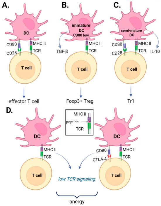
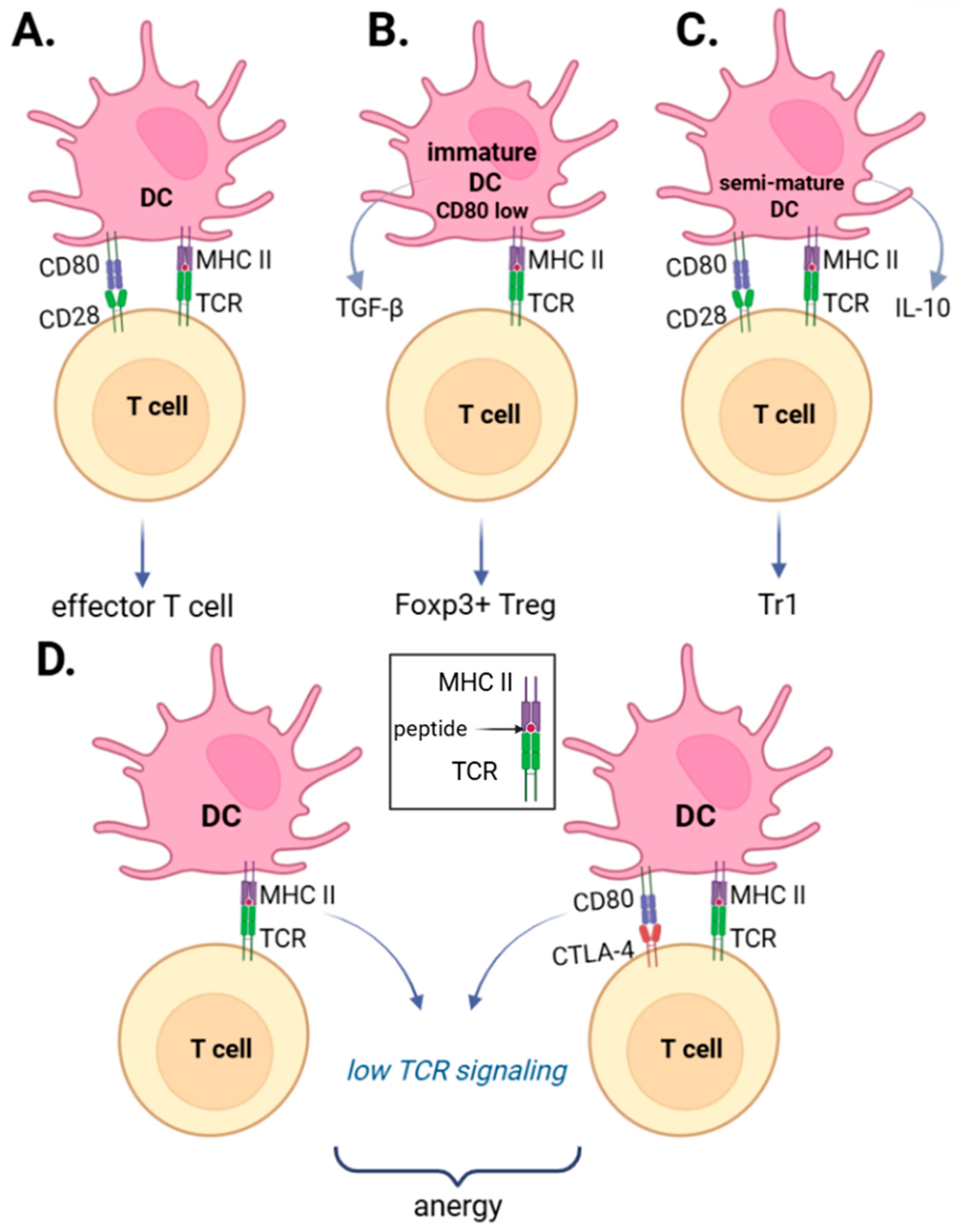
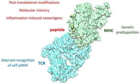
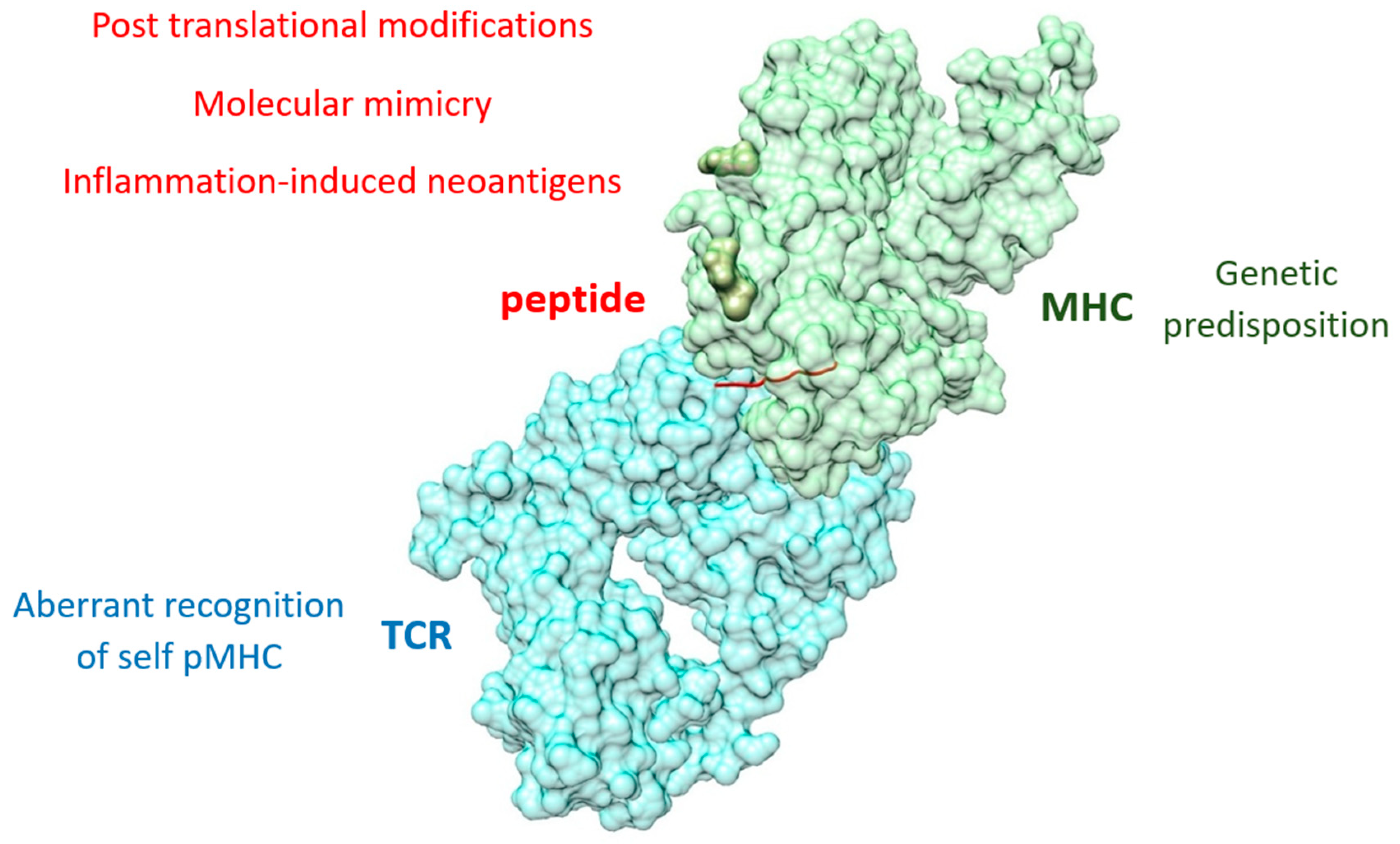
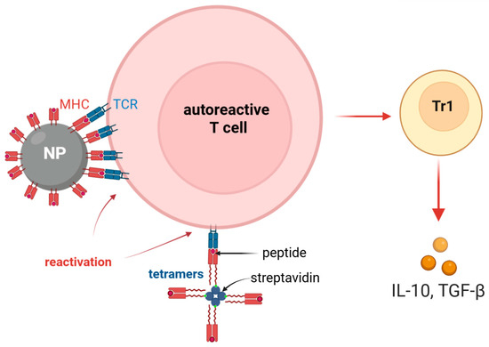
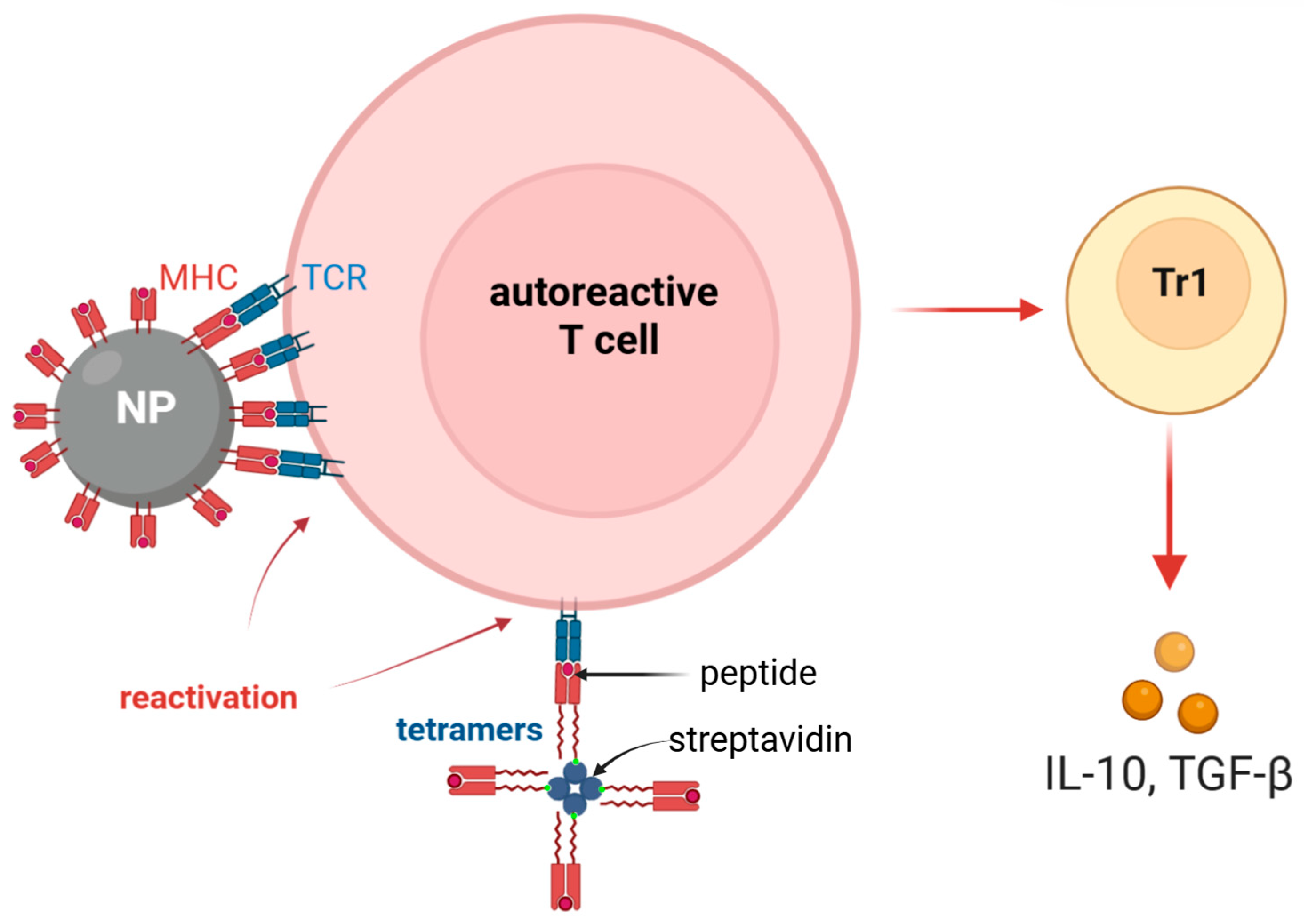
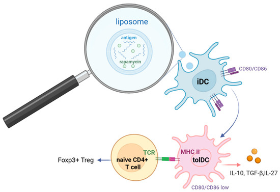
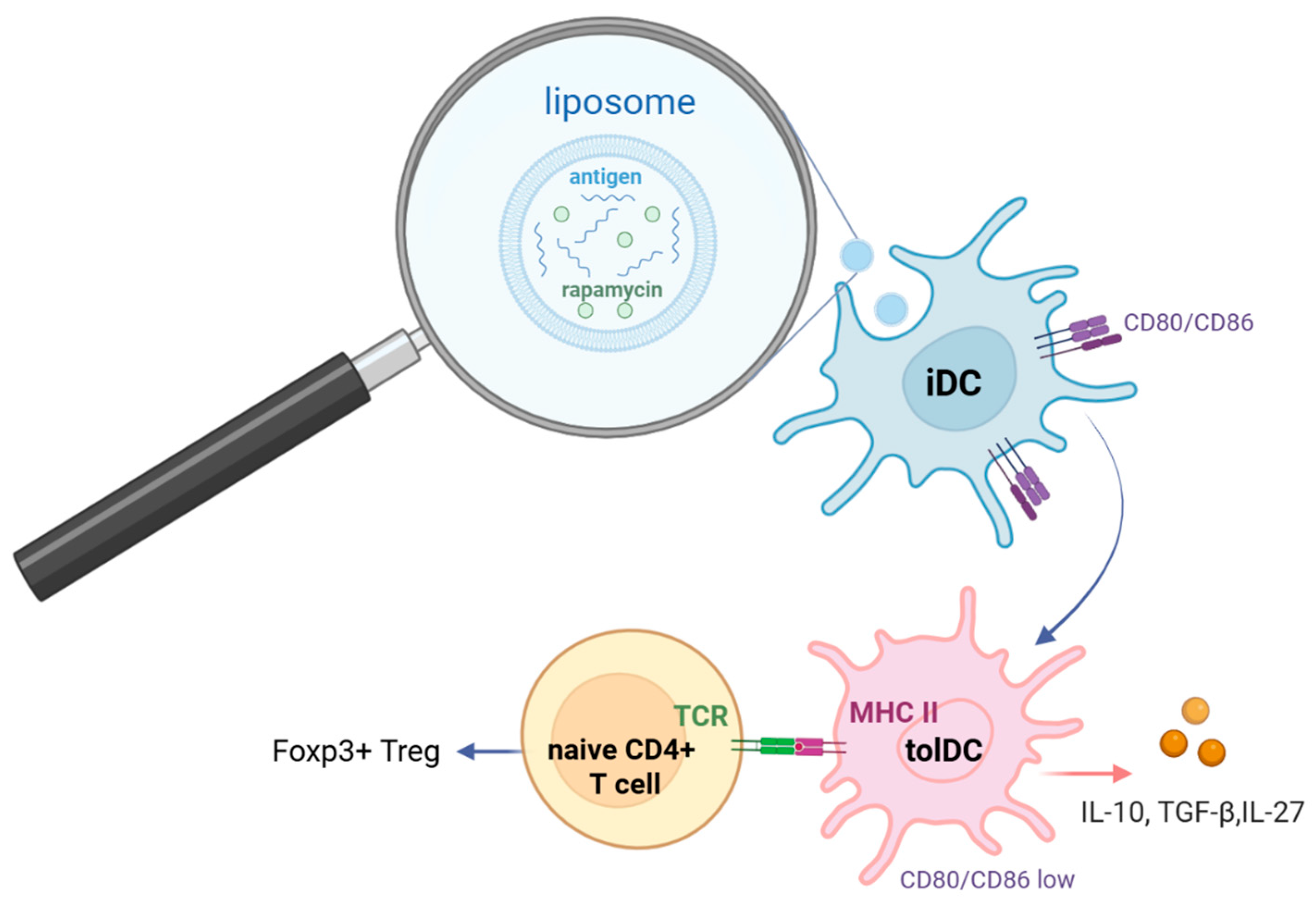
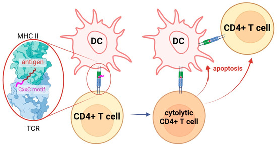
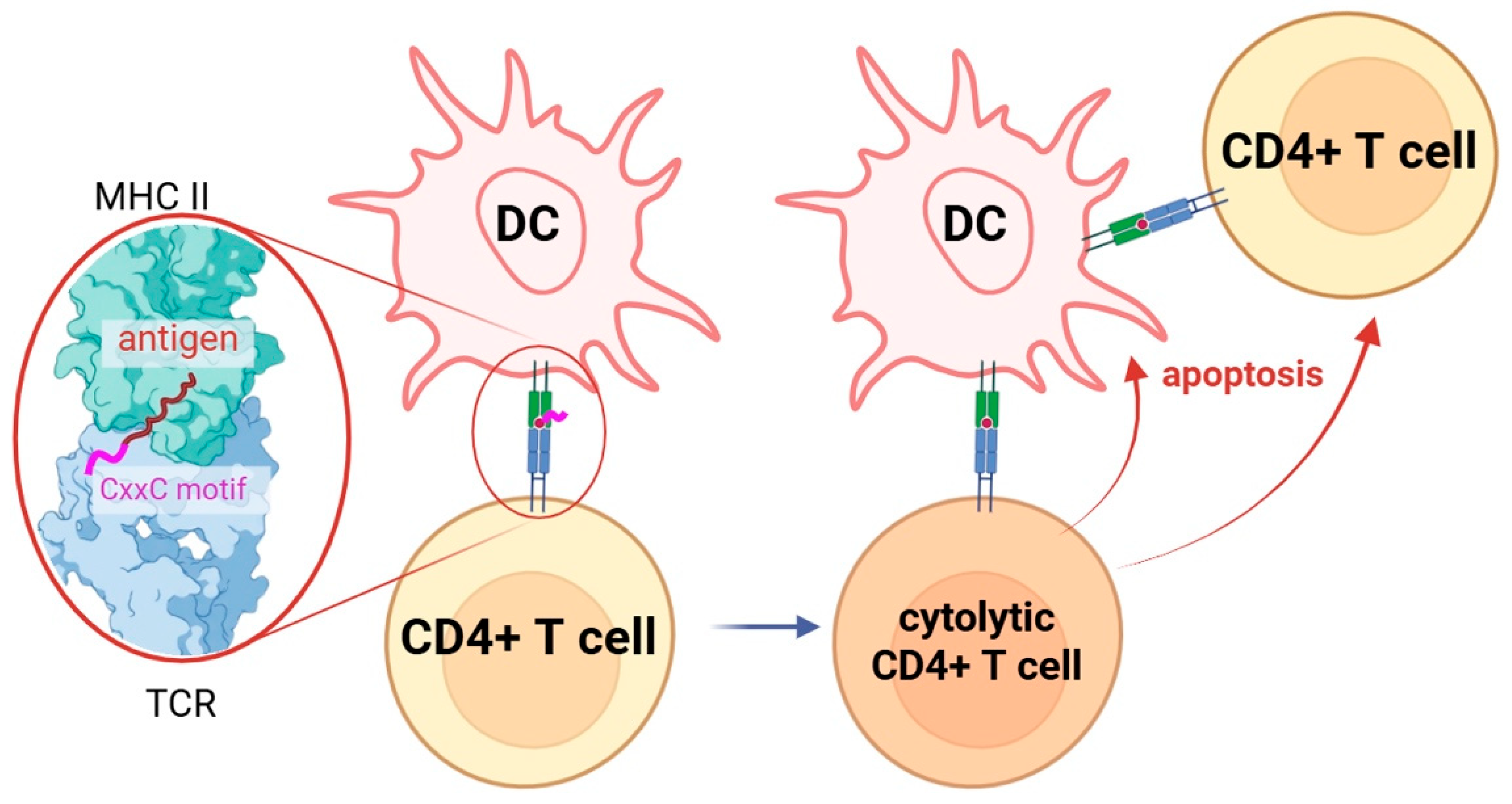

Disclaimer/Publisher’s Note: The statements, opinions and data contained in all publications are solely those of the individual author(s) and contributor(s) and not of MDPI and/or the editor(s). MDPI and/or the editor(s) disclaim responsibility for any injury to people or property resulting from any ideas, methods, instructions or products referred to in the content. |
© 2026 by the authors. Licensee MDPI, Basel, Switzerland. This article is an open access article distributed under the terms and conditions of the Creative Commons Attribution (CC BY) license.