Short Note

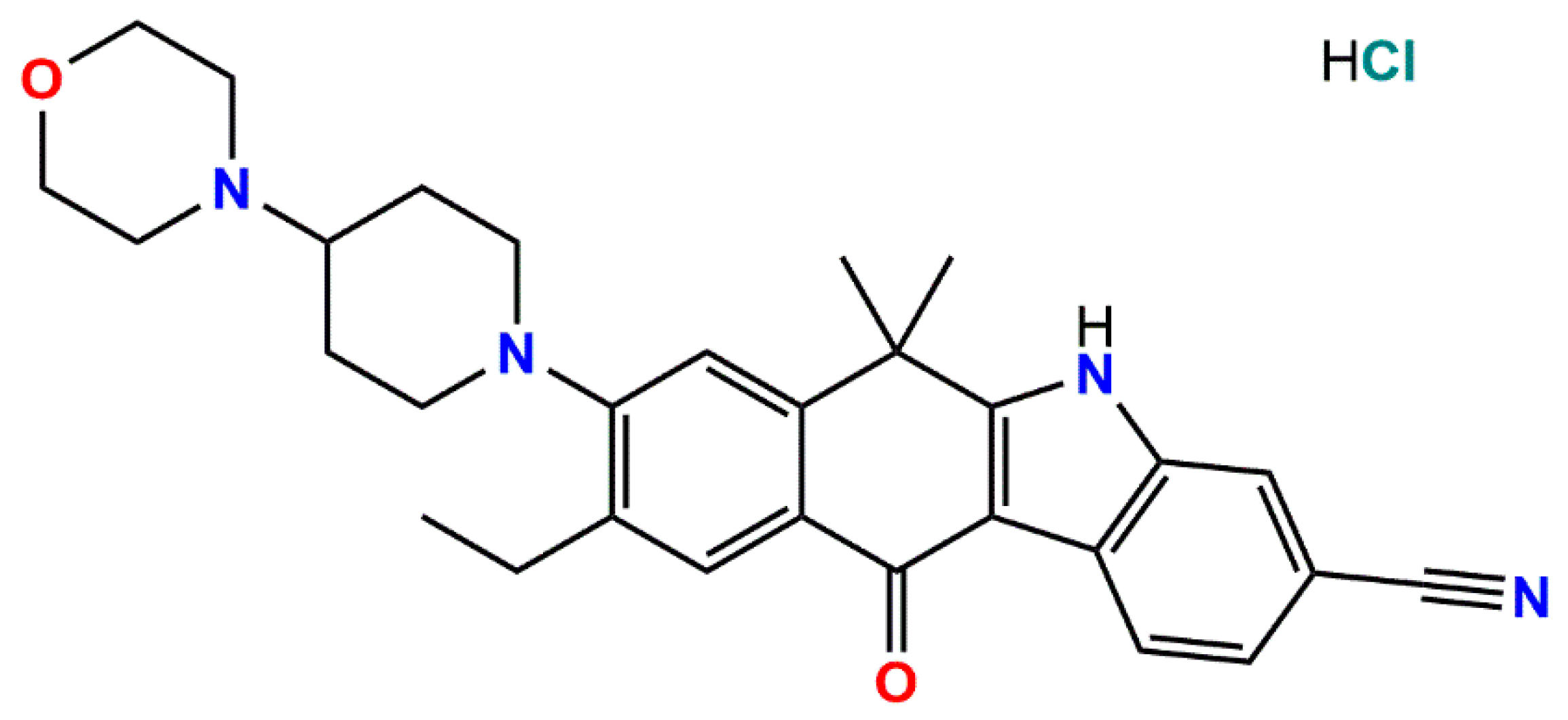
9-Ethyl-6,6-dimethyl-8-[4-(morpholin-4-yl)piperidin-1-yl]-11-oxo-6,11-dihydro-5H-benzo[b]carbazole-3-carbonitrile Hydrochloride

by
Petr A. Buikin
1,2,
Anna V. Vologzhanina
1,
Roman A. Novikov
3 and
Alexander A. Korlyukov
1,*
1
A. N. Nesmeyanov Institute of Organoelement Compounds, Russian Academy of Sciences, 28 Vavilov St., 119334 Moscow, Russia
2
N. S. Kurnakov Institute of General and Inorganic Chemistry, Russian Academy of Sciences, 31 Leninskii prosp., 119991 Moscow, Russia
3
N. D. Zelinsky Institute of Organic Chemistry, Russian Academy of Ssciences, 47 Leninsky prosp., 119991 Moscow, Russia
*
Author to whom correspondence should be addressed.
Molbank 2024, 2024(1), M1759; https://doi.org/10.3390/M1759
Submission received: 29 November 2023 / Revised: 25 December 2023 / Accepted: 28 December 2023 / Published: 5 January 2024
(This article belongs to the Section Structure Determination)

Abstract

:
Alectinib hydrochloride is an anticancer medication used for the first-line treatment of non-small cell lung cancer. Although it was approved for medical use ten years ago, and three polymorphs of this substance were proposed based on X-ray diffraction patterns, their crystal structures remained unknown to date. The main problem was the preparation of high quality single crystals due to the very low solubility of the salt. Herein, we report on the molecular and crystal structure of form I of alectinib hydrochloride as obtained using powder X-ray diffraction data from a laboratory source. Short Cl…N distances between the anion and the nitrogen atoms of the morpholine and benzo[b]carbazole rings indicate the positions of the H(N) atoms. As a result, the cation and anion form infinite Cl…H(N)-bonded chains.

1. Introduction

Alectinib (brand name Alecensa, Drug Bank No DB11363) is an inhibitor of anaplastic lymphoma kinase activity [1]. In 2014, it was approved for the treatment of non-small cell lung cancer in Japan and it is now used worldwide. It has the systematic name 9-ethyl-6,6-dimethyl-8-[4-(morpholin-4-yl)piperidin-1-yl]-11-oxo-6,11-dihydro-5H-benzo[b]carbazole-3-carbonitrile. Its solid form, which is used in medications, is alectinib hydrochloride for which three solid forms have been detected based on powder X-ray diffraction (XRD) data [2], although none of these patterns have been indexed and the corresponding molecular and crystal structures remain unknown. Unfortunately, this is typical of a significant fraction of pharmaceuticals, despite the pharmaceutical industry’s need for high-quality reference powder patterns for phase identification and purity control. However, recent progress in algorithms for structure solutions and in refining from powder XRD data has significantly increased the number of crystal structures of multi-component API solids obtained from powder XRD (see, for example, besifloxacin hydrochloride [3], butenafine hydrochloride [4], meglumine diatrizoate [5] and danofloxacin mesylate [6]).
Within our study of the crystal and molecular structures of the solid forms of active pharmaceutical ingredients [7,8,9], the powder pattern of alectinib hydrochloride was measured using CuKα radiation. Room temperature was maintained to overcome the effects of thermal contraction and to avoid possible phase transitions. This corresponded to the previously patented type I crystals of alectinib hydrochloride ((C30H35N4O2)Cl, 1) [2]. The pattern was indexed and the crystal structure of 1 was solved and refined. The two-dimensional molecular diagram of this compound is shown in Scheme 1.

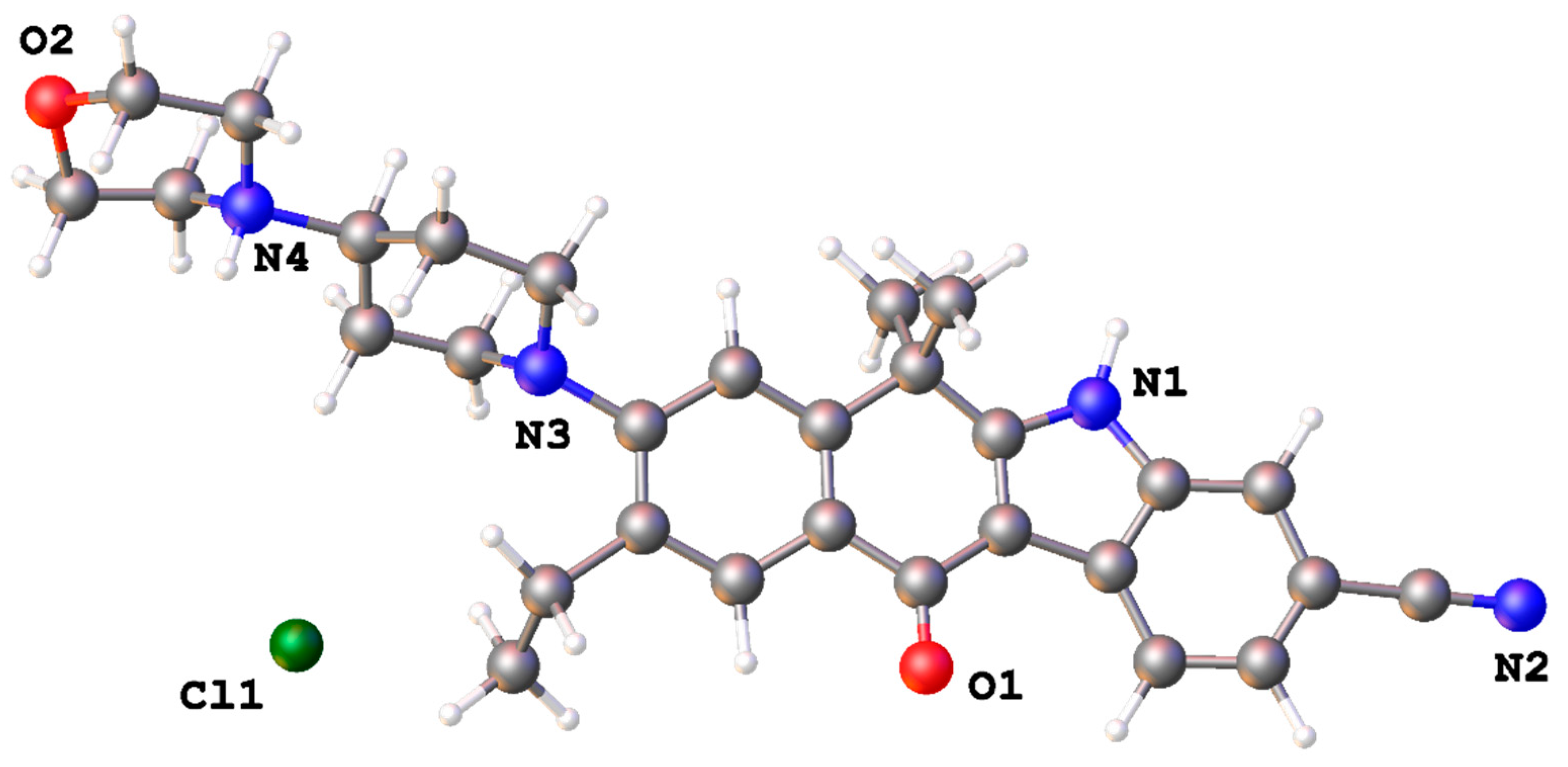
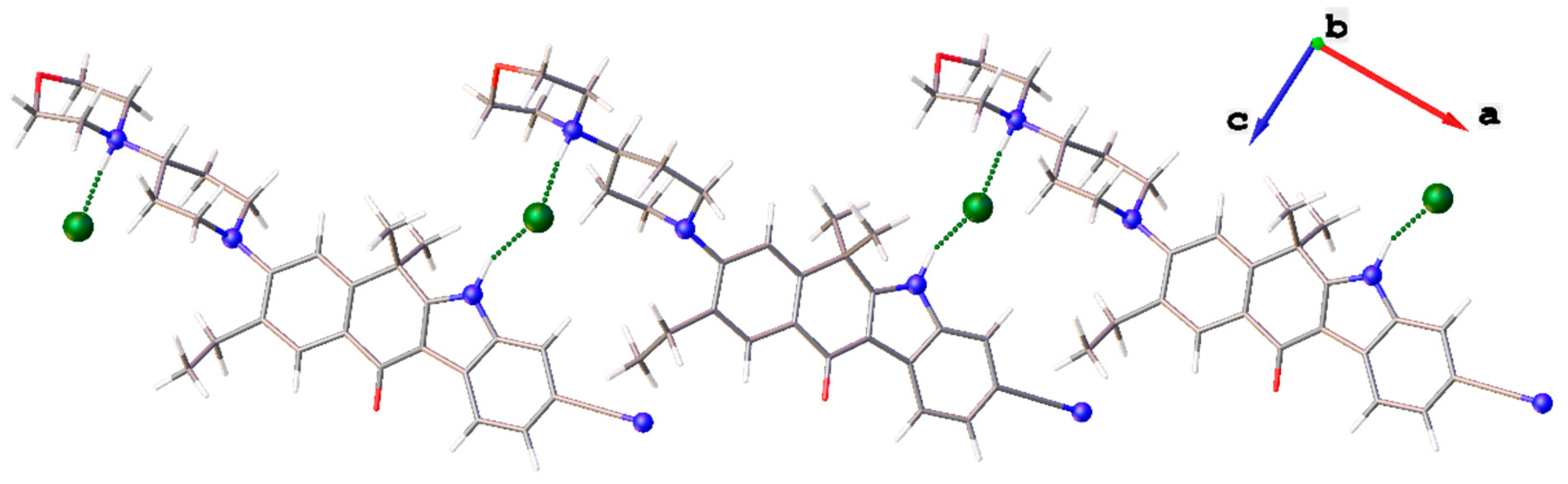
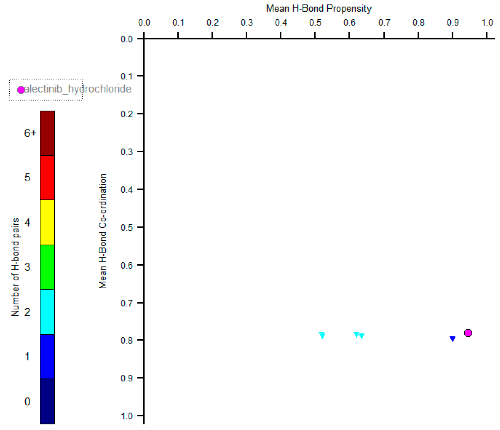
2. Results and Discussion

A sample of alectinib hydrochloride was purchased from Cdymax and used without purification and recrystallization. The sample was characterized using powder X-ray diffraction, FTIR, 1H, 13C and 13C-1H HSQC NMR spectra (see Figures S1–S5, Supporting Information).
The powder pattern was obtained at room temperature; thus, it can be used in industry for phase identification (Figure S5). It was indexed using Topas 5.0 package [10], which indicates the sample purity. The systematic absences suggested the space group P21/c, which was confirmed by the successful solution and refinement of the structure. A simulated annealing algorithm of Topas 5.0 was applied to find the positions of the non-hydrogen atoms of alectinib and an independent chlorine atom in an asymmetric unit. The Rietveld refinement was carried out to refine the coordinates of all atoms [11]. The asymmetric unit of 1 is depicted in Figure 1. It contains one alectinib molecule and an anion. The morpholine and piperidine rings are in the chair conformation. The condensed cycles are nearly coplanar; the average deviation of the atoms, with an exception of the (CH3)2C fragment, is equal to 0.07(8) Å. The C15 atom of (CH3)2C is situated 0.040(4) Å above the plane of its six-membered ring. Although the positions of the hydrogen atoms cannot be located during the refinement of powder XRD data, the most likely system of H-bonds corresponds to location of the additional H(N) proton on the nitrogen atom of the morpholinyl cycle.
Both acidic H(N) atoms of the five-membered cycle and morpholinyl cycle take part in N–H⋯Cl interactions. The parameters of the H-bonds are listed in Table 1. As both a cation and anion take part in the two hydrogen bonds, infinite H-bonded chains are observed in solid 1. The chains are depicted in Figure 2. Patent [2] contains information about the different polymorphs of alectinib hydrochloride; thus, it is of interest to reveal if type I crystals correspond to a stable or metastable polymorph. The crystal structures of other polymorphs remain unknown; however, the H-bond propensity tool implemented in the Mercury package [12] can be used to estimate if the most likely hydrogen bonds are present in a solid [13,14]. Within this method, it is assumed that the most stable polymorph also contains the most likely hydrogen bonds. Stable polymorphs are characterized by points in the right bottom corner of the H-bond Coordination/H-bond Propensity Plot. In accordance with such calculations, the experimentally observed N–H⋯Cl interactions are more likely than any of the theoretically possible N–H⋯O or N–H⋯N bonds. All donors of H-bonds take part in H-bonding and the solid is characterized by the point in the right bottom corner of the plot (Figure 3). Thus, based on this approach, the system of H-bonds in 1 is the most likely, and the structure is expected to be the most stable polymorph.
The Rietveld plot of 1, as depicted in Figure S5, and its convergence factors demonstrate that some experimental data are insufficiently described by the obtained model. This is probably an effect of the preferred orientation. Thus, we used the HUW (half uncertainty window [15]) parameter in order to estimate the quality of our model. The HUW parameter identifies the range where the restraints in the model can be varied without the refined bond lengths deviating from the target values in a statistically significant way. For highly crystalline powders and synchrotron data, the HUW can be lower than 0.05 Å, while unacceptable models are characterized by a HUW > 0.3 Å. For our model and data, HUW = 0.091 Å. This is indicative of good refinement.
To sum up, the first crystal and molecular structure of a solid containing alectinib was obtained. This corresponds to the type I crystals patented in 2017. The model was obtained via the Rietveld refinement of powder XRD data taken from a laboratory diffractometer at room temperature. Nevertheless, the HUW parameter indicates the good quality of the refinement.

3. Materials and Methods

Fine powder of 1 was obtained from Jiangsu Cdymax Pharmaceuticals Co., Ltd. (Nantong, China) and used without further purification. NMR spectra in dimethyl sulfoxide solution were obtained for 1H at 400 MHz and for 13C at 100 MHz using Bruker AVANCE III WB 400 spectrometer (Bruker, Billerica, MA, USA). The FTIR spectrum was recorded on an IR spectrometer with a Fourier transformer Shimadzu IRTracer100 (Kyoto, Japan) in the range of 4000–600 cm−1 at a resolution of 1 cm−1 (Nujol mull, KBr pellets). The powder XRD data were recorded using a Bruker D8 Advance diffractometer (Bruker, Billerica, MA, USA) equipped with a LynxEye detector and Ge(111) monochromator in transmission mode. CuKα radiation with a wavelength of 1.544493 Å was used. The 2θ range was 3.50–90.0° with a step size of 0.1431°.

X-ray Diffraction

The indexing of the powder pattern and the subsequent structure solution was performed with the Topas 5.0 software [10]. The pattern was indexed in the P-centred monoclinic unit cell. The systematic absences suggested the space group P21/c, which was confirmed by the successful solution and refinement of the structure. A model of alectinib was taken from PubChem [16]. A simulated annealing algorithm of the Topas 5.0 was applied to find positions of non-hydrogen atoms of alectinib and an independent chlorine atom in an asymmetric unit. The solution result was used as the starting geometry for the periodic DFT calculations at the PBE exchange–correlation functional level with a fixed unit cell using VASP 5.4.1 [17,18,19]. Atomic cores were described using PAW potentials [20,21]. Valence electrons were described in terms of a plane-wave basis set.
The optimization result with the fixed unit cell was used as the starting geometry and the sources of bond and angle restraints in the Rietveld refinement. Anisotropic displacement parameters were refined as equal for all of carbon atoms, all nitrogen, all oxygen, all hydrogen atoms and a chlorine atom. The positions of the hydrogen atoms were calculated geometrically and refined in the riding model. The Rietveld plot recorded is given in Figure S5 of the Supplementary Materials.
Crystal Data for C30H35ClN4O2 (M = 519.06 g/mol): monoclinic, space group P21/n (no. 14), a = 20.3873(10), b = 10.4405(4), c = 12.6827(5) Å, α = 90, β = 93.105(2), γ = 90°, V = 2698.2(2) Å3, Z = 4, µ = 1.521 mm−1, Dcalc = 1.278 g cm−3, F(000) = 1104, 3.5° ≤ 2Θ ≤ 90.0° range was used in all calculations. The final Rp/RWP/Rwp′/RBragg/GOF were 3.60/4.69/1.37/1.72/3.43.

Supplementary Materials

The following supporting information can be downloaded online, NMR and FTIR spectra, Rietveld plot, and crystallographic data in Crystallographic Information File (CIF) format.

Author Contributions

Conceptualization, A.A.K.; methodology, P.A.B.; investigation, R.A.N. and P.A.B.; writing, A.A.K. and A.V.V.; funding acquisition, A.V.V. All authors have read and agreed to the published version of the manuscript.

Funding

This research was funded by the Russian Science Foundation, grant number 23-73-00027.

Data Availability Statement

The X-ray data are available at CCDC under ref. code CCDC 2310527.

Acknowledgments

We acknowledge the Ministry of Science and Higher Education of the Russian Federation for providing access to the scientific literature. NMR experiments were performed using the NMR facility at the Department of Structural Studies of the N. D. Zelinsky Institute of Organic Chemistry of the RAS, Moscow.

Conflicts of Interest

The authors declare no conflicts of interest.

References

  1. McKeage, K. Alectinib: A Review of Its Use in Advanced ALK-Rearranged Non-Small Cell Lung Cancer. Drugs 2015, 75, 75–82. [Google Scholar] [CrossRef] [PubMed]
  2. Tanaka, K.; Ueto, T. Crystal of Tetracyclic Compound. Patent US9714229B2, 25 July 2017. Available online: https://patents.google.com/patent/US9714229B2/en?oq=alectinib++WO2015163447 (accessed on 21 December 2023).
  3. Kaduk, J.A.; Gates-Rector, S.; Blanton, T.N. Crystal Structure of Besifloxacin Hydrochloride, C19H22ClFN3O3Cl. Powder Diffr. 2023, 38, 43–52. [Google Scholar] [CrossRef]
  4. Kaduk, J.A.; Gates-Rector, S.; Blanton, T.N. Crystal Structure of Butenafine Hydrochloride, C23H28NCl. Powder Diffr. 2023, 38, 30–36. [Google Scholar] [CrossRef]
  5. Ens, T.M.; Kaduk, J.A.; Dosen, A.V.; Blanton, T.N. Crystal Structure of Meglumine Diatrizoate, (C7H18NO5)(C11H8I3N2O4). Powder Diffr. 2023, 38, 185–193. [Google Scholar] [CrossRef]
  6. Ens, T.M.; Kaduk, J.A.; Dosen, A.; Blanton, T.N. Crystal Structure of Danofloxacin Mesylate (C19H21FN3O3)(CH3O3S). Powder Diffr. 2023, 38, 194–200. [Google Scholar] [CrossRef]
  7. Korlyukov, A.A.; Buikin, P.A.; Dorovatovskii, P.V.; Vologzhanina, A.V. Synthesis, NoSpherA2 Refinement, and Noncovalent Bonding of Abiraterone Bromide Monohydrate. Struct. Chem. 2023, 34, 1927–1934. [Google Scholar] [CrossRef]
  8. Korlyukov, A.A.; Dorovatovskii, P.V.; Vologzhanina, A.V. N-(4-Methyl-3-((4-(Pyridin-3-Yl)Pyrimidin-2-Yl)Amino)Phenyl)-4-((4-Methylpiperazin-1-Yl)Methyl)Benzamide. Molbank 2022, 2022, M1461. [Google Scholar] [CrossRef]
  9. Volodin, A.D.; Vologzhanina, A.V.; Peresypkina, E.V.; Korlyukov, A.A. Conformational Polymorphism of Solidum Salt of Elsulfavirin. J. Struct. Chem. 2024, 65, 123238. [Google Scholar] [CrossRef]
  10. Coelho, A.A. TOPAS and TOPAS-Academic: An Optimization Program Integrating Computer Algebra and Crystallographic Objects Written in C++. J. Appl. Cryst. 2018, 51, 210–218. [Google Scholar] [CrossRef]
  11. Rietveld, H.M. A Profile Refinement Method for Nuclear and Magnetic Structures. J. Appl. Cryst. 1969, 2, 65–71. [Google Scholar] [CrossRef]
  12. Macrae, C.F.; Sovago, I.; Cottrell, S.J.; Galek, P.T.A.; McCabe, P.; Pidcock, E.; Platings, M.; Shields, G.P.; Stevens, J.S.; Towler, M.; et al. Mercury 4.0: From Visualization to Analysis, Design and Prediction. J. Appl. Cryst. 2020, 53, 226–235. [Google Scholar] [CrossRef] [PubMed]
  13. Galek, P.T.A.; Allen, F.H.; Fábián, L.; Feeder, N. Knowledge-Based H-Bond Prediction to Aid Experimental Polymorph Screening. CrystEngComm 2009, 11, 2634–2639. [Google Scholar] [CrossRef]
  14. Delori, A.; Galek, P.T.A.; Pidcock, E.; Jones, W. Quantifying Homo- and Heteromolecular Hydrogen Bonds as a Guide for Adduct Formation. Chem. Eur. J. 2012, 18, 6835–6846. [Google Scholar] [CrossRef] [PubMed]
  15. Dmitrienko, A.O.; Bushmarinov, I.S. Reliable Structural Data from Rietveld Refinements via Restraint Consistency. J. Appl. Cryst. 2015, 48, 1777–1784. [Google Scholar] [CrossRef]
  16. PubChem Alectinib. Available online: https://pubchem.ncbi.nlm.nih.gov/compound/49806720 (accessed on 29 November 2023).
  17. Kresse, G.; Hafner, J. Ab Initio Molecular Dynamics for Liquid Metals. Phys. Rev. B 1993, 47, 558–561. [Google Scholar] [CrossRef] [PubMed]
  18. Kresse, G.; Hafner, J. Ab Initio Molecular-Dynamics Simulation of the Liquid-Metal--Amorphous-Semiconductor Transition in Germanium. Phys. Rev. B 1994, 49, 14251–14269. [Google Scholar] [CrossRef] [PubMed]
  19. Kresse, G.; Furthmüller, J. Efficient Iterative Schemes for Ab Initio Total-Energy Calculations Using a Plane-Wave Basis Set. Phys. Rev. B 1996, 54, 11169–11186. [Google Scholar] [CrossRef] [PubMed]
  20. Kresse, G.; Furthmüller, J. Efficiency of Ab-Initio Total Energy Calculations for Metals and Semiconductors Using a Plane-Wave Basis Set. Compt. Mat. Sci. 1996, 6, 15–50. [Google Scholar] [CrossRef]
  21. Blöchl, P.E. Projector Augmented-Wave Method. Phys. Rev. B 1994, 50, 17953–17979. [Google Scholar] [CrossRef] [PubMed]
Scheme 1. Schematic representation of alectinib hydrochloride.
Molbank 2024 m1759 sch001
Figure 1. Asymmetric unit of 1.
Molbank 2024 m1759 g001
Figure 2. H-bonded dimers in 1.
Molbank 2024 m1759 g002
Figure 3. H-bond Propensity/Coordination plot for theoretically possible systems of H-bonds in 1. Fuchsia circle denotes experimentally observed data.
Molbank 2024 m1759 g003
Table 1. Parameters of H-bonds in 1 (Å, °).
D–H…AD–H, ÅH…A, ÅD…A, Å∠ (DHA), °
N1–H1⋯Cl1 i1.042.203.107(7)145
N4–H4⋯Cl1 ii0.902.032.918(6)170
Symmetry codes: (i) 1 − x, 1 − y, 1 − z; (ii) 1/2 − x, 1/2 + y, 3/2 − z.
Disclaimer/Publisher’s Note: The statements, opinions and data contained in all publications are solely those of the individual author(s) and contributor(s) and not of MDPI and/or the editor(s). MDPI and/or the editor(s) disclaim responsibility for any injury to people or property resulting from any ideas, methods, instructions or products referred to in the content.

