Article

Characterization of a Pseudokeronopsis Strain (Ciliophora, Urostylida) and Its Bacterial Endosymbiont “Candidatus Trichorickettsia” (Alphaproteobacteria, Rickettsiales)

1
Department of Biology and Biotechnology, University of Pavia, 27100 Pavia, Italy
2
Department of Biology, University of Pisa, 56126 Pisa, Italy
3
Centro de Ciências Naturais e Humanas, Universidade Federal do ABC, Santo Andre 09210-580, Brazil
4
CIME, Centro Interdipartimentale di Microscopia Elettronica, University of Pisa, 56100 Pisa, Italy
5
CISUP, Centro per l’Integrazione Della Strumentazione, University of Pisa, 56100 Pisa, Italy
*
Authors to whom correspondence should be addressed.
These authors contributed equally to this work.
Diversity 2022, 14(12), 1032; https://doi.org/10.3390/d14121032
Submission received: 21 August 2022 / Revised: 4 November 2022 / Accepted: 23 November 2022 / Published: 25 November 2022
(This article belongs to the Special Issue Integrated Taxonomy of Protists: Morphology, Genes and Symbionts)

Abstract

:
Symbiotic associations between bacteria and ciliate protists are rather common. In particular, several cases were reported involving bacteria of the alphaproteobacterial lineage Rickettsiales, but the diversity, features, and interactions in these associations are still poorly understood. In this work, we characterized a novel ciliate protist strain originating from Brazil and its associated Rickettsiales endosymbiont by means of live and ultrastructural observations, as well as molecular phylogeny. Though with few morphological peculiarities, the ciliate was found to be phylogenetically affiliated with Pseudokeronopsis erythrina, a euryhaline species, which is consistent with its origin from a lagoon with significant spatial and seasonal salinity variations. The bacterial symbiont was assigned to “Candidatus Trichorickettsia mobilis subsp. hyperinfectiva”, being the first documented case of a Rickettsiales associated with urostylid ciliates. It resided in the host cytoplasm and bore flagella, similarly to many, but not all, conspecifics in other host species. These findings highlight the ability of “Candidatus Trichorickettsia” to infect multiple distinct host species and underline the importance of further studies on this system, in particular on flagella and their regulation, from a functional and also an evolutionary perspective, considering the phylogenetic proximity with the well-studied and non-flagellated Rickettsia.

1. Introduction

Protists and bacteria display a wide range of relationships, and may form complex microbial communities, with variable levels of interaction and integration [1,2]. Such associations may play important roles in the ecology and evolution of the involved bacteria [3], as well as on their hosts. Indeed, as underlined in a recent work in which bacterial endosymbionts were treated as taxonomic descriptors for a ciliate species of the genus Euplotes, symbionts can deeply influence their hosts in many aspects, even in their resulting morphology [4].
In general, protists harbour abundant and diverse bacterial endosymbionts (i.e., intracellular bacteria), including several representatives of lineages that also encompass human pathogens, such as Rickettsiales, Legionellales, and Chlamydiae [5,6,7]. Some protists are even able to host, at least temporarily, human pathogenic bacteria such as Legionella [8,9], thus potentially representing natural reservoirs. Those features have led several authors to infer a role of protists as melting pots for the evolution of potentially pathogenic bacteria, able to invade multicellular eukaryotes, hence potentially acting as Trojan horses [10,11,12]. Several studies provided relevant contributions in this sense, investigating the mechanisms and dynamics of establishment of intracellular symbioses in protists such as ciliates [13,14,15,16].
In particular, multiple representatives of Rickettsiales are hosted by ciliate protists. Among those, the cases of “Candidatus (Ca.) Trichorickettsia mobilis” and “Ca. Gigarickettsia flagellata” are noteworthy. “Ca. Trichorickettsia” was found in association with multiple strains of several ciliate species (Paramecium multimicronucleatum, P. nephridiatum, P. calkinsi, Euplotes aediculatus) [17,18,19], while “Ca. Gigarickettsia” only in a single population of Spirostomum minus [17]. These two symbiont species are sister groups, and, in turn, together form the sister group of the lineage of Rickettsia and the recently described genus “Ca. Tisiphia” [20]. Genus Rickettsia in particular includes many pathogens vectored by hematophagous arthropods, causing various diseases in humans and other vertebrates, such as endemic typhus, epidemic typhus, and Rocky Mountain spotted fever [21].
Until recently, as still reported in the description in Bergey’s Manual of Systematics of Archaea and Bacteria [22], all Rickettsiales were considered devoid of flagella. However, in the last decade, unexpectedly, several Rickettsiales were found to harbour flagella [23], including “Ca. Trichorickettsia” and “Ca. Gigarickettsia” [17,19], or flagellar genes [20]. Flagella likely date back to the Rickettsiales ancestor [24,25], but their role in strictly obligate intracellular symbionts, such as the vast majority of characterised Rickettsiales (though with at least one significant exception [26]) or even Chlamydiae [6], still needs to be elucidated. Multiple non-mutually exclusive functions have been hypothesised, either as actual motile organelles [5,24,27], possibly during horizontal transmission, or as mediators of host-symbiont interactions [23,24,28]
Among “flagellated” Rickettsiales, the case of “Ca. Trichorickettsia” and “Ca. Gigarickettsia” is peculiar, as, to our best knowledge, they are the only Rickettsiales bacteria for which flagellar-driven motility was actually observed [17]. Moreover, “Ca. Trichorickettsia mobilis” presents intraspecific variability for the presence of flagella, which resulted up to now quite consistent with genetic diversity [17,19].
In this work, we report the characterisation of a novel “Ca. Trichorickettsia” representative, discovered as endosymbiont of the hypotrich ciliate Pseudokeronopsis (Spirotrichea, Urostylida). This finding provides further information for understanding the intraspecific phenotypic variability of “Ca. Trickorickettsia” regarding host species specificity and flagellar repertoire. Additionally, the ciliate host itself is noteworthy and, according to morphological and molecular analyses, it likely represents a novel record of an euryhaline [29] Pseudokeronopsis species, i.e., Pseudokeronopsis erythrina, though with few distinctive morphological features, and is also the first member of urostylid ciliates recorded so far for hosting a Rickettsiales bacterium.

2. Materials and Methods

Pseudokeronopsis sp. strain PSqRJ01 was isolated from a water sample collected in Jacarepiá lagoon, located in Saquarema city, 107 km east from Rio de Janeiro, Brazil (22°54′53″ S, 42°25′43″ W). The strain was maintained in the laboratory inside an incubator at 19 ± 1 °C, with a 12 h light and a 12 h dark. It was fed as previously described [30] on the diatom Phaeodactylum tricornutum, in turn propagated as a monoclonal culture at 5‰ salinity.
Cells of Pseudokeronopsis sp. PSqRJ01 were examined in vivo for their behaviour and their morphological features under a stereomicroscope (WILD HEERBRUGG, Switzerland) and a Leitz Orthoplan (Weitzlar, Germany) Differential Interference Contrast (DIC) microscope equipped with a digital camera (Canon PowerShot S45), at a magnification of 10–50× and 100–1250×, respectively.
Approximately 100 cells were fixed in 70% ethanol, and DNA extraction was performed employing the NucleoSpinTM Plant II kit (Macherey-Nagel, Düren, Germany).
The 18S rRNA gene of Pseudokeronopsis sp. PSqRJ01 was amplified and sequenced with universal eukaryotic primers (18S F9 Euk—[31]—18S R1513 Hypo—[32]), as previously described [19]. The internal transcribed spacer (ITS) was amplified with primers 18S F919 [33] and RGD2 [34] as previously described [19].
Based on preliminary fluorescence microscopy observations, the 16S rRNA gene of the bacterial symbiont of Pseudokeronopsis sp. PSqRJ01 was amplified with primers RickFla_F69 and Rick_R1455, as previously described [17], and sequenced with internal primers [35].
Pseudokeronopsis sp. PSqRJ01 cells were washed in sterile water prior to fixation in 4% (v/v) formaldehyde (in PBS 1×) on microscope slides. The protocol by Manz and co-authors [36] was followed for the subsequent hybridisation, using multiple combinations of probes with different specificities, in particular the probe EUB338 [37] (5′-GCTGCCTCCCGTAGGAGT-3′), targeting over 90% of domain Bacteria, the ALF1b (5′-CGTTCGYTCTGAGCCAG-3′) [36], targeting Alphaproteobacteria, and the “Candidatus Trichorickettsia”-specific probe TrichoRick_142 (5′-GTTTCCAAATGTTATTCCATAC-3′) [17]. Fixed cells were simultaneously investigated under UV-light after staining with DAPI dye.
Pseudokeronopsis sp. PSqRJ01 cells were fixed in 2.5% glutaraldehyde in 0.1 M cacodylate buffer for 45 min, briefly rinsed in the same buffer, and post-fixed in 1.5% aqueous osmium tetroxide in distilled water for 45 min at room temperature. Then, cells were dehydrated and processed as elsewhere described [38]. Briefly, cells were embedded in an Epon-araldite mixture and the obtained blocks sectioned with an RMC PowerTome X ultra-microtome; sections were placed on copper grids and stained with uranyl acetate and lead citrate. Samples were observed using a JEM-100SX-TEM and a JEM-F200 HR FEG-TEM (both JEOL Ltd., Tokyo, Japan).
The 18S rRNA gene sequence of Pseudokeronopsis sp. PSqRJ01 was aligned with the automatic aligner of the ARB software package version 5.5 [39] on the SSU ref NR99 SILVA database [40], manually updated with latest released sequences.
For the phylogenetic analysis, 32 18S rDNA sequences belonging to representatives of the genus Pseudokeronopsis (or closely related lineages) were selected for the ingroup, and nine sequences belonging to other urostylids were selected for the outgroup, for a total of 41 sequences.
After manual editing to optimise base pairing in the predicted rRNA stem regions, the alignment was trimmed at both ends to the length of the shortest sequence. The resulting matrix contained 1675 nucleotides and was used for phylogenetic reconstructions.
The optimal substitution model was selected with jModelTest 2.1 [41] according to the Akaike Information Criterion (AIC). The maximum likelihood (ML) tree was inferred with PHYML version 2.4 [42] from the ARB package, performing 100 pseudo-replicates. Bayesian inference (BI) tree was inferred with MrBayes 3.2 [43], using three runs, each with one cold and three heated Monte Carlo Markov chains, iterating for 1,000,000 generations with a burn-in of 25%.
For the phylogenetic analysis of the endosymbiont, the 16S rRNA gene of this bacterium and closely related sequences retrieved from NCBI nucleotide were aligned with the automatic aligner of the ARB software package to the SSU ref NR99 123 SILVA database, and the alignment was manually refined to optimise base pairing in the predicted rRNA structure. The final selection for the phylogeny included 32 total organisms, namely eight “Ca. Trichorickettsia mobilis”, twenty other representatives of family Rickettsiaceae, and, as outgroup, four other members of the order Rickettsiales. The alignment of selected sequences was trimmed at both ends to the length of the shortest sequence and keeping all internal positions, resulting in 1360 nucleotide columns. The best substitution model was selected with jModeltest according to the AIC. ML and BI inference (BI) phylogenies were then estimated, respectively, with PHYML, with 1000 pseudo-replicates, and MrBayes, with three runs of three heated and one cold chain iterating for 1,000,000, applying a burn-in of 25%. Identity values within “Ca. Trichorickettsia” and “Ca. Gigarickettsia” were calculated with the same full matrix as phylogeny, and with a modified matrix excluding their typical gene insert [17,19] (1095 total nucleotide sites).

3. Results

3.1. Characterization and Identification of the Host

Pseudokeronopsis sp. PSqRJ01 cells (Figure 1) were 140–240 × 17–37 µm in vivo (mean value: 203 × 23 µm, measured on more than 20 undeformed individuals). A buccal field occupied one third to one fourth of the cell length (Figure 1A). Cells appeared dorsoventrally flattened, elliptical in shape and elongated, with rounded ends, the posterior one sometimes larger than the anterior one; cell body was quite flexible (Figure 1A). A single contractile vacuole was present in the posterior half of the cell (Figure 1A). Numerous spherical pigment granules (coloured in red blood to brick red to brownish; diameter: ~1.7 µm) were present in the cortex (Figure 1). They were especially, but not exclusively, distributed around the different cell ciliature components (i.e., each ciliary unit of adoral zone membranelles, cirri, and dorsal kineties) (Figure 1B–D). Additionally, numerous ellipsoidal colourless granules (~1.5–1.8 × 0.9 µm), were observed densely distributed beneath the cortex and in the cytoplasm (Figure 1C,D). Cells spent most of the time crawling on the substrate
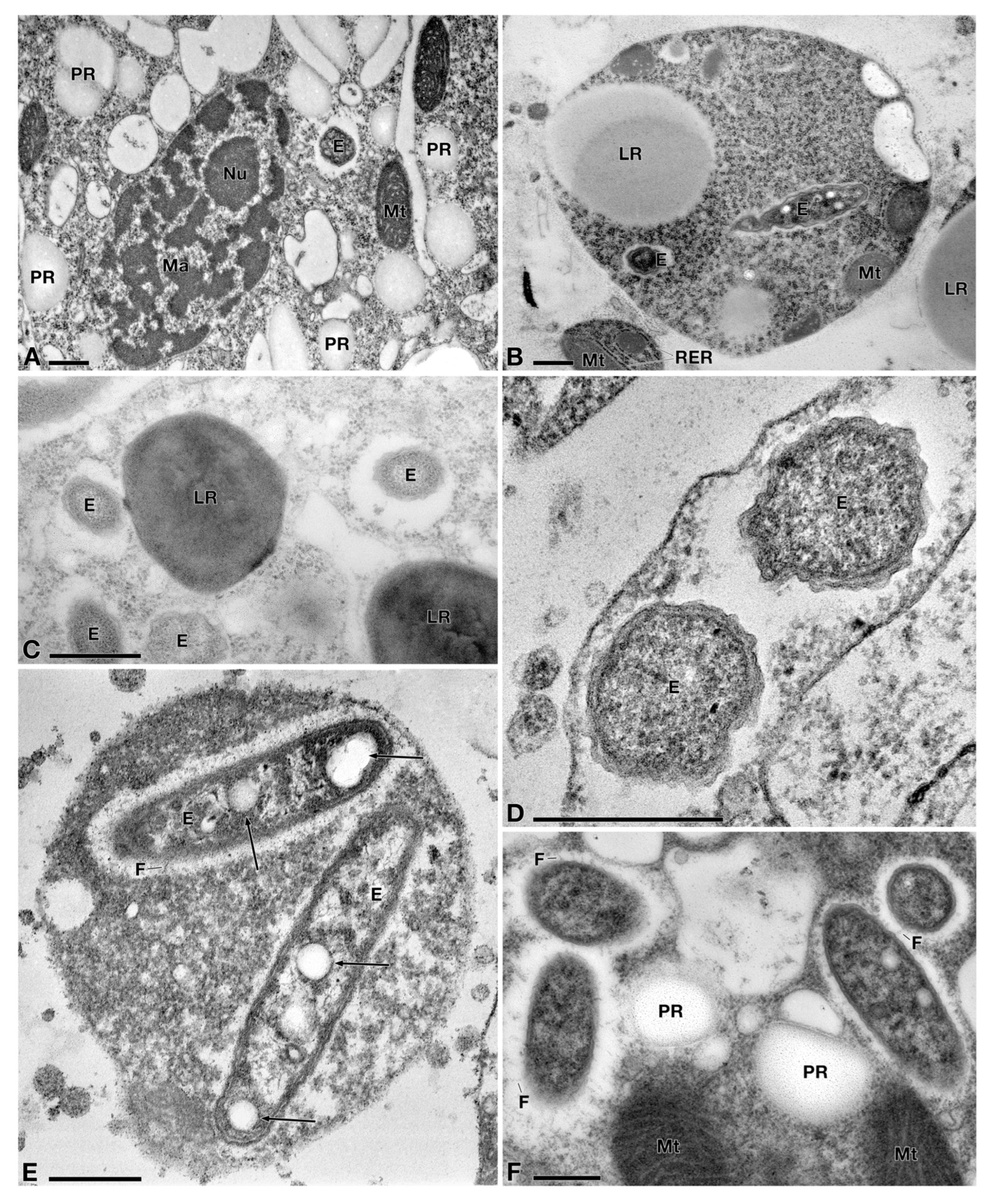
Transmission electron microscopy showed additional features (Figure 2), such as: 1. the structure of the macronuclei (size: ~4 × 2 µm), which showed irregular chromatin bodies and large nucleoli (diameter: ~0.75 µm) (Figure 2A); 2. ovoidal mitochondria (length: ~1–1.5 µm), (Figure 2A,B), sometimes associated with ribosomes (Figure 2B) (likely belonging to the rough endoplasmic reticulum cisternae); 3. medium to large lipid droplets (diameter: ~1.0–2.0 µm) and numerous bodies of polysaccharide reserve (Figure 2A–C); 4. several sparse, endosymbiotic bacteria exhibiting a common single morphotype (see below for details), encircled by a white halo and embedded in the host’s cytoplasm (i.e., not enclosed in any symbiosomal vacuole) (Figure 2).
The combined partial 18S rRNA gene, ITS and partial 28S rRNA gene of Pseudokeronopsis sp. PSqRJ01 were 2842 bp in length. The highest identity (~99.8%, three mismatches and no gaps over 1732 aligned positions) was found with an 18S rRNA gene sequence labelled as Pseudokeronopsis erythrina strain TL-1 (KX459375.1), while, considering the full available 18S-ITS-28S sequence, the best hit was Pseudokeronopsis rubra INHC29 (HQ228548.1 overall identity 97.7% with 59 mismatches and 6 gaps, while the corresponding identity in the 18S was 99.5%, with 8 mismatches and no gaps).
Phylogenetic analyses showed coherent topologies between ML and BI trees (Figure 3). The relationships among the Pseudokeronopsis species were not fully resolved by the present analysis, considering the relatively poor support for some nodes. Nevertheless, Pseudokeronopsis sp. PSqRJ01 resulted within the monophyletic P. erythrina clade with high support (92 ML|0.99 BI), and closely related to P. rubra and P. flava.
To sum up, the results of the morphological investigation on both in vivo and TEM processed specimens, showing that most of our specimen’s features matched with those of P. erythrina as originally described by [44] (see Discussion below), and the findings of the molecular analyses support each other. Thus, from now on we will refer to PSqRJ01 strain as Pseudokeronopsis cfr. erythrina PSqRJ01.

3.2. Characterization of the Bacterial Symbiont

Fluorescence in situ hybridisation experiments revealed the presence of several cells of an alphaproteobacterial endosymbiont in the cytoplasm of Pseudokeronopsis cfr. erythrina PSqRJ01 (Supplementary Figure S1).
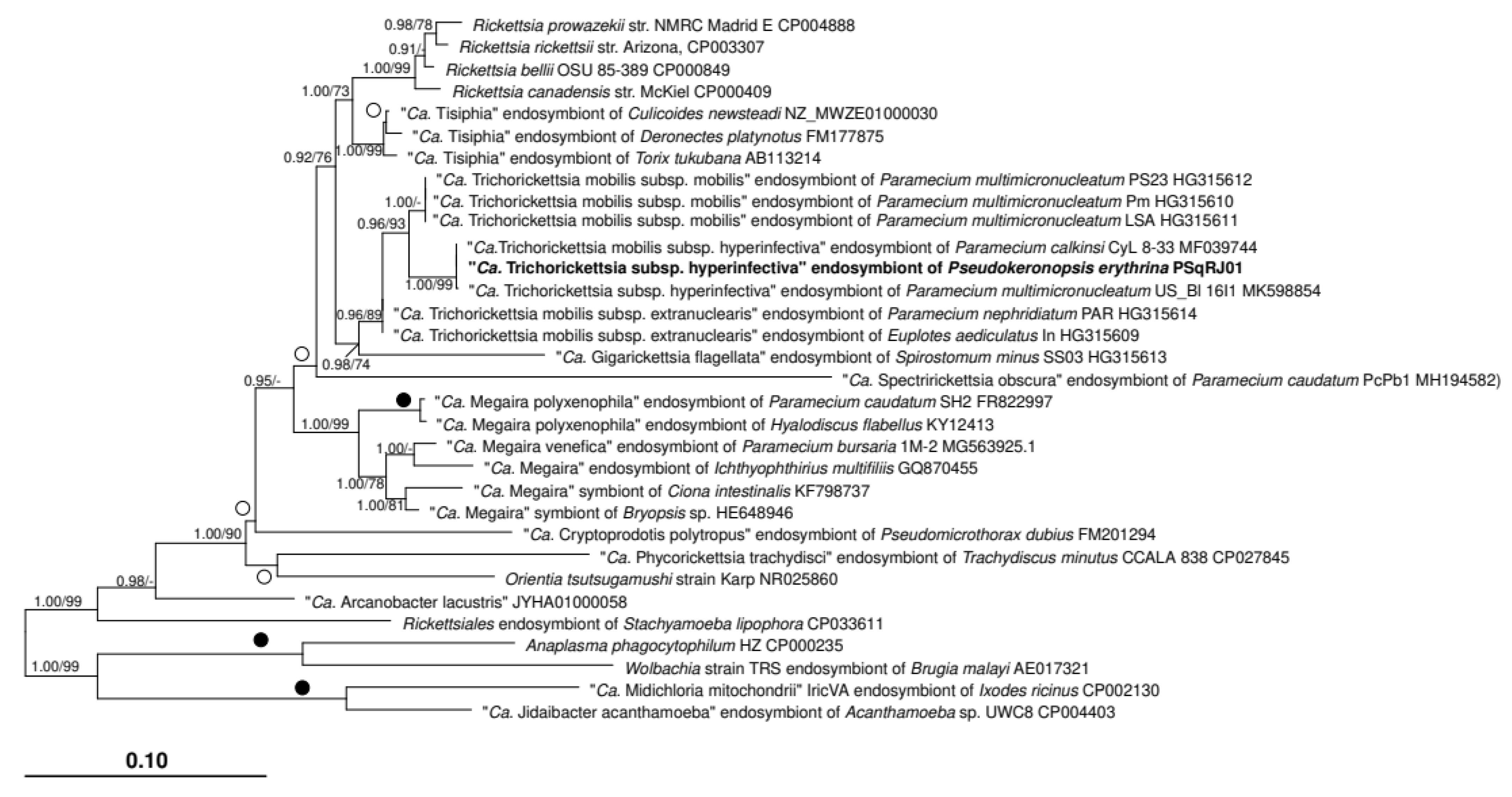
The obtained bacterial 16S rRNA gene sequence was 1527 bp long with a 100% identity with “Ca. Trichorickettsia mobilis subsp. hyperinfectiva” endosymbiont of P. calkinsi strain CyL 8-33 (MF039744.1). Phylogenetic analyses fully confirmed the assignment of the PSqRJ01’s symbiont to “Ca. Trichorickettsia mobilis subsp. hyperinfectiva” (1.00 BI|99 ML. Figure 4). Moreover, phyletic relationships between “Ca. Trichorickettsia mobilis” subspecies, i.e., in particular “Ca. Trichorickettsia mobilis subsp. mobilis” and “Ca. Trichorickettsia mobilis subsp. hyperinfectiva” as sister groups (0.96 BI/93 ML), as well as within Rickettsiaceae in general, were consistent with other 16S rRNA gene-based and phylogenomic studies [19,45,46,47,48,49].
In terms of sequence identity, the 16S rRNA genes of “Ca. Trichorickettsia mobilis” were highly homogeneous within each subspecies, with 100% identities both in the “full sequence” and the “no insert matrix” (Table 1 and Table 2). On the other side, most of the observed divergence between subspecies is concentrated in the insert regions (Table 1), which is putatively non-functional, as identities not considering the insert are all rather high (≥99.5%) (Table 2), consistent with previous studies [17,19], and with the species threshold of 98.65–98.7% [50].
Unfortunately, after some initial experiments, Pseudokeronopsis cfr. erythrina PSqRJ01 was lost and FISH experiments with more specific probes could no longer be performed. However, TEM investigations disclosed that 1, the endosymbionts exhibited a common morphology, uniform between them, including a two-membrane cell wall typical of Gram-negative bacteria (Figure 2) and a rod-shaped silhouette (size: ~2.0 × 0.4 µm) with generally rounded ends (Figure 2B,E,F); 2, bacterial cytoplasm was not homogeneous, i.e., it included areas with variable electron density, electron-lucid granules (one to three in number per bacterium) (diameter: ~0.5–0.7 µm), and filaments (DNA) (Figure 2B,E,F); 3, on several occasions thin flagella (diameter: ∼12–15 nm; length ∼60–90 nm) were visible (Figure 2E,F), often distributed all around the bacterial cell (Figure 2F). Thus, the appearance of the cytoplasmic bacterial endosymbionts of PSqRJ01 was consistent with previous reports of “Ca. Trichorickettsia mobilis” [17,18,19,38,51].

4. Discussion

In this work, we characterised by a multidisciplinary approach (in vivo and ultrastructural observations, molecular phylogeny) a strain of the ciliate protist Pseudokeronopsis, isolated from a sample coming from Brazil, and its associated Rickettsiales bacterial symbiont, affiliated to “Ca. Trichorickettsia”. While reports of Rickettsiales (including “Ca. Trichorickettsia) in association with ciliates and other protists are rather common [5,26,45,47,49,52,53,54,55], to our best knowledge, this represents the first record of a host belonging to the genus Pseudokeronopsis, and, in general, to urostylid ciliates. It also represents the first record of a “Ca. Trichorickettsia” from South America. The characterisation of the host and of the symbiont will be discussed below separately, in a comparison with known relative organisms.
For what concerns the host taxonomy, it is well known that species attribution in the genus Pseudokeronopsis is difficult, due to a high interspecific morphological similarity [44,56,57,58,59,60]. Indeed, as evidenced in Figure 3, several sequences attributed to different species do not form monophyletic clades, suggesting a possible misidentification of few isolates (or the use of slightly different identification criteria by different researchers). Moreover, within the genus Pseudokeronopsis 18S rDNA sequences are poorly informative in resolving phylogenetic relationships, due to extremely low distances. Nevertheless, in the case of the herein characterised Pseudokeronopsis sp. PSqRJ01, 18S rRNA gene data clearly indicated a strongly supported affiliation to the P. erythrina clade. Unfortunately, due to an incomplete reference database, we could not use the obtained ITS and 28S rRNA gene sequences for species identification and for an improved phylogenetic resolution within the genus. Nevertheless, our data may offer such a possibility in future studies, being the ITS more and more used for species delimitation in ciliates, e.g., [61,62,63,64,65]. As for the morphological analyses, they were overall supportive for a relatedness to P. erythrina (Chen et al., 2011). Only the following two differences with the original description of P. erythrina [44] were noticed: 1, colourless granules were found to be densely distributed in the PSqRJ01 cytoplasm (likely not coincident with the blood cell-shaped granules found in other species of the genus; see [57,58]), while they are absent in P. erythrina; 2, a single contractile vacuole was observed, as opposed to, generally, two in P. erythrina. Taking all the above together and considering that a careful taxonomic revision of genus Pseudokeronopsis would be beyond the aims of the present study, we opted to conservatively refer to the novel strain as to Pseudokeronopsis cfr. erythrina PSqRJ01. Pseudokeronopsis erythrina is considered a euryhaline species [29], having been retrieved from both brackish and freshwater environments in China and Italy, respectively [29,44]. Interestingly, the finding of Pseudokeronopsis cfr. erythrina PSqRJ01 in the Saquarema lagoon, which shows significant spatial and seasonal variations in salinity [66], can be seen as consistent with previous observations on the autoecology of P. erythrina.
For what concerns the presence of intracellular bacterial symbionts in Pseudokeronopsis cfr. erythrina PSqRJ01, molecular analyses clearly indicate the presence of “Ca. Trichorickettsia mobilis”. Specifically, the symbiont is affiliated with full support to “Ca. Trichorickettsia mobilis subsp. hyperinfectiva” (1.00 BI|99 ML). Such finding expands the known host range for this bacterium. Indeed, “Ca. Trichorickettsia mobilis subsp. hyperinfectiva” was up to now retrieved in natural association only with members of genus Paramecium [19,38], similarly to, in general, most representatives of the other subspecies of “Ca. Trichorickettsia mobilis” as well [17]. Therefore, it is implied that this bacterium displays the ability to shift horizontally between different host species in natural conditions, suggesting a certain flexibility in terms of host preference. This is consistent with general inferences on other Rickettsiales species, e.g., “Ca. Megaira polyxenophila” [20,47,52], and, interestingly, also with previous laboratory experiments on another “Ca. Trichorickettsia mobilis subsp. hyperinfectiva”, naturally hosted by Paramecium multimicronucleatum, and able to invade, at least transiently, a metazoan host (the planarian Dugesia japonica) [38].On the other hand, interestingly, the association of “Ca. Trichorickettsia mobilis” with the ciliate P. multimicronucleatum was reported to be quite stable in laboratory conditions, even in the case of antibiotic/antimicrobial treatments [18,51]. The unfortunate loss of the host strain Pseudokeronopsis cfr. erythrina PSqRJ01 did not allow us to perform further investigations that could rule out the presence of additional, phylogenetically distinct, intracellular bacteria. Nevertheless, combined together, multiple available evidence, in particular the preliminary FISH experiments with alphaproteobacterial probes (Supplementary Figure S1) and the uniform morphology and ultrastructure (Figure 3), consistent with multiple previous studies on “Ca. Trichorickettsia” [17,18,19,38,51], indicate that such possibility is unlikely.
Similarly to several other “Ca. Trichorickettsia” [17,18,19,38,51], the PSqRJ01 symbiont was often found to bear flagella (Figure 2). The presence/absence pattern of flagella within “Ca. Trichorickettsia mobilis” is peculiar, so far with all specimens of “Ca. Trichorickettsia mobilis subsp. mobilis” and “Ca. Trichorickettsia mobilis subsp. hyperinfectiva” (including the herein characterised symbiont) being flagellated, while those of “Ca. Trichorickettsia mobilis subsp. extranuclearis” are non-flagellated [17,18,19,38,51]. There might be also a potential variability in flagellar abundance (both in terms of proportion of flagellated cells, and of number of flagella per cell), with the current specimen possibly poorer than others, even within the same subspecies [19,38]. However, this variation might be only apparent, considering that different methods can have differential sensitivity (e.g., in the current study negative stain was not employed) and that we did not perform a quantitative analysis of the flagellar abundance.
In any case, we underline that the precise function(s) of flagella in “Ca. Trichorickettsia mobilis” and in Rickettsiales in general still need(s) to be elucidated. Despite being absent in several long-time studied representatives (e.g., Rickettsia, Orientia, Anaplasma, Ehrlichia, Wolbachia), it has become evident that flagella [17,23,47] or at least flagellar genes [24,27,46,67,68] are present in many Rickettsiales. They are now considered an ancestral trait [24,25,26,69], independently lost in many extant lineages, possibly concurrently with the adaptation to terrestrial hosts. Indeed, it has been speculated that flagella-driven motility could be important in particular in aquatic environments, for possible intermediate “free-living” phases during horizontal transfer between different hosts [27]. Anyhow, motility might be also involved in as-yet-unexplored stages of the life cycle of “terrestrial” Rickettsiales in vertebrate hosts [24]. Alternative roles have been proposed for flagella in Rickettsiales [23], in particular as a potential secretion system [24,28]. Indeed, they share a common type III secretion system component with the injectisome [70], which is important in many bacteria for delivering effectors mediating the interaction with host cells. Thus, the flagellum may actually add up to the already known repertoire of apparatuses for delivering effectors to the hosts in Rickettsiales [71]. It has also been proposed that the flagellum could be directly mediating the interaction with host cells in the Rickettsiales bacterium “Ca. Midichloria” [28], with potential analogies with other Alphaproteobacteria [72].
In this context of elucidating the role of flagella in Rickettsiales, the case of “Ca. Trichorickettsia mobilis” is highly interesting for several reasons. First, to our knowledge, its flagellated members are the only ones for which actual motility due to flagella has been observed among Rickettsiales. Second, the fact that flagella were observed only in certain strains and subspecies (see references [19,38] and present study) is quite a relevant aspect that still needs to be elucidated, namely whether this is due to the presence/absence of the respective genetic repertoire, or by other causes. For example, it could be speculated that a contingent expression could be triggered only in some conditions, possibly related to host species and host subcellular compartment. In this regard, it is worth noting that the observed presence of flagella seems to correlate more with the bacterial phylogeny, rather than with other features, which might be considered in favour of a genetic explanation. However, any conclusions on this point are clearly premature, and further targeted studies, including in particular genome sequencing, would be necessary to address it. Last but not least, the interest in studying flagella in “Ca. Trichorickettsia mobilis” resides in its phylogenetic proximity with Rickettsia [17], which includes some of the most medically relevant, and thus deeply studied, Rickettsiales [71,73,74]. Investigations on “Ca. Trichorickettsia mobilis”, especially but not exclusively focused on flagella, may provide relevant insights on the evolutionary origin of Rickettsia, in particular for what concerns the adaptation to terrestrial metazoan hosts, such as arthropods and vertebrates.
In sum, due to the features exposed above, in our view, “Ca. Trichorickettsia mobilis” may represent an ideal system to study flagella and their regulation in Rickettsiales, from a functional and evolutionary perspective. Thus, we call for further investigations on this bacterium and its interaction with host organisms, including host species-dependent variability, and with a special focus on the role of flagella.

Supplementary Materials

The following supporting information can be downloaded at: https://www.mdpi.com/article/10.3390/d14121032/s1, Figure S1: Fluorescence in situ hybridisation pictures of Pseudokeronopsis cfr. erythrina PSqRJ01.

Author Contributions

Conceptualization, M.C., V.S., M.V.X.S., L.M. and G.P.; formal analysis, M.C. and V.S.; investigation, M.V.X.S., M.C. and L.M.; resources, G.P.; data curation, M.V.X.S., M.C., L.G. and L.M.; writing—original draft preparation, M.C., V.S. and L.M.; writing—review and editing, G.P., M.V.X.S. and L.G.; visualization, L.M. and V.S.; supervision, M.C., L.M. and G.P.; project administration, G.P.; funding acquisition, G.P. All authors have read and agreed to the published version of the manuscript.

Funding

This research was funded by the European Community’s H2020 Programme H2020-MSCA-RISE 2019: grant agreement n° 872767. The present study was also supported by the Fondazione Cassa di Risparmio di Pistoia e Pescia—giovani@ricercascientifica2019 fellowship (n° 2019.0380).

Institutional Review Board Statement

Not applicable.

Data Availability Statement

The sequences newly obtained in this study were deposited to the NCBI GenBank repository, under the following accession numbers: combined partial 18S rRNA gene, ITS and partial 28S rRNA gene of Pseudokeronopsis cfr. erythrina PSqRJ01 (ON331772), “Candidatus Trichorickettsia mobilis subsp. hyperinfectiva” endosymbiont of Pseudokeronopsis cfr. erythrina PSqRJ01 (ON355343).

Acknowledgments

The authors would like to thank Franco Verni and Claudia Vannini for the support in TEM preparation and observation, Simone Gabrielli for figure-editing support, E. Mugnaioli (CISUP) and P. Lucchesi (CIME) for the assistance with HR FEG-TEM observation, and Katrin Grosser for the support in molecular biology experiments.

Conflicts of Interest

The authors declare no conflict of interest. The funders had no role in the design of the study; in the collection, analyses, or interpretation of data; in the writing of the manuscript, or in the decision to publish the results.

References

  1. Husnik, F.; Tashyreva, D.; Boscaro, V.; George, E.E.; Lukeš, J.; Keeling, P.J. Bacterial and Archaeal Symbioses with Protists. Curr. Biol. 2021, 31, R862–R877. [Google Scholar] [CrossRef] [PubMed]
  2. Nowack, E.C.M.; Melkonian, M. Endosymbiotic Associations within Protists. Philos. Trans. R. Soc. Lond. B Biol. Sci. 2010, 365, 699–712. [Google Scholar] [CrossRef] [PubMed] [Green Version]
  3. Dziallas, C.; Allgaier, M.; Monaghan, M.T.; Grossart, H.-P. Act Together—Implications of Symbioses in Aquatic Ciliates. Front. Microbiol. 2012, 3, 288. [Google Scholar] [CrossRef] [Green Version]
  4. Serra, V.; Gammuto, L.; Nitla, V.; Castelli, M.; Lanzoni, O.; Sassera, D.; Bandi, C.; Sandeep, B.V.; Verni, F.; Modeo, L.; et al. Morphology, Ultrastructure, Genomics, and Phylogeny of Euplotes Vanleeuwenhoeki Sp. Nov. and Its Ultra-Reduced Endosymbiont “Candidatus Pinguicoccus Supinus” Sp. Nov. Sci. Rep. 2020, 10, 20311. [Google Scholar] [CrossRef] [PubMed]
  5. Castelli, M.; Sassera, D.; Petroni, G. Biodiversity of “Non-Model” Rickettsiales and Their Association with Aquatic Organisms. In Rickettsiales; Thomas, S., Ed.; Springer: Berlin/Heidelberg, Germany, 2016; pp. 59–91. [Google Scholar]
  6. Collingro, A.; Köstlbacher, S.; Mussmann, M.; Stepanauskas, R.; Hallam, S.J.; Horn, M. Unexpected Genomic Features in Widespread Intracellular Bacteria: Evidence for Motility of Marine Chlamydiae. ISME J. 2017, 11, 2334–2344. [Google Scholar] [CrossRef]
  7. Hugoson, E.; Guliaev, A.; Ammunét, T.; Guy, L. Host Adaptation in Legionellales Is 1.9 Ga, Coincident with Eukaryogenesis. Mol. Biol. Evol. 2022, 39, msac037. [Google Scholar] [CrossRef]
  8. Watanabe, K.; Nakao, R.; Fujishima, M.; Tachibana, M.; Shimizu, T.; Watarai, M. Ciliate Paramecium Is a Natural Reservoir of Legionella pneumophila. Sci. Rep. 2016, 6, 24322. [Google Scholar] [CrossRef]
  9. Richards, A.M.; Von Dwingelo, J.E.; Price, C.T.; Abu Kwaik, Y. Cellular Microbiology and Molecular Ecology of Legionella-Amoeba Interaction. Virulence 2013, 4, 307–314. [Google Scholar] [CrossRef] [PubMed] [Green Version]
  10. Mungroo, M.R.; Siddiqui, R.; Khan, N.A. War of the Microbial World: Acanthamoeba Spp. Interactions with Microorganisms. Folia Microbiol. 2021, 66, 689–699. [Google Scholar] [CrossRef]
  11. Wang, Z.; Wu, M. Comparative Genomic Analysis of Acanthamoeba Endosymbionts Highlights the Role of Amoebae as a “Melting Pot” Shaping the Rickettsiales Evolution. Genome Biol. Evol. 2017, 9, 3214–3224. [Google Scholar] [CrossRef]
  12. Barker, J.; Brown, M.R. Trojan Horses of the Microbial World: Protozoa and the Survival of Bacterial Pathogens in the Environment. Microbiology 1994, 140 Pt 6, 1253–1259. [Google Scholar] [CrossRef] [PubMed] [Green Version]
  13. Watanabe, K.; Higuchi, Y.; Shimmura, M.; Tachibana, M.; Fujishima, M.; Shimizu, T.; Watarai, M. Peculiar Paramecium Peculiar Hosts Fail to Establish a Stable Intracellular Relationship with Legionella pneumophila. Front. Microbiol. 2020, 11, 596731. [Google Scholar] [CrossRef] [PubMed]
  14. Fujishima, M.; Kodama, Y. Mechanisms for Establishing Primary and Secondary Endosymbiosis in Paramecium. J. Eukaryot. Microbiol. 2022, 69, e12901. [Google Scholar] [CrossRef] [PubMed]
  15. Siegmund, L.; Schweikert, M.; Fischer, M.S.; Wöstemeyer, J. Bacterial Surface Traits Influence Digestion by Tetrahymena Pyriformis and Alter Opportunity to Escape from Food Vacuoles. J. Eukaryot. Microbiol. 2018, 65, 600–611. [Google Scholar] [CrossRef] [PubMed]
  16. Siegmund, L.; Burmester, A.; Fischer, M.S.; Wöstemeyer, J. A Model for Endosymbiosis: Interaction between Tetrahymena Pyriformis and Escherichia Coli. Eur. J. Protistol. 2013, 49, 552–563. [Google Scholar] [CrossRef] [PubMed]
  17. Vannini, C.; Boscaro, V.; Ferrantini, F.; Benken, K.A.; Mironov, T.I.; Schweikert, M.; Görtz, H.-D.; Fokin, S.I.; Sabaneyeva, E.V.; Petroni, G. Flagellar Movement in Two Bacteria of the Family Rickettsiaceae: A Re-Evaluation of Motility in an Evolutionary Perspective. PLoS ONE 2014, 9, e87718. [Google Scholar] [CrossRef]
  18. Mironov, T.; Sabaneyeva, E. A Robust Symbiotic Relationship Between the Ciliate Paramecium multimicronucleatum and the Bacterium Ca. Trichorickettsia Mobilis. Front. Microbiol. 2020, 11, 603335. [Google Scholar] [CrossRef]
  19. Sabaneyeva, E.; Castelli, M.; Szokoli, F.; Benken, K.; Lebedeva, N.; Salvetti, A.; Schweikert, M.; Fokin, S.; Petroni, G. Host and Symbiont Intraspecific Variability: The Case of Paramecium calkinsi and “Candidatus Trichorickettsia Mobilis”. Eur. J. Protistol. 2018, 62, 79–94. [Google Scholar] [CrossRef]
  20. Davison, H.R.; Pilgrim, J.; Wybouw, N.; Parker, J.; Pirro, S.; Hunter-Barnett, S.; Campbell, P.M.; Blow, F.; Darby, A.C.; Hurst, G.D.D.; et al. Genomic Diversity across the Rickettsia and “Candidatus Megaira” Genera and Proposal of Genus Status for the Torix Group. Nat. Commun. 2022, 13, 2630. [Google Scholar] [CrossRef]
  21. Blanton, L.S. The Rickettsioses: A Practical Update. Infect. Dis. Clin. N. Am. 2019, 33, 213–229. [Google Scholar] [CrossRef]
  22. Dumler, J.S.; Walker, D.H. Rickettsiales. In Bergey’s Manual of Systematics of Archaea and Bacteria; Wiley: Hoboken, NJ, USA, 2015; p. 1. [Google Scholar]
  23. Boscaro, V.; Schrallhammer, M.; Benken, K.A.; Krenek, S.; Szokoli, F.; Berendonk, T.U.; Schweikert, M.; Verni, F.; Sabaneyeva, E.V.; Petroni, G. Rediscovering the Genus Lyticum, Multiflagellated Symbionts of the Order Rickettsiales. Sci. Rep. 2013, 3, 3305. [Google Scholar] [CrossRef] [PubMed] [Green Version]
  24. Sassera, D.; Lo, N.; Epis, S.; D’Auria, G.; Montagna, M.; Comandatore, F.; Horner, D.; Peretó, J.; Luciano, A.M.; Franciosi, F.; et al. Phylogenomic Evidence for the Presence of a Flagellum and cbb(3) Oxidase in the Free-Living Mitochondrial Ancestor. Mol. Biol. Evol. 2011, 28, 3285–3296. [Google Scholar] [CrossRef] [PubMed]
  25. Schön, M.E.; Martijn, J.; Vosseberg, J.; Köstlbacher, S.; Ettema, T.J.G. The Evolutionary Origin of Host Association in the Rickettsiales. Nat. Microbiol. 2022, 7, 1189–1199. [Google Scholar] [CrossRef] [PubMed]
  26. Castelli, M.; Sabaneyeva, E.; Lanzoni, O.; Lebedeva, N.; Floriano, A.M.; Gaiarsa, S.; Benken, K.; Modeo, L.; Bandi, C.; Potekhin, A.; et al. Deianiraea, an Extracellular Bacterium Associated with the Ciliate Paramecium, Suggests an Alternative Scenario for the Evolution of Rickettsiales. ISME J. 2019, 13, 2280–2294. [Google Scholar] [CrossRef] [PubMed]
  27. Schulz, F.; Martijn, J.; Wascher, F.; Lagkouvardos, I.; Kostanjšek, R.; Ettema, T.J.G.; Horn, M. A Rickettsiales Symbiont of Amoebae with Ancient Features. Environ. Microbiol. 2016, 18, 2326–2342. [Google Scholar] [CrossRef] [PubMed]
  28. Floriano, A.M.; Biffignandi, G.B.; Castelli, M.; Olivieri, E.; Clementi, E.; Comandatore, F.; Rinaldi, L.; Opara, M.; Plantard, O.; Palomar, A.M.; et al. The Origin and Evolution of Mitochondrial Tropism in Midichloria Bacteria. bioRxiv 2022. [Google Scholar] [CrossRef]
  29. Anesi, A.; Buonanno, F.; di Giuseppe, G.; Ortenzi, C.; Guella, G. Metabolites from the Euryhaline CiliatePseudokeronopsis Erythrina. Eur. J. Org. Chem. 2016, 2016, 1330–1336. [Google Scholar] [CrossRef]
  30. Guella, G.; Frassanito, R.; Mancini, I.; Sandron, T.; Modeo, L.; Verni, F.; Dini, F.; Petroni, G. Keronopsamides, a New Class of Pigments from Marine Ciliates. Eur. J. Org. Chem. 2010, 2010, 427–434. [Google Scholar] [CrossRef]
  31. Medlin, L.; Elwood, H.J.; Stickel, S.; Sogin, M.L. The Characterization of Enzymatically Amplified Eukaryotic 16S-like rRNA-Coding Regions. Gene 1988, 71, 491–499. [Google Scholar] [CrossRef] [Green Version]
  32. Petroni, G.; Dini, F.; Verni, F.; Rosati, G. A Molecular Approach to the Tangled Intrageneric Relationships Underlying Phylogeny in Euplotes (Ciliophora, Spirotrichea). Mol. Phylogenet. Evol. 2002, 22, 118–130. [Google Scholar] [CrossRef]
  33. Rosati, G.; Modeo, L.; Melai, M.; Petroni, G.; Verni, F. A Multidisciplinary Approach to Describe Protists: A Morphological, Ultrastructural, and Molecular Study on Peritromus kahli Villeneuve-Brachon, 1940 (Ciliophora, Heterotrichea). J. Eukaryot. Microbiol. 2004, 51, 49–59. [Google Scholar] [CrossRef] [PubMed]
  34. Boscaro, V.; Fokin, S.I.; Verni, F.; Petroni, G. Survey of Paramecium duboscqui Using Three Markers and Assessment of the Molecular Variability in the Genus Paramecium. Mol. Phylogenet. Evol. 2012, 65, 1004–1013. [Google Scholar] [CrossRef] [PubMed]
  35. Vannini, C.; Rosati, G.; Verni, F.; Petroni, G. Identification of the Bacterial Endosymbionts of the Marine Ciliate Euplotes magnicirratus (Ciliophora, Hypotrichia) and Proposal of “Candidatus Devosia Euplotis”. Int. J. Syst. Evol. Microbiol. 2004, 54, 1151–1156. [Google Scholar] [CrossRef] [PubMed] [Green Version]
  36. Manz, W.; Amann, R.; Ludwig, W.; Wagner, M.; Schleifer, K.-H. Phylogenetic Oligodeoxynucleotide Probes for the Major Subclasses of Proteobacteria: Problems and Solutions. Syst. Appl. Microbiol. 1992, 15, 593–600. [Google Scholar] [CrossRef]
  37. Amann, R.I.; Binder, B.J.; Olson, R.J.; Chisholm, S.W.; Devereux, R.; Stahl, D.A. Combination of 16S rRNA-Targeted Oligonucleotide Probes with Flow Cytometry for Analyzing Mixed Microbial Populations. Appl. Environ. Microbiol. 1990, 56, 1919–1925. [Google Scholar] [CrossRef] [PubMed] [Green Version]
  38. Modeo, L.; Salvetti, A.; Rossi, L.; Castelli, M.; Szokoli, F.; Krenek, S.; Serra, V.; Sabaneyeva, E.; Di Giuseppe, G.; Fokin, S.I.; et al. Trichorickettsia Mobilis", a Bacterium, Can Be Transiently Transferred from the Unicellular Eukaryote to the Planarian. PeerJ 2020, 8, e8977. [Google Scholar] [CrossRef] [PubMed] [Green Version]
  39. Westram, R.; Bader, K.; Prüsse, E.; Kumar, Y.; Meier, H.; Glöckner, F.O.; Ludwig, W. ARB: A Software Environment for Sequence Data. In Handbook of Molecular Microbial Ecology I: Metagenomics and Complementary Approaches; Wiley-Blackwell: Hoboken, NJ, USA, 2011; pp. 399–406. [Google Scholar]
  40. Quast, C.; Pruesse, E.; Yilmaz, P.; Gerken, J.; Schweer, T.; Yarza, P.; Peplies, J.; Glöckner, F.O. The SILVA Ribosomal RNA Gene Database Project: Improved Data Processing and Web-Based Tools. Nucleic Acids Res. 2013, 41, D590–D596. [Google Scholar] [CrossRef] [PubMed]
  41. Darriba, D.; Taboada, G.L.; Doallo, R.; Posada, D. jModelTest 2: More Models, New Heuristics and Parallel Computing. Nat. Methods 2012, 9, 772. [Google Scholar] [CrossRef] [PubMed] [Green Version]
  42. Guindon, S.; Gascuel, O. A Simple, Fast, and Accurate Algorithm to Estimate Large Phylogenies by Maximum Likelihood. Syst. Biol. 2003, 52, 696–704. [Google Scholar] [CrossRef] [PubMed] [Green Version]
  43. Ronquist, F.; Teslenko, M.; van der Mark, P.; Ayres, D.L.; Darling, A.; Höhna, S.; Larget, B.; Liu, L.; Suchard, M.A.; Huelsenbeck, J.P. MrBayes 3.2: Efficient Bayesian Phylogenetic Inference and Model Choice across a Large Model Space. Syst. Biol. 2012, 61, 539–542. [Google Scholar] [CrossRef] [PubMed]
  44. Chen, X.; Clamp, J.C.; Song, W. Phylogeny and Systematic Revision of the Family Pseudokeronopsidae (Protista, Ciliophora, Hypotricha), with Description of a New Estuarine Species of Pseudokeronopsis. Zool. Scr. 2011, 40, 659–671. [Google Scholar] [CrossRef]
  45. Yurchenko, T.; Ševčíková, T.; Přibyl, P.; El Karkouri, K.; Klimeš, V.; Amaral, R.; Zbránková, V.; Kim, E.; Raoult, D.; Santos, L.M.A.; et al. A Gene Transfer Event Suggests a Long-Term Partnership between Eustigmatophyte Algae and a Novel Lineage of Endosymbiotic Bacteria. ISME J. 2018, 12, 2163–2175. [Google Scholar] [CrossRef] [PubMed] [Green Version]
  46. Martijn, J.; Schulz, F.; Zaremba-Niedzwiedzka, K.; Viklund, J.; Stepanauskas, R.; Andersson, S.G.E.; Horn, M.; Guy, L.; Ettema, T.J.G. Single-Cell Genomics of a Rare Environmental Alphaproteobacterium Provides Unique Insights into Rickettsiaceae Evolution. ISME J. 2015, 9, 2373–2385. [Google Scholar] [CrossRef] [PubMed] [Green Version]
  47. Lanzoni, O.; Sabaneyeva, E.V.; Modeo, L.; Castelli, M.; Lebedeva, N.; Verni, F.; Schrallhammer, M.; Potekhin, A.; Petroni, G. Diversity and Environmental Distribution of the Cosmopolitan Endosymbiont “Candidatus Megaira”. Sci. Rep. 2019, 9, 1179. [Google Scholar] [CrossRef] [PubMed] [Green Version]
  48. Castelli, M.; Serra, V.; Senra, M.V.X.; Basuri, C.K.; Soares, C.A.G.; Fokin, S.I.; Modeo, L.; Petroni, G. The Hidden World of Rickettsiales Symbionts: “Candidatus Spectririckettsia Obscura,” a Novel Bacterium Found in Brazilian and Indian Paramecium caudatum. Microb. Ecol. 2019, 77, 748–758. [Google Scholar] [CrossRef]
  49. Muñoz-Gómez, S.A.; Hess, S.; Burger, G.; Lang, B.F.; Susko, E.; Slamovits, C.H.; Roger, A.J. An Updated Phylogeny of the Reveals That the Parasitic and Have Independent Origins. Elife 2019, 8, e42535. [Google Scholar] [CrossRef]
  50. Kim, M.; Oh, H.-S.; Park, S.-C.; Chun, J. Towards a Taxonomic Coherence between Average Nucleotide Identity and 16S rRNA Gene Sequence Similarity for Species Demarcation of Prokaryotes. Int. J. Syst. Evol. Microbiol. 2014, 64, 346–351. [Google Scholar] [CrossRef]
  51. Mironov, T.; Yakovlev, A.; Sabaneyeva, E. Together Forever: Inseparable Partners of the Symbiotic System Paramecium multimicronucleatum/Ca. Trichorickettsia Mobilis. Symbiosis 2022, 87, 19–30. [Google Scholar] [CrossRef]
  52. Hess, S. Description of Hyalodiscus flabellus Sp. Nov. (Vampyrellida, Rhizaria) and Identification of Its Bacterial Endosymbiont, “Candidatus Megaira Polyxenophila” (Rickettsiales, Alphaproteobacteria). Protist 2017, 168, 109–133. [Google Scholar] [CrossRef]
  53. George, E.E.; Husnik, F.; Tashyreva, D.; Prokopchuk, G.; Horák, A.; Kwong, W.K.; Lukeš, J.; Keeling, P.J. Highly Reduced Genomes of Protist Endosymbionts Show Evolutionary Convergence. Curr. Biol. 2020, 30, 925–933.e3. [Google Scholar] [CrossRef]
  54. Boscaro, V.; Manassero, V.; Keeling, P.J.; Vannini, C. Single-Cell Microbiomics Unveils Distribution and Patterns of Microbial Symbioses in the Natural Environment. Microb. Ecol. 2022. [Google Scholar] [CrossRef] [PubMed]
  55. Boscaro, V.; Husnik, F.; Vannini, C.; Keeling, P.J. Symbionts of the Ciliate Euplotes: Diversity, Patterns and Potential as Models for Bacteria-Eukaryote Endosymbioses. Proc. Biol. Sci. 2019, 286, 20190693. [Google Scholar] [CrossRef]
  56. Berger, H. Monograph of the Urostyloidea (Ciliophora, Hypotricha); Springer Science & Business Media: Berlin/Heidelberg, Germany, 2007; ISBN 9781402052736. [Google Scholar]
  57. Li, J.; Xu, K. Morphology and Taxonomy of Pseudokeronopsis rubra (Ehrenberg, 1836) and Pseudokeronopsis parasongi Sp. Nov. (Ciliophora, Hypotrichia, Urostylida) from the Yellow Sea. Eur. J. Protistol. 2020, 76, 125737. [Google Scholar] [CrossRef]
  58. Li, J.; Zhan, Z.; Xu, K. Systematics and Molecular Phylogeny of the Ciliate Genus Pseudokeronopsis (Ciliophora, Hypotrichia). J. Eukaryot. Microbiol. 2017, 64, 850–872. [Google Scholar] [CrossRef]
  59. Chen, X.; Li, J.; Xu, K. Insights into the Phylogeny of Three Systematically Controversial Subfamilies of Urostylid Ciliates Based on rDNA. Zool. Scr. 2021, 50, 383–395. [Google Scholar] [CrossRef]
  60. Xu, Y.; Shen, Z.; Gentekaki, E.; Xu, J.; Yi, Z. Comparative Transcriptome Analyses during the Vegetative Cell Cycle in the Mono-Cellular Organism Pseudokeronopsis erythrina (Alveolata, Ciliophora). Microorganisms 2020, 8, 108. [Google Scholar] [CrossRef] [PubMed] [Green Version]
  61. Boscaro, V.; Carducci, D.; Barbieri, G.; Senra, M.V.X.; Andreoli, I.; Erra, F.; Petroni, G.; Verni, F.; Fokin, S.I. Focusing on Genera to Improve Species Identification: Revised Systematics of the Ciliate Spirostomum. Protist 2014, 165, 527–541. [Google Scholar] [CrossRef] [PubMed]
  62. Melekhin, M.; Yakovleva, Y.; Lebedeva, N.; Nekrasova, I.; Nikitashina, L.; Castelli, M.; Mayén-Estrada, R.; Romanovich, A.E.; Petroni, G.; Potekhin, A. Cryptic Diversity in Revealed with a Polyphasic Approach. Microorganisms 2022, 10, 974. [Google Scholar] [CrossRef]
  63. Obert, T.; Vďačný, P. Delimitation of Five Astome Ciliate Species Isolated from the Digestive Tube of Three Ecologically Different Groups of Lumbricid Earthworms, Using the Internal Transcribed Spacer Region and the Hypervariable D1/D2 Region of the 28S rRNA Gene. BMC Evol. Biol. 2020, 20, 37. [Google Scholar] [CrossRef]
  64. Zhang, T.; Shao, C.; Zhang, T.; Song, W.; Vd’ačný, P.; Al-Farraj, S.A.; Wang, Y. Multi-Gene Phylogeny of the Ciliate Genus Trachelostyla (Ciliophora, Hypotrichia), With Integrative Description of Two Species, Trachelostyla multinucleata Spec. Nov. and T. Pediculiformis (Cohn, 1866). Front. Microbiol. 2022, 12, 775570. [Google Scholar] [CrossRef]
  65. Shazib, S.U.A.; Vďačný, P.; Kim, J.H.; Jang, S.W.; Shin, M.K. Molecular Phylogeny and Species Delimitation within the Ciliate Genus Spirostomum (Ciliophora, Postciliodesmatophora, Heterotrichea), Using the Internal Transcribed Spacer Region. Mol. Phylogenet. Evol. 2016, 102, 128–144. [Google Scholar] [CrossRef] [PubMed]
  66. Belart, P.; Habib, R.; Raposo, D.; Clemente, I.; Alves Martins, M.V.; Frontalini, F.; Figueiredo, M.S.L.; Lorini, M.L.; Laut, L. Seasonal Dynamics of Benthic Foraminiferal Biocoenosis in the Tropical Saquarema Lagoonal System (Brazil). Estuaries Coast. 2019, 42, 822–841. [Google Scholar] [CrossRef]
  67. Carrier, T.J.; Leigh, B.A.; Deaker, D.J.; Devens, H.R.; Wray, G.A.; Bordenstein, S.R.; Byrne, M.; Reitzel, A.M. Microbiome Reduction and Endosymbiont Gain from a Switch in Sea Urchin Life History. Proc. Natl. Acad. Sci. USA 2021, 118, e2022023118. [Google Scholar] [CrossRef]
  68. Kwan, J.C.; Schmidt, E.W. Bacterial Endosymbiosis in a Chordate Host: Long-Term Co-Evolution and Conservation of Secondary Metabolism. PLoS ONE 2013, 8, e80822. [Google Scholar] [CrossRef] [PubMed]
  69. Castelli, M.; Nardi, T.; Gammuto, L.; Bellinzona, G.; Sabaneyeva, E.; Potekhin, A.; Serra, V.; Petroni, G.; Sassera, D. Host Association and Intracellularity Evolved Multiple Times Independently in the Rickettsiales. bioRxiv 2022. [Google Scholar] [CrossRef]
  70. Diepold, A.; Armitage, J.P. Type III Secretion Systems: The Bacterial Flagellum and the Injectisome. Philos. Trans. R. Soc. B Biol. Sci. 2015, 370, 20150020. [Google Scholar] [CrossRef] [Green Version]
  71. Gillespie, J.J.; Kaur, S.J.; Rahman, M.S.; Rennoll-Bankert, K.; Sears, K.T.; Beier-Sexton, M.; Azad, A.F. Secretome of Obligate Intracellular Rickettsia. FEMS Microbiol. Rev. 2015, 39, 47–80. [Google Scholar]
  72. Eicher, S.C.; Dehio, C. Bartonella Entry Mechanisms into Mammalian Host Cells. Cell. Microbiol. 2012, 14, 1166–1173. [Google Scholar] [CrossRef]
  73. Perlman, S.J.; Hunter, M.S.; Zchori-Fein, E. The Emerging Diversity of Rickettsia. Proc. Biol. Sci. 2006, 273, 2097–2106. [Google Scholar] [CrossRef] [Green Version]
  74. Parola, P.; Paddock, C.D.; Socolovschi, C.; Labruna, M.B.; Mediannikov, O.; Kernif, T.; Abdad, M.Y.; Stenos, J.; Bitam, I.; Fournier, P.-E.; et al. Update on Tick-Borne Rickettsioses around the World: A Geographic Approach. Clin. Microbiol. Rev. 2013, 26, 657–702. [Google Scholar] [CrossRef]
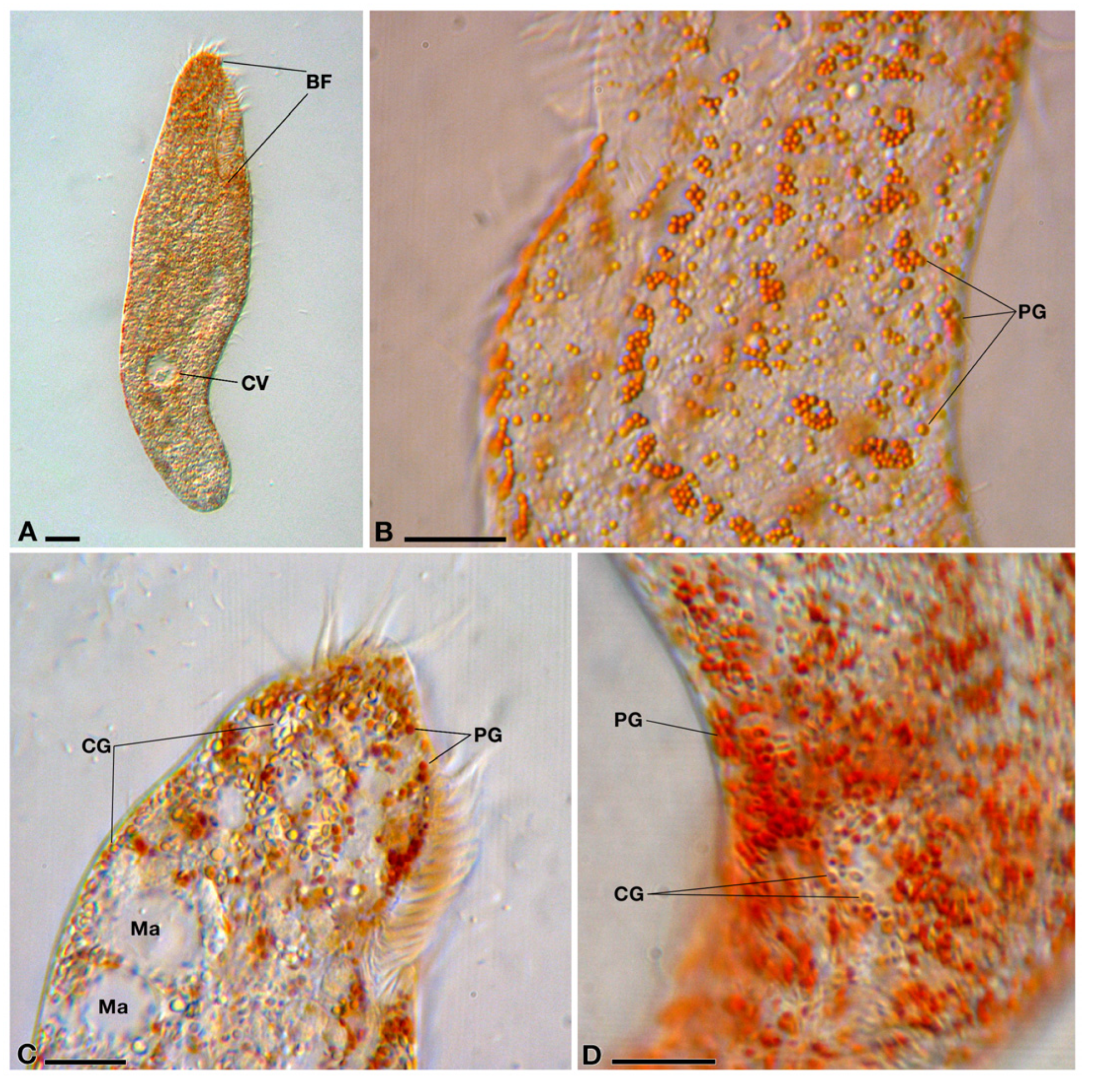
Figure 1. DIC images of Pseudokeronopsis cfr. erythrina PSqRJ01 in vivo. (A) A whole cell. The buccal field (BF) occupies between a third and a fourth of the cell length. A single contractile vacuole (CV) is present in the posterior half of the cell; (B) a lateral view just below the BF level, between the ventral (on the left) and the dorsal (on the right) cell side: pigment granules (PG) are generally associated with each component (i.e., ciliary unit) of the cell ciliature; (C) the anterior cell region. Both PG and ellipsoidal colourless granules (CG) are present. Two macronuclear beads (Ma) are also visible; (D) many PG are also distributed randomly throughout the cell cortical region. Bars stand for 10 µm.
Diversity 14 01032 g001
Figure 2. TEM images of Pseudokeronopsis cfr. erythrina PSqRJ01 and its bacterial endosymbionts “Ca. Trichorickettsia mobilis”. (A) In the cytoplasm, a macronucleus (Ma) with a large nucleolus (Nu), several ovoidal mitochondria (Mt) and polysaccharide reserves (PR), and endosymbionts (E) are visible; (B) Mt sometimes are associated with ribosomes likely belonging to the rough endoplasmic reticulum (RER) cisternae; (C) lipid reserves (LR) often correspond to medium to large-sized droplets. Three endosymbiotic bacteria in the neighbourhood of two LR. They are encircled by a white halo and embedded in the host’s cytoplasm; (D) two cross-sectioned endosymbiotic bacteria showing their two-membrane cell wall; (E) bacteria are rod shaped, and include areas with variable electron density, electron-lucid granules (arrows), and filaments (DNA); (F) sometimes bacteria show flagella (F) distributed all around the cell. Bars stand for 0.5 µm.
Diversity 14 01032 g002
Figure 3. Maximum likelihood tree of the genus Pseudokeronopsis based on 18S rDNA sequences. The phylogenetic position of Pseudokeronopsis cfr. erythrina PSqRJ01 is shown. Numbers associated with nodes represent posterior probability from Bayesian inference (BI) and bootstrap value from maximum likelihood (ML) analyses, respectively (only values of BI ≥ 0.80 and ML ≥ 70% are shown). Black dots represent the highest statistical support (BI = 1.00 and ML = 100); white dots indicate non-significant statistical support (BI < 0.80 and ML < 70%). The sequence obtained in the present work is in bold.
Diversity 14 01032 g003
Figure 4. Maximum likelihood tree of the family Rickettsiaceae based on 16S rDNA sequences. The phylogenetic position of the bacterial symbiont of Pseudokeronopsis cfr. erythrina PSqRJ01 is shown. Numbers associated with nodes represent posterior probability from BI and bootstrap value from ML analyses, respectively (only values of BI ≥ 0.80 and ML ≥ 70% are shown). Black dots represent the highest statistical support (BI = 1.00 and ML = 100); white dots indicate non-significant statistical support (BI < 0.80 and ML < 70%). The sequence obtained in the present work is in bold.
Diversity 14 01032 g004
Table 1. Percent identity values of the 16S rRNA genes of “Ca. Trichorickettsia” on the “full-sequence” matrix (1360 nucleotide positions). The newly characterized sequence of the endosymbiont of Pseudokeronopsis cfr. erythrina PSqRJ01 is highlighted in bold. Full identity values are highlighted in bold.
Organism1.2.3.4.5.6.7.8.
1.Ca. Trichorickettsia mobilis subsp. mobilis”—Paramecium multimicronucleatum PS23 HG31561210010010097.797.797.698.498.4
2.Ca. Trichorickettsia mobilis subsp. mobilis”—Paramecium multimicronucleatum Pm HG315610 10010097.797.797.698.498.4
3.Ca. Trichorickettsia mobilis subsp. mobilis”—Paramecium multimicronucleatum LSA HG315611 10097.797.797.698.498.4
4.Ca. Trichorickettsia mobilis subsp. hyperinfectiva”—Paramecium calkinsi CyL 8-33 MF039744 10010010097.397.3
5. “Ca. Trichorickettsia mobilis subsp. hyperinfectiva”Pseudokeronopsis erythrina PSqRJ01 10010097.397.3
6.Ca. Trichorickettsia mobilis subsp. hyperinfectiva”—Paramecium multimicronucleatum US_Bl 16I1 MK598854 10097.297.2
7.Ca. Trichorickettsia mobilis subsp. extranuclearis”—Paramecium nephridiatum PAR13 HG315614 100100
8.Ca. Trichorickettsia mobilis subsp. extranuclearis”—Euplotes aediculatus In HG315609 100
Table 2. Percent identity values of the 16S rRNA genes of “Ca. Trichorickettsia” on the “no insert” matrix (i.e., excluding the typical insert present in these bacterial endosymbionts: 1095 nucleotide positions). The newly characterized sequence of the endosymbiont of Pseudokeronopsis cfr. erythrina PSqRJ01 is highlighted in bold. Full identity values are highlighted in bold.
Organism1.2.3.4.5.6.7.8.
1.Ca. Trichorickettsia mobilis subsp. mobilis”—Paramecium multimicronucleatum PS23 HG31561210010010099.599.599.599.799.7
2.Ca. Trichorickettsia mobilis subsp. mobilis”—Paramecium multimicronucleatum Pm HG315610 10010099.599.599.599.799.7
3.Ca. Trichorickettsia mobilis subsp. mobilis”—Paramecium multimicronucleatum LSA HG315611 10099.599.599.599.799.7
4.Ca. Trichorickettsia mobilis subsp. hyperinfectiva”—Paramecium calkinsi CyL 8-33 MF039744 10010010099.599.5
5. “Ca. Trichorickettsia mobilis subsp. hyperinfectiva”Pseudokeronopsis erythrina PSqRJ01 10010099.599.5
6.Ca. Trichorickettsia mobilis subsp. hyperinfectiva”—Paramecium multimicronucleatum US_Bl 16I1 MK598854 10099.599.5
7.Ca. Trichorickettsia mobilis subsp. extranuclearis”—Paramecium nephridiatum PAR13 HG315614 100100
8.Ca. Trichorickettsia mobilis subsp. extranuclearis”—Euplotes aediculatus In HG315609 100
Publisher’s Note: MDPI stays neutral with regard to jurisdictional claims in published maps and institutional affiliations.