Article

Insights into Virus–Prokaryote Relationships in a Subtropical Danshui River Estuary of Northern Taiwan in Summer

1
Institute of Marine Environment and Ecology, National Taiwan Ocean University, Keelung 202-24, Taiwan
2
Center of Excellence for the Oceans, National Taiwan Ocean University, Keelung 202-24, Taiwan
3
A.O. Kovalevsky Institute of Biology of the Southern Seas, Russian Academy of Sciences, 299011 Sevastopol, Russia
4
Laboratory of Marine Ecosystems, Institute for Advanced Research, Sevastopol State University, 299053 Sevastopol, Russia
*
Author to whom correspondence should be addressed.
Diversity 2022, 14(4), 237; https://doi.org/10.3390/d14040237
Submission received: 22 February 2022 / Revised: 20 March 2022 / Accepted: 23 March 2022 / Published: 24 March 2022
(This article belongs to the Special Issue Ecology of Microbes in Marine and Estuarine Ecosystems)

Abstract

:
In spite of the fact that the interactions between environmental parameters and prokaryotic and viral abundance have been explored in various aquatic environments, only a few independent estimates of viral production and decay in the estuarine region have been explored. In this study, data were analyzed for viral and prokaryotic abundance, viral production, and viral decay in a subtropical Danshui estuary in summer 2021. Prokaryotic abundance varied from 2.4 ± 0.6 × 10⁵ to 12 ± 2.3 × 10⁵ cells mL−1, and viral abundance ranged from 2.3 ± 0.9 × 10⁵ to 6.9 ± 1.3 × 10⁵ viruses mL−1 during the study period. Viral abundance was significantly correlated with prokaryotic abundance and chlorophyll a concentration. Furthermore, studies of changes in viral to prokaryotic abundance ratio (VPR) ranged from 0.42 ± 0.11 to 2.0 ± 0.25. Viral decay values were 2.1 ± 0.5 and 2.1 ± 0.3 × 10⁴ virus mL−1h−1, and non-significant differences were observed between the inner estuary and coastal water region. Viral decay almost balanced gross viral production in this study. The dilution experiments revealed non-significant net viral production in July; thus, a lower VPR might be explained in this estuarine environment.

1. Introduction

Viral infection is one of the most important factors regulating prokaryotic abundance and diversity, food web carbon, and nutrient fluxes in aquatic ecosystems [1,2,3]. Furthermore, a variety of environmental parameters, including oxygen concentration, temperature, and UV light intensity, seem to influence viral abundances and activities [4,5]. However, to understand the function and role of viruses in controlling prokaryotic mortality and the effects on biogeochemical cycling in different aquatic environments, it is essential to investigate the prokaryotic and viral abundance and distributions in the study region.
VPR has been proposed as a measure of viral dynamic in aquatic ecosystems. To the best of our knowledge, VPR measures are widely discussed as indicators of virus activity in aquatic environments [1,6,7,8], yet there is no generally accepted theory that describes the mechanisms controlling VPR. Parikka et al. [8] assessed data from more than 210 articles and information on VPR and opined that the viral and host abundance can be monitored over time through VPR in specific, controlled conditions. Furthermore, Parikka et al. [8] also gathered data from 210 publications and reported VPR values ranged from 0.008 [9] to 2150 [10] in aquatic ecosystems, giving an overall average of 26.5. Basically, the VPR is higher in more productive environments, meaning nutrients-rich environments promote higher growth rates of prokaryotes, which influence viral production and thus increase the VPR [11]. However, values of VPR differed in the investigated marine environments, and these data exhibited general trends, suggesting higher mean VPR values in the open ocean and offshore when compared to coastal and estuarine waters [8]. In coastal and estuarine water, viral loss is higher than in open ocean waters, which may explain the lower VPR. Furthermore, the knowledge on viral production or viral decay rates in these coastal and estuarine waters is scarce, and little is known about the factors that regulate the virus–host interactions in these coastal and estuarine environments. Thus, further information on viral production and decay rates in these coastal and estuarine environments could provide insights into the reasons for the differences in their VPR values.
It is likely that differences in viral production and loss rates contribute to the different relationships between viral and prokaryotic abundance found in the investigated marine environments. In the present study, we investigated the variations in viral and prokaryotic abundance along natural salinity gradients in an estuarine system in summer and tried to estimate the viral production and the decay rates. In addition, we attempted to use these data to examine the role of viruses in influencing prokaryotic mortality.

2. Materials and Methods
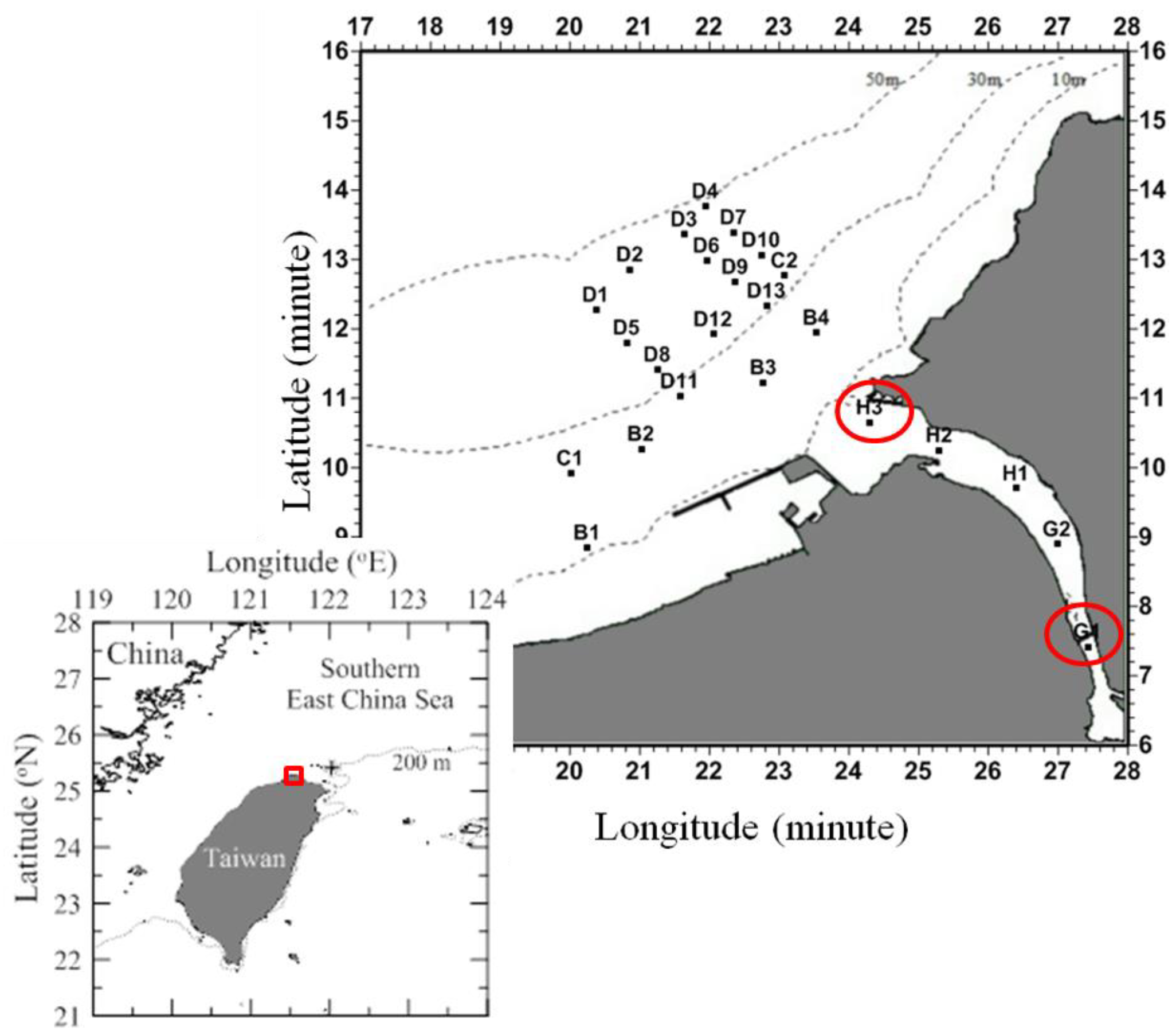
2.1. Sampling

The Danshui River estuarine system, located in northern Taiwan (25°09′42″ N, 121°26′55″ E, Figure 1), is formed by the confluence of the Tahan Stream, the Hsintien Stream, and the Keelung River. According to the annual water quality report published by the nearby Guandu Nature Reserve, river water temperatures range from 12.9 to 32 °C, and salinities range from 7 to 12 PSU (Practical Salinity Units) at low tide and 25 PSU at high tide [12].
We collected surface water samples (1 m) in July 2021 at four stations within Danshui River by boat and at twenty stations in the Danshui River estuary by New R/V Ocean Research II vessel (Figure 1). Using a SeaBird General Oceanic Rosette, the surface temperatures and salinities were measured at each location. Chl a was measured using in vitro fluorometry (Turner Design 10-AU-005) after samples were filtered (25 mm GF/F) for analysis [13]. Previously described methods were used to measure nutrient concentrations in seawater samples [13].

2.2. Viral Production and Viral Decay Rate Experiments

Time-course experiments were carried out at two stations (Station G1: inner estuary; Station H3: coastal water) for measuring viral production and viral decay rate. At the first step, we prepared the grazing-free water by vacuum filtering 1 L of surface seawater through a polycarbonate track-etched filter membrane with a pore size of 2 μm and a diameter of 47 mm (Whatman). For viral dilution, a Minimate TFF Capsule (Pall) was used to filter 200 mL of grazer-free seawater with a molecular weight cut-off of 30 kDa and produce virus-free filtered water. By mixing 200 mL of virus-free water with 50 mL of grazer-free water, prokaryotic and viral abundance of the seawater was reduced by approximately 20%. The diluted incubation water was thoroughly mixed and filled in 50 mL polycarbonate incubation bottles. Then, immediately after preparation, the polycarbonate bottles were moved outside the laboratory close to the sampling site and then incubated for 24 h under natural light in a thermo-controlled incubator with in situ temperature; the treatments were performed in triplicate. In this study, linear regression between viral abundance and incubation time was used for calculating net viral production (NVP) (viruses mL−1 h−1). The viral decay rate (VD) was assessed according to the method of Noble and Furhman [14]. Water samples were filtered through polycarbonate filters with pores of 0.2 μm to exclude bacteria and particles larger than 0.2 μm. To determine prokaryotic and viral abundance, subsamples of 1 mL were taken at the beginning of the experiment and every 2 h for a 24 h period. From the increase or decrease in viral abundance over time, the net viral production and viral decay constant k was calculated by fitting a linear regression to the viral abundance versus time. The slope of the line is the net viral production (NVP) and decay (VD) constant k h−1 [15]. Based on our NVP and VD patterns in the experiments conducted in July, gross viral production (GVP) was estimated as the sum of NVP and VD (the equation is “Net viral production (NVP) = Gross viral production (GVP) − Decay rate (VD)”).

2.3. Determination of Prokaryotic Growth Rates

In this study, the growth rate of prokaryotes was estimated in these 20% diluted samples during the study period, where the prokaryotic abundance was monitored over time. The prokaryotic growth rate was calculated as follows:
μ = ln (Nt/N0) t
where μ is the growth rate (h−1), N0 and Nt are the abundances of prokaryotes at the beginning and end of the incubation experiment, and t is the incubation time (24 h).

2.4. Fluorescence Cytometry (FCM) for the Estimation of Viral and Prokaryotic Abundance

We collected viral subsamples of 0.5 mL every two hours from each incubation set up and fixed these samples in glutaraldehyde (0.5% final concentration) for 15 min at 4 °C and subsequently deep froze them in liquid nitrogen. Prokaryotic subsamples (1 mL) were also collected every 2 h from each incubation and fixed in paraformaldehyde (1% final concentration). Virus and prokaryotic samples were preserved at −80 °C until FCM analysis.
CytoFLEX S flow cytometer (Beckman Coulter, Indianapolis, IN, US) equipped with a 488 nm air-cooled argon-ion laser, a standard 525 nm filter, and an SYBR signal trigger was used to analyze a triplicate water sample for viral and prokaryotic abundance. Prior to staining, viral samples were diluted 1:10 in Tris-EDTA (TE) buffer (pH 8.0, EM grade) to minimize the interference from high particle density. Incubation at 80 °C in the dark for 10 min stained the diluted samples with SYBR Green I (1:50,000 of commercial stock). Samples were stained, cooled in an ice bath to 25 °C and processed with FCM according to the method of Brussaard [16]. Blank controls of TE buffer stained with the same concentration of SYBR Green I were run for detecting and eliminating any noise from the buffer. In accordance with the protocol of Hammes and Egli [17], prokaryotic samples were stained with SYBR Green I (final concentration 1:10,000) for 15 min in darkness and processed through FCM.

2.5. Statistical Analysis

The relationship between viral abundance and incubated time for triplicate incubations was analyzed using linear regression analysis. ANOVA was used to test for statistical differences between the viral production and viral decay rates estimated by the slopes of the linear regressions during time-course experiments. Any associations between the measured biological (Chl a) and environmental parameters (temperature, salinity, NO3, NH4, PO4, dissolved oxygen, and total suspended matter) and their combined effect on prokaryotic and viral abundance were explored along the estuary (24 stations) using principal component analysis (PCA) with a correlation matrix. All data were log transformed to satisfy the assumptions of this analysis. STATISTICA 7.0 software was used for all statistical operations. A probability value of <0.05 was considered significant.

3. Results

3.1. Environmental Parameters

Surface water temperature remained relatively constant (30.5 °C to 31 °C) in the inner estuary, and significantly lower values (28.3 °C to 29.3 °C) were observed (Figure 2A) in the coastal waters. Surface water salinity ranged between 9.5 and 33.5 PSU and increased from inner estuary to coastal water, showing significant spatial differences in salinity in this study (Figure 2A). In terms of nutrient distribution, nitrate concentrations ranged between 1.2 and 19.2 μM, and there was a 16-fold decrease in nitrate from the inner estuary to coastal water (Figure 2B). The correlation between nitrate concentrations and salinity (r = 0.95, p = 0.008, n = 19) (Figure 2B) was significant. Chlorophyll a concentration varied between 0.53 and 1.03 mg m−3; however, spatial variations in chlorophyll a concentration were found to be non-significant in this study (Figure 2C).

3.2. Prokaryotic and Viral Abundance in the Surface Water

Prokaryotic abundance varied from 2.4 ± 0.6 × 10⁵ to 12 ± 2.3 × 10⁵ cells mL−1 and viral abundance from 2.3 ± 0.9 × 10⁵ to 6.9 ± 1.3 × 10⁵ viruses mL−1 during the study period (Figure 3A). Non-significantly higher prokaryotic and viral abundance was observed in the inner estuary in this study (Figure 3A). On the entire dataset, the correlation between salinity and prokaryotic abundance was not significant (r = 0.23, p = 0.115, n = 19) (Figure 3A), but the correlation between chlorophyll a and prokaryotic abundance (r = 0.53, p = 0.007, n = 19) (Figure 3B) was significant.
PCA ordination with vectors for prokaryotic and viral abundance and environmental variables was shown in Figure 4 and shows the ordination of the first two principal components (PC1 and PC2). Based on the biplot, PC1 (51.6%) and PC2 (18.1%), accounted for 69.7% of the total variability of the environmental indices. Results indicate that PC1 pertains to the gradient of physical-chemical variables from estuaries to coasts, while PC2 pertains to the mode of variation of biological variables uncorrelated with physical-chemical parameters. During the study period, there were similar spatial fluctuations in viral abundance, prokaryotic abundance, and chlorophyll a concentration (Figure 4).

3.3. Prokaryotic Growth Rate, Viral Production, and Decay Rates

The effectiveness of the dilution technique is dependent on the efficiency of the 30 kDa filtration step to remove viruses from the 0.2 μm filtrate. In this study, we estimated prokaryotic growth rate in the 20% diluted samples. Prokaryotic abundance increased from 2.0 ± 0.4 × 10⁵ to 38.7 ± 2.8 × 10⁵ cells mL−1 and 0.3 ± 0.1 × 10⁵ to 1.2 ± 0.4 × 10⁵ cells mL−1 during the incubation at the inner and coastal stations, respectively (Figure 5). In this situation, the growth rate of prokaryotes at the inner and coastal stations was measured at 0.12 ± 0.3 h−1 and 0.07 ± 0.02 h−1, respectively (Figure 5).
Net viral production and decay rates are shown in Figure 6. From the dilution experiments, we found non-significant changes in viral abundance at both sampling stations in NVP experiments (Figure 6A,C), and non-significant NVP was estimated in July (ANOVA, p > 0.05). Further, all values for viral decay were nearly 2.1 ± 0.5 × 10⁴ virus mL−1h−1 (Figure 6B,D), and differences between both stations were non-significant. Viral decay almost balanced gross viral production in this study; based on this calculation, the gross viral production was, in all cases, determined to be nearly 2.1 ± 0.5 and 2.1 ± 0.3 × 10⁴ virus mL−1h−1 at both stations.

4. Discussion

In the current study, we investigated the association of viral abundance with prokaryotic abundance and environmental variables in a subtropical Danshui estuary in summer. This study also provided new data on various viral parameters (viral production and viral decay rate) in the estuary. Viral production and decay, as well as the viral balance, must be carefully examined in aquatic environments to further our understanding of the viral dynamics.
Temperature, salinity, and nutrients within estuarine environments are characterized by steep gradients [18]. Since prokaryotes in the estuarine region are affected by complex interactions among these environmental factors, it is difficult to determine which environmental factors control prokaryotic abundance. Numerous studies have been conducted along salinity gradients, especially within estuaries, which have shown that salinity dynamics influence the distribution and composition of prokaryotic communities [19,20]. Figure 4 presents a visual summary of the contributions from environmental factors and compares them with prokaryotic and viral abundance at all stations in this study. However, prokaryotic and viral abundance in a subtropical Danshui estuary in summer is not related to physical-chemical parameters. Furthermore, as shown in Figure 3B, about 28% of the variability in prokaryotic abundance could be explained by phytoplanktons. Generally, the Danshui River estuary has a water residence time of the order of 1–2 days [21], and thus, coastal waters in this estuary receive little allochthonous organic matter and nutrients. Therefore, the spatial distribution of prokaryotes might be more dependent on the distribution of phytoplankton. These findings are similar to those observed by Shiah and Ducklow [22] and suggest that allochthonous organic matter supply might be the major factor regulating prokaryotic growth in the estuarine environment in summer. Furthermore, from the PCA results it appears that prokaryotic abundance was unrelated with the salinity gradient, as it should be expected that allochthonous organic matter supply might be the major factor regulating prokaryotic growth in coastal waters. Indeed, in this study, the dynamics of prokaryotic growth increased significantly from the coastal station (0.07 h−1) to the inner station (0.12 h−1) (Figure 4); these were likely influenced by substrate supply, with higher prokaryotic growth rates associated with higher allochthonous organic matter and nutrients at the inner station. Besides the effect of substrate supply on the spatial relationships with prokaryotic abundance in this study, top-down controls, such as grazing or viral lysis, also set limits on prokaryotic abundance [23,24,25]. Overall, viral abundance was related to prokaryotic abundance in the present study (Figure 3A and Figure 4); thus, viruses are potentially important and have a key role in controlling prokaryotic in this estuarine environment. In most cases, viral abundance correlates significantly with prokaryotic abundance changes [4,26]. Aside from this, it is widely acknowledged that lysed prokaryotic cells enhance prokaryotic production by releasing nutrients and carbon into the dissolved fraction, and experimental evidence indicates that viral lysis may be important for nutrient regeneration in some environments [3]. In addition, it may also explain why viral and prokaryotic abundance in a subtropical Danshui estuary in summer is not correlated to physical-chemical parameters. Moreover, viral abundance and chlorophyll a concentration showed similar patterns of spatial fluctuations (Figure 4). Chl a has been used as an index for photoautotrophic biomass, and viruses infecting marine phytoplankton can be significant agents of mortality [26]. Although picophytoplankton was not determined in this study, Bettarel et al. [27] discovered a higher relative abundance of viruses other than bacteriophage viruses, such as cyanophage viruses, in productive environments. However, the VPR (0.42 to 2.0) in this estuarine environment was low in July. The VPR index has been reported for more than 30 years and is used as a measure of viral influence in aquatic environments [1,7]. High VPR typically represents high and ongoing viral dynamics. In contrast, low ratios tend to indicate high viral decay rates, diminished viral activity, or the absence of viruses. In this situation, a potential mechanism for this pattern may be the grazing by nanoflagellates, an important causative agent of prokaryotic mortality, thus controlling prokaryotic carbon production in this estuarine environment in summer [28].
A review of 210 articles by Parikka et al. [8] indicates that the overall average VPR values in coastal and estuarine waters were 20.7 and 11.4, respectively. However, the apparent trend reported by the same group [8] showed that, compared with coastal/estuarine waters, offshore waters suggested higher mean VPR values. Parikka et al. [8] also pointed out that coastal and estuary waters are more productive than offshore waters and that high viral abundances accompany disproportionately high bacterial abundance, resulting in a lower VPR. SYBR Green I was used to stain prokaryotic samples, and picophytoplankton could not be distinguished from these samples, thus, a higher picophytoplankton community in coastal and estuary waters than in offshore regions [10] makes it possible that there is an overestimation of prokaryotic abundance, causing a lower VPR. Furthermore, it is also possible that VPR is lower in coastal and estuarine waters due to a higher viral loss than in open ocean waters. Similar results have been reported by Tsai et al. [29]. Lower VPR were also observed at semi-enclosed coastal waters (0.9–6.1), probably as a result of higher suspended matter causing removal of viruses from the surface waters. Viruses tend to sediment at higher rates when they are attached to particles with more suspended matter [14]. Our knowledge of experiments of viral loss in these environments is scarce, thus, information on viral decay in these habitats could provide insights into the reasons behind the differences in their VBR values.
Data from our studied stations in the Danshui estuary showed that VPR (0.42 ± 0.11 to 2.0 ± 0.25) was lower than the average value obtained by Parikka et al. [8]. In order to accurately assess viral dynamics, it is imperative to measure viral production and decay rates independently. With this analysis and an evaluation of viral life strategies, a complete picture of the fate of viruses can be revealed, and factors that influence their removal can be identified. Our results throughout this study indicate that viral losses through viral decay counterbalance gross viral production (Figure 6). In the present study, we estimated viral decay by filtration-based method [13] that is based on the removal of prokaryotes, aggregates, and suspended particles and may result in a lower rate of virus removal through this incubation system. Hence, viral decay rates in this study may have been partially underestimated with this method. If we consider these environmental factors for controlling viral losses (suspended particles, UV light), a negative net balance of viral abundance may be caused in this estuarine region. It is, therefore, reasonable to observe lower VPR values (0.42 ± 0.11 to 2.0 ± 0.25) in this study. Furthermore, we estimated that the gross viral productions were nearly 2.1 × 10⁴ virus mL−1h−1 at both stations. Based on reports of Parada et al. [30] from a variety of different aquatic environments, the mean burst size was calculated to be 24 and 34 for marine and freshwater environments, respectively. In this situation, the cause for 9 × 10² and 6 × 10² cells mL−1 of prokaryotic abundance was direct lysis and viral infection, resulting in an average 1% decrease in prokaryotic production. In this regard, the influence of viral activity on prokaryotes could be ignored in our study during summer, which might help explain the lower VPR seen in this estuarine environment.
Viruses and protozoa are thought to be two major factors that control prokaryotic mortality in oceanic systems [31]. Our findings in this study suggest that nanoflagellate grazing can exert a relevant top-down control on prokaryotes during summer in this estuarine region. Similar results of Tsai et al. [26] revealed generally higher ratios of nanoflagellate grazing in summer periods. No significant viral lysis rates were reported in subtropical waters of the western Pacific during these periods. Furthermore, according to the study of Bettarel et al. [32], the nanoflagellates are indirectly responsible for the decrease in viral populations by grazing on the infected prokaryotes. On the other hand, the nanoflagellates studied by González and Suttle [33] were found to be able to consume and digest viruses in the marine environment and suggested there is a trophic relationship between nanoflagellates and viruses.

5. Conclusions

In conclusion, our data indicate a significant correlation of viral abundance with prokaryotic abundance and chlorophyll a concentration in the Danshui estuary. Data from our studied stations show that low VBR (0.42 to 2.0) value and viral decay almost balance gross viral production. We suggest that the influence of viral activity on prokaryotes could be ignored in this study during the summer period, which might help to explain the lower VPR seen in this estuarine environment.

Author Contributions

Conceptualization: A.-Y.T.; methodology: A.-Y.T.; validation: A.-Y.T.; formal analysis: P.W.-Y.C., A.-Y.T. and G.-C.G.; investigation: A.-Y.T.; resources: A.-Y.T. and G.-C.G.; data curation: A.-Y.T.; Writing—Original draft preparation: A.-Y.T. and V.M.; Writing—Review and editing: A.-Y.T. and V.M.; funding acquisition: A.-Y.T. and V.M. All authors have read and agreed to the published version of the manuscript.

Funding

The research was conducted in the frame of the Russian state assignments No. 121040600178-6, AAAA-A21-121121700354-9 and supported by RFBR projects 18-44-920026 (works on the GAF phenomenon) and 21-55-52001, and the Ministry of Science and Technology, ROC (Taiwan), grant number NSC 109-2611-M-019-013.

Institutional Review Board Statement

Not applicable.

Data Availability Statement

All data are provided in the main text.

Conflicts of Interest

The authors declare no conflict of interest.

References

  1. Wommack, K.E.; Colwell, R.R. Virioplankton: Viruses in aquatic ecosystems. Microbiol. Mol. Biol. Rev. 2000, 64, 69–114. [Google Scholar] [CrossRef] [PubMed] [Green Version]
  2. Weinbauer, M.G. Ecology of prokaryotic viruses. FEMS Microbiol. Rev. 2004, 28, 127–181. [Google Scholar] [CrossRef] [PubMed] [Green Version]
  3. Corinaldesi, C.; Rastelli, E.; Canensi, S.; Tangherlini, M.; Danovaro, R.; Dell’Anno, A. High rates of viral lysis stimulate prokaryotic turnover and C recycling in bathypelagic waters of a Ligurian canyon (Mediterranean Sea). Prog. Oceanogr. 2019, 171, 70–75. [Google Scholar] [CrossRef]
  4. Mathias, C.B.; Kirschner, K.T.; Velimirov, B. Seasonal variations of virus abundance and viral control of the bacterial population in backwater system of the Danube River. Appl. Environ. Microbiol. 1995, 61, 3734–3740. [Google Scholar] [CrossRef] [Green Version]
  5. Weinbauer, M.G.; Fuks, D.; Puskaric, S.; Peduzzi, P. Diel, seasonal and depth-related variability of viruses and dissolved DNA in the northern Adriatic Sea. Microb. Ecol. 1995, 30, 25–41. [Google Scholar] [CrossRef]
  6. Ogunseitan, O.A.; Sayler, G.S.; Miller, R.V. Dynamic interactions of Pseudomonas aeruginosa and bacteriophages in lake water. Microb. Ecol. 1990, 19, 171–185. [Google Scholar] [CrossRef]
  7. Wigington, C.H.; Sonderegger, D.; Brussaard, C.P.D.; Buchan, A.; Finke, J.F.; Fuhrman, J.A.; Lennon, J.T.; Middelboe, M.; Suttle, C.A.; Stock, C.; et al. Re-examination of the relationship between marine virus and microbial cell abundance. Nat. Microbiol. 2016, 1, 15024. [Google Scholar] [CrossRef] [Green Version]
  8. Parikka, K.J.; Romancer, M.L.; Wauters, N.; Jacquet, S. Deciphering the virus-to-prokaryote ratio (VPR): Insights into virus–host relationships in a variety of ecosystems. Biol. Rev. 2017, 92, 1081–1100. [Google Scholar] [CrossRef]
  9. Proctor, L.M.; Fuhrman, J.A. Viral mortality of marine bacteria and cyanobacteria. Nature 1990, 343, 60–62. [Google Scholar] [CrossRef]
  10. Clasen, J.L.; Brigden, J.S.; Payet, J.P.; Suttle, C. Evidence that viral abundance across oceans and lakes is driven by different biological factors. Freshw. Biol. 2008, 53, 1090–1100. [Google Scholar] [CrossRef]
  11. Jacquet, S.; Miki, T.; Noble, R.T.; Peduzzi, P.; Wilhelm, S. Viruses in aquatic ecosystems: Important advancements of the last 20 years and prospects for the future in the field of microbial oceanography and limnology. Adv. Oceanogra. Limnol. 2010, 1, 71–101. [Google Scholar] [CrossRef]
  12. Lin, H.J.; Shao, K.J.; Chiu, W.L.; Ma, C.J.W.; Hsieh, H.L.; Wu, W.L.; Severinhaus, L.L.; Wang, Y.T.; Chang, W.L. Biotic communities of freshwater marshes and mangroves in relation to salinity: Implications for wetland regulation. Biodiver. Conser. 2003, 12, 647–665. [Google Scholar] [CrossRef] [Green Version]
  13. Gong, G.C.; Shiah, F.-K.; Liu, K.-K.; Wen, Y.-H.; Liang, M.-H. Spatial and temporal variation of chlorophyll a, primary productivity and chemical hydrography in the southern East China Sea. Cont. Shelf. Res. 2000, 20, 411–436. [Google Scholar] [CrossRef]
  14. Noble, R.T.; Fuhrman, J.A. Viral decay and its causes in coastal waters. Appl. Environ. Microbiol. 1997, 63, 77–83. [Google Scholar] [CrossRef] [PubMed] [Green Version]
  15. Mei, M.L.; Danovaro, R. Virus production and life strategies in aquatic sediments. Limnol. Oceanogr. 2004, 49, 459–470. [Google Scholar] [CrossRef]
  16. Brussaard, C.P.D. Optimization of procedures for counting viruses by flow cytometry. Appl. Environ. Microbiol. 2004, 70, 1506–1513. [Google Scholar] [CrossRef] [PubMed] [Green Version]
  17. Hammes, F.; Egli, T. Cytometric methods for measuring bacteria in water: Advantages, pitfalls, and applications. Anal. Bioanal. Chem. 2010, 397, 1083–1095. [Google Scholar] [CrossRef]
  18. Day, J.W.; Hall, C.A.S.; Kemp, W.M.; Yanez-Arancibia, A. Estuarine Ecology; John Wiley and Sons: New York, NY, USA, 1989. [Google Scholar]
  19. Kan, J.; Crump, B.C.; Wang, K.; Chen, F. Bacterioplankton community in Chesapeake Bay: Predictable or random assemblages. Limnol. Oceanogr. 2006, 51, 2157–2169. [Google Scholar] [CrossRef] [Green Version]
  20. Schapira, M.; Buscot, M.J.; Leterme, S.C.; Pollet, T.; Chapperon, C.; Seuront, L. Distribution of heterotrophic bacteria and virus-like particles along a salinity gradient in a hypersaline coastal lagoon. Aquat. Microb. Ecol. 2009, 54, 171–183. [Google Scholar] [CrossRef]
  21. Wang, C.F.; Hsu, M.H.; Kuo, A.Y. Residence time of the Danshuei River estuary, Taiwan. Estuar. Coast. Shelf. Sci. 2004, 60, 381–393. [Google Scholar] [CrossRef]
  22. Shiah, F.K.; Ducklow, H.W. Temperature regulation of heterotrophic bacterioplankton abundance, production, and specific growth rate in Chesapeake Bay. Limnol. Oceanogr. 1994, 39, 1243–1258. [Google Scholar] [CrossRef]
  23. Fuhrman, J.A.; Noble, R.T. Viruses and protists cause similar bacterial mortality in coastal seawater. Limnol. Oceanogr. 1995, 40, 1236–1243. [Google Scholar] [CrossRef]
  24. Weinbauer, M.G.; Suttle, C.A. Lysogeny and prophage induction in coastal and offshore bacterial communities. Aquat. Microb. Ecol. 1999, 18, 217–225. [Google Scholar] [CrossRef] [Green Version]
  25. Christaki, U.; Giannakourou, A.; Van Wambeke, F.; Gregori, G. Nanoflagellate predation on auto- and heterotrophic picoplankton in the oligotrophic Mediterranean Sea. J. Plankton. Res. 2001, 23, 1297–1310. [Google Scholar] [CrossRef] [Green Version]
  26. Cochlan, W.P.; Wikner, J.; Steward, G.R.; Smith, D.C.; Azam, F. Spatial distribution of viruses, bacteria and chlorophyll a in nentic, oceanic and estuarine environments. Mar. Ecol. Prog. Ser. 1993, 92, 77–87. [Google Scholar] [CrossRef]
  27. Bettarel, Y.; Sime-Ngando, T.; Amblard, C.; Dolan, J. Viral activity in two contrasting lake ecosystems. Appl. Environ. Microbiol. 2004, 70, 2941–2951. [Google Scholar] [CrossRef] [Green Version]
  28. Tsai, A.Y.; Gong, G.C.; Hung, J. Seasonal variations of virus- and nanoflagellate-mediated mortality of heterotrophic bacteria in the coastal ecosystem of subtropical western Pacific. Biogeosciences 2013, 10, 3055–3065. [Google Scholar] [CrossRef] [Green Version]
  29. Tsai, A.Y.; Gong, G.C.; Liu, H. Seasonal variations in virioplankton and picoplankton in semi-enclosed and open coastal waters. Terr. Atmos. Ocean. Sci. 2018, 29, 465–472. [Google Scholar] [CrossRef] [Green Version]
  30. Parada, V.; Herndl, G.J.; Weinbauer, M.G. Viral burst size of heterotrophic prokaryotes in aquatic systems. J. Mar. Biol. Assoc. UK 2006, 86, 613–621. [Google Scholar] [CrossRef]
  31. Wilhelm, S.W.; Brigden, S.M.; Suttle, C.A. A dilution technique for the direct measurement of viral production: A comparison in stratified and tidally mixed coastal waters. Microb. Ecol. 2002, 43, 168–173. [Google Scholar] [CrossRef]
  32. Bettarel, Y.; Sime-Ngando, T.; Amblard, C.; Carrias, J.F.; Portelli, C. Virioplankton and microbial communities in aquatic systems: A seasonal study in two lakes of differing trophy. Freshw. Biol. 2003, 48, 810–822. [Google Scholar] [CrossRef]
  33. González, J.M.; Suttle, C.A. Grazing by marine nanoflagellates on viruses and virus-sized particles: Ingestion and digestion. Mar. Ecol. Prog. Ser. 1993, 94, 1–10. [Google Scholar] [CrossRef]
Figure 1. Map of sampling stations. Stations where viral production and decay experiments were carried out are indicated on the map as G1 (inner station) and H3 (coastal station). In the lower image, the study area is indicated by a red box.
Diversity 14 00237 g001
Figure 2. Relationships between salinity and temperature (A), NO3 (B), and Chl a concentrations (C) during the study period. The line indicates linear regression fitted to the data of the relationship between salinity and NO3 concentration.
Diversity 14 00237 g002
Figure 3. Relationships between salinity and prokaryotic (○), and between salinity and viral abundance (×) (A) during the study period. Furthermore, the relationship between Chl a concentrations and prokaryotic abundance is shown in (B).
Diversity 14 00237 g003
Figure 4. Principal component analysis (PCA) ordination diagram of prokaryotes, virus (V), virus-to- prokaryotes ratios (VPR), and environment variables at 24 stations (T: temperature; S: salinity; DO: dissolved oxygen; TSM: total suspended matter; and nutrients: NO3, NH4 and PO4) during the study period.
Diversity 14 00237 g004
Figure 5. Temporal variations of prokaryotic abundance during 24 h incubations in the inner estuary (●) and coastal waters (□).
Diversity 14 00237 g005
Figure 6. Temporal variations of viral abundance during 24 h incubations for net viral production and viral decay experiments in the inner estuary (A,B) and coastal waters (C,D), respectively.
Diversity 14 00237 g006
Publisher’s Note: MDPI stays neutral with regard to jurisdictional claims in published maps and institutional affiliations.

