Article

Taxonomic Composition of Protist Communities in the Coastal Stratified Lake Kislo-Sladkoe (Kandalaksha Bay, White Sea) Revealed by Microscopy

by
Yulia V. Mindolina
1,
Elena A. Selivanova
1,
Marina E. Ignatenko
1,
Elena D. Krasnova
2,
Dmitry A. Voronov
3 and
Andrey O. Plotnikov
1,*
1
Orenburg Federal Research Center, Institute for Cellular and Intracellular Symbiosis, Ural Branch of Russian Academy of Sciences, 460000 Orenburg, Russia
2
Pertsov White Sea Biological Station, Biological Faculty, Lomonosov Moscow State University, 119234 Moscow, Russia
3
Kharkevich Institute for Information Transmission Problems, Russian Academy of Sciences, 127051 Moscow, Russia
*
Author to whom correspondence should be addressed.
Diversity 2023, 15(1), 44; https://doi.org/10.3390/d15010044
Submission received: 7 November 2022 / Revised: 23 December 2022 / Accepted: 25 December 2022 / Published: 29 December 2022
(This article belongs to the Special Issue Ecology of Microbes in Marine and Estuarine Ecosystems)

Abstract

:
Lake Kislo-Sladkoe is a stratified water body partly isolated from the White Sea. Perennial meromixis in the lake irregularly alternates with mixing events. Taking into account that the protists of Arctic coastal stratified water bodies are understudied, we evaluated for the first time the vertical structure, species richness, and diversity of protists assigned to different taxonomic groups in Lake Kislo-Sladkoe using light, luminescent, and scanning electron microscopy. To test the research hypothesis that a mixing event affects the vertical stratification and species composition of protists in a stratified lake, we compared the protist communities of Lake Kislo-Sladkoe in two extremely different states: strong meromixis vs. full vertical mixing. A total of 97 morphologically distinct phototrophic, heterotrophic, and mixotrophic protists were revealed with the most diverse supertaxa SAR (59), Obazoa (9), and Excavates (14). The hidden diversity of protists (43 species) was a bit less than the active diversity (54 species). A taxonomic list and micrographs of cells for the observed protists are provided. The majority of species revealed are cosmopolitan or widespread in the northern sea waters. The vertical patterns of protist communities were absolutely different in 2018 and 2021. In July 2018, clearly distinct protist communities inhabited different layers of the lake. Bloom of cryptophyte Rhodomonas cf. baltica was detected in chemocline, whereas the maximum density of its grazers was observed in adjacent layers, mainly dinoflagellates Gymnodinium sp. and Scrippsiella trochoidea, as well as a ciliate Prorodon sp. In 2021 due to the recent mixing of lake and seawater, there were no distinct communities in the water column except the superficial 0–1 m layer of fresh water.

Graphical Abstract

1. Introduction

Meromixis is a stage of the lake evolution determined by various external or internal factors providing persistent stratification of a water column with the formation of two main layers: the upper aerobic layer (mixolimnion) and the lower anaerobic layer (monimolimnion) [1]. Despite plenty of lakes on Earth, only about 200 of them have been recognized as meromictic [2,3]. On the White Sea shore, numerous lagoons and lakes differing in their stage of isolation from the sea have arisen due to post-glacial uplift [4]. Some of the lagoons feature a stable stratification including multiyear meromixis with euxinic anoxia in the monimolimnion enriched by hydrogen sulphide [4,5]. In the stratified lakes and lagoons, gradients of physicochemical parameters formed in a water column from top to bottom determine the transformation of originally marine microbial communities into vertically stratified multilayer biocenoses [6,7].
Although the meromictic lakes are very attractive for specialists in paleolimnology [3,8], biological and ecological research in such water bodies is often fragmentary, whereas bacterial communities remain the main target of microbiological studies in the meromictic lakes [9]. Approximately 350 reports have been published since 1985 on bacterial communities of meromictic lakes [9]. Fewer articles have been published on the phytoplankton of meromictic lakes and lagoons [9], and a small number of reports on heterotrophic protists, mainly ciliates, may be found in available sources [6,7,10,11,12,13,14,15,16,17]. Thus, protists including all unicellular eukaryotes represented by algae, protozoa, and zoosporic fungi [18] remain understudied in meromictic water bodies.
The first study of ciliate distribution in meromictic waters based on their microscopic identification to the genus and species levels was carried out by Fenchel et al. in 1990 in two Danish meromictic fjords [6]. In this report, for the first time, a strong vertical zonation of heterotrophic protist communities has been disclosed, as well as a biomass maximum of ciliates in the oxycline [6]. In 1995, Fenchel et al. reported the taxonomic composition of protists from the meromictic Mariager Fjord, analyzed their vertical distribution, diversity using both fresh samples and enrichment cultures, and described several new genera and species [10]. At the end of the XX century and later, several papers have been published based on the vertical distribution of protists studied with microscopy methods in meromictic marine basins and lakes with bottom anoxia. Some of them have described a diversity of autotrophic and mixotrophic protists in phytoplankton or phytobenthos [19,20,21,22,23,24,25]. Other reports were devoted to heterotrophic protists, mainly ciliates [7,11,15,16,26,27,28]. A few studies provided data on both phototrophic and heterotrophic-dominated protists, without a thorough examination of the protist diversity and richness [4,13,14,29,30,31,32]. All the reports mentioned above, except [10], were devoted to the active diversity of protists, which were numerous in the water column at the moment of sampling or just after it. The hidden or cryptic diversity of protists includes a number of rare or dormant (encysted) species that are often overlooked during microscopy of fresh environmental samples but can be easily revealed in enrichment cultures [33]. Estimation of hidden diversity has a great ecological significance because it is able to disclose much more species, especially bacterivorous ciliate, amoeboid, and flagellated protists that are delicate, inconspicuous, or encysted, and usually overlooked when studied through direct microscopy of water samples without enrichment cultures [33,34]. Unfortunately, the hidden diversity of protists in meromictic waters has not been investigated recently, except for a study of the taxonomic composition of heterotrophic flagellates in the inland Siberian meromictic Lake Shira [11].
Microscopic identification of protists is a laborious and time-consuming procedure. For this reason, new molecular techniques were applied in the XXI century for the evaluation of protist diversity in meromictic waters. The first studies were based on molecular cloning of the 18S rRNA gene from the total DNA isolated from a water sample, followed by Sanger sequencing of clone DNA libraries [12,15,35,36,37,38,39,40,41]. Later high-throughput techniques of next-generation DNA sequencing (NGS) have been used in a few studies for DNA metabarcoding of protists in meromictic water bodies and basins [40,42,43,44,45,46,47,48]. Though DNA sequencing methods give a list of exact genetic signatures for the majority of protists that are present in the sample studied, the species richness cannot be evaluated correctly due to the inconsistency between DNA sequences and the morphospecies of protists resulting from a lack of data on intra- and interspecific sequence variation for most protists [49]. In addition, DNA data cannot identify the vegetative or dormant state of the protist cell and characterize the active and hidden diversity of protists [50]. That is why microscopic methods remain essential in the evaluation of the species richness of protists.
Strong stratification of meromictic water bodies and basins may be affected by some meteorological events or climatic conditions [7,30,51]. Lake Kislo-Sladkoe located on the White Sea coast is one such water body, in which a period of meromixis usually lasts for a few years, and then is replaced by an event of full vertical mixing [52]. Obviously, a disturbance of meromixis may affect gradients of physicochemical parameters and ecological processes. However, the vertical distribution of protists in meromictic waters after such events has been demonstrated only in three reports [7,40,52]. Moreover, these studies described only certain protist groups of interest, e.g., ciliates [7] or algae [52]. In the Saanich Inlet, a seasonally anoxic fjord on the coast of Vancouver Island (Canada) the community shifts between major taxa of protists have been detected using NGS, as well as dominant sequences of the 18S rRNA gene that were not assigned to certain protist genera and species [40].
Thus, despite the rarity and curiosity of the meromictic water bodies, the diversity and species richness of protists inhabiting such waters, especially coastal lakes and lagoons, have been studied insufficiently. There are few reports on the species composition of protists in such water bodies covering all ecological groups including microalgae, protozoa, and groups with diverse trophic strategies, e.g., Dinoflagellata including photosynthetic, heterotrophic, mixotrophic, and predator species. The hidden diversity of protists in the coastal meromictic waters has not been studied yet. Taking into account the significance of meromixis for the development of stratified microbial communities, very little is known about the influence of stratification disturbance on the vertical distribution and the species richness of protist communities in meromictic waters, especially coastal lakes and lagoons.
Lake Kislo-Sladkoe (Kandalaksha Bay, White Sea) is a coastal meromictic water body at an early stage of separation from the sea [53]. Although physicochemical and hydrological parameters in Lake Kislo-Sladkoe have been being monitored since 2010 [53,54], research on protist communities started only a few years ago. Particularly, a bloom of cryptophyte Rhodomonas sp. in the redox zone has been recorded [55]. A clear vertical zonality of the composition, number, and biomass of phytoplankton has been described in this lake [52]. However, the biodiversity of protists is not limited to phytoplankton only. Heterotrophic protists have remained outside of attention so far due to the issues of their identification. In the present study, we evaluated for the first time the vertical structure, species richness, and diversity of protists assigned to different taxonomic groups in Lake Kislo-Sladkoe using light, luminescent, and scanning electron microscopy. It is the first study of protist communities in a stratified water body on the Arctic coast of Russia and in water bodies separating from the sea. To test the research hypothesis that a mixing event affects the vertical stratification and species composition of protists in a stratified lake, we compared the protists communities of Lake Kislo-Sladkoe in two extremely different states: strong meromixis with chemocline and anaerobic monimolimnion vs. full vertical mixing after recent high tidal and wind surges. In addition, we supposed that the study of hidden diversity would enlarge the species list of recorded protists due to mainly small-cell, rare, or encysted species.

2. Materials and Methods

2.1. Description of the Studied Water Body

Lake Kislo-Sladkoe (66°32′54″ N, 33°8′05″ E) is located at the shore of Kandalaksha Bay of the White Sea, 1.5 km east of the White Sea Biological Station of the Lomonosov Moscow State University. It has an area of 1.6 thousand m2, average depth of 1 m, and a small bottom depression with a depth of 4.5 m. The lake is separated from the sea with a rocky bar as high as the average high tide level.
According to the reconstruction of the lake formation, it passed through four stages: the stage of a sea bottom depression (1.5 thousand years ago); a wide strait between the mainland and a low discontinuous ridge of moraine hills (until the middle of the XIX century); a semi-isolated lagoon (until the middle of the 20th century); a meromictic water body at an early stage of separation from the sea (current state) [53]. Currently, the lake gets water from the sea through a single intermittent channel [54,56]. At high tides, in syzygy (every lunar month for several tidal cycles), or during high surges, which most often occur in late autumn shortly before freezing, salt water from the sea flows over the narrow rocky barrier. The lake also accumulates fresh runoff from a drainage basin 10 times larger than the water area of the lake, which leads to the dilution of the surface layer of the lake. The largest volume of freshwater comes due to snow melting. During a low-water summer period, the inflow of fresh water is limited by filtration from a neighboring bog and a tiny stream with a flow rate of less than 1.5 m3 per day.
The adjacency of salt water from the sea and freshwater from the surrounding area forms a stable vertical stratification that persists for several years. This allows to qualify it as the meromictic lake of ectogenic coastal marine type (Ib) [1,3,57]. In the bottom depression, due to bacterial sulfate reduction, hydrogen sulfide is formed and accumulated, but the upper bound of the sulfide zone varies in different years [58].

2.2. Measurement of Physicochemical Parameters and Sampling

Lake Kislo-Sladkoe has been monitored since 2010. In this study, sampling and measurement of hydrological characteristics were carried out on 6 July 2018 and 11 September 2021. All measurements were taken at the deepest point of the lake from an inflatable boat. Salinity, temperature, and redox potential were determined with a YSI PRO 30 conductivity meter (YSI Inc., Yellow Springs, OH, USA). Oxygen concentration was registered with a ProODO Optical Dissolved Oxygen Instrument probe (YSI, USA). Illuminance was evaluated with a digital AS803 LCD Display Digital Lux Meter illuminometer (Dongguan Wanchuang Electronic Products Co., Ltd, Dongguan, China) modified manually for immersing underwater. Samples were taken with a GP1352 Whale Premium Submersible pump (Wale Marine, Bangor, Northern Ireland, UK) and a cable with distances marked out. All measurements were provided every 0.5 m in depth and with an interval of 0.1 m in the zone with a sharp gradient. Water samples were taken in sterile 500 mL plastic bottles from the surface to the bottom with an interval of 0.5 m and additionally form the redox horizon.

2.3. Treatment of Samples, Microscopy, and Identification of Protists

The active diversity of protists was studied just after sampling at the Pertsov White Sea Biological Station of the Lomonosov Moscow State University (Poselok Primorskiy, Republic Karelia, Russia). A 15 mL aliquot of water from every water horizon was concentrated for 10 min at 3000 rpm in an Eppendorf 5804 centrifuge (Eppendorf, Hamburg, Germany). The supernatant was carefully removed, leaving a deposit in water of 50 µL volume approximately. Active diversity of protists was observed under a light and luminescent microscope Leica DM2500 (Leica Microsystems GmbH, Wetzlar, Germany) with N2.1 filter set (excitation/transmission 515–560/580 nm). We recorded all colored photosynthetic protists or microalgae, including phytoplankton and nanophytoplankton, as well as heterotrophic protists represented by heliozoans, amoebae, flagellates and nanoflagellates, and ciliates. Cysts and zoosporic fungi were not registered.
The hidden diversity of protists was studied in the “Persistence of microorganisms” Science Resource Center of the Institute for Cellular and Intracellular Symbiosis of the Ural Branch of the Russian Academy of Sciences (Orenburg, Russia). The water samples were transferred to 60 mm plastic Petri dishes, enriched with a suspension of feed bacterium Pseudomonas fluorescens, and incubated under conditions similar to the physicochemical parameters of each water horizon (temperature and oxygen availability). Heterotrophic protists, namely flagellates, amoebae, and heliozoans multiplied in the enrichment cultures during the next two weeks. The hidden diversity of phototrophic protists was studied in 60 mm plastic Petri dishes with the water samples incubated at +15 °C under a luminescent lamp (20 W). In such conditions, certain species of diatoms, green algae, and dinoflagellates multiplied. The cultures were examined at 3, 6, 9, 12, 15, and 20 days to observe protists, which were rare or invisible in fresh samples [34] using direct light Axio Scope A1 microscopes (Carl Zeiss Vision GmbH, Aalen, Germany) with DIC objectives, phase contrast, and water immersion, equipped with Axiocam 105 color and Axiocam 208 color cameras (Carl Zeiss Vision GmbH, Aalen, Germany).
Species identification of some protists with a dense external cover, shell, or plates, such as diatoms, cryptophytes, euglenozoans, heliozoans, testate amoebae, and heterotrophic flagellates was carried out using a Tescan Mira 3 scanning electron microscope (Tescan Brno, s.r.o, Brno, Czech Republic) in the Center for Revealing and Support of Talented Children “Gagarin” (Orenburg, Russia). For SEM, the aliquots of fresh samples were concentrated by centrifugation and fixed with 1% acid Lugol’s solution, 2% formaldehyde, or 4% glutaraldehyde. The cells were washed from a fixing reagent using three-time centrifugation for 5 min at 3000 rpm in a Microspin 12 centrifuge (BioSan, Riga, Latvia). Washed cells were placed on aluminum stubs, dried on air, and coated with gold for 60 s. Cleaning of diatom shells from organic matter was carried out using cold acid treatment [59]. Single cells of testate amoebae and heliozoa were picked out under light microscope from the samples using glass pipette, placed on the cover glasses, and dried on air.
Heterotrophic protists were identified using relevant taxonomic keys and articles [60,61,62,63,64,65,66,67,68,69,70,71,72,73,74,75,76,77,78,79,80,81]. Algae were identified using a number of relevant taxonomic keys and articles [82,83,84,85,86,87,88,89,90,91,92,93,94]. Taxonomy and nomenclature of protists are given according to the last revision of eukaryotes [95]. Nomenclature of Bacillariophyta and Euglenophyceae is given according to online database Algaebase [96]. Vertical distribution graphs of protists were built with GraphPad Prism Version 9.4.1 for Windows (GraphPad Software, San Diego, CA, USA, www.graphpad.com, accessed on 1 July 2022).

3. Results

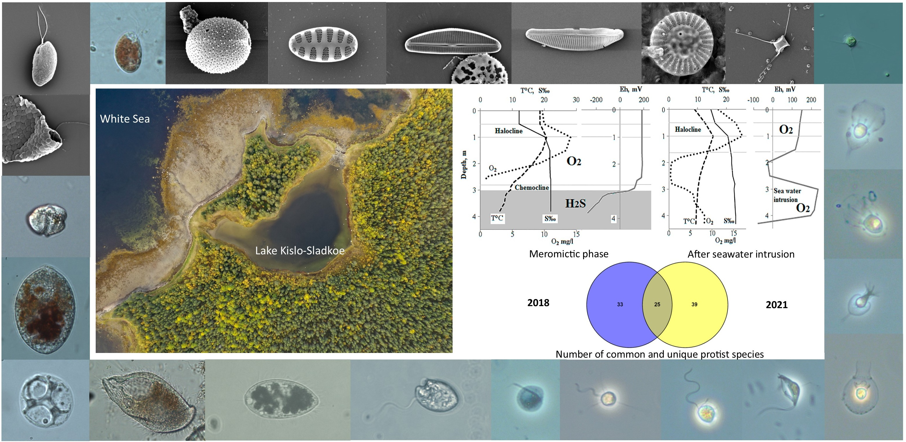
3.1. Hydrological Features of Lake Kislo-Sladkoe, Hydro-Chemical and Hydro-Physical Parameters in the Water Column

The hydrological phase of Lake Kislo-Sladkoe differed greatly in 2018 and 2021. By July 2018 the lake had persisted in the meromictic phase without complete mixing for five years. In July 2018 it consisted of five layers from surface to bottom: (1) a brackish layer of wind mixing up to 0.5 m, (2) a narrow halocline 0.5–1 m, (3) a stagnant saline aerobic layer, oversaturated with oxygen due to phytoplankton photosynthesis 1–3 m, (4) chemocline (transition from aerobic zone to anoxic) 3.0–3.1 m, and (5) near-bottom anaerobic zone 3.1–4.5 m (Figure 1A).
In September 2021, the inflow of seawater occurred after summer meromixis. On the sampling day, seawater with a higher density dropped to a depth of 3.0–3.5 m, and the bottom anaerobic layer lifted to a level of 2.0–3.0 m. At the same time, near-bottom anaerobic water has mixed partially with infused aerobic sea water, and aerobic conditions have been formed inside this layer (Figure 1B).
The light profiles followed the vertical structure of the lake (Figure 1A, B). In 2018, dense microbial suspension prevented the penetration of light through the chemocline resulting in the formation of an aphotic layer below 3.1 m. In 2021, the absence of the chemocline with the dense microbial layer led to a gradual decrease in illumination along the water column and sunlight passed up to the bottom.

3.2. Taxonomic Composition of Protist Communities

In the studied water body, 97 morphologically distinct phototrophic (38) and heterotrophic (59) protists were revealed, 56 of which were identified at the species or infraspecies level, whereas 29 were identified at the genus level only. In addition, some unidentified representatives of Cryptophyta, Haptophyta, Chlorophyta, Crysophyceae, Euglenozoa, and Amoebozoa were found, as well as two heterotrophic protists with an unclear taxonomic position (Figure 2, Figure 3, Figure 4, Figure 5, Figure 6, Figure 7, Figure 8 and Figure 9, Supplementary Table S1).
The greatest number of identified protists belongs to macrotaxonomic groups SAR, Obazoa, and Excavates. SAR is the most diverse (59 species) and composed of heterotrophic flagellates Bicosoecida (4), Developea (1), and Colponemida (1); phototrophic (2) and heterotrophic (6) flagellates Chrysophyceae; diatoms (26); heterotrophic flagellated members of Dictiochophyceae (2); ciliates (9); phototrophic Dinoflagellata (4); and Cercozoa (4) represented by heterotrophic flagellates (3) and a testate amoeba. Obazoa (9) includes heterotrophic flagellates Apusomonadida (2), Choanoflagellata (6), and Filasterea (1). Excavates (14) is represented by heterotrophic (3) and phototrophic (1) flagellates Euglenozoa, heterotrophic flagellates Kinetoplastea (5) and Jacobida (1), anaerobic heterotrophic flagellates Symbiontida (1), and Metamonada (3). The lower number of protists was assigned to Amoebozoa (2), Archaeplastida (2) represented by green algae Chlorophyta, Cryptista (4) including two heterotrophic and two phototrophic flagellates, and Haptista (2) represented by an alga Haptophyta and a heterotrophic heliozoan Centroplasthelida. Finally, some heterotrophic protists of uncertain taxonomic position are present in the taxonomic list such as flagellates Telonemia (1), Ancyromonadida (1), Eoramonas cf. jungensis, and two unidentified flagellated protists.
Active diversity of protists exceeds hidden diversity; 54 species of protists were found in both fresh samples and enrichment cultures, or in fresh samples only. However, a significant number of protists (43 species), mainly heterotrophic flagellates, amoebae, heliozoa, and several diatoms were found only in the enrichment cultures (Supplementary Table S1).

3.3. Vertical Distribution of Protists in July 2018

In July 2018, during the meromictic phase of the lake, distinct protist communities were formed in the superficial freshwater, marine aerobic, chemocline, and marine anaerobic layers (Figure 10, Supplementary Table S1). In particular, only in the superficial brackish layer of wind mixing 0–0.5 m two species of heterotrophic flagellates were revealed: freshwater Reclinomonas Americana, and ubiquitous bodonid Neobodo designis. An amoeba Cochliopodium sp. and the filasterian protist Ministeria sp. were found in enrichment cultures from the superficial samples. They are typical protists for the North Atlantic Ocean and the British coastal waters [97,98]. Diatoms such as freshwater Gomphonema parvulum, marine Amphora micrometra, and Navicula sp. were recorded only in this layer.
Some protists were revealed in both the superficial layer and the aerobic marine one from 0 to 2–2.5 m, such as unidentified Haptophyta, heterotrophic Spumella-like flagellates, Paraphysomonas sp., Monosiga ovata, Actinomonas mirabilis, and Bodo sp. The centric diatom Cyclotella choctawhatcheeana and the araphid diatom Nanofrustulum trainorii were abundant in the same horizons. Recently, N. trainorii has been recorded in this lake [52].
The widespread marine predatory cercozoan Ebria tripartita was found in fresh samples from 0 to 2.0 m, and its mass concentration was noted below the picnocline at the 1–1.5 m depth. This protist feeds on phytoplankton, especially on diatoms and dinoflagellates [99]. Several protists were observed in the lower layer of the aerobic zone only, e.g., the widespread marine heterotrophic flagellates Pseudobodo tremulans, Bicosoeca cf. maris, Cafeteria sp., unidentified Amoebozoa, small unidentified Chlorophyta, marine diatoms Podosira stelligera, Chaetoceros sp. 2, and freshwater diatom Nitzschia paleacea.
A bloom of Cryptophyta represented by Rhodomonas cf. baltica was detected in the chemocline at 3.0–3.1 m, but it was also found at 2.5 m. The specific protist community of Rhodomonas grazers inhabited the chemocline and adjacent layers, including the near-bottom anaerobic zone, such as dinoflagellates Gymnodinium sp., Prorocentrum sp., Scrippsiella trochoidea, and a ciliate Prorodon sp.
The anaerobic heterotrophic protists, such as ciliate Metopus sp., an unidentified representative of heterotrophic euglenozoa Symbiontida, the metamonadian flagellates Hexamita sp. and Carpediemonas membranifera, were observed at the near-bottom depth (4 m) in the anoxic sulphide zone. The great diversity of diatom frustules assigned to 17 species was found in the bottom layer 4 m compared to only 8 species in the overlying layers.

3.4. Vertical Distribution of Protists in September 2021

In September 2021, the mixing event disturbed meromixis due to the intrusion of seawater that had happened just before sampling. As a result, a clear vertical stratification-determining formation of distinct protist communities along the water column was damaged. However, both the brackish superficial layer from 0 to 1 m and the 2 m layer representing the lower boundary of aerobic marine water had protist communities with the most specific taxonomic composition. In particular, an amoeba Cochliopodium sp., heterotrophic flagellates Stephanoeca sp. 2, Salpingoeca sp., Ministeria sp., Amastigomonas caudata, Neobodo curvifilus, unidentified Haptophyta, ciliates Oxytricha sp., Vorticella sp. were revealed only in the superficial layer.
The protist community at the 2 m depth specifically included heterotrophic flagellates Actinomonas mirabilis, Telonema sp.; a testacean amoeba Cyphoderia aff. littoralis; a green unidentified alga Chlorophyta 2; diatoms Melosira nummuloides, Cyclotella meneghiniana, Amphora micrometra, Martyana atomus, Nitzschia microcephala, Planothidium sp., Pseudostaurosira cf. elliptica; euglenophyte Trachelomonas intermedia var. minor.
Many protists inhabited only aerobic and microaerophilic zones of the upper layer 0–2.5 m, such as heterotrophic flagellates Monosiga ovata, Clathromonas butcheri, diverse species of Paraphysomonas, Discocelis saleuta, Goniomonas truncata, Petalomonas poosilla; dinoflagellates Gymnodinium sp. and Scrippsiella trochoidea; diatom Chaetoceros sp. 1; a cercozoan predatory flagellate Ebria tripartita; a Heterophrys-like heliozoan; and unidentified cryptophyte.
The community of bottom aerobic horizon between 4.0 and 4.5 m, resulting from mixing the original anaerobic layer with seawater, comprised of species mainly inhabiting the upper layers and only a few specific protists, such as a diatom Gyrosigma sp., a metamonad flagellate Kipferlia bialata, ciliates Uronema sp., and Euplotes sp. Euplotes sp. was also recorded at 2 m.
Many protist species were found at different depths from surface to bottom: unidentified Amoebozoa; heterotrophic flagellates Pteridomonas danica, Spumella-like flagellates, Oxyrrhis marina, Neobodo designis, Rhynchomonas nasuta, Ancyromonas sigmoides, Stephanoeca paludosa; diatoms Cyclotella choctawhatcheeana and Entomoneis var. subsalina; ciliates Cyclidium sp. and Mesodinium sp.

3.5. Comparison of the Protist Communities in 2018 and 2021

The species lists of protists revealed in Lake Kislo-Sladkoe in 2018 and 2021 were compared using a Venn diagram (Figure 11). A number of common species was less than a number of unique ones. The shared species included Ebria tripartita, Monosiga ovata, Salpingoeca sp., Actinomonas mirabilis, Oxyrrhis marina, Gymnodinium sp., Petalomonas poosilla, Neobodo curvifilus, Neobodo designis, Reclinomonas americana, Cochliopodium sp., unidentified Haptophyta and Amoebozoa, Ministeria sp., and Spumella-like flagellates. Several diatom species were found both in 2018 and 2021, such as Cyclotella choctawhatcheeana, Amphora micrometra, Cocconeis placentula var. euglypta, Martyana atomus, Nanofrustulum trainorii, Navicula antonii, Planothidium sp., and Pseudostaurosira cf. elliptica.
Anaerobic ciliates Plagiopyla sp. and Metopus sp., flagellates Carpediemonas membranifera, Hexamita sp., unidentified Symbiontida, as well as chrysophyte Kephyrion sp. and an unidentified chrysophyte, were unique for the samples of 2018. Rhodomonas cf. baltica, the distinctive species for the chemocline of Lake Kislo-Sladkoe, was observed only in 2018, together with diatom species Paralia sp., Podosira stelligera, Chaetoceros sp. 1, Cocconeis placentula, Gomphonema parvulum, Navicula gregaria, Navicula sp., Nitzschia paleacea, Nitzschia sigma, Staurosirella pinnata, Gyrosigma sp., Cylindrotheca closterium, and Surirella striata. Only five diatom species Melosira nummuloides, Chaetoceros sp. 2, Cyclotella meneghiniana, Entomoneis paludosa var. subsalina, and Nitzschia microcephala were unique for 2021. Ciliates Cyclidium sp., Uronema sp., Vorticella sp., Oxytricha sp., Euplotes sp., heterotrophic flagellates Caecitellus parvulus, Developayella cf. elegans, Clathromonas butcheri, Pteridomonas danica, Bodomorpha reniformis, Cyphoderia aff. littoralis, Discocelis saleuta, Goniomonas amphinema, Goniomonas truncata, Neometanema exaratum, Rhynchomonas nasuta, Kipferlia bialata, Ancyromonas sigmoides, Eoramonas cf. jungensis, and a centrohelid heliozoan Heterophrys sp. were observed only in 2021.

4. Discussion

Lake Kislo-Sladkoe is one of the most extensively studied stratified water bodies on the coast of the Kandalaksha Bay of the White Sea [52,54,55,56,58,100,101,102]. Despite the fact that many phototrophic protists have been found in the lake recently [52,55,100], we managed to record a wide diversity of protists in Lake Kislo-Sladkoe including 97 heterotrophic and phototrophic taxa, although sampling was performed only twice in 2018 and 2021. Such a significant result was provided with different methods of microscopy (bright field microscopy, DIC, phase contrast, luminescent microscopy, SEM), as well as the study of both active and hidden diversity of protists. Recently, a seasonal study of algae in Lake Kislo-Sladkoe was published [52]. Unfortunately, a comparison of our list with the aforementioned study is not possible due to the absence of a taxonomic list. However, the marked diversity of Bacillariophyta and Dinoflagellata in contrast to a poor variety of Chlorophyta and Dictiochophyta was also noted in the lake as in our results [52]. The diversity of heterotrophic flagellates and amoebae in the Lake Kislo-Sladkoe (50 species) is similar to 44 species observed by Fenchel et al. in the Danish meromictic Mariager Fjord [10]. Heterotrophic flagellates were the most diverse, whereas amoebae and heliozoans were represented by five species only in both surveys. Fewer species of heterotrophic flagellates, namely 36, were found in the brackish inland meromictic Shira Lake located in Siberia [11]. Possible reasons may include the lower diversity of inland waters in contrast to marine sites or an artificial decrease in the registered species because of the estimation of hidden diversity with light microscopy only. As to ciliates, their diversity in Lake Kislo-Sladkoe includes only nine species, which is substantially poorer than in the meromictic Mariager Fjord (37 species) and the coastal meromictic Faro Lake (25 species) connected to the Straits of Messina by a shallow channel [10,31]. At the same time, finding seven ciliate species in inland meromictic Shira Lake [7] is very similar to our lake, possibly due to the mutual absence of a permanent connection to the sea.
A general decrease in protist species richness in the lake from surface to bottom in both 2018 and 2021 (Figure 11) is congruent with other observations of vertical patterns of protist communities in meromictic waters [10,11]. In 2018, at the depth of 4 m, the species richness of protists rose again, having exceeded the number of species on the surface horizon. A high diversity of diatoms was revealed in a near-bottom water layer, especially in 2018 when Lake Kislo-Sladkoe was in a phase of meromixis. Particularly, from 21 diatom species registered, 17 were found in the 4 m bottom layer of water, and more than half (13 species) were found exclusively in this layer. This phenomenon may be related to the persistence of intact valves of dead diatoms that had developed in the lake before and then sank to the lake bottom. A widely known resistance of siliceous diatom frustules to adverse environmental conditions provides their storage for centuries in bottom sediments, and usage in paleolimnological reconstructions [53]. Otherwise, transparent exopolymeric particles have been confirmed recently to be the main transport particles determining the transfer of diatoms from plankton to deeper layers in a lake [103].
In the enrichment cultures, we revealed 45 species of mainly heterotrophic protists that correspond to approximately half of all protist species observed. Hidden diversity is determined by mass growth in enrichment cultures of those protists that are in an active state, but rare in the natural habitat or being in the encysted state, such as some heterotrophic flagellates, amoebae, and ciliates. For example, the amoeboid protists Cochliopodium sp. and unidentified Amoebozoa were revealed only in the cultures. Perhaps they tend to be encysted in the natural habitat where insufficient particulate matter to attach, locomote, and feed. In addition, certain physicochemical parameters obviously induce their encystments in an environment, such as changes in temperature and salinity. Therefore, some protists that come up in a culture may have been in the encysted state in the lake water. Unfortunately, enrichment cultures do not distinguish between cryptic active stages of the protists versus encysted stages that emerge under the more favorable conditions of laboratory enrichment. However, revealing the hidden species is crucial for the comprehensive estimation of species richness in the biotope. In our study, 10 species of phototrophic protists including green algae, diatoms, dinoflagellates, and euglenozoans were detected only in enrichment cultures. This fact supports the assumption that the real diversity of both heterotrophic and phototrophic protists remains underestimated, being evaluated only in fresh samples without the assessment of hidden diversity.
Most of the protists revealed in Lake Kislo-Sladkoe are euryhaline and have been found in both marine and freshwater sites, e.g., heterotrophic protists M. ovata, Salpingoeca sp., Ministeria sp., A. mirabilis, P. cylicophora, C. butcheri, Telonema sp., N. exaratum, P. poosilla, G. truncata, G. amphinema, A. sigmoides, R. nasuta, N. designis, N. curvifilus, B. reniformis, and Cochliopodium sp. Other protists had been observed recently only in marine and inland saline waters, such as Stephanoeca sp., P. foraminifera, D. saleuta, E. tripartita, A. crassum, K. bialata, D. grandis, S. parva, A. caudata, P. danica, O. marina, C. parvulus, and D. elegans. Flagellate R. americana was the only representative of freshwater heterotrophic protists. Phototrophic protists or algae were represented mostly by euryhaline and freshwater species, e.g., G. parvulum, C. choctawhatcheeana, M. atomus, N. trainorii, P. cf. elliptica. Marine species of phototrophic protists include M. nummuloides, Paralia sp., A. micrometra, P. stelligera, and R. cf. baltica.
In general, the majority of protists revealed in this study are either cosmopolitan or species widespread in the northern sea waters. For instance, heterotrophic flagellates M. ovata, P. danica, P. poosilla, A. sigmoides, R. nasuta, as well as members of genera Stephanoeca, Salpingoeca, and Spumella, have been found previously in a lake located on the White Sea coast [104]. The species listed above and other heterotrophic flagellates such as A. caudata, A. mirabilis, A. crassum, C. parvulus, D. saleuta, G. amphinema, N. exaratum, N. designis, and N. curvifilus have been noted recently in the protist communities of sandy deposits in the White Sea coastline, in the estuary of the Chernaya River, and in the Velikaya Salma Strait of the Kandalaksha Bay of the White Sea [105,106]. Algae M. nummuloides and P. sulcata have been recorded as dominant and subdominant algae of the White Sea [94,107]. M. nummuloides was rather numerous in the samples studied, and multiplied intensively in the enrichment cultures. Single valves of Paralia sp. and P. stelligera suggest that these species presumably are not common inhabitants of the Lake Kislo-Sladkoe, and are transferred here with inflow of the seawater. Many species of diatoms revealed in Lake Kislo-Sladkoe had been found in the well-studied meromictic Lake Mogilnoe on the Kildin Island in the Barents Sea including M. atomus, M. nummuloides, N. trainorii, N. gregaria, N. microcephala, and P. cf. elliptica [84,85,86].
All ciliates genera found in Lake Kislo-Sladkoe have been found in other meromictic waters. E.g., Cyclidium, Mesodinium, Euplotes, Oxytricha, and Vorticella, have been recorded previously in the inland meromictic Shira Lake (Siberia, Russia) [7]. Cyclidium has been found in the chemocline and the anaerobic zone, whereas Euplotes sp. has been distributed in both the littoral and pelagic zones of the lake [7]. Ciliate Cyclidium also has been reported in the aerobic zone of another meromictic Lake Suigetsu (Japan) [41]. In the meromictic Mariager Fjord, seven ciliate genera from our list have been found except for Oxytricha and Vorticella [10]. At last, Cyclidium, Mesodinium, Euplotes, Metopus, and Plagiopyla have been recorded in the coastal meromictic Faro Lake [31].
All the dinoflagellate genera found in Lake Kislo-Sladkoe have been recorded recently as dominant taxa of phytoplankton in this lake (Gymnodinium, S. trochoidea) [52], or as typical inhabitants of the White Sea littoral sites (Gymnodinium, Prorocentrum) [104,106]. Particularly, G. arcticum dominated at 3.5 and 4.5 m horizons in June 2019, whereas S. trochoidea was the most abundant at 2.5 m in August 2019 [52]. Dinoflagellates have been revealed to dominate in other meromictic lakes, such as the mountain Tibetan Baishihai Lake (Tovellia diexiensis), Japanese Lake Suigetsu (Prorocentrales), the tropical East African Great Lake Kivu, the iron-rich karstic Lake La Cruz in central Spain (Gymnodinium, Peridinium sp., Ceratium hirundinella, and Parvodinium umbonatum), Antarctic Lake Ace (Gyrodinium glaciale, Gymnodinium spp.), and Lake Alatsee in Southwest Germany (Peridiniphycidae and Gymnodiniphycidae) [46,48,108,109,110,111]. A woloszynskioid dinoflagellate T. diexiensis was the causative organism of regular “summer reddening” due to the production of a high amount of astaxanthin-related carotenoids, which are rare for dinoflagellates [108].
Lake Kislo-Sladkoe was in different hydrological phases in 2018 and 2021. Species lists demonstrate differences in the taxonomic composition of the protist communities. Particularly, in July 2018, distinct protist assemblages inhabited different layers of the lake (0–2, 2.5–3.5, and 4 m) following the meromictic stratification. At the same time, more marine species were revealed in the samples of 2021 than in 2018. This phenomenon was presumably determined by the intrusion of a large volume of seawater into the lake due to the syzygy tide, which had happened just before sampling. The mixing of sea and lake waters resulted in the spreading of many protists along almost the whole water column from surface to bottom (Figure 10, Supplementary Table S1). The differences in the taxonomic composition of protist communities are illustrated in a Venn diagram demonstrating that the sum of specific species in 2018 and 2021 was almost three times more than the number of shared species for both years (Figure 11).
For many years of observation, when Lake Kislo-Sladkoe was in a meromictic state, a bloom of the cryptophyte flagellate Rhodomonas sp. appeared in the chemocline, forming a highly dense population there [100]. The same phenomenon was recorded in July 2018, resulting in the formation of an approximately 10 cm layer of pink water in the chemocline. Blooms of different cryptophyte species similar to the bloom of Rhodomonas sp. in Lake Kislo-Sladkoe have been a typical trait for coastal and inland meromictic lakes Ace (Antarctica), Piburger See (Alps), Shira (Siberia), and Shunet (Siberia) [14,22,23,32]. In 2018, the chemocline and adjacent water layers of Lake Kislo-Sladkoe contained a great number of Rhodomonas sp. and its grazers such as a dinoflagellate Gymnodinium sp., and a ciliate Prorodon sp. A similar vertical distribution of protists was described in the meromictic Shunet Lake (Siberia, Russia), where Cryptomonas bloomed in a 5–10 cm water layer in the chemocline, above the anoxic zone or in the anoxic zone (about 5 m) [14]. Ciliates adapted to anoxia and the presence of hydrogen sulfide, such as Strombidium sp., Cyclidium sp., Euplotes sp., Oxytricha sp., Prorodon sp., and Balanion sp. were concentrated in the same layer [14].
This study provides records of rare and poorly described protists found for the first time not only in Lake Kislo-Sladkoe but in Russia too. For instance, a diatom A. micrometra has never been revealed in Russia so far. A. micrometra is known as the brackish and marine alga [82,96]. The first finding of A. micrometra was reported in 1967 from the marine littoral region at Kidd’s Beach, Cape Province, South Africa [82]. In later studies, this species has been revealed in all continents, but only a few of those records were reliably confirmed by SEM [82]. All morphological characteristics of the revealed diatom valves were in good agreement with the emended morphological description of A. micrometra done by Acs et al. (2011) [82]. Only one distinction was found, namely a higher density of dorsal striae (51–55 in 10 µm) in the observed valves in contrast to the original description (44–52 in 10 µm). Perhaps, this is one of the peculiar diatom species for Lake Kislo-Sladkoe, because the valves assigned to this species were revealed in both 2018 and 2021.
An example of a poorly described protist that requires a more thorough examination is the cryptophyte flagellate R. cf. baltica, which bloomed in the chemocline in 2018. The cells examined with light microscopy, luminescent microscopy, and SEM had morphological and ultrastructure features that are very similar to the description and cell appearance of R. baltica (Karsten) Butcher [112]. The single difference is a larger length of the furrow (1/5 cell length) on the anterior ventral surface in the examined cells compared to the taxonomic summary of R. baltica (1/8 cell length) [113]. However, taking into account the paraphyletic origin of this genus, as well as the absence of clear morphological criteria for species differentiation, genetic features are required for clear identification, at least nuclear and nucleomorph sequences of the 18S rRNA gene and nuclear ITS2 [114]. According to the current recommendations for the description of taxa assigned to the Pyrenomonadaceae family, the combination of SEM with gene sequencing allows for avoiding taxonomic confusion and uncertainty related to ambiguous morphological characters of different species [114].
Although in this study a large taxonomic diversity of protists was described, few eukaryotic microorganisms have not been identified yet at the species or even genus level. We cannot exclude that they represent very rare or new taxa, which have never been described before. This limitation of our study could be resolved further by a more comprehensive investigation including cell cloning of the lake protists, their cultivation, light microscopy, SEM, and DNA sequencing.

Supplementary Materials

The following supporting information can be downloaded at: https://www.mdpi.com/article/10.3390/d15010044/s1, Table S1: List of protist species and infraspecies taxa found in the Lake Kislo-Sladkoe via microscopy.

Author Contributions

Conceptualization, E.D.K. and A.O.P.; methodology, E.A.S. and D.A.V.; investigation, Y.V.M., E.A.S., M.E.I., E.D.K., D.A.V. and A.O.P.; resources, E.D.K., D.A.V. and A.O.P.; writing—original draft preparation, Y.V.M., E.A.S., M.E.I., E.D.K. and A.O.P.; writing—review and editing, E.A.S., E.D.K. and A.O.P.; visualization, Y.V.M., E.A.S., M.E.I. and E.D.K.; supervision, A.O.P.; project administration, E.D.K. All authors have read and agreed to the published version of the manuscript.

Funding

This research received no external funding.

Institutional Review Board Statement

Not applicable.

Data Availability Statement

Not applicable.

Acknowledgments

Authors are cordially thankful to Emma Hocking (Northumbria University, UK) for scan copies of a rare book published in 1967. The authors are very grateful to Vladimir Kataev for his help with the building of graphs showing the vertical distribution of protist species, and to Daria Ivanova for help with the identification of Dinoflagellata.

Conflicts of Interest

The authors declare no conflict of interest.

References

  1. Walker, K.F.; Likens, G.E. Meromixis and a reconsidered typology of lake circulation patterns. SIL Proc. 1922–2010 1975, 19, 442–458. [Google Scholar] [CrossRef]
  2. Zadereev, E.S.; Boehrer, B.; Gulati, R.D. Introduction: Meromictic lakes, their terminology and geographic distribution. In Ecology of Meromictic Lakes; Gulati, R.D., Zadereev, E., Degermendzhi, A.G., Eds.; Springer International Publishing: Berlin/Heidelberg, Germany, 2017; Volume 228, pp. 1–11. ISBN 978-3-319-49141-7. [Google Scholar]
  3. Hakala, A. Meromixis as a part of lake evolution-observations and a revised classification of true meromictic lakes in Finland. Boreal Environ. Res. 2004, 9, 37–53. [Google Scholar]
  4. Krasnova, E.D.; Kharcheva, A.V.; Milyutina, I.A.; Voronov, D.A.; Patsaeva, S.V. Study of microbial communities in redox zone of meromictic lakes isolated from the White Sea using spectral and molecular methods. J. Mar. Biol. Assoc. United Kingd. 2015, 95, 1579–1590. [Google Scholar] [CrossRef]
  5. Krasnova, E.D. Ecology of meromictic lakes of Russia. 1. Coastal marine waterbodies. Water Resour. 2021, 48, 427–438. [Google Scholar] [CrossRef]
  6. Fenchel, T.; Kristensen, L.D.; Rasmussen, L. Water column anoxia: Vertical zonation of planktonic protozoa. Mar. Ecol. Prog. Ser. 1990, 62, 1–10. [Google Scholar] [CrossRef]
  7. Khromechek, E.B.; Barkhatov, Y.V.; Rogozin, D.Y. Community structure and vertical distribution of planktonic ciliates in the saline meromictic lake Shira during breakdown of meromixis. Ecohydrol. Hydrobiol. 2021, 21, 142–152. [Google Scholar] [CrossRef]
  8. Drljepan, M.; McCarthy, F.M.G.; Hubeny, J.B. Natural and cultural eutrophication of Sluice Pond, Massachusetts, USA, recorded by algal and protozoan microfossils. Holocene 2014, 24, 1731–1742. [Google Scholar] [CrossRef]
  9. Zadereev, E.S.; Gulati, R.D.; Camacho, A. biological and ecological features, trophic structure and energy flow in meromictic lakes. In Ecology of Meromictic Lakes; Gulati, R.D., Zadereev, E., Degermendzhi, A.G., Eds.; Springer International Publishing: Berlin/Heidelberg, Germany, 2017; pp. 61–86. [Google Scholar]
  10. Fenchel, T.; Bernard, C.; Esteban, G.; Finlay, B.J.; Hansen, P.J.; Iversen, N. Microbial diversity and activity in a Danish Fjord with anoxic deep water. Ophelia 1995, 43, 45–100. [Google Scholar] [CrossRef]
  11. Kopylov, A.I.; Kosolapov, D.B.; Romanenko, A.V.; Degermendzhy, A.G. Structure of planktonic microbial food web in a brackish stratified siberian lake. Aquat. Ecol. 2002, 36, 179–204. [Google Scholar] [CrossRef]
  12. Behnke, A.; Bunge, J.; Barger, K.; Breiner, H.-W.; Alla, V.; Stoeck, T. Microeukaryote community patterns along an O2/H2S gradient in a supersulfidic anoxic fjord (Framvaren, Norway). Appl. Environ. Microbiol. 2006, 72, 3626–3636. [Google Scholar] [CrossRef] [Green Version]
  13. Degermendzhy, A.G.; Zadereev, E.S.; Rogozin, D.Y.; Prokopkin, I.G.; Barkhatov, Y.V.; Tolomeev, A.P.; Khromechek, E.B.; Janse, J.H.; Mooij, W.M.; Gulati, R.D. Vertical stratification of physical, chemical and biological components in two saline Lakes Shira and Shunet (South Siberia, Russia). Aquat. Ecol. 2010, 44, 619–632. [Google Scholar] [CrossRef]
  14. Khromechek, E.B.; Barkhatov, Y.V.; Rogozin, D.Y. Densities and distribution of flagellates and ciliates in the chemocline of saline, meromictic Lake Shunet (Siberia, Russia). Aquat. Ecol. 2010, 44, 497–511. [Google Scholar] [CrossRef]
  15. Edgcomb, V.P.; Pachiadaki, M. Ciliates along oxyclines of permanently stratified marine water columns. J. Eukaryot. Microbiol. 2014, 61, 434–445. [Google Scholar] [CrossRef] [Green Version]
  16. Bykova, S.V. Ecological Specificity and Spatial and Temporal distribution of ciliates from the pelagic plankton of a fresh meromictic waterbody. Inl. Water Biol. 2015, 8, 166–176. [Google Scholar] [CrossRef]
  17. Umanskaya, M.V.; Bykova, S.V.; Gorbunov, M.Y. Stratification and vertical distribution of ciliates and phototrophic bacteria in boreal ferruginous lake. Water Chem. Ecol. 2018, 117, 74–83. [Google Scholar]
  18. Adl, S.M.; Simpson, A.G.; Farmer, M.A.; Andersen, R.A.; Anderson, O.R.; Barta, J.R.; Bowser, S.S.; Brugerolle, G.; Fensome, R.A.; Fredericq, S.; et al. The new higher level classification of eukaryotes with emphasis on the taxonomy of protists. J. Eukaryot. Microbiol. 2005, 52, 399–451. [Google Scholar] [CrossRef] [PubMed]
  19. Croome, R.L.; Tyler, P.A. Taxonomy and ecology of the phytoplankton of Lake Fidler and Sulphide Pool, meromictic Tasmanian Lakes. Hydrobiologia 1986, 140, 135–141. [Google Scholar] [CrossRef]
  20. Croome, R.L.; Tyler, P.A. Phyloflagellates and their ecology in Tasmanian polyhumic lakes. Hydrobiologia 1988, 161, 245–253. [Google Scholar] [CrossRef]
  21. Bowling, L.C.; Tyler, P.A. Lake Chisholm, a polyhumic forest lake in Tasmania. Hydrobiologia 1988, 161, 55–67. [Google Scholar] [CrossRef]
  22. Burch, M.D. Annual Cycle of phytoplankton in Ace Lake, an ice covered, saline meromictic lake. In Biology of the Vestfold Hills, Antarctica. Developments in Hydrobiology; Ferris, J.M., Burton, H.R., Johnstone, G.W., Bayly, I.A.E., Eds.; Springer: Dordrecht, The Netherlands, 1988; Volume 34, pp. 59–75. [Google Scholar]
  23. Rott, E. Some aspects of the seasonal distribution of flagellates in Mountain Lakes. In Flagellates in Freshwater Ecosystems. Developments in Hydrobiology; Jones, R.I., Ilmavirta, V., Eds.; Springer: Dordrecht, The Netherlands, 1988; Volume 45, pp. 159–170. [Google Scholar]
  24. Tardio, M.; Tolotti, M.; Novarino, G.; Cantonati, M. Ecological and taxonomic observations on the flagellate algae characterising four years of enclosure experiments in Lake Tovel (Southern Alps). Hydrobiologia 2003, 502, 285–296. [Google Scholar] [CrossRef]
  25. McCarthy, F.M.G.; Drljepan, M.; Hubeny, J.B.; Krueger, A.M.; Pilkington, P.M.; Riddick, N.L.; MacKinnon, M.D. The influence of dissolved oxygen on dinoflagellate cyst distribution across Sluice Pond, a meromictic lake in NE Massachusetts, USA. Palynology 2017, 41. [Google Scholar] [CrossRef]
  26. Psennerl, R.; Schlott-Idl, K. Trophic relationships between bacteria and protozoa in the hypolimnion of a meromictic mesostrophic lake. Hydrobiologia 1985, 121, 111–120. [Google Scholar] [CrossRef]
  27. Carey, P.G.; Sargent, A.J.; Taberner, A.M.; Ramón, G.; Moyà, G. Ecology of cavernicolous ciliates from the anchihaline lagoons of Mallorca. Hydrobiologia 2001, 448, 193–201. [Google Scholar] [CrossRef]
  28. Stoeck, T.; Bruemmer, F.; Foissner, W. Evidence for local Ciliate endemism in an alpine anoxic lake. Microb. Ecol. 2007, 54, 478–486. [Google Scholar] [CrossRef] [PubMed]
  29. Laybourn-Parry, J.; Quayle, W.; Henshaw, T. The Biology and evolution of Antarctic saline lakes in relation to salinity and trophy. Polar Biol. 2002, 25, 542–552. [Google Scholar] [CrossRef]
  30. Saccà, A.; Guglielmo, L.; Bruni, V. Vertical and temporal microbial community patterns in a meromictic coastal lake influenced by the Straits of Messina upwelling system. Hydrobiologia 2008, 600, 89–104. [Google Scholar] [CrossRef]
  31. Sacca, A.; Borrego, C.M.; Renda, R.; Triado-Margarit, X.; Bruni, V.; Guglielmo, L. Predation impact of ciliated and flagellated protozoa during a summer bloom of brown sulfur bacteria in a meromictic coastal lake. FEMS Microbiol. Ecol. 2009, 70, 42–53. [Google Scholar] [CrossRef]
  32. Prokopkin, I.G.; Barkhatov, Y.V.; Khromechek, E.B. A one-dimensional model for phytoflagellate distribution in the meromictic lake. Ecol. Model. 2014, 288, 1–8. [Google Scholar] [CrossRef]
  33. Fenchel, T.; Esteban, D.F.; Finaly, B.J. Local versus global diversity of microorganisms: Cryptic diversity of ciliated protozoa. Oikos 1997, 80, 220–225. [Google Scholar] [CrossRef]
  34. Vørs, N. Heterotrophic Amoebae, Flagellates and Heliozoa from the Tvärminne Area, Gulf of Finland, in 1988–1990. Ophelia 1992, 36, 1–109. [Google Scholar] [CrossRef]
  35. Stoeck, T.; Taylor, G.T.; Epstein, S.S. Novel eukaryotes from a permanently anoxic Cariaco Basin (Caribbean Sea). Appl. Environ. Microbiol. 2003, 69, 5656–5663. [Google Scholar] [CrossRef] [Green Version]
  36. Edgcomb, V.; Orsi, W.; Leslin, C.; Epstein, S.S.; Bunge, J.; Jeon, S.; Yakimov, M.M.; Behnke, A.; Stoeck, T. Protistan community patterns within the brine and halocline of deep hypersaline anoxic basins in the eastern Mediterranean Sea. Extremophiles 2009, 13, 151–167. [Google Scholar] [CrossRef]
  37. Stock, A.; Jürgens, K.; Bunge, J.; Stoeck, T. Protistan diversity in suboxic and anoxic waters of the Gotland Deep (Baltic Sea) as revealed by 18S rRNA clone libraries. Aquat. Microb. Ecol. 2009, 55, 267–284. [Google Scholar] [CrossRef]
  38. Behnke, A.; Barger, K.J.; Bunge, J.; Stoeck, T. Spatio-temporal variations in protistan communities alongan O2/H2S gradient in theanoxic Framvaren Fjord (Norway). FEMS Microbiol. Ecol. 2010, 72, 89–102. [Google Scholar] [CrossRef] [Green Version]
  39. Wylezich, C.; Jürgens, K. Protist diversity in suboxic and sulfidic waters of the Black Sea. Environ. Microbiol. 2011, 13, 2939–2956. [Google Scholar] [CrossRef]
  40. Orsi, W.; Song, Y.C.; Hallam, S.; Edgcomb, V. Effect of oxygen minimum zone formation on communities of marine protists. ISME J. 2012, 6. [Google Scholar] [CrossRef] [Green Version]
  41. Okamura, T.; Mori, Y.; Nakano, S.I.; Kondo, R. Abundance and bacterivory of heterotrophic nanoflagellates in the meromictic Lake Suigetsu, Japan. Aquat. Microb. Ecol. 2012, 66, 149–158. [Google Scholar] [CrossRef] [Green Version]
  42. Takishita, K.; Tsuchiya, M.; Kawato, M.; Oguri, K.; Kitazato, H.; Maruyama, T. Genetic diversity of microbial eukaryotes in anoxic sediment of the saline meromictic Lake Namako-Ike (Japan): On the detection of anaerobic or anoxic-tolerant lineages of eukaryotes. Protist 2007, 158, 51–64. [Google Scholar] [CrossRef]
  43. Edgcomb, V.; Orsi, W.; Jeon, S.; Leslin, C.; Bunge, J.; Taylor, G.T.; Varela, R.; Epstein, S. Protistan microbial observatory in the Cariaco Basin, Caribbean. II. Habitat specialization. ISME J. 2011, 5, 1357–1373. [Google Scholar] [CrossRef] [Green Version]
  44. Charvet, S.; Vincent, W.F.; Comeau, A.; Lovejoy, C. Pyrosequencing analysis of the protist communities in a High Arctic meromictic lake: DNA preservation and change. Front. Microbiol. 2012, 3. [Google Scholar] [CrossRef] [Green Version]
  45. Triadó-Margarit, X.; Casamayor, E.O. High protists diversity in the plankton of sulfurous lakes and lagoons examined by 18S rRNA gene sequence analyses. Environ. Microbiol. Rep. 2015, 7, 908–917. [Google Scholar] [CrossRef] [PubMed]
  46. Oikonomou, A.; Filker, S.; Breiner, H.-W.; Stoeck, T. Protistan diversity in a permanently stratified meromictic lake (Lake Alatsee, SW Germany). Environ. Microbiol. 2015, 17, 2144–2157. [Google Scholar] [CrossRef] [PubMed]
  47. Lepère, C.; Domaizon, I.; Hugoni, M.; Vellet, A.; Debroas, D. Diversity and dynamics of active small microbial eukaryotes in the anoxic zone of a freshwater meromictic lake (Pavin, France). Front. Microbiol. 2016, 7. [Google Scholar] [CrossRef] [PubMed] [Green Version]
  48. Kataoka, T.; Kondo, R. Protistan Community composition in anoxic sediments from three salinity- disparate Japanese lakes. Estuar. Coast. Shelf Sci. 2019, 224, 34–42. [Google Scholar] [CrossRef]
  49. Santoferrara, L.; Burki, F.; Filker, S.; Logares, R.; Dunthorn, M.; McManus, G.B. Perspectives from ten years of protist studies by High-throughput metabarcoding. J. Eukaryot. Microbiol. 2020, 67, 612–622. [Google Scholar] [CrossRef]
  50. Zou, S.; Fu, R.; Deng, H.; Zhang, Q.; Gentekaki, E.; Gong, J. Coupling between ribotypic and phenotypic traits of protists across life cycle stages and temperatures. Microbiol. Spectr. 2021, 9, e01738-21. [Google Scholar] [CrossRef]
  51. Rodrigo, M.A.; Miracle, M.R.; Vicente, E. The Meromictic lake La Cruz (Central Spain). Patterns of stratification. Aquat. Sci. 2001, 63, 406–416. [Google Scholar] [CrossRef]
  52. Ivanova, D.A.; Krasnova, E.D.; Voronov, D.A.; Radchenko, I.G. Seasonal dynamics of algal flora in the stratified Lake Kislo-Sladkoe partially separated from the White Sea. Oceanology 2022, 62, 207–220. [Google Scholar] [CrossRef]
  53. Repkina, T.; Shilova, O.; Krasnova, E.; Entin, A.; Grigoriev, V.; Vakhrameyeva, E.; Losyuk, G.; Kublitskiy, Y.; Leontiev, P.; Lugovoy, N.; et al. From the sea strait to the meromictic lake: Evolution and ecosystem of a water body at the Fiard Coast (Lake Kislo-Sladkoe at the Karelian Coast of the Kandalaksha Bay, the White Sea, Russia). Quat. Int. J. 2022; in press. [Google Scholar] [CrossRef]
  54. Repkina, T.; Krasnova, E.; Leontev, P.; Entin, A.; Alyautdinov, A.; Efimova, L.; Frolova, N.; Lugovoy, N.; Romanenko, F.; Voronov, D. From the strait to the meromictic lake: Water bodies of fjard and skerry coasts, their relief, hydrological features, and ecological communities (on the example of Lake Kislo-Sladkoe, Karelian Coast of the White Sea, Russia). Limnol. Freshw. Biol. 2020, 495–497. [Google Scholar] [CrossRef]
  55. Krasnova, E.D.; Pantyulin, A.N.; Matorin, D.N.; Todorenko, D.A.; Belevich, T.A.; Milyutina, I.A.; Voronov, D.A. Cryptomonad alga Rhodomonas Sp. (Cryptophyta, Pyrenomonadaceae) bloom in the redox zone of the basins separating from the White Sea. Microbiol. 2014, 83, 270–277. [Google Scholar] [CrossRef]
  56. Shilova, O.; Leontiev, P.; Vakhrameeva, E.; Losyuk, G.; Grigoriev, V.; Krasnova, E.; Repkina, T.; Kublitskiy, U. From the lagoon to the meromictic lake: A case study of lake-bottom sediments of Lake Kislo-Sladkoe (the Karelian Coast of White Sea, Russia). Limnol. Freshw. Biol. 2020, 490–491. [Google Scholar] [CrossRef]
  57. Stewart, K.M.; Walker, K.F.; Likens, G.E. Meromictic lakes. In Encyclopedia of Inland Waters; Acad. Press: Oxford, UK, 2009; pp. 589–602. [Google Scholar]
  58. Losyuk, G.N.; Kokryatskaya, N.M.; Krasnova, E.D. Hydrogen sulfide contamination of coastal lakes at different stages of isolation from the White Sea. Oceanology 2021, 61, 351–361. [Google Scholar] [CrossRef]
  59. Balonov, M.I. Preparation of Algae for Electron Microscopy. In Methods for the Study of Biocenosis; Nauka: Moscow, Russia, 1975; pp. 87–89. (In Russian) [Google Scholar]
  60. Preisig, H.R.; Hibberd, D.J. Ultrastructure and taxonomy of Paraphysomonas (Chrysophyceae) and related genera 2. Nord. J. Bot. 1982, 2, 601–638. [Google Scholar] [CrossRef]
  61. Patterson, D.J.; Fenchel, T. Insights into the evolution of heliozoa (Protozoa, Sarcodina) as provided by ultrastructural studies on a new species of flagellate from the genus Pteridomonas. Biol. J. Linn. Soc. 1985, 24, 381–403. [Google Scholar] [CrossRef]
  62. Prokina, K.I.; Turbanov, I.S.; Tikhonenkov, D.V.; Mylnikov, A.P. Free-living heterotrophic flagellates from Bays of Sevastopol (the Black Sea Littoral). Protistology 2018, 12, 202–222. [Google Scholar] [CrossRef]
  63. Mylnikov, A.P.; Tikhonenkov, D.V.; Karpov, S.A.; Wylezich, C. Microscopical Studies on Ministeria vibrans Tong, 1997 (Filasterea) highlight the cytoskeletal structure of the common ancestor of Filasterea, Metazoa and Choanoflagellata. Protist 2019, 170, 385–396. [Google Scholar] [CrossRef]
  64. Vørs, N. Discocelis saleuta Gen. Nov. et Sp. Nov. (Protista Incertae Sedis)—A new heterotrophic marine flagellate. Eur. J. Protistol. 1988, 23, 297–308. [Google Scholar] [CrossRef]
  65. Lee, W.J. Small Free-Living Heterotrophic flagellates from marine intertidal sediments of the Sydney Region, Australia. Acta Protozool. 2019, 58, 167–189. [Google Scholar] [CrossRef]
  66. Jung, M.K.; Yin, T.Y.; Moon, S.J.; Park, J.; Yoon, E.Y. Taxonomy and physiology of Oxyrrhis marina and Oxyrrhis maritima in Korean Waters. Water 2021, 13, 2057. [Google Scholar] [CrossRef]
  67. Andersen, P. Functional biology of the Choanoflagellate Diaphanoeca grandis Ellis. Mar. Microb. Food Webs 1988, 3, 35–49. [Google Scholar]
  68. Flavin, M.; Nerad, T.A. Reclinomonas americana N. G., N. Sp., a new freshwater heterotrophic flagellate. J. Eukaryot. Microbiol. 1993, 40, 172–179. [Google Scholar] [CrossRef]
  69. Tong, S.M. Developayella elegans Nov. Gen., Nov. Spec., a new type of heterotrophic flagellate from marine plankton. Eur. J. Protistol. 1995, 31, 24–31. [Google Scholar] [CrossRef]
  70. Patterson, D.J.; Simpson, A.G.B. Heterotrophic flagellates from coastal marine and hypersaline sediments in Western Australia. Eur. J. Protistol. 1996, 32, 423–448. [Google Scholar] [CrossRef]
  71. Tong, S.M.; Nygaard, K.; Bernard, C.; Vørs, N.; Patterson, D.J. Heterotrophic flagellates from the water column in Port Jackson, Sydney, Australia. Eur. J. Protistol. 1998, 34, 162–194. [Google Scholar] [CrossRef]
  72. Leadbeater, B.S.C. Choanoflagellate lorica construction and assembly: The nudiform condition. I. Savillea Species. Protist 2008, 159, 259–268. [Google Scholar] [CrossRef]
  73. Lee, W.J.; Patterson, D.J. Heterotrophic flagellates (protista) from marine sediments of Botany Bay, Australia. J. Nat. Hist. 2000, 34, 483–562. [Google Scholar] [CrossRef]
  74. LeRoi, J.M.; Hallegraeff, G.M. Scale-bearing nanoflagellates from Southern Tasmanian Coastal Waters, Australia. II. Species of Chrysophyceae (Chrysophyta), Prymnesiophyceae (Haptophyta, Excluding Chrysochromulina) and Prasinophyceae (Chlorophyta). Bot. Mar. 2006, 49, 216–235. [Google Scholar] [CrossRef]
  75. Todorov, M.; Golemansky, V.; Mitchell, E.A.D.; Heger, T.J. Morphology, biometry, and taxonomy of freshwater and marine interstitial Cyphoderia (Cercozoa: Euglyphida). J. Eukaryot. Microbiol. 2009, 56, 279–289. [Google Scholar] [CrossRef]
  76. Leadbeater, B.S.C.; Cheng, R. Costal strip production and lorica assembly in the large tectiform choanoflagellate Diaphanoeca grandis Ellis. Eur. J. Protistol. 2010, 46, 96–110. [Google Scholar] [CrossRef]
  77. Scoble, J.M.; Cavalier-Smith, T. Scale Evolution in Paraphysomonadida (Chrysophyceae): Sequence phylogeny and revised taxonomy of Paraphysomonas, new genus Clathromonas, and 25 New Species. Eur. J. Protistol. 2014, 50, 551–592. [Google Scholar] [CrossRef] [Green Version]
  78. Gómez, F.; Artigas, L.F. High Diversity of dinoflagellates in the intertidal sandy sediments of Wimereux (north-east English Channel, France). J. Mar. Biol. Assoc. United Kingd. 2014, 94, 443–457. [Google Scholar] [CrossRef] [Green Version]
  79. Zlatogursky, V.V. There and back again: Parallel evolution of cell coverings in centrohelid heliozoans. Protist 2015, 167, 51–66. [Google Scholar] [CrossRef]
  80. Prokina, K.I.; Mylnikov, A.P.; Zelalem, W. First data on heterotrophic flagellates and heliozoans of Ethiopia. Biol. Bull. 2017, 44, 896–912. [Google Scholar] [CrossRef]
  81. Prokina, K.I.; Mylnikov, A.P. Heterotrophic flagellates of sphagnum bogs and lakes in Usman Pine Forest, Voronezh Oblast. Inl. Water Biol. 2017, 10, 182–191. [Google Scholar] [CrossRef]
  82. Ács, É.; Ector, L.; Kiss, K.T.; Cserháti, C.; Morales, E.A.; Levkov, Z. Morphological observations and emended description of Amphora Micrometra from the Bolivian Altiplano, South America. Diatom Res. 2011, 26, 199–212. [Google Scholar] [CrossRef]
  83. Alves-da-Silva, S.M.; de Mattos Bicudo, C.E. Taxonomic and ultrastructural survey of Trachelomonas (Euglenophyceae) of a shallow subtropical reservoir in the Municipality of Triunfo, Southern Brazil. Rev. Bras. Bot. 2013, 36, 223–246. [Google Scholar] [CrossRef]
  84. Gogorev, R.M.; Lange, E.K. Centric and araphid diatoms (Bacillariophyta) in water column of the relict Lake Mogilnoye (Kildin Island, Barents Sea). Nov. Sist. Nizshikh Rastenii 2014, 48, 66–80. [Google Scholar] [CrossRef]
  85. Gogorev, R.M.; Lange, E.K. Naviculoid diatoms (Bacillariophyta) of relict Lake Mogilnoye (Kildin Island, Barents Sea). Nov. Sist. Nizshikh Rastenii 2018, 52, 13–32. [Google Scholar] [CrossRef]
  86. Gogorev, R.M.; Lange, E.K. Amphoroid and canal-raphid diatoms (Bacillariophyta) of relict Lake Mogilnoye (Kildin Island, Barents Sea). Nov. Sist. Nizshikh Rastenii 2019, 53, 15–38. [Google Scholar] [CrossRef]
  87. Krammer, K.; Lange, B.H. Bacillariophyceae. 1. Teil: Naviculaceae. Süßwasserflora von Mitteleuropa. Bd. 2/1.; Spektrum Akademischer: Stuttgart, Germany; Jena, Germany, 1986. (In German) [Google Scholar]
  88. Krammer, K.; Lange, B.H. Bacillariophyceae. 2. Teil: Bacillariaceae, Epithemiaceae, Surirellaceae. Süßwasserflora von Mitteleuropa. Bd. 2/2.; Spektrum Akademischer: Stuttgart, Germany; Jena, Germany, 1988. (In German) [Google Scholar]
  89. Krammer, K.; Lange, B.H. Bacillariophyceae. 3. Teil: Centrales, Fragilariaceae, Eunotiaceae. Süßwasserflora von Mitteleuropa Bd. 2/3.; Spektrum Akademischer: Stuttgart, Germany; Jena, Germany, 1991. (In German) [Google Scholar]
  90. Krammer, K.; Lange, B.H. Bacillariophyceae 4. Teil: Achnanthaceae, Kritische Ergänzungen Zu Navicula (Lineolatae) Und Gomphonema Gesamtliteraturverzeichnis Teil 1–4. Süsswasserflora von Mitteleuropa. Bd 2/4.; Spektrum Akademischer: Stuttgart, Germany; Jena, Germany, 1991. (In German) [Google Scholar]
  91. Kulikovskiy, M.S.; Glushchenko, A.M.; Genkal, S.I.; Kuznetsova, I.V. Identification Book of Diatoms from Russia; Filigran: Yaroslavl, Russia, 2016. (In Russian) [Google Scholar]
  92. Chudaev, D.A.; Gololobova, M.A. Diatoms of Lake Glubokoe (Moscow Region); KMK Scientific Press Ltd.: Moscow, Russia, 2015. (In Russian) [Google Scholar]
  93. Genkal, S.I.; Kulikovskiy, M.S.; Kuznetsova, I.V. The Recent Freshwater Centric Diatoms of Russia; Filigran: Yaroslavl, Russia, 2020. (In Russian) [Google Scholar]
  94. Gogorev, R.M. Seasonal changes in phythoplankton from the Chupa Inlet of the White Sea. Nov. Sist. Nizshikh Rastenii 2005, 38, 38–47. (In Russian) [Google Scholar]
  95. Adl, S.M.; Bass, D.; Lane, C.E.; Lukeš, J.; Schoch, C.L.; Smirnov, A.; Agatha, S.; Berney, C.; Brown, M.W.; Burki, F.; et al. Revisions to the classification, nomenclature, and diversity of eukaryotes. J. Eukaryot. Microbiol. 2019, 66, 4–119. [Google Scholar] [CrossRef] [PubMed] [Green Version]
  96. Guiry, M.D.; Guiry, G.M. AlgaeBase. World-Wide Electronic Publication, National University of Ireland, Galway. Available online: https://www.algaebase.org/ (accessed on 6 November 2022).
  97. Patterson, D.J.; Nygaard, K.; Steinberg, G. Heterotrophic flagellates and other protists associated with oceanic detritus throughout the water column in the Mid North Atlantic. J. Mar. Biol. Assoc. United Kingd. 1993, 73, 67–95. [Google Scholar] [CrossRef]
  98. Tong, S.M. Heterotrophic flagellates and other protists from Southampton Water, U.K. Ophelia 1997, 47, 71–131. [Google Scholar] [CrossRef]
  99. Hoppenrath, M.; Leander, B.S. Ebriid phylogeny and the expansion of the Cercozoa. Protist 2006, 157, 279–290. [Google Scholar] [CrossRef]
  100. Krasnova, E.D.; Matorin, D.N.; Belevich, T.A.; Efimova, L.E.; Kharcheva, A.V.; Kokryatskaya, N.M.; Losyuk, G.N.; Todorenko, D.A.; Voronov, D.A.; Patsaeva, S.V. The characteristic pattern of multiple colored layers in coastal stratified lakes in the process of separation from the White Sea. J. Oceanol. Limnol. 2018, 36, 1962–1977. [Google Scholar] [CrossRef]
  101. Lunina, O.N.; Savvichev, A.S.; Kuznetsov, B.B.; Pimenov, N.V.; Gorlenko, V.M. Anoxygenic phototrophic bacteria of the Kislo-Sladkoe stratified lake (White Sea, Kandalaksha Bay). Mikrobiol. 2013, 82, 815–832. [Google Scholar] [CrossRef]
  102. Lunina, O.N.; Savvichev, A.S.; Krasnova, E.D.; Kokryatskaya, N.M.; Veslopolova, E.F.; Kuznetsov, B.B.; Gorlenko, V.M. Succession processes in the anoxygenic phototrophic bacterial community in Lake Kislo-Sladkoe (Kandalaksha Bay, White Sea). Microbiol. 2016, 85, 570–582. [Google Scholar] [CrossRef]
  103. Arnous, M.-B.; Courcol, N.; Carrias, J.-F. The significance of transparent exopolymeric particles in the vertical distribution of bacteria and heterotrophic nanoflagellates in Lake Pavin. Aquat. Sci. 2010, 72, 245–253. [Google Scholar] [CrossRef]
  104. Tikhonenkov, D.V.; Mazei, Y.A.; Mylnikov, A.P. Species diversity of heterotrophic flagellates in White Sea littoral sites. Eur. J. Protistol. 2006, 42, 191–200. [Google Scholar] [CrossRef]
  105. Tikhonenkov, D.V.; Mazei, Y.A. Distribution of heterotrophic flagellates at the littoral of estuary of Chernaya River (Kandalaksha Bay, White Sea). Russ. J. Mar. Biol. 2006, 32, 276–283. [Google Scholar] [CrossRef]
  106. Azovsky, A.; Saburova, M.; Tikhonenkov, D.; Khazanova, K.; Esaulov, A.; Mazei, Y. Composition, diversity and distribution of microbenthos across the intertidal zones of Ryazhkov Island (the White Sea). Eur. J. Protistol. 2013, 49, 500–515. [Google Scholar] [CrossRef]
  107. Ilyash, L.V.; Radchenko, I.G.; Shevchenko, V.P.; Lisitzin, A.P.; Paka, V.T.; Burenkov, V.I.; Novigatskiy, A.N.; Chul’tsova, A.L.; Pantyulin, A.N. Spatial distribution of phytoplankton in the White Sea in the late summer period with regard to the water structure and dynamics. Oceanology 2011, 51, 993–1003. [Google Scholar] [CrossRef]
  108. Zhang, Q.; Zhu, H.; Hu, Z.; Liu, G. Blooms of the woloszynskioid dinoflagellate Tovellia diexiensis sp. nov. (Dinophyceae) in Baishihai Lake at the eastern edge of Tibetan Plateau. Algae 2016, 31, 205–217. [Google Scholar] [CrossRef]
  109. Fermani, P.; Metz, S.; Balague, V.; Descy, J.P.; Morana, C.; Logares, R.; Massana, R.; Sarmento, H. Microbial eukaryote assemblages and potential novel diversity in four tropical East African Great Lakes. FEMS Microbiol. Ecol. 2021, 97, fiab114. [Google Scholar] [CrossRef]
  110. Camacho, A.; Miracle, M.R.; Romero-Viana, L.; Picazo, A.; Vicente, E. Lake La Cruz, an iron-rich karstic meromictic lake in Central Spain. In Ecology of Meromictic Lakes; Gulati, R.D., Zadereev, E., Degermendzhi, A.G., Eds.; Springer International Publishing: Berlin/Heidelberg, Germany, 2017; pp. 187–233. [Google Scholar]
  111. Rengefor, K.; Laybourn-Parry, J.; Logares, R.; Marshall, W.A.; Hansen, G. Marine-derived dinoflagellates in antarctic saline lakes: Community composition and annual dynamics. J. Phycol. 2008, 44, 592–604. [Google Scholar] [CrossRef]
  112. Hill, D.R.A. Rhodomonas baltica Karsten (Cryptophyceae). Baltic Sea Phytoplankton identification Sheet No. 9. Ann. Bot. Fenn. 1992, 29, 167–168. [Google Scholar]
  113. Hill, D.R.A.; Wetherbee, R. A reappraisal of the genus Rhodomonas (Cryptophyceae). Phycologia 1989, 28, 143–158. [Google Scholar] [CrossRef]
  114. Majaneva, M.; Remonen, I.; Rintala, J.M.; Belevich, I.; Kremp, A.; Setälä, O.; Jokitalo, E.; Blomster, J. Rhinomonas nottbecki n. Sp. (Cryptomonadales) and molecular phylogeny of the family Pyrenomonadaceae. J Eukaryot Microbiol 2014, 5, 480–492. [Google Scholar] [CrossRef]
Figure 1. Vertical hydrological, hydro-chemical (O2 concentration, Eh), and hydro-physical profiles (temperature, salinity, illuminance) in Lake Kislo-Sladkoe on 6 July 2018 (A) and 11 September 2021 (B).
Diversity 15 00044 g001
Figure 2. Morphology of the Obazoa flagellates observed (LM—(af,h,i,k,l); SEM—(g,j)): (a) Amastigomonas caudata; (b) Amastigomonas cf. mutabilis; (c) Ministeria sp.; (d) Monosiga ovata; (e) Salpingoeca sp.; (f) Savillea parva; (g,h) Stephanoeca sp. 1 ((g)—lorica; (h)—alive cell); (i) Stephanoeca sp. 2; (jl) Diaphanoeca grandis ((j)—lorica; (k,l)—alive cell). Scale bars: 10 µm.
Diversity 15 00044 g002
Figure 3. Light microscopy of the heterotrophic flagellates observed: (a) Pteridomonas danica; (b) Actinomonas mirabilis; (c) Caecitellus parvulus; (d) Bicosoeca cf. maris; (e) Discocelis saleuta; (f) Ebria tripartita; (g) Telonema sp.; (h) Ancyromonas sigmoides; (i) Eoramonas cf. jungensis; (j) Kipferlia bialata; (k) Reclinomonas americana; (l) Neobodo designis; (m) Rhynchomonas nasuta; (n,o) unidentified protist; (p) Petalomonas poosilla; (q) Neometanema exaratum. Scale bars: 10 µm.
Diversity 15 00044 g003
Figure 4. Morphology of the observed protists with external scales: (a) Paraphysomonas sp. (LM); (b) Paraphysomonas vestita; (c) Paraphysomonas foraminifera; (d,e) Clathromonas butcheri; (f) Paraphysomonas cylicophora; (gi) Cyphoderia aff. littoralis (LM—(g); SEM—(h,i); (h)—shell; (i)—detail of the scales arrangement); (j,k) Heterophrys-like organism. Scale bars: 1 µm—(d,f); 2 µm—(b,c,e,k); 5 µm—(i); 10 µm—(a,g,h,j).
Diversity 15 00044 g004
Figure 5. Morphology of the Dinoflagellata, Cryptophyta, and Colponemida flagellates observed: (a) Prorocentrum sp.; (b) Amphidinium crassum; (c) Gymnodinium sp.; (d,e) Oxyrrhis marina; (f,g) Scrippsiella trochoidea; (h,i,j) Rhodomonas cf. baltica (LM—(h); SEM—(i,j)). Scale bars: 1 µm—(i); 5 µm—(j); 20 µm—(ah).
Diversity 15 00044 g005
Figure 6. Light microscopy of the ciliates observed: (a) Metopus sp.; (b) Plagiopyla sp.; (c) Uronema sp.; (d) Mesodinium sp.; (e) Cyclidium sp.; (f) Prorodon sp. (fluorescent microscopy); (g) Euplotes sp.; (h) Oxytricha sp. Scale bars: 20 µm.
Diversity 15 00044 g006
Figure 7. Morphology of the diatoms observed (LM (eg); SEM (ad,hk): (a,b) Cyclotella choctawhatcheeana ((a)—external view of valve; (b)—internal view of valve, arrows indicate central fultoportula with three satellite pores, marginal fultoportulae with two satellite pores, and rimoportula (arrow on the left)); (c) Cyclotella meneghiniana; (d,e) Chaetoceros sp. 1; (f) Chaetoceros sp. 2; (gj) Melosira nummuloides; (k) Podosira stelligera. Scale bars: 2 µm—(a,b); 5 µm—(c,ik); 10 µm—(df,h); 20 µm—(g).
Diversity 15 00044 g007
Figure 8. Morphology of the diatoms observed (LM—(o), SEM—(an,pt)): (a) Martyana atomus; (b) Nanofrustulum trainorii; (c,d) Pseudostaurosira cf. elliptica; (e) Staurosirella pinnata; (f,g) Nitzschia sigma; (h) Nitzschia paleacea; (i,j,m,n) Planothidium sp.; (k) Paralia sp.; (l) Nitzschia microcephala; (o) Entomoneis paludosa var. subsalina, (p) Gomphonema parvulum; (q) Cocconeis placentula var. euglypta; (r) Cocconeis placentula; (s,t) Amphora micrometra ((s)—external view of valve; (t)—internal view of valve, arrows indicate portulae). Scale bars: 2 µm—(ae,s,t); 5 µm—(g,k,pr); 20 µm—(f,o).
Diversity 15 00044 g008
Figure 9. Morphology of protists observed (LM—(d,fp) SEM—(ac,e)): (a) Navicula antonii; (b) Navicula gregaria; (c) Navicula sp.; (d) Surirella striatula; (e) Trachelomonas intermedia var. minor; (f) Cylindrotheca closterium; (g) Gyrosigma sp; (h) unidentified Chlorophyta 2; (i) Kephyrion sp.; (j) unidentified Haptophyta; (k) unidentified Symbiontida; (l) unidentified Euglenozoa; (m) unidentified flagellate; (n,o) Hexamita sp.; (p) Carpediemonas membranifera. Scale bars: 5 µm—(ac,e,h,i,j,m,p); 10 µm—(f,k,l,n,o); 20 µm—(d,g).
Diversity 15 00044 g009
Figure 10. Vertical distribution in the water column of all protist species recorded in Lake Kislo-Sladkoe in 2018 and 2021. Blue squares—heterotrophic flagellates, orange triangles—diatoms, green diamonds—other phototrophic protists, red circles—ciliates, grey circles—amoebae and heliozoa.
Diversity 15 00044 g010
Figure 11. Venn diagram demonstrating number of common and unique protist species in Lake Kislo-Sladkoe in 2018 and 2021.
Diversity 15 00044 g011
Disclaimer/Publisher’s Note: The statements, opinions and data contained in all publications are solely those of the individual author(s) and contributor(s) and not of MDPI and/or the editor(s). MDPI and/or the editor(s) disclaim responsibility for any injury to people or property resulting from any ideas, methods, instructions or products referred to in the content.

