A New Webbing Aberoptus Species from South Africa Provides Insight in Silk Production in Gall Mites (Eriophyoidea)
Abstract
:1. Introduction
2. Materials and Methods
3. Results
3.1. Taxonomy: Morphological Description of Aberoptus Schotiae n. sp.
3.2. Field Observations and Seasonal Distribution of Morphotypes A and B in Samples
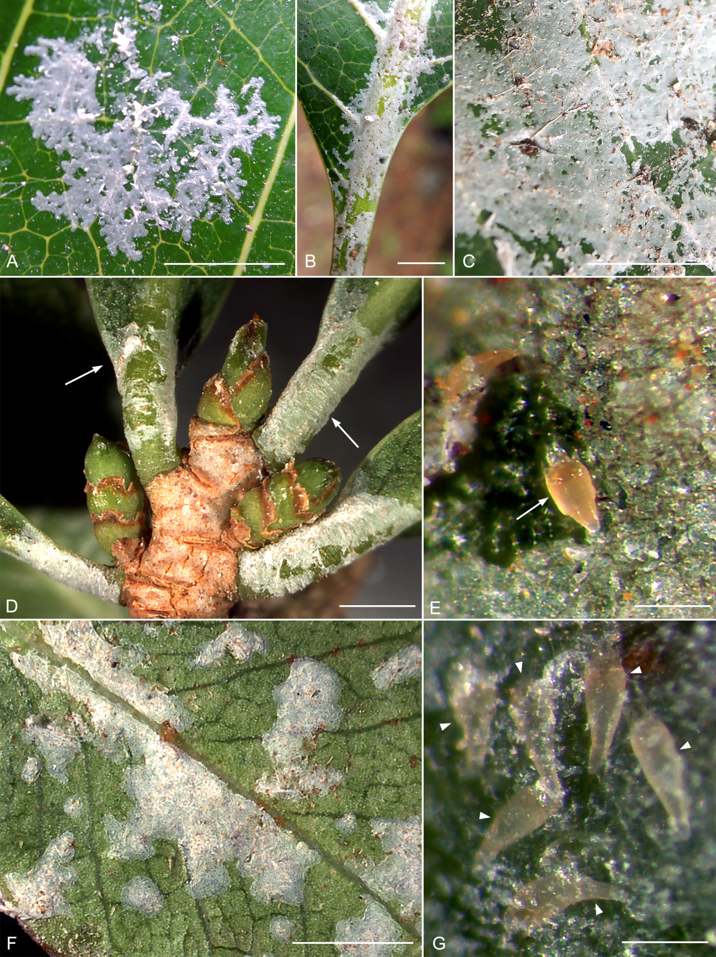
3.3. Web-Spinning in Aberoptus schotiae n. sp.
3.3.1. Web Structure and Appearance
3.3.2. Silk Producing Anal Secretory Apparatus (ASA) of Web-Spinning Females of Aberoptus schotiae n. sp.
3.4. Molecular Phylogenetics
3.4.1. GenBank Data and 28S Sequence Diversity
3.4.2. Molecular Phylogenetic Analyses (Figure 14)

4. Discussion
5. Conclusions
Supplementary Materials
Author Contributions
Funding
Institutional Review Board Statement
Data Availability Statement
Acknowledgments
Conflicts of Interest
References
- Craig, C.L. Spiderwebs and Silk: Tracing Evolution from Molecules to Genes to Phenotypes; Oxford University Press: New York, NY, USA, 2003; pp. 1–230. [Google Scholar]
- Sutherland, T.D.; Young, J.H.; Weisman, S.; Hayashi, C.Y.; Merritt, D.J. Insect silk: One name, many materials. Annu. Rev. Entomol. 2010, 55, 171–188. [Google Scholar] [CrossRef] [PubMed]
- Capinera, J.L. Silk. In Encyclopedia of Entomology; Capinera, J.L., Ed.; Springer: Dordrecht, The Netherlands, 2008; pp. 3373–3374. [Google Scholar] [CrossRef]
- Schoeser, M. Silk; Yale University Press: New Haven, CT, USA, 2007; pp. 1–256. [Google Scholar]
- Allmeling, C.; Jokuszies, A.; Reimers, K.; Kall, S.; Vogt, P.M. Use of spider silk fibres as an innovative material in a biocompatible artificial nerve conduit. J. Cell Mol. Med. 2006, 10, 770–777. [Google Scholar] [CrossRef] [Green Version]
- Kluge, J.A.; Rabotyagova, O.; Leisk, G.G.; Kaplan, D.L. Spider silks and their applications. Trends Biotechnol. 2008, 26, 244–251. [Google Scholar] [CrossRef]
- Lateef, A.; Ojo, S.A.; Elegbede, J.A. The emerging roles of arthropods and their metabolites in the green synthesis of metallic nanoparticles. Nanotechnol. Rev. 2016, 5, 601–622. [Google Scholar] [CrossRef]
- Lozano-Pérez, A.A.; Pagán, A.; Zhurov, V.; Hudson, S.D.; Hutter, J.L.; Pruneri, V.; Pérez-Moreno, I.; Grbić, V.; Cenis, J.L.; Grbić, M.; et al. The silk of gorse spider mite Tetranychus lintearius represents a novel natural source of nanoparticles and biomaterials. Sci. Rep. 2020, 10, 1–14. [Google Scholar] [CrossRef] [PubMed]
- Arakawa, K.; Mori, M.; Kono, N.; Suzuki, T.; Gotoh, T.; Shimano, S. Proteomic evidence for the silk fibroin genes of spider mites (Order Trombidiformes: Family Tetranychidae). J. Proteom. 2021, 239, 104195. [Google Scholar] [CrossRef] [PubMed]
- Li, X.; Liu, R.; Li, G.; Jin, D.; Guo, J.; Ochoa, R.; Yi, T. Identification of the fibroin of Stigmaeopsis nanjingensis by a nanocarrier-based transdermal dsRNA delivery system. Exp. Appl. Acarol. 2022, 87, 31–47. [Google Scholar] [CrossRef]
- Sombke, A.; Müller, C.H. When SEM becomes a deceptive tool of analysis: The unexpected discovery of epidermal glands with stalked ducts on the ultimate legs of geophilomorph centipedes. Front. Zool 2021, 18, 1–19. [Google Scholar] [CrossRef]
- Kovoor, J. Comparative structure and histochemistry of silk-producing organs in arachnids. In Ecophysiology of Spiders; Nentwig, W., Ed.; Springer: Berlin/Heidelberg, Germany, 1987; pp. 160–186. [Google Scholar] [CrossRef]
- Hilbrant, M.; Damen, W.G. The embryonic origin of the ampullate silk glands of the spider Cupiennius salei. Arthropod Struct. Dev. 2015, 44, 280––288. [Google Scholar] [CrossRef]
- Alberti, G.; Coons, L.B. Acari: Mites. In Microscopic Anatomy of Invertebrates: Chelicerate Arthropoda; Harrison, F.W., Foelix, R.F., Eds.; Wiley-Liss: New York, NY, USA, 1999; Volume 8, pp. 515–1265. [Google Scholar]
- Shatrov, A.B.; Soldatenko, E.V. Organization of dermal glands and characteristic of secretion in the freshwater mite, Limnesia maculata (O. F. Müller, 1776) (Acariformes, Limnesiidae). J. Morphol. 2022, 283, 346–362. [Google Scholar] [CrossRef]
- Kluge, N.J. Insect Systematics and Principles of Cladoendesis, 3rd ed.; KMK Scientific Press: Moscow, Russia, 2020; Volume 2, pp. 1–1037. (In Russian) [Google Scholar]
- Weygoldt, P. Spermatophore web formation in a pseudoscorpion. Science 1966, 153, 1647–1649. [Google Scholar] [CrossRef]
- Nuzzaci, G.; Alberti, G. Internal anatomy and physiology. In Eriophyoid Mites: Their Biology, Natural Enemies and Control. World Crop Pests; Lindquist, E.E., Sabelis, M.W., Bruin, J., Eds.; Elsevier Science Publishing: Amsterdam, The Netherlands, 1996; Volume 6, pp. 101–150. [Google Scholar] [CrossRef]
- Chetverikov, P.E.; Bolton, S.J.; Gubin, A.I.; Letukhova, V.Y.; Vishnyakov, A.E.; Zukoff, S. The anal secretory apparatus of Eriophyoidea and description of Phyllocoptes bilobospinosus n. sp.(Acariformes: Eriophyidae) from Tamarix (Tamaricaceae) from Ukraine, Crimea and USA. Syst. Appl. Acarol. 2019, 24, 139–157. [Google Scholar] [CrossRef]
- Norton, R.A.; Florian, M.L.E.; Manning, L.E. Ecdysial cleavage line in Paralycus sp. (Acari: Oribatida: Pediculochelidae). Int. J. Acarol. 2001, 27, 97–99. [Google Scholar] [CrossRef]
- Shatrov, A.B.; Soldatenko, E.V.; Gavrilova, O.V. Observation on silk production and morphology of silk in water mites (Acariformes: Hydrachnidia). Acarina 2014, 22, 133–148. [Google Scholar]
- Gerson, U. Webbing. In Spider Mites: Their Biology, Natural Enemies, and Control; Helle, W., Sabelis, M.W., Eds.; Elsevier Science Publishers: Amsterdam, The Netherlands, 1985; Volume 2, pp. 223–230. [Google Scholar]
- Ray, H.A.; Hoy, M.A. Role of silk webbing in the biology of Hemicheyletia wellsina (Acari: Cheyletidae). Int. J. Acarol. 2014, 40, 577–581. [Google Scholar] [CrossRef]
- Lindquist, E.E. External anatomy and notation of structures. In Eriophyoid Mites: Their Biology, Natural Enemies and Control. World Crop Pests; Lindquist, E.E., Sabelis, M.W., Bruin, J., Eds.; Elsevier Science Publishing: Amsterdam, The Netherlands, 1996; Volume 6, pp. 3–31. [Google Scholar] [CrossRef]
- Bolton, S.J.; Chetverikov, P.E.; Klompen, H. Morphological support for a clade comprising two vermiform mite lineages: Eriophyoidea (Acariformes) and Nematalycidae (Acariformes). Syst. Appl. Acarol. 2017, 22, 1096–1131. [Google Scholar] [CrossRef]
- Klimov, P.B.; OConnor, B.M.; Chetverikov, P.E.; Bolton, S.J.; Pepato, A.R.; Mortazavi, A.L.; Tolstikov, A.V.; Bauchan, G.R.; Ochoa, R. Comprehensive phylogeny of acariform mites (Acariformes) provides insights on the origin of the four-legged mites (Eriophyoidea), a long branch. Mol. Phylogenet. Evol. 2018, 119, 105–117. [Google Scholar] [CrossRef]
- Klimov, P.B.; Chetverikov, P.E.; Dodueva, I.E.; Vishnyakov, A.E.; Bolton, S.J.; Paponova, S.S.; Lutova, L.A.; Tolstikov, A.V. Symbiotic bacteria of the gall-inducing mite Fragariocoptes setiger (Eriophyoidea) and phylogenomic resolution of the eriophyoid position among Acari. Sci. Rep. 2022, 12, 3811. [Google Scholar] [CrossRef]
- Amrine, J.W., Jr.; Stasny, T.A.H.; Flechtmann, C.H.W. Revised Keys to the World Genera of the Eriophyoidea (Acari: Prostigmata); Indira Publishing House: West Bloomfield, MI, USA, 2003; pp. 1–244. [Google Scholar]
- de Lillo, E.; Pozzebon, A.; Valenzano, D.; Duso, C. An intimate relationship between eriophyoid mites and their host plants—A review. Front. Plant Sci. 2018, 9, 1–14. [Google Scholar] [CrossRef] [Green Version]
- Neravathu, R. Feeding impact of Cisaberoptus kenyae Keifer (Acari: Eriophyidae) on photosynthetic efficiency and biochemical parameters of Mangifera indica L. Acarol. Stud. 2019, 1, 84–94. [Google Scholar]
- Ivanova, L.A.; Chetverikov, P.E.; Ivanov, L.A.; Tumurjav, S.; Kuzmin, I.V.; Desnitskiy, A.G.; Tolstikov, A.V. The effect of gall mites (Acariformes, Eriophyoidea) on leaf morphology and pigment content of deciduous trees in West Siberia. Acarina 2022, 30, 89–98. [Google Scholar] [CrossRef]
- Manson, D.C.M.; Gerson, U. Web spinning, wax secretion and liquid secretion by eriophyoid mites. In Eriophyoid Mites: Their Biology, Natural Enemies and Control. World Crop Pests; Lindquist, E.E., Sabelis, M.W., Bruin, J., Eds.; Elsevier Science Publishing: Amsterdam, The Netherlands, 1996; Volume 6, pp. 251–258. [Google Scholar] [CrossRef]
- Britto, E.P.; Gondim, M.G., Jr.; Navia, D.; Flechtmann, C.H.W. A new deuterogynous eriophyid mite (Acari: Eriophyidae) with dimorphic males from Caesalpinia echinata (Caesalpiniaceae) from Brazil: Description and biological observations. Int. J. Acarol. 2008, 34, 307–316. [Google Scholar] [CrossRef]
- Knorr, L.C.; Phatak, H.C.; Keifer, H.H. Web-spinning eriophyid mites. J. Wash. Acad. Sci. 1976, 66, 228–234. [Google Scholar]
- Keifer, H.H. Eriophyid Studies XVII. Bull. Calif. State Dept. Agric. 1951, 40, 93–104. [Google Scholar]
- Hassan, E.F.O.; Keifer, H.H. Mango leaf-coating mite, Cisaberoptus kenyae K. (Eriophyidae, Aberoptinae). Pan. Pac. Entomol. 1978, 54, 185–193. [Google Scholar]
- Navia, D.; Flechtmann, C.H.W. Eriophyid mites (Acari: Prostigmata) from mango, Mangifera indica L., in Brazil. Int. J. Acarol. 2000, 26, 73–80. [Google Scholar] [CrossRef]
- Flechtmann, C.H.W. Aberoptus cerostructor n. sp., a deuterogynous species from Brazil (Acari: Eriophyidae). Int. J. Acarol. 2001, 27, 199–204. [Google Scholar] [CrossRef]
- Smith Meyer, M.K.P. African Eriophyoidea: On species of the subfamily Aberoptinae (Acari: Eriophyidae). Phytophylactica 1989, 21, 271–274. [Google Scholar]
- Elhalawany, A.S.; Amrine, J.W.; Ueckermann, E.A. A new species and new record of eriophyoid mites (Trombidiformes: Eriophyoidea) from mango in Egypt with a note on the population dynamics of four eriophyoid species. Acarines 2021, 15, 1–22. [Google Scholar] [CrossRef]
- Chetverikov, P.E.; Desnitskaya, E.A.; Efimov, P.G.; Bolton, S.J.; Cvrković, T.; Petanović, R.U.; Zukoff, S.; Amrine, J.W., Jr.; Klimov, P. The description and molecular phylogenetic position of a new conifer-associated mite, Setoptus tsugivagus n. sp. (Eriophyoidea, Phytoptidae, Nalepellinae). Syst. Appl. Acarol. 2019, 24, 683–700. [Google Scholar] [CrossRef]
- Chetverikov, P.E.; Craemer, C.; Cvrković, T.; Efimov, P.G.; Klimov, P.B.; Petanović, R.U.; Sukhareva, S.I. First pentasetacid mite from Australasian Araucariaceae: Morphological description and molecular phylogenetic position of Pentasetacus novozelandicus n. sp. (Eriophyoidea, Pentasetacidae) and remarks on anal lobes in eriophyoid mites. Syst. Appl. Acarol. 2019, 24, 1284–1309. [Google Scholar] [CrossRef]
- Chetverikov, P.E.; Bertone, M. First rhyncaphytoptine mite (Eriophyoidea, Diptilomiopidae) parasitizing american hazelnut (Corylus americana): Molecular identification, confocal microscopy, and phylogenetic position. Exp. Appl. Acarol. 2022, 88, 75–95. [Google Scholar] [CrossRef] [PubMed]
- Amrine, J.W., Jr.; Manson, D.C.M. Preparation, mounting and descriptive study of eriophyoid mites. In Eriophyoid Mites: Their Biology, Natural Enemies and Control. World Crop Pests; Lindquist, E.E., Sabelis, M.W., Bruin, J., Eds.; Elsevier Science Publishing: Amsterdam, The Netherlands, 1996; Volume 6, pp. 383–396. [Google Scholar] [CrossRef]
- Chetverikov, P.E. Video projector: A digital replacement for camera lucida for drawing mites and other microscopic objects. Syst. Appl. Acarol. 2016, 21, 1278–1280. [Google Scholar] [CrossRef]
- Echlin, P.; Paden, R.; Dronzek, B.; Wayte, R. Scanning electron microscopy of labile biological material maintained under controlled conditions. Scan. Electron. Micros. 1970, 49–56. [Google Scholar]
- Kirejtshuk, A.G.; Chetverikov, P.E.; Azar, D. Libanopsinae, new subfamily of the family Sphindidae (Coleoptera, Cucujoidea) from Lower Cretaceous Lebanese amber, with remarks on using confocal microscopy for the study of amber inclusions. Cretac. Res. 2015, 52, 461–479. [Google Scholar] [CrossRef]
- Chetverikov, P.E. Confocal laser scanning microscopy technique for the study of internal genitalia and external morphology of eriophyoid mites (Acari: Eriophyoidea). Zootaxa 2012, 3453, 56–68. [Google Scholar] [CrossRef]
- Schneider, C.; Rasband, W.; Eliceiri, K. NIH Image to ImageJ: 25 years of image analysis. Nat. Methods 2012, 9, 671–675. [Google Scholar] [CrossRef]
- Humphrey, C.D.; Pittman, F.E. A simple methylene blue-azure II-basic fuchsin stain for epoxy-embedded tissue sections. Stain. Technol. 1974, 49, 9–14. [Google Scholar] [CrossRef]
- Katoh, K.; Misawa, K.; Kuma, K.; Miyata, T. MAFFT: A novel method for rapid multiple sequence alignment based on fast Fourier transformation. Nucleic Acids Res. 2002, 30, 3059–3066. [Google Scholar] [CrossRef] [Green Version]
- Katoh, K.; Rozewicki, J.; Yamada, K.D. MAFFT online service: Multiple sequence alignment, interactive sequence choice and visualization. Brief. Bioinform. 2017, 20, 1160–1166. [Google Scholar] [CrossRef] [Green Version]
- Minh, B.Q.; Schmidt, H.A.; Chernomor, O.; Schrempf, D.; Woodhams, M.D.; von Haeseler, A.; Lanfear, R. IQ-TREE 2: New Models and Efficient Methods for Phylogenetic Inference in the Genomic Era. Mol. Biol. Evol. 2020, 37, 1530–1534. [Google Scholar] [CrossRef] [PubMed]
- Kalyaanamoorthy, S.; Minh, B.Q.; Wong, T.K.F.; von Haeseler, A.; Jermiin, L.S. ModelFinder: Fast model selection for accurate phylogenetic estimates. Nat. Methods 2017, 14, 587–589. [Google Scholar] [CrossRef] [Green Version]
- Chase, M.W.; Christenhusz, M.J.M.; Fay, M.F.; Byng, J.W.; Judd, W.S.; Soltis, D.E.; Mabberley, D.J.; Sennikov, A.N.; Soltis, P.S.; Stevens, P.F. An update of the Angiosperm Phylogeny Group classification for the orders and families of flowering plants: APG IV. Bot. J. Linn. Soc. 2016, 181, 1–20. [Google Scholar] [CrossRef] [Green Version]
- Koenen, E.J.; Ojeda, D.I.; Steeves, R.; Migliore, J.; Bakker, F.T.; Wieringa, J.J.; Kidner, C.; Hardy, O.J.; Pennington, R.T.; Bruneau, A.; et al. Large-scale genomic sequence data resolve the deepest divergences in the legume phylogeny and support a near-simultaneous evolutionary origin of all six subfamilies. New. Phytol. 2020, 225, 1355–1369. [Google Scholar] [CrossRef] [PubMed] [Green Version]
- Chetverikov, P.E.; Craemer, C.; Neser, S. New pseudotagmic genus of acaricaline mites (Eriophyidae, Acaricalini) from a South African palm Hyphaene coriacea and remarks on lateral opisthosomal spines and morphology of deutogynes in Eriophyoidea. Syst. Appl. Acarol. 2018, 23, 1073–1101. [Google Scholar] [CrossRef]
- Krantz, G.W. Habit and habitats. In A Manual of Acarology, 3rd ed.; Krantz, G.W., Walter, D.E., Eds.; Texas Tech University Press: Lubbock, TX, USA, 2009; pp. 64–82. [Google Scholar]
- Buzatto, B.A.; Machado, G. Male dimorphism and alternative reproductive tactics in harvestmen (Arachnida: Opiliones). Behav. Process. 2014, 109, 2–13. [Google Scholar] [CrossRef] [Green Version]
- Cho, G.; Malenovský, I.; Burckhardt, D.; Inoue, H.; Lee, S. DNA barcoding of pear psyllids (Hemiptera: Psylloidea: Psyllidae), a tale of continued misidentifications. B Entomol. Res. 2020, 110, 521–534. [Google Scholar] [CrossRef]
- Manson, D.C.M.; Oldfield, G.N. Life forms, deuterogyny, diapause and seasonal development. In Eriophyoid Mites: Their Biology, Natural Enemies and Control. World Crop Pests; Lindquist, E.E., Sabelis, M.W., Bruin, J., Eds.; Elsevier Science Publishing: Amsterdam, The Netherlands, 1996; Volume 6, pp. 173–183. [Google Scholar] [CrossRef]
- Chetverikov, P.E.; Petanović, R.U. New observations on early-derivative mite Pentasetacus araucariae (Schliesske)(Acariformes: Eriophyoidea) infesting relict gymnosperm Araucaria araucana. Syst. Appl. Acarol. 2016, 21, 1157–1190. [Google Scholar] [CrossRef] [Green Version]
- Chetverikov, P.E.; Rector, B.G.; Tonkel, K.; Dimitri, L.; Cheglakov, D.S.; Romanovich, A.E.; Amrine, J. Phylogenetic position of a new Trisetacus mite species (Nalepellidae) destroying seeds of North American junipers and new hypotheses on basal divergence of Eriophyoidea. Insects 2022, 13, 201. [Google Scholar] [CrossRef]
- Keifer, H.H. Eriophyid Studies XII. Bull. Calif. State Dept. Agric. 1942, 31, 117–129. [Google Scholar]
- Yin, Y.; Yao, L.F.; Zhang, Q.; Hebert, P.D.; Xue, X.F. Using multiple lines of evidence to delimit protogynes and deutogynes of four-legged mites: A case study on Epitrimerus sabinae s.l. (Acari: Eriophyidae). Invertebr. Syst. 2020, 34, 757–768. [Google Scholar] [CrossRef]
- Chetverikov, P.E.; Klimov, P.B.; He, Q. Vertical transmission and seasonal dimorphism of eriophyoid mites (Acariformes, Eriophyoidea) parasitic on the Norway maple: A case study. Royal Soc. Open Sci. 2022, 9, 220820. [Google Scholar] [CrossRef] [PubMed]
- Chetverikov, P.E.; Craemer, C.; Bolton, S. Exoskeletal transformations in Eriophyoidea: New pseudotagmic taxon Pseudotagmus africanus ng & n. sp. from South Africa and remarks on pseudotagmosis in eriophyoid mites. Syst. Appl. Acarol. 2017, 22, 2093–2118. [Google Scholar] [CrossRef]
- Doğan, S.; Çağlar, M.; Çağlar, B.; Çirak, Ç.; Zeytun, E. Structural characterization of the silk of two-spotted spider mite Tetranychus urticae Koch (Acari: Tetranychidae). Syst. Appl. Acarol. 2017, 22, 572–583. [Google Scholar] [CrossRef] [Green Version]
- Guo, J.F.; Li, H.S.; Wang, B.; Xue, X.F.; Hong, X.Y. DNA barcoding reveals the protogyne and deutogyne of Tegolophus celtis sp. nov. (Acari: Eriophyidae). Exp. Appl. Acarol. 2015, 67, 393–410. [Google Scholar] [CrossRef]
- Sabelis, M.W.; Bruin, J. 1996 Evolutionary ecology: Life history patterns, food plant choice and dispersal. In Eriophyoid Mites: Their Biology, Natural Enemies and Control. World Crop Pests; Lindquist, E.E., Sabelis, M.W., Bruin, J., Eds.; Elsevier Science Publishing: Amsterdam, The Netherlands, 1996; Volume 6, pp. 329–366. [Google Scholar]
- Michalska, K.; Skoracka, A.; Navia, D.; Amrine, J.W. Behavioural studies on eriophyoid mites: An overview. Exp. Appl. Acarol. 2010, 51, 31–59. [Google Scholar] [CrossRef] [Green Version]
- Navia, D.; Flechtmann, C.H.W.; Lindquist, E.E.; Aguilar, H. A new species of Abacarus (Acari: Prostigmata: Eriophyidae) damaging sugarcane, Sacharrum officinarum L., from Costa Rica—The first eriophyoid mite described with a tibial seta on leg II. Zootaxa 2011, 3025, 51–58. [Google Scholar] [CrossRef]
- Nuzzaci, G. Contributo alla conoscenza dell’anatomia degli acari eriofidi. Entomologica 1976, 12, 21–55. [Google Scholar] [CrossRef]
- Silvere, A.P.; Stein-Margolina, V. Tetrapodili. Microanatomy, Problems of Evolution and Interrelationship with Pathogens of Plant Diseases; Valgus Press: Tallinn, Estonia, 1976; pp. 1–164. [Google Scholar]
- Wipfler, B.; Schneeberg, K.; Löffler, A.; Hünefeld, F.; Meier, R.; Beutel, R.G. The skeletomuscular system of the larva of Drosophila melanogaster (Drosophilidae, Diptera)—A contribution to the morphology of a model organism. Arthropod. Struct. Dev. 2013, 42, 47–68. [Google Scholar] [CrossRef]


Disclaimer/Publisher’s Note: The statements, opinions and data contained in all publications are solely those of the individual author(s) and contributor(s) and not of MDPI and/or the editor(s). MDPI and/or the editor(s) disclaim responsibility for any injury to people or property resulting from any ideas, methods, instructions or products referred to in the content. |
© 2023 by the authors. Licensee MDPI, Basel, Switzerland. This article is an open access article distributed under the terms and conditions of the Creative Commons Attribution (CC BY) license (https://creativecommons.org/licenses/by/4.0/).
Share and Cite
Chetverikov, P.E.; Craemer, C.; Gankevich, V.D.; Vishnyakov, A.E.; Zhuk, A.S. A New Webbing Aberoptus Species from South Africa Provides Insight in Silk Production in Gall Mites (Eriophyoidea). Diversity 2023, 15, 151. https://doi.org/10.3390/d15020151
Chetverikov PE, Craemer C, Gankevich VD, Vishnyakov AE, Zhuk AS. A New Webbing Aberoptus Species from South Africa Provides Insight in Silk Production in Gall Mites (Eriophyoidea). Diversity. 2023; 15(2):151. https://doi.org/10.3390/d15020151
Chicago/Turabian StyleChetverikov, Philipp E., Charnie Craemer, Vladimir D. Gankevich, Andrey E. Vishnyakov, and Anna S. Zhuk. 2023. "A New Webbing Aberoptus Species from South Africa Provides Insight in Silk Production in Gall Mites (Eriophyoidea)" Diversity 15, no. 2: 151. https://doi.org/10.3390/d15020151
APA StyleChetverikov, P. E., Craemer, C., Gankevich, V. D., Vishnyakov, A. E., & Zhuk, A. S. (2023). A New Webbing Aberoptus Species from South Africa Provides Insight in Silk Production in Gall Mites (Eriophyoidea). Diversity, 15(2), 151. https://doi.org/10.3390/d15020151


