Article

Diversity and Regional Variation of Endosymbionts in the Green Peach Aphid, Myzus persicae (Sulzer)

1
Bio21 Institute, School of BioSciences, The University of Melbourne, Parkville, VIC 3010, Australia
2
Cesar Australia, 293 Royal Parade, Parkville, VIC 3052, Australia
3
Institute of Plant Protection, Beijing Academy of Agriculture and Forestry Sciences, Beijing 100097, China
4
College of Life and Environmental Sciences, Biosciences, University of Exeter, Penryn Campus, Penryn, Cornwall TR10 9FE, UK
5
MOA Key Laboratory of Integrated Management of Pests on Crops in Southwest China, Institute of Plant Protection, Sichuan Academy of Agricultural Sciences, Chengdu 610066, China
*
Authors to whom correspondence should be addressed.
Diversity 2023, 15(2), 206; https://doi.org/10.3390/d15020206
Submission received: 18 November 2022 / Revised: 16 January 2023 / Accepted: 24 January 2023 / Published: 1 February 2023

Abstract

:
The green peach aphid, Myzus persicae, is globally distributed and an important pest of many economically valuable food crops, largely due to its ability to transmit plant viruses. Almost all aphids, including M. persicae, carry the obligate symbiont Buchnera aphidicola, which provides essential amino acids that aphids cannot obtain from the phloem of plants themselves. Many aphids also harbor facultative (secondary) endosymbionts, which provide benefits under specific ecological conditions. In this study, we screened for secondary endosymbionts in M. persicae, with a particular focus on Australian populations where this species is growing in status as a major agricultural pest. We compared 37 Australian M. persicae populations with other populations, including 21 field populations from China and 15 clones from the UK, France, Italy, Greece, USA, Spain, South Korea, Chile, Japan and Zimbabwe. No secondary endosymbionts were identified in M. persicae samples outside of China, despite samples covering a wide geographic range and being collected from several host plant families. We detected two secondary endosymbionts (Rickettsia, Spiroplasma) in Chinese samples, although diversity appeared lower than detected in a recent study. We also found very high clonal diversity in Chinese samples based on DNA microsatellite markers in comparison with lower clonal diversity from Australia. These patterns may indicate a higher diversity of secondary endosymbionts (and clonal diversity) in the native range of M. persicae when compared to its invasive range.

1. Introduction

Bacterial symbionts are widespread in insects, and their symbiotic associations range from obligate mutualism to facultative parasitism [1]. In aphids, the primary (also called obligate) symbiont Buchnera aphidicola provides essential amino acids that aphids cannot obtain from the phloem of host plants themselves [2,3]. Secondary endosymbionts (also called facultative) can influence ecologically important traits in aphids, including resistance to parasitoid wasps [4], tolerance to heat stress [5], and host plant utilisation patterns [6]. An association between endosymbiont persistence and colonization of new plant species has been described by Henry et al. [7] in the pea aphid, Acyrthosiphon pisum (Harris) (Homoptera: Aphididae). Endosymbionts are spread within populations through vertical transmission, whereby the bacteria are passed from the mother directly to her offspring (which is the main pathway) and through horizontal transmission, for example through host feeding and via parasitoids (e.g., [8]).
The green peach aphid, Myzus persicae (Sulzer) (Hemiptera: Aphididae), is globally distributed and one of the most economically important aphid crop pests [9]. It has a host range of more than 400 plant species [10], multiple methods of causing plant damage [11], and widespread resistance to insecticides [12,13]. In addition, M. persicae is an important vector of many plant viruses, that can cause considerable harm to host plants [14]. Myzus persicae is thought to be of Chinese origin, similar to its primary host, the peach, Prunus persica, and has invaded many countries on every continent, except Antarctica [15,16]. Studies of genetic variation within Chinese M. persicae populations collected from their primary host plant P. persica using DNA microsatellite markers have shown that the genetic structure of these populations involved a split into a southern group and a northern group divided by the Yangtse River. However, the historical demography of M. persicae in China remains unknown [17]. The subsequent invasion of this species has included Australia, where M. persicae was first detected in 1893 [18], and where it is now found in every state and territory. Singh et al. [15] recently used a high-quality chromosome-scale genome assembly with resequenced genomes of 127 globally sampled M. persicae to provide evidence of migration/gene flow between Australia and some populations in Europe and Asia, suggesting multiple incursions.
Artificial endosymbiont infections in aphids can be generated through microinjection of hemolymph across aphid strains or species [19,20], and there is increasing interest in the use of such endosymbiont transfections in pest control. There may be opportunities to release aphid strains generated through transinfection with favourable traits and with endosymbionts able to spread these through wild populations. When applying endosymbiont technology, it is important to understand the status of natural infections in populations of the target aphid, particularly when secondary endosymbionts can vary in incidence between geographic locations as demonstrated in the pea aphid [21]. In M. persicae, a few studies have reported that secondary endosymbiont infections may be very rare [22,23], but Xu et al. [24] observed a high diversity of secondary endosymbionts in several Chinese M. persicae populations.
In order to better understand the diversity and regional variation of endosymbionts in M. persicae and to investigate the ecological and evolutionary factors that might influence the ability of endosymbionts to invade local populations, we characterized the secondary endosymbiont diversity in M. persicae populations and some laboratory clones using 16S metabarcoding and quantitative PCR. We undertook broad-scale sampling of aphids from different host plants throughout Australia, China and several other countries to provide baseline data for the exploitation of endosymbiont technology in M. persicae control. Our work suggests variation in the incidence of secondary endosymbionts between M. persicae within China and elsewhere.

2. Methods

2.1. Aphids

Thirty-two M. persicae samples were collected by direct searching for aphids from a variety of host plants from around Australia between 2019 and 2021, while several historical samples, which were collected from the field and had been placed in culture, were also included (Table 1). Ten individuals from each sample were stored in 100% ethanol and frozen at −20 °C for later molecular analysis. The samples in culture were maintained as asexual lines established from a single female in the laboratory on single bok choy (Brassica napus subsp. chinensis) leaves that were placed in 60 mm petri dishes containing agar (1%). Petri dishes were kept in a controlled temperature cabinet at 11 °C, with a 16L:8D h photoperiod. Twenty-one aphid samples were collected from China (n = 2 to 25) from eight different plant hosts between 2016 and 2021. An additional 15 clones from Europe, the USA, Chile, Zimbabwe, South Korea and Japan (N = 5) were included in this study. These clones were established from aphids collected as part of a global study investigating the evolution of resistance in M. persicae [15], with these aphids being maintained as asexual lineages on individual wombok (Brassica rapa var. pekinensis) leaves in small plastic cups at 20 °C, with a 16L:8D h photoperiod prior to this study.
A map displaying the location of all M. persicae samples used here is provided in Figure 1.

2.2. Clonal Assignment of M. persicae

Aphids from 27 Australian samples and 13 Chinese samples were genotyped to determine the clonal make-up of each sample. Two to 12 aphids from each population were genotyped using 10 previously described DNA microsatellite loci: M35, M37, M40, M49, M55, M63, M86, myz2, myz9 and myz25 [25,26]. DNA was extracted by homogenising individual aphids in a 200 μL solution containing 5% Chelex 100 resin (Bio-Rad Laboratories, Hercules, CA, USA) according to methods described previously [24]. Samples were centrifuged for 2 min at 20,800 g (Eppendorf Centrifuge 5417 C, Hamburg, Germany) and 2 μL of the supernatant was used as template in polymerase chain reactions. Loci were pooled into three groups, labelled with unique fluorophores (FAM, NED, VIC, and PET) and coamplified by multiplex PCR using a Qiagen multiplex kit and an Eppendorf Mastercycler S gradient PCR machine. Genotyping was subsequently performed using a 3730 capillary analyzer (Applied Biosystems, Melbourne, Australia) and product lengths were scored manually using GeneMapper version 4.0 (Applied Biosystems).

2.3. 16S rRNA Gene Metabarcoding and Quantitative PCR

We used DNA metabarcoding to characterise the microbiome of some M. persicae samples listed in Table 1 (all Australian populations and a few Chinese populations). For these samples, individuals were pooled to provide sufficient DNA for next generation sequencing. Two replicate DNA extractions, each containing a pool of 5 individuals, were performed for each sample using a DNeasy® Blood & Tissue kit (Qiagen, Hilden, Germany). Metabarcoding targeted the hypervariable V3-V4 region of the bacterial 16S rRNA gene and was carried out by Novogene (Novogene (HK) Co. Ltd., Hong Kong, China), using the universal primers 341F and 806R. Sequence analysis was performed using a standard QIIME2 pipeline [27]. Firstly, primer sequences were trimmed from reads with the cutadapt plugin. Sequence quality filtering and error correction, assembly of paired-end reads, and chimera removal were performed with the DADA2 plugin. DADA2 was then used to group reads into amplicon sequence variants (ASVs), which are analogous to Operational Taxonomic Units (OTUs) clustered at a 100% identity threshold. Background filtering was performed on a sample-wise basis. ASVs that made up less than 0.1% of the reads in a sample were removed from that sample. Taxonomic identity was assigned to ASVs with the classify-sklearn plugin, using a naïve Bayes classifier that had been trained against the SILVA 16S rRNA database [28] (release 132; 99% identity criterion). The identity of endosymbiont ASVs was further investigated with blastn searches (nr/nt database). LFN (low-frequency noise) filters were used to discard variants with low read counts and sequencing contamination (Yang et al., unpublished). An average of 283,084 reads per sample were retained after each of the quality filtering and assembly steps from our 16S metabarcoding data.
Quantitative PCR (qPCR) assays were used to screen for secondary endosymbionts in M. persicae samples which comprised a limited number of individuals and/or had insufficient DNA amounts for metabarcoding. DNA was extracted from individual aphids (variable numbers per population; Table 1) using 150 μL of 5% Chelex 100 resin as described previously, with PCRs undertaken using a LightCycler® 480 High Resolution Melting Master (HRMM) kit (Roche Diagnostics Australia Pty. Ltd., Castle Hill, Australia) and IMMOLASETM DNA polymerase (5 U/µL) (Bioline; Cat. No. BIO-21047). The PCR conditions for DNA amplification began with a 10-min pre-incubation at 95 °C (Ramp Rate = 4.8 °C/s), followed by 40 cycles of 95 °C for 5 s (Ramp Rate = 4.8 °C/s), 58 °C for 15 s (Ramp Rate = 2.5 °C/s), and 72 °C for 30 s (Ramp Rate = 4.8 °C/s). Two primer sets were applied to amplify markers to confirm the quality of aphid DNA (β-actin as reference gene) [29] and the presence or absence of the target endosymbiont infection (Table 2). Crossing point (Cp) values of three consistent replicate runs were averaged. Differences in Cp values between the actin and the target endosymbiont markers were transformed by 2n to produce relative endosymbiont density measures.
Presence of the endosymbiont amplicon was confirmed by running a standard PCR using the same program and primers with qPCR, running 10 μL of PCR product on a 2% molecular biology grade agarose gel (Scientifix, Springvale, Australia) and observing a clear band with appropriate size for each primer pair. PCR products were then sent for Sanger sequencing (Macrogen, Inc., Geumcheongu, Seoul, South Korea). Sequencing chromatograms were examined and processed with Geneious 9.18 software (Biomatters, Inc., Auckland, New Zealand). The 16S rRNA gene sequencing data were analysed with the program Geneious. A phylogenetic tree was constructed with a neighbour-joining model applied to a genetic distance matrix with the Tamura-Nei2 model implemented with 1000 bootstrap replications in Geneious.

3. Results and Discussion

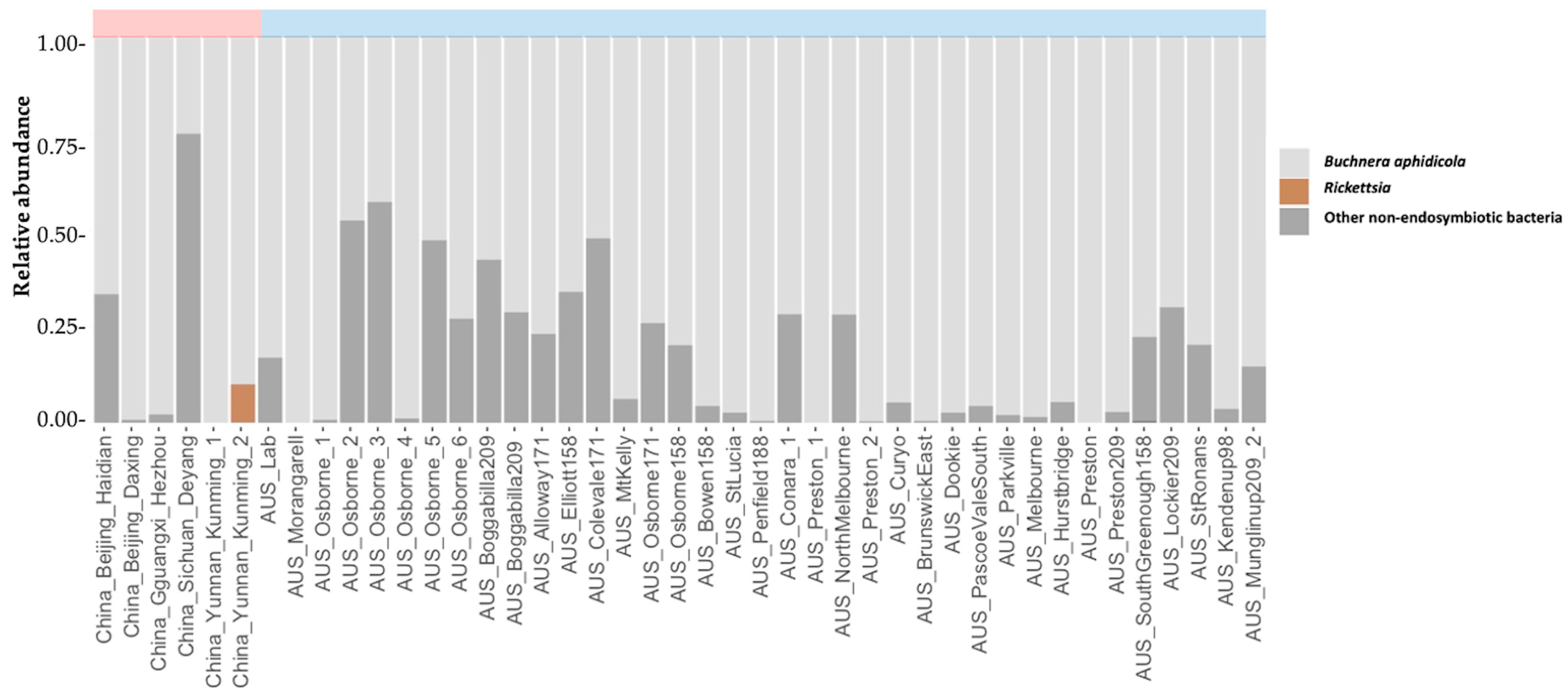
In this study, we examined endosymbiont diversity in 73 M. persicae samples from 18 different host plants, focusing on Australia but also including samples collected more widely. Importantly, this included samples from China, which is believed to represent the region where this species originated [17]. Genotyping across 10 microsatellite DNA loci identified 10 distinct clones among the Australian M. persicae populations sampled here and 47 clones among the Chinese populations sampled (Table 1). No secondary endosymbionts were detected within the 37 Australian samples (Table 3, Figure 2), even though individuals from Australian samples represented multiple clones as characterized by microsatellites, were sampled over a broad geographic range, collected from multiple plant families and included multiple color morphs. These patterns are consistent with the lack of secondary endosymbionts in the 15 clones from the UK, France, Italy, Greece, the USA, Spain, South Korea, Chile, Japan and Zimbabwe, and also from other studies of M. persicae outside of China [22,23,33].
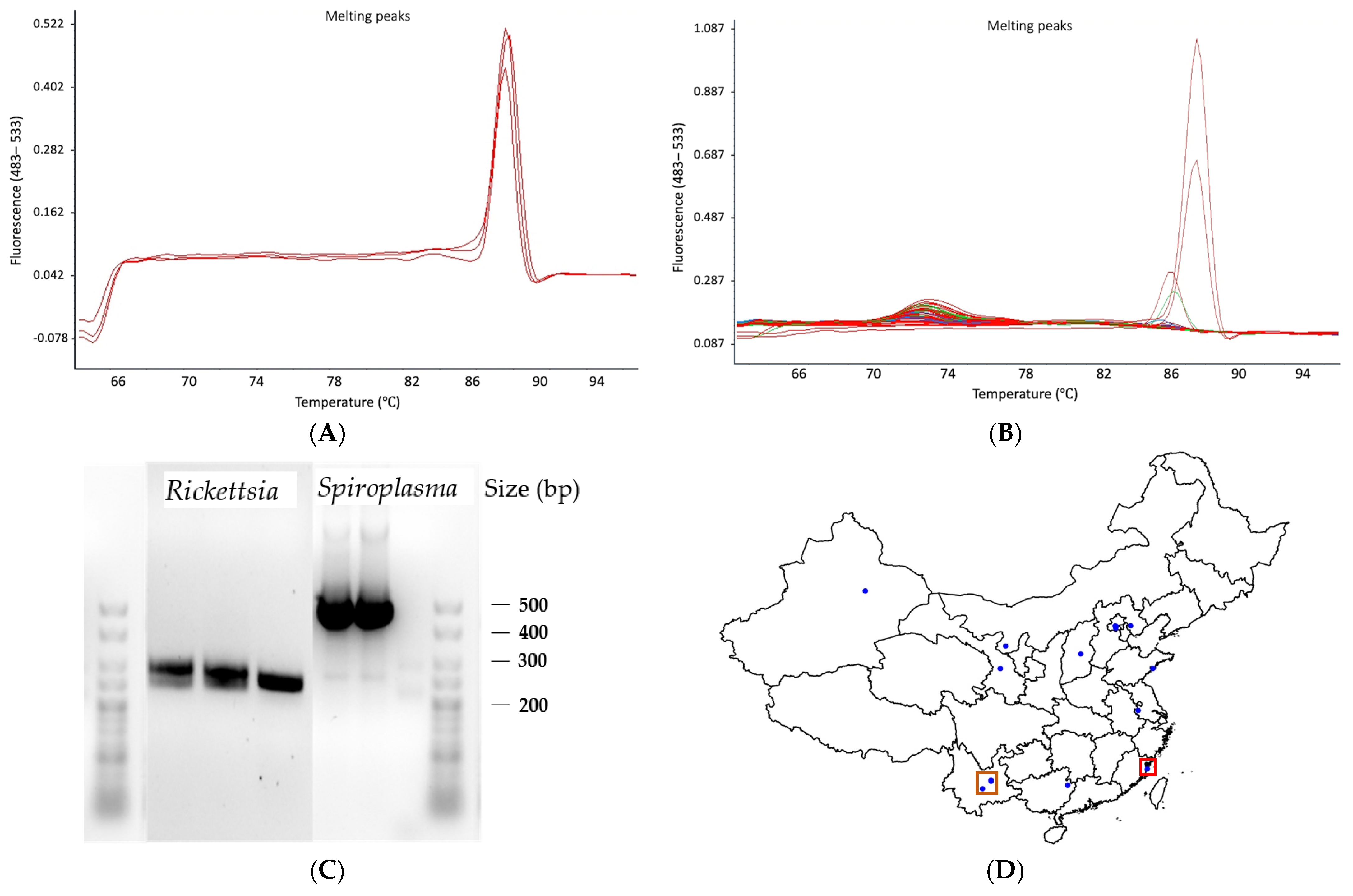
We then screened 21 M. persicae populations from China using qPCR, the putative native range, in order to provide a comparison to invaded populations. Rickettsia was detected in four out of 21 samples, each of which was collected from tobacco, Nicotiana tabacum. We also detected Spiroplasma from a single population collected on radish, Raphanus sativus (Figure 3, Table 3). The incidence of endosymbiont detection in the Chinese samples (5/21) differs from the incidence of detection in Australian samples (0/37) (Fisher’s exact test, p = 0.004, IBM Statistics SPSS version 26), even though the sample size of clones from China was smaller (Table 1). A previous screen of diversity in secondary endosymbionts from Chinese populations also identified Rickettsia and Spiroplasma from samples of M. persicae. In addition, another five endosymbionts were detected including Wolbachia, which was abundant across the populations (Table 3) [24]. Although the sample sizes of field collections were not specified in this paper, it is unclear why we failed to detect these other endosymbionts and particularly Wolbachia in our field samples from China.
It is possible that some detections in previously published work represents contaminants. Note that we set a sensitive cut-off at 0.1% to remove ASVs with low relative abundance and validated all our positive detections through PCR and Sanger sequencing, whereas previous work has relied on metabarcoding data and set a much lower cut off at 0.005%, with the exception of qPCR screening for Rickettsia. It is also possible that there is geographic or seasonal variation in the distribution of endosymbionts. Although we had multiple sampling points in China, there was only one that overlapped with the samples scored as Wolbachia infected by Xu et al. (M37103, Amygdalus persica, Beijing) [24]. Seasonal changes in Wolbachia infection have been documented in some insects [34] and may occur for other endosymbionts.
Nevertheless, the results from our survey combined with the data from Xu et al. [24] suggest that secondary endosymbionts are more common in the native range of this species than the introduced range, which is also consistent with our failure to detect secondary endosymbionts in a sample of the global clonal collection from Singh et al. [15]. Note that Singh et al. [15] also failed to detect any secondary endosymbionts in whole genome sequence data from >110 fully sequenced globally sampled clonal lines of M. persicae, although this method is less sensitive than metabarcoding of the microbiome.
The phylogenetic analyses of endosymbionts based on sequences obtained from both the 16S data and the Sanger data indicates that the Rickettsia we detected is not closely linked to Rickettsia from other aphids, but instead connects to other arthropod groups including weevils (e.g., Sitona obsoletusand and Liophloeus sp.) and green lacewings (Pseudomallada ventralis) (Figure S1, Supplementary Data). However, the phylogenic analysis of the Spiroplasma sequences indicated that the secondary endosymbiont we identified was closely connected to an aphid-specific group, which was separate from the Spiroplasma sequences from Drosophila and several other insects, including the mealybug Antonina crawii (Figure S2, Supplementary Data). Perhaps Rickettsia has a high rate of horizontal transmission which may explain its similarity across disparate taxonomic groups, but at this stage we are unaware of any literature on horizontal transmission in this group and additional molecular analyses based on more comprehensive sequence data are required.
Several reasons might explain the absence of secondary endosymbionts outside of China. Firstly, it may be the case that the colonization process of invasive M. persicae involved a low number of individuals and that by chance these lacked secondary endosymbionts. If this happened, we might expect lower nuclear genetic diversity in colonized regions (or lower clonal diversity in cases where there is no sex). Singh et al. [15] found that both host plant and geography play a significant role in partitioning genetic variation in M. persicae global populations. However, genetic divergence between Chinese M. persicae populations was not higher than divergence detected between Australian M. persicae populations. This may reflect limited sampling of Chinese clones by Singh et al. [15] given that they characterized only 9 opportunistically collected clones from China. Based on microsatellite data, we detected much higher clonal diversity in Chinese populations, with 47 clones detected from 53 individuals, compared to only 10 clones from 270 individuals detected in Australia (binomial test comparing two proportions, z = 14.6, p < 0.001).
Secondly, it may be that secondary endosymbionts have been lost in colonized regions. This might occur if there is imperfect maternal transmission and no selective advantage of individual aphids with these endosymbionts in newly colonized environments. Numerous phenotypes have been associated with aphid endosymbionts that could contribute to selective differences between host aphid lineages, including resistance to parasitoid wasps [4], tolerance to heat stress [5], and host plant utilisation patterns [6], although very few of these have been examined in M. persicae. One exception is the secondary endosymbiont Regiella, which has been associated with parasitoid resistance in M. persicae [35]. Interestingly the natural Regiella strain used in that study was collected from Bacchus Marsh, Australia on wild mustard, Hirschfeldia incana, in 2003 [36]. Clearly this was a serendipitous finding given that our much more extensive collection indicated an absence of Regiella in all 32 M. persicae field populations tested. We have also failed to collect this endosymbiont from aphids from this host plant in subsequent work (Yang et al., unpublished data). Perhaps the Regiella strain was a recent introduction into Australia which has been lost because of a selective disadvantage and/or transmission leakage. Or perhaps the abundance of secondary endosymbiont seasonally changes. In China, there was no association between the presence of secondary endosymbionts and host plant, but Regiella was commonly detected [24].
It is not yet clear if the loss of endosymbionts from the invaded range of a species is a general finding or specific to M. persicae. Perhaps the most extensively investigated comparison of native and invasive ranges involves endosymbionts in the mosquito Aedes albopictus. In this species, Yang et al. [37] showed that two Wolbachia strains were widely distributed across both its native and introduced ranges, with only minor population differences in endosymbiont frequency. In other species, variation in the distribution of endosymbionts has been found across populations, such as the pea aphid A. pisum [21], the spider mite Tetranychus truncates [38], and various plant parasitic nematodes [39]. These comparisons do not provide a contrast between invaded versus native range populations, but there are other instances (for instance in fire ants [40] and in thrips [41]) where there has been a loss of symbionts in the invasive range.
These results have implications for the introduction of secondary endosymbionts for pest control. This is an area of increasing interest, particularly from the perspective of blocking plant viral transmission [42,43] and changing pesticide susceptibility [44]. Based on the current work, a lack of endosymbionts in the invasive range of this species means that any secondary endosymbionts artificially introduced into M. persicae populations from this range will not have to compete with other secondary endosymbionts already present in field populations. Interactions among endosymbionts are known from other work [45] and could otherwise lead to a suppression of newly introduced endosymbionts in a native host. We have recently introduced two endosymbionts from other aphid species into M. persicae (Gu et al., under review) and into the oat aphid, Rhapalosiphum padi [46] from their invasive range. These have proven to be stable introductions, and we are currently exploring if they have favorable characteristics (such as blocking plant virus transmission or decreasing host fitness) that may make them suitable for introductions into pest aphid populations in Australia and elsewhere.
Endosymbionts have also been implicated in color variation among aphids [47], including in a recent study where the endosymbiont Rickettsiella viridis was transferred to M. persicae which modified the aphid body color from light to dark green (Gu et al. under review). Although color morphs were not specifically considered in this study, we did test individuals from Australia that varied considerably in color, ranging from light green, to orange, pink and red. Since we did not find secondary endosymbionts associated with these color morphs, it appears that environmental factors generate color variation in Australian M. persicae.

4. Conclusions

In summary, there appear to be differences in the diversity (number and variety) of secondary endosymbionts found in M. persicae from invaded and native range (China) populations. Secondary endosymbionts in colonized regions may have been lost due to imperfect maternal transmission, founder events and/or selection associated with different conditions in the invaded region. The lack of secondary endosymbionts in the invaded regions open up the possibility of manipulating populations in these regions through the introduction of novel endosymbionts.

Supplementary Materials

The following supporting information can be downloaded at: https://www.mdpi.com/article/10.3390/d15020206/s1, Figure S1: Phylogenetic analysis of Rickettsia based on 16S rRNA gene variation; Figure S2: Phylogenetic analysis of Spiroplasma based on 16S rRNA gene variation; Supplementary Data: Chromatogram from Sanger sequencing.

Author Contributions

Conceptualization, Q.Y. and A.A.H.; methodology, Q.Y., A.G., K.L.R., W.Y. and A.v.R.; software, Q.Y., K.L.R. and D.Z.; validation, Q.Y.; formal analysis, Q.Y.; investigation, Q.Y., P.A.U. and A.A.H.; resources, P.A.U., C.B., S.W., W.Y., A.v.R. and S.E.W.; data curation, Q.Y. and A.A.H.; writing—original draft preparation, Q.Y. and A.A.H.; writing—review and editing, P.A.U., C.B., K.L.R., A.v.R., A.G. and S.E.W.; visualization, Q.Y.; supervision, A.A.H.; project administration, Q.Y.; funding acquisition, A.A.H. and P.A.U. All authors have read and agreed to the published version of the manuscript.

Funding

Grains Research Development Corporation (Australia), grant no. UOM1906-002RTX.

Institutional Review Board Statement

Not applicable.

Informed Consent Statement

Not applicable.

Data Availability Statement

The data presented in this study are available from the corresponding authors upon reasonable request.

Acknowledgments

This work was funded by the Grains Research Development Corporation (Australia) as part of the Australian Grains Pest Innovation Program, and with support from the University of Melbourne and Sichuan Science and Technology Program (2021YFH0112). We thank Sonia Sharma, Nick Bell, Ashley MacMahon and Kelly Richardson for technical assistance.

Conflicts of Interest

The authors declare no conflict of interest.

References

  1. Kikuchi, Y. Endosymbiotic bacteria in insects: Their diversity and culturability. Microbes Environ. 2009, 24, 195–204. [Google Scholar] [CrossRef]
  2. Baumann, P. Biology bacteriocyte-associated endosymbionts of plant sap-sucking insects. Annu. Rev. Microbiol. 2005, 59, 155–189. [Google Scholar] [CrossRef]
  3. Douglas, A.E. Nutritional interactions in insect-microbial symbioses: Aphids and their symbiotic bacteria Buchnera. Annu. Rev. Entomol. 1998, 43, 17–37. [Google Scholar] [CrossRef]
  4. Oliver, K.M.; Russell, J.A.; Moran, N.A.; Hunter, M.S. Facultative bacterial symbionts in aphids confer resistance to parasitic wasps. Proc. Natl. Acad. Sci. USA 2003, 100, 1803–1807. [Google Scholar] [CrossRef]
  5. Chen, D.Q.; Montllor, C.B.; Purcell, A.H. Fitness effects of two facultative endosymbiotic bacteria on the pea aphid, Acyrthosiphon pisum, and the blue alfalfa aphid, A. kondoi. Entomol. Exp. Appl. 2003, 95, 315–323. [Google Scholar] [CrossRef]
  6. Tsuchida, T.; Koga, R.; Fukatsu, T. Host plant specialization governed by facultative symbiont. Science 2004, 303, 1989. [Google Scholar] [CrossRef]
  7. Henry, L.M.; Peccoud, J.; Simon, J.C.; Hadfield, J.D.; Maiden, M.J.; Ferrari, J.; Godfray, H.C.J. Horizontally transmitted symbionts and host colonization of ecological niches. Curr. Biol. 2013, 23, 1713–1717. [Google Scholar] [CrossRef]
  8. Kaech, H.; Vorburger, C. Horizontal transmission of the heritable protective endosymbiont Hamiltonella defensa depends on titre and haplotype. Front. Microbiol. 2021, 11, 628755. [Google Scholar] [CrossRef]
  9. Van Emden, H.F.; Harrington, R. Aphids as Crop Pests; CABI: Wallingford, UK, 2007. [Google Scholar]
  10. Blackman, R.L.; Eastop, V.F. Aphids on the World’s Crops: An Identification and Information Guide, 2nd ed.; John Wiley & Sons, Ltd.: Chichester, UK, 2000. [Google Scholar]
  11. Dedryver, C.A.; Le Ralec, A.; Fabre, F. The conflicting relationships between aphids and men: A review of aphid damage and control strategies. Comptes Rendus Biol. 2010, 333, 539–553. [Google Scholar] [CrossRef]
  12. Bass, C.; Puinean, A.M.; Zimmer, C.T.; Denholm, I.; Field, L.M.; Foster, S.P.; Gutbrod, O.; Nauen, R.; Slater, R.; Williamson, M.S. The evolution of insecticide resistance in the peach potato aphid, Myzus persicae. Insect Biochem. Mol. Biol. 2014, 51, 41–51. [Google Scholar] [CrossRef] [Green Version]
  13. Edwards, O.R.; Franzmann, B.; Thackray, D.; Micic, S. Insecticide resistance and implications for future aphid management in Australian grains and pastures: A review. Aust. J. Exp. Agric. 2008, 48, 1523–1530. [Google Scholar] [CrossRef]
  14. Liang, Y.; Ma, K.S.; Liang, P.Z.; Yang, L.W.; Zhang, L.; Gao, X.W. Combined transcriptomic and proteomic analysis of Myzus persicae, the green peach aphid, infected with cucumber mosaic virus. Insects 2021, 12, 372. [Google Scholar] [CrossRef]
  15. Singh, K.S.; Cordeiro, E.M.; Troczka, B.J.; Pym, A.; Mackisack, J.; Mathers, T.C.; Duarte, A.; Legeai, F.; Robin, S.; Bielza, P.; et al. Global patterns in genomic diversity underpinning the evolution of insecticide resistance in the aphid crop pest Myzus persicae. Commun. Biol. 2021, 4, 847. [Google Scholar] [CrossRef]
  16. Saguez, J.; Giordanengo, P.; Vincent, C. Insect pests of potato: Global perspectives on biology and management. In Aphids as Major Potato Pests; Elsevier: Amsterdam, The Netherlands, 2013; pp. 31–63. [Google Scholar]
  17. Li, J.; Cao, J.; Niu, J.; Liu, X.; Zhang, Q. Identification of the population structure of Myzus persicae (Hemiptera: Aphididae) on peach trees in China using microsatellites. J. Insect. Sci. 2015, 15, 73. [Google Scholar] [CrossRef]
  18. Wilson, A.C.C.; Sunnucks, P.; Blackman, R.L.; Hales, D.F. Microsatellite variation in cyclically parthenogenetic populations of Myzus persicae in south-eastern Australia. Heredity 2002, 88, 258–266. [Google Scholar] [CrossRef]
  19. Scarborough, C.L.; Ferrari, J.; Godfray, H.C.J. Aphid protected from pathogen by endosymbiont. Science 2005, 310, 1781. [Google Scholar] [CrossRef]
  20. Moran, N.A.; Yun, Y. Experimental replacement of an obligate insect symbiont. Proc. Natl. Acad. Sci. USA 2015, 112, 2093–2096. [Google Scholar] [CrossRef]
  21. Tsuchida, T.; Koga, R.; Fujiwara, A.; Fukatsu, T. Phenotypic effect of “Candidatus Rickettsiella viridis”, a facultative symbiont of the pea aphid (Acyrthosiphon pisum), and its interaction with a coexisting symbiont. Appl. Environ. Microbiol. 2014, 80, 525–533. [Google Scholar] [CrossRef]
  22. Gallo-Franco, J.J.; Duque-Gamboa, D.; Toro-Perea, N. Bacterial communities of Aphis gossypii and Myzus persicae (Hemiptera: Aphididae) from pepper crops (Capsicum sp.). Sci. Rep. 2019, 9, 5766. [Google Scholar] [CrossRef]
  23. Henry, L.M.; Maiden, M.C.J.; Ferrari, J.; Godfray, H.C.J. Insect life history and the evolution of bacterial mutualism. Ecol. Lett. 2015, 18, 516–525. [Google Scholar] [CrossRef]
  24. Xu, S.; Jiang, L.; Qiao, G.; Chen, J. Diversity of bacterial symbionts associated with Myzus persicae (Sulzer) (Hemiptera: Aphididae: Aphidinae) revealed by 16S rRNA Illumina sequencing. Microb. Ecol. 2020, 81, 784–794. [Google Scholar] [CrossRef]
  25. Sloane, M.A.; Sunnucks, P.; Wilson, A.C.C.; Hales, D.F. Microsatellite isolation, linkage group identification and determination of recombination frequency in the peach-potato aphid, Myzus persicae (Sulzer) (Hemiptera: Aphididae). Genet. Res. 2001, 77, 251–260. [Google Scholar] [CrossRef] [PubMed]
  26. Umina, P.A.; Edwards, O.; Carson, P.; Van Rooyen, A.; Anderson, A. High levels of resistance to carbamate and pyrethroid chemicals widespread in Australian Myzus persicae (Hemiptera: Aphididae) populations. J. Econ. Èntomol. 2014, 107, 1626–1638. [Google Scholar] [CrossRef]
  27. Bolyen, E.; Rideout, J.R.; Dillon, M.R.; Bokulich, N.A.; Abnet, C.C.; Al-Ghalith, G.A.; Alexander, H.; Alm, E.J.; Arumugam, M.; Asnicar, F.; et al. Author Correction: Reproducible, interactive, scalable and extensible microbiome data science using QIIME 2. Nat. Biotechnol. 2019, 37, 1091. [Google Scholar] [CrossRef]
  28. Quast, C.; Pruesse, E.; Yilmaz, P.; Gerken, J.; Schweer, T.; Yarza, P.; Peplies, J.; Glöckner, F.O. The SILVA ribosomal RNA gene database project: Improved data processing and web-based tools. Nucleic. Acids Res. 2013, 41, D590–D596. [Google Scholar] [CrossRef]
  29. Tsuchida, T.; Koga, R.; Shibao, H.; Matsumoto, T.; Fukatsu, T. Diversity and geographic distribution of secondary endosymbiotic bacteria in natural populations of the pea aphid, Acyrthosiphon pisum. Mol. Ecol. 2002, 11, 2123–2135. [Google Scholar] [CrossRef]
  30. Ayoubi, A.; Talebi, A.A.; Fathipour, Y.; Mehrabadi, M. Coinfection of the secondary symbionts, Hamiltonella defensa and Arsenophonus sp. contribute to the performance of the major aphid pest, Aphis gossypii (Hemiptera: Aphididae). Insect Sci. 2018, 27, 86–98. [Google Scholar] [CrossRef]
  31. Tsuchida, T.; Koga, R.; Meng, X.; Matsumoto, T.; Fukatsu, T. Characterization of a Facultative Endosymbiotic Bacterium of the Pea Aphid Acyrthosiphon pisum. Microb. Ecol. 2005, 49, 126–133. [Google Scholar] [CrossRef]
  32. Rao, R.U.; Williams, S.; Susapu, M.; Weil, G.J.; Ramzy, R.; Helmy, H.; Bockarie, M.J.; Farid, H.A.; Atkinson, L.J.; Laney, S.J. A real-time PCR-based assay for detection of Wuchereria bancrofti DNA in blood and mosquitoes. Am. J. Trop. Med. Hyg. 2006, 74, 826–832. [Google Scholar] [CrossRef]
  33. Zytynska, S.E.; Weisser, W. The natural occurrence of secondary bacterial symbionts in aphids. Ecol. Èntomol. 2015, 41, 13–26. [Google Scholar] [CrossRef] [Green Version]
  34. Zhao, D.; Zhang, Z.; Niu, H.; Guo, H. Win by quantity: A striking Rickettsia-Bias symbiont community revealed by seasonal tracking in the whitefly Bemisia tabaci. Microb. Ecol. 2020, 81, 523–534. [Google Scholar] [CrossRef] [PubMed]
  35. Vorburger, C.; Gehrer, L.; Rodriguez, P. A strain of the bacterial symbiont Regiella insecticola protects aphids against parasitoids. Biol. Lett. 2009, 6, 109–111. [Google Scholar] [CrossRef] [PubMed]
  36. Von Burg, S.; Ferrari, J.; Müller, C.B.; Vorburger, C. Genetic variation and covariation of susceptibility to parasitoids in the aphid Myzus persicae: No evidence for trade-offs. Proc. Biol Sci. 2008, 275, 1089–1094. [Google Scholar] [CrossRef] [PubMed]
  37. Yang, Q.; Chung, J.; Robinson, K.L.; Schmidt, T.L.; Ross, P.A.; Liang, J.; Hoffmann, A.A. Sex-specific distribution and classification of Wolbachia infections and mitochondrial DNA haplogroups in Aedes albopictus from the Indo-Pacific. PLoS Negl. Trop. Dis. 2022, 16, e0010139. [Google Scholar] [CrossRef] [PubMed]
  38. Zhu, Y.-X.; Song, Y.-L.; Zhang, Y.-K.; Hoffmann, A.; Zhou, J.-C.; Sun, J.-T.; Hong, X.-Y. Incidence of facultative bacterial endosymbionts in spider mites associated with local environments and host plants. Appl. Environ. Microbiol. 2018, 84, e02546-17. [Google Scholar] [CrossRef]
  39. Wasala, S.K.; Brown, A.M.V.; Kang, J.; Howe, D.K.; Peetz, A.B.; Zasada, I.A.; Denver, D.R. Variable abundance and distribution of Wolbachia and Cardinium endosymbionts in plant-parasitic nematode field populations. Front. Microbiol. 2019, 10, 964. [Google Scholar] [CrossRef]
  40. Yang, C.-C.; Yu, Y.-C.; Valles, S.M.; Oi, D.H.; Chen, Y.-C.; Shoemaker, D.; Wu, W.-J.; Shih, C.-J. Loss of microbial (pathogen) infections associated with recent invasions of the red imported fire ant Solenopsis invicta. Biol. Invasions 2010, 12, 3307–3318. [Google Scholar] [CrossRef]
  41. Nguyen, D.T.; Spooner-Hart, R.N.; Riegler, M. Loss of Wolbachia but not Cardinium in the invasive range of the Australian thrips species, Pezothrips kellyanus. Biol. Invasions 2015, 18, 197–214. [Google Scholar] [CrossRef]
  42. Gong, J.-T.; Li, Y.; Li, T.-P.; Liang, Y.; Hu, L.; Zhang, D.; Zhou, C.-Y.; Yang, C.; Zhang, X.; Zha, S.-S.; et al. Stable introduction of plant-virus-inhibiting Wolbachia into planthoppers for rice protection. Curr. Biol. 2020, 30, 4837–4845.e5. [Google Scholar] [CrossRef]
  43. Higashi, C.H.V.; Nichols, W.L.; Chevignon, G.; Patel, V.; Allison, S.E.; Kim, K.L.; Strand, M.R.; Oliver, K.M. An aphid symbiont confers protection against a specialized RNA virus, another increases vulnerability to the same pathogen. Mol. Ecol. 2022, 1–15. [Google Scholar] [CrossRef]
  44. Guo, S.-K.; Cao, L.-J.; Song, W.; Shi, P.; Gao, Y.-F.; Gong, Y.-J.; Chen, J.-C.; Hoffmann, A.A.; Wei, S.-J. Chromosome-level assembly of the melon thrips genome yields insights into evolution of a sap-sucking lifestyle and pesticide resistance. Mol. Ecol. Resour. 2020, 20, 1110–1125. [Google Scholar] [CrossRef] [PubMed]
  45. Oliver, K.M.; Moran, A.N.; Hunter, M. Costs and benefits of a superinfection of facultative symbionts in aphids. Proc. R. Soc. B Boil. Sci. 2006, 273, 1273–1280. [Google Scholar] [CrossRef] [PubMed]
  46. Chirgwin, E.; Yang, Q.; Umina, A.P.; Gill, A.; Soleimannejad, S.; Gu, X.; Ross, P.; Hoffmann, A.A. Fungicides have transgenerational effects on Rhopalosiphum padi but not their endosymbionts. Pest Manag. Sci. 2022, 78, 4709–4718. [Google Scholar] [CrossRef] [PubMed]
  47. Tsuchida, T. Molecular basis and ecological relevance of aphid body colors. Curr. Opin. Insect Sci. 2016, 17, 74–80. [Google Scholar] [CrossRef] [PubMed]
Figure 1. Geographic origin of M. persicae samples used in this study.
Diversity 15 00206 g001
Figure 2. Secondary endosymbionts in M. persicae populations tested via 16S metabarcoding. Mean relative abundances are shown for each sample included within the study (calculated from 2 technical replicates). Known secondary endosymbionts are shown in color, while the primary endosymbiont, Buchnera aphidcola, and non-endosymbiotic bacteria are shown in grey. The color label on the top of the bar chart represents China, and blue represents Australia. Note that sample codes can be found in Table 1. The group of ‘other non-endosymbionts’ includes all the bacterial detected except for Buchnera and 8 secondary endosymbionts recorded in aphids (Serratia, Hamiltonella, Regiella, Rickettsia, Rickettsiella, Spiroplasma, Wolbachia and Arsenophonus).
Diversity 15 00206 g002
Figure 3. qPCR and PCR amplification of Rickettsia and Spiroplasma. (A). Melting curve of Rickettsia in qPCR showing distinct Tm value of 87 degrees. (B). Melting curve of Spiroplasma in qPCR. (C). Agarose gel images of Rickettsia and Spiroplasma amplicons in traditional PCR. (D) Map showing the 21 sampling points of M. persicae in China (blue dots) with Rickettsia points (brown square) and Spiroplasma points (red square) indicated.
Diversity 15 00206 g003
Table 1. Summary of aphid samples tested. Collection country and host plant are given, along with numbers tested and microsatellite-defined clonal types where available.
M. persicae SampleLatitudeLongitudeHost PlantDate CollectedAphids TestedMethodType of SampleMicrosatellite Defined Clones
AUS_Alloway171−24.955152.394Celosia sp.5 April 20201016SField sample171
AUS_Boggabilla209−28.718150.033Brassica napus16 September 20201016SField sample209
AUS_Bowen158−20.010148.188Solanum melongena16 August 20211016SField sample158
AUS_BrunswickEast_1−37.776144.975Capsicum sp. 13 May 20221016SField sample
AUS_BrunswickEast_2−37.776144.975Solanum lycopersicum29 September 20211016SField sample
AUS_Colevale171−19.505147.328Capsicum chinense27 August 20211016SField sample171
AUS_Conara−41.833147.464Brassica napus23 October 20191016SField sample4, 36, 78, 157, 209
AUS_Curyo−35.848142.780Brassica napus21 September 20191016SField sample
AUS_Dookie−36.344145.654Brassica napus23 September 20131016SField sample209, 211
AUS_Elliott158−24.983152.304Capsicum annum4 October 20171016SField sample158
AUS_Hurstbridge−37.642145.198Solanum betaceum16 May 20211016SField sample
AUS_Kendenup98−34.480117.404Brassica napus17 September 20191016SField sample98
AUS_Lab−36.723142.175Trifolium sp.7 October 20191016SLaboratory colony
AUS_Lockier209−29.155115.360Brassica napus22 September 20201016SField sample209
AUS_Melbourne−37.817144.965Helianthus annuus26 March 20211016SField sample
AUS_Morangarell209−34.220147.714Brassica napus9 September 20191016SField sample209
AUS_MtKelly171−19.696147.319Cucurbita sp.10 August 20211016SField sample171
AUS_Munglinup209_2−33.681120.820Brassica napus29 August 20181016SField sample209
AUS_NorthMelbourne 1−37.795144.949Plantago sp.15 April 20201016SField sample
AUS_Osborne_1 2−19.706147.361Capsicum frutescens26 August 20201016SField sample158, 171
AUS_Osborne_2 2−19.706147.361Capsicum frutescens26 August 20201016SLaboratory colony158
AUS_Osborne_3 2−19.706147.361Capsicum frutescens26 August 20201016SLaboratory colony171
AUS_Osborne_4 3−19.706147.361Capsicum frutescens26 August 20201016SField sample158, 171
AUS_Osborne_5 3−19.706147.361Capsicum frutescens26 August 20201016SLaboratory colony158
AUS_Osborne_6 3−19.706147.361Capsicum frutescens26 August 20201016SLaboratory colony171
AUS_Osborne171−19.706147.361Capsicum frutescens26 August 20201016SField sample171
AUS_Osborne158−19.706147.361Capsicum frutescens26 August 20201016SField sample158
AUS_SouthGreenough158−29.028114.832Capsicum sp. 30 October 20191016SField sample158
AUS_StLucia209−27.496153.009Brassica oleracea16 November 20211016SField sample209
AUS_StRonans209−31.909116.703Brassica napus28 September 20211016SField sample209
AUS_Parkville−37.780144.940Brassica oleracea12 March 20211016SField sample
AUS_PascoeValeSouth−37.738144.935Capsicum sp.11 March 20211016SField sample
AUS_Penfield188−34.687138.633Solanum melongena10 October 20141016SField sample188
AUS_Preston−37.742145.000Prunus persica10 October 20201016SField sample
AUS_Preston158_1 4−37.733145.009Brassica oleracea22 April 20201016SField sample158
AUS_Preston158_2 5−37.733145.009Brassica oleracea22 April 20201016SField sample158
AUS_Preston209−37.742145.000Solanum betaceum23 November 20211016SField sample209
China_Beijing_Haidian39.944116.288Capsicum sp.22 October 20212516SField sample
China_Beijing_Daxing39.733116.349Capsicum sp.21 March 20211016SField sample
China_Beijing_139.903116.401Arabidopsis thaliana201610qPCRField sample253, 254, 256, 257, 258, 259
China_Beijing_239.903116.401Amygdalus persica20165qPCRField sample103, 244, 307, 315
China_Beijing_339.903116.401Amygdalus persica20165qPCRField sample237, 244, 304, 313
China_Fujian_Ningde26.160119.767Brassica napus20055qPCRClone_laboratory
China_Gansu_Lanzhou_136.062103.832Nicotiana tabacum20166qPCRField sample110, 260
China_Gansu_Lanzhou_236.061103.832Nicotiana tabacum20166qPCRField sample223, 249, 291, 309
China_Guangxi_Hezhou24.468111.130Solanum melongena21 June 20181016SField sample
China_Hebei_Tangshan39.958117.967Prunus persica20165qPCRClone_laboratory
China_Jiangsu_Nanjing32.060118.791Raphanus sativus201612qPCRField sample241, 245, 247, 256, 302, 303, 310, 316
China_Shanxi_Jinzhong37.421112.545Brassica oleracea201612qPCRField sample231, 255, 256
China_Shandong_Qindao36.066120.378Amygdalus persica20164qPCRField sample105, 140, 266, 314
China_Sichuan_Deyang38.142104.417Brassica napus12 March 20211016SField sample
China_Xinjiang_Tulufan_142.94189.183Prunus persica20164qPCRField sample6, 59, 121, 292
China_Xinjiang_Tulufan_242.94189.183Prunus persica20168qPCRField sample10, 86, 120, 141, 143, 228, 308, 317
China_Yunnan_Kunming_125.009102.825Brassica oleracea19 July 20201016SField sample
China_Yunnan_Yuxi24.094101.910Nicotiana tabacum20 July 20201016SField sample
China_Yunnan_Kunming_224.883102.832Nicotiana tabacum20165qPCRField sample117, 290
China_YunnanKunming_324.883102.832Nicotiana tabacum20165qPCRField sample117, 261, 290
China_Y_Kunming_424.883102.832Nicotiana tabacum20162qPCRField sample117
Chile_Duao35.55871.588Prunus persica20185qPCRClone_laboratory
FranceUnknownUnknownPrunus persica20095qPCRClone_laboratory
Greece_ Tyrnavos39.75922.286Prunus persica20185qPCRClone_laboratory
Greece_ Neo Keramidi40.28622.463Nicotiana tabacum20185qPCRClone_laboratory
Italy_Salvo42.04814.734Prunus persica20125qPCRClone_laboratory
Italy_Benevento41.13014.783Nicotiana tabacum19995qPCRClone_laboratory
JapanUnknownUnknownSolanum melongena19835qPCRClone_laboratory
South Korea_ North Gyeongsang35.848129.202Brassica oleraceaUnknown5qPCRClone_laboratory
Spain37.7551.103Capsicum sp.Unknown5qPCRClone_laboratory
UK_ Worcestershire52.2552.267Chrysanthemum19825qPCRClone_laboratory
UK_1UnknownUnknownBrassica oleracea20045qPCRClone_laboratory
UK_2UnknownUnknownBeta vulgaris19745qPCRClone_laboratory
UK_3UnknownUnknownSolanum tuberosum20075qPCRClone_laboratory
USA_ North Carolina35.88377.665Nicotiana tabacum20155qPCRClone_laboratory
ZimbabweUnknownUnknownNicotiana tabacum20105qPCRClone_laboratory
1 Orange color morph from AUS_NorthMelbourne. 2 Green color morph from samples preserved in 100% ethanol immediately after field collection or from lab colony established from AUS_Osborne. 3 Red color morph from AUS_Osborne. 4 Green color morph from AUS_Preston158. 5 Pink color morph from AUS_Preston158.
Table 2. Primers used for the detection of endosymbionts in this study.
Target
Endosymbiont
Primer NamePrimer SequenceReference
ArsenophonusArsen_yaeT_FAATATGCCTGTTCGGGTAGG[30]
Arsen_yaeT_RGTTGGCCGCTCTTTTACTTG
Hamiltonella
defensa
Ham_16Sl_F1AGGAGGAAGCGATAAATGCThis study
Ham_16Sl_R1CCCTCTAGAAAACTCTAGCGAC
Regiella
insecticola
U99FATCGGGGAGTAGCTTGCTAC[31]
16SB4CTAGAGATCGTCGCCTAGGTA
RickettsiaRickettsia_16S_F1GTGCGTAGGCGGTTTAGTAThis study
Rickettsia_16S_R1TTGTAGCCCAGATGACCG
Rickettsiella
viridis
RCL16S-211FGGGCCTTGCGCTCTAGGT[31]
RCL16S-470RTGGGTACCGTCACAGTAATCGA
Serratia
symbiotica
Serr_16S_F1TTGTTGCCAGCGATAAAGThis study
Serr_16S_R1CCATTGTAGCACGTGTGT
WolbachiaWol_16S_FCCAGCAGCCGCGGTAAT[32]
Wol_16S_RCGCCCTTTACGCCCAAT
Wol_probeCGGAGAGGGCTAGCGTTATTCGGAATT
Reference geneactin_aphid_F1GTGATGGTGTATCTCACACTGTCThis study
actin_aphid_R1AGCAGTGGTGGTGAAACTG
Table 3. Infections by endosymbionts detected in M. persicae samples tested in this study and in Xu et al. (2019) [24].
EndosymbiontNo. Aphids Infected/Sample Size
This StudyXu et al. (2019) [24]
ChinaAustraliaOther CountriesChina
Buchnera aphidicola21/2137/3715/1592/92
Serratia symbiotica0/210/370/1515/92
Rickettsiella viridis0/210/370/15NA
Hamiltonella defensa0/210/370/154/92
Rickettsia4/210/370/1515/92
Regiella insecticola0/210/370/1512/92
Wolbachia0/210/370/1553/92
Arsenophonus0/210/370/1515/92
Spiroplasma1/210/370/153/92
Disclaimer/Publisher’s Note: The statements, opinions and data contained in all publications are solely those of the individual author(s) and contributor(s) and not of MDPI and/or the editor(s). MDPI and/or the editor(s) disclaim responsibility for any injury to people or property resulting from any ideas, methods, instructions or products referred to in the content.