Article

Feeding Behaviour of the Mite Blattisocius mali on Eggs of the Fruit Flies Drosophila melanogaster and D. hydei

by
Katarzyna Michalska
1,*,
Agnieszka Mrowińska
1,
Marcin Studnicki
2 and
Manoj Kumar Jena
1
1
Section of Applied Entomology, Department of Plant Protection, Institute of Horticulture Sciences, Warsaw University of Life Sciences, Nowoursynowska 159, 02-776 Warsaw, Poland
2
Department of Biometry, Institute of Agriculture, Warsaw University of Life Sciences, Nowoursynowska 159, 02-776 Warsaw, Poland
*
Author to whom correspondence should be addressed.
Diversity 2023, 15(5), 652; https://doi.org/10.3390/d15050652
Submission received: 21 March 2023 / Revised: 22 April 2023 / Accepted: 9 May 2023 / Published: 11 May 2023
(This article belongs to the Special Issue Host-Parasitic Mite Interactions and Co-evolution)

Abstract

:
Many predatory mites use insects for dispersal; however, their possible negative effects on insect hosts during transportation and on insect offspring while preying in the hosts’ habitats are still poorly understood. A recent study has revealed that the predatory mite Blattisocius mali can not only spread by means of drosophilid fruit flies but also feed on their bodies during dispersal. The aim of this study was to examine the capability of B. mali to prey upon the eggs of their fruit fly hosts and determine the effect of the egg’s age on the voracity of this predator. Drosophila melanogaster oviposited on agar media for 1 h and D. hydei for 8 or 16 h. During 10-h experiments with fifteen fly eggs per cage, a single female predator totally consumed on average 3.62 ± 0.673 “1-h” D. melanogaster eggs and 3.00 ± 0.612 “8-h” eggs of D. hydei, while it partially consumed 2.75 ± 0.586 and 3.00 ± 0.612 eggs of each fly species. In the experiments involving D. hydei, the predator totally destroyed a similar number of “8-h” and “16-h” eggs, but it partially consumed significantly more younger eggs than older eggs. Ethological observations showed that mites returned to some partially fed eggs, usually from the side where the first puncture was made, and only then did they consume them whole.

1. Introduction

Many mesostigmatic predatory mites use insects for dispersal to habitats that are potentially rich in food but discontinuous and ephemeral habitats [1,2]. These mite-insect associations can be phoretic, where a mite is entirely passive while being attached to its host [3,4,5]. Not infrequently, however, the mite feeds on insects during transportation, and, as an ectoparasite, it can exert a negative impact on their fitness [6,7,8,9]. Moreover, when foraging in the habitat of the insect host, mesostigmatic predators can also prey on its immature stages [1,10]. Thus, when investigating the possible negative effects of the mite on insect hosts, aspects related to the mite feeding on the juvenile offspring of the host should not be neglected.
Drosophilidae is a family of cosmopolitan flies that comprises two subfamilies, Drosophilinae and Steganinae, with approximately 4400 species [11]. The majority of these fruit flies are adapted to feed on bacteria, yeast, and other fungi and inhabit such substrates as rotting fruit, decaying herbs, or parts of logs. Others are phytophagous, or, as in the Steganinae subfamily, they are predatory as larvae and necrophilic or zoophilic (lachryphagous) as adults [12].
Both field and laboratory observations have shown that drosophilids can also be carriers for mesostigmatic predatory mites [8,9,13,14,15,16,17]. With reference to this, the relationships of Macrocheles subbadius (Berlese) and M. muscadomesticae (Scopoli) (Macrochelidae) as well as Blattisocius (Paragarmania) mali (Oudemans) (Blattisociidae) have been studied in detail. Both macrochelids turned out to be ectoparasitic in relation to the adult fruit fly hosts; they fed on them during transportation, deleteriously affecting their body condition and flight ability [6,8,18,19]. Moreover, the ectoparasitism of M. subbadius also adversely influenced fruit fly reproduction and survival [6,20]. It has not been examined yet whether the mites also prey on drosophilid fruit flies in their immature stages. However, it is known that both those mites can be carried by the housefly and also readily prey on its eggs and 1st instar larvae [20,21].
Blattisocius mali is a polyphagous and cosmopolitan mite feeding on some acarid mites, nematodes, or eggs of moths [9,22,23,24]. It is the only representative of Blattisocius sp. recorded on drosophilid flies, although other species such as B. keegani Fox, B. dendriticus (Berlese), B. patagorium Treat, and B. tarsalis (Berlese) can also be spread by insects (pyralids and noctuid moths). Blattisocius mali has been found on Drosophilinae fruit flies Drosophila littoralis Meigen, D. montana Stone, D. ezoana Takada and Okada, D. lummei Hackman [15], and D. hexastigma Patterson and Mainland [16], as well as on a Steganinae fruit fly, Phortica semivirgo Máca [17].
A recent study by Michalska et al. [9] indicates the ectoparasitic association of B. mali with drosophilids. The hungry predatory females not only attached themselves to the bodies of the flightless forms of D. melanogaster Meigen and D. hydei Sturtevant but also fed on them during transportation, and their presence resulted in an increase in fly mortality.
Our preliminary observations also revealed that B. mali can consume drosophilid eggs, both those inserted into an agar medium as well as those laid on a substate surface (Figure 1). Drosophilids tend to insert their eggs into the substrate, while the hatched larvae tend to burrow into it. However, some fruit fly eggs, even the majority, are also oviposited on a food surface, and some larvae may crawl on it [25,26,27], where they can be easily preyed upon by predatory mites.
Studies on the predation of B. mali on the eggs of the Phthorimaea operculella moth (Zeller) and B. tarsalis on the eggs of the moths Ephestia kuehniella Zeller and Plodia interpunctella (Hübner) have shown that some of the prey eggs were only partially fed on by these predators, and all punctures were lethal to the prey [23,28,29,30]. Moreover, the age of E. kuehniella eggs affected the extent to which the egg content was utilised by B. tarsalis [29]. We expect similar phenomena in B. mali in relation to drosophilid eggs. It should be noted that in addition to differences in the quantity and quality of nutrients, there are also morphological differences in insect eggs, including chorion thickness and the presence of chorion structures such as, e.g., dorsal appendages of drosophilid eggs [31,32] that may significantly affect the predator’s handling time and also the number of eggs eaten. However, so far, no detailed observations on the feeding behaviour of Blattisocius mites on insect eggs have been carried out.
The aim of this study was to examine B. mali predation upon eggs laid on the substrate surface in two fruit fly species, D. hydei and D. melanogaster. Apart from the repertoire of behaviours associated with B. mali feeding on fruit fly eggs, we also examined the predation rate on these eggs as well as the impact of the fly eggs’ age on predator voracity.

2. Materials and Methods

2.1. Biological Materials and General Methodology

The primary population of B. mali was maintained on various stages of the mould mite Tyrophagus putrecentiae (Schrank) in wheat bran in the laboratory of the Department of Plant Protection at Warsaw University of Life Sciences (WULS) [9]. The rearing unit consisted of soaked foam platforms (22 cm × 15 cm × 2.5 cm), which were covered with foil and placed within broader vessels filled with a 1.5 cm layer of water, which provided humidity for mites and prevented them from escaping. The cultures of B. mali were maintained in a climatic room at 21–23 °C with a photoperiod of 16/8 h (L/D). The colonies of T. putrescentiae were kept in Erlenmeyer flasks with instant yeast pellets and closed with cotton plugs. They were maintained in a desiccator (Chemland, Stargard, Poland) in darkness at 26 °C and 75–80% RH. The desiccator was filled up in a lower deposit with saturated potassium chloride solution to provide constant and required humidity [1,33]. Blattisocius mali colonies were supplied with mould mites ad libitum on a weekly basis.
In this study, we used the eggs of two flightless drosophilid species, D. melanogaster Meigen and D. hydei Sturtevant, which are distributed commercially as live pet food [34]. The stock populations of these flies were obtained from the Amustela Zoological Centre, Warsaw, Poland, and reared on a standard fruit fly medium based on cornmeal, molasses, yeast, and propionic acid in an incubator (Panasonic, Osaka, Japan) at 25 °C and a 12/12 (L/D) photoperiod [9]. The species of these flightless fruit fly forms were identified in a previous study by Michalska et al. [9] using DNA barcoding and methods developed by Dabert et al. [35,36] and Mironov et al. [37].
All experiments were carried out using B. mali females that were randomly selected from a stock population and starved for 24 h before the experiments started.
As in the case of the mass rearing of T. putrescentiae (see above), we used desiccators to provide sufficient humidity for starving and then tested predators and fruit fly eggs.
For the starvation of predators and further experiments, we used glass cages, as described by Michalska et al. [9]. They possessed a drilled conical hole with an upper diameter of 0.8 cm and a lower diameter of 0.3 cm, and the bottom was made of white filter paper. Cages with individually starving predators were kept in a desiccator in a growth chamber at 23 °C with a 16/8 h (L/D) photoperiod.
In order to obtain the fruit fly eggs, 20–30 adults of D. hydei or D. melanogaster were collected from the stock cultures, which were under a light phase, and released into Petri dishes of 8.5 cm in diameter filled in half with medium based on grape juice, yeast, and 1% agar [38]. In the middle of each Petri dish, there was a single strip of fresh yeast paste (2 cm wide), which provided food for the flies and stimulated them to lay eggs. Since yeast “grows” at higher temperatures, the edges of the yeast strip were protected with two hard foil partitions measuring 8.5 cm × 1 cm. The partitions were placed in the medium before it solidified completely. As shown by Shaffer et al. [38], drosophilid fruit flies willingly lay eggs in the darkness, especially just after a transition from light conditions. Thus, the Petri dishes with flies (previously kept in a light phase) were transferred into a dark incubator at 25 °C and kept there, depending on the fly species, for 1 h (D. melanogaster), 8 h, or 14 h (D. hydei).
Since the fruit fly eggs were embedded within the agar medium, they were collected from Petri dishes using a needle. They were then “cleaned” off the medium residues in a drop of distilled water and carefully transferred into glass cages with the aid of a brush. We individually placed 15 eggs of D. melanogaster or D. hydei in each cage. To protect the eggs from drying out, the filter paper at the bottom of the cage was heavily moistened and additionally sealed with scotch tape to maintain humidity inside the cage. Immediately after transferring the eggs, the starved female of B. mali was released, and then the cage was closed with a coverslip sealed with paraffin at the edges. In the control cages, 15 fruit fly eggs were maintained without predators. To estimate the B. mali predation rate on drosophilid eggs and larvae hatching, cages were kept in a desiccator at 23 °C, 70–80% RH, under light conditions.

2.2. Assay for Feeding Rates of B. mali on Fruit Fly Eggs

In this assay, we compared the feeding rate of 24-h starved B. mali females on “1-h” eggs of D. melanogaster and “8-h” eggs of D. hydei. The eggs of both species are similar in size, although D. hydei has four and D. melanogaster only two dorsal appendages [31,32] (Figure 1).
As eggs of both species develop quickly, and after 12–15 h at 25 °C, most D. melanogaster larvae had hatched, with D. hydei larvae hatching only several hours later, we conducted the tests for 10 h using relatively young eggs of both fly species. Our preliminary experiments showed that a 10-h period was sufficient to estimate the feeding rate of B. mali because, during that time, the predator usually consumed several fruit fly eggs. In the case of D. melanogaster, we were able to obtain a sufficient number of eggs already after 1 h of the fly females’ oviposition in agar-grape-juice medium. Although in the following days of life, D. hydei has a higher fecundity and a higher rate of oviposition than D. melanogaster, our preliminary tests have shown that D. hydei begins oviposition on the medium later than D. melanogaster does, usually after 4–5 h from the moment of the fruit flies’ release into Petri dishes. Therefore, to obtain a sufficient number of D. hydei eggs, we kept them on the media for a longer period than D. melanogaster females, i.e., for 8 h. As a consequence, some eggs of D. hydei were a few hours older than those of D. melanogaster and may have also differed to some extent from D. melanogaster eggs in their embryonic development. The eggs were then transferred into cages, and the predatory females were released into half of them. For both “1-h” eggs of D. melanogaster and “8-h” eggs of D. hydei, N = 16 replications, either with or without predators, were performed. After 10 h, the predators were removed from the cages, and the number of damaged eggs was counted. While we did not find any visibly damaged eggs in the control cages, in cages with a predator, apart from undamaged eggs, there were some totally flattened or slightly sunken eggs, which we categorised as totally consumed or partially consumed by the predator, respectively.
Since it was not known whether the slightly collapsed eggs were viable or not or what the influence of the presence of a predator in the cage could have been on other, apparently undamaged eggs, an additional test was performed to check the hatching of D. melanogaster larvae from the slightly collapsed or undamaged eggs in treatment cages and from undamaged eggs in control cages. To separate undamaged eggs from slightly collapsed eggs, the eggs of each type were either left in the cage or transferred to a new cage. This resulted in N = 7 cages with transferred eggs and N = 7 cages with remaining, slightly collapsed eggs, and similarly, N = 7 cages with transferred eggs and N = 7 cages with remaining, undamaged eggs. There were also N = 14 control cages, all with remaining, undamaged eggs. The cages with eggs from each category were kept in an incubator for 10 h, after which hatched larvae were counted.
To estimate the effect of the age of drosophilid eggs on the feeding rate of B. mali, we compared the feeding rate of 24-h starved B. mali females on “8-h” eggs (N = 16) and “14-h” eggs of D. hydei (N = 12). The eggs were obtained from Petri dishes with the media (see above) on which D. hydei females previously oviposited for 8 or 14 h. For each combination, we also prepared control cages with eggs from each age category but without predatory females. The experiment was conducted in an incubator for 10 h.
The effect of fly species and egg age on the mean numbers of eggs totally consumed, partially consumed, or undamaged by the predator was analysed using the one-factor generalised linear model (GLM) with a Poisson distribution. To compare the mean proportions of larvae hatching from undamaged eggs, which were either transferred to new cages or left in the experimental cages, as well as from the eggs kept in control cages, we applied the one-factor generalised linear model (GLM) with gamma distribution. GLM analysis was performed using R 4.2.1 software [39]. The data are given as mean ± SE.

2.3. Observations of B. mali Feeding Behaviour on Fruit Fly Eggs

In our assay on B. mali feeding rates, some drosophilid eggs were only slightly flattened, but no larvae hatched from such eggs. To confirm that such an egg injury was caused by a predator, detailed observations were conducted on the feeding behaviour of B. mali females. As in the tests on B. mali feeding rates, 15 “8-h” eggs of D. hydei and ”1-h” eggs of D. melanogaster were inserted per cage (N = 10), and a single 24-h starved B. mali female was released into each cage. Each predator was observed by one and the same person, at room temperature, continuously from the moment of predator release for 10 h, directly under a stereo dissecting microscope connected to a cool light source.
Apart from details on B. mali female behaviour, the moment of attack and the beginning and end of feeding for each egg, as well as the site where the egg was pierced, were recorded. As each attack of an egg was very quick (1–2 s) for both fly species, its time was not measured separately but estimated jointly with the time of egg consumption. A stopwatch was used for all-time measurements.
To compare B. mali predation on eggs of each fruit fly species, the mean time of pause before attack and consumption of the first egg, the mean time of attack and egg consumption, the mean time of the first and second attack and partial egg consumption, and the mean time of pause after attack and egg consumption were estimated. (1) The mean time of pause before attack and consumption of the first egg was the average length of time spent by the predator from the moment it was released into the cage until the attack of the first egg in the cage. (2) The mean time of attack and egg consumption concerned both eggs that were eaten by the predator once and consumed completely or only partially as well as eggs eaten twice. For the latter, the time was calculated as a sum of the time of the first and second attacks and partial egg consumption. (3) The mean time of first attack and partial egg consumption was calculated for eggs that were attacked once and consumed partially and for eggs that were attacked and consumed twice. (4) The mean time of the second attack and partial egg consumption refers only to eggs that were attacked and consumed twice. (5) The mean time of pause after attack and egg consumption refers to the time spent by the predator in pause after feeding on each egg until either the same or another egg was attacked, or until the end of a 10-h observation session. The mean time was estimated for all records of predator attack and egg consumption or pauses noted in each fruit fly species.
The statistical analyses were performed using R 4.2.1 software [39]. The proportions of eggs of D. melanogaster and D. hydei that were pierced either at the dorsal appendages, in the middle, or at the back in relation to all eggs pierced during 10-h observations were compared using the Z test of proportion. We also compared the differences between attack and consumption times and pauses before and after attack and consumption of D. melanogaster and D. hydei eggs using a t-test. Before the t-test was applied, the normality of the time distribution was tested using the Shapiro–Wilk test.

3. Results

3.1. Assay for Feeding Rates of B. mali on Fruit Fly Eggs

3.1.1. Feeding Rates of B. mali Females on Eggs of D. hydei and D. melanogaster

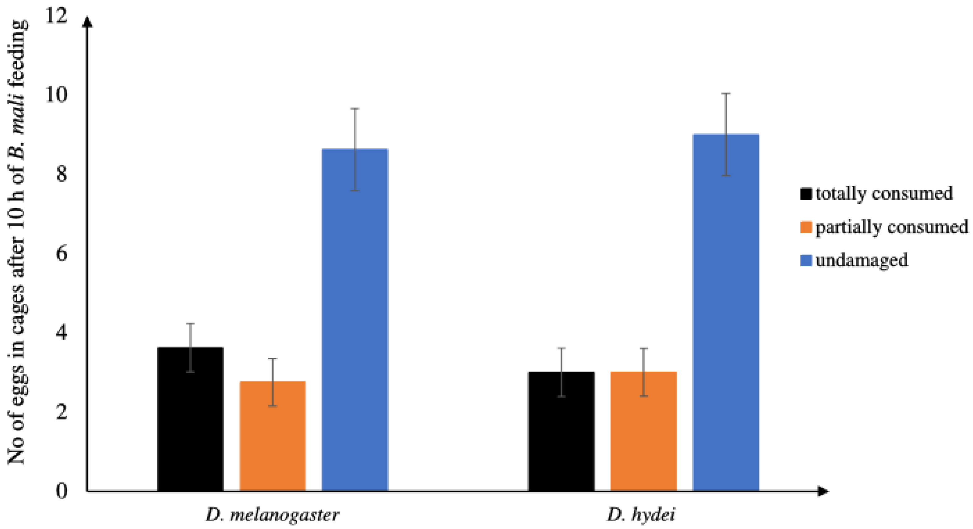
Statistical analysis showed no significant effect of the species of fruit fly on the degree of damage to their eggs by the predator (GLM: χ2 = 0.8171, df = 2, p = 0.5642). When we offered single B. mali females fifteen “1-h” eggs of D. melanogaster or “8-h” eggs of D. hydei, during the following 10 h, they totally consumed 3.62 ± 0.673 (N = 16) D. melanogaster eggs and 3.00 ± 0.612 (N = 16) eggs of D. hydei (GLM: χ2 = 0.4724, df = 14, p = 0.4919) on average (Figure 2). In the cages with the predators, we also found partially collapsed eggs, i.e., 2.75 ± 0.586 (N = 16) and 3.00 ± 0.612 (N = 16) eggs of each fly species (GLM: χ2 = 0.063835, df = 14, p = 0.8005). In the control combinations, however, with either fifteen D. melanogaster (N = 16) or D. hydei (N = 16) eggs, no eggs became flattened or dried until the end of the test.

3.1.2. Hatching of D. melanogaster Larvae from Partially Consumed or Undamaged Eggs

No larvae hatched from D. melanogaster eggs previously partially consumed by B. mali and then left for the next 10 h for hatching (Table 1). The presence of a predator in the cage did not have any destructive effect on the later emergence of D. melanogaster larvae from undamaged eggs. Similarly, transferring the undamaged eggs to new cages had no negative effect on the later hatching of larvae. As statistical analysis showed, there were no significant differences between the proportions of larvae hatching from undamaged eggs, which were either transferred to new cages or left in the experimental cages (after the removal of the predator), and from eggs kept in the control cages (GLM: χ2 = 0.2374, df = 2, p = 0.7904).

3.1.3. Effect of the Age of D. hydei Eggs on the Feeding Rate of B. mali Females

In our 10-h-long test, B. mali females damaged a similar number of “8-h” and “14-h” eggs of D. hydei (GLM: χ2 = 0.15, df = 1, p = 0.6982). However, the age of D. hydei eggs had a significant impact on the degree of their damage (GLM: χ2 = 3.009, df = 2, p = 0.0445). Although the mites totally consumed similar numbers of “8-h” and “14-h” eggs (GLM: χ2 = 0.03523, df = 12; p = 0.8511), they partially consumed significantly more younger than older eggs (GLM: χ2 = 4.5342, df = 12, p = 0.0332) (Figure 3).

3.2. Behavioural Observations on the Feeding of B. mali Females on Fruit Fly Eggs

3.2.1. Repertoire of Feeding Behaviours

The sequence of B. mali behaviours accompanying feeding on the “1-h” eggs of D. melanogaster or the “8-h” D. hydei eggs was similar. After release into the cage, a 24-h-starved predatory female initially moved for some time all over the cage chamber, usually with the first pair of legs raised up. She did not use her legs while attacking an egg or consuming the egg’s contents. After approaching the egg, the female would stop by it, touch it once or several times with her pedipalps, and immediately cut the chorion with its chelicerae and insert them into the egg. The attack of the egg was very fast, taking one or two seconds. While feeding on the egg, the mite was motionless. Its pedipalps were slightly widened and laid over the egg, touching it. In some cases, after removing the chelicerae from the egg, the predator cleaned the chelicerae with the help of pedipalps. Sometimes, it also cleaned the pedipalps using the first pair of legs. However, grooming was not specifically associated with the predator feeding on the egg. It has also been observed in other situations, such as during mite exploration or resting.

3.2.2. The Site of the Insertion of Chelicerae into an Egg

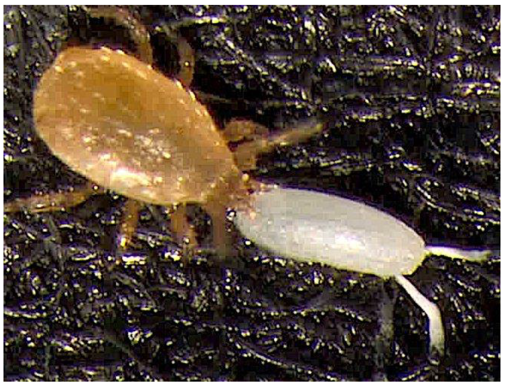
The predator approached an egg either from the back, puncturing it terminally, or from the side, and then burrowed the chelicerae either in the middle part of the egg or closer to its dorsal appendages (Figure 4).
In the case of both D. hydei (Z test of proportion: χ2 = 13.849, df = 2, p < 0.0001) and D. melanogaster eggs (Z test of proportion: χ2 = 22.671, df = 2, p < 0.0001), the proportion of eggs pierced by B. mali near the dorsal appendages was significantly lower than those pierced at the back (p < 0.05) or in the middle part (p < 0.05) (Figure 5). Moreover, when a female touched the dorsal appendages by chance, she immediately left that egg without feeding. We observed three such situations in the trial with D. melanogaster eggs.

3.2.3. Time of Attack and Consumption of an Egg, Partial Egg Consumptions, and Pauses before and after Attack and Egg Consumptions

As in the assay for feeding rates, during our 10-h observations, B. mali females did not always consume the fruit fly eggs completely. Some eggs were attacked and only partially fed, which was manifested by a slight collapse of the chorion of the egg. In total, we registered nine out of 59 consumed eggs of D. melanogaster and 18 out of 66 consumed eggs of D. hydei that were only partially fed by the mites. During our observations, B. mali would partially consume the first encountered eggs as well as those encountered later, after consuming some other eggs. We also noted 12 D. melanogaster eggs and seven D. hydei eggs that were attacked and partially consumed twice. The predator would initially consume the egg, then leave it for some time, and after a pause from feeding or after feeding on another egg, it would return and complete its consumption. We “arbitrary” named these two behaviours as first attack and partial egg consumption and second attack and partial egg consumption, respectively (see Table 2). When the predator fed on the egg repeatedly, it usually returned to the egg and pierced it from a similar side as at the first partial feeding. We observed nine such situations out of 12 cases of repeated feedings on D. melanogaster eggs and six out of eight cases of repeated feedings on D. hydei eggs. Moreover, the mites would, though very rarely, insert the chelicerae into an egg using the same hole as before. Only two B. mali females that fed on D. melanogaster eggs once used a previously made hole.
As shown in Table 2, the average time spent by a starved B. mali female from the moment of release into the cage until the attack and consumption of the first egg was significantly longer for D. hydei than for D. melanogaster eggs during the 10-h observation period. By contrast, the average time of attack and egg consumption and the first attack and partial egg consumption were significantly shorter for D. hydei eggs than for D. melanogaster eggs. The statistical analysis also showed no significant differences in the meantime between the second attack and partial egg consumption and the pauses after the attack and egg consumption of D. melanogaster and D. hydei eggs.

4. Discussion

Our study has shown that starved B. mali females can feed on both D. melanogaster and D. hydei eggs. However, not all the eggs destroyed by the mites were consumed completely. Some of the fruit fly eggs were only fed partially, and even if the chorion of those eggs was only slightly collapsed, no larvae hatched from them. Occasionally, predatory females returned to partially feed eggs, usually from the side where the first puncture was made, and then they consumed them completely. In the test with D. hydei eggs, the age of the eggs had no effect on the number of eggs destroyed by a predator. The mites damaged similar numbers of “8-h” and “14-h” eggs of this fruit fly. However, they exhibited different patterns of egg utilisation, partially consuming a greater number of younger than older D. hydei eggs.
These findings support our hypothesis that B. mali may partially consume fruit fly eggs and that predators’ partial feeding has a lethal effect on their hatchability. Moreover, as in the case of B. tarsalis feeding on E. kuehniella moth eggs [29], the age of the fruit fly egg may affect the degree to which its content is eaten by B. mali. Contrary to our expectation, however, partially consumed eggs of fruit flies can still be “attractive” for B. mali, and the remaining content of the eggs can also be fed on by this predator at a later time.
Our study is the first report of a predator of the genus Blattisocius feeding on the eggs of drosophilid fruit flies. A number of species of mesostigmatic mites, including species from Ascidae, Laelapidae, Melicharidae, Parasitidae, and Blattisocidae, have so far been reported to predate on eggs of drosophilid fruit flies, namely on D. melanogater and D. suzukii (Matsumura) [40,41,42,43]. Furthermore, Lasioseius alli Chant (Blattisocidae) developed and reproduced on D. melanogaster [44].
It should be emphasised that this study examined the predation of B. mali on fruit fly eggs for 10 h only. We also used random predatory females that had previously starved for 24-h and were thus generally highly motivated to feed. However, the rates of B. mali predation could have been substantially different if the predators were of similar age, e.g., young and gravid females, as practised in some studies, and fed by drosophilid eggs for 24 h or a longer time.
A characteristic feature of the eggs of drosophilids is the presence of so-called dorsal appendages, in varying numbers depending on the drosophilid species, which support the respiration of these eggs, especially when they are immersed in fluid. They are located near the micropyle and the so-called operculum, through which the larvae hatch from the egg [31,32].
In our study, the mites did not insert the chelicerae between the appendages but only from the side of an egg, close to these structures, and they did it less often than at the back or in the middle of the egg. It has to be stressed that the chorion within the operculum of a drosophilid egg has a different structure in some layers than that surrounding the main body of the egg [45]. That, plus the presence of the appendages, could have made egg piercing difficult for predatory mites. However, we did not observe any B. mali attempts to penetrate the eggs at this site. Strikingly, accidental touching of the appendages by the mites resulted in their escaping from the egg without feeding. This possible “discouraging” effect of the egg appendages could also explain the delay in B. mali feeding on the first egg of D. hydei (in comparison with D. melanogaster eggs), which has twice as many such structures compared to the D. melanogaster egg. This does not change the fact, however, that in general, the feeding rate on the eggs of both fruit fly species was similar during the “10-h” test. Moreover, starved B. mali females can eagerly feed on D. melanogaster eggs embedded in agar medium from which only those dorsal appendages protrude (Michalska K., unpublished).
Further research is needed to examine whether B. mali’s feeding on fruit fly eggs buried in the substrate or present on its surface would be similar. A study by Esteca [43] on predation on D. suzukii eggs appears especially interesting in this case. All five tested species of predatory mites (from Melicharidae and Lealepidae) fed on D. suzukii eggs present on the substrate, consuming them at different rates, from 2–10 eggs per day. However, when the fruit fly eggs were embedded in the fruit, only Stratiolaelaps scimitus (Womersley) fed on those eggs. According to Esteca [43], among the predators studied, S. scimitius had the longest chelicerae, which probably allowed it to pierce the fruit fly egg in the fruit and feed on it. The results presented in her work indicate, however, that this predatory mite fed on embedded eggs at a much lower rate than on unburied eggs. This may mean that it could have been more difficult for this mite to detect the eggs within the fruit, but also that the presence of protruding appendages could have made it difficult to feed on those eggs and/or that they acted aversively.
When describing predatory behaviour, the time of handling is often estimated, and according to Holling’s definition, it includes the time a predator spends pursuing, subduing, and eating a prey item [46]. In our study, B. mali females devoted the greatest part of the handling time of either “1-h” eggs of D. melanogaster or “8-h” eggs of D. hydei to egg consumption. Their attack consisted of approaching the egg, touching it with pedipalps, and inserting the chelicerae, and lasted very briefly, 1–2 s only, similarly as described for Glyptholaspis americana (Berl.) (Macrochelidae) and Parasitus coleoptratorum (L.) (Parasitidae) feeding on the house fly eggs [47]. Interestingly, while rates of B. mali feeding on D. hydei and D. melanogaster eggs were similar, the mean time of attack and egg consumption and the mean time of first attack and partial consumption of D. hydei eggs were definitely shorter than those of D. melanogaster eggs. It suggests that D. hydei eggs may be more nutritionally valuable for B. mali, and a shorter feeding period (with a smaller amount of egg content) could be enough for predator satiation and/or gut filling. On the other hand, however, predators may simply need more time to suck out a similar portion of food from D. melanogaster eggs than D. hydei eggs due to their biochemical properties. Therefore, to fully explain this phenomenon, more detailed tests are needed.
Partial prey consumption is common among vertebrates and invertebrates, including predatory mites [23,30,48,49,50,51]. This phenomenon is associated with increased prey availability and is internally regulated by the state of the predator’s hunger and/or gut filling [52,53]. This is also in line with predictions of foraging optimalization, according to which the predator should maximise net energy intake, handling time should be shorter, and partial consumption should be favoured when the prey is abundant [54].
Similarly to our study, partial egg consumption has also been reported for B. mali females feeding the eggs of the moth Phthorimaea operculella (Zeller) [23] as well as for B. tarsalis consuming the eggs of the moth Ephestia kuehniella Zeller [29,30]. In the study of Gallego et al. [23], the rate of P. operculella eggs partially consumed was low (3.59 ± 12.03%), while in our test, B. mali females partially consumed nearly 50% of all destroyed eggs. It should be noted, however, that in addition to differences in prey species and the connected food quantity and quality per egg, different prey densities may have been at play. In a test by Gallego et al. [23], five moth eggs were offered to a single predatory female for 48 h, while our study involved 15 fruit fly eggs for 10 h. Interestingly, our behavioural observations showed that some completely consumed eggs were, in fact, fed twice. Not only did the predators approach and feed on the eggs from the same side as at the first partial feeding, but they could also, though rarely, continue to feed precisely at the same spot. Returns to partially consumed prey were also observed in some phytoseiid mites feeding on spider mites [49,55]. Sandness and McMurtry [49] observed up to seven returns of the predatory female Amblyseius largoensis (Muma) to partially fed female spider mites, Oligonychus punicae (Hirst). The relocation of the initially fed prey was presumably based on olfaction, considering that the removal of a dead spider mite caused the predator excitement after returning to the place where the prey had been [45]. The capability of locating and feeding on wounded, initially eaten, and dead prey seems to have significant selective value for mesostigmatic mites, as it may allow them to reduce the time and energy costs of handling related to chasing or subduing prey, but it may also enable them to survive through better utilisation of food resources and scavenging when potential prey is scarce. In B. mali, possible scavenging has been recently indicated by observations of the mite feeding on the dead bodies of the fruit flies D. melanogaster and D. hydei [9].
In many animals, eggs contain large amounts of nutritious yolk, lipid droplets, and glycogen, which are then absorbed in the process of embryogenesis [56]. In D. melanogaster, significant changes in the distribution of the yolk inside the egg and in the external appearance of the developing embryo can already be observed in the first hours after oviposition [57]. Our study showed that even a few hours’ difference in the age of D. hydei eggs (eggs laid within an 8-h vs. 14-h period) had an impact on whether the predator consumed them partially or completely. Although the age of the eggs had no effect on the rate of feeding, the predators partially consumed a greater number of younger than older eggs. Interestingly, a similar phenomenon was observed by Nielsen [29], who fed B. tarsalis females with E. kuehniella eggs. Although there was no difference in the number of moth eggs consumed between the age categories from 0–1 days old to 3–4 days old, larger portions of older moth eggs were fed by the predator. Nielsen [29] believed that older eggs of E. kuehniella were presumably nutritionally inferior to young ones and that B. tarsalis mites compensated for this by eating a larger amount of each single egg.
It should be emphasized here that the older prey eggs may be eaten more willingly by some predatory mites. As shown by Akyazi et al. [58], the starved phytoseiid mites Neoseiulus californicus (McGregor) and Amblyseius swirskii Athias-Henriot consumed higher numbers of older eggs of T. urticae Koch than the younger eggs of this spider mite. Interestingly, in an experiment by Cavalcante et al. [59], A. swirski consumed more younger eggs (up to 24 h of age) than older eggs of Bemisia tabaci (Gennadius). Moreover, when feeding on the younger eggs of this hemipteran insect, it had a higher oviposition rate than on its older eggs. However, when given a choice, the predator was clearly attracted to the older B. tabaci eggs despite the higher suitability of the younger eggs. Therefore, in order to confirm the hypothesis of the greater nutritional value of the younger D. hydei eggs and their possible attractiveness for B. mali further tests are necessary to examine the fecundity of the predator fed on these eggs and their choice by the predator.
Our research to date has shown that B. mali can not only be ectoparasitically associated with drosophilid fruit flies during dispersal [9], but, as it has been demonstrated in this study, they can also prey upon eggs laid by their fruit fly host. However, to assess how much egg predation by B. mali, negatively affects the fitness of their insect hosts, further investigations are needed, including studies on the food preferences of B. mali not only in relation to drosophilid fruit fly eggs, larvae (which can be hidden within the substrate), or pupae but also other potential prey of this predator co-occurring in colonies of these flies.

Author Contributions

Conceptualization, K.M.; methodology, K.M., A.M. and M.K.J.; software, M.S.; validation, K.M., M.S., A.M. and M.K.J.; formal analysis, M.S. and K.M.; Investigation, A.M., K.M. and M.K.J.; Resources, K.M. and M.S.; data curation, K.M. and M.S.; writing—Original Draft Preparation, K.M. and M.S.; writing—review and editing, K.M.; visualization, K.M., M.S., A.M. and M.K.J.; supervision, K.M.; project administration, K.M.; funding acquisition, K.M. All authors have read and agreed to the published version of the manuscript.

Funding

This research received no external funding.

Institutional Review Board Statement

Not applicable.

Data Availability Statement

The data used in this study are available by email request to the corresponding author.

Acknowledgments

We thank D.J. Gwiazdowicz (Poznań University of Life Sciences) for identifying Blattisocius mali. We also thank M. Wit and E. Jabłońska (Warsaw University of Life Sciences) for their help in mite macrophotography.

Conflicts of Interest

The authors declare no conflict of interest.

References

  1. Hunter, P.E.; Rosario, R.M.T. Associations of Mesostigmata with Other Arthropods. Annu. Rev. Entomol. 1988, 33, 393–417. [Google Scholar] [CrossRef]
  2. Szymkowiak, P.; Gorski, G.; Bajerlein, D. Passive dispersal in arachnids. Biol. Lett. 2007, 44, 75–101. [Google Scholar]
  3. Huck, K.; Schwarz, H.H.; Schmid-Hempel, P. Host choice in the phoretic mite Parasitellus fucorum (Mesostigmata: Parasitidae): Which bumblebee caste is the best? Oecologia 1998, 115, 385–390. [Google Scholar] [CrossRef]
  4. De Gasparini, O.; Kilner, R.M. Friend or foe: Inter-specific interactions and conflicts of interest within the family. Ecol. Entomol. 2015, 40, 787–795. [Google Scholar] [CrossRef] [PubMed]
  5. Kvifte, G.M.; Kaczmarek, S.; Marquardt, T.; Seniczak, A. Linked seasonality between a phoretic mite and its moth fly host (Parasitiformes: Mesostigmata and Diptera: Psychodidae). Acarologia 2022, 62, 956–964. [Google Scholar] [CrossRef]
  6. Polak, M.; Markow, T.A. Effect of Ectoparasitic Mites on Sexual Selection in a Sonoran Desert Fruit Fly. Evolution 1995, 49, 660–669. [Google Scholar] [CrossRef]
  7. Horn, C.J.; Luong, L.T. Proximity to parasites reduces host fitness independent of infection in a DrosophilaMacrocheles system. Parasitology 2018, 145, 1564–1569. [Google Scholar] [CrossRef]
  8. Durkin, E.S.; Proctor, H.; Luong, L.T. Life history of Macrocheles muscaedomesticae (Parasitiformes: Macrochelidae): New insights on life history and evidence of facultative parasitism on Drosophila. Exp. Appl. Acarol. 2019, 79, 309–321. [Google Scholar] [CrossRef]
  9. Michalska, K.; Mrowińska, A.; Studnicki, M. Ectoparasitism of the Flightless Drosophila melanogaster and D. hydei by the Mite Blattisocius mali (Acari: Blattisociidae). Insects 2023, 14, 146. [Google Scholar] [CrossRef]
  10. Flanders, S.E.; Badgley, M.E. Prey-predator interactions in self-balanced laboratory populations. Hilgardia 1963, 35, 145–183. [Google Scholar] [CrossRef]
  11. Toda, M.J. DrosWLD: Taxonomic Information Database for World Species of Drosophilidae. Available online: https://bioinfo.museum.hokudai.ac.jp/db/index.php (accessed on 20 March 2023).
  12. Máca, J.; Otranto, D. Drosophilidae feeding on animals and the inherent mystery of their parasitism. Parasit. Vectors 2014, 7, 516. [Google Scholar] [CrossRef] [PubMed]
  13. Ashburner, M. Drosophila: A Laboratory Handbook and Manual; Two Volumes; Cold Spring Harbor Laboratory: New York, NY, USA, 1989; ISBN 9780879693213. [Google Scholar]
  14. Poinar, G.O.; Grimaldi, D.A. Fossil and Extant Macrochelid Mites (Acari: Macrochelidae) Phoretic on Drosophilid Flies (Diptera: Drosophilidae). J. N. Y. Entomol. Soc. 1990, 98, 88–92. [Google Scholar]
  15. Lehtinen, P.T.; Aspi, J. Aspi, J. A Phytoseiid mite Paragarmania mali, associated with drosophilid flies. In Acari Physiological and Ecological Aspects of Acari-Host Relationships; European Association of Acarologists: Krynica, Poland, 1992; pp. 537–544. [Google Scholar]
  16. Perez-Leanos, A.; Loustalot-Laclette, M.R.; Nazario-Yepiz, N.; Markow, T.A. Ectoparasitic mites and their Drosophila hosts. Fly 2017, 11, 10–18. [Google Scholar] [CrossRef] [PubMed]
  17. Kerezis, V.; Kiss, B.; Deucht, F.; Kontschan, J. First record of Blattisocius mali (Oudemans, 1929) in Hungary associated with the drosophilid fly Phortica semivirgo (Máca, 1977). Redia 2019, 102, 69–72. [Google Scholar] [CrossRef]
  18. Polak, M. Effects of Ectoparasitism on Host Condition in the DrosophilaMacrocheles System. Ecology 1998, 79, 1807–1817. [Google Scholar] [CrossRef]
  19. Luong, L.T.; Penoni, L.R.; Horn, C.J.; Polak, M. Physical and physiological costs of ectoparasitic mites on host flight endurance. Ecol. Entomol. 2015, 40, 518–524. [Google Scholar] [CrossRef]
  20. Polak, M. Ectoparasitic Effects on Host Survival and Reproduction: The DrosophilaMacrocheles Association. Ecology 1996, 77, 1379–1389. [Google Scholar] [CrossRef]
  21. Axtell, R.C. New Records of North American Macrochelidae (Acarina: Mesostigmata) and Their Predation Rates on the House Fly. Ann. Entomol. Soc. Am. 1961, 54, 748. [Google Scholar] [CrossRef]
  22. de Moraes, G.J.; Venancio, R.; dos Santos, V.L.V.; Paschoal, A.D. Potential of Ascidae, Blattisociidae and Melicharidae (Acari: Mesostigmata) as Biological Control Agents of Pest Organisms. In Prospects for Biological Control of Plant Feeding Mites and Other Harmful Organisms; Springer International Publishing: Cham, Switzerland, 2015; pp. 33–75. [Google Scholar]
  23. Gallego, J.R.; Gamez, M.; Cabello, T. Potential of the Blattisocius mali Mite (Acari: Blattisociidae) as a Biological Control Agent of Potato Tubermoth (Lepidoptera: Gelechiidae) in Stored Potatoes. Potato Res. 2020, 63, 241–251. [Google Scholar] [CrossRef]
  24. Pirayeshfar, F.; Moayeri, H.R.S.; Da Silva, G.L.; Ueckermann, E.A. Comparison of biological characteristics of the predatory mite Blattisocius mali (Acari: Blattisocidae) reared on frozen eggs of Tyrophagus putrescentiae (Acari: Acaridae) alone and in combination with cattail and olive pollens. Syst. Appl. Acarol. 2022, 27, 399–409. [Google Scholar] [CrossRef]
  25. Carton, Y.; David, J.R. Relation between the genetic variability of digging behavior of Drosophila larvae and their susceptibility to a parasitic wasp. Behav. Genet. 1985, 15, 143–154. [Google Scholar] [CrossRef] [PubMed]
  26. Albornoz, J.; Dominguez, A. Genetic analysis of Drosophila melanogaster egg insertion behavior. Behav. Genet. 1987, 17, 257–262. [Google Scholar] [CrossRef] [PubMed]
  27. Troncoso, B.; Godoy-Herrera, R.; Mora, W. The development of larval movement patterns in Drosophila. Heredity 1987, 58, 321–329. [Google Scholar] [CrossRef]
  28. Darst, P.H.; King, E.W. Biology of Melichares tarsalis in association with Plodia interpunctella. Ann. Entomol. Soc. Am. 1969, 62, 747–749. [Google Scholar] [CrossRef]
  29. Nielsen, S. The use of Blattisocius tarsalis (Acari: Ascidae) for biological control in flour mills. In Proceedings of the 7th Intertuuional Working Conference on Stored-Product Protection, Beijing, China, 14–19 October 1998; Volume 2, pp. 1265–1268. [Google Scholar]
  30. Nielsen, P.S. Predation by Blattisocius tarsalis (Berlese) (Acari: Ascidae) on eggs of Ephestia kuehniella Zeller (Lepidoptera: Pyralidae). J. Stored Prod. Res. 2003, 39, 395–400. [Google Scholar] [CrossRef]
  31. Osterfield, M.; Berg, C.A.; Shvartsman, S.Y. Epithelial Patterning, Morphogenesis, and Evolution: Drosophila Eggshell as a Model. Dev. Cell 2017, 41, 337–348. [Google Scholar] [CrossRef] [PubMed]
  32. Horne-Badovinac, S. The Drosophila micropyle as a system to study how epithelia build complex extracellular structures. Philos. Trans. R. Soc. B Biol. Sci. 2020, 375, 20190561. [Google Scholar] [CrossRef]
  33. Kopecký, J.; Nesvorná, M.; Hubert, J. Bartonella-like bacteria carried by domestic mite species. Exp. Appl. Acarol. 2014, 64, 21–32. [Google Scholar] [CrossRef]
  34. Lourenço, F.; Calado, R.; Medina, I.; Ameixa, O.M.C.C. The Potential Impacts by the Invasion of Insects Reared to Feed Livestock and Pet Animals in Europe and Other Regions: A Critical Review. Sustainability 2022, 14, 6361. [Google Scholar] [CrossRef]
  35. Dabert, J.; Mironov, S.V.; Dabert, M. The explosive radiation, intense host-shifts and long-term failure to speciate in the evolutionary history of the feather mite genus Analges (Acariformes: Analgidae) from European passerines. Zool. J. Linn. Soc. 2022, 195, 673–694. [Google Scholar] [CrossRef]
  36. Dabert, M.; Witalinski, W.; Kazmierski, A.; Olszanowski, Z.; Dabert, J. Molecular phylogeny of acariform mites (Acari, Arachnida): Strong conflict between phylogenetic signal and long-branch attraction artifacts. Mol. Phylogenet. Evol. 2010, 56, 222–241. [Google Scholar] [CrossRef] [PubMed]
  37. Mironov, S.V.; Dabert, J.; Dabert, M. A new feather mite species of the genus Proctophyllodes Robin, 1877 (Astigmata: Proctophyllodidae) from the Long-tailed Tit Aegithalos caudatus (Passeriformes: Aegithalidae)—Morphological description with DNA barcode data. Zootaxa 2012, 3253, 54. [Google Scholar] [CrossRef]
  38. Shaffer, C.D.; Wuller, J.M.; Elgin, S.C.R. Raising Large Quantities of Drosophila for Biochemical Experiments. Methods Cell Biol. 1994, 44, 99–108. [Google Scholar] [PubMed]
  39. R Core Team. Team, R.D.C. R: A Language and Environment for Statistical Computing; R Core Team: Vienna, Austria, 2023. [Google Scholar]
  40. Binns, E.S. Phoresy as Migration—Some Functional Aspects of Phoresy in Mites. Biol. Rev. 1982, 57, 571–620. [Google Scholar] [CrossRef]
  41. Bowman, C.E. Studies on feeding in the soil predatory mite Pergamasus longicornis (Berlese) (Mesostigmata: Parasitidae) using dipteran and microarthropod prey. Exp. Appl. Acarol. 1987, 3, 201–206. [Google Scholar] [CrossRef]
  42. Houck, M.A.; Clark, J.B.; Peterson, K.R.; Kidwell, M.G. Possible Horizontal Transfer of Drosophila Genes by the Mite Proctolaelaps regalis. Science 1991, 253, 1125–1128. [Google Scholar] [CrossRef] [PubMed]
  43. Esteca, F.; de Cassia Neves, F. Environmental Management Strategies for Pest Control in Strawberry Crop. Ph.D. Thesis, Universidade de São Paulo, Piracicaba, Brazil, 2021. [Google Scholar]
  44. Nawar, M.S.; Rakha, M.A.; Ali, F.S. Laboratory studies on the predaceous mite, Lasioseius bispinosus Evans (Acari: Mesostigmata: Ascidae) on various kinds of food substances. Bull. Soc. Entomol. 1990, 69, 247–255. [Google Scholar]
  45. Margaritis, L.H.; Kafatos, F.C.; Petri, W.H. The eggshell of Drosophila melanogaster. I. Fine structure of the layers and regions of the wild-type eggshell. J. Cell Sci. 1980, 43, 1–35. [Google Scholar] [CrossRef]
  46. Papanikolaou, N.E.; Kypraios, T.; Moffat, H.; Fantinou, A.; Perdikis, D.P.; Drovandi, C. Predators’ Functional Response: Statistical Inference, Experimental Design, and Biological Interpretation of the Handling Time. Front. Ecol. Evol. 2021, 9, 749. [Google Scholar] [CrossRef]
  47. Wernz, J.G.; Krantz, G.W. Studies on the function of the tritosternum in selected Gamasida (Acari). Can. J. Zool. 1976, 54, 202–213. [Google Scholar] [CrossRef]
  48. Hoyt, S.C. Effect of Short Feeding Periods by Metaseiulus occidentalis on Fecundity and Mortality of Tetranychus mcdanieli. Ann. Entomol. Soc. Am. 1970, 63, 1382–1384. [Google Scholar] [CrossRef]
  49. Sandness, J.N.; McMurtry, J.A. Prey consumption behavior of Amblyseius largoensis in relation to hunger. Can. Entomol. 1972, 104, 461–470. [Google Scholar] [CrossRef]
  50. Flechtmann, C.H.W.; McMurtry, J.A. Studies on how Phytoseiid mites feed on spider mites and pollen. Int. J. Acarol. 1992, 18, 157–162. [Google Scholar] [CrossRef]
  51. Fantinou, A.A.; Perdikis, D.C.; Maselou, D.A.; Lambropoulos, P.D. Prey killing without consumption: Does Macrolophus pygmaeus show adaptive foraging behaviour? Biol. Control 2008, 47, 187–193. [Google Scholar] [CrossRef]
  52. Holling, C.S. The functional response of predators to prey density and its role in mimicry and population regulation. Mem. Entomol. Soc. Can. 1965, 45, 5–60. [Google Scholar] [CrossRef]
  53. Johnson, D.M.; Akre, B.G.; Crowley, P.H. Modeling arthropod predation: Wasteful killing by damselfly naiads. Ecology 1975, 56, 1081–1093. [Google Scholar] [CrossRef]
  54. Samu, F. Wolf spider feeding strategies: Optimality of prey consumption in Pardosa hortensis. Oecologia 1993, 94, 139–145. [Google Scholar] [CrossRef]
  55. Ballard, R.C. The Biology of the Predacious Mite Typhlodromus Fallacis (Garman) (Phytoseiidae) at 78 degrees F. Ohio J. Sci. 1954, 54, 175–179. [Google Scholar]
  56. Jeong, Y.E.; Chung, H.M.; Ahn, T.I. Utilization of a storage protein in the embryonic development of Drosophila and Xenopus. Korean J. Biol. Sci. 2001, 5, 85–90. [Google Scholar] [CrossRef]
  57. Bownes, M. A photographic study of development in the living embryo of Drosophila melanogaster. J. Embryol. Exp. Morphol. 1975, 33, 789–801. [Google Scholar] [CrossRef]
  58. Akyazi, R.; Altunc, Y.E.; Soysal, M. Amblyseius swirskii ve Neoseiulus californicus (Mesostigmata: Phytoseiidae)’un Tüketim Kapasitesine Avın Yumurta Yaşının Etkisi. Türk Tarım Doğa Bilim. Derg. 2018, 5, 628–633. [Google Scholar] [CrossRef]
  59. Cavalcante, A.C.C.; Borges, L.R.; Lourenção, A.L.; de Moraes, G.J. Potential of two populations of Amblyseius swirskii (Acari: Phytoseiidae) for the control of Bemisia tabaci biotype B (Hemiptera: Aleyrodidae) in Brazil. Exp. Appl. Acarol. 2015, 67, 523–533. [Google Scholar] [CrossRef] [PubMed]
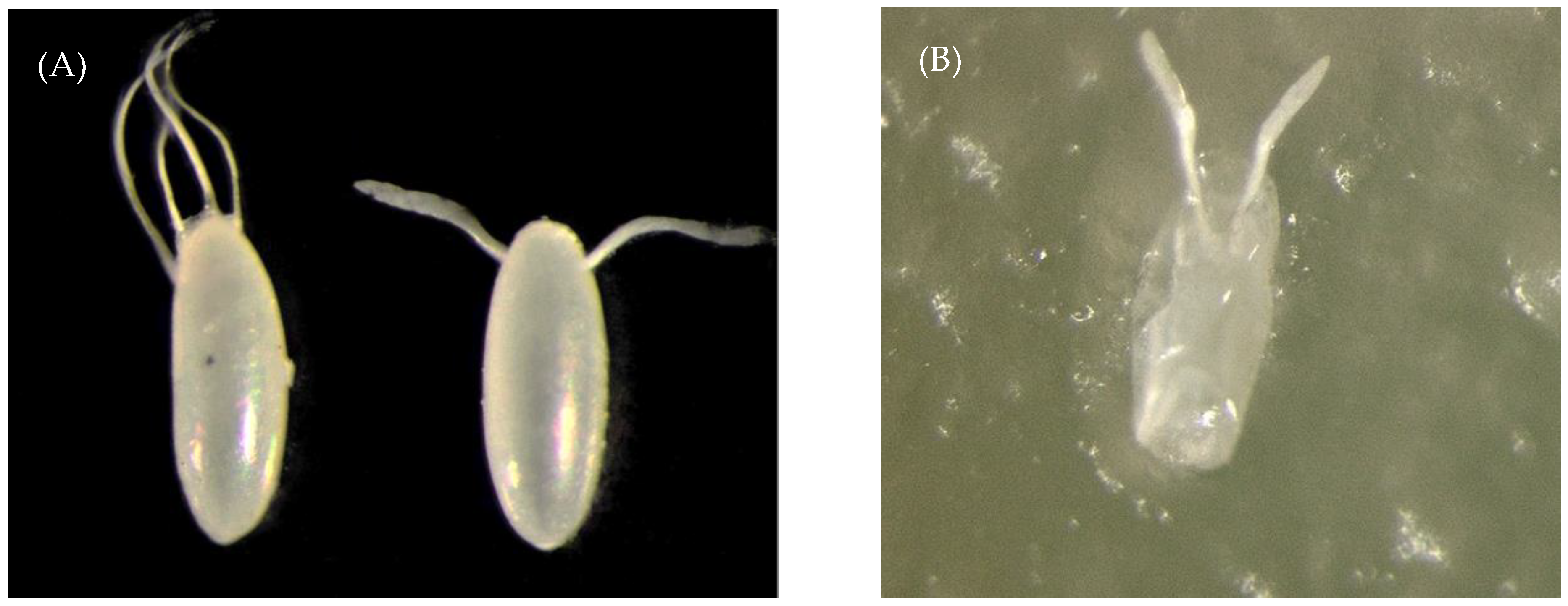
Figure 1. (A). Eggs of Drosophila hydei (left) and D. melanogaster (right). (B). The egg of D. melanogaster is embedded in agar medium and consumed by a Blattisocius mali female.
Diversity 15 00652 g001
Figure 2. Feeding of 24-h starved females of Blattisocius mali on “1-h” Drosophila melanogaster eggs and “8-h” D. hydei eggs. Each cage (N = 16) contained a single predator and 15 fruit fly eggs.
Diversity 15 00652 g002
Figure 3. Feeding of 24-h starved females of Blattisocius mali on “8-h” (N = 16) and “14-h” D. hydei eggs (N = 12) during 10 h. Each cage contained a single predator and 15 fruit fly eggs. * p < 0.05.
Diversity 15 00652 g003
Figure 4. Blattisocius mali female feeding at the back of the egg of Drosophila melanogaster.
Diversity 15 00652 g004
Figure 5. The proportion of eggs of D. melanogaster and D. hydei that were pierced either at the dorsal appendages, in the middle, or at the back by starved Blattisocius mali females in relation to all eggs pierced during the 10-h observational period. Bars with the repeated letters “a” or “b” mean no significant differences between means (p > 0.05).
Diversity 15 00652 g005
Table 1. Mean proportion (±SE; N—number of replications) of D. melanogaster larvae that hatched after 10 h from eggs partially consumed or undamaged by Blattisocius mali or from eggs kept in control cages. Partially consumed or undamaged eggs were either transferred to new cages or left in the experimental cages. All the eggs in the control cages remained in the experimental cages.
Eggs Partially Consumed by a PredatorEggs Undamaged by a PredatorEggs from Control Cages
TransferredLeftTransferredLeft
000.56 ± 0.0950.58 ± 0.0310.53 ± 0.022
(N = 7)(N = 7)(N = 7)(N = 7)(N = 14)
Table 2. The mean time (s) (SE, min-max) of attack and egg consumption, first and second attack and egg consumption, and pauses before and after attack and egg consumption of “1-h” eggs of D. melanogaster and “8-h” eggs of D. hydei by 24-h starved B. mali females during the 10-h observation period. N—number of replications. As the time of attack was very short, it was measured and calculated jointly with the time of egg consumption. For a detailed description of all time parameters, see Section 2.3.
Mean TimeFruit Fly SpeciesStatistics
D. melanogasterD. hydeit TestDegree of Fredomp-Value
Pause before attack and consumption of the first egg302.80 ± 49.36
(93–556)
N = 10
429.63 ± 57.31
(159–689)
N = 8
−3.68160.0115
Attack and egg consumption89.41 ± 7.68
(6–286)
N = 58
67.98 ± 6.34
(5–187)
N = 65
2.151210.0335
First attack and partial egg consumption44.80 ± 8.45
(4–121)
N = 22
21.36 ± 4.69
(5–120)
N = 23
2.55430.0143
Second attack and partial egg consumption83.00 ± 20.91
(6–224)
N = 11
57.88 ± 14.77
(15–137)
N = 9
0.88180.3394
Pause after attack and egg consumption5218.43 ± 527.30
(88–16,445)
N = 68
4447.59 ± 518.62
(21–18,883)
N = 71
1.041370.2991
Disclaimer/Publisher’s Note: The statements, opinions and data contained in all publications are solely those of the individual author(s) and contributor(s) and not of MDPI and/or the editor(s). MDPI and/or the editor(s) disclaim responsibility for any injury to people or property resulting from any ideas, methods, instructions or products referred to in the content.