Article

Metabarcoding Extends the Distribution of Porphyra corallicola (Bangiales) into the Arctic While Revealing Novel Species and Patterns for Conchocelis Stages in the Canadian Flora

by
Gary W. Saunders
1,* and
Cody M. Brooks
1,2
1
Department of Biology, Centre for Environmental & Molecular Algal Research, University of New Brunswick, Fredericton, NB E3B 5A3, Canada
2
Bedford Institute of Oceanography, Department of Fisheries and Oceans, Dartmouth, NS B2Y 4A2, Canada
*
Author to whom correspondence should be addressed.
Diversity 2023, 15(5), 677; https://doi.org/10.3390/d15050677
Submission received: 23 March 2023 / Revised: 4 May 2023 / Accepted: 15 May 2023 / Published: 18 May 2023
(This article belongs to the Special Issue Marine Nearshore Biodiversity)

Abstract

:
Porphyra corallicola was described based on a filamentous red alga inadvertently introduced into culture from a crustose coralline alga. This species is known only in its sporophyte (Conchocelis) stage, being possibly asexual and lacking the charismatic and “collectable” gametophyte stage. Consequently, little is known of its range and distribution. Taxon-targeted metabarcoding was explored as a pathway to gain insights into the vertical (intertidal versus subtidal) and biogeographical distribution of this species, as well as to assess host diversity. We also wanted to ascertain if other species occur in only the Conchocelis stage in the Canadian flora. Primers targeting a short (521 bp) region of the plastid rbcL gene in the Bangiales were used to screen DNA from 285 coralline crusts collected throughout Canada and adjacent waters. In addition to confirming the presence of P. corallicola in the Bay of Fundy, this species was recovered from coralline crusts along the coast of Nova Scotia (n = 1) and in the low Arctic (Labrador; n = 2), greatly extending its range and suggesting it is a cold-water taxon. We have confirmed its presence in both the low intertidal and subtidal (to 10 m), and its occurrence in three different coralline species, suggesting that it lacks host specificity. In total, nine genetic groups of Bangiales were uncovered in our survey, six matching entries currently in GenBank and three apparently novel genetic groups—two from the northeast Pacific and one from the low Arctic. Notable host and ecological patterns are discussed. This method, when further developed, will facilitate the study of Conchocelis stages in nature, which will greatly enhance ecological knowledge of bangialean species.

1. Introduction

For many, Bangiales conjures images of beautiful filmy rose to purple blades, while others may be inclined to think of the typically dark red to golden multiseriate filaments assigned to the form genus Bangia [1]; however, these represent only the gametophyte stage. In 1949, Drew [2] published results linking the endolithic filaments of Conchocelis rosea Batters to the leafy Porphyra umbilicalis Kützing, adding to previous studies that linked germination of spores from other Porphyra spp. with a filamentous stage now known to be the sporophytic stage in a life history with an alternation of heteromorphic generations [1]. Despite the significance of these and subsequent discoveries, especially for the aquaculture of nori, there have been few studies on the actual ecology of Conchocelis stages in situ including aspects of host range, as well as vertical and biogeographical distribution. Although there is a fair level of interest in Conchocelis stages inhabiting corals (e.g., [3] and references therein), our knowledge of cold-temperate systems is more limited.
For European waters, Drew [4] provided the first ecological insights. Although not the focus of her manuscript, what little was known on the ecology of the sporophyte relative to the gametophyte was discussed. In terms of habitat, Drew [4] notes that in addition to calcareous invertebrate shells, the Conchocelis stage grows in calcareous stone and crustose coralline algae (notably Lithothamnion laevigatum Foslie, now Phymatolithon laevigatum (Foslie) Foslie, although there has been considerable confusion in the identification of crustose corallines e.g., [5]) (Figure 1). With so few studies, the view of Conchocelis as a shell-boring filament has become the default in general texts, which is only partially compatible with the work of Drew [4] and others, notably their occurrence in crustose coralline algae [4,6]. Although subsequent works have looked at thermal tolerances for growth and reproduction (summarized in [6]), little is known of the distribution, diversity and habitat specificity of the many species in nature.
In terms of distribution, Drew [4] reports that although the Conchocelis stage “occurs occasionally in the intertidal belt, it is usually found by dredging in water up to 32 m in depth”. Indeed, whereas Porphyra umbilicalis is widely regarded as an intertidal species [6,7], Conchocelis rosea is considered largely subtidal in distribution (although the unequivocal linking of these two species remains uncertain given the cryptic habit of the Conchocelis stages [6]). For the NW Atlantic, detailed phenological observations for Porphyra linearis Greville reported that the Conchocelis stage was absent from the intertidal band of the gametophyte stage but found subtidally at ~9 m depth [8]. In short, Conchocelis stages are typically considered subtidal in distribution (see [6] for a summary), which has caused some to ponder how the spores contribute to the recruitment of the intertidal gametophytic stage. In considering this conundrum, Drew [4] posited that the Conchocelis stage may be more prevalent in the intertidal than realized, considering that “minute pieces” of shell (washed up by storms into the intertidal) and barnacles were likely hosts for Conchocelis filaments and sources for the recruitment of the intertidal gametophyte stages.
A complicating factor is the difficulty, perhaps even inability, to identify various Conchocelis stages in the field to their respective species (i.e., link to a known gametophyte stage) as these filaments are largely cryptic [4,6,9]. As well, although asexual species of Bangiales are reported for the erect (gametophyte) stage, this does not appear to have been considered as a possibility for the Conchocelis stage. The description of Porphyra corallicola H.Kucera & G.W.Saunders (Figure 1) provided a departure in that it was possibly the only species of the Bangiales intentionally described based on its Conchocelis stage in the absence of knowledge of the gametophyte, if one even exists [7]. This species was accidentally introduced into culture when one of the authors (GWS) attempted to culture the red crust Peyssonnelia rosenvingei F.Schmitz. Finding it odd that the culture was filamentous and subsequently that molecular data indicated that it was a Conchocelis, a re-examination of the voucher revealed bangialean filaments growing within the tissue of a calcified coralline crust that had been overgrown by P. rosenvingei (Figure 1). Thus P. corallicola was described based on a single low intertidal collection growing in a crustose coralline alga from the lower Bay of Fundy [7].
The primary aim of this study was to assess P. corallicola considering host preference, vertical distribution (intertidal versus subtidal) and biogeography (especially in light of the connectivity of the Canadian flora through the Arctic e.g., [10,11]) by screening archived crustose coralline DNA at UNB. The secondary aims included assessing how many other species of Conchocelis were living in coralline crusts, to identify any other putatively asexual filament-only species and to look for any other trends of host specificity, vertical distribution and/or geographical distribution of Conchocelis stages relative to their gametophyte stages.
To accomplish these aims while appreciating the cryptic nature of Conchocelis stages, a marker system specific to the Bangiales but excluding coralline algae (and as many other epi-endophytic organisms that inhabit them as possible) was designed. Although not the best barcode marker for species discrimination among red algae, the rbcL provides reasonable species resolution among Bangiales [7] and excludes a wide variety of potential non-photosynthetic contaminants [12]. Primers were developed to amplify 521 bp of this marker for Bangiales, but attempts at Sanger sequencing revealed multiple bangialean taxa in some hosts (ambiguities in the data consistent with two or more bangialean taxa being present), which resulted in the application of NextGen sequencing—in essence taxon-targeted metabarcoding—to further reveal Conchocelis’ diversity and ecology in coralline crusts. Although a preliminary survey, we have uncovered some interesting patterns as well as four putative new species of Bangiales in Canadian waters.

2. Materials and Methods

A total of 285 collections were selected for this study from archival crustose coralline DNAs (Table S1). In total, 237 collections were from the northwest Atlantic (ranging from Connecticut to Newfoundland and Labrador in the low Arctic), 41 from British Columbia (plus an additional collection was from Washington), three from Hudson Bay, two from Nunavut and a single collection from Norway (Table S1). The previously extracted DNA followed published protocols [13]. Amplification targeted a 521 bp region of the rbcL gene using the reverse primer TLR6 (5′ GTATAACCAATWACAAGRTC 3′ [12]) and the novel forward primer ConcF3 (5′ GWGTIGATCCAGTTCCRAAYGTTG 3′) and a published PCR profile for red algal rbcL [12]. ConcF3 was designed to amplify Bangiales to the exclusion of other red algae by aligning 35 and 441 rbcL sequences, respectively, to identify a suitable primer. Successful amplicons were sent to the Integrated Microbiome Resource (IMR) at Dalhousie University for short-read amplicon sequencing on an Illumina MiSeq machine following [14].
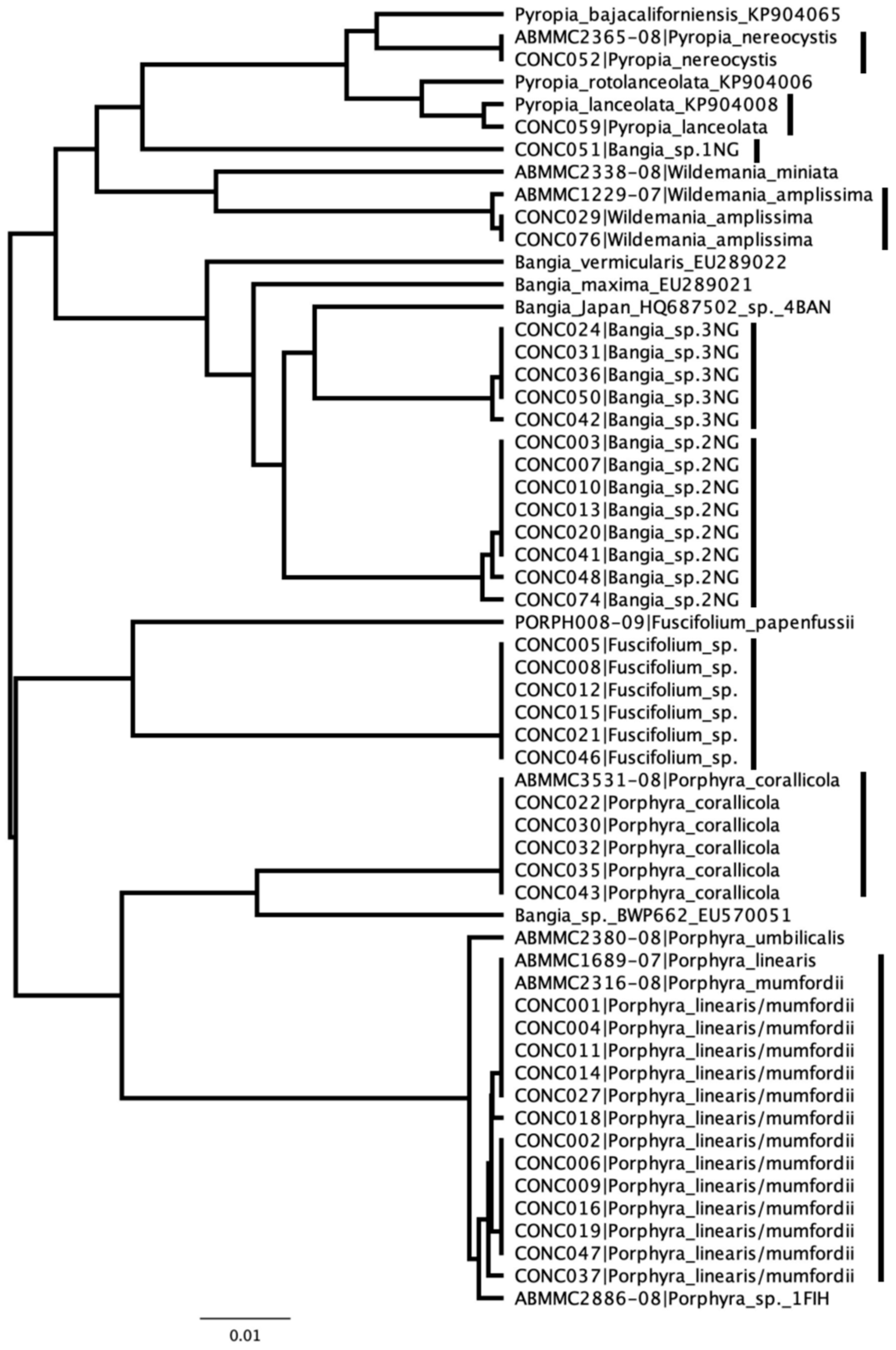
Raw data were processed using QIIME2 [15] and DADA2 software [16], generally following the Microbiome Helper standard operating procedures [14,17], with a relaxed expected error rate of three during read trimming. Current reference libraries used for metabarcoding analyses are generally lacking in red algal coverage and were found unsuitable for adequately identifying species within the Bangiales, so a custom reference library was created of 65 sequences (Table S2) amalgamated from publicly available data in GenBank and supplemented with newly generated sequences following established protocols [12]. All sequences used were generated at UNB and thus unequivocally linked to a voucher. To visualize the genetic groups obtained through metabarcoding in context of the reference library, all sequences were aligned by eye and subsequently subjected to UPGMA cluster analyses (Jukes and Cantor corrected distances) in Geneious Prime 2023.1.1 (https://www.geneious.com accessed on 7 March 2023).

3. Results

Of the original 285 specimens, 56 were successfully amplified and sequenced resulting in a total of 47,847 raw reads. Following denoising, dereplication and chimera filtering steps in DADA2, these raw reads were reduced to just 13,510 (28.2%), representing 49 unique operational taxonomic units (OTUs), which were assigned names by the Microbiome Helper pipeline using the custom reference database. Novel OTUs (not in the reference database) were compared against publicly available data in GenBank to search for their best match. Forward reads were truncated at a length of 294 bp with a median Phred score of 35, and reverse reads at a length of 283 bp with a median Phred score of 24 to allow adequate overlap for the merging of paired-end reads. A review by eye uncovered that seven of these 49 OTUs were chimeric, despite previous filtering. Using a ~0.5% threshold (allowing for 2–3 substitutions owing to variation within a species and/or PCR and sequencing errors), the remaining 42 sequences resolved into nine genetic species groups (Figure 2). Five of the previous matched known species for Canadian waters, one matched an unnamed species of Fuscifolium in GenBank (KP781730) while the remaining three were newly encountered species tentatively assigned to Bangia sensu lato (Figure 2 and Table 1). These nine genetic species groups were distributed among 28 positive Conchocelis identification events (CIEs) (Table 1 and Table S3).
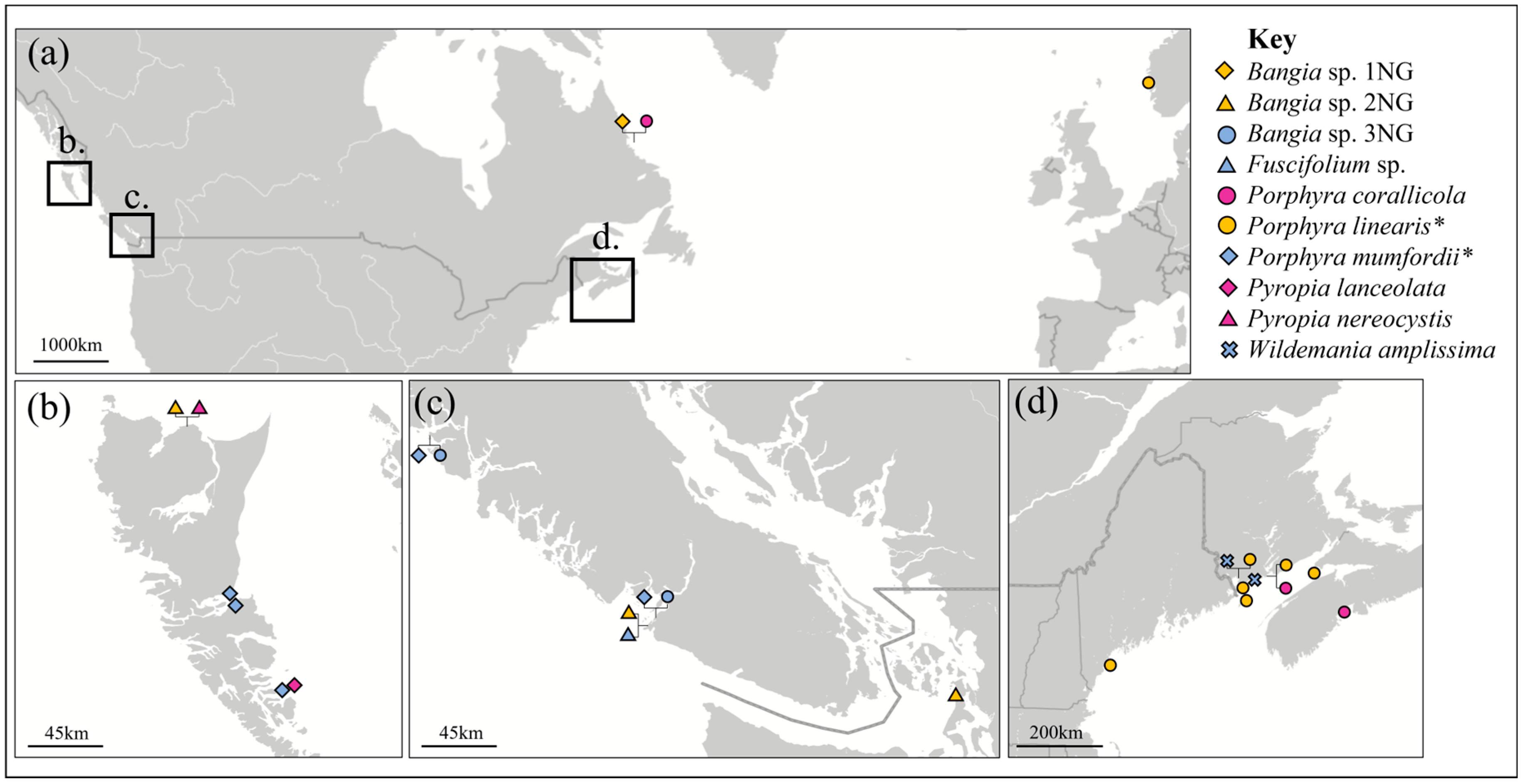
Of primary consideration here is that four of the CIEs were for Porphyra corallicola, extending the range from the Bay of Fundy into the low Arctic (Labrador; Table 1 and Figure 3). The Arctic also returned a new Bangia sp. (sp. 1NG; Table 1 and Figure 3). Although Wildemania amplissima grows in both the Pacific and Atlantic, our two positive CIEs were from New Brunswick (Table 1 and Figure 3). Our marker region could not distinguish between Porphyra linearis and Porphyra mumfordii; however, the former is considered an Atlantic species and the latter a Pacific species, accounting for seven and five of the twelve CIEs, respectively (Table 1 and Figure 3). Pyropia lanceolata and Pyropia nereocystis were recovered from NE Pacific crusts, consistent with the expected range of these species, as were two of the new Bangia spp. (sp. 2NG and sp. 3NG) and a range extension for Fuscifolium sp., which was previously reported from Chile (KP781730) (Table 1 and Figure 3).
Of the crusts tested, 19% (54 of 283 (2 of the 285 lacked distributional data; Table S1)) were collected from the intertidal while the CIEs returned were 29% intertidal (8 of 28; Table 1). In total, 85% of the specimens tested were Atlantic/Arctic in distribution (243 of 285), but only 50% of the CIEs were from this region (14 of 28; Table 1). Ten of the twenty-eight CIEs were recovered from coralline crusts that were growing on shells or invertebrates, opening the possibility that the actual host may not have been the coralline crust but the latter’s host (Table 1). At least 11 host coralline species in six divergent genera were uncovered with no obvious patterns of host specificity (Table 1).

4. Discussion

The primary objective of this study was to assess the utility of taxon-targeted metabarcoding in extending knowledge on the range of Porphyra corallicola, which is currently known only from the type culture isolated from the low intertidal zone at Maces Bay along the lower coast of the Bay of Fundy in New Brunswick [7]. Our new data identified a second low intertidal collection from near the type location at Musquash Head (host GWS046571), but also three subtidal records: one from Nova Scotia (host GWS011835) and two from the low Arctic in Labrador (hosts GWS040344 and GWS040346) (Table 1 and Figure 3). These CIEs were recovered from three hosts—an unidentified Clathromorphum sp., Lithothamnion glaciale and L. lemoineae—all relatively robust species and all growing directly on rock (GWS040346 was partially overgrowing a dead crustose coralline, Table 1), consistent with the crusts being the host for Porphyra corallicola. Our results have thus extended the vertical, host and biogeographical range of this species.
In the NW Atlantic, we also uncovered two CIEs for Wildemania amplissima, both intertidal in the Bay of Fundy. We have collected the gametophyte stage of this species widely in Canadian waters from the intertidal to shallow subtidal and it is common in the lower Bay of Fundy during spring and summer based on records in our database [18]. The most encountered CIEs in this area matched Porphyra mumfordii/linearis (our marker region cannot distinguish between these two species), which were found from the low intertidal to subtidal in a variety of hosts (Table 1). Interestingly, this was also the most common CIE in British Columbia, suggesting that these two closely related species may prefer crustose coralline algae as habitat for their Conchocelis stages. As Porphyra mumfordii is a Pacific species (although see [19]) and P. linearis an Atlantic species, we have used this biogeographical pattern to make tentative species assignments (Table 1 and Figure 3). While we have collected the gametophyte stage of P. mumfordii widely in British Columbia from the intertidal, we have only collected Porphyra linearis during winter in the intertidal at a few exposed locations [18]. There are two notable exceptions, but both are presumptive Conchocelis stages. A single subtidal collection from Massachusetts was reported as growing in a coralline crust (MK185874), and our only previous Bay of Fundy collection for this species was also subtidal (GWS041780; OQ706563), growing in undetermined “calcified substrata”. Despite the limited vertical, ecological and seasonal distribution of the gametophyte, the Conchocelis stage appears to be more widely distributed in all categories. Notably, we are yet to encounter the gametophyte stage in the Bay of Fundy, but the previous record (GWS041780) and five of the seven Conchocelis stages detected here were from this area (Table 1 and Figure 3). Thus, there may be a generalization that Conchocelis stages have broader biogeographical and ecological ranges than their gametophytic counterparts as has been noted in other red algae with alternations of heteromorphic generations (e.g., [11]).
In the NE Pacific, in addition to the known species Pyropia lanceolata and Pyropia nereocystis, we uncovered three novel species: what appear to be newly discovered species Bangia sp. 2NG and Bangia sp. 3NG, as well as a range extension for Fuscifolium sp. currently reported from Chile (Table 1). To determine if these species represent stages in a sexual life history with an alternation of generations (e.g., Porphyra linearis) or novel asexual Conchocelis-only species (e.g., Porphyra corallicola) will require more study. We can note that through our work [18] and that of colleagues (e.g., [1]), hundreds of specimens of bangialean gametophytes have been genetically screened from British Columbia and have not turned up matches to these species. We do have a gametophytic Bangia sp. (GWS008341; UPA, JN029024) from northern British Columbia that currently lacks rbcL data, which may be a match to one of these species. Prior to this study we had only encountered a single genetic group for Fuscifolium in British Columbia, Fuscifolium papenfussii (V.Krishnamurthy) S.C.Lindstrom (e.g., JN028940), and indeed only one other species is included in this genus (Fuscifolium tasa (Yendo) S.C.Lindstrom) for which there are rbcL data in GenBank [1]. The novel Fuscifolium sp. may or may not have an erect gametophytic stage in British Columbia but is reported as a blade (gametophyte) in Chile [20]. More collections are needed from British Columbia to determine if this species undergoes an alternation of heteromorphic generations in that region or simply persists as an asexual Conchocelis stage.
It is also notable that while only 15% of the crusts screened were from the Pacific (Table S1), 50% of the CIEs were from this region (14 of 28; Table 1). In looking at our own records for gametophyte stages, there are more species of Bangiales in the NE Pacific than NW Atlantic (~25 vs. 17 [18]), but this slight difference does not appear to account for the discrepancy. It could be that more Pacific species are sexual and/or prefer coralline algae as hosts for their Conchocelis stages (there is considerably more crustose coralline diversity in the NE Pacific, perhaps providing more opportunity; compare [21,22]). More study is needed.
The novel entity designated here as Bangia sp. 1NG was also from the low Arctic. It was only a distant match (93.61%) to a genetic group that we call Bangia sp. 2Ban, that unequivocally has a gametophyte with the Bangia morphology and is widely distributed in the North Atlantic with genetic matches from Rhode Island to Norway [18]. Thus, if Bangia sp. 1NG does have an alternation of generations, the gametophyte is likely to have this morphology and again more collections are needed from northern waters. Although there is a general notion that the Conchocelis stages are subtidal in distribution (e.g., [8]), Drew [4] suggested that they may be more common in the intertidal than realized. Only 19% of the crusts we tested were collected from the intertidal, while the CIEs were 29% intertidal (Table 1). This supports Drew’s assertion, but it is important to recognize that they are also abundant subtidally with some of our records from as deep as 10 m (Table 1).
The three novel taxa uncovered here are all in the genus Bangia. Currently in Canada, floristic guides recognize only Bangia atropurpurea (Roth) C.Agardh [21] or have defaulted to Bangia spp., reporting three genetic groups in need of taxonomic study [22]. Our own lab has uncovered two genetic groups in BC and two in the NW Atlantic [18], with those numbers increasing to three (or four) and three, respectively, following this survey (Figure 2). None of the genetic groups discussed here from Canada are a genetic match for bona fide B. atropurpurea, which is a freshwater species [1]. Considerable taxonomic research remains for this genus in our waters and indeed the genus itself is not monophyletic [1].
This study has limitations as it was opportunistic in the use of existing archived crustose coralline DNA. Nonetheless, it has merit in joining the few studies that use archival DNA for purposes other than taxonomy (for examples, see [23,24]), and it serves as a proof of concept that this technique can be used to identify Conchocelis stages in the field, which sets the foundation for a more structured survey of coralline crusts (and other calcareous substrata) based on targeted sampling. In carrying out such a project, care must be taken in acquiring the crusts to ensure that any Conchocelis stages identified are unequivocally growing in the coralline alga. For example, 10 of the 28 CIEs that we recovered here were from coralline crusts that were growing on shells or invertebrates, opening the possibility that the host may not have been the actual coralline crust (Table 1). Although we always sample with care to avoid contamination from underlying or host material, it can be difficult to obtain “clean” samples from nature. However, this last-mentioned caveat raises the potential of under sampling as we intentionally avoided old or what appeared to be infested pieces of material during sampling to facilitate acquiring a clean target sequence for coralline crusts during our routine DNA barcode surveys. A survey for Conchocelis stages would do the opposite, focusing on old and infested pieces of hosts. It is also notable that the Conchocelis stages have been cultured without the use of calcareous substrata (e.g., [4,9]), raising the possibility that they may grow in noncalcareous substrata. Hence, the screening of fleshy macroalgae may return unexpected results.
The 521 bp region of the rbcL used as a marker here failed to resolve all the target species. The rbcL-3P is considered a suitable secondary barcode marker for red algae [12], but that region is 800 bp in length and outside the range for the Illumina technology used here. Further, it is more variable than the region used here, which obstructed efforts at primer design for a shorter fragment from this region of the gene. For red algae, the COI-5P and ITS are recognized as better barcode markers in terms of species resolution [25], but they come with other shortfalls with regard to the current study. Being highly variable, COI-5P primer design has been a challenge for red algae [12], and we were unable to design primers to include all Bangiales to the exclusion of other red algae. The ITS is potentially more amenable to primer design but can be highly variable in length in red algae, which could result in taxa being missed with Illumina. Both of these markers are also found in a wider variety of taxa (e.g., animals, fungi, etc.) than the rbcL, which would further complicate primer design and could invite further PCR bias and lead to reduced Conchocelis read counts and underestimating species richness [26]. In future, a longer fragment of the rbcL could be used (if primers can be developed), but this would require a different sequencing technology. In the end, the marker used here separated all but two of the taxa included in our reference database (Table S2), which is good resolution by metabarcoding standards.
Metabarcoding can struggle with high false-negative rates [27], owing in part to the necessary discarding of large quantities of “junk data” by the denoising program (DADA2 in this case, although this is a universal feature). Typically, “junk data” are rare reads, particularly singletons and doubletons, which are notoriously difficult to discern from sequencing artefacts [16]. In this study, old stocks of DNA were screened for minute traces of microscopic endophytes belonging to a single order, so unsurprisingly our quantity of raw reads was low (~43,000) and the quantity of rare reads was high. This paucity of data was exacerbated when denoising removed the singletons, doubletons and rare reads (71.8% of raw reads), many of which could be true, rare species [28]. The authors in [29] observed (albeit in fungal systems) that up to 44% of the discarded singletons and doubletons alone could be true rare species rather than sequencing artefacts, to say nothing of other low-abundance reads also discarded by DADA2. Although the results of this study include several novel groups, the true diversity of Conchocelis present in these crustose hosts is likely under-represented here. In contrast, had we not carefully examined the resulting alignment for chimeric sequences post bioinformatics pipeline, we would have had seven additional OTUs and would be reporting on the remarkable levels of undiscovered bangialean diversity in Canadian waters. Clearly, better methods for analyzing these types of data are needed for metabarcoding surveys to reach their full potential.
As a final consideration, without detailed microscopy it can be difficult to confirm that the sequences are from an endophytic Conchocelis stage and not simply from juveniles of the gametophytic stage. We note that we have never collected the gametophyte of Porphyra linearis in the Bay of Fundy, and yet five of the seven positive CIEs were from this region (Table 1 and Figure 3). Further, the P. linearis gametophyte is a mid to upper intertidal winter annual [8], while all of our CIEs (including the two discussed above as being encountered during routine DNA barcode screening) were low intertidal (n = 3) or subtidal (n = 6) and collected from April to September (Table S3). Finally, in contrast to the gametophyte stage of Porphyra linearis, which is seemingly rare in the Bay of Fundy and confined to winter and the mid to upper intertidal, the closely allied Porphyra umbilicalis Kützing (Figure 2) is common in the Bay of Fundy (we have 78 archived collections [18]), and occurs from the upper intertidal to the shallow subtidal and in all seasons (we have collections from March to December; we rarely collect in December to March) in this area [18,30]). However, in our experience, Porphyra umbilicalis is asexual and confined to the blade morphology in this region, which may account for this species not being encountered in our study, consistent with our CIEs (Table 1) being legitimately from Conchocelis stages. On the other hand, we did not encounter the Conchocelis for the less closely related and presumably sexual Porphyra purpurea (Roth) C.Agardh, which is also common in this region [7,18]. Does its Conchocelis grow exclusively in calcareous substrata of animal origin, or is it actually asexual in the study region? Unlike Porphyra linearis, we have not unexpectedly encountered the sporophyte of P. purpurea in our routine barcode surveys (which include abundant coralline crusts, but not animals), which is consistent with both the previous hypotheses. With the tools developed here we can begin to resolve these uncertainties and further shed light on the ecology of the Conchocelis stages and species in the Bangiales.

Supplementary Materials

The following supporting information can be downloaded at https://www.mdpi.com/article/10.3390/d15050677/s1, Table S1: Listing of coralline DNAs screened for Conchocelis stages with their associated collection data; Table S2: List of all species included in the custom Bangiales reference database and their associated GenBank accession numbers; Table S3: Resulting CIEs with their assigned name, match and associated collection data.

Author Contributions

G.W.S. conceptualized the project and developed the marker system used to generate sequence data, as well as completed the original writing of a draft preparation. C.M.B. managed the short-read amplicon sequencing, including pipeline assembly and data analyses. Both authors contributed to project administration and data management, preparation of the final document for submission and the interpretation of the data. All authors have read and agreed to the published version of the manuscript.

Funding

This research was funded by the Natural Sciences and Engineering Research Council of Canada, through a Discovery Grant to G.W. Saunders (170151-2013), with infrastructure support from the Canada Foundation for Innovation and the New Brunswick Innovation Foundation.

Institutional Review Board Statement

Not applicable.

Data Availability Statement

Sequences generated for the rbcL reference database are available through the Barcode of Life Data System published dataset DS-BANGIAL1 Bangiales DNA Barcode Survey Data release [18], as well as GenBank (Table S2). The Illumina data are available in GenBank PRJNA951650.

Acknowledgments

This work was only possible through access to a number of archived DNA extractions from coralline crusts collected by numerous members of the Saunders Laboratory over the years, all of whom are thanked for their contribution. T. Moore generated some of the reference bangialean rbcL sequences used in this study (Table S2) and is thanked for her assistance in other technical aspects of this project.

Conflicts of Interest

The authors declare no conflict of interest. The funders had no role in the design of the study; in the collection, analyses, or interpretation of data; in the writing of the manuscript; or in the decision to publish the results.

References

  1. Sutherland, J.E.; Lindstrom, S.C.; Nelson, W.A.; Brodie, J.; Lynch, M.D.J.; Hwang, M.S.; Choi, H.G.; Miyata, M.; Kikuchi, N.; Oliveira, M.C.; et al. A new look at an ancient order: Generic revision of the Bangiales (Rhodophyta). J. Phycol. 2011, 47, 1131–1151. [Google Scholar] [CrossRef] [PubMed]
  2. Drew, K.M. Conchocelis-phase in the life-history of Porphyra umbilicalis (L.) Kütz. Nature 1949, 164, 748–749. [Google Scholar] [CrossRef]
  3. Tribollet, A.; Pica, D.; Puce, S.; Radtke, G.; Campbell, S.E.; Golubic, S. Euendolithic Conchocelis stage (Bangiales, Rhodophyta) in the skeletons of live stylasterid reef corals. Mar. Biodiv. 2018, 48, 1855–1862. [Google Scholar] [CrossRef]
  4. Drew, K.M. Studies in the Bangioideae III. The life-history of Porphyra umbilicalis (L.) Kütz. var. laciniata (Lightf.) J. Ag.: A. The Conchocelis-phase in culture. Ann. Bot. 1954, 18, 183–211. [Google Scholar]
  5. Peña, V.; Bélanger, D.; Gagnon, P.; Richards, J.L.; Le Gall, L.; Hughey, J.R.; Saunders, G.W.; Lindstrom, S.C.; Rinde, E.; Husa, V.; et al. Lithothamnion (Hapalidiales, Rhodophyta) in the Arctic and Subarctic: DNA sequencing of type and recent specimens provides a systematic foundation. Eur. J. Phycol. 2021, 56, 468–493. [Google Scholar] [CrossRef]
  6. Brodie, J.A.; Irvine, L.M. Seaweeds of the British Isles. Volume 1. Rhodophyta. Part 3B. Bangiophycidae; Intercept: Andover, UK, 2003; pp. i–xiii, 1–167. [Google Scholar]
  7. Kucera, H.; Saunders, G.W. A survey of Bangiales (Rhodophyta) based on multiple molecular markers reveals cryptic diversity. J. Phycol. 2012, 48, 869–882. [Google Scholar] [CrossRef]
  8. Bird, C.J. Aspects of the life history and ecology of Porphyra linearis. (Bangiales, Rhodophyceae) in nature. Can. J. Bot. 1973, 51, 2371–2379. [Google Scholar] [CrossRef]
  9. Bird, C.J.; Chen, L.C.-M.; McLachlan, J. The culture of Porphyra linearis (Bangiales, Rhodophyceae). Can. J. Bot. 1972, 50, 1859–1863. [Google Scholar] [CrossRef]
  10. Bringloe, T.T.; Saunders, G.W. DNA barcoding of the marine macroalgae from Nome, Alaska (Northern Bering Sea) reveals many trans-Arctic species. Polar Biol. 2019, 42, 851–864. [Google Scholar] [CrossRef]
  11. Bringloe, T.T.; Verbruggen, H.; Saunders, G.W. Unique diversity in Arctic marine forests is shaped by diverse recolonisation pathways and far northern glacial refugia. Proc. Natl. Acad. Sci. USA 2020, 117, 22590–22596. [Google Scholar] [CrossRef] [PubMed]
  12. Saunders, G.W.; Moore, T.E. Refinements for the amplification and sequencing of red algal DNA barcode and RedToL phylogenetic markers: A summary of current primers, profiles and strategies. Algae 2013, 28, 31–43. [Google Scholar] [CrossRef]
  13. Saunders, G.W.; McDevit, D.C. Methods for DNA barcoding photosynthetic protists emphasizing the macroalgae and diatoms. Methods Molec. Biol. 2012, 858, 207–222. [Google Scholar]
  14. Comeau, A.M.; Douglas, G.M.; Langille, G.I. Microbiome helper: A custom and streamlined workflow for microbiome research. Methods Protoc. 2017, 2, e00127-16. [Google Scholar] [CrossRef]
  15. Bolyen, E.; Rideout, J.R.; Dillon, M.R.; Bokulich, N.A.; Abnet, C.C.; Al-Ghalith, G.A.; Alexander, H.; Alm, E.J.; Arumugam, M.; Asnicar, F.; et al. Reproducible, interactive, scalable and extensible microbiome data science using QIIME2. Nat. Biotechnol. 2019, 37, 852–857. [Google Scholar] [CrossRef] [PubMed]
  16. Callahan, B.J.; McMurdie, P.J.; Rosen, M.J.; Han, A.W.; Johnson, A.J.A.; Holmes, S.P. DADA2: High-resolution sample inference from Illumina amplicon data. Nat. Methods 2016, 13, 581–583. [Google Scholar] [CrossRef] [PubMed]
  17. Martin, M. Cutadapt removes adapter sequences from high-throughput sequencing reads. Embnet. J. 2011, 17, 10–12. [Google Scholar] [CrossRef]
  18. Saunders, G.W.; Moore, T.E. Dataset—DS-BANGIAL1 Bangiales DNA Barcode Survey Data Release. 2023. Available online: http://www.boldsystems.org/index.php/Public_SearchTerms?query=DS-BANGIAL1 (accessed on 7 March 2023).
  19. Saunders, G.W.; Brooks, C.M.; Scott, S. Preliminary DNA barcode report on the marine red algae (Rhodophyta) from the British Overseas Territory of Tristan da Cunha. Cryptogam. Algol. 2019, 40, 105–117. [Google Scholar] [CrossRef]
  20. Guillemin, M.-L.; Contreras-Porcia, L.; Ramírez, M.E.; Macaya, E.C.; Contador, C.B.; Woods, H.; Wyatt, C.; Brodie, J. The bladed Bangiales (Rhodophyta) of the South Eastern Pacific: Molecular species delimitation reveals extensive diversity. Mol. Phylogenet. Evol. 2016, 94, 814–826. [Google Scholar] [CrossRef]
  21. Sears, J.R. NEAS Keys to Benthic Marine Algae of the Northeastern Coast of North America from Long Island Sound to the Strait of Belle Isle; Northeast Algal Society Contr. No. 2. Univ. Mass. Dartmouth Campus Book Store: Dartmouth, MA, USA, 2002; pp. i–xvii, 1–161. [Google Scholar]
  22. Gabrielson, P.W.; Lindstrom, S.C. Keys to the Seaweeds and Seagrasses of Southeast Alaska, British Columbia, Washington, and Oregon; Phycological Contribution Number 9; Island Blue/Printorium Bookworks: Victoria, BC, USA, 2018; pp. i–iv, 1–180. [Google Scholar]
  23. Datlof, E.M.; Amend, A.S.; Earl, K.; Hayward, J.; Morden, C.W.; Wade, R.; Zahn, G.; Hynson, N.A. Uncovering unseen fungal diversity from plant DNA banks. PeerJ 2017, 5, e3730. [Google Scholar] [CrossRef]
  24. Heberling, J.M.; Burke, D.J. Utilizing herbarium specimens to quantify historical mycorrhizal communities. Appl. Plant Sci. 2019, 7, e01223. [Google Scholar] [CrossRef]
  25. Saunders, G.W. Applying DNA barcoding to red macroalgae: A preliminary appraisal holds promise for future applications. Phil. Trans. R. Soc. B. 2005, 360, 1879–1888. [Google Scholar] [CrossRef]
  26. Tedersoo, L.; Nilsson, R.H.; Abarenkov, K.; Jairus, T.; Sadam, A.; Saar, I.; Bahram, M.; Bechem, E.; Chuyong, G.; Kõljalg, U. 454 Pyrosequencing and Sanger sequencing of tropical mycorrhizal fungi provide similar results but reveal substantial methodological biases. New Phytol. 2011, 188, 291–301. [Google Scholar] [CrossRef]
  27. Harper, L.R.; Handley, L.L.; Hahn, C.; Boonham, N.; Rees, H.C.; Gough, K.C.; Lewis, E.; Adams, I.P.; Brotherton, P.; Phillips, S.; et al. Needle in a Haystack? A comparison of eDNA metabarcoding and targeted qPCR for detection of the great crested newt (Triturus cristatus). Ecol. Evol. 2017, 8, 6330–6341. [Google Scholar] [CrossRef] [PubMed]
  28. Zhan, A.; Hulák, M.; Sylvester, F.; Huang, X.; Adebayo, A.A.; Abbot, C.L.; Adamowicz, S.J.; Heath, D.D.; Cristescu, M.E.; MacIsaac, H.J. High-sensitivity pyrosequencing for detection of rare species in aquatic communities. Methods Ecol. Evol. 2013, 4, 558–565. [Google Scholar] [CrossRef]
  29. Brown, S.P.; Veach, A.M.; Rigdon-Huss, A.R.; Grond, K.; Lickteig, S.K.; Lothamer, K.; Oliver, A.K.; Jumpponen, A. Scraping the bottom of the barrel: Are rare high throughput sequences artifacts? Fungal Ecol. 2019, 13, 221–225. [Google Scholar] [CrossRef]
  30. Saunders, G.W. Seaweed of Canada. Porphyra umbilicalis Kützing (A). Available online: https://seaweedcanada.wordpress.com/pophyra-umbilicalis-linnaeus-kutzing-a/ (accessed on 7 March 2023).
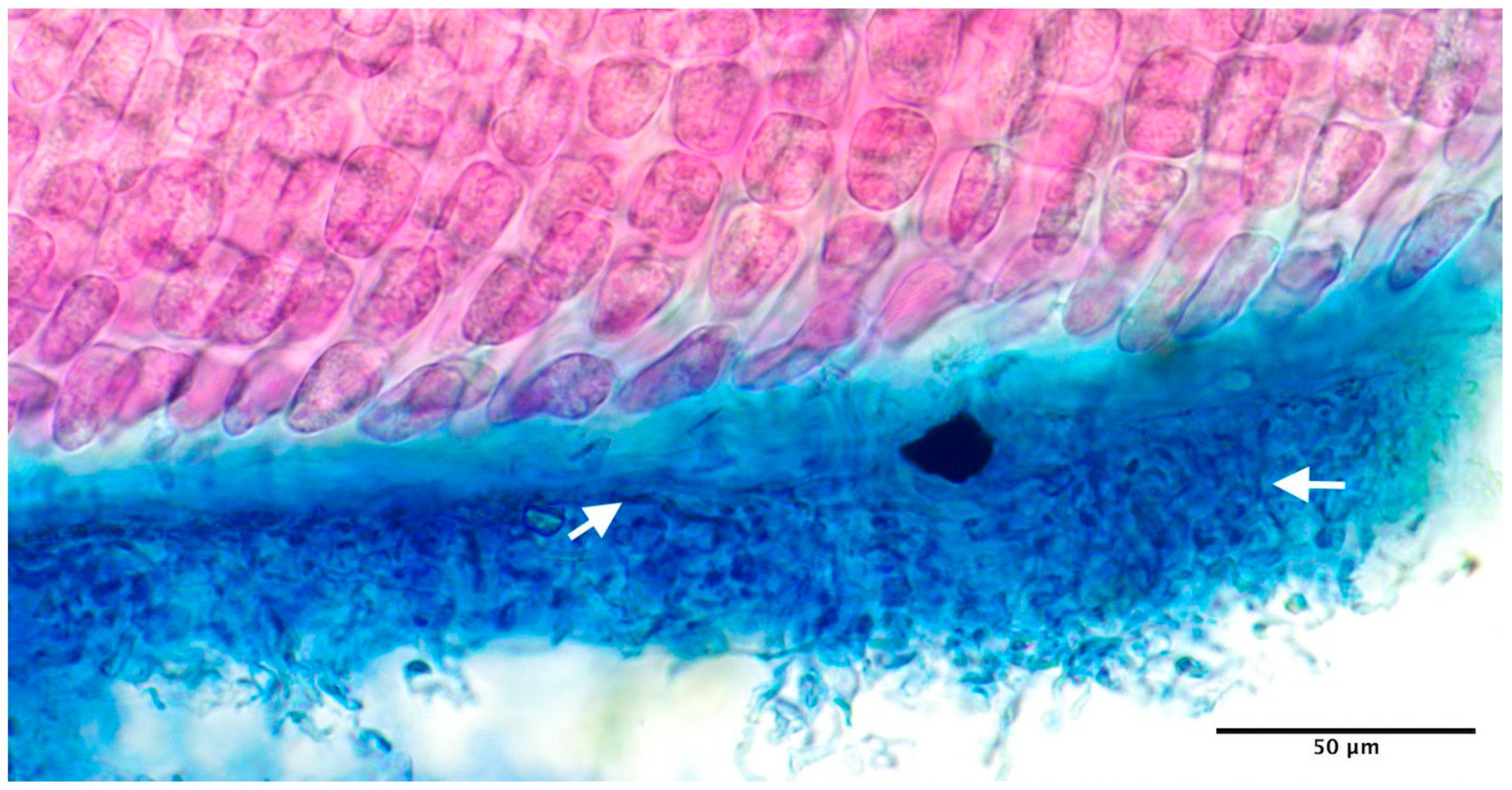
Figure 1. The original voucher from the collection that yielded the culture of Porphyra corallicola H. Kucera & G. W. Saunders; the filaments (white arrows) growing among cells of a coralline crust overgrown by Peyssonnelia rosenvingei F. Schmitz (aniline blue stained).
Diversity 15 00677 g001
Figure 2. UPGMA clustering of our 42 unique Conchocelis OTUs (CONC###) with their closest matches from the reference database (Table S2) resolved as nine genetic species groups (vertical lines). Note that Fuscifolium sp. matched (100%) a GenBank entry from Chile (KP781730), which was not included in our reference database.
Diversity 15 00677 g002
Figure 3. Maps showing the locations of our CIEs: entire study area (a) with inserts for Haida Gwaii (b), central to southern Vancouver Island (c) and the Maritime Provinces/Maine (d). Asterisks (*) indicate that Porphyra linearis and Porphyra mumfordii sequences are identical throughout the target region, so these species have been delineated based on their respective biogeographic ranges.
Diversity 15 00677 g003
Table 1. List of positive Conchocelis identification events (CIEs) acquired, including species assignment, DNA match and host and distributional data. For details see Table S3.
Species AssignmentDNA MatchHab.Location
Bangia sp. 1NGBangia sp. 2Ban (93.61%)Subtidal (10 m) in Clathromorphum sp. (GWS040344)Labrador
Bangia sp. 2NGBangia Japan HQ687502 (~95%)Subtidal (6 m) in Lithophyllum sp. 2BCcrust (GWS021028)British Columbia
Bangia sp. 2NGBangia Japan HQ687502 (~95%)Subtidal (10 m) in inverts or Leptophytum sp. 1SanJuan (GWS014430)British Columbia
Bangia sp. 2NGBangia Japan HQ687502 (~95%)Subtidal (7 m) in shell or Leptophytum sp. 1SanJuan (GWS036253)Washington
Bangia sp. 3NGBangia Japan HQ687502 (~96%)Low intertidal pool in Lithothamnion sp. 6BCcrust (GWS009940)British Columbia
Bangia sp. 3NGBangia Japan HQ687502 (~96%)Subtidal (10 m) in shell or Lithothamnion sp. 2glaciale (GWS014308)British Columbia
Bangia sp. 3NGBangia Japan HQ687502 (~96%)Subtidal (10 m) in Lithophyllum sp. 2BCcrust (GWS019653)British Columbia
Fuscifolium sp.Fuscifolium sp. CHa Chile (100%)Subtidal (10 m) in inverts or Leptophytum sp. 1SanJuan (GWS014430)British Columbia
Porphyra corallicolaPorphyra corallicola (99.47%)Subtidal (4 m) in Lithothamnion glaciale (GWS011835)Nova Scotia
Porphyra corallicolaPorphyra corallicola (99.47%)Subtidal (10 m) in Clathromorphum sp. (GWS040344)Labrador
Porphyra corallicolaPorphyra corallicola (99.65%)Subtidal (10 m) in Lithothamnion lemoineae (overgrowing a dead crust) (GWS040346)Labrador
Porphyra corallicolaPorphyra corallicola (99.65%)Low intertidal pool in Lithothamnion lemoineae (GWS046571)New Brunswick
Porphyra linearisPorphyra mumfordii/linearis (99.29–99.82%)Subtidal (5 m) in Lithothamnion glaciale (GWS003728)New Brunswick
Porphyra linearisPorphyra mumfordii/linearis (99.29–99.82%)Subtidal (10 m) in Phymatolithon sp. 6ATcrust (GWS008908)New Brunswick
Porphyra linearisPorphyra mumfordii/linearis (99.29–99.82%)Subtidal (10 m) in Lithothamnion glaciale (GWS011765)New Brunswick
Porphyra linearisPorphyra mumfordii/linearis (99.29–99.82%)Subtidal (3 m) in mussel or Lithothamnion glaciale (GWS018163)Maine
Porphyra linearisPorphyra mumfordii/linearis (99.29–99.82%)Low intertidal in Phymatolithon laevigatum (GWS039831)Norway
Porphyra linearisPorphyra mumfordii/linearis (99.29–99.82%)Low intertidal in Phymatolithon laevigatum (GWS045271)New Brunswick
Porphyra linearisPorphyra mumfordii/linearis (99.29–99.82%)Low intertidal pool in Lithothamnion lemoineae (GWS046571)New Brunswick
Porphyra mumfordiiPorphyra mumfordii/linearis (99.29–99.82%)Low intertidal pool in Lithothamnion sp. 6BCcrust (GWS009940)British Columbia
Porphyra mumfordiiPorphyra mumfordii/linearis (99.29–99.82%)Subtidal (10 m) in shell or Lithothamnion sp. 2glaciale (GWS014308)British Columbia
Porphyra mumfordiiPorphyra mumfordii/linearis (99.29–99.82%)Subtidal (6 m) in invert or Leptophytum sp. 1SanJuan (GWS020757)British Columbia
Porphyra mumfordiiPorphyra mumfordii/linearis (99.29–99.82%)Subtidal (6 m) in invert or Leptophytum sp. 1SanJuan (GWS020843)British Columbia
Porphyra mumfordiiPorphyra mumfordii/linearis (99.29–99.82%)Subtidal (3 m) in Crusticorallina adhaerens (GWS030995)British Columbia
Pyropia lanceolataPyropia lanceolata (99.11%)Subtidal (5 m) in snail or Lithothamnion sp. 2glaciale (GWS028036)British Columbia
Pyropia nereocystisPyropia nereocystis (99.47%)Subtidal (6 m) in Lithophyllum sp. 2BCcrust (GWS021028)British Columbia
Wildemania amplissimaWildemania amplissima (99.47%)Lowest intertidal in Lithothamnion glaciale (GWS008885)New Brunswick
Wildemania amplissimaWildemania amplissima (99.64%)Low intertidal in periwinkle or Clathromorphum sp. 1circumscriptum (GWS039527)New Brunswick
Disclaimer/Publisher’s Note: The statements, opinions and data contained in all publications are solely those of the individual author(s) and contributor(s) and not of MDPI and/or the editor(s). MDPI and/or the editor(s) disclaim responsibility for any injury to people or property resulting from any ideas, methods, instructions or products referred to in the content.