Article

Resurrection of Diplostomum numericum Niewiadomska, 1988 (Digenea, Diplostomatoidea: Diplostomidae) Based on Novel Molecular Data from the Type-Host

by
Sergey G. Sokolov
1,
Evgeny P. Ieshko
2 and
Daria I. Lebedeva
2,*
1
Alexey Nikolaevich Severtsov Institute of Ecology and Evolution, RAS, 119071 Moscow, Russia
2
Institute of Biology, Karelian Research Centre, RAS, 185000 Petrozavodsk, Russia
*
Author to whom correspondence should be addressed.
Diversity 2023, 15(7), 840; https://doi.org/10.3390/d15070840
Submission received: 25 May 2023 / Revised: 27 June 2023 / Accepted: 7 July 2023 / Published: 9 July 2023
(This article belongs to the Special Issue Taxonomy, Biodiversity and Ecology of Parasites of Aquatic Organisms)

Abstract

:
Diplostomum is a taxonomically problematic genus of trematodes, with many members harmful at the metacercarial stage to fish in aquaculture. We found metacercariae in the eye vitreous humor of the rudd, Scardinius erythrophthalmus; the bleak, Alburnus alburnus; and the roach, Rutilus rutilus, in Karelia (Russia) and identified them as Diplostomum numericum based on a combination of morphological and ecological data. Previously, D. numericum was thought to be conspecific with Diplostomum gavium. However, our phylogenetic analysis based on the sequences of the cox1 mitochondrial DNA gene did not support this hypothesis. We demonstrated the conspecificity of D. numericum with the larval trematode Diplostomum sp. Lineage 3 of Blasco-Costa et al. (2014) from the eye vitreous humor of European salmonid and cyprinid fishes. Therefore, we resurrected the species D. numericum and identified Diplostomum sp. Lineage 3 of Blasco-Costa et al. (2014) as D. numericum. Finally, we hypothesized that D. numericum was, in fact, conspecific with Diplostomum colymbi and that the latter was distinct from D. gavium sensu stricto.

1. Introduction

After the 1832 proposal of the genus Diplostomum Nordmann by the Russian naturalist Alexander von Nordmann [1], numerous papers have been published on this taxon, covering a wide range of issues from the taxonomy of these parasites to their modulating influence on host behavior and their ecological role in aquatic ecosystems (e.g., [2,3,4,5,6,7]). Metacercariae of many Diplostomum spp. cause eye disease in fish (e.g., [8,9]). Intensive molecular phylogenetic studies of Diplostomum spp. in the last two decades have exposed the extensive species richness of these parasites (e.g., [10,11,12,13,14,15,16,17,18,19,20,21,22,23,24,25]). Unfortunately, all modern researchers, with few exceptions [15], in their taxonomic conclusions on Palearctic Diplostomum spp. adhere to the synonymy of species proposed by Niewiadomska [26] and lack a balanced approach to this author’s criticism of Shigin’s taxonomic ideas [4,5].
Diplostomum numericum Niewiadomska, 1988, described metacercariae from the eye vitreous humor and the eye fluid of the rudd, Scardinius erythrophthalmus (Linnaeus, 1758), the European perch, Perca fluviatilis Linnaeus, 1758, and the ruffe, Gymnocephalus cernua (Linnaeus, 1758), from Poland [27]. The type host of this species is the rudd; the type site of infection is the eye vitreous humor [27,28]. Later, Shigin and Stanislavetz [29] found Diplostomum metacercariae in the eye vitreous humor of the roach, Rutilus rutilus (Linnaeus, 1758), the bleak, Alburnus alburnus (Linnaeus, 1758), and the vendace, Coregonus albula (Linnaeus, 1758), from Lake Oster (Karelia, Russia). Being unaware of the study by Niewiadomska [27], Shigin and Stanislavetz [29] described these metacercariae as belonging to a new species, Diplostomum vitreophilum Shigin and Stanislavetz, 1989.
Soon after that discovery, Shigin [5] proposed to consider D. numericum and D. vitreophilum as junior synonyms of Diplostomum gavium (Guberlet, 1922) described based on adult specimens found in the common loon, Gavia immer (Brunnich, 1764), in Oklahoma, the USA [30]. Metacercariae of D. gavium were unknown at that time. The reasons for the synonymization proposed by Shigin [5] were the similarity of the metacercariae of D. numericum and D. vitreophilum in morphology and localization in the host body and the similarity of the adults experimentally reared in ducklings infected with D. vitreophilum metacercariae with the adults of D. gavium. Recently, Achatz et al. [24] determined the cox1 gene sequences of D. gavium adults from the type-host captured in North Dakota (USA). However, the relationship between D. gavium and D. numericum has not been tested by genetic methods because of the lack of molecular data for the latter species.
After Niewiadomska [27] and Shigin and Stanislavetz [29], Diplostomum spp. metacercariae have also been recorded in the eye vitreous humor of European fishes by Brady [31], Valtonen and Gibson [32], Behrmann-Godel [33], Georgieva et al. [15], Blasco-Costa et al. [17], Faltýnková et al. [25], Soldánová et al. [19], and Landeryou et al. [34]. In all cases, however, the authors identified these metacercariae as either Diplostomum baeri Dubois, 1937, Diplostomum gasterostei Williams, 1966; or Diplostomum sp. The phylogenetic analyses have shown that the metacercariae found by Behrmann-Godel [33], Georgieva et al. [15], Blasco-Costa et al. [17], Faltýnková et al. [25], Soldánová et al. [19], and Landeryou et al. [34] are distributed across two species-level lineages, Diplostomum sp. Lineage 3 of Blasco-Costa et al. (2014) and Diplostomum sp. Lineage 4 of Blasco-Costa et al. (2014), with most of them belonging to the former lineage [17,22,35]. The metacercariae of Diplostomum sp. Lineage 4 of Blasco-Costa et al., 2014 have been shown to be conspecific with Diplostomum petromyzifluviatilis (Diesing, 1850) [22]. As for the metacercariae of Diplostomum sp., Lineage 3 of Blasco-Costa et al. (2014), no nomenclatural proposals concerning them have been made so far.
The aim of this study was to elucidate the taxonomic status of D. numericum on the basis of morphological and molecular data.

2. Materials and Methods

2.1. Sample Collection and Morphological Study

Diplostomum spp. metacercariae were isolated from the eye vitreous humor of the rudd, S. erythrophthalmus, the bleak, A. alburnus, and the roach, R. rutilus, from various water bodies in Karelia, Russia. Twelve specimens of the rudd were caught in Lake Vanchozero (64°04′ N, 32°49′ E) in June 2022. One specimen of the bleak was caught in Lake Koivas (64°48′ N, 31°00′ E) in June 2012 and one in Lake Kostomukshskoye (64°47′ N, 30°21′ E) in June 2018. Ten, four, one, and one specimens of the roach originated from Lake Gimoilampi (62°17′ N, 33°53′ E) in April 2023, Lake Onega near Kondopoga (62°16′ N, 34°31′ E) in June 2022, and Lake Koivas and Lake Kostomukshskoye in June 2012, respectively. The eyes of fish were dissected and examined under a stereomicroscope for the presence of Diplostomum spp. metacercariae. The parasites were counted and processed for morphological and/or phylogenetic analyses.
Metacercariae from the rudd and the roach intended for morphometric study were processed as suggested by Shigin [4]. Live metacercariae were transferred to acetocarmine for fixation and simultaneous staining, then dehydrated in the graded ethanol series, cleared in dimethyl phthalate, and mounted in Canada balsam. According to Sudarikov and Shigin [36], fixation of Diplostomum metacercariae with acetocarmine reduces the dispersion of morphometric features, which makes it preferable to other fixation options (using ethanol or formalin). Drawings were made with the aid of a camera lucida.
A few live metacercariae were fixed in 96% ethanol and later used for counts of the excretory bodies (only specimens from the rudd) and/or for the molecular study (specimens from all three host species). In order to count the number of excretory bodies, the three ethanol-fixed metacercariae from the rudd were placed in distilled water for 1 min and then transferred into a 0.5% silver nitrate solution for 2 min. They were then sequentially transferred into a saline, 3% sodium thiosulfate solution, dehydrated in the graded ethanol series, cleared in dimethyl phthalate, and mounted in Canada balsam [36]. The excretory body count from metacercariae isolated from the roach (two trematode specimens from Lake Gimoilampi) was carried out on live specimens.
In the course of fixing metacercariae from the rudd with both acetocarmine and ethanol, we paid attention to the diversity of the metacercariae body shapes, which ranged from oval to elongate-oval. Both oval and elongate-oval trematode specimens from this host were used for all morphometric and molecular procedures. Metacercariae from the remaining fish species were relatively homogeneous in shape.

2.2. Molecular Data and Phylogenetic Analysis

Genomic DNA was isolated from individual ethanol-fixed metacercariae using DNA-Extran kits (Synthol, Moscow, Russia). A total of fifteen specimens, six from the rudd (three oval and three elongate-oval metacercariae), two from the bleak, and seven from the roach (three trematode specimens from Lake Gimoilampi, 2 from Lake Onega, and 2 from Lake Kostomukshskoye and Lake Koivas), were studied molecularly. For each of these larvae, we amplified a fragment of the mtDNA cox1 gene using the primers Cox1_schist_5′ (5′-TCT TTR GAT CAT AAG CG-3′) and Cox1_schist_3′ (5′-TAA TGC ATM GGA AAA AAA CA-3′) of Lockyer et al. (2003) [37].
The PCR assay was carried out in 20 μL of reaction mixture containing ready 5X ScreenMix (Evrogen, Moscow, Russia), 1.5 pmol of each primer, and 2 µL of DNA; an annealing temperature of 50 °C was used for amplification [37].
PCR products were purified using the Cleanup Standard Extraction Kit (Evrogen, Moscow, Russia) following the manufacturer’s instructions and then sequenced directly with the automatic sequencing system ABI PRISM 3100-Avant (Applied Biosystems Inc., Foster City, CA, USA). Consensus sequences (1170 bp length) were assembled in MEGA v. 10 [38] and deposited in GenBank with accession numbers OQ214204–OQ214209 (metacercariae ex S. erythrophthalmus), OQ257342, OR044094 (metacercariae ex A. alburnus), and OR044091–OR044093, OR044095–OR044097 (metacercariae ex R. rutilus).
The identity of the newly generated sequences was checked with the Basic Local Alignment Search Tool (BLASTn) (www.ncbi.nih.gov/BLAST/, accessed on 15 January 2023 ). The novel sequences were aligned with those of 172 representatives of the genus Diplostomum (Table S1; [10,12,15,17,18,19,20,21,22,23,24,33,34,35,39,40,41,42,43,44,45,46,47,48]) in MEGA v. 10 [38] and trimmed to the shortest length, 384 nt. The nomenclature of Diplostomum spp. followed the latest studies [22,23,24,35]. The cox1 sequence for Tylodelphys clavata (Nordmann, 1832) (JX986909) was used as an outgroup (Table S1; [15]).
To assess the phylogenetic relationships of the newly found Diplostomum metacercariae, we applied Bayesian inference analysis to the dataset. Prior to the analyses, the best-fitting GTR + G+ I model was identified with jModelTest v2.1.2 [49]. Bayesian inference analyses were conducted using MrBayes (v3.2.3) [50]. Markov chain Monte Carlo (MCMC) simulations were run for 3,000,000 generations, log-likelihood scores were plotted, and only the final 75% of trees were used to produce the consensus tree. Posterior probability was calculated to estimate nodal support. FigTree v1.4 [51] was used to visualize the tree. The genetic divergence (as pairwise distances) between the taxa was estimated with the bootstrap method (1000 replicates) and with a uniform nucleotide substitution (transition + transversion) rate using MEGA v. 10 [38].

3. Results

Taxonomy
Family Diplostomidae Poirier, 1886
Genus Diplostomum Nordmann, 1832
Diplostomum numericum Niewiadomska, 1988, metacercaria (Figure 1 and Figure 2)
Synonyms:Diplostomum sp. Lineage 3 of Blasco-Costa et al., 2014; Diplostomum baeri 1 of Georgieva et al., 2013; Diplostomum baeri of Landeryou et al., 2020; Diplostomum gavium of Shigin, 1993, part.; Diplostomum vitreophilum Shigin & Stanislavetz, 1989.
Type-host: Scardinius erythrophthalmus (Linnaeus, 1758) (Actinopterygii, Cyprinidae).
Other hosts: metacercariae—Alburnus alburnus (Linnaeus, 1758), Rutilus rutilus (Linnaeus, 1758), Gobio gobio (Linnaeus, 1758) (Actinopterygii, Cyprinidae), Salnelinus alpinus (Linnaeus, 1758), Salmo trutta (Linnaeus, 1758), and Coregonus albula (Linnaeus, 1758) (Actinopterygii, Salmonidae); adults (only in experiment)—Anas platyrhynchos domesticus Linnaeus, 1758 (Aves, Anatidae).
Type-site of infection: Eye vitreous humor.
Type-locality: Lake Warniak, Poland.
Other localities: Lake Vanchozero (64°04′ N, 32°49′ E), Karelia, Russia (S. erythrophthalmus); Lake Gimoilampi (62°17′ N, 33°53′ E); Lake Koivas (64°48′ N, 31°00′ E); Lake Kostomukshskoye (64°47′ N, 30°21′ E); and Lake Onega (62°16′ N, 34°31′ E) in Karelia, Russia (R. rutilus); Lakes Koivas and Kostomukshskoye in Karelia, Russia (A. alburnus); as well as Iceland [17,25], Germany [15], Norway [19], Scotland [34], and other water bodies in Karelia, Russia [29].
Specimens deposited: Three slides with whole-mounted metacercariae from S. erythrophthalmus (IPEE RAS 14332–14334), paragenophores.
Sequences deposited: Six cox1 sequences, GenBank NCBI, as OQ214204–OQ214209 (ex S. erythrophthalmus from Lake Vanchozero); two as OQ257342 and OR044094 (ex A. alburnus from Lake Koivas and Lake Kostomukshskoye, respectively); three as OR044095–OR044097 (ex R. rutilus from Lake Gimoilampi); two as OR044093 and OR044096 (ex R. rutilus from Lake Koivas and Lake Kostomukshskoye, respectively); and two as OR044091 and OR044092 (ex R. rutilus from Lake Onega).
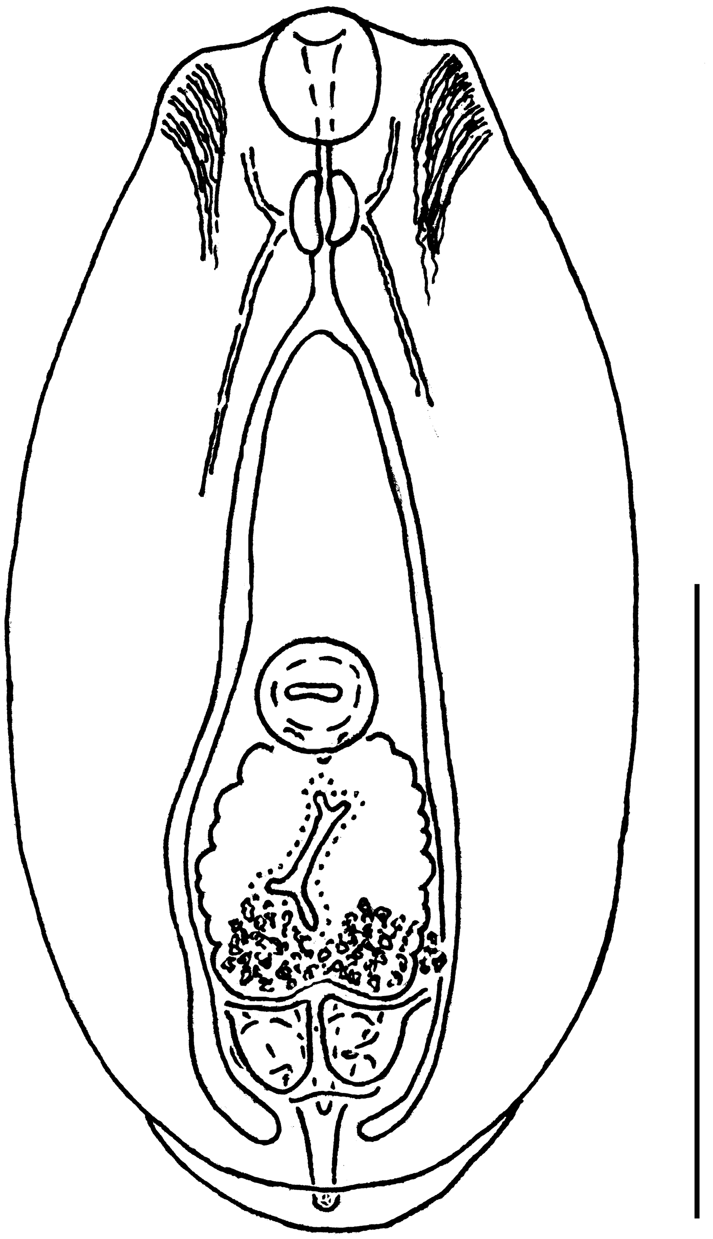
Description (based on 15 acetocarmine-stained paragenophores from S. erythrophthalmus and the same number from R. rutilus; measurements in Table 1): Body is oval to elongate-oval, distinctly bipartite, with a relatively large, concave prosoma and a small, conical opisthosoma. Oral sucker is muscular, longer than wide, and mouth subterminal. Pseudosuckers are two, well developed, lateral, on either side of the oral sucker, completely protruded or partly introverted. Ventral sucker muscles are muscular, transversely oval or rounded, and slightly post-equatorial. Holdfast organs are suboval or rounded, with a longitudinal fissure just posterior to the ventral sucker. Prepharynx distinct. Pharynx elongate-oval, muscular; oesophagus distinct, shorter than pharynx. Cerebral ganglia with suprapharyngeal commissure, posterior longitudinal ventral nerve cords, and anterior nerves clearly visible. Caeca long, terminating in opisthosoma. Proteolytic gland bilobed, dorsal to posterior end of holdfast organ. Gonad primordium posterior to the holdfast organ. The excretory vesicle is V-shaped and reaches to the holdfast organ.

Phylogenetic Analysis

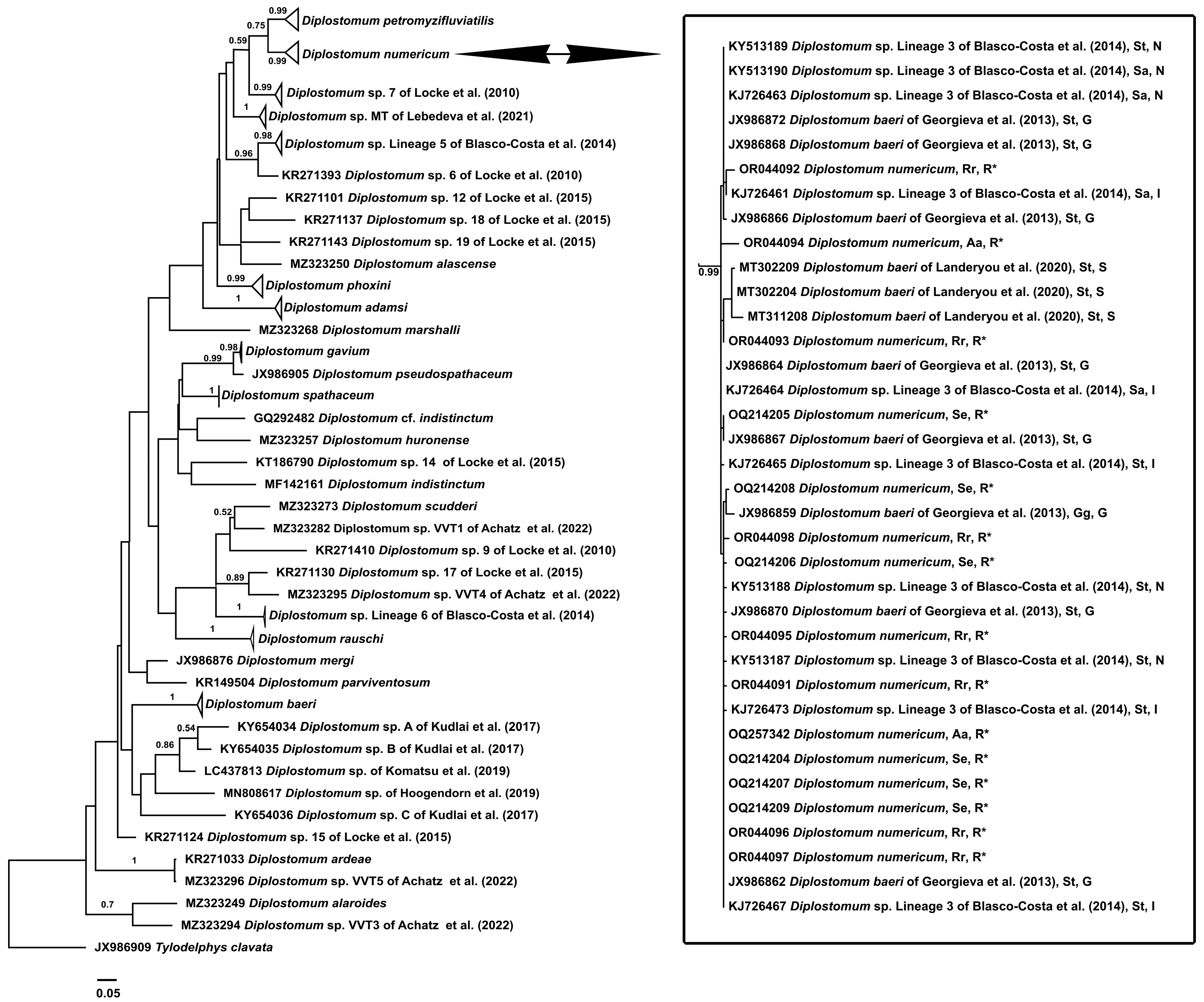
All 15 newly obtained sequences were grouped together with the sequences of the metacercariae identified previously as Diplostomum sp. Lineage 3 of Blasco-Costa et al. (2014) (=Diplostomum baeri 1 of Georgieva et al., 2013 and D. baeri of Landeryou et al., 2020) is a strongly supported clade (Figure 3). The p-distance between members of the clade was 0–1% for a sequence length of 388 nt. For the full length (1170 bp), p-distance ranged from 0 to 2%. Hereafter, we refer to this clade as D. numericum.
Diplostomum numericum appeared as a poorly supported sister taxon to D. petromyzifluviatilis. The p-distance values between these species were 6%. The D. numericum + D. petromyzifluviatilis clade occupied a terminal position in the large group of Diplostomum spp. with brain or non-lens eye localization, which also contained Diplostomum sp. 7 of Locke et al., 2010, Diplostomum sp. of Lebedeva et al., 2021, Diplostomum sp. Lineage 5 of Blasco-Costa et al., 2014, Diplostomum sp. 6 of Locke et al., 2010, Diplostomum sp. 12 of Locke et al., 2015, Diplostomum sp. 18 of Locke et al., 2015, Diplostomum sp. 19 of Locke et al., 2015, Diplostomum alascense Dubois, 1969, Diplostomum phoxini (Faust, 1919), Diplostomum adamsi Lester & Huizinga, 1977 and Diplostomum marshalli Chandler, 1954 (Figure 3).
Diplostomum gavium occupied a well-supported sister position to Diplostomum pseudospathaceum Niewiadomska, 1984. The D. gavium + D. pseudospathaceum groups had no direct phylogenetic connections with the aforementioned large clade of Diplostomum spp. The p-distance values between D. numericum and D. gavium were 15%.

4. Discussion

The metacercariae from the rudd and the roach (Lake Gimoilampi) examined in our study are similar to each other in morphological and ecological (site of infection, cyprinid host) features, as well as molecularly. In turn, they are close to those of D. numericum and D. vitreophilum in a number of the key characters, namely, the site of infection, the number of excretory bodies, and the mean and/or range values of the indexes describing the body proportions or organ ratios. (Table 1). However, the specimens in our study were slightly larger (in mean and maximum values) than the metacercariae described by Niewiadomska [27] and Shigin and Stanislavetz [29]. These authors fixed metacercariae using different techniques, specifically fixation in 75% ethanol of metacercariae killed by hot water [27] vs. fixation of live metacercariae in acetocarmine [29]. Different methods of fixation are known to affect the morphometric characteristics of Diplostomum metacercariae [31,36]. However, the differences in size indicated above do occur despite the identity of the techniques used by us and by Shigin and Stanislavetz [29].
Shigin [5] has suggested that D. vitreophilum is conspecific with D. numericum because they are very similar in general morphology and have an identical localization. In turn, as mentioned above, both D. vitreophilum and D. numericum were considered by this author to be synonymous with D. gavium. Niewiadomska [28] agreed with these conclusions. Our morphological and phylogenetic data confirm only the conspecificity of D. vitreophilum to D. numericum and allow us to identify all the specimens we examined, including those from the bleak and the roach (Lakes Koivas, Onega, and Kostomukshskoye), as D. numericum.
The molecular results of our study allow us to conclude with certainty that D. numericum is conspecific with the metacercariae of Diplostomum sp., Lineage 3 of Blasco-Costa et al. (2014) from the eye vitreous humor of European salmonid and cyprinid fishes. Therefore, we identify this lineage as D. numericum. Morphometric data on the metacercariae of this lineage from the brown trout, Salmo trutta Linnaeus, 1758, and the Arctic char, Salvelinus alpinus (Linnaeus, 1758) from Iceland were provided by Faltýnková et al. [25]. However, the measurements in that study were performed on live larvae and specimens fixed with 96% ethanol without pre-treatment by the standard staining, cleaning, and mounting techniques.Therefore, we cannot compare the morphometrics of specimens from Faltýnková et al. [25] with those examined in our study. It would be correct to compare them only in terms of the number of excretory bodies. Metacercariae of Diplostomum sp. Lineage 3 of Blasco-Costa et al. (2014) studied by Faltýnková et al. [25] had fewer excretory bodies than the specimens of D. numericum examined by us and by Shigin and Stanislavetz [29]: 450–600 vs. 638–1150. Combined with data published by Faltýnková et al. [25] and Lebedeva et al. [22] on metacercariae of D. petromyzifluviatilis (=Diplostomum sp. Lineage 4 of Blasco-Costa et al., 2014), these findings indicate considerable intraspecific variability in the number of excretory bodies in Diplostomum metacercariae and contradict Shigin’s [4] notion of the high taxonomic importance of this trait for species identification in these parasites.
Niewiadomska [27] noted the great morphological similarity between the metacercariae of D. numericum and those of Diplostomum baeri of Niewiadomska, 1988 (=Diplostomum volvens of Shigin, 1986) from the retina, choroid, and eye fluid of percid, lotid, gasterosteid, salmonid, and, less frequently, cyprinid fishes. Shigin and Stanislavetz [29] report great similarities between D. vitreophilum (=D. numericum), D. volvens of Shigin, 1986, and D. petromyzifluviatilis. It is worth noting that D. petromyzifluviatilis is probably conspecific to D. volvens of Shigin, 1986. This hypothesis is supported by the similarities between metacercariae of D. volvens of Shigin, 1986, and Diplostomum sp. Lineage 4 of Blasco-Costa et al. (2014), in terms of localization and hosts, and recent data on the conspecificity of D. petromyzifluviatilis with Diplostomum sp. Lineage 4 of Blasco-Costa et al., 2014 [22]. At this point, however, we regard D. volvens of Shigin, 1986 and D. petromyzifluviatilis as separate species. Morphological differences between metacercariae of D. numericum, on the one hand, and metacercariae of D. volvens of Shigin, 1986, and D. petromyzifluviatilis, on the other hand, are the size of the ventral sucker compared to that of the body and the oral sucker: mean values of the [BL × BW]/[VL × VW] and [OL × OW]/[VL × VW] indexes are 53.10–58.40 vs. 37.20–45.26 and 1.13–1.22 vs. 0.98–1.05, respectively ([4,27,29], present data).
Of the metacercariae found in the eye vitreous humor of European fishes, morphological data are also available for Diplostomum sp. type 2 of Brady [31]. These larvae have been described from the farmed rainbow trout, Oncorhynchus mykiss (Walbaum, 1792), from Scotland and, despite the perfunctory similarity to D. numericum, differ significantly from it in body size (302–397 × 155–262 μm vs. 440–651 × 210–336 μm). Brady [31] suggested that the metacercariae she described probably belonged to Diplostomum mergi Dubois, 1932. However, given the known facts on the confinement of D. mergi metacercariae to the lens [4], this taxonomic conclusion seems doubtful. The taxonomic status of Diplostomum sp. type 2 of Brady [31] remains unclear.
Niewiadomska ([27], Figure 3) noted the variability of the body shape of D. numericum metacercariae. Both elongate-oval and oval specimens were represented in her material. Our data, supported by the results of phylogenetic analysis, also demonstrate the variability of body shape in metacercariae of this species we isolated from the rudd (Figure 1). This is probably because both the previous author and we were dealing with a mixture of specimens at different stages of development (early and fully formed metacercariae). It is known that the morphology of Diplostomum metacercariae varies notably across development stages [4].
We can now reject the hypothesis that D. numericum is conspecific with D. gavium [5]. Our molecular genetic data showed that the p-distance values between D. numericum and D. gavium were 15%, which corresponds to a level of difference between species. In addition, D. numericum and D. gavium sensu stricto seem to be different ecologically at the main site within the host where their metacercariae reside. According to the molecular data of Achatz et al. [24], adults of D. gavium sensu stricto are conspecific with the metacercariae of Diplostomum sp. 3 of Moszczynska et al. (2009) from the eyes of various North American fishes and amphibians and the metacercariae of Diplostomum specimens from the northern hog sucker, Hypentelium nigricans (Lesueur, 1817), also from North America ([10,11,12,24,37,52]; Achatz, personal communication). In fish, a majority of metacercariae molecularly conspecific with D. gavium sensu stricto were found in the lens (for amphibians, the specific localization of these metacercariae within the eye was not indicated). Only two specimens of metacercariae of D. gavium sensu stricto have ever been found outside the lens in fish: one in an unspecified site in the brown bullhead Ameurus nebulosus (Lesueur, 1819) (D.J. Marcogliese and S. Locke, personal communications) and one in the eye vitreous humor in the northern hog sucker (T. Achatz, personal communication). Marcogliese and Locke [42] also identified metacercariae of D. gavium sensu stricto in the lens of the invasive round goby Neogobius melanostomus (Pallas, 1814) in North America.
However, it is noteworthy that the adult specimen experimentally reared by Shigin [5] from metacercaria of D. vitreophilum (=D. numericum) was similar in morphological form to D. gavium formed under conditions of high infection intensity in the final host (492 specimens per bird). Shigin [5] adhered to the hypothesis of Dubois and Rausch [53] about the conspecificity of D. gavium sensu stricto with Diplostomum colymbi (Dubois, 1928), described based on adult specimens from Gavia arctica (Linnaeus, 1758) in Switzerland. It cannot be ruled out that D. numericum is, in fact, conspecific with D. colymbi and that the latter is distinct from D. gavium sensu stricto. If so, it is likely that D. gavium sensu stricto is widespread in North America, while D. colymbi inhabits Eurasia. Molecular evidence is necessary to test this hypothesis. Nevertheless, at this point, the species D. numericum must be resurrected.
To date, metacercariae molecularly identified as D. numericum (=Diplostomum sp., Lineage 3 of Blasco-Costa et al., 2014) have been found only in the eye vitreous humor of European salmonids and cyprinids. Niewiadomska [27] reported the localization of some specimens of D. numericum metacercaria in the “eyeball outside eyelens” of European percid fishes. However, these data need molecular verification because it has already been proven that Diplostomum metacercariae localized in the eye vitreous humor of European percids are conspecific with D. petromyzifluviatilis [22,33].

Supplementary Materials

The following supporting information can be downloaded at: https://www.mdpi.com/article/10.3390/d15070840/s1, Table S1: Summary data for cox1 sequences of Diplostomum spp. retrieved from GenBank.

Author Contributions

Conceptualization and morphological study—S.G.S.; writing—original draft preparation, S.G.S. and D.I.L.; sample collection, S.G.S. and E.P.I.; molecular and phylogenetic analyses, D.I.L.; writing—editing, S.G.S., D.I.L. and I E.P.I. All authors have read and agreed to the published version of the manuscript.

Funding

The study was funded by the Russian Ministry of Science and Higher Education: FFER-2021-0005 (SS) and FMEN-2022-0005 (DL, EI).

Institutional Review Board Statement

No applicable.

Data Availability Statement

The data presented in this study are available on request.

Acknowledgments

Special thanks to Olga Novokhatskaya (Unicaf University, Cyprus) for help with proofreading the text.

Conflicts of Interest

The authors declare no conflict of interest.

References

  1. Nordmann, A.v. Mikrographische Beiträge zur Naturgeschichte der Wirbellosen Thiere; Zweites Heft. Mit zehn Kupfertafeln, G. Reimer: Berlin, Germany, 1832; pp. 1–150. [Google Scholar]
  2. Dubois, G. Synopsis des Strigeidae et des Diplostomatidae (Trematoda). Douxième Partie. Mem. Soc. Neuchl. Sci. Nat. 1970, 10, 259–727. [Google Scholar]
  3. Shigin, A.A. Trematodes of the genus Diplostomum in the biocenosis of the trout farm “Skhodnia”. Trudy Gel'mintologicheskoi Lab. Akad. Nauk SSSR 1980, 30, 140–202. [Google Scholar]
  4. Shigin, A.A. Trematodes in the Fauna of the USSR: The Genus Diplostomum. Metacercariae; Nauka: Moscow, Russia, 1986; p. 252. [Google Scholar]
  5. Shigin, A.A. Trematodes of the Fauna of Russia and Neighbouring Regions. Genus Diplostomum. Adults; Nauka: Moscow, Russia, 1993; p. 207. [Google Scholar]
  6. Mikheev, V.N.; Pasternak, A.F.; Taskinen, J.; Valtonen, E.T. Parasite-induced aggression and impaired contest ability in a fish host. Parasites Vectors 2010, 3, 17. [Google Scholar] [CrossRef] [Green Version]
  7. Karvonen, A.; Marcogliese, D.J. Diplostomiasis (Diplostomum spathaceum and related species). In Climate Change and Infectious Fish Diseases; Woo, P.T.K., Leong, J.-A., Buchmann, K., Eds.; CAB International: Wallingford, UK, 2020; pp. 434–456. [Google Scholar]
  8. Stables, J.N.; Chappell, L.H. The epidemiology of diplostomiasis in farmed rainbow trout from north-east Scotland. Parasitology 1986, 92, 699–710. [Google Scholar] [CrossRef] [PubMed]
  9. Avsever, M.L.; Selver, M.M.; Yazicioğlu, Ö.; Tokşen, E.; Tay, S.; Erdal, G.; Günen, M.Z. The first report of diplostomiasis from cultured rainbow trout (Oncorhynchus mykiss) in Turkey. Ank. Üniv. Vet. Fak. Derg. 2016, 63, 377–381. [Google Scholar]
  10. Locke, S.A.; McLaughlin, J.D.; Dayanandan, S.; Marcogliese, D.J. Diversity and specificity in Diplostomum spp. metacercariae in freshwater fishes revealed by cytochrome c oxidase I and internal transcribed spacer sequences. Int. J. Parasitol. 2010, 40, 333–343. [Google Scholar] [CrossRef]
  11. Locke, S.A.; McLaughlin, J.D.; Marcogliese, D.J. DNA barcodes show cryptic diversity and a potential physiological basis for host specificity among Diplostomoidea (Platyhelminthes: Digenea) parasitizing freshwater fishes in the St. Lawrence River, Canada. Mol. Ecol. 2010, 19, 2813–2827. [Google Scholar] [CrossRef]
  12. Locke, S.A.; Al-Nasiri, F.S.; Caffara, M.; Drago, F.; Kalbe, M.; Lapierre, A.R.; McLaughlin, J.D.; Nie, P.; Overstreet, R.M.; Souza, G.T.R.; et al. Diversity, specificity and speciation in larval Diplostomidae (Platyhelminthes: Digenea) in the eyes of freshwater fish, as revealed by DNA barcodes. Int. J. Parasitol. 2015, 45, 841–855. [Google Scholar] [CrossRef]
  13. Locke, S.A.; Van Dam, A.; Caffara, M.; Pinto, H.A.; Blanard, C.A. Validity of the Diplostomoidea and Diplostomida (Digenea, Platyhelminthes) upheld in phylogenomic analysis. Int. J. Parasitol. 2018, 48, 1043–1059. [Google Scholar] [CrossRef]
  14. Locke, S.A.; Drago, F.B.; Núñez, V.; Souza, G.T.R.E.; Takemoto, R.M. Phylogenetic position of Diplostomum spp. from New World herons based on complete mitogenomes, rDNA operons, and DNA barcodes, including a new species with partially elucidated life cycle. Parasitol. Res. 2020, 119, 2129–2137. [Google Scholar] [CrossRef]
  15. Georgieva, S.; Soldánová, M.; Pérez-del-Olmo, A.; Dangel, R.D.; Sitko, J.; Sures, B.; Kostadinova, A. Molecular prospecting for European Diplostomum (Digenea: Diplostomidae) reveals cryptic diversity. Int. J. Parasitol. 2013, 43, 57–72. [Google Scholar] [CrossRef]
  16. Chibwana, F.D.; Blasco-Costa, I.; Georgieva, S.; Hosea, K.M.; Nkwengulila, G.; Scholz, T.; Kostadinova, A. A first insight into the barcodes for African diplostomids (Digenea: Diplostomidae): Brain parasites in Clarias gariepinus (Siluriformes: Clariidae). Infect. Genet. Evol. 2013, 17, 62–70. [Google Scholar] [CrossRef] [PubMed]
  17. Blasco-Costa, I.; Faltýnková, A.; Georgieva, S.; Skírnisson, K.; Scholz, T.; Kostadinova, A. Fish pathogens near the Arctic Circle: Molecular, morphological and ecological evidence for unexpected diversity of Diplostomum (Digenea: Diplostomidae) in Iceland. Int. J. Parasitol. 2014, 44, 703–715. [Google Scholar] [CrossRef] [PubMed]
  18. Pérez-del-Olmo, A.; Georgieva, S.; Pula, H.J.; Kostadinova, A. Molecular and morphological evidence for three species of Diplostomum (Digenea: Diplostomidae), parasites of fishes and fish-eating birds in Spain. Parasites Vectors 2014, 7, 502. [Google Scholar] [CrossRef] [PubMed]
  19. Soldánová, M.; Georgieva, S.; Rohacova, J.; Knudsen, R.; Kuhn, J.A.; Henriksen, E.H.; Siwertsson, A.; Shaw, J.C.; Kuris, A.M.; Amundsen, P.A.; et al. Molecular analyses reveal high species diversity of trematodes in a sub-Arctic lake. Int. J. Parasitol. 2017, 47, 327–345. [Google Scholar] [CrossRef]
  20. Hoogendoorn, C.; Smit, N.J.; Kudlai, O. Resolution of the identity of three species of Diplostomum (Digenea: Diplostomidae) parasitising freshwater fishes in South Africa, combining molecular and morphological evidence. Int. J. Parasitol. Parasites Wildl. 2020, 11, 50–61. [Google Scholar] [CrossRef]
  21. Lebedeva, D.I.; Chrisanfova, G.G.; Ieshko, E.P.; Guliaev, A.S.; Yakovleva, G.A.; Mendsaikhan, B.; Semyenova, S.K. Morphological and molecular differentiation of Diplostomum spp. metacercariae from brain of minnows (Phoxinus phoxinus L.) in four populations of northern Europe and East Asia. Infect. Genet. Evol. 2021, 92, 104911. [Google Scholar] [CrossRef]
  22. Lebedeva, D.I.; Popov, I.Y.; Yakovleva, G.A.; Zaicev, D.O.; Bugmyrin, S.V.; Makhrov, A.A. No strict host specificity: Brain metacercariae Diplostomum petromyzifluviatilis Müller (Diesing, 1850) are conspecific with Diplostomum sp. Lineage 4 of Blasco-Costa et al. (2014). Parasitol. Int. 2022, 91, 102654. [Google Scholar] [CrossRef]
  23. Schwelm, J.; Georgieva, S.; Grabner, D.; Kostadinova, A.; Sures, B. Molecular and morphological characterisation of Diplostomum phoxini (Faust, 1918) with a revised classification and an updated nomenclature of the species-level lineages of Diplostomum (Digenea: Diplostomidae) sequenced worldwide. Parasitology 2021, 148, 1648–1664. [Google Scholar] [CrossRef]
  24. Achatz, T.J.; Martens, J.R.; Kostadinova, A.; Pulis, E.E.; Orlofske, S.A.; Bell, J.A.; Fecchio, A.; Oyarzún-Ruiz, P.; Syrota, Y.Y.; Tkach, V.V. Molecular phylogeny of Diplostomum, Tylodelphys, Austrodiplostomum and Paralaria (Digenea: Diplostomidae) necessitates systematic changes and reveals a history of evolutionary host switching events. Int. J. Parasitol. 2022, 52, 47–63. [Google Scholar] [CrossRef]
  25. Faltýnková, A.; Georgieva, S.; Kostadinova, A.; Blasco-Costa, I.; Scholz, T.; Skírnisson, K. Diplostomum von Nordmann, 1832 (Digenea: Diplostomidae) in the sub-Arctic: Descriptions of the larval stages of six species discovered recently in Iceland. Syst. Parasitol. 2014, 89, 195–213. [Google Scholar] [CrossRef] [PubMed]
  26. Niewiadomska, K. The genus Diplostomum—Taxonomy, morphology and biology. Acta Parasitol. 1996, 41, 55–66. [Google Scholar]
  27. Niewiadomska, K. Diplostomum metacercariae (Digenea) in fish of the Dgal Wielki and Warniak lakes: D. numericum sp. n. and D. baeri Dubois, 1937, with comments on the synonymy of this species. Acta Parasitol. 1988, 33, 7–24. [Google Scholar]
  28. Niewiadomska, K. Fauna słodkowodna Polski. 34A. In Przywry (Trematoda).Część ogólna; Część systematyczna—Aspidogastrea, Digenea: Strigeida; Wydawnictwo Uniwersytetu Łódzkiego: Łódź, Poland, 2010; p. 388. [Google Scholar]
  29. Shigin, A.A.; Stanislavetz, A.N. On the species validity of metacercariae of the genus Diplostomum from the vitreous body of the eyes of freshwater fish. Mater. Nauchnoi Conf. Vsesoyuznogo Obschestva Gel'mintolgov 1989, 38, 253–259. [Google Scholar]
  30. Guberlet, J.E. Three new species of Holostomidae. J. Parasitol. 1922, 9, 6–14. [Google Scholar] [CrossRef]
  31. Brady, A. Studies on the taxonomy and biology of Diplostomum species (Digenea). Ph.D. Thesis, University of Stirling, Stirling, UK, 1989; p. 277. [Google Scholar]
  32. Valtonen, E.T.; Gibson, D.I. Aspects of the Biology of Diplostomid Metacercarial (Digenea) Populations Occurring in Fishes in Different Localities of Northern Finland. Ann. Zool. Fenn. 1997, 34, 47–59. Available online: http://www.jstor.org/stable/23735430 (accessed on 1 June 2023).
  33. Behrmann-Godel, J. Parasite identification, succession and infection pathways in perch fry (Perca fluviatilis): New insights through a combined morphological and genetic approach. Parasitology 2013, 140, 509–520. [Google Scholar] [CrossRef]
  34. Landeryou, T.; Kett, S.M.; Ropiquet, A.; Wildeboer, D.; Lawton, S.P. Characterization of the complete mitochondrial genome of Diplostomum baeri. Parasitol. Int. 2020, 79, 102166. [Google Scholar] [CrossRef]
  35. Faltýnková, A.; Kudlai, O.; Pantoja, C.; Yakovleva, G.; Lebedeva, D. Another plea for ‘best practice’ in molecular approaches to trematode systematics: Diplostomum sp. clade Q identified as Diplostomum baeri Dubois, 1937 in Europe. Parasitology 2022, 149, 503–518. [Google Scholar] [CrossRef]
  36. Sudarikov, V.E.; Shigin, A.A. On the methods for working with metacercariae of the Strigeidida. Trudy Gel'mintologicheskoi Lab. Akad. Nauk SSSR 1965, 15, 158–166. [Google Scholar]
  37. Lockyer, A.E.; Olson, P.D.; Stergaard, P.; Rollinson, D.; Johnston, D.A.; Attwood, S.W.; Southgate, V.R.; Horak, P.; Snyder, S.D.; Le, T.H.; et al. The phylogeny of the Schistosomatidae based on three genes with emphasis on the interrelationships of Schistosoma Weinland, 1858. Parasitology 2003, 126, 203–224. [Google Scholar] [CrossRef] [Green Version]
  38. Kumar, S.; Stecher, G.; Li, M.; Knyaz, C.; Tamura, K. MEGA X: Molecular Evolutionary Genetics Analysis across Computing Platforms. Mol. Biol. Evol. 2018, 35, 1547–1549. [Google Scholar] [CrossRef]
  39. Ubels, J.L.; DeJong, R.J.; Hoolsema, B.; Wurzberger, A.; Nguyen, T.T.; Blankespoor, H.D.; Blankespoor, C.L. Impairment of retinal function in yellow perch (Perca flavescens) by Diplostomum baeri metacercariae. Int. J. Parasitol. 2018, 7, 171–179. [Google Scholar] [CrossRef] [PubMed]
  40. Gordy, M.A.; Kish, L.; Tarrabain, M.; Hanington, P.C. A comprehensive survey of larval digenean trematodes and their snail hosts in central Alberta, Canada. Parasitol. Res. 2016, 115, 3867–3880. [Google Scholar] [CrossRef]
  41. Gordy, M.A.; Hanington, P.C. A fine-scale phylogenetic assessment of digenean trematodes in central Alberta reveals we have yet to uncover their total diversity. Ecol. Evol. 2019, 9, 3153–3238. [Google Scholar] [CrossRef] [PubMed] [Green Version]
  42. Marcogliese, D.J.; Locke, S.A. Infection of Diplostomum spp. in invasive round gobies in the St Lawrence River, Canada. J. Helminthol. 2021, 95, e64. [Google Scholar] [CrossRef] [PubMed]
  43. Selbach, C.; Soldánová, M.; Georgieva, S.; Kostadinova, A.; Sures, B. Integrative taxonomic approach to the cryptic diversity of Diplostomum spp. in lymnaeid snails from Europe with a focus on the ‘Diplostomum mergi’ species complex. Parasites Vectors 2015, 8, 300. [Google Scholar] [CrossRef] [Green Version]
  44. Rahn, A.K.; Krassmann, J.; Tsobanidis, K.; MacColl, A.D.C.; Bakker, T.C.M. Strong neutral genetic differentiation in a host, but not in its parasite. Infect. Gen. Evol. 2016, 44, 261–271. [Google Scholar] [CrossRef]
  45. Kuhn, J.A.; Kristoffersen, R.; Knudsen, R.; Jakobsen, J.; Marcogliese, D.J.; Locke, S.A.; Primicerio, R.; Amundsen, P.-A. Parasite communities of two three-spined stickleback populations in subarctic Norway—Effects of a small spatial-scale host introduction. Parasitol. Res. 2015, 114, 1327–1339. [Google Scholar] [CrossRef] [Green Version]
  46. Brabec, J.; Kostadinova, A.; Scholz, T.D.; Littlewood, T.J. Complete mitochondrial genomes and nuclear ribosomal RNA operons of two species of Diplostomum (Platyhelminthes: Trematoda): A molecular resource for taxonomy and molecular epidemiology of important fish pathogens. Parasites Vectors 2015, 8, 336. [Google Scholar] [CrossRef] [Green Version]
  47. Komatsu, N.; Itoh, N.; Ogawa, K. Worm Cataract of Hatchery-Reared Japanese Dace Tribolodon hakonensis Caused by Diplostomum sp. (Digenea: Diplostomidae). Fish Pathol. 2019, 54, 1–11. [Google Scholar] [CrossRef] [Green Version]
  48. Kudlai, O.; Oros, M.; Kostadinova, A.; Georgieva, S. Exploring the diversity of Diplostomum (Digenea: Diplostomidae) in fishes from the river Danube using DNA mitochondrial barcodes. Parasites Vectors 2017, 10, 592. [Google Scholar] [CrossRef] [PubMed] [Green Version]
  49. Darriba, D.; Taboada, G.L.; Doallo, R.; Posada, D. jModelTest 2: More models, new heuristics and parallel computing. Nat. Methods 2012, 9, 772. [Google Scholar] [CrossRef] [PubMed] [Green Version]
  50. Ronquist, F.; Teslenko, M.; Van Der Mark, P.; Ayres, D.L.; Darling, A.; Höhna, S.; Huelsenbeck, J.P. MrBayes 3.2: Efficient Bayesian phylogenetic inference and model choice across a large model space. Syst. Biol. 2012, 61, 539–542. [Google Scholar] [CrossRef] [Green Version]
  51. Rambaut, A. FigTree v1. 4. Molecular Evolution, Phylogenetics and Epidemiology. 2012. Available online: http://tree.bio.ed.ac.uk/software/figtree/ (accessed on 1 January 2023).
  52. Moszczynska, A.; Locke, S.A.; McLaughlin, J.D.; Marcogliese, D.J.; Crease, T.J. Development of primers for the mitochondrial cytochrome c oxidase I gene in digenetic trematodes (Platyhelminthes) illustrates the challenge of barcoding parasitic helminths. Mol. Ecol. Resour. 2009, 9, 75–82. [Google Scholar] [CrossRef]
  53. Dubois, G.; Raush, R. Les Strigeata (Trematoda) des Gaviides nord-americans. Rev. Suisse Zool. 1967, 74, 399–407. [Google Scholar] [CrossRef]
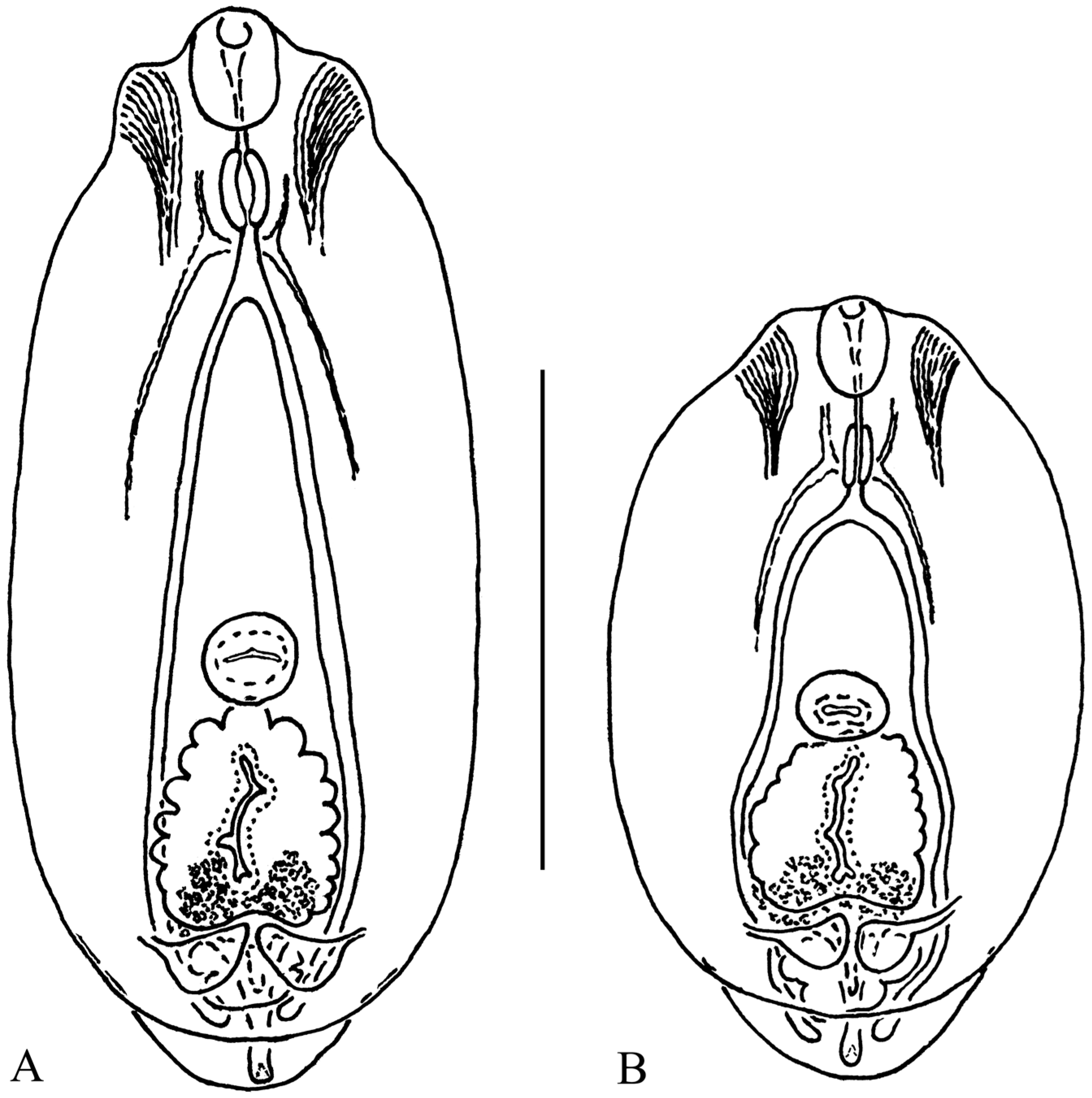
Figure 1. Metacercariae of Diplostomum numericum ex Scardinius erythrophthalmus from Lake Vanchozero, Karelia, Russia, with elongate-oval (A) and oval (B) body shapes at the same scale. Scale bar = 300 μm.
Diversity 15 00840 g001
Figure 2. Metacercaria of Diplostomum numericum ex Rutilus rutilus from Lake Gimoilampi, Karelia, Russia. Scale bar = 300 μm.
Diversity 15 00840 g002
Figure 3. Phylogenetic tree of Diplostomum spp. based on partial cox1 (388 bp) inferred using Bayesian inference analysis. Posterior probability values lower than 0.5 are not shown. Newly obtained sequences are marked with an asterisk (*). Abbreviations: AaAlburnus alburnus; RrRutilus rutilus; SaSalvelinus alpinus; SeScardinius erythrophthalmus; StSalmo trutta; GgGobio gobio; I—Iceland; G—Germany; N—Norway; R—Russia; S—Scotland.
Diversity 15 00840 g003
Table 1. Measurements of Diplostomum numericum metacercariae.
CharactersOur Material;
Range (Mean)
Niewiadomska [27];
Range (Mean)
Shigin and Stanislavetz [29] [as D. vitreophilum];
Range (Mean)
HostScardinius erythrophthalmusRutilus rutilusScardinius erythrophthalmusRutilus rutilus, *Coregonus albula, *Alburnus alburnus *
Total body length (BL), μm440–651 (545)543–644 (578)451–547 (512)465–587 (526)
Maximum body width (BW), μm242–336 (289)256–298 (280)244–288 (270)210–265 (239)
Oral sucker length (OL), μm52–69 (64)58–72 (64)51–68 (62)48–62 (56)
Oral sucker width (OW), μm45–55 (50)48–58 (52)40–54 (47)45–58 (51)
Pharynx length (PL), μm31–48 (40)34–52 (42)30–42 (37)28–40 (33)
Pharynx width (PB), μm22–31 (27)26–34 (32)17–30 (24)25–35 (29)
Ventral sucker length (VL), μm41–58 (49)50–58 (53)40–57 (47)43–51 (47)
Ventral sucker width (VW), μm52–62 (56)52–58 (56)40–64 (51)46–55 (51)
Distance from anterior margin of body to center of ventral sucker (O), μm242–402 (304)298–367 (322)234–306 (275)
Holdfast organ length (HL), μm107–144 (124)110–138 (120)81–119 (98)100–130 (112)
Holdfast organ width (HW), μm103–120 (112)103–120 (113)74–108 (87)92–135 (107)
Proteolytic gland length, μm28–55 (44)41–69 (54)23–40 (30)
Proteolytic gland width, μm76–110 (94)79–110 (97)68–102 (81)
Number of excretory bodies 638, 659, 813957, 1088~ 700760–1150 (965)
BW/BL, %42.62–61.54 (53.32)43.09–53.75 (48.52)40.67–58.42 (52.39)41.70–50.50 (45.50)
O/BL, %50.63–61.70 (55.61)52.49–58.33 (55.62)50.38–55.94 (53.74)51.30–56.60 (54.50)
[BL × BW]/[VL × VW]43.43–77.17 (57.69)45.05–63.27 (54.52)41.89–78.64 (58.40)43.20–58.90 (53.10)
[BL × BW]/[HL × HW]9.87–14.22 (11.38)10.54–13.21 (11.99)11.35–20.46 (16.51)8.37–12.89 (10.58)
[OL × OW]/[VL × VW]0.94–1.34 (1.15)0.96–1.31 (1.13)0.94–1.84 (1.22)1.06–1.39 (1.21)
[HL × HW]/[VL × VW]4.13–6.25 (5.10)3.76–5.47 (4.56)2.63–5.04 (3.63)3.89–7.02 (5.02)
[OL × OW]/[PL × PW]2.08–3.72 (2.94)1.90–3.36 (2.57)2.13–4.21 (3.22)2.29–3.79 (3.02)
Note: * It is unclear whether Shigin and Stanislavetz (1989) measured metacercariae from the bleak and the vendace.
Disclaimer/Publisher’s Note: The statements, opinions and data contained in all publications are solely those of the individual author(s) and contributor(s) and not of MDPI and/or the editor(s). MDPI and/or the editor(s) disclaim responsibility for any injury to people or property resulting from any ideas, methods, instructions or products referred to in the content.