Article

Hemolymph Parameters Are a Useful Tool for Assessing Bivalve Health and Water Quality

1
A.V. Zhirmunsky National Scientific Center of Marine Biology, Far Eastern Branch of Russian Academy of Sciences, 690041 Vladivostok, Russia
2
School of Medicine and Life Sciences, Far Eastern Federal University, 690922 Vladivostok, Russia
*
Author to whom correspondence should be addressed.
Diversity 2024, 16(7), 404; https://doi.org/10.3390/d16070404
Submission received: 13 May 2024 / Revised: 5 July 2024 / Accepted: 11 July 2024 / Published: 13 July 2024
(This article belongs to the Special Issue Biodiversity as Tools to Assess Impacts on Coastal Ecosystems)

Abstract

:
Bivalves play a key role in aquatic ecosystems and are a valuable commercial resource. The prosperity of these aquatic organisms depends mainly on the effectiveness of their immune defense, in which the hemolymph plays a central role. Hemolymph may be used as an effective non-lethal criterion of health. However, the predictive value of hemolymph analysis depends on the comparison between the obtained results and reference data from healthy individuals living in natural aquatic environments. We collected hemolymph from 15 commercially important species from wild populations at stations located in non-impacted and impacted water areas of the Sea of Japan. Of the 11 hemolymph parameters we analyzed, the total hemocyte count, percentage of hemocyte types, phagocytic activity, presence of reactive oxygen species, and protein concentration differed significantly between populations from non-impacted and impacted water areas. The most responsive species to pollution were Magallana gigas, Crenomytilus grayanus, Mizuhopecten yessoensis, and Mactra chinensis. This work is the first to examine a large number of commercially important species simultaneously. The results of this study are the basis for establishing the health status criteria of commercial bivalves for veterinary control in aquaculture and biomonitoring.

1. Introduction

Bivalves are a widespread group of invertebrates that are ecologically and economically important. Coastal sites are their prime habitat and cultivation areas, which experience tremendous pressure. Improving aquaculture efficiency and protecting the health of aquatic organisms are closely related issues that involve managing bioresources in marine and freshwater ecosystems. One of the effective ways to solve these problems is the development of reliable technologies for diagnosing the conditions of aquatic organisms and their habitats. Currently, histological analysis is a key method for diagnosing the condition of bivalves, but it is time-consuming («Aquatic Animal Health Code»). While it provides comprehensive information about the condition of mollusks and their pathogens, it only reflects the morphological changes and may not clearly indicate disturbances in physiological functions. To develop a reliable, comprehensive, and integrated indicator of health conditions in diagnostics, functional tests are necessary. Changes in internal environmental parameters are the most appropriate reflection of an organism’s adaptation to changes in its environment and within the organism itself. For vertebrates and humans, evaluation of hematological parameters is a key aspect of diagnosing physiological conditions. Furthermore, in fish aquaculture, hematological analysis is a recognized, reliable tool for monitoring their health status and a standard in diagnosing pathologies [1,2,3]. The blood analysis provides a clear picture of fish’s health, even at the earliest stages of disease development and changes in habitat conditions [4,5,6]. Mollusk hemolymph is an analog of blood and plays a crucial role in physiological adaptations, maintaining internal balance and providing immune protection. It is also a promising and accessible tool for the health diagnosis of these organisms. Collecting hemolymph from bivalves is a simple and relatively non-invasive process. Numerous studies [7,8,9,10,11,12] have shown that immune parameters in bivalve hemolymph are generally stable under normal environmental conditions but become more imbalanced under stress. This imbalance reflects their high sensitivity and reactivity, which is an obligatory criterion for health diagnostics. Research conducted in an almost perfect habitat [7,13] (with optimal food supply, no pollution, and minimal infections) has identified key factors influencing hemolymph parameters, including water temperature, salinity, and the reproductive status of the animals [13]. By comparing hemolymph parameters under stress to those in normal conditions, it is possible to monitor the health of individual organisms and the entire population, as well as to reliably detect changes in environmental conditions.
The number of studies and the variety of methods using hemolymph for ecotoxicological analysis are increasing annually [14,15,16,17], confirming its effectiveness as a reliable tool. Laboratory investigations have advantages, particularly the ability to control conditions. However, the interpretation of laboratory results in relation to natural systems with several fluctuating variables is challenging. Field research allows for the analysis of parameters under conditions present at a particular moment, considering all the factors that affect both the ecosystem and the organisms within it. Currently, extensive data has been collected on assessing the parameters of the hemolymph of mollusks in laboratory conditions, while there is comparatively less data from field studies (see Discussion). This issue is especially relevant for the Sea of Japan waters in Russia, which are among the main fishing areas of the country. These waters are involved not only in the import but also in the export of seafood to neighboring countries [18,19,20]. Peter the Great Bay, the largest of the bays in the northwestern part of the Sea of Japan, is one of the richest areas of the Far East in terms of the abundance and diversity of animals and plants inhabiting it. Unfortunately, over the past few decades, there has been a deterioration in the environmental conditions in certain areas, mainly associated with pollution from coastal sources. Expert estimates [21] indicate that the greatest anthropogenic load is experienced by the water areas of the bay adjacent to the city of Vladivostok—Amur Bay and Zolotoy Rog Bay—as well as Nakhodka Bay near Nakhodka city [21,22,23]. Along the coastal waters, chronic pollution is observed with a wide range of pollutants, including organic waste, pesticides, heavy metals, oils, petroleum products, surfactants, etc. The level and nature of the distribution of these pollutants are not the same along the coastal strip of the bays [21,24,25,26], and there are areas with good environmental conditions [24]. Studies have shown that the entry of pollutants into water areas negatively affects various structures of aquatic organisms, causing dysfunctions and pathologies. Extensive studies of the benthos of Peter the Great Bay by Moshchenko and colleagues in 2019 [26] and Galysheva in 2009 [24] revealed varying degrees of transformation of biocenoses, with the most serious damage observed in Zolotoy Rog Bay, Amur Bay, and Nakhodka Bay. Analyses of the heavy metals content in oyster tissues by Kiku and Kovekovdova [25] showed their greatest accumulation in mollusks from Amur Bay and Nakhodka Bay. Additionally, investigations by Zhuravel and Podgurskaya [27] and Vaschenko and colleagues [28] in Scaphechinus mirabilis and Strongylocentrotus intermedius revealed anomalies in the development of the gonads and the reproductive cycle in certain aquatic organisms caught in Amur Bay and Nakhodka Bay. Similar anomalies were found in the bivalve Modiolus kurilensis by Yurchenko and Vaschenko [29] and in gastropods Littorina and Nucella by Syasina and Shcheblykina [30].
A comprehensive investigation of the histopathological and immune status of the bivalve M. kurilensis carried out by our team in 2018 [12] showed a pronounced negative impact of pollutants on the health of mollusks. However, large-scale monitoring research that covers the water areas of Peter the Great Bay with different ecology and various species of mollusks has not yet been conducted. Therefore, the aim of this research was to determine reference ranges for selected cellular and humoral parameters of bivalves’ hemolymph from water areas with different ecological conditions to assess the health of commercial mollusks in this region.

2. Materials and Methods

2.1. Animals

In this study, 15 commercial bivalve species were examined (Mactra chinensis, Mercenaria stimpsoni, Spisula sibyllae, Chlamys farreri, Mizuhopecten yessoensis, Swiftopecten swiftii, Magallana gigas, Crenomytilus grayanus, Tetrarca boucardi, Mytilus trossulus, Mya arenaria, Saxidomus purpurata, Ruditapes philippinarum, Anadara broughtonii, and Corbicula japonica). Sexually mature individuals of average age (4–7 years) and shell sizes were collected using the light diving method at a depth of up to 10 m during the period of sexual inertia in September and October of 2023. Each sample contained at least 30 individuals of each species from each station (900 individuals in total). Next, the caught animals were immediately transferred to the laboratory in containers with aerated seawater and used in the experiment on the day of collection.

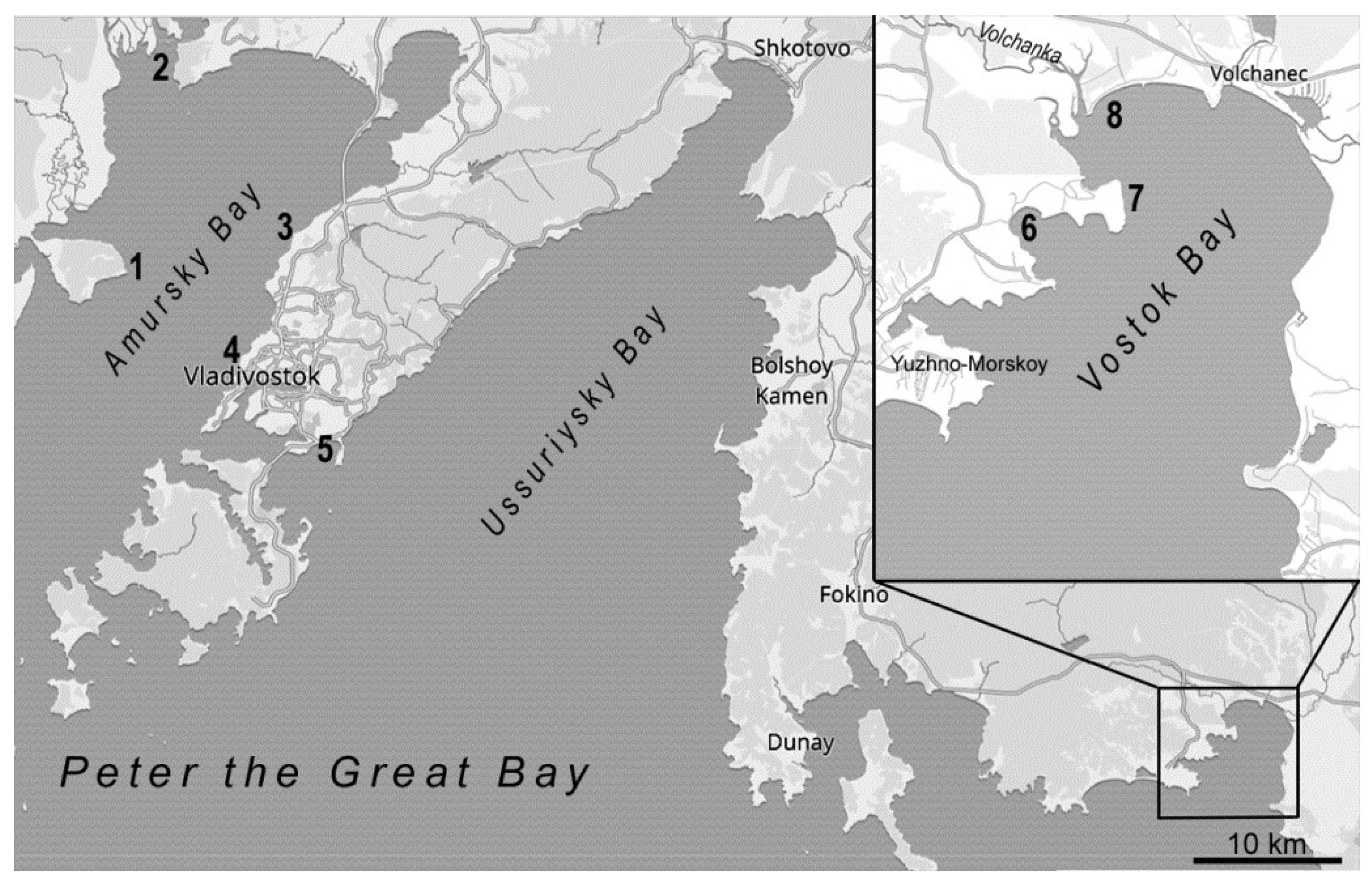
2.2. Collection Areas

Bivalves were collected from wild populations at stations in non-impacted/reference (near the biological station «Vostok» in Vostok Bay, Cape Peschany, Tavrichansky Estuary) and impacted (Cape Krasny, Sportivnaya Gavan Bay, Patrokl Bay, Sredneya Bay, and Vostok Cove) water bodies of the Sea of Japan (Table 1, Figure 1). The collection of certain species of mollusks from water areas was based on the similar geographical and hydrological conditions of the water areas and the occurrence of a particular species.
Moreover, according to the literature [24], the impacted water areas of the Peter the Great Bay of the Sea of Japan are exposed to various degrees of pollution (Table 1) and are characterized by different levels of pesticides, pollutants, phenols, petroleum products and diesel fuel, enterococci, heterotrophic microorganisms, Escherichia coli, and bacteria resistant to heavy metals. The pollution level here exceeds the natural background and maximum permissible concentration (MPC) for at least one of the indicators [26,31,32,33]. Patrokl Bay is an extremely polluted water area where concentrations of heavy metals and hydrocarbons are hundreds of times higher than natural geochemicals and MPCs [24,34]. This water area is used for ship mooring, raids, wastewater discharge, permanent transportation lines, fishing, and commercial ports. Furthermore, Patrokl Bay experiences periodic pollution from oil products, which was particularly intense from 1990 to 1997. The sources of this pollution are ships moored at the roadstead and an oil storage facility in the nearby Promezhytochnay Bay. Additionally, the condition of Patrokl Bay is undoubtedly affected by its proximity to the Eastern Bosphorus [34]. After the military withdrawal, the bay became a popular recreation spot for residents of Vladivostok, which unfortunately led to the destruction of coastal aquatic species. Due to the weakened hydrodynamics and the bay’s enclosure, intensive siltation of the upper sublittoral is observed here [26]. In addition to microbiological and hydrochemical indicators, Patrokl Bay has a poorer benthic population in terms of total number of species compared to the nearby water areas near Russky Island (71 and 104 species belonging to 10 and 9 taxonomic groups, respectively) [26].
Vostok Cove is also characterized as one of the most polluted water areas due to the exceeding content of Pb and Zn, hundreds of times higher than the permissible levels [31,32,33,35,36]. In addition to the above-mentioned pollution, and for all other typical water area types of pollution, this area also exhibits high levels of Ni, Cu, Cd, Si, ammonium, phosphorus, and phosphates with reduced dissolved oxygen levels [31,33,35]. This is caused by residential, agricultural, and industrial wastewater from the settlement of Volchanec [33,36].
The third place in terms of pollution belongs to Sredneya Bay, which contains a slightly lower concentration of heavy metals but has high Ni pollution, which indicates a specialized anthropogenic impact caused by hydrocarbon fuel combustion [31,33,35,37]. Pollution from ship repair and fish processing factories occurs in the nearby Gaidamak Bay, as well as from domestic sewage from the settlement of Yuzhno-Morskoy and Livadia spreading in this bay via constant and tidal currents from the south [38]. Similarly to Patrokl Bay, in Sredneya Bay and Vostok Cove a decrease in the number of benthic groups was observed [37,38].
Despite the urban agglomeration of Vladivostok being nearby, the Sportivnaya Gavan Bay [27,39] and the water area near Cape Krasny, located to the north of the city [24], are affected by strong and moderate levels of pollution, respectively, according to the literature data [40,41]. The main sources of pollutants in these water areas are the city’s sewage, the seaport, rail, and road transport, as well as stormwater runoff and the aeolian transport of pollutants with dust particles. In Sportivnaya Gavan, an increase in the cases of intensive development of microalgae with replacement of the diatom complex by the flagellate community, as well as filamentous cyanobacteria, was noted. This indicates eutrophication of the water area, presumably due to the uncontrolled inflow of untreated sewage [42]. It is also shown that the condition of the water area adversely affects the condition of some aquatic organisms [39,43,44,45], in particular, affecting the physiological status [12] and spermatogenesis [29] of M. kurilensis. One study demonstrated a more pronounced disruption of sea urchin embryogenesis when incubated in water from the Patrokl Bay area compared to water from the area near Cape Krasny [27].
Despite the localization of the key sampling station (near the biological station “Vostok”) in Vostok Bay (which includes Srednyaya Bay and Vostok Cove), this water area is deemed non-impacted. This area of the bay is more open, exposed to stronger action of south and east winds and runoff currents, and is characterized by specific hydrodynamic processes that contribute to the dispersion of pollutants [31,32,35]. Part of the water area of Vostok Bay is protected by the State Natural Complex Marine Reserve “Vostok Bay.” The marine biota of Peter the Great Bay has been studied for more than 30 years [24,32,38] at the marine biological station “Vostok,” a part of NSCMB, located on the shore of this bay. As for Cape Peschaniy [25,46] and the Tavrichanskiy Estuary [47], despite recreational impact in the summertime and the close location of settlements, the water areas show minimal content of enterococci and concentrations of the above-mentioned metals, which do not exceed the MPC. Additionally, all reference water areas have a high biodiversity of macrobenthos, according to the literature data [36,37,42].

2.3. Hemolymph Sampling

Hemolymph was collected from the posterior adductor muscle sinus using a 1 or 5 mL sterile syringe into precooled (4 °C) microtubes to avoid hemocyte aggregation.
For total hemocyte count, an aliquot of hemolymph (500 µL) was immediately mixed with an equal volume of a 4% paraformaldehyde (PFA) solution prepared in artificial seawater (ASW) (pH 7.5) at an osmolality of 1090 mOsm. The remaining hemolymph samples were centrifuged at 800× g for 12 min at 15 °C. Then, the supernatant was frozen at −85 °C for further analysis of humoral immunity activity assays (protein concentration and hemolytic and hemagglutination activity). The hemocyte pellet was resuspended in ASW and used to assess the parameters of hemolymph cellular factors.

2.4. Determination of Hemolytic Activity

Hemolytic activity was evaluated according to a method described in detail by Grinchenko and colleagues [48] with modifications for 96-well microplate assay. Volumes were proportionally reduced by five times. An incubation was carried out in a 96-well microplate for 1 h with periodic shaking every 15 min at 15 °C. After stopping the reaction and centrifugation, 200 µL of supernatant was transferred to a new flat-bottomed 96-well microplate for measurement of optical density (OD) by a Bio-Rad xMark Microplate Absorbance Spectrophotometer at 415 nm.

2.5. Hemagglutination Reaction

To assess hemagglutination, a protocol described by Grinchenko and colleagues [48] was used. First, the most appropriate type of erythrocytes was selected for each mollusk species. The following types of erythrocytes were used: a mouse for M. arenaria, M. chinensis, S. swifti, and C. farreri; a rat for A. broughtonii, T. boucardi, M. trossulus, and C. japonica; a dog for S. sibyllae, M. yessoensis, and M. stimpsoni; a human for C. grayanus; bottle-nosed dolphin for R. philippinarum; a walrus for S. purpurata; a Baikal seal for M. gigas.

2.6. Protein Concentration Assay

Protein concentration measurement was performed in 96-well UV-transparent microplates (200 µL of sample per well) using a Bio-Rad xMark Microplate Absorbance Spectrophotometer with default settings for the UV method.

2.7. Haemocyte Oxygen-Dependent Systems Estimation

To detect reactive oxygen species (ROS) in hemocytes, the unstimulated nitroblue tetrazolium (NBT) reduction test in formazan was performed. For this purpose, hemocyte suspensions from each individual were plated in triplicate in a 96-well black plate (Greiner Bio-One, Frickenhausen, Germany) and incubated for 20 min at 15 °C for cell adhesion to the substrate. Afterward, hemocytes were incubated with 0.1% NBT diluted ASW for 30 min at 15 °C. Then, the samples were washed three times with ASW. Intracellular formazan was solubilized with a 2 M KOH-DMSO solution and analyzed using a Bio-Rad xMark Microplate Absorbance Spectrophotometer at 630 nm.

2.8. Phagocytosis Assay

Phagocytic activity was detected using the Spark 10M multimode plate reader (Tecan Trading AG, Männedorf, Switzerland) with 485 nm excitation and 535 nm emission filters, as described previously by Grinchenko and colleagues [48].

2.9. Hemocyte Count and Flow Cytometry Analysis

To differentiate hemocyte counts, 500 µL of fixed hemolymph was washed three times with ASW, followed by centrifugation at 800× g for 12 min at 15 °C. Samples were then analyzed using a BD Accuri C6 flow cytometer (Becton Dickinson, Franklin Lakes, NJ, USA) equipped with an argon excitation laser (488 nm) and detectors of forward (FSC) and side (SSC) light scatter. A total of 100,000 events per sample were recorded. A size (FSC) against internal complexity (SSC) density plot was measured using the computer program CellQuest. It was used to distinguish the hemocyte subpopulations based on their relative cell size and granularity. Debris was gated out by using forward scatter (FS)/side scatter (SS) data and the red fluorescence of hemocyte nuclei associated with cell prestaining with 0.001% propidium iodide (Thermo Fisher Scientific, Waltham, MA, USA).

2.10. Data Analysis

The Kolmogorov–Smirnov test showed that the distributions of data do not follow the normal distribution law (p < 0.05) for all samples, so the analysis was carried out using nonparametric statistics. The Mann–Whitney U rank test was used to compare the studied parameters of the bivalve mollusks from the non-impacted and impacted water areas.

3. Results

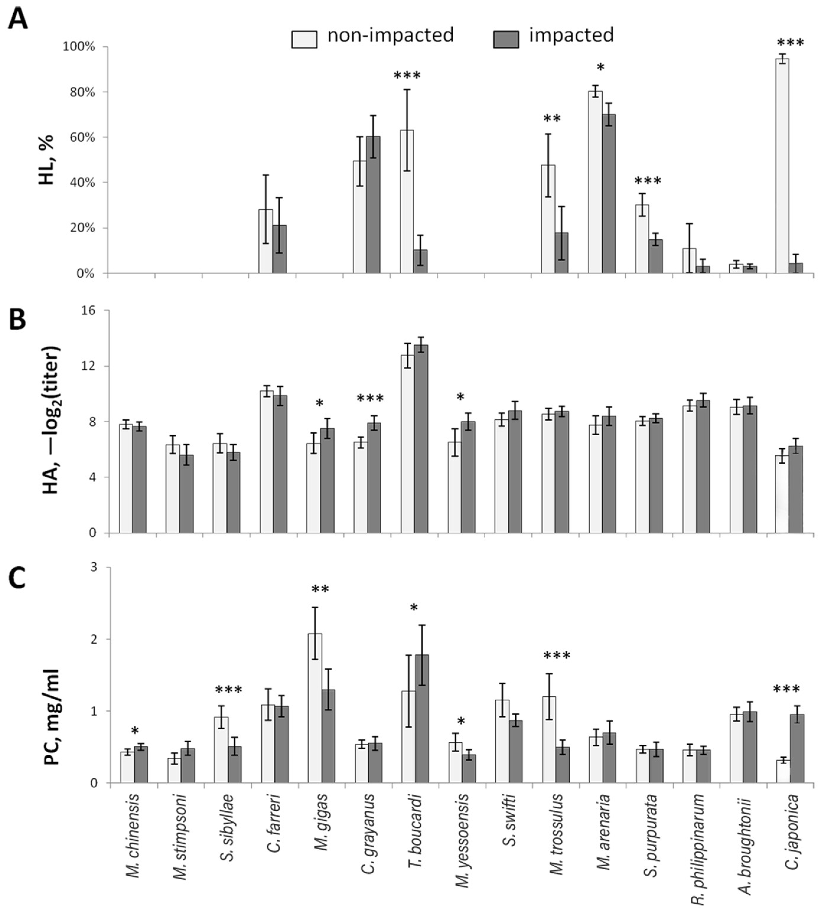
3.1. Hemolytic Activity

The most notable and significant differences in hemolytic activity (HL) of plasma were found in T. boucardi, S. purpurata, M. arenaria, M. trossulus., and especially in C. japonica (Figure 2A). The plasma of M. chinensis, M. stimpsoni, S. sibyllae, M. gigas, M. yessoensis, and S. swifti did not show any hemolytic activity against human erythrocytes. The plasma of R. philippinarum and A. broughtonii had extremely low HL values, with no significant differences between impacted and non-impacted water areas. Meanwhile, C. farreri and C. grayanus exhibited moderate HL values with no significant differences between water areas. It is worth noting that HL is a highly variable parameter, with values for most species reaching 90% or more, except for M. arenaria (41%), S. purpurata (49%), and A. broughtonii (14%).

3.2. Hemagglutination

The initial selection of erythrocytes allowed for the relevant assessment of hemagglutination (HA) for all investigated mollusk species. The widest range of HA values (−log2(titer) from two to eleven) was observed for M. yessoensis (Figure 2B). In other cases, the range was significantly narrower and began at higher values. T. boucardi displayed extremely high HA (−log2(titer) from nine to sixteen), which was evident not only with the selected type of erythrocytes but also with all types used during the selection process. Significantly higher HA values were only found for M. gigas, C. grayanus, and M. yessoensis collected from the impacted water area (Figure 2B).

3.3. Protein Concentration

The highest mean protein concentration (PC) was identified for M. gigas (Figure 2C), ranging from 0.44 to 3.69 mg/mL. However, some individuals of T. boucardi had even higher (from 4.43 mg/mL) and lower (from 0.22 mg/mL) values, characterizing T. boucardi as the species with the most variable PC. A significant increase in the PC was observed for M. chinensis, T. boucardi, and C. japonica (Figure 2C) from the impacted water area. However, C. japonica had mean values that differed more than three times. A significant decrease in the PC was only observed for S. sibyllae, M. gigas, M. yessoensis, and M. trossulus from the impacted water areas (Figure 2C).

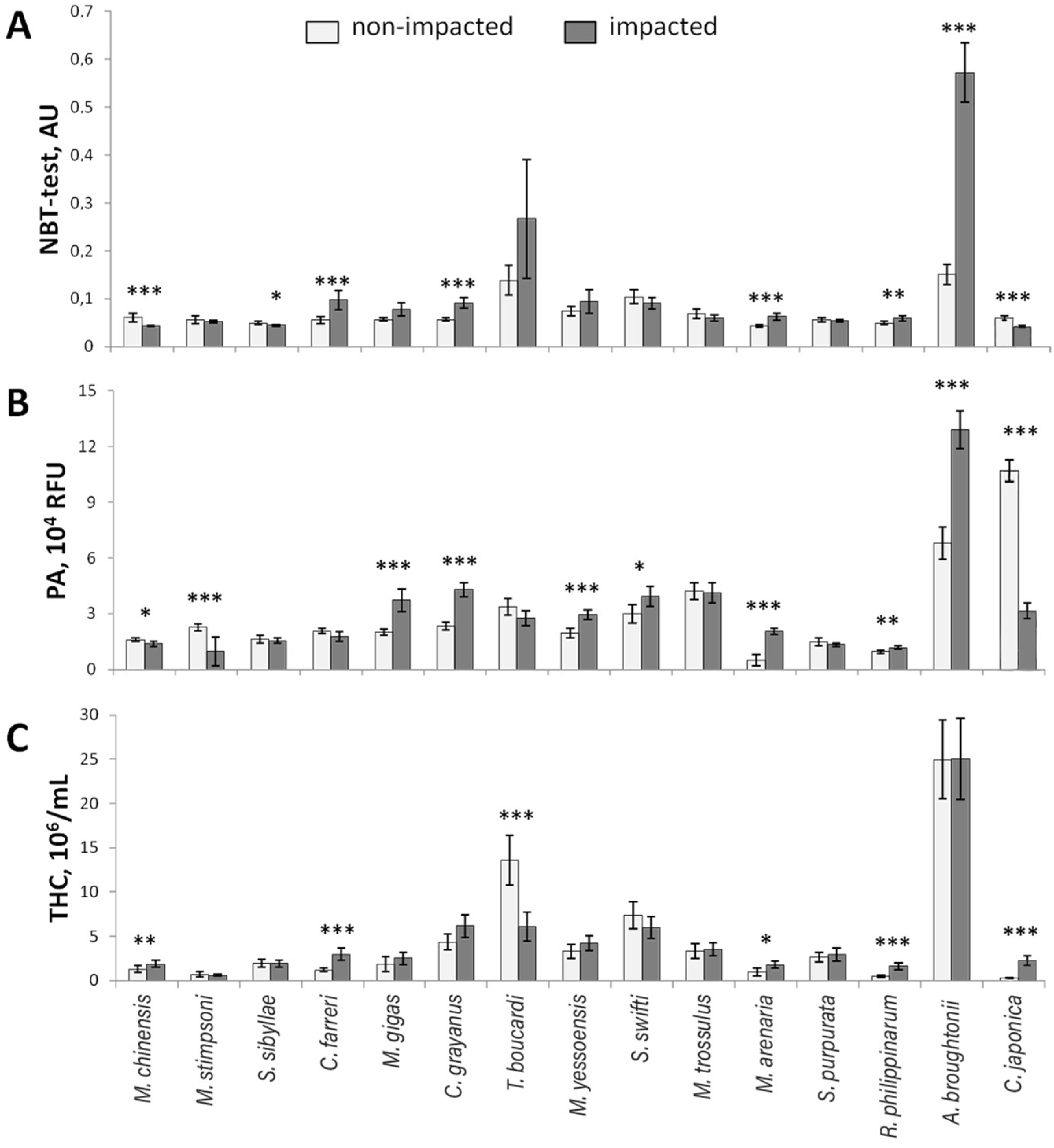
3.4. Nitroblue Tetrazolium Reduction Test

The nitroblue tetrazolium reduction test (NBT-test) showed the highest values for T. boucardi and A. broughtonii (Figure 3A). These species also displayed the greatest range of values. Additionally, the increasing NBT-test values led to a two-fold significant difference for T. boucardi from the impacted water area. For all other species, NBT-test values ranged from 0.04 to 0.26 AU and remained relatively stable for individuals of the same species and water area. A significant increase in NBT-test values was observed for C. farreri, C. grayanus, M. arenaria, R. philippinarum, and A. broughtonii (Figure 3A). Similarly to humoral immune factors, NBT-test values were often higher in animals from impacted water areas, and a decrease in this parameter was only detected for M. chinensis, S. sibyllae, and C. japonica (Figure 3A).

3.5. Phagocytic Activity

The hemocytes of A. broughtonii and C. japonica showed the highest phagocytic activity (PA) values (Figure 3B). For all other mollusk species, PA values ranged from 0.63 to 7.30 RFU, except for M. arenaria collected from a non-impacted water area (Figure 3B). The PA values for M. arenaria were the lowest among the species, and this difference was found to be highly significant. Moreover, significant decreases in PA values of mollusks from the impacted water area were only observed for M. chinensis, M. stimpsoni, and C. japonica (Figure 3B).

3.6. Total Hemocyte Count

The total hemocyte count (THC) was generally a stable parameter and showed an insignificant range of values across most species, with the exception of T. boucardi and A. broughtonii, which had the highest THC values (Figure 3C). Similarly to other immune parameters, the THC was increased significantly in mollusks from the impacted water area, especially in M. chinensis, C. farreri, M. arenaria, R. philippinarum, and C. japonica. However, T. boucardi exhibited a pronounced decrease in THC (Figure 3C).

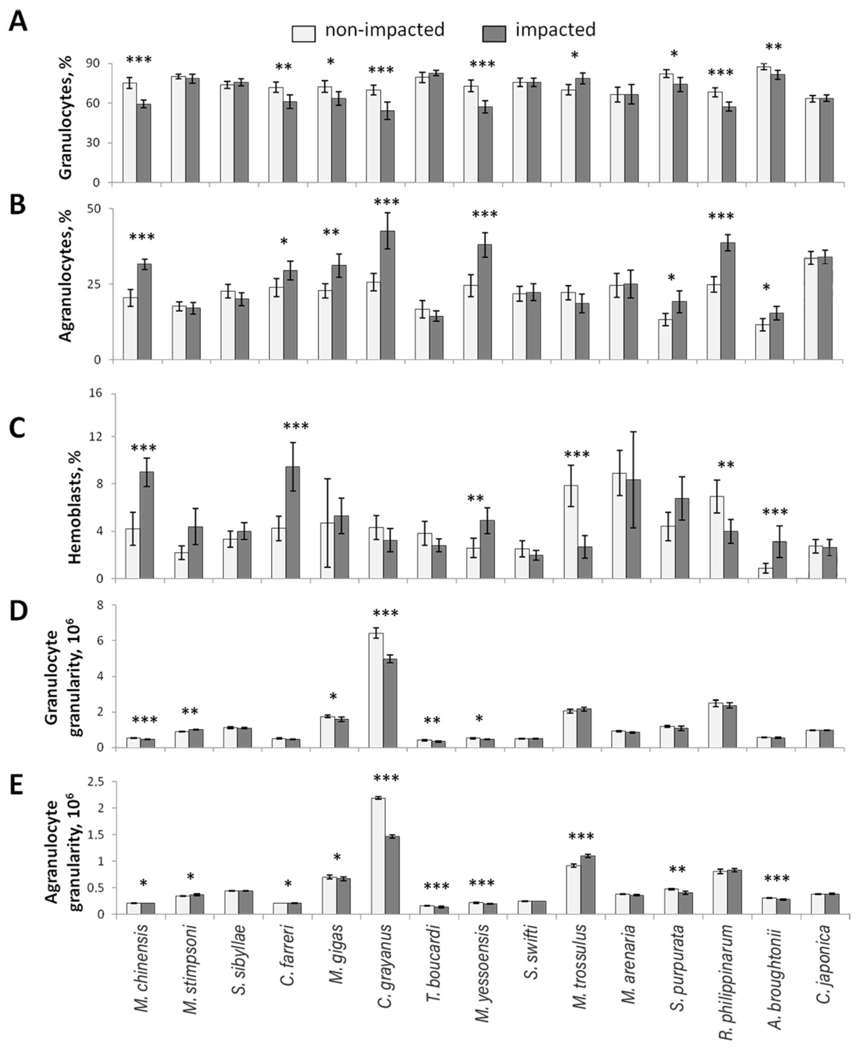
3.7. Hemocyte Populations

Granulocytes were the predominant cell type in the hemolymph of all studied species, except in T. boucardi and A. broughtonii, where erythrocytes were more prevalent (Figure 4A). Despite this, T. boucardi and A. broughtonii, compared to other species, had the highest content of granulocytes among hemolymph amoebocytes, averaging 81% and 84%, respectively (Figure 4A). The lowest percentage of granulocytes was observed in C. grayanus at 62%. The content of granulocytes varied the most in C. grayanus and M. arenaria, with a total range of up to 57% and 60%, respectively. Unlike most of the previously described parameters of cellular immunity, the proportion of granulocytes generally decreased in bivalves from the impacted water area (in M. chinensis, C. farreri, M. gigas, C. grayanus, M. yessoensis, S. purpurata, R. philippinarum, and A. broughtonii), except M. trossulus, where it increased (Figure 4A).
Agranulocytes were the second largest population of hemocytes, averaging from 15 to 34% of total hemocytes (Figure 4B). Significant differences were observed regarding the proportion of agranulocytes and granulocytes. The opposite effect was found in the above-mentioned species of bivalves from the impacted water area, except M. trossulus, where there was an increase in the proportion of agranulocytes. This parameter varied significantly more in C. grayanus (total range across two water areas up to 53%) than in other species (Figure 4B).
Hemoblasts represented the smallest population of hemocytes, with a mean of less than 10% of hemocytes (Figure 4C). M. arenaria had the highest concentration at 8%, while A. broughtonii and S. swifti had the lowest at around 2% each. There were fewer species with significant differences in the hemoblast concentration between water areas, and the pattern was different (Figure 4C). A decrease in hemoblast concentrations in the hemolymph of bivalves from the impacted water area was observed in M. trossulus and R. thilippinarum, while an increase was noted in M. chinensis, C. farreri, M. yessoensis, and A. broughtonii (Figure 4C).
An additional parameter characterizing the main cellular populations of granulocytes and agranulocytes was their granularity (Figure 4D,E). A decrease in the granularity was observed in both granulocytes and agranulocytes in M. chinensis, M. gigas, C. grayanus, T. boucardi, and M. yessoensis from the impacted water area. However, a decrease in the granularity of agranulocytes was observed only in S. purpurata and A. broughtonii. An increase in the granularity was observed in both cell types in M. stimpsoni, while only agranulocytes were increased in M. trossulus.
Since the hemolymph of T. boucardi and A. broughtonii contained erythrocytes in addition to the amoebocytes described above, a comparative analysis was performed for them. The analysis showed a decrease in the proportion of erythrocytes in T. boucardi (p < 0.001, Mann–Whitney U Test) and an increase in their granularity in A. broughtonii (p < 0.05, Mann –Whitney U Test) in non-impacted water areas.

4. Discussion

Bivalves serve as natural filters and accumulators, playing a crucial role in the structure and functioning of marine benthic communities. Besides their commercial value, they are also significant for the environment, serving as bioindicators of marine environment quality [11,49,50,51,52]. With increasing impacts on aquatic ecosystems, there is a growing need to improve research in environmental toxicology using predictive systems. Reliable technologies for assessing the health of marine organisms and their habitats are essential for ensuring environmental safety and the sustainable development of the economy.
Over the past three decades, researchers have studied various physiological parameters of fish and mussel species, which are important in aquaculture. Despite this, only a few of these parameters have been incorporated into monitoring programs as test systems by organizations such as NOAA, ICES, UNEP, MED POL II, North Sea, WGBEC, NMMP, WHO-FAO, WFD, BEST, and BEEP of the EU [53,54]. The reason for this is that the most promising indicators are still in the experimental refinement and validation stage and must meet several requirements before being used outside of a research environment.
In 2006, Newton and Cope [55] analyzed research papers on various biomarkers of unionid mussels in the context of toxicology, using the biomarker classification system developed for fish [56]. While many groups of biomarkers have been successfully tested on mussels, the authors [55] highlighted that most of the analyzed works had shortcomings and required additional studies focusing on the health of the organism in natural conditions without pollutants and characterizing its initial state. However, studies on the influence of environmental factors in natural conditions on mollusk immunity are limited, especially regarding the immunity of bivalves in water areas with varying degrees of anthropogenic load. Based on the available information, it can be concluded that various pollutants have a negative impact on the organism, resulting in direct damage to certain tissues and organs, as well as indirect changes in the organism’s overall physiological status, affecting the systems responsible for maintaining homeostasis, including the immune system. The few studies in this area indicate that different pollutants can alter the immune state of mollusks, with the effect depending on the type and concentration of pollutant [57,58,59]. Generally, low pollutant concentrations have a stimulating effect, while high concentrations have an inhibitory effect [57,58,60,61,62,63,64,65], leading to an immune-deficient state and the increased susceptibility of mollusks to pathogens [66].
The majority of mollusk species in our study, regardless of their lifestyle and substrate preference, displayed similar immune system patterns when collected from impacted waters that do not meet sanitary and epidemiological standards for levels of phenols, petroleum products, pathogenic bacteria (such as enterococci), oxygen, phosphorus, heavy metals, and other parameters [26,31,32,33]. Most bivalve species under anthropogenic pressure exhibited a decreased plasma HL and PC, as well as granulocyte concentration, granularity degree, and NBT test parameters. On the other hand, other parameters, such as HA, PA, THC, agranulocyte, and hemoblast concentrations increased. Mytilids (M. trossulus and C. grayanus) from the water area near Cape Krasny in Amur Bay showed slightly different immune status from other species, likely due to their different lifestyle. The differences mainly concerned cellular parameters of immunity, particularly in M. trossulus. Bivalve species (M. chinensis, M. stimpsoni, and S. sibyllae) from the highly polluted water areas of Vostok Cove exhibited an opposite pattern of immune parameter activity compared to the other species, possibly due to chronic immune system suppression caused by heavy pollution. A similar phenomenon was observed in a study by Fisher and colleagues [67], where Crassostrea virginica, relocated to polluted areas (with high content of metals, polycyclic aromatic hydrocarbons, and polychlorinated biphenyls), showed increased THC and bactericidal activity after 12 weeks. However, when the experiment was reversed, the results varied, indicating a mixed response; lysozyme concentration was reduced, but THC and phagocytic index did not change. The authors suggest that these results may indicate the adaptation of oysters to chronic pollution [67,68]. Another study conducted in 2018 revealed a significant decrease in plasma hemolytic activity and total protein concentration in M. kurilensis from the chronically polluted waters of Sportivnaya Gavan Bay [12]. Similar results in the response of humoral factors in the hemolymph of C. virginica and R. philippinarum were also described by Chu, Peyre [69], and Allam and colleagues [70]. Additionally, a study on the effect of a high density of heterotrophic microflora on the physiological state of freshwater bivalves Elliptio complanata showed a significant negative correlation of PA of hemocytes with the concentration of microorganisms and a significant decrease in the concentration of lysozyme with maximum bacterial impact [71].
The immune response pattern we observed at varying levels of contamination indicated that the immune system was activated in cases of weak or moderate pollution but suppressed in cases of strong pollution. Some studies [61,63] have shown that exposing Mytilus edulis to low copper concentrations stimulates the production of reactive oxygen species (ROS) by hemocytes, while high concentrations inhibit this process, making the animals more susceptible to bacterial infections. In our study, we observed a similar pattern; mollusks from moderately or slightly impacted water areas (Cape Krasny and Sportivnaya Gavan Bay) had increased ROS in hemocytes, while mollusks from heavily impacted Vostok Cove had decreased ROS compared to non-impacted areas. It was shown that in R. philippinarum, copper inhibited the phagocytic activity (PA) and the superoxide dismutase content in hemocytes. Cadmium, on the other hand, did not affect the cell parameters but increased cytochrome oxidase activity and caused the destabilization of lysosomal membranes [57]. As previously reported, in M. edulis, cadmium did not affect PA and ROS of hemocytes, but it did affect the total hemocyte count (THC); low concentrations of the pollutant decreased the number of cells, while increasing concentrations increased the count, indicating cell migration from the tissues to the hemolymph, as authors suggested [62]. In our study, we observed an increase in the THC only in M. chinensis from Vostok Cove, which had excessive cadmium content (according to the literature data), as well as in all mollusk species from the most polluted Patrokl Bay. Additionally, an increase in the proportion of hemoblasts among circulating hemocytes was observed in mollusks from Vostok Cove. It was shown that in C. virginica, exposure to high concentrations of cadmium increased the concentration of circulating cells and their PA while also increasing the content of metallothionein in hemocytes and inhibiting ROS generation [72]. Longer exposure (for 21 days) led to an increase in the number of dead hemocytes and increased mortality in mollusks, with the effects being strongest at low concentrations of cadmium. However, higher concentrations increased the viability and functional activity of hemocytes [73]. Similar effects were observed in Mytilus galloprovincialis exposed to higher doses of cadmium and copper. The production of ROS by hemocytes was altered only in the presence of copper and manifested in the suppression of their synthesis [74]. Hemocyte viability and PA decreased in M. arenaria under similar conditions (28 days) [75]. Similar alterations in PA and the THC were observed in M. arenaria from Vostok Cove.
However, it appears that heavy metals may not necessarily have a toxic effect on bivalves. The hemocytes of C. virginica can actually accumulate metals such as copper and zinc to help with antimicrobial defense [68] and shell formation [67]. For instance, when M. edulis was infected with Vibrio tubiashii, the combined effect of copper and temperature resulted in a significant decrease in the THC compared to the effect of either factor alone. Additionally, the ROS content in the presence of different concentrations of copper decreased significantly at 10 °C while it increased at 15 °C. In the case of copper concentrations, at low metal concentrations, PA increased [76]. In studies by Cheng [77,78], oysters exposed to 1 ppm copper sulfate showed a lower percentage of hyalinocytes, while those exposed to 1 ppm cadmium chloride showed a significantly higher percentage of hyalinocytes. In our study, mollusks from the most impacted areas showed a decrease in granulocytes and a slight increase in hyalinocytes and hemoblasts due to the degranulation and diapedesis of hemocytes. As previously reported in M. kurilensis, pollution resulted in a significant decrease in the percentage of agranulocytes and phagocytic activity [12]. One year after the catastrophic oil spill in Korea, a decrease in the number of granulocytes was recorded in the oyster C. gigas [79], whereas a higher percentage of granulocytes in the clam R. philippinarum was recorded two years after the spill [80]. Cd exposure increased the proportion of granulocytes in the Perna canaliculus [81] and the hemocyte type ratio in M. galloprovincialis [82]. Metal contamination also caused a decrease in the number of hyalinocytes in P. canaliculus [83], while in Ruditapes decussatus, higher PA was reported in hyalinocytes than in granulocytes when the cells were exposed to CuSO4 in vitro [84]. The different responses of hemocyte types to Cd in the freshwater mussel Dreissena polymorpha led to the consideration of granulocytes with a higher capacity to regulate oxidative stress and a greater involvement in the transport of base metals or heavy metal sequestration [85,86]. Crassostrea hongkongensis cultivated with Zn showed an increase in granulocyte mortality, suggesting that granulocytes are the most sensitive cell type in response to Zn; moreover, the number of granulocytes decreased, whereas the number of semigranulocytes and agranulocytes increased [87]. The number of granulocytes and PA decreased, whereas the number of hyalinocytes increased in C. farreri after exposure to polychlorinated biphenyls [88]. Exposure of M. galloprovincialis to benzo[a]pyrene and polychlorinated biphenyls increased the number of granulocytes [89]. In another study, polychlorinated biphenyls caused a decrease in granulocytes [90]. Prolonged exposure to persistent organic pollutants in M. edulis can reduce the proportion of granulocytes, suggesting that this type of hemocyte may be more sensitive to these pollutants [91]. When the effects of polycyclic aromatic hydrocarbons, a constituent of crude oil, were investigated on M. arenaria [59] and C. gigas [92], a suppression of hemocyte PA was found.
The studies conducted on Pecten maximus showed that exposure to phenanthrene at concentrations of 100 and 200 µg/L for 7 days resulted in an increase in the number of hemocytes but a decrease in their cell membrane stability and PA [93]. Exposure to phenol led to an increase in PA in M. edulis [94], while Mercenaria mercenaria exhibited an inhibition of hemocyte PA [95]. Chamelea gallina exposed to a benzo[a]pyrene concentration of 0.5 mg/L for 7 and 12 days experienced a significant decrease in the lysozyme activity, adhesion, and PA of hemocytes [96]. Benzo[a]pyrene had practically no effect on M. galloprovincialis except for an increase in ROS [74]. C. gigas exposed in vitro to benzo[a]pyrene and phenanthrene showed that these compounds significantly increased the proportion of granulocytes, esterase, and lysosome concentrations but suppressed cell death [73]. Exposure of Pinctada imbricata to No. 6 fuel oils for 7 days did not significantly alter immunological parameters such as the THC, PA, and lysozyme concentration, except for cell viability [97]. It was suggested that exposure to polycyclic aromatic hydrocarbons may increase susceptibility to infections due to the decreased immunocompetence of hemocytes caused by increased ROS. The highest petroleum carbon content was observed in Vostok Cove [24,32], Sredneya Bay [35], and Patrokl Bay [34], and animals caught in these water areas had reduced PA, HA, HL, ROS, and THC. Vostok Cove showed a decrease in dissolved oxygen content from all water areas, potentially impacting the immune system of mollusks [13]. It was previously reported that keeping M. galloprovincialis under hypoxia for 24 h resulted in a significant increase in the number of granulocytes but a decrease in the number of agranulocytes and ROS in granulocytes [98]. The responses of bivalves to simulated impacts aim to reveal possible negative effects of human activities on the physiology of these animals and their survival rates. In our study, most immunity indices and the studied mollusk species showed sensitivity to pollutant factors. However, a few bivalves (Merceraria, Spisula, Saxidomus, Swiftopecten, and Mya) showed insignificant alterations in the state of immunity under conditions of chronic pollution, indicating that these mollusk species are not sensitive to the existing level of pollution. The most sensitive bivalves were Mactra, Magallana, Crenomytilus, and Mizuhopecten, in which most of both cellular and humoral parameters were significantly changed. Each of the listed species was caught in different impacted water areas and can be recommended as a surveillance species for these areas. Among the immune indicators, the most frequently significantly altered parameters under the influence of pollution were plasma PC, THC, percentage of hemocyte types, ROS content, and PA, which can be recommended as representative parameters of the state of immunity of mollusks and their habitat.
We would like to emphasize that the biomarkers mentioned above indicate either the presence of environmental stressors or the consequences of their impact. Additionally, predictive biomarkers that can anticipate future exposure to stressors at higher levels of organization would be especially valuable for biomonitoring. Techniques like metabolomics and transcriptomics, which provide real-time data on how mollusks respond to current environmental conditions, could be instrumental in this regard [99,100]. However, at this stage in the development of molecular diagnostic methods, it is not yet possible to completely replace cellular, physiological, behavioral, and other methods for assessing organisms’ health. Comparative studies are essential for understanding the relationships between molecular markers and responses at the cellular and organism levels. An integrated approach to analysis will allow us to identify variations in the studied parameters under pathogenetic conditions from the norm. It will also help us to accurately determine the minimum necessary yet sufficient indicators for the development of an effective test system for the veterinary control of the mollusk′s physiological state. Additionally, by monitoring the mollusk’s health alongside genomic analysis, we can identify individuals that are disease-resistant or resilient to environmental stressors [101].

5. Conclusions

The hemolymph parameters studied proved to be a useful tool for assessing the health of mollusks in accordance with the environmental conditions of the water area they inhabit. The most significant changes were observed in the number of hemolymph cells, percentage of hemocyte types, phagocytic activity, reactive oxygen species, and protein concentration. The species most affected by pollution were M. chinensis, M. yessoensis, M. gigas, and C. grayanus, with most of the 11 studied parameters showing significant changes. We have established reference ranges for these parameters, which may indicate changes in both the external and internal environment of bivalves and can be measured non-lethally.
The reference values for the variability of health indicators of bivalve mollusks of different lifestyles and habitats may allow for further research in a wide range of factors, including testing other species from different regions. Based on the data obtained, it is possible to survey water bodies, conduct rapid diagnostic assessments of the actual state of marine organisms, provide forecasts of their condition, and make recommendations to counteract the negative impact of various biotic and abiotic factors. It also allows for dynamic control over the adaptation to various factors. In the future, a combination of the assessment of hemolymph parameters and molecular analysis to reflect the health status of aquatic organisms will lead to greater efficiency in their cultivation. This improvement will lead to an increased quality of life, a better physiological state, higher survival rates, and increased biological productivity of mollusks.

Author Contributions

Conceptualization, A.G. and Y.S.; methodology, A.G. and Y.S.; validation, A.G. and Y.S.; formal analysis, V.K.; investigation, E.T., A.T., I.B., M.M. and M.O.; data curation, A.G.; writing—original draft preparation, A.G., E.T. and Y.S.; writing—review and editing, Y.S. and M.M.; visualization, A.G. and Y.S.; supervision, A.G. and Y.S.; funding acquisition, Y.S. and V.K. All authors have read and agreed to the published version of the manuscript.

Funding

This research was funded by the Russian Science Foundation grant, grant number 23-76-10051 (https://rscf.ru/en/project/23-76-10051/ accessed on 13 July 2024).

Institutional Review Board Statement

Not applicable.

Data Availability Statement

Data are contained within the article.

Acknowledgments

Authors are grateful to the personnel of the Vostok Marine Biological Station (121082600038-3) and the Primorsky Aquarium Shared Equipment Facility of the A.V. Zhirmunsky National Scientific Center of Marine Biology, Far Eastern Branch, Russian Academy of Sciences for mollusk collection and screening.

Conflicts of Interest

The authors declare no conflicts of interest.

References

  1. Witeska, M.; Kondera, E.; Lugowska, K.; Bojarski, B. Hematological Methods in Fish—Not Only for Beginners. Aquaculture 2022, 547, 737498. [Google Scholar] [CrossRef]
  2. Ivanc, A.; Haskovic, E.; Jeremic, S.; Dekic, R. Hematological Evaluation of Welfare and Health of Fish. Prax. Vet. 2005, 53, 191–202. [Google Scholar]
  3. Clauss, T.M.; Dove, A.D.M.; Arnold, J.E. Hematologic Disorders of Fish. Vet. Clin. N. Am.—Exot. Anim. Pract. 2008, 11, 445–462. [Google Scholar] [CrossRef] [PubMed]
  4. Grant, K.R. Fish Hematology and Associated Disorders. Vet. Clin. N. Am.—Exot. Anim. Pract. 2015, 18, 83–103. [Google Scholar] [CrossRef] [PubMed]
  5. Docan, A.; Grecu, I.; Dediu, L. Use of Hematological Parameters as Assessment Tools in Fish Health Status. J. Agroaliment. Process. Technol. 2018, 24, 317–324. [Google Scholar]
  6. Fazio, F. Fish Hematology Analysis as an Important Tool of Aquaculture: A Review. Aquaculture 2019, 500, 237–242. [Google Scholar] [CrossRef]
  7. Oliver, L.M.; Fisher, W.S. Appraisal of Prospective Bivalve Immunomarkers. Biomarkers 1999, 4, 510–530. [Google Scholar]
  8. Donaghy, L.; Lambert, C.; Choi, K.S.; Soudant, P. Hemocytes of the Carpet Shell Clam (Ruditapes decussatus) and the Manila Clam (Ruditapes philippinarum): Current Knowledge and Future Prospects. Aquaculture 2009, 297, 10–24. [Google Scholar] [CrossRef]
  9. Gestal, C.; Roch, P.; Renault, T.; Pallavicini, A.; Paillard, C.; Novoa, B.; Oubella, R.; Venier, P.; Figueras, A. Study of Diseases and the Immune System of Bivalves Using Molecular Biology and Genomics. Rev. Fish. Sci. 2008, 16, 131–154. [Google Scholar] [CrossRef]
  10. Renault, T. Immunotoxicological Effects of Environmental Contaminants on Marine Bivalves. Fish Shellfish Immunol. 2015, 46, 88–93. [Google Scholar] [CrossRef]
  11. Sokolnikova, Y.; Trubetskaya, E.; Beleneva, I.A.; Grinchenko, A.V.; Kumeiko, V.V. Fluorescent in Vitro Phagocytosis Assay Differentiates Hemocyte Activity of the Bivalve Molluscs Modiolus kurilensis (Bernard, 1983) Inhabiting Impacted and Non-Impacted Water Areas. Russ. J. Mar. Biol. 2015, 41, 118–126. [Google Scholar] [CrossRef]
  12. Kumeiko, V.V.; Sokolnikova, Y.N.; Grinchenko, A.V.; Mokrina, M.S.; Kniazkina, M.I. Immune State Correlates with Histopathological Level and Reveals Molluscan Health in Populations of Modiolus kurilensis by Integral Health Index (IHI). J. Invertebr. Pathol. 2018, 154, 42–57. [Google Scholar] [CrossRef] [PubMed]
  13. Gerdol, M.; Gomez-Chiarri, M.; Castillo, M.G.; Figueras, A.; Fiorito, G.; Moreira, R.; Novoa, B.; Pallavicini, A.; Ponte, G.; Roumbedakis, K.; et al. Immunity in Molluscs: Recognition and Effector Mechanisms, with a Focus on Bivalvia. In Advances in Comparative Immunology; Springer International Publishing: Berlin/Heidelberg, Germany, 2018; pp. 225–341. ISBN 9783319767680. [Google Scholar]
  14. Yang, H. Immunological Assays of Hemocytes in Molluscan Bivalves as Biomarkers to Evaluate Stresses for Aquaculture. Bull. Jpn. Fish. Res. Educ. Agency 2021, 50, 31–45. [Google Scholar]
  15. de la Ballina, N.R.; Maresca, F.; Cao, A.; Villalba, A. Bivalve Haemocyte Subpopulations: A Review. Front. Immunol. 2022, 13, 826255. [Google Scholar] [CrossRef] [PubMed]
  16. Balbi, T.; Auguste, M.; Ciacci, C.; Canesi, L. Immunological Responses of Marine Bivalves to Contaminant Exposure: Contribution of the -Omics Approach. Front. Immunol. 2021, 12, 618726. [Google Scholar] [CrossRef] [PubMed]
  17. Girón-Pérez, M.I. Relationships between Innate Immunity in Bivalve Molluscs and Environmental Pollution. Invertebr. Surviv. J. 2010, 7, 149–156. [Google Scholar]
  18. Leljuhin, S.; Shirjakov, D.; Razumova, J. The Structure and Raw Materials Base of Fisheries Industry in the Far East. Reg. Econ. Manag. Electron. Sci. J. 2018, 3, 1–9. [Google Scholar]
  19. Saltykov, M.; Komogortseva, Z.; Kalyagin, M. International Trade of Aquatic Biological Resources with the Countries of Northeast Asia as a Factor of the Economic Growth of the Far Eastern Federal District Territories. Tomsk. State Univ. J. Econ. 2024, 65, 32–46. [Google Scholar] [CrossRef]
  20. Barkhudarova, S.M.; Gusakova, A.P.; Saltykov, M.A. Analysis of Development of the Fish Market of the Primorsk Territory and Vladivostok. Bull. Sci. Educ. 2018, 1, 14–18. [Google Scholar]
  21. Lukyanova, O.; Cherkashin, S.; Simokon, M. Overview of the Current Ecological State of the Peter the Great Bay (2000–2010). Vestn. Far East. Branch Russ. Acad. Sci. 2012, 2, 1–9. [Google Scholar]
  22. Kozhenkova, S.; Chernova, E. Assessment of Heavy Metal Pollution of the Peter the Great Bay (North-West Pacific Region) Using Brown Algae. J. Geosci. Environ. Prot. 2020, 8, 134–146. [Google Scholar] [CrossRef]
  23. Rostov, I.D.; Rudykh, N.I.; Rostov, V.I.; Vorontsov, A.A. Tendencies of Climatic and anthropogenic Changes of the Marine Environments in the Coastal Areas of Russia in the Japan for the Last Decades. Izv. TINRO 2016, 186, 163–181. [Google Scholar] [CrossRef]
  24. Galysheva, Y.A. Biological Consequences of Organic Pollution for Marine Coastal in the Russian Part of the Japan Sea. Izv. TINRO 2009, 158, 209–227. [Google Scholar]
  25. Kiku, D.P.; Kovekovdova, L.T. Trace Elements in the Oyster Crassostrea gigas from Peter the Great Bay (Japan Sea). Izv. TINRO 2007, 150, 400–407. [Google Scholar]
  26. Moshchenko, A.V.; Belan, T.A.; Borisov, B.M.; Lishavskaya, T.S.; Sevastianov, A.V. Modern Contamination of Bottom Sediments and Ecological State of Macrozoobenthos in the Coastal Zone at Vladivostok (Peter the Great Bay, Japan Sea). Izv. TINRO 2019, 196, 155–181. [Google Scholar] [CrossRef]
  27. Zhuravel, E.V.; Podgurskaya, O.V. Early Development of Sand Dollar Scaphechinus Mirabilis in the Water from Different Areas of Peter the Great Bay (Japan Sea). Izv. TINRO 2014, 178, 206–216. [Google Scholar] [CrossRef]
  28. Vaschenko, M.A.; Almyashova, T.N.; Zhadan, P.M. Long-Term and Seasonal Dynamics of the Gonad State in the Sea Urchin Strongylocentrotus intermedius under Anthropogenic Pollution (Amursky Bay, Sea of Japan). Vestn. Far East. Branch Russ. Acad. Sci. 2005, 1, 32–42. [Google Scholar]
  29. Yurchenko, O.V.; Vaschenko, M.A. Morphology of Spermatogenic and Accessory Cells in the Mussel Modiolus kurilensis under Environmental Pollution. Mar. Environ. Res. 2010, 70, 171–180. [Google Scholar] [CrossRef] [PubMed]
  30. Syasina, I.G.; Shcheblykina, A.V. Morphofunctional Characterization of the Reproductive System of the Gastropods Littorina brevicula, L. Mandshurica, and Nucella heyseana from Uncontaminated and Contaminated Areas of Peter the Great Bay, Sea of Japan. Russ. J. Mar. Biol. 2007, 33, 399–404. [Google Scholar] [CrossRef]
  31. Khristoforova, N.K.; Naumov, Y.A.; Arzamastsev, I.S. Heavy Metals in Bottom Sediments of Vostok Bay (Japan Sea). Izv. TINRO 2004, 136, 278–289. [Google Scholar]
  32. Khristoforova, N.K.; Lazaryuk, A.Y.; Zhuravel, E.V.; Boychenko, T.V.; Emelyanov, A.A. Vostok Bay: Interseasonal Changes in Hydrological, Hydrochemical and Microbiological Properties. Izv. TINRO 2023, 203, 906–924. [Google Scholar] [CrossRef]
  33. Barysheva, V.S.; Chernova, E.N.; Patrusheva, O.V. Pollution of the Marine Environment of the Vostok Bay (Japan Sea) by Organic Matter in 2016–2018. Vestn. Far East. Branch Russ. Acad. Sci. 2019, 2, 87–94. [Google Scholar] [CrossRef]
  34. Tarasov, V.G.; Kasyanov, V.L.; Adrianov, A.V.; Chernyshev, A.V.; Shulkin, V.M.; Semykina, G.I. The Ecological State and Bottom Communities in the Patrokl Bight and Sobol Bight (Peter the Great Bay, Sea of Japan): The Past and the Present. Vestn. Far East. Branch Russ. Acad. Sci. 2005, 1, 3–18. [Google Scholar]
  35. Khristoforova, N.K.; Boychenko, T.V.; Kobzar, A.D. Hydrochemical and Microbiological Assessment of the Current State of the Vostok Bay. Vestn. Far East. Branch Russ. Acad. Sci. 2020, 210, 64–72. [Google Scholar] [CrossRef]
  36. Buzoleva, L.S.; Bogatyrenko, E.A.; Golozubova, Y.S.; Kim, A.V. The Influence of Anthropogenous Pollution on Quality of Coastal Waters of Recreational Zones of Primorsky Kray. Fundam. Res. 2014, 11, 2423–2425. [Google Scholar]
  37. Galysheva, Y.A.; Khristoforova, N.K. Environments and Macrobenthos in the Vostok Bay (Japan Sea) in Conditions of Anthropogenic Impact. Izv. TINRO 2007, 149, 270–309. [Google Scholar]
  38. Grigoryeva, N.I. Environmental Conditions in Coastal Biotopes of Vostok Bay (Sea of Japan) as Habitats for Gastropods and Bivalves. Bull. Russ. Far East. Malacol. Soc. 2022, 26, 128–142. [Google Scholar] [CrossRef]
  39. Vashchenko, M.A.; Zhadan, P.M.; Almyashova, T.N.; Kovalyova, A.L.; Slinko, E.N. Assessment of the Contamination Level of Bottom Sediments of Amursky Bay (Sea of Japan) and Their Potential Toxicity. Russ. J. Mar. Biol. 2010, 36, 359–366. [Google Scholar] [CrossRef]
  40. Nigmatulina, L.V. Assessment of Anthropogenic Load of Coastal Sources on the Amursky Bay (Sea of Japan). Vestn. Far East. Branch Russ. Acad. Sci. 2007, 1, 73–77. [Google Scholar]
  41. Chernyaev, A.P.; Nigmatulina, L.V. Quality Monitoring of Coastal Waters in Peter the Great Bay (Japan Sea). Izv. TINRO 2013, 173, 230–238. [Google Scholar]
  42. Tevs, K.O.; Shevchenko, O.G. Dynamics of Phytoplankton in the Coastal Waters at Vladivostok in 2019–2021. Izv. TINRO 2022, 202, 880–893. [Google Scholar] [CrossRef]
  43. Vaschenko, M.A.; Zhadan, P.M. Developmental Disturbances in the Progeny of Sea Urchins as an Index of Environmental Pollution. Russ. J. Ecol. 2003, 34, 418–424. [Google Scholar] [CrossRef]
  44. Syasina, I.G.; Vaschenko, M.A.; Zhadan, P.M. Morphological Alterations in the Digestive Diverticula of Mizuhopecten Yessoensis (Bivalvia: Pectinidae) from Polluted Areas of Peter the Great Bay, Sea of Japan. Mar. Environ. Res. 1997, 44, 85–98. [Google Scholar] [CrossRef]
  45. Markina, Z.V.; Aizdaicher, N.A. Biotesting of Water from Peter the Great Bay, Sea of Japan, Using the Microalga Dunaliella Salina. Russ. J. Ecol. 2008, 39, 183–187. [Google Scholar] [CrossRef]
  46. Boychenko, T.V.; Khristohorova, N.K.; Buzoleva, L.S. Microbial indication of Coastal Waters in the Northern Amur Bay. Izv. TINRO 2009, 158, 324–332. [Google Scholar]
  47. Dimitrieva, G.Y.; Bezverbnay, I.P.; Semikina, G.I. Microbiological Monitoring of Heavy Metal Pollution in Coastal Waters of the Peter the Great Bay. Izv. TINRO 2000, 127, 657–676. [Google Scholar]
  48. Grinchenko, A.; Sokolnikova, Y.; Korneiko, D.; Kumeiko, V. Dynamics of the Immune Response of the Horse Mussel Modiolus kurilensis (Bernard, 1983) Following Challenge with Heat-Inactivated Bacteria. J. Shellfish Res. 2015, 34, 909–917. [Google Scholar] [CrossRef]
  49. Vidal-Liñán, L.; Bellas, J. Practical Procedures for Selected Biomarkers in Mussels, Mytilus galloprovincialis—Implications for Marine Pollution Monitoring. Sci. Total Environ. 2013, 461–462, 56–64. [Google Scholar] [CrossRef]
  50. Moschino, V.; Del Negro, P.; De Vittor, C.; Da Ros, L. Biomonitoring of a Polluted Coastal Area (Bay of Muggia, Northern Adriatic Sea): A Five-Year Study Using Transplanted Mussels. Ecotoxicol. Environ. Saf. 2016, 128, 1–10. [Google Scholar] [CrossRef]
  51. Beyer, J.; Green, N.W.; Brooks, S.; Allan, I.J.; Ruus, A.; Gomes, T.; Bråte, I.L.N.; Schøyen, M. Blue Mussels (Mytilus edulis Spp.) as Sentinel Organisms in Coastal Pollution Monitoring: A Review. Mar. Environ. Res. 2017, 130, 338–365. [Google Scholar] [CrossRef]
  52. Faggio, C.; Tsarpali, V.; Dailianis, S. Mussel Digestive Gland as a Model Tissue for Assessing Xenobiotics: An Overview. Sci. Total Environ. 2018, 636, 220–229. [Google Scholar] [CrossRef]
  53. Melwani, A.R.; Gregorio, D.; Jin, Y.; Stephenson, M.; Ichikawa, G.; Siegel, E.; Crane, D.; Lauenstein, G.; Davis, J.A. Mussel Watch Update: Long-Term Trends in Selected Contaminants from Coastal California, 1977–2010. Mar. Pollut. Bull. 2014, 81, 291–302. [Google Scholar] [CrossRef]
  54. Farrington, J.W.; Tripp, B.W.; Tanabe, S.; Subramanian, A.; Sericano, J.L.; Wade, T.L.; Knap, A.H.; Edward, D. Goldberg’s Proposal of “the Mussel Watch”: Reflections after 40 Years. Mar. Pollut. Bull. 2016, 110, 501–510. [Google Scholar] [CrossRef]
  55. Newton, T.; Gregory Cope, W. Biomarker Responses of Unionid Mussels to Environmental Contaminants. In Freshwater Bivalve Ecotoxicology; CRC Press: Boca Raton, FL, USA, 2006; pp. 257–284. [Google Scholar]
  56. van der Oost, R.; Beyer, J.; Vermeulen, N.P.E. Fish Bioaccumulation and Biomarkers in Environmental Risk Assessment: A Review. Environ. Toxicol. Pharmacol. 2003, 13, 57–149. [Google Scholar] [CrossRef] [PubMed]
  57. Matozzo, L.B.D.M.P.V. Effects of Copper and Cadmium Exposure on Functional Responses of Hemocytes in the Clam, Tapes Philippinarum. Arch. Environ. Contam. Toxicol. 2001, 41, 163–170. [Google Scholar] [CrossRef] [PubMed]
  58. Gagnaire, B.; Soletchnik, P.; Faury, N.; Kerdudou, N.; Le Moine, O.; Renault, T. Analysis of Hemocyte Parameters in Pacific Oysters, Crassostrea gigas, Reared in the Field—Comparison of Hatchery Diploids and Diploids from Natural Beds. Aquaculture 2007, 264, 449–456. [Google Scholar] [CrossRef]
  59. Frouin, H.; Pellerin, J.; Fournier, M.; Pelletier, E.; Richard, P.; Pichaud, N.; Rouleau, C.; Garnerot, F. Physiological Effects of Polycyclic Aromatic Hydrocarbons on Soft-Shell Clam Mya Arenaria. Aquat. Toxicol. 2007, 82, 120–134. [Google Scholar] [CrossRef]
  60. Cheng, T.C.; Sullivan, J.T. Effects of Heavy Metals on Phagocytosis by Molluscan Hemocytes. Mar. Environ. Res. 1984, 14, 305–315. [Google Scholar] [CrossRef]
  61. Pipe, R.K.; Coles, J.A. Environmental Contaminants Influencing Immunefunction in Marine Bivalve Molluscs. Fish Shellfish Immunol. 1995, 5, 581–595. [Google Scholar] [CrossRef]
  62. Coles, J.; Farley, S.; Pipe, R. Alteration of the Immune Response of the Common Marine Mussel Mytilus edulis Resulting from Exposure to Cadmium. Dis. Aquat. Organ. 1995, 22, 59–65. [Google Scholar] [CrossRef]
  63. Pipe, R.K.; Coles, J.A.; Carissan, F.M.M.; Ramanathan, K. Copper Induced Immunomodulation in the Marine Mussel, Mytilus edulis. Aquat. Toxicol. 1999, 46, 43–54. [Google Scholar] [CrossRef]
  64. Dyrynda, E.A.; Pipe, R.K.; Burt, G.R.; Ratcliffe, N.A. Modulations in the Immune Defences of Mussels (Mytilus edulis) from Contaminated Sites in the UK. Aquat. Toxicol. 1998, 42, 169–185. [Google Scholar] [CrossRef]
  65. Dyrynda, E.; Law, R.; Dyrynda, P.; Kelly, C.; Pipe, R.; Ratcliffe, N. Changes in Immune Parameters of Natural Mussel Mytilus edulis Populations Following a Major Oil Spill (“Sea Empress”, Wales, UK). Mar. Ecol. Prog. Ser. 2000, 206, 155–170. [Google Scholar] [CrossRef]
  66. Auffret, M.; Duchemin, M.; Rousseau, S.; Boutet, I.; Tanguy, A.; Moraga, D.; Marhic, A. Monitoring of Immunotoxic Responses in Oysters Reared in Areas Contaminated by the “Erika” Oil Spill. Aquat. Living Resour. EDP Sci. 2004, 17, 297–302. [Google Scholar] [CrossRef]
  67. Fisher, W.S. Relationship of Amebocytes and Terrestrial Elements to Adult Shell Deposition in Eastern Oysters. J. Shellfish Res. 2004, 23, 353–367. [Google Scholar]
  68. Fisher, W.S. Antimicrobial Activity of Copper and Zinc Accumulated in Eastern Oyster Amebocytes. J. Shellfish Res. 2004, 23, 321–351. [Google Scholar]
  69. Chu, F.-L.E.; La Peyre, J.F. Effect of Environmental Factors and Parasitism on Hemolymph Lysozyme and Protein of American Oysters (Crassostrea virginica). J. Invertebr. Pathol. 1989, 54, 224–232. [Google Scholar] [CrossRef]
  70. Allam, B.; Paillard, C.; Auffret, M. Alterations in Hemolymph and Extrapallial Fluid Parameters in the Manila Clam, Ruditapes philippinarum, Challenged with the Pathogen Vibrio Tapetis. J. Invertebr. Pathol. 2000, 76, 63–69. [Google Scholar] [CrossRef] [PubMed]
  71. Bouchard, B.; Gagné, F.; Fortier, M.; Fournier, M. An In-Situ Study of the Impacts of Urban Wastewater on the Immune and Reproductive Systems of the Freshwater Mussel Elliptio Complanata. Comp. Biochem. Physiol. Part C Toxicol. Pharmacol. 2009, 150, 132–140. [Google Scholar] [CrossRef]
  72. Butler, R.A.; Roesijadi, G. Metallothionein (MT) Gene Expression and Cadmium-Induced Immunotoxicity in Hemocytes of the Eastern Oyster Crassostrea virginica. Mar. Environ. Res. 2000, 50, 470. [Google Scholar] [CrossRef]
  73. Bouilly, K.; Gagnaire, B.; Thomas-Guyon, H.; Bonnard, M.; Renault, T.; Miramand, P.; Lapègue, S. Effects of Cadmium on Aneuploidy and Hemocyte Parameters in the Pacific Oyster, Crassostrea gigas. Aquat. Toxicol. 2006, 78, 149–156. [Google Scholar] [CrossRef] [PubMed]
  74. Gómez-Mendikute, A.; Cajaraville, M.P. Comparative Effects of Cadmium, Copper, Paraquat and Benzo[a]Pyrene on the Actin Cytoskeleton and Production of Reactive Oxygen Species (ROS) in Mussel Haemocytes. Toxicol. Vitr. 2003, 17, 539–546. [Google Scholar] [CrossRef] [PubMed]
  75. Fournier, M.; Pellerin, J.; Clermont, Y.; Morin, Y.; Brousseau, P. Effects of in Vivo Exposure of Mya Arenaria to Organic and Inorganic Mercury on Phagocytic Activity of Hemocytes. Toxicology 2001, 161, 201–211. [Google Scholar] [CrossRef] [PubMed]
  76. Parry, H.E.; Pipe, R.K. Interactive Effects of Temperature and Copper on Immunocompetence and Disease Susceptibility in Mussels (Mytilus edulis). Aquat. Toxicol. 2004, 69, 311–325. [Google Scholar] [CrossRef] [PubMed]
  77. Cheng, T. In Vivo Effects of Heavy Metals on Cellular Defense Mechanisms of Crassostrea virginica: Total and Differential Cell Counts. J. Invertebr. Pathol. 1988, 51, 207–214. [Google Scholar] [CrossRef]
  78. Cheng, T. In Vivo Effects of Heavy Metals on Cellular Defense Mechanisms of Crassostrea virginica: Phagocytic and Endocytotic Indices. J. Invertebr. Pathol. 1988, 51, 215–220. [Google Scholar] [CrossRef]
  79. Donaghy, L.; Hong, H.-K.; Lee, H.-J.; Jun, J.-C.; Park, Y.-J.; Choi, K.-S. Hemocyte Parameters of the Pacific Oyster Crassostrea gigas a Year after the Hebei Spirit Oil Spill off the West Coast of Korea. Helgol. Mar. Res. 2010, 64, 349–355. [Google Scholar] [CrossRef]
  80. Hong, H.-K.; Donaghy, L.; Kang, C.-K.; Kang, H.-S.; Lee, H.-J.; Park, H.-S.; Choi, K.-S. Substantial Changes in Hemocyte Parameters of Manila Clam Ruditapes philippinarum Two Years after the Hebei Spirit Oil Spill off the West Coast of Korea. Mar. Pollut. Bull. 2016, 108, 171–179. [Google Scholar] [CrossRef] [PubMed]
  81. Chandurvelan, R.; Marsden, I.D.; Gaw, S.; Glover, C.N. Waterborne Cadmium Impacts Immunocytotoxic and Cytogenotoxic Endpoints in Green-Lipped Mussel, Perna Canaliculus. Aquat. Toxicol. 2013, 142–143, 283–293. [Google Scholar] [CrossRef]
  82. Nardi, A.; Benedetti, M.; Gorbi, S.; Regoli, F. Interactive Immunomodulation in the Mediterranean Mussel Mytilus galloprovincialis under Thermal Stress and Cadmium Exposure. Front. Mar. Sci. 2021, 8, 751983. [Google Scholar] [CrossRef]
  83. Chandurvelan, R.; Marsden, I.D.; Glover, C.N.; Gaw, S. Assessment of a Mussel as a Metal Bioindicator of Coastal Contamination: Relationships between Metal Bioaccumulation and Multiple Biomarker Responses. Sci. Total Environ. 2015, 511, 663–675. [Google Scholar] [CrossRef] [PubMed]
  84. Ladhar-Chaabouni, R.; Ayadi, W.; Sahli, E.; Mokdad-Gargouri, R. Establishment of Primary Cell Culture of Ruditapes decussatus Haemocytes for Metal Toxicity Assessment. Vitr. Cell Dev. Biol. Anim. 2021, 57, 477–484. [Google Scholar] [CrossRef] [PubMed]
  85. Evariste, L.; Rioult, D.; Brousseau, P.; Geffard, A.; David, E.; Auffret, M.; Fournier, M.; Betoulle, S. Differential Sensitivity to Cadmium of Immunomarkers Measured in Hemocyte Subpopulations of Zebra Mussel Dreissena polymorpha. Ecotoxicol. Environ. Saf. 2017, 137, 78–85. [Google Scholar] [CrossRef] [PubMed]
  86. Evariste, L.; Auffret, M.; Audonnet, S.; Geffard, A.; David, E.; Brousseau, P.; Fournier, M.; Betoulle, S. Functional Features of Hemocyte Subpopulations of the Invasive Mollusk Species Dreissena polymorpha. Fish Shellfish Immunol. 2016, 56, 144–154. [Google Scholar] [CrossRef] [PubMed]
  87. Luo, Y.; Wang, W.-X. Immune Responses of Oyster Hemocyte Subpopulations to In Vitro and In Vivo Zinc Exposure. Aquat. Toxicol. 2022, 242, 106022. [Google Scholar] [CrossRef] [PubMed]
  88. Liu, J.; Zhao, Y. Morphological and Functional Characterization of Clam Ruditapes philippinarum Haemocytes. Fish Shellfish Immunol. 2018, 82, 136–146. [Google Scholar] [CrossRef] [PubMed]
  89. Pittura, L.; Avio, C.G.; Giuliani, M.E.; d’Errico, G.; Keiter, S.H.; Cormier, B.; Gorbi, S.; Regoli, F. Microplastics as Vehicles of Environmental PAHs to Marine Organisms: Combined Chemical and Physical Hazards to the Mediterranean Mussels, Mytilus galloprovincialis. Front. Mar. Sci. 2018, 5, 103. [Google Scholar] [CrossRef]
  90. Avio, C.G.; Gorbi, S.; Milan, M.; Benedetti, M.; Fattorini, D.; d’Errico, G.; Pauletto, M.; Bargelloni, L.; Regoli, F. Pollutants Bioavailability and Toxicological Risk from Microplastics to Marine Mussels. Environ. Pollut. 2015, 198, 211–222. [Google Scholar] [CrossRef] [PubMed]
  91. Jiang, Y.; Tang, X.; Sun, T.; Wang, Y. BDE-47 Exposure Changed the Immune Function of Haemocytes in Mytilus edulis: An Explanation Based on ROS-Mediated Pathway. Aquat. Toxicol. 2017, 182, 58–66. [Google Scholar] [CrossRef]
  92. Bado-Nilles, A.; Gagnaire, B.; Thomas-Guyon, H.; Le Floch, S.; Renault, T. Effects of 16 Pure Hydrocarbons and Two Oils on Haemocyte and Haemolymphatic Parameters in the Pacific Oyster, Crassostrea gigas (Thunberg). Toxicol. Vitr. 2008, 22, 1610–1617. [Google Scholar] [CrossRef]
  93. Hannam, M.L.; Bamber, S.D.; Galloway, T.S.; John Moody, A.; Jones, M.B. Effects of the Model PAH Phenanthrene on Immune Function and Oxidative Stress in the Haemolymph of the Temperate Scallop Pecten Maximus. Chemosphere 2010, 78, 779–784. [Google Scholar] [CrossRef]
  94. Renwrantz, L. Internal Defence System of Mytilus edulis. In Neurobiology of Mytilus edulis; Stefano, G.B., Ed.; Manchester University Press: Manchester, UK, 1990; pp. 256–275. [Google Scholar]
  95. Fries, C.R.; Tripp, M.R. Depression of Phagocytosis in Mercenaria Following Chemical Stress. Dev. Comp. Immunol. 1980, 4, 233–244. [Google Scholar] [CrossRef]
  96. Matozzo, V. Aspects of Eco-Immunology in Molluscs. Invertebr. Surviv. J. 2016, 13, 116–121. [Google Scholar]
  97. Nusetti, O.A.; Marcano, L.; Zapata, E.; Nusetti, S.; Esclapes, M.; Lodeiros, C. Immunological and Antioxidant Enzyme Responses in the Pearl Oyster Pinctada Imbricata (Mollusca: Pteridae) Exposed to Sublethal Levels of Fuel Oil No 6. Interciencia 2004, 29, 324–328. [Google Scholar]
  98. Andreyeva, A.Y.; Efremova, E.S.; Kukhareva, T.A. Morphological and Functional Characterization of Hemocytes in Cultivated Mussel (Mytilus galloprovincialis) and Effect of Hypoxia on Hemocyte Parameters. Fish Shellfish Immunol. 2019, 89, 361–367. [Google Scholar] [CrossRef]
  99. Gómez-Chiarri, M.; Warren, W.C.; Guo, X.; Proestou, D. Developing Tools for the Study of Molluscan Immunity: The Sequencing of the Genome of the Eastern Oyster, Crassostrea virginica. Fish Shellfish Immunol. 2015, 46, 2–4. [Google Scholar] [CrossRef]
  100. Waller, D.L.; Cope, W.G. The Status of Mussel Health Assessment and a Path Forward. Freshw. Mollusk Biol. Conserv. 2019, 22, 26–42. [Google Scholar] [CrossRef]
  101. Houston, R.D. Future Directions in Breeding for Disease Resistance in Aquaculture Species. Rev. Bras. Zootec. 2017, 46, 545–551. [Google Scholar] [CrossRef]
Figure 1. Location of sampling stations in the Peter the Great Bay of the Sea of Japan: (1) Cape Peschany, (2) Tavrichansky Estuary, (3) Cape Krasny, (4) Sportivnaya Gavan Bay, (5) Patrokl Bay, (6) Sredneya Bay, (7) Vostok Bay (near biological station «Vostok»), and (8) Vostok Cove.
Diversity 16 00404 g001
Figure 2. Parameters of humoral immunity of bivalves from the non-impacted and impacted water areas: (A) hemolytic activity (HL); (B) hemagglutination (HA); (C) protein concentration (PC). * p < 0.05, ** p < 0.01, *** p < 0.001 (Mann-Whitney U Test); mean ± 95% confidence interval (CI).
Diversity 16 00404 g002
Figure 3. Parameters of cell-mediated immunity of bivalves from the non-impacted and impacted water areas: (A) reactive oxygen species measured by nitroblue tetrazolium reduction test (NBT-test); (B) phagocytic activity (PA); (C) total hemocyte count (THC). * p < 0.05, ** p < 0.01, *** p < 0.001 (Mann-Whitney U Test); mean ± 95% CI.
Diversity 16 00404 g003
Figure 4. Parameters of cell populations in the hemolymph of bivalves from the non-impacted and impacted water areas. (A) percentage of granulocytes; (B) percentage of agranulocytes; (C) percentage of hemoblasts; (D) granulocyte granularity (internal complexity, SSC); (E) agranulocyte granularity (internal complexity, SSC). * p < 0.05, ** p < 0.01, *** p < 0.001 (Mann-Whitney U Test); mean ± 95% CI.
Diversity 16 00404 g004
Table 1. Bivalve collection stations.
SpeciesAge, Years
(Shell Length, mm)
StationsDegree of Impact
Non-ImpactedImpacted
C. farreri4–5 (75–90)Vostok BayPatrokl Bayextremely
M. arenaria6–7 (40–50)Vostok BayPatrokl Bay
S. sibyllae5–6 (95–100)Vostok BayVostok Cove
M. chinensis5–6 (60–70)Vostok BayVostok Cove
M. stimpsoni6–7 (50–55)Vostok BayVostok Cove
C. japonica4–7 (20–30)Tavrichansky EstuaryVostok Cove (Volchanka Estuary)
S. swifti4–6 (80–115)Vostok BaySredneya Baystrongly
T. boucardi4–5 (35–40)Cape PeschanySportivnaya Gavan Bay
M. yessoensis5–7 (110–130)Vostok BaySportivnaya Gavan Bay
M. gigas6–7 (100–115)Vostok BayCape Krasnymoderate
C. grayanus6–7 (50–80)Vostok BayCape Krasny
M. trossulus4–5 (50–60)Vostok BayCape Krasny
S. purpurata5–7 (50–55)Vostok BayCape Krasny
R. philippinarum4–5 (35–40)Vostok BayCape Krasny
A. broughtonii5–6 (80–90)Cape PeschanyCape Krasny
Disclaimer/Publisher’s Note: The statements, opinions and data contained in all publications are solely those of the individual author(s) and contributor(s) and not of MDPI and/or the editor(s). MDPI and/or the editor(s) disclaim responsibility for any injury to people or property resulting from any ideas, methods, instructions or products referred to in the content.

