Article

New Evidence of the Feeding Behaviors of Coronodon and the Origin of Filter Feeding in Mysticetes (Mammalia: Cetacea) Revisited

by
Jonathan H. Geisler
1,2,*,
Brian L. Beatty
1,2 and
Robert W. Boessenecker
3
1
Department of Anatomy, New York Institute of Technology, Old Westbury, NY 11568, USA
2
Department of Paleobiology, National Museum of Natural History, Washington, DC 20024, USA
3
University of California Museum of Paleontology, University of California, Berkeley, CA 94720, USA
*
Author to whom correspondence should be addressed.
Diversity 2024, 16(9), 549; https://doi.org/10.3390/d16090549
Submission received: 7 June 2024 / Revised: 7 August 2024 / Accepted: 16 August 2024 / Published: 5 September 2024
(This article belongs to the Special Issue Evolution of Crown Cetacea)

Abstract

:
Coronodon includes species of basal toothed mysticetes that were initially interpreted as engaging in raptorial feeding and dental filtration. Here, the feeding of this extinct genus is revisited based on recently described specimens and species. Associations between tooth position and types of dental wear were tested, and evidence for feeding behaviors was tabulated using scores from 14 craniodental characters, each mapped onto five alternate phylogenetic hypotheses. Individual character states were interpreted as being supportive, neutral, or contradictory evidence to raptorial feeding, suction feeding, baleen filtration, or dental filtration. Wear in Coronodon was found to be significantly more concentrated on mesial teeth, mesial cusps, higher cusps, and upper teeth. Upper teeth also had mesial cusps more worn than distal cusps, inconsistent with predictions of the dental filtration hypothesis. Wear in notches was correlated with wear on neighboring cusps, and side wear was concentrated on occlusal sides, suggesting both were caused by raptorial feeding. These observations raise the possibility that raptorial feeding was the primary, and maybe even the only, mode of feeding for Coronodon. The feeding scores of reconstructed ancestors leading to crown mysticetes typically display a stepwise decrease in raptorial feeding, a stepwise increase in baleen filtration, and, occasionally, an intermediate but weakly supported stage of dental filtration. For most toothed mysticetes, there is little evidence for or against suction feeding. The method we have developed for studying the origin of baleen can be expanded and allows for multiple hypotheses to be tested without undue emphasis on any particular taxon or set of characters.

1. Introduction

Coronodon is a genus of extinct whale only known from Oligocene deposits of South Carolina. Specimens were first mentioned in a meeting abstract [1] and then subsequently included in phylogenetic analyses [2,3,4,5,6,7,8,9,10] before being formally named [11]. Typically, phylogenetic analyses have placed Coronodon as the most basal or one of the most basal lineages of toothed whales within Mysticeti [11,12,13], the clade including extant, toothless, baleen-bearing whales, although some studies have placed it somewhat higher on the mysticete stem [7], whereas another one positioned it outside of Mysticeti and even Neoceti [12,14]. The type species, C. havensteini, was based on a nearly complete skull with a partial skeleton from the Rupelian Ashley Formation (28–30 Ma). More recently, three additional specimens of this species have been described, two of which are juveniles, as well as two additional species within this genus, i.e., C. planifrons and C. newtonorum [12]. Together, this material makes Coronodon one of the best-known toothed mysticetes, and the only one known from distinct ontogenetic stages [12]. Coronodon is the only named genus within the family Coronodonidae, which includes at least two other unnamed species, one of which is only known from a partial skull that lacks specific characters (CCNHM 8745; [12]) and another one represented by two skulls with ample diagnostic material (ChM PV5720, and CCNHM 214). All taxa within Coronodonidae have mesiodistally expanded but labiolingually compressed multi-cusped molars and premolars with sharp carinae and deep notches. When viewed from the side, the cusps resemble a crown, thus the origin of its name, i.e., crown-tooth, or Coronodon in Greek. Given its likely position near the base of Mysticeti, Coronodon has been thought to be critical for understanding the origin of filter feeding in whales. Specifically, Geisler et al. [11] suggested that it engaged in raptorial and filter feeding with its enlarged teeth.
The idea that cetaceans could use multi-cusped teeth for filter feeding was first suggested by Fordyce [15] for the toothed mysticete Mammalodon. This idea was further bolstered by the discovery of Llanocetus, a late Eocene mysticete with radial cusps that resemble those of the extant filter-feeding seals Hyrdurga and Lobodon [16]. Dental filtration provides a plausible route by which baleen could have evolved, with an expectation that larger baleen would coincide with a reduction in the number and complexity of teeth, eventually leading to the loss of teeth entirely. More recent studies have focused on the discovery of palatal foramina in aetiocetids [17], which, in extant mysticetes, convey branches of the superior alveolar artery that supply the epithelium from which baleen grows [18,19,20]. The teeth of aetiocetids do not resemble those of filter-feeding seals, either having rudimentary accessory cusps or large cusps with adjacent shear facets. Although palatal foramina occur in some terrestrial artiodactyls and non-mysticete cetaceans [21], the data provided so far suggest that these foramina convey branches of the greater palatine nerve and/or artery and are thus not homologous with those in extant mysticetes [18,22]. Regardless, aetiocetids have other features associated with baleen filter feeding, including a loose mandibular symphysis and convex maxillary and mandibular margins, which, together with the palatal foramina, suggest they had teeth and baleen [17,18,20].
Most recently, a series of studies have suggested that baleen filter feeding evolved from suction feeding, with the loss of teeth in mysticetes being the result of teeth having little role in suction feeding and thus exposed to little selective pressure [23,24,25]. This hypothesis is directly at odds with the dental filtration hypothesis, and supporters of the suction feeding hypothesis have typically focused on how the teeth of toothed mysticetes are effective for raptorial feeding [23] or exhibit wear consistent with this behavior [24,26]. Most recently, lingual wear on the teeth of a skull of an unnamed species of aetiocetid has been described [24]. Similar wear in a variety of extant marine mammals has subsequently been named glossowear and hydrowear and is hypothesized to be caused by contact with a sediment-covered tongue or sediment-laden water being ejected from the mouth [27]. Scratches on purported glossowear surfaces are typically oriented mesiodistally, and with hydrowear, are thought to be reflective of suction feeding [27]. The use of macroscopic wear to infer feeding behavior has rarely been applied to infer the feeding of extinct cetaceans [28,29,30], and the recognition of glossowear and hydrowear suggests new avenues with which to infer the feeding of extinct taxa. However, the successful application of macroscopic wear to extinct cetaceans requires a strong understanding of the relationships among wear, feeding behaviors, and prey in extant taxa, including how these three factors varies within species. Although the cetacean fossil record is remarkable [31], the vast majority of species are represented by single skulls, and this is particularly true of toothed mysticetes [12]. Of the 19 named taxa, 16 (or 84%) consist of a single specimen only. This fact presents a major challenge to inferring the feeding of extinct mysticetes. Although this state of affairs largely remains unchanged, the recent description of additional specimens of Coronodon havensteini, as well as the description of two additional species, i.e., C. planifrons and C. newtonorum, provides an opportunity to develop and apply a more formal approach to understanding the diet of one genus of extinct cetacean that is situated near to the transition from raptorial feeding to baleen-assisted filter feeding.
There are several challenges to reconstructing the diets of extinct cetaceans, some of which are unique to marine mammals, but many challenges are related to the dietary inferences of any extinct group. The aforementioned problem with limited sample sizes for fossil cetaceans is compounded by the fact that our knowledge of the feeding behavior of many extant cetaceans is limited. In a few cases, there is observational evidence of one species employing different feeding strategies, and in all likelihood, this flexibility is not the exception but the norm. For example, some populations of killer whales (Orcinus orca) primarily engage in suction feeding, whereas others use raptorial feeding on sharks or a grip-and-tear approach to feed on other marine mammals [32,33]. Not surprisingly, these very different behaviors can result in different types of dental wear [30] as well as genetic, and to a limited extent, morphological differences in what are currently considered populations but may be more appropriately viewed as species [34,35,36]. A potentially more relevant example includes the leopard seal (Hydrurga), which uses its anterior teeth for grip-and-tear feeding and its posterior teeth, which are very differently shaped, for dental filtration [37]. These examples underscore the caution one should have when reconstructing the diet of a single species or macroevolutionary changes in a single clade based on a single specimen. They also illustrate the dangers of viewing a single source of data as infallible. Although wear does reflect actual dietary behaviors of the individual being examined, those recorded behaviors may not be typical for that individual, that population, or the species at large. Morphological features might be more indicative of behaviors at the species level; however, such features may reflect an evolutionary lag where the morphology is the result of the selective pressures on their ancestors [38] instead of their current behaviors.
Another challenge that toothed mysticetes present is the lack of extant analogs. Although filter feeding has evolved on multiple occasions among vertebrates [39,40,41,42,43,44], none of these separate innovations has anything quite like baleen. While it is logical to look at extant taxa to understand extinct ones, the effectiveness of this approach largely comes down to extinction and the extent to which extant species are representative of past morphological diversity. Some toothed mysticetes are particularly difficult to interpret. For example, Llanocetus has teeth that resemble those of filter-feeding seals [16], but the diastema are so wide and the teeth so small (in a relative sense) that it is hard to envision these teeth acting like effective filters [26]. The following question then arises: how do we interpret extinct species that explored areas of morphospace that are completely “uninhabited” by extant species?
Given the fact the evolutionary origins of baleen and filter feeding are far from settled questions [7,11,17,18,20,21,22,23,24,25,27,45] and the many challenges we outlined above, we undertook the current study to accomplish three objectives. First, we aim to better characterize the different types of dental wear observed in Coronodon, with an emphasis on recently described specimens and species [12]. Second, we aim to test different hypotheses for the feeding behavior of Coronodon using patterns of wear across the dentition of different specimens based on the reasoning that different feeding behaviors should result in different patterns of macroscopic wear. Third, we seek to reconstruct the feeding of extinct mysticetes, as well as ancestral taxa along the mysticete stem, using previously proposed craniodental features that correlate with different feeding strategies in extant taxa. For the last objective, we aim to develop a transparent repeatable method to explore alternative hypotheses and integrate new fossil taxa.

2. Materials and Methods

2.1. Dental Wear in Coronodon

2.1.1. Specimens Studied for Dental Wear

This part of our study relates to the three specimens of Coronodon that have multiple teeth preserved and for which the positions of most teeth can be determined. Specifically, we include the holotype and only known specimen of C. planifrons (CCNHM 166), which consists of 17 teeth that could be measured, and two specimens of C. havensteini (CCNHM 108, 164), for a total of 32 teeth.

2.1.2. Measurement of Wear

It has long been challenging to quantify wear, even outside of dental microwear studies. Macroscopic wear has often been measured as a change in crown height among animals (ungulates and rodents) whose teeth tend to wear along a flat occlusal plane or one that flattens with age [46]. However, for the cusps of conical or complex teeth, wear quantification is more difficult because the wear surface may not be planar, can be oriented at angle not correlated with standard anatomical structures, and/or the material removed may have formed a shape whose volume cannot be easily calculated (i.e., not a cylinder or rectangle). Here, we aim to maximize repeatability by defining four qualitative categories of macroscopic dental wear. These categories are treated as ordinated categories, and by no means should be considered as equally distributed measures along a continuum. The process of going from unworn (category 0) to dentine exposure (1) to a cusp being half gone (2) to a cusp being completely worn away (3) are not separated by physically or temporally (i.e., the amount of time to generate such wear) equivalent amounts.
Additionally, observations on the wear of the notches between cusps were limited to the presence or absence of notch wear, where the wear exposed some amount of dentine immediately between two adjacent cusps on the labial and/or lingual sides. To describe tooth position, we assigned each tooth a whole number between 0 and 8, where 0 included all caniniform teeth (i.e., incisors and canines, positions that could not typically be distinguished using tooth morphology), 8 was assigned to the lower fourth molar, and other teeth were assigned a number based on the number of positions posterior to the first premolar. Under this scheme, p1–4 are 1–4 and m1–4 are 5–8. In preparation for comparisons of apical wear to notch wear, we calculated average apical wear for each notch (varying between 0 and 3), which is the average of the apical wear on the two cusps on either side of the notch being measured.
In addition to these methods of quantifying wear, we also made qualitative observations to better understand the origin of different types of wear. Wear surfaces were observed and photographed using a Canon Rebel T5 DSLR camera with a 100 mm Tokina AT-X Pro macro lens with extension tubes and a Laowa 25 mm f/2.8 2.5–5X ultra macro lens. Additional details on microwear, its distribution across individual teeth, and statistical analyses of microwear data will be covered in a future study.

2.1.3. Hypotheses and Statistical Tests for Wear Patterns

To better understand the feeding behavior of Coronodon, we made a series of predictions for alternate hypotheses and then tested these hypotheses through statistical analysis of patterns of wear across the teeth using the computer application SPSS [47]. Given that all our observations were either qualitative, dichotomous, or ordinal, we considered all our variables as categorical. Each variable was tested for normality, with all of them exhibiting non-normal distributions (p < 0.05; Kolmogorov-Smirnov and Shapiro-Wilk tests in SPSS), except for the variable that represents cusp position relative to the apex. Considering the fact that the teeth of Coronodon are typically symmetrical in lingual and labial views, we expected a normal distribution for this variable. Non-normal data distributions require non-parametric tests.
For each test we considered wear as the dependent variable, and it was considered nominal (i.e., presence or absence) or ordinal (i.e., for apical wear: unworn (0), dentine exposure (1), cusp being half gone (2), or cusp entirely gone (3); for notch wear: no side (0), one side (1); or lingual and labial side (2)). Our sample consisted of all cusps in three specimens and two species of Coronodon (CCNHM 108, 164, and 166), all notches in these specimens, or all sides of these teeth (Supplementary Materials File S1). The following null hypotheses for the broader hypothesis that Coronodon engaged in raptorial feeding were tested:
(1)
There is no association between apical wear and position in the toothrow (if Coronodon engaged in raptorial feeding, we predict that anterior teeth would display more apical wear because they would have more uncontrolled interactions with the hard parts of prey).
(2)
There is no association between apical wear and the height of the cusp (if Coronodon engaged in raptorial feeding, we predict that the higher cusps would impact prey more than lower cusps).
In addition, there was one null hypothesis for the hypothesis that Coronodon engaged in filter-feeding.
(1)
There is no association between apical wear and whether a cusp is on the mesial or distal side of the tooth (if Coronodon engaged in dental filtration, as outlined by Geisler et al. [11], the mesial cusps of the lower teeth should have more apical wear than the distal cusps of the lower teeth, as well as more wear than the mesial and distal cusps of the upper teeth).
In testing these null hypotheses, we used Pearson’s Chi-square if one variable was nominal, but if all variables were ordinal, a Spearman’s Rank Correlation was used instead. The ordinal range for the position in the toothrow was 0–8; canines and incisors, which could not be differentiated, were given a rank of 0, and then, each subsequent locus was given a higher number, culminating in m4 being given a rank of 8. For each Chi-square test, we report the degree of the association with Cramer’s V, which is scaled to vary between 0 and 1, with 1 indicating the strongest possible association. We initially tried to use our four ordinal states to represent apical wear in the Chi-square tests, but some cells in the contingency tables of some analyses had an expected count less than 5. These violations of the Chi-square test were mitigated by combining states 2 and 3 (i.e., cusp half gone and cusp entirely gone) and rerunning the test, although in a few instances, this problem could not be entirely addressed. In addition to Chi-square and Spearman’s Rank Correlation Coefficient, we controlled for multiple variables through binomial logistic regression analyses, although in this case, apical wear and notch wear were reduced to simple presence or absence. In these tests, we included all the previously described variables, as well as the nominal variables of position in the jaw (upper or lower) and side (right or left). We report the proportion of the total variation in the data explained by these regressions through two measures, namely, Cox and Snell R2 and Nagelkerke R2. In addition, we report the % of observations predicted correctly, the odds ratio as represented by Exp(B), and the upper and lower bounds of the 95% confidence interval for Exp(B).
Geisler et al. [11] noted the presence of wear in the notches between the cusps of the lower teeth of the holotype of Coronodon havensteini, and they hypothesized that this formed as a result of dental erosion. We tested an alternative hypothesis for the formation of this wear, as well as the occurrence of wear on the sides of the cusps, specifically, that both are the result of raptorial feeding. The resulting null hypotheses are as follows:
(1)
There is no association between notch wear and the average apical wear of the two adjacent cusps (if notch wear formed by portions of prey being forced into the notch, we would expect that it would be associated with greater apical wear on adjacent cusps).
(2)
There is no association between notch wear and the height of the notch (if Coronodon engaged in raptorial feeding, we predict that the higher cusps would impact prey more than lower cusps).
(3)
There is no association between notch wear and whether the side it occurs on is occlusal or non-occlusal (if the notch wear formed when prey was wedged between the upper and lower teeth, we predict it would be more common on occlusal sides).
(4)
There is no association between the presence of side wear and whether a side is occlusal or non-occlusal (if Coronodon processed prey with its postcanines, we predict that side wear occurred as portions of the prey were wedged between the upper and lower teeth).
These null hypotheses were tested by considering notch wear or side wear as the dependent variable, with average apical wear, notch height, and occlusal/non-occlusal side as the independent variables. The first two hypotheses for notch wear listed above were tested using a Spearman Rank Correlation; notch wear was considered ordinal, with the three states being notch wear absent (0), notch wear on one side of the tooth (1), or notch wear on labial and lingual sides of the tooth (2). For the third hypothesis, notch wear was recoded as a nominal variable that is either present or absent, labial and lingual sides were recoded as occlusal or non-occlusal sides, and the association was evaluated using a Chi-square test. An association between side wear, which was always coded as simply present or absent, and occlusal side was also tested using a Chi-square test. Similar to apical wear, we also evaluated multiple associations with separate binomial logistic regressions for notch wear and side wear. The two states for the binomial regression were notch wear being present or absent (notch wear being on the labial, lingual, or both sides was not distinguished), and independent variables were as follows: species, position in the toothrow (varies from 0–8), whether its corresponding tooth was an upper or lower, side (left or right), whether a notch was on the mesial or distal side of the tooth, the height of a notch, and average apical wear. For side wear, the independent variables were as follows: species, position in the toothrow, whether its corresponding tooth was an upper or lower, side, and occlusal or non-occlusal side.
As described in the Results section, some of our tests showed significant differences between C. havensteini and C. planifrons. Thus, we also explored variations within the genus Coronodon by conducting separate binomial regressions for each species for apical wear, notch wear, and side wear.

2.2. Evolution of Feeding Behaviors in Toothed Mysticetes

2.2.1. Taxa Included

For this part of our study, we used the same taxa and specimens that were used in a recent phylogenetic analysis of Mysticeti [12], including the aforementioned specimens of Coronodon listed in Section 2.1. Similar to Boessenecker et al. [12], we included a total of 127 taxa, including three basilosaurids, one stem neocete (i.e., Kekenodon onamata), 10 odontocetes, 23 toothed mysticetes, 10 eomysticetids (some or all of which may retain a few anterior teeth, [2]), and 83 extant and extinct chaeomysticetes (i.e., toothless mysticetes). The study by Boessenecker et al. [12] lists sources for character codings; details on specimens examined can be found in the supplemental material of the original source matrix (i.e., [48]).

2.2.2. Character Selection

We selected 14 morphological characters for the current study; these previously published features are associated with specific feeding behaviors in extant taxa and/or can be used to infer the feeding behaviors of extinct taxa (Supplementary Materials File S2). Our goal was to include all craniodental features that have been suggested to be relevant to the feeding behaviors of toothed mysticetes, and characters that do not occur in this clade but might otherwise be relevant to feeding behavior were excluded (e.g., the precoronoid process of ziphiids; [49]). Based on our own observations, we believe there are additional features that could be used for this purpose, but we only included data that have been published.
We developed a rubric for how each character state relates to the following inferences of feeding behavior: raptorial feeding, dental filter feeding, baleen filter feeding, and suction feeding. As much as possible, we tried to follow the original authors who described the importance of these features for behavioral and/or functional inference, but in most cases, individual features were discussed for one or two, but not all, of these different feeding behaviors. We denote a character state that supports an inference as +1, one that neither supports nor contradicts as 0, and one that contradicts as −1. In cases where the character is multistate and ordered, the degree of support or contradiction was treated as fractional. For example, in character 1, which codes for the amount of apical wear, any form of wear is considered supportive of raptorial feeding, but the degree of wear is considered progressively stronger evidence. Thus, wear with dentine exposed is counted as +0.33, wear with about half the cusp gone is counted as +0.66, and total removal of the cusp is counted as +1 for raptorial feeding. In this example, the range of 0 to +1 was divided into three equal and successive intervals, and similarly, we assumed equal intervals for other multistate, ordered characters. All character states and their evidentiary value relative to different feeding hypotheses are detailed in Supplementary Materials File S2—Characters for inferring feeding behaviors.

2.2.3. Characters for Inferring Feeding Behaviors and Their Interpretations

Apical Wear (Character 1): Wear on the apices of the cusps of teeth has long been viewed as a key indicator of raptorial feeding in marine mammals, and it likely forms as the apices of the teeth impact prey and typically increases with ontogenetic age [50]. Another possibility is that apical wear occurs when the upper and lower teeth impact each other [50], such as in taxa where the teeth are tightly imbricated, but this seems less likely in Eocene and Oligocene cetaceans, where the teeth are typically less crowded. To our knowledge, apical wear has not been used as a character in phylogenetic analyses of Mysticeti, although its presence has been cited as evidence for raptorial feeding in various toothed mysticetes (e.g., [24,26]). There are two forms of apical wear, although in some respects, they are endmembers of a broad continuum. On one end, there is apical breakage, where a large piece of the cusp fails and breaks off. At the other end, there is slow microscopic wear, which is restricted to the cusp and typically appears as a small, oval, and flat wear facet perpendicular to the direction of the bite force (i.e., horizontal). Apical breakage tends to be much more irregular, with more of one side of the crown being removed than the other and, in extant carnivorans, is more common on the canines and in taxa that display aggressive behaviors [51]. Chipped crowns and vertical root fractures in teeth in the teeth of Scaldicetus carreti have been interpreted as the result of macroraptorial feeding, specifically repeated impacts of the crown with bone [52].
Another important feature of apical wear includes differences between such wear on the mesial versus the distal teeth. In raptorial cetaceans, the anterior teeth play an important role in prey capture and, thus, often experience the most wear [29,53]. With respect to the four hypotheses investigated, we consider apical wear to be relevant to the raptorial and suction behavioral hypotheses. Specifically, large apical wear facets are viewed as being fully supportive of a raptorial inference (+1) and fully contradictory of a suction inference, whereas small apical facets are of intermediate evidentiary value (+0.5 or −0.5). Although the apices could be worn during suction feeding, we would expect wear from suction feeding to be spread across the labial sides of the teeth [24,27], create a more planar surface across multiple teeth [54], and potentially be greater on more posterior teeth, the opposite of what would be expected from raptorial feeding. Wear often varies across the toothrow; so, for the present character, the most common condition was taken as the condition for the taxon as a whole (if individuals in a species have, on average, 23 teeth with apical wear and 21 without, then the species was considered to have apical wear).
Shearing Occlusion in Molars (Character 2): Molar shear refers to attritional wear facets that are formed by consistent occlusion of the upper and lower teeth. This feature specifically refers to shear between the molars and does not include attritional wear facets between the anterior dentition, which is quite common in extant odontocetes [50]. Shear between the molars is a hallmark of how tribosphenic mammals process prey in preparation for deglutition. In archaeocete cetaceans, the occlusal facets, like those in other carnivorous mammals, are nearly vertical, but they differ by often extending beyond the crown and onto the root [55]. The same is seen in some basilosaurids, but in those taxa, wear can also occur between the mesial face of the lowers and the distal face of the uppers, particularly on the lower m1 and m2 [56]. In xenorophids, the uppers and lowers are aligned in occlusion, forming a single mesiodistal row instead of the uppers being labial to the lowers. As a result, they only bear attritional wear between the mesial and distal sides, and the wear on the lingual faces of the uppers and the labial faces of the lower is lost [57]. We consider molar shear to be evidence of raptorial feeding, with large shear facets being fully supportive (+1) and small facets being partially supportive (+0.5). We do not consider the presence of such facets, regardless of size, to be evidence against any other feeding hypotheses.
Rostral Proportion and Palatal Shape (Character 3): Boessenecker et al. [58] developed the rostral proportion index (RPI) to describe the length of the maxillary portion of the rostrum relative to the width of the rostral base. It is related to, and often correlated with, the mandibular bluntness index [59] but was developed to be calculated for a much broader array of extinct cetaceans. Most fossil species are not represented by complete mandibles, and many others preserve the maxilla but not the entire premaxilla. We used the assignments of feeding behaviors in extant cetaceans, as listed by Johnston and Berta [60], and note that an RPI of 0.74 to 1.4 corresponds to species that specialize in suction feeding, whereas an RPI > 1.5 indicates raptorial feeding. Taxa with shorter rostra are able to shape their lips into a tight oval, ideal for concentrating suction [59,61]. By contrast, taxa with long rostra typically engage in raptorial feeding of small prey, potentially through sweeping of the long rostrum [62]. Taxa with extremely long rostra may have stunned prey during sweeping of the rostrum, instead of catching prey with their anterior teeth, much like billfish [63], but this specialized morphology and behavior does not apply to the taxa in the current study. Low RPI values are considered positive evidence for suction feeding (+1), whereas more intermediate values are considered neutral (0). The cutoffs between character states we chose are conservative; taxa with an RPI > 1.5 are almost certainly raptorial whereas those with an RPI < 1.4 are almost suction-feeding specialists, but there are both raptorial feeders as well as suction feeders in between.
Glossowear (Character 4): This wear occurs on the lingual side of the teeth of some marine mammals and is characterized by macro- and microscopic mesiolingual scratches [27]. This wear was first noted in the extant walrus (Odobenus) and presumably forms during suction feeding as the tongue moves fore and aft [64]. The wear is likely not from the tongue itself but from suspended sediment caught between the tongue and the teeth, brought into the mouth during benthic suction feeding [64]. It has been observed in a variety of marine mammals, including various odontocetes, and while it is fairly consistent within species, it does display intraspecific variation [27]. Among fossils, this wear has only been observed in an unnamed aetiocetid (NMV P252567) and two possible mammalodontids [24,27]. The former specimen shows pronounced lingual, but typically not labial, wear on the crowns with pronounced mesiodistal striations. We consider glossowear to be fully supportive of suction feeding (+1) but neutral with respect to the other hypotheses because such wear should not impede or be incompatible with other feeding behaviors. However, extreme wear, which is also seen in Mammalodon and Mystacodon, which involves the destruction of the entire crown, clearly impedes tooth performance (discussed further below; character 14).
A second type of wear, hydrowear, was also reported by Marx et al. [27]. Hydrowear consists of horizontal wear facets and scratches along the mesial and distal edges of the crown base that extend onto the labial surface of the tooth [27]; they reported this type of wear in extant Hydrurga leptonyx and in extinct toothed mysticetes (cf. Janjucetus, Aetiocetidae indet.). These authors attributed this wear to suspended abrasive particles (e.g., sediment or prey remains) expelled, along with water, between the teeth during feeding. Hydrowear could be indicative of either suction feeding or dental filtration [27]. We did not include hydrowear in the present study because it is absent in Coronodon (see Dental Filtration in the Discussion section) and all other cetaceans coded into our matrix. Although we have not studied hydrowear in any detail, we suggest that it might be caused by grip-and-tear feeding. Specifically, once a whale bites large prey and begins to shake and twist, the tooth might rotate against the prey, causing wear that wraps around the tooth. This alternative explanation could be tested by a more thorough survey of this wear among extant odontocetes. We note that Marx et al. ([27]: Figure 5) provided an excellent example of hydrowear from Pseudorca crassidens, a species that engages in grip-and-tear feeding [28].
Carinae on Primary Cusp (Character 5): Carinae, their sharpness, and their implications for feeding behaviors were the focus of a paper by Hocking et al. [23]. Specifically, they quantified the sharpness of the primary cusps in an array of marine mammals and terrestrial carnivores using surface scans to infer how fossil mysticetes used their teeth. Their primary findings are that the postcanine teeth of seals that filter feed (i.e., Hydrurga and Lobodon) are blunt and rounded, that those of terrestrial carnivores have pronounced carinae and deep carnassial notches, and that the teeth of toothed mysticetes are as sharp as those of extant carnivorous mammals. Their findings largely hinge on the morphologies of Hydrurga and Lobodon. Both have fairly uniform, multi-cusped, postcanine teeth that trap prey, but not water, in the mouth. The latter can be expelled from the mouth through gaps in the dentition, with the gaps bordered mesially and distally by cusps of the same tooth, basally by the body of the tooth, and apically by the opposing dentition. Hocking et al. [23] explained the lack of sharpness (i.e., low carinae) in filter-feeding seals as a result of the interplay between cusp and gap morphologies; if carinae were prominent, the gaps would be smaller and less effective at expelling water. The key to understanding their reasoning is their concept of a dedicated filter [37], which refers to teeth that are specialized only for dental filtration. They defined a dedicated filter as a portion of the oral fissure (either teeth or baleen) specialized for filtering but not for any function. The presence of a dedicated filter does not preclude other types of feeding as long as the individual utilizes a different portion of the toothrow; thus, Hydrurga performs raptorial feeding with its incisors and canines while using its postcanine teeth for filtering (i.e., a dedicated filter) [37].
Carinae have long been understood as a specialization that aids in the cutting of flesh, particularly when serrated [65,66], and we agree with Hocking et al. [23] that the presence of carinae in marine mammals is strong evidence of raptorial feeding. However, we differ from Hocking et al. [23] in the interpretation of teeth that lack carinae (i.e., are not sharp). Although we have not conducted precise morphometrics studies of tooth shape in marine mammals, many raptorial marine mammals have teeth that lack carinae. Examples include many delphinoids, including the grip-and-tear feeders Orcinus and Pseudorca, as well as the pinnipeds Ommatophoca (Ross seal) or Leptonychotes (Weddell seal). Unfortunately, these taxa were not included in the morphometric study of Hocking et al. [23], and we suspect that many of these taxa would plot in their PCA morphospace between and among the raptorial and the filter-feeding seals, blurring the correlation between tooth sharpness and feeding behavior. The consideration of the phylogenetic relationships among these taxa is potentially even more important. Leptonychotes is the sister group to Hydrurga [67] and Lobodon and Ommatophoca usually appear as sister taxa to the Hydrurga + Leptonychotes clade [67,68]. Thus, the dull cusps in filter-feeding seals likely evolved prior to filter feeding, further eroding the correlation between filter feeding and sharpness. Despite these problems, we consider dull cusps to be evidence of a dedicated dental filter in our table of feeding inferences (+1), similar to Hocking et al. [23]. However, unlike those authors, we consider sharp cusps to be weak, not strong, evidence against filter feeding (−0.5).
Dental filtration in Coronodon havensteini was originally hypothesized by Geisler et al. [11] to have been permitted by the interdental slots formed by overlapping cheek teeth, rather than the morphology of the accessory cusps and intervening gaps. Under this hypothesis, water would flow posteriorly from the oral cavity between the overlapping cheek teeth, and a second mode of dental filtration may have been permitted by the diamond-shaped openings between the upper and lower teeth when the mouth was partially open [11]. Hocking et al. ([23]:3) acknowledged the role of interdental slots but wrote “Nevertheless, water still has to pass the cusps and notches framing each gap during both types of filtration, with the cusps themselves thought to maximize prey retention.” We note that interdental slots in Coronodon permit water flow in a direction parallel to the long axis of teeth (mesial-distal), rather than perpendicular (labial-lingual), potentially rendering cusp morphology irrelevant to filter feeding using interdental slots (contra [23]).
Shape of Intercusp Notches (Character 6): Hocking et al. [23] also collected data on the shape of the notches between cusps, specifically the most apical notches between the largest and most prominent cusps. They noted that pinnipeds that engage in dental filtration have very open notches, which contrasts strongly with the very deep carnassial notches seen in many carnivorans. This character is related to the previous one; taxa that have carinae tend to have deep narrow notches, whereas those that lack carinae have wide open notches. However, they are treated as separate characters here because character 5 relates to the presence of carina on the primary cusp, including at its apex, whereas character 6 only deals with the carinae in and around the notches and their effect on the shape of the notch.
In the present study, we take a qualitative approach to coding the shape of the intercusp notches. The wide notches of Hydrurga and Lobodon have mesial and distal margins that are concave and, as demonstrated by Hocking et al. [23], are associated with dental filtration (+1). Prominent carinae with deep narrow notches that have convex mesial and distal edges, also called carnassial notches, are considered evidence for raptorial feeding (+1). Experimental work focusing on biological materials confirms that a notched blade reduces the amount of work required to fracture prey [69] and that the trapping aspect of deep notches is particularly important for extensible material, such as flesh [70]. Although experimental data do not suggest that notches in upper and lower teeth are more efficient than notches in the upper teeth or the lower teeth only [70], this could change if further morphological details are incorporated into experimental designs. Mellet [71] described the “every effect” whereby bowing of carnassial blades towards a common occlusal shearing plane facilitated the trapping of prey between the opposing blades instead of forcing the blades apart. Regardless, there is a clear association between deep carnassial notches and the ability to cut prey into smaller pieces prior to deglutition.
While we consider notch shape to be positive evidence for dental filtration or raptorial feeding, we do not think any such morphology can count against a particular feeding hypothesis. For example, many extant odontocetes engage in raptorial feeding, but none have accessory cusps; they either swallow prey whole or use grip-and-tear feeding to process prey into pieces [72]. Thus, wide notches are not considered evidence against raptorial feeding (0). Hocking et al. [23] argued that deep intercusp notches are evidence against dental filtration, but we argue this is an overinterpretation. While this might be true if all filtering occurs between cusps, we argued in our original description of Coronodon that dental filtration in that genus was more likely to occur between teeth. Furthermore, some toothed mysticetes have substantial diastema between teeth (e.g., Aetiocetus weltoni and Llanocetus); thus, the space between teeth might be more important than the space between cusps. Finally, there are taxa that have a morphology intermediate between those of filter-feeding pinnipeds and those in carnivorans. In general, these taxa have a notch with nearly straight margins in mesial and distal views. In the present study, we consider this morphology as ambiguous with respect to various hypothesized feeding behaviors.
Palatal Foramina (Character 7): The importance of palatal foramina as an osteological indicator of baleen is an ongoing controversy in studies on the evolution of filter feeding in mysticetes [17,18,19,20,21,22,25,26,45]. All adult extant mysticetes lack teeth but have baleen, although during development, teeth progress through the bud and cap stages before reabsorbing at the bell stage [73]. Teeth appear to slightly overlap with an early stage in baleen development [74,75], although the overlap is brief and more data are needed to confirm that this is generally the case for extant mysticetes. As was recently described for the Minke whale (Balaenoptera acutorostrata), a denser tissue that later contributed to baleen forms inside the alveolar groove and then erupts onto the palate to further develop into baleen [75]. The alveolar groove then closes, but numerous foramina for branches of the superior alveolar nerve, artery, and veins remain [19]. Although these neurovascular structures are present in most, if not all, mammals, the condition in extant mysticetes is unique in several ways: (1) they are quite large, (2) they emerge onto the palate instead of ending within alveoli, and (3) are quite numerous [17,18,19,22].
Deméré et al. [17] reported eight lateral palatal foramina just medial to the toothrow between C1 and M2 in Aetiocetus weltoni (a total of 15 on the entire palate). Unlike the foramina of extant mysticetes, the foramina are quite small, about 1 to 1.5 mm in diameter (with 5.5–15.5 mm long sulci), but subsequent CT data have demonstrated that, like extant mysticetes, they are branches of the canal for the superior alveolar neurovasculature [18]. After this pioneering work, similar foramina have been found in other aetiocetids, including Morawanocetus [17,76], Aetiocetus cotylalveus, Fucaia goedertorum [17], and even Coronodon havensteini [12]. Although other studies have noted that palatal foramina occur in other mammals, including terrestrial artiodactyls, archaeocetes, and odontocetes [21], the reported foramina are smaller, far less abundant, and/or are not connected to the canal for the superior alveolar neurovascular structures [22]. In fact, although Peredo et al. [21] initially found the number of foramina between mysticetes and terrestrial artiodactyls to be similar and not significantly different, a more accurate count of these structures demonstrates that mysticetes do in fact have significantly more foramina [22]. Furthermore, some purported palatal foramina in archaeocetes occurred within the premaxilla ([21]: Figure 4), which is not involved with baleen in extant mysticetes, and others in fossil odontocetes turned out to be misidentified fractures ([18,22]: supporting information). The presence of large palatal foramina in physeterids in the absence of baleen (Geisler, per. obs.) is more problematic for the use of palatal foramina as osteological correlates of baleen. Like mysticetes, Physeter lacks upper teeth [77], although there are rare occurrences of the retention of a few maxillary teeth (e.g., [78]). Instead of teeth, the palate has numerous foramina well medial to the palatal margin, and if the narrow lower jaw is used as a guide, they appear to be situated in a position homologous to the toothrow [79]. Many of the foramina are partially connected, forming an alveolar groove, and this is somewhat different from the closed alveolar groove and more distinct foramina seen on the posterior palate of many mysticetes [17]. The morphology of Physeter notwithstanding, the presence of large palatal foramina that lead to the superior alveolar canal and/or infraorbital canal are considered strong evidence for baleen filter feeding (+1); homologous but smaller foramina are considered partial evidence (+0.5), whereas the absence of foramina is considered ambiguous. Although some authors have considered the absence of palatal foramina as evidence that baleen is absent (e.g., [45]), it is often difficult to definitively determine that all palatal foramina are absent in fossil skulls (e.g., owing to poor preservation); the number of foramina varies substantially in extant mysticetes, all of which have baleen [17], and it is possible that more superficial vasculature, like the greater palatine arteries, could have helped supply baleen in extinct taxa that have few palatal foramina.
Mandibular Symphysis (Character 8): The presence of a mandibular symphysis or its loss through the fusion of the mandibular bodies is important in maintaining jaw stability and precise occlusion of the teeth [80,81,82]. Thus, loss of the mandibular symphysis is viewed as evidence against raptorial feeding (−1). In fact, in all extant odontocetes, a synchondrosis marks the mandibular symphysis, although the length of the symphysis varies considerably [6]. The only extinct odontocetes that appear to lack a symphysis are Simocetus [83] and an unnamed taxon described by Barnes et al. [84]. By contrast, in all extant mysticetes, the mandibular symphysis is absent. Instead, the anterior tips of the mandibles are joined by a synovial joint capsule in Balaenidae [85,86] and a mass of fibrocartilage in Eschrichtius and Balaenopteridae [87]; Balaenopteridae further possess a novel sensory structure within the cartilage, hypothesized to coordinate lunge feeding [88]. The fibrocartilage within the symphyseal joint is further connected to a Y-shaped fibrocartilage body that extends posteriorly and diverges into two branches that run medial to and parallel to the mandibles into the ventral groove blubber in Balaenopteridae [89]. The absence of a symphysis in extant Mysticeti is not just a result of the loss of the teeth and greater freedom that transformation allows but is also functional as mandibles, at least in balaenopterids, rotate during the closing and opening of the mouth to facilitate the engulfment of water during filter feeding [86,88,90]. Thus, the absence of a mandibular symphysis is considered positive evidence for baleen-assisted filter feeding (+1).
Mandibular Curvature (Character 9): All extant mysticetes have enlarged oral cavities, which is a crucial adaptation for filter feeding. The most extreme morphology is seen in balaenopterids, which can rapidly expand their distensible throat pouch to engulf an enormous volume of water—71 m3 in a 20 m long Balaenoptera physalus [91]. One osteological feature that correlates with the enlarged oral cavity is a laterally bowed mandibular body, most visible in dorsal or ventral views [86,92]. Thus, the presence of this feature is viewed as positive evidence for baleen filtration (+1). By contrast, in all extant odontocetes, the mandibles are either straight or laterally concave. Concave mandibles are associated with a longer mandibular symphysis, an expansion of the housing of the anterior dentition, and longirostrine taxa (RPI > 2.5). Examples of extant taxa with this morphology are Platanista and Pontoporia, and it is generally thought that the teeth along the anterior end of the rostrum are used to snag prey during raptorial feeding, possibly by sweeping the rostrum from side to side [62]. Thus, the presence of a medially concave mandibular body is viewed as evidence of raptorial feeding (+1) and evidence against baleen filtration (−1). A notable exception to this pattern is Physeteroidea, whose members have low RPIs [58] but concave mandibles. The disconnect occurs because the rostrum is much wider than the mandible, presumably because the former hosts an exceptionally large melon/junk [93]. A straight mandible occurs in many odontocetes with ‘mesorostrine’ and brevirostrine rostral proportions (RPI: 0.8–2); those that have a relatively short rostrum and mandibles correspond to mandibular bluntness indices of 0.5–1 (e.g., Monodontidae, Phocoenidae, and many Delphinidae) [59]. These taxa are predominantly suction, not raptorial, feeders. Thus, a straight mandible is considered partial evidence against raptorial feeding (−1). The hypothesis for dental filtration is scored the same as for baleen filtration, under the assumption that a large oral cavity is important for filter feeding, regardless of the anatomical structure that does the filtering.
With respect to suction feeding, the curvature of the mandible potentially affects the size of the oral fissure. Suction feeding is most effective where the area of the oral fissure is small but large enough to allow prey to enter the mouth, and the oral cavity is large [59]. Both straight or laterally concave mandibles will create a narrow or pointed anterior end of the mandible, at least when observing osteology alone, whereas laterally convex mandibles join anteriorly to form a broad rounded apex, along with a broad oral fissure. Based on this relationship, convex mandibles are considered evidence against suction feeding (−1).
Number of Mandibular Teeth (Character 10): The presence of mandibular teeth is not very informative with respect to inferring the feeding behavior of cetaceans. However, the absence of teeth is quite informative. Taxa that lack teeth cannot effectively filter feed or engage in raptorial feeding [59]; thus, the absence of teeth is strong evidence against both of these behaviors (−1). The situation is more complicated if one views the absence of teeth as positive evidence for a particular feeding behavior. Some extant cetaceans with low tooth counts engage in suction feeding (e.g., physeterids, ziphiids, and Globicephala), but extant mysticetes, which lack teeth as adults, engage in baleen-mediated filtration through lunge feeding or skimming [59,94]. There is no simple way to account for this ambiguity in our scoring scheme. Although admittedly subjective, we decided to weigh the absence of teeth as partial positive evidence for suction feeding or baleen filtration in extinct taxa (+0.5).
Postcanine Tooth Spacing (Character 11): We consider tooth spacing to be potential evidence for or against dental filtration. If diastemata are very wide, so wide that they are not filled by the opposing dentition when the mouth is closed, small prey could escape from the mouth through these gaps. We consider such wide gaps as evidence against dental filtration (−1), although, for extinct species, it is possible that these gaps were filled by soft tissues. As suggested in our original description of Coronodon havensteini [11], we consider the overlapping lower molars and premolars in Coronodon to be evidence of dental filtration (+1), with the overlap contributing to the formation of oblique gaps ringed by the radially projecting cusps of successive teeth. A small gap, specifically one where the diastema is <33% the length of adjacent teeth is considered neutral, i.e., neither evidence for nor against the dental filtration hypothesis (+0).
Size of the Principal Cusp in Postcanine Teeth (Character 12): Raoellids, the sister group to cetaceans [95,96,97,98], have multi-cusped upper and lower molars with a trigon (upper) or trigonid and talonid (lower) basins [99]. Extant cetaceans typically have single cusped teeth only, and it might be tempting to infer that during the transition from terrestrial to aquatic diets, cusps were lost, one by one. The fossil record shows a more complicated pattern; after an initial phase of cusp loss, basin reduction, and transverse compression of the lower teeth in pakicetids and protocetids, basilosaurids greatly increased the number of cusps [100]. The largest teeth in the dentition of basilosaurids are the upper and lower fourth premolar; in these taxa, the central cusp is much larger and taller than the accessory cusps. This morphology is most apparent in Basilosaurus, which is interpreted as a macropredator that fed on smaller whales [101,102], and would result in high occlusal loads on the central cusp in feeding [103]. Thus, it is not surprising that spalling of the central cusps occurs in some individuals [104]. While this morphology results in the primary cusp being more vulnerable to catastrophic failure, particularly if the crown has a narrow base [105], it also maximizes the effectiveness of the bite force by concentrating it on a single apex [103]. Although the central cusp is still higher than accessory cusps in all teeth in Coronodon, as noted by Geisler et al. [11], the accessory cusps are larger and higher than those in basilosaurids. This would reduce the risk of breakage of the central cusp and the ability of that cusp to puncture prey [105]. It may not be necessary to fully puncture prey in raptorial feeding as long as the prey can be snagged by the teeth and worked back into the mouth for swallowing. Thus, with respect to raptorial feeding, we consider low cusps to be positive evidence (+1) but high cusps to be uninformative (0). High accessory cusps are essential for framing filter-feeding slots in dental filtration, and both the number of cusps and the height of the accessory cusps have been associated with zooplankton consumption in phocid seals [106]. Thus, we view them as positive evidence of this feeding behavior (+1).
Relative Size of Premolars and Molars (Character 13): Primitively in Cetacea, the molars bear shear facets [55] and are subequal in length to the p4 (90–97%, [107]), indicating that the molars played an important role in breaking down prey. In basilosaurids, the lower molars are much smaller than the posterior premolars, typically about 55% of the mesiodistal length of the p3 or p4 [8,56], and the upper M3 is absent. The simplest interpretation is that molar reduction indicates that this part of the toothrow has a diminished role in prey processing. Even in pakicetids, where the molars are similar in size to the p4/P4, the molars differ from the last premolar; the p4 lacks a hypoconid and distinct talonid basin, and the P4 lacks a metacone and has a much higher paracone [108]. Churchill and Clementz [109] noted that similarly sized and shaped premolars and molars are evidence of dental filtration; this similarity is seen in the extant filter-feeding seals Lobodon and Hydrurga. The functional explanation is that the molars’ shape and size are a consequence of selection for a common morphology for dental filtration and that the molarization of the posterior premolars increases the battery of teeth that can function this way. This is parallel to the molarization of premolars in many herbivores, although in that case, the molar morphology facilitates the grinding of plant matter. In the current study, we consider small molars to be evidence against dental filtration (−1) and large molars subequal to the posterior premolars to be evidence for dental filtration (+1).
Extensive Tooth Wear (Character 14): Some extant odontocetes experience extreme wear that removes the entire crown down to the level of the gingiva. This type of wear is most closely associated with benthic suction feeding and presumably results from interactions between the teeth and sediment-covered prey or a sediment-covered tongue, as documented in Odobenus [27]. It should be noted that similar wear has been noted in populations of Orcinus orca that feed on sharks [30] and is interpreted to be caused by contact with the skin of sharks, which is covered by enameloid-capped orthodentine cusps [110]. In fact, pronounced wear is widely distributed across mammals, and dental senescence is a common cause of death, regardless of the preferred feeding style. That said, the highly planar form of wear in some suction-feeding odontocetes is atypical, and we consider the complete loss of the crowns to be fully supportive of a suction-feeding inference (+1) and fully contradictory of raptorial behavior or dental filtration (−1). Examples of extinct mysticetes that have heavily worn crowns include Mystacodon [7] and Mammalodon [5]. Certainly, the loss of teeth would compromise the ability of organisms that rely on their teeth for prey capture or processing.

2.2.4. Trees Used to Infer the Evolution of Feeding Behaviors

To explore the effect of phylogeny on the inferred changes in feeding behaviors across the archaeocete to mysticete transition, we mapped our 14 characters on a suite of different phylogenetic hypotheses (Supplementary Materials File S3). We primarily focused on trees we previously published [12], but they presented some challenges. The equal weights (EW) analysis yielded >10,000 trees, maxed out the ram we provided to the phylogenetic software, and thus, the trees saved represent a subsample of the universe of all the most parsimonious trees. An inspection of a subset of these most parsimonious trees revealed that most differences among these trees involved species within crown Mysticeti and/or different positions among taxa that could not be coded for our data matrix of 14 characters. With this in mind, we subjected the total saved pool of the most parsimonious trees to multiple rounds of tree filtering in the application TNT [111] to gain a better understanding of the different hypotheses for the relationships among toothed mysticetes. Specifically, we used a constraint tree as a filter, with the following taxa set as floating: all taxa not coded for any of our 14 characters (i.e., Mammalodon hakataramea, Aetiocetus tomitai, Chonecetus sookensis, Tohoraata spp., Tlaxcallicetus, Whakakai waipata, Toipahautea Waitaki, and Kaaucetus thesaurus) as well as all taxa more closely related to crown Mysticeti than to Eomysticetidae. For the constraint tree, we used the first tree in the EW tree file of Boessenecker et al. [12], which was also consistent with a majority-rule consensus of the entire tree file (>10,000 trees). Using this constraint tree, we, one at a time, selected a single node that did not occur in all MP trees (i.e., <100% in majority rule), removed all trees consistent with that constraint (Trees > Tree Buffer > filter, selected constraint), and then saved the remaining trees in a separate tree file. In several cases, this resulted in the same subset of trees being saved, and in fact, all meaningful variation could surprisingly be summed up in just two trees. That is not to say that there are no additional most parsimonious trees, but they vary in the positions of taxa that could not be coded for our 14 characters and/or are positioned higher up the tree after teeth were lost and baleen evolved.
In addition to investigating the evolution of feeding behaviors on the two sets of trees by Boessenecker et al. [12] (i.e., those from implied weighting analyses and those with equal weights), we also wanted to explore how these interpretations varied under different phylogenetic hypotheses. Fossil mysticete systematics is an area of intensive and fast-paced study, and it is neither practical nor useful to include every study, particularly because different labs iteratively take one phylogenetic dataset they are working with, improve it, and use it for the next study. Thus, successive phylogenetic studies from the same group are often not independent. With these caveats in mind, we aimed to select studies with the following criteria: (1) the phylogenetic analysis was conducted by a group of authors that is largely different from any other we selected, (2) the analysis was the most recently published of that particular lab, (3) and the study included most, if not all, toothed mysticetes, including Coronodon, Mystacodon, and Llanocetus. We found that only three other studies fit these three criteria: Marx et al. [112], Muizon et al. [13], and Bisconti et al. [113]. Some older impactful studies by other teams of researchers (e.g., [114]) were excluded because they did not include the more recently described Mystacodon [7] or the full remains of the holotype of Llanocetus [26], making it difficult to compare the impact of tree topology on feeding inferences. Even with these three criteria, the taxon sample in our five phylogenetic hypotheses did not match; many of the taxa in the study by Boessenecker et al. [12] were not sampled in the other studies. The only solutions to this dilemma are (1) to prune trees from each study so that they share a common set of taxa, (2) to choose the study with the most toothed mysticetes (i.e., [12]) and then prune trees from other studies so that they only have taxa represented in the best sampled study, or (3) to use the best sampled matrix (i.e., [12]) and then use backbone constraint trees to enforce topologies from other studies. In the current study, we chose the second option because this most closely aligns with the hypotheses of the original studies, whereas option 3 would create novel hypotheses and option 1 would discard substantial aspects of those hypotheses.

2.2.5. Calculating Support for Alternate Feeding Behavior in Extinct Taxa

Characters were optimized onto each of the phylogenetic hypotheses using parsimony and maximum likelihood with the computer application Mesquite [115]. For parsimony reconstructions, we ordered multistate characters in cases where some states resembled others, and this similarity could be represented by a linear series. In cases where there was ambiguity (i.e., multiple character states were equally parsimonious at a given node), either because of missing data or due to multiple equally parsimonious optimizations, we considered the extremes that would lead to the strongest or weakest raptorial score (Supplementary Materials File S4). Although we did not enumerate all optimizations, we are confident that we were able to select character states that would represent the highest and lowest scores for each feeding hypothesis.
To conduct ancestral state character reconstructions using maximum likelihood, we first generated branch lengths for each of the six phylogenetic hypotheses using the cal3TimePaleoPhy function in the R package paleotree [116]. This package requires the first and last appearances for each taxon in the analysis; we started with the dataset compiled by Marx and Fordyce [10], with updates and adjustments from other studies as detailed in Supplementary Materials File S5. We generated ten sample trees, each with different branch lengths, using paleotree, and then selected the single tree that was most consistent with the fossil record. These six trees were then imported into Mesquite [115]; any zero-length branches were set to a minimum length of 0.1 Ma, and the 15 morphological characters were optimized onto the tree using the Mk1 model of evolution. To maximize the number of nodes and characters that could be reconstructed, all polymorphisms were changed to “?” and characters 1, 6, and 10 were recoded so that all states were represented and sequential (Supplementary Materials File S2). For each character and for each node, we selected the character state reconstructed with the highest likelihood, which varied from 0.33 to near 1. Individual cells in the spreadsheet were left blank if the ancestral state could not be calculated, usually a result of missing data or, in a few cases, where multiple character states were tied for the highest likelihood.
We used Microsoft Excel to calculate the score for each of the feeding behaviors for each taxon, as well as each reconstructed ancestor on the mysticete stem between the root of the tree and the most inclusive clade that included crown Mysticeti but excluded Eomysticetidae (Supplementary Materials Files S4 and S5). To accomplish this, the entire data matrix of 14 characters was copied from Mesquite and pasted into Excel. Additional lines were added for ancestral taxa on each phylogenetic hypothesis, and then additional sheets were created for each feeding behavior. We created simple logical equations that transformed the character codes into the scores for each feature in each taxon or reconstructed ancestor. Then, the scores for all 14 characters were summed to create the total score for a given feeding hypothesis for a specific taxon. To account for the uncertainty implied by the low likelihoods for some reconstructions, we conducted a second calculation of scores for our maximum likelihood reconstructions. Specifically, the score for each character state was multiplied by the proportional likelihood of the respective state at a given node (Supplementary Materials File S5). For example, if the likelihood for that state was near 1, the score was unchanged, but if the likelihood was 0.33, then the score was 0.33 (+ or −, depending on the interpretation of that state for a particular feeding hypothesis).

3. Results

3.1. Dental Wear in Coronodon

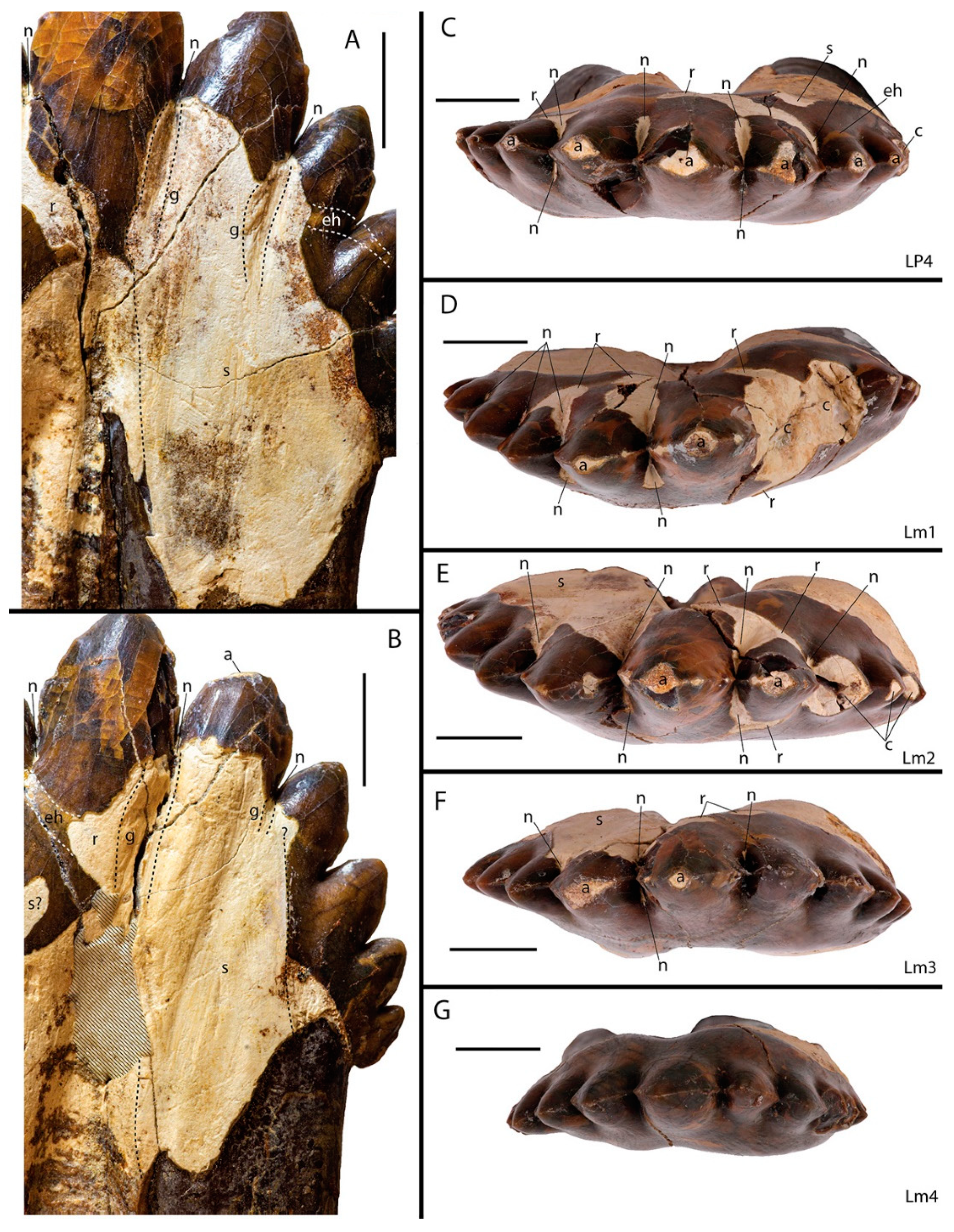
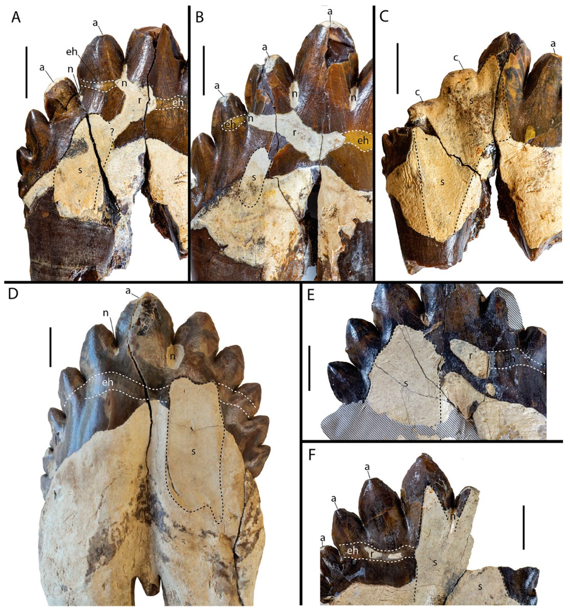
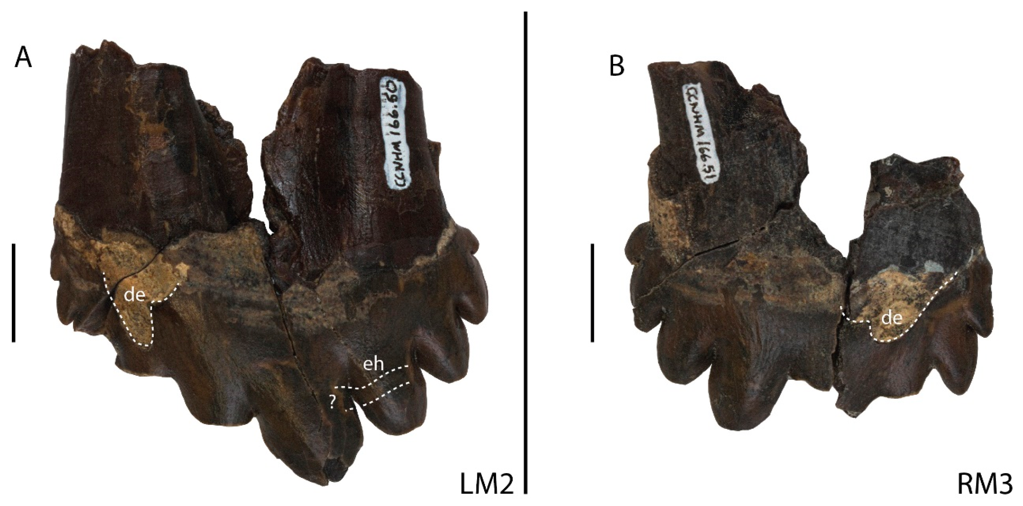
3.1.1. Types of Dental Wear in Coronodon

Apical Wear: Extreme apical wear, where the cusp is mostly, if not entirely, removed, is uncommon in Coronodon, but at least one tooth in every described adult specimen exhibits this trait. In the holotype of C. havensteini, the crown in one upper tooth has been removed by wear or a combination of spalling with subsequent wear (right P2), and the apical half of the primary cusp in the right m2 has been similarly removed. In an additional specimen of C. havensteini (CCNHM 164), the two largest mesial cusps on the right M1 have been completely removed by wear.
Apical wear is much more common in the holotype of C. planifrons (CCNHM 166), including extreme apical wear where most or all of a cusp has been removed. Of the five caniniform teeth, two have intact crowns (probable canine or first premolar) and two have crowns that have been removed by wear (probable incisors). In one of these, i.e., CCNHM 166.42, the crown has been replaced by a steeply sloping gouged wear surface that potentially received the opposing dentition. Among the postcanine dentition, the distal accessory cusps on the right and left P3 have been completely worn away. Similarly, on the left m1, the two highest mesial accessory cusps have been removed (Figure 1D). The left m2 has one mesial cusp worn off (Figure 1E).
Although one might expect a pattern of extreme wear in Coronodon that reflects the strength of occlusal forces or the frequency of impact with prey items, we see none. As the above summary indicates, extreme apical wear is rare but occurs throughout the dental row. There are other instances of missing cusps in specimens of Coronodon (particularly in upper molars of C. planifrons), but these are better described as the result of occlusal wear (see below).
Occlusal Shear Facets: Most posterior lower premolars and all lower molars, except for the m4, of Coronodon have occlusal shear facets [11,12]. These shear facets are centered near the base of the crown, on the labial side, and apical to and extending onto the distal root (Figure 1A,B and Figure 2D). As described by Boessenecker et al. [12], m4 does not have a counterpart in the upper jaw of C. havensteini or C. planifrons, thus explaining the absence of shear facets on these teeth (Figure 1G). The holotype of C. havensteini preserves only three upper postcanine teeth, i.e., the right P2, left P3, and left M2. Only M2 has an occlusal wear facet; it is a small (occupies 15% of the crown’s lingual side), oval, concave patch of exposed dentin, presumably formed by occlusion with the primary cusp of m2. Otherwise, the lingual sides of the preserved upper teeth in the holotype are intact. This is quite different from the referred specimen of C. havensteini (CCNHM 164) and the holotype of C. planifrons. In CCNHM 164, the left and right P3 bear an occlusal facet on the mesial half of the lingual face, which has removed approximately a third of the enamel. The left P4 (CCNHM 164.3) and right M1 (CCNHM 164.8) do not have clear occlusal facets, although portions of the enamel are missing, which is described below in the section entitled Side Wear. The right M2 (164–6) has a large shear facet on the lingual side, which has removed nearly half of the enamel, exposing the underlying dentin (Figure 2F).
Within the genus Coronodon, occlusal wear is most pronounced in the holotype and only known specimen of Coronodon planifrons (CCNHM 166). On the lower left m2 and m3, the occlusal facet on the labial side of the tooth has removed about a third of the enamel (Figure 1A,B). The dentin on each one bears macroscopic, elongate, apicobasal furrows and scratches. On the left m2, the occlusal shear facet is continuous with enamel removed through notch wear (see below). On the left M1, about 20% of the enamel on the lingual side of the tooth is gone and appears to have been worn off through occlusion with the m1. This is supported by the fact that there is a beveled edge of flattened and worn enamel that borders the irregular zone of exposed dentin and extends apically in two rectangular strips: one to the primary cusp and another to the highest mesial cusp. Similar and much larger shear facets occur on the lingual sides of the upper teeth, including the right M1, left M2 (Figure 2C), and right M3. These pronounced lingual shear facets have removed all mesial cusps in some teeth (CCNHM 166.34); most of the mesial cusps in others, leaving behind their bases (Figure 2C); or at least one mesial cusp, but not all of them (CCNHM 166.51).
Notch Wear: Small (typically, 1–3 mm in mesiodistal diameter and 2–5 mm in apicobasal length, but up to 10 mm in CCNHM 166.27) ovoid, areas of exposed dentine in the notches between the accessory cusps were termed “notch wear” by Geisler et al. [11]. They suggested that the enamel was removed through dental erosion, based largely on a published report of erosion in a similar position in domesticated dogs [117]. In the holotype of C. havensteini, these were observed on the lower posterior premolars and molars, typically in the highest notches, and on both labial and lingual sides [11]. Here, we note the presence of notch wear in the upper teeth as well, including in C. havensteini (CCNHM 164.8, right M1) as well as C. planifrons (CCNHM 166, right P3, left P4, and left M1) (Figure 1 and Figure 2). Typically, the notches on the upper teeth are much narrower, mesiodistally, than those on the lower teeth (Figure 2A,B,D). Under magnification, we observed that the dentin exposed in notch wear has subparallel furrows that are oriented apicobasally, especially in the lower postcanines of C. planifrons (e.g., left m1, left m2, and left m3). The furrows are subparallel because they slightly radiate out from the apical-most and narrowest part of the notch wear and, in some cases, continue basally onto occlusal shear facets (e.g., left m1, m2, and m3 of C. planifrons). The notch wear is typically deeply entrenched and, although at a smaller scale, reminiscent of the profile of a glacial-carved valley (Figure 1A,B). The edges of the missing enamel appear chipped, not dissolved, with the broken edges planar and nearly 90° to the outer surface of the enamel. Taken together, these features contradict the initial assessment of Geisler et al. [11] that this “wear” formed by dental erosion and instead suggest that the enamel was removed through impact with prey during mastication. A taphonomic origin for notch wear is contradicted by the prominent apicobasal furrows in the dentine.
Side Wear: Other areas of missing enamel and exposed dentine that cannot be ascribed to the aforementioned categories are grouped together as “side wear”. The most apparent pattern with respect to side wear is that it tends to be concentrated along a line of enamel hypoplasia that is visible in all known specimens of Coronodon except ChM PV4745. This line usually appears as a zone of discolored enamel (Figure 2) but, in some cases, appears as a zone of lower/thinner enamel under reflected light. The hypoplasia, when present, occurs on all teeth in an individual and is in similar positions in multiple specimens. On the lower molars, it typically starts in the notch between the highest and second-highest mesial cusps, is subhorizontal (with a basal dip on the labial side), and then terminates in the notch between the second and third-highest cusps on the distal side (Figure 2D). The best example of side wear being concentrated along the line of enamel hypoplasia is the left m1 of C. planifrons (Figure 1D). It occurs on both sides of this tooth, but it is more widespread on the labial side and merges with multiple areas of notch wear. The highest and second-highest mesial cusps of the m1 have been entirely removed, and the notch between these cusps is aligned with the zone of missing enamel and line of enamel hypoplasia. It is tempting to suggest that this area of weakness might have contributed to the loss of these two cusps in life. For example, on the left m2 (CCNHM 166.27), the highest mesial cusp, although largely intact, is nearly totally encircled around its base by a zone of missing enamel (Figure 1E). Some of the missing enamel can be attributed to notch wear, but other areas occur along the line of enamel hypoplasia. Additional examples of side wear being concentrated along the line of enamel hypoplasia are the lingual sides of the right M2 of C. havensteini (Figure 2F) as well as the left P4 of C. planifrons (Figure 2B).
Some of the missing enamel grouped under side wear is likely caused by occlusion with the opposing dentition, even though prominent shear facets are missing. On the lingual side of some upper postcanines and the labial side of some lower postcanines is an area of exposed dentine near the base of the crown adjacent to the mesial root. This oblong area of missing enamel extends apicodistally, forming an adjacent short (Figure 2B) or long spur of enamel (Figure 2A) along the crown base. This oblong area of missing enamel connects apically with a larger area of side wear that extends mesiodistally along the line of enamel hypoplasia on the right P3 in the holotype of C. planifrons. At least on the uppers, this smaller area of missing enamel could be a precursor to the large occlusal shear facets that often form on the mesial half of the crown (Figure 2C).
Dental Erosion: Geisler et al. [11] reported extensive dental erosion near the crown base of the labial side of the P4 and M2 in C. havensteini. Elsewhere along the toothrow of the holotype, the base of the crown is often undulatory, but convincing evidence of more extensive dental erosion is lacking. They also reported possible dental erosion in the notches between accessory cusps, but this pathology is now recognized as the result of notch wear (see discussion above). Among the more recently described species and specimens of Coronodon [12], we were only able to find two areas of missing enamel that we could convincingly ascribe to dental erosion, and both occur in C. planifrons. On the left M2 (CCNHM 166.50), there is an ovoid patch of missing enamel on the labial side of the tooth, near the crown base and close to the mesial border. The same portion of the tooth in the right M3 (CCNHM 166.51) is also missing enamel, and the simplest interpretation for both teeth is that this is due to dental erosion in a gingival pocket (Figure 3).

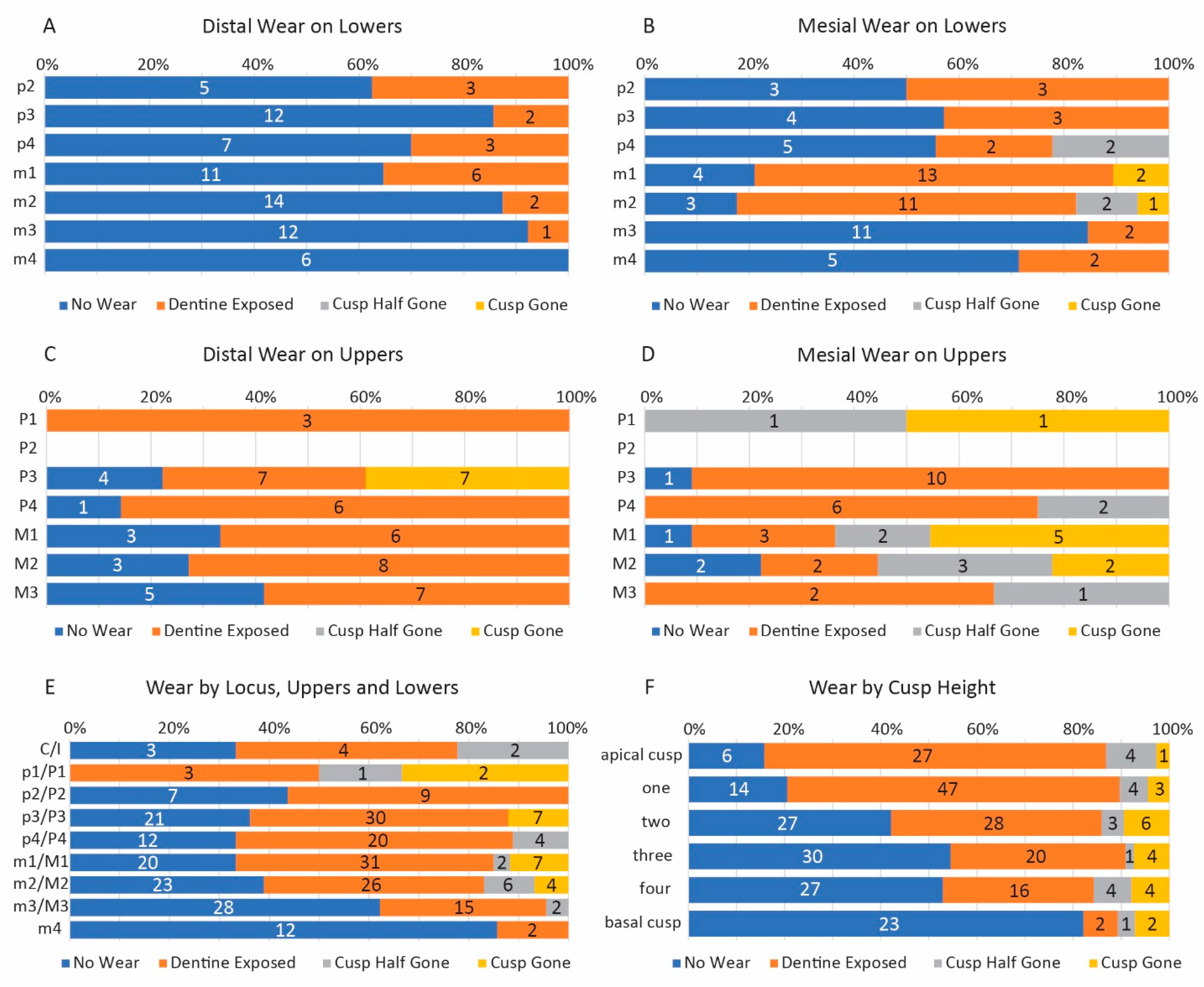
3.1.2. Statistical Analyses of Wear Patterns in Coronodon

The two species of Coronodon are significantly different in two of the three types of wear; the presence and degree of apical wear are moderately associated with species (Pearson’s Chi-square = <0.001; Cramer’s V = 0.276), as is the presence of notch wear between cusps (Pearson’s Chi-square = <0.001; Cramer’s V = 0.223), but not the presence of side wear on each tooth (Pearson’s Chi-square = 0.201) (Table 1). Inspection of the contingency tables and z-tests shows that C. planifrons has more wear than C. havensteini, and the significant differences are concentrated in the frequency of apical cusps that have no visible wear, the frequency of cusps that are entirely gone, the frequency of notches without wear, and the frequency of notches that exhibit wear on the labial and lingual sides. Given these differences, the species were also analyzed separately in the binomial logistic regressions described below. These binomial regression analyses support a distinction between the species with respect to notch wear (significance = <0.001) but not with respect to apical wear (significance = 0.365) (Table 2). These logistic regressions were able to explain between a third to half of the variation in wear, with the most variation explained in the test of side wear in C. havensteini (Cox and Snell R2 = 0.537 and Nagelkerge R2 = 0.725) and the least variation explained in the test for side wear in C. planifrons (Cox and Snell R2 = 0.234 and Nagelkerge R2 = 0.316).
Statistical support was found for an association between the degree (Chi-square or Spearman rank correlation) or the presence/absence (binomial logistic regression) of apical wear and the position of a tooth in the toothrow (Pearson’s Chi-square = <0.001; logistic regression = 0.014; Figure 4E); whether a cusp was a mesial, distal, or primary cusp (Pearson’s Chi-square = <0.001, Cramer’s V = 0.253; logistic regression = <0.001); and the height of a cusp (Pearson’s Chi-square = <0.001, logistic regression = <0.001). Unexpectedly, we also found an association with whether a tooth was in the upper or lower jaw (logistic regression = <0.001) (Table 2). If upper and lower teeth are tested separately, mesial cusps are associated with more wear than distal cusps on both (upper = 0.006, Cramer’s V = 0.249; lower = <0.001, Cramer’s V = 0.35; Figure 4A–D); however, the contingency table shows that in the uppers, this signal is driven largely by wear that removed half or all of a cusp. For some teeth (i.e., CCNHM 166, RM1, LM2, RM3), the removal of the cusp could be due to occlusal, not apical, wear; removing the mesial cusps of these teeth from the analysis significantly reduces this association (Pearson Chi-square = 0.954). The contingency tables, z-tests, and odds ratios (ORs) show that apical wear is less likely on distal teeth (0.796 OR, Spearman correlation −0.196), that mesial cusps have more apical wear than distal cusps (OR = 6.43), and that primary cusps are more worn than both, but with different degrees of statistical significance between tests (mesial to primary cusp comparison, logistic regression significance = 0.106; Chi-square z-tests indicate significant differences between mesial and primary cusps for distributions of “no wear” and wear where cusp is half or all gone). Lower cusps (i.e., those closer to the base of the crown) are associated with less apical wear than higher cusps (OR = 0.513; Spearman correlation = −0.306; Figure 4F), and upper teeth are associated with more apical wear than lower teeth (OR = 8.112). Not surprisingly, side (left or right) is not associated with the presence of apical wear (logistic regression = 0.248). If samples are restricted to species, similar results are found, although no association is found between the position in the toothrow and apical wear in C. havensteini (significance in logistic regression = 0.348) (Table 2).
Strong statistical support was found for an association of side wear and whether that side is an occlusal side (Chi-square = <0.001, Cramer’s V = 0.636; logistic regression = <0.001); specifically, wear was more common on occlusal sides (Table 1 and Table 2). As noted above, side wear includes areas of missing enamel that could not be directly attributed to occlusion with an opposing tooth. The binomial logistic regression did not support an association between side wear and the position in the toothrow (significance = 0.156), whether the tooth is an upper or a lower (significance = 0.211), or right or left side (significance = 0.69). Analyses that restrict the sample to individual species do not meaningfully differ from the analysis that groups both species together.
The presence of wear in notches between cusps was associated with the amount of apical wear on adjacent cusps, specifically the average of the two (logistic regression = <0.001), as well as with the height of the notch (logistic regression = <0.001) (Table 2). Similar results are found in Spearman rank correlations that treat notch wear as a 3-state ordinal variable (i.e., absent, present on one side, or present on labial and lingual sides): an association was found with apical wear (0.330; significance = <0.001) as well as with notch height (−0.492; significance = <0.001) (Table 1). An inspection of bar charts generated by SPSS shows that notch wear, when present, is more likely to occur on both sides of the tooth and is positively correlated with the amount of apical wear. A binomial logistic regression did not find an association with position in the toothrow (significance = 0.761), whether a tooth is an upper or a lower (significance = 0.098), side (significance = 0.552), or whether a cusp is the primary cusp or positioned on the mesial or distal side of the crown (significance = 0.226). We also found support for an association between the presence of notch wear on occlusal and non-occlusal sides of a tooth (Pearson Chi-square = <0.001. Cramer’s V = 0.543), with the contingency table indicating that wear on one side typically co-occurs with wear on the other (i.e., labial and lingual) (Table 1). Analyses at the species level showed a significant association between apical wear and notch height, and an association was also found with position in the toothrow in C. planifrons only (logistic regression = 0.012) and with whether the tooth is in the upper or lower jaw in C. havensteini only (logistic regression = 0.029). In C. planifrons, notch wear is more common on mesial teeth, whereas in C. havensteini, notch wear is more common on the lower teeth.

3.2. Evolution of Feeding Behaviors in Toothed Mysticetes

3.2.1. Scores for Extant and Extinct Taxa

An inspection of scores for the evidence of different feeding behaviors across all taxa included in the study by Boessenecker et al. [12] reveals important generalizations and overall patterns. The range between the lowest and highest scores for a particular feeding behavior indicates the total amount of evidence that can be brought to bear on a given hypothesis. Not surprisingly, raptorial feeding can most easily be evaluated (observed range of 5.33 to −3) as nearly all evidence comes from teeth, which are commonly preserved elements in the fossil record (Table 3). However, not far behind is evidence for and against baleen filtration, with a maximum score of +4 and a minimum score of −3. The spread of scores is much smaller for dental filtration (+2 to −3), reflecting in part the very limited diversity and disparity of extant taxa that employ this feeding strategy. Evidence for and against suction feeding is most limited, with an observed range of just +2 to −1. From a testability standpoint, these ranges indicate that a hypothesis of suction feeding in an extinct taxon is the hardest to test (minimum of −1), whereas a hypothesis of raptorial feeding is the easiest to corroborate (maximum of 5.33). The scores themselves are not independent because many features that support one feeding hypothesis concurrently contradict others. This is most apparent with raptorial feeding versus baleen filtration; scores for these hypotheses are inversely correlated for two characters (i.e., characters 9 and 10), and taxa that have negative scores for raptorial feeding typically have positive scores for baleen filtration. A more complex relationship occurs between raptorial feeding and dental filtration. Both behaviors require teeth and thus scores in some taxa are inversely correlated with the score for baleen filtration; otherwise, these hypotheses are associated with different morphologies.
Of the 127 taxa included in the study by Boessenecker et al. [12], 28 taxa could not be scored for any of the 14 characters related to feeding behaviors, and thus, the feeding scores could not be calculated. The vast majority of these are chaeomysticetes, but this list also includes Chonecetus sookensis and Aetiocetus tomitae. Two other taxa, i.e., Ashleycetus planicapitis and Miocaperea pulchra, have a score of 0, even though they could be scored for one character each. For the remaining 84 taxa, the scores appear to be largely driven by the characters themselves and are not distorted by missing data. The strongest evidence for raptorial feeding (i.e., >+4) occurs in the three basilosaurids as well as in Kekenodon onamata (Table 3), a taxon thought to be the most basal neocete or the most derived archaeocete [14]. Moderate evidence for raptorial feeding (+2 to +4) is seen among a few taxa, including the mysticetes C. havensteini (+1 to +2.33), C. planifrons (+2.08 to +2.33), the unnamed coronodonid represented by ChM PV5720 (+3.33), Janjucetus (+2.33), and an unnamed taxon represented by ZMT 62 (+2 to −2), as well as the odontocetes Agorophius (+2.33 to +3.33), Ankylorhiza (+2.33), Physeter (+2.33), and Xenorophus sloanii (+3). Other taxa with weak positive evidence for raptorial feeding (i.e., + 0.5 to +2) include the toothed mysticetes Borealodon (+1.5 to +0.75), Aetiocetus cotylalveus (+1.33), Fucaia buelli (+1), and Mystacodon selenesis (+1). The strongest evidence for dental filtration occurs in Coronodon; C. havensteini has a score of +2 and some optimizations of C. planifrons also yield a score of +2. Many other toothed mysticetes (e.g., Janjucetus, Fucaia goedertorum, and Morawanocetus) and some eomysticetids that may have anterior teeth but lack posterior ones (e.g., Tokarahia) [2,3] have a weak score of +1. Strong evidence for baleen filtration (i.e., +4) was calculated for all extant mysticetes in the matrix as well as numerous extinct taxa, including Pelocetus calvertensis, Herpetocetus morrowi, Aglaocetus moreni, and Parietobalaena palmeri. Most other chaeomysticetes with lower scores for baleen filtration (i.e., +2 to +4) are represented by partial remains where one or more key character states that support baleen filtration could not be coded. In terms of eomysticetids, taxa better represented in the fossil record have scores of + 3.16 for baleen filtration (e.g., Yamatocetus, Waharoa ruwhenua, and Maiabalaena) whereas taxa represented by more fragmentary remains have lower scores (i.e., +1.66 to +2.66), including Eomysticetus whitmorei, Tokarahia lophocephalus, and Matapanui waihao.
Overall, there was little craniodental evidence in support of suction feeding. The taxa with the highest scores of +2 are Mammalodon colliveri and Mystacodon selenensis (Table 3). Other taxa with some support (i.e., +2 to +1.75) include Janjucetus, Coronodon newtonorum, Metasqualodon symmetricus, Mammalodon hakataramea, Basilosaurus, Ziphiidae, and Matapanui waihao. Not all feeding strategies are mutually exclusive, and some extant raptorial feeders also engage in dental filtration (e.g., [37]), whereas others engage in suction feeding or behaviors that combine aspects of raptorial and suction feeding [118]. Some extinct taxa display evidence for multiple types of feeding. For example, in Coronodon havensteini, there is evidence for raptorial feeding (+2.33 to +1) and dental filtration (+2), supporting the inference by Geisler et al. [11] that this taxon engaged in both types of feeding. In eomysticetids that have putative anterior teeth (e.g., Tokarahia) as well as in the aetiocetid Fucaia goedertorum, there is evidence for dental filtration and baleen filtration, although the evidence for the latter is stronger than the former (Tokarahia +2.5 to +2 vs. +1; F. goedertorum +1.5 vs. +1).
Some taxa, even though they could be coded for most characters in our matrix, are particularly challenging to interpret. Llanocetus, which can be coded for 12 of the 14 characters, has scores that vary from −1.25 to +1 for the different feeding inferences (Table 3). Previously, Llanocetus was interpreted as a raptorial or suction feeder [26], although the score for the former is just −1.25 to +0.33, and the score for the latter is 0 to +0.25. Dental filtration scores are not any better, with a range of −1 to +1.25. The best evidence, albeit quite weak, is for baleen filtration in Llanocetus, which has a score of +1. This interpretation is consistent with some previous suggestions [4,13], and it has important implications depending upon where Llanocetus falls in mysticete phylogeny. If it is fairly basal, as is the case in many recent phylogenetic hypotheses (e.g., [10,26]), then this implies baleen evolved quite early and was widespread among toothed mysticetes. At a minimum, the presence of baleen would indicate that baleen and teeth co-occurred in this taxon, as has been suggested for aetiocetids [17,18]. Other taxa that score low for all feeding hypotheses include the toothed mysticetes Metasqualodon and Borealodon. The best evidence for Borealodon is that it was a raptorial feeder, but with a score between +1.5 to +0.75, the evidence for this behavior is quite meager. Metasqualodon scores highest on the suction feeding score, but only with a score of +1.

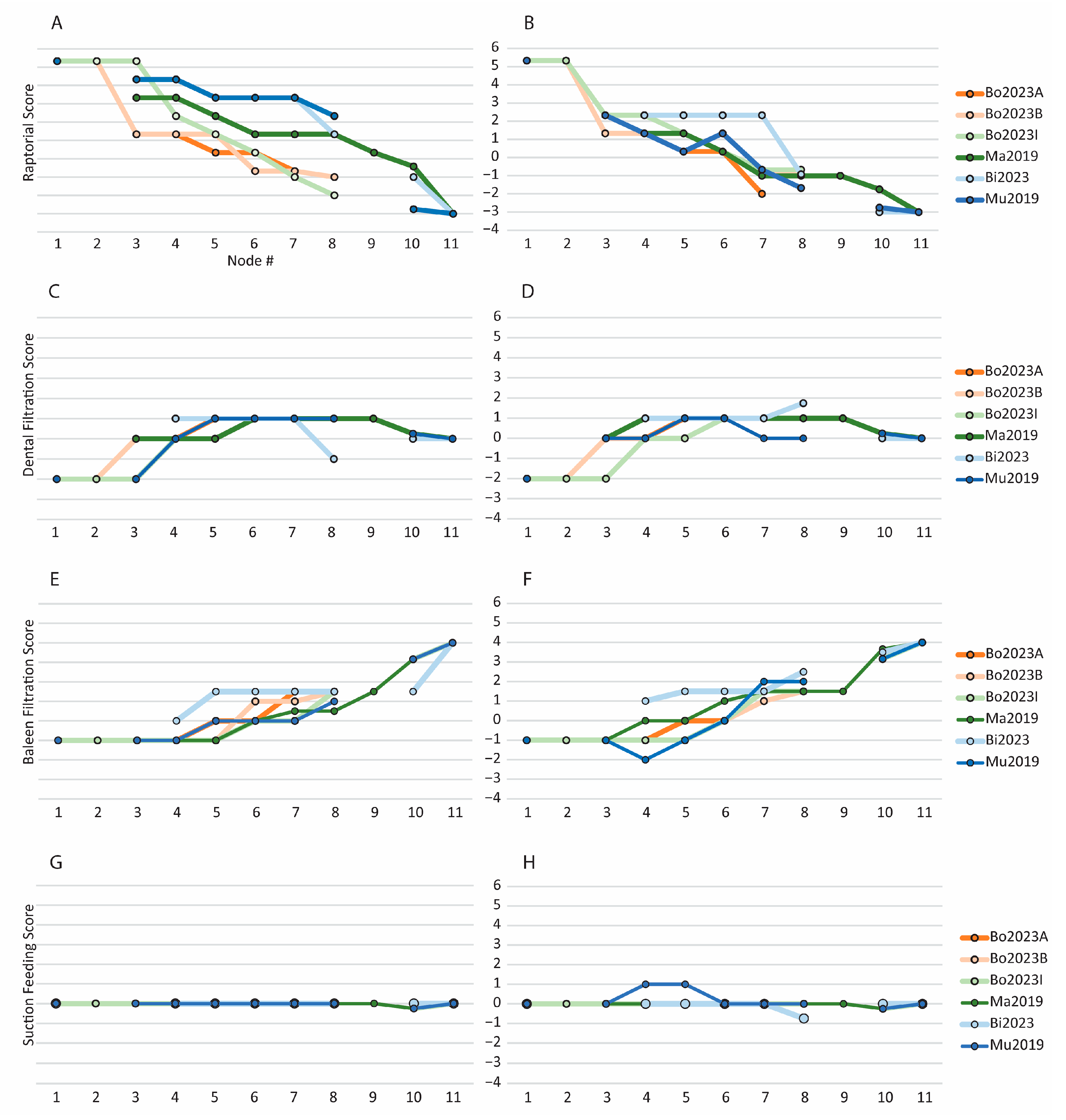
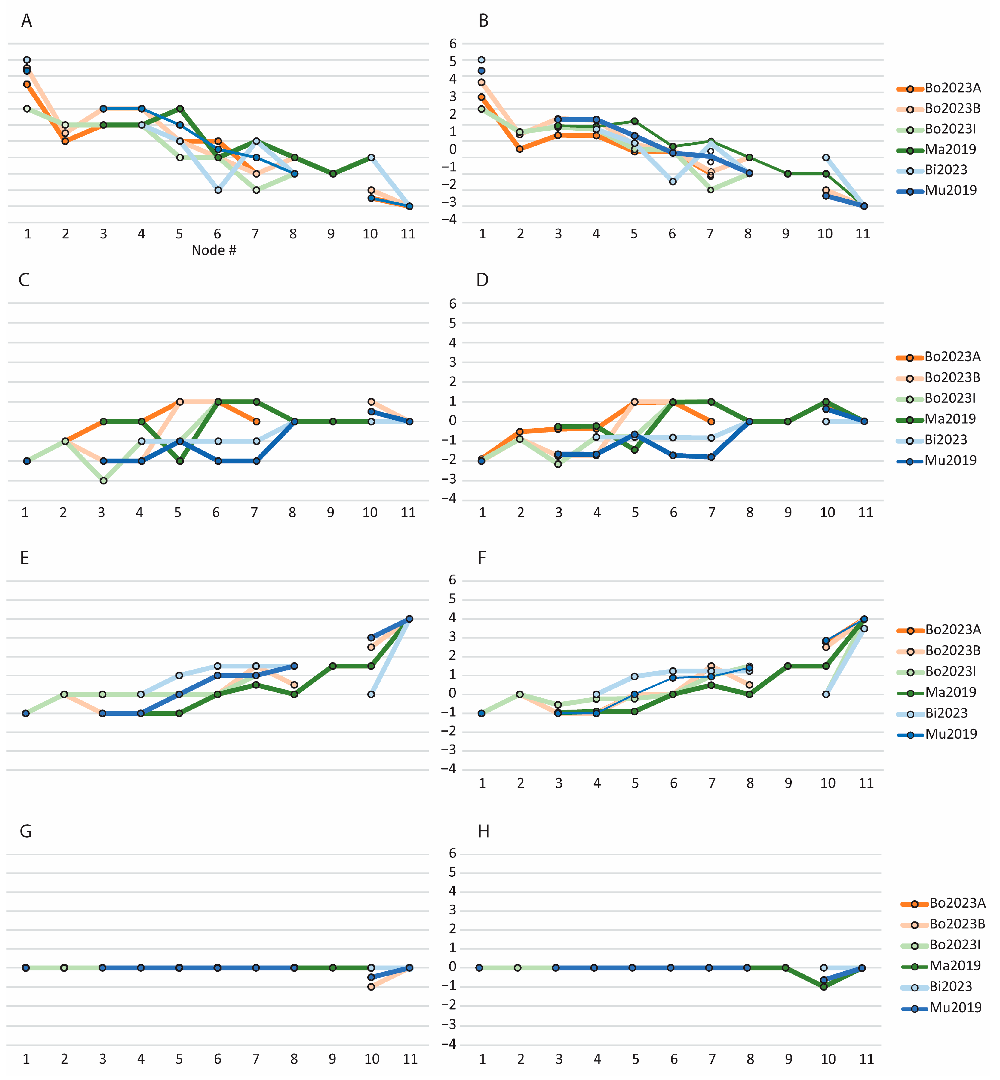
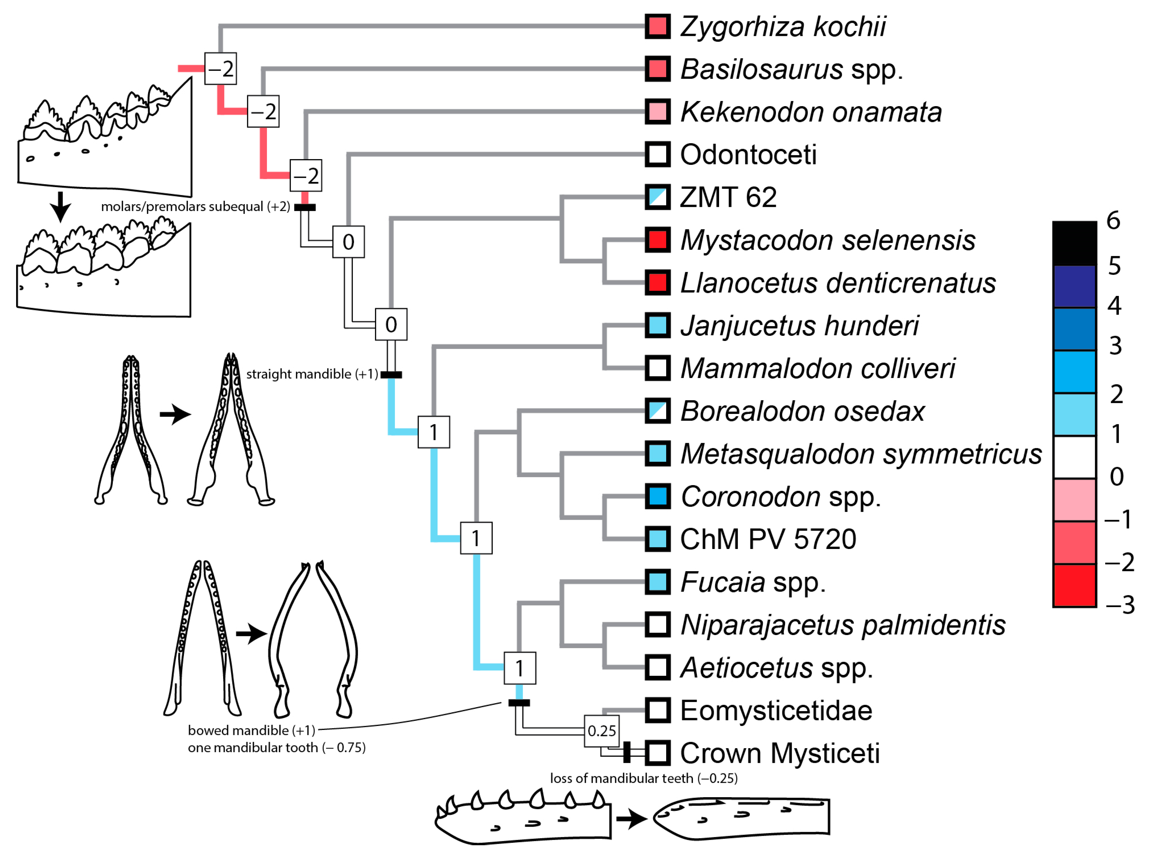
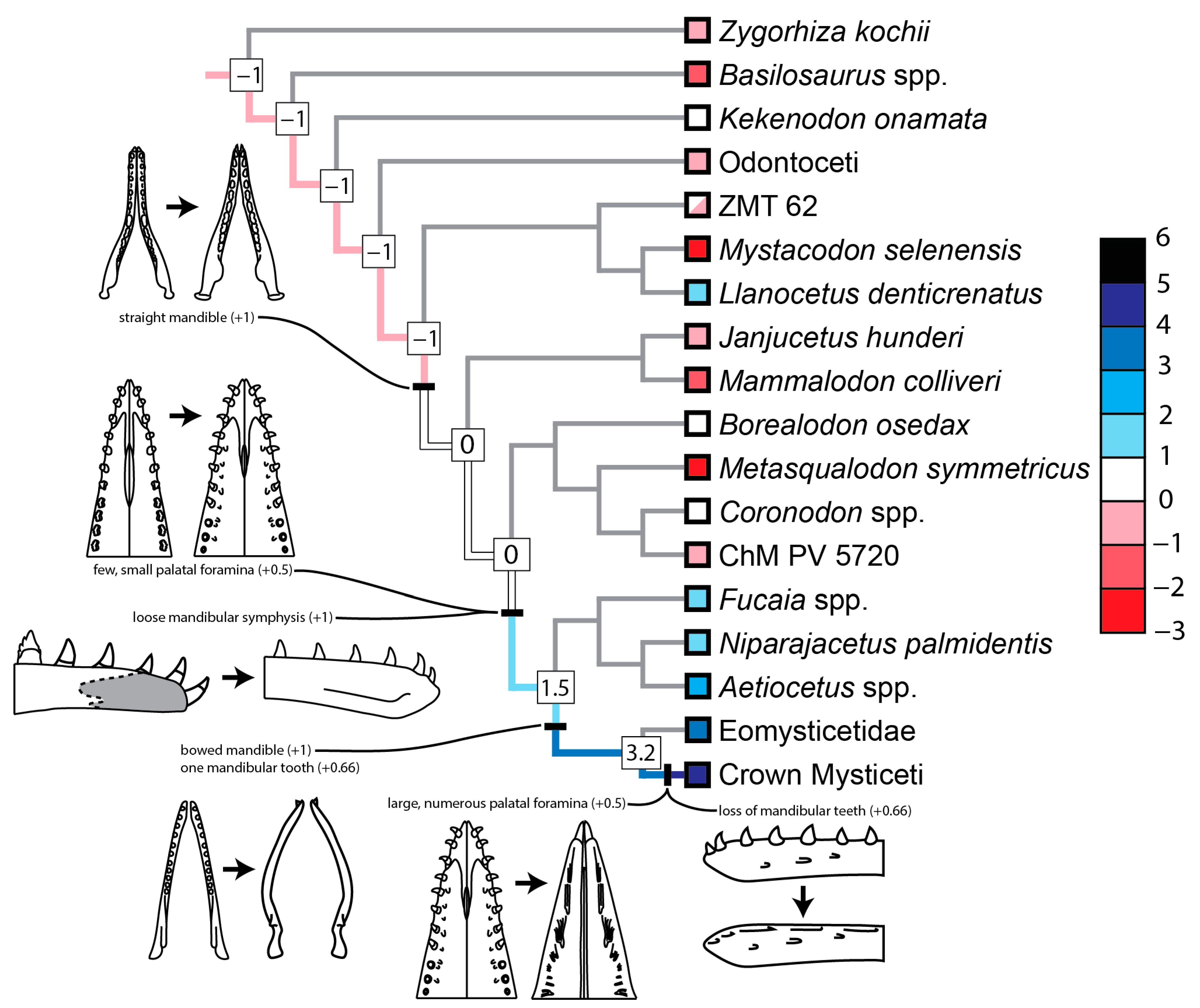
3.2.2. Scores of Reconstructed Ancestral Toothed Mysticetes

When the raptorial scores for the hypothetical ancestors across all phylogenetic hypotheses are plotted, there is, not surprisingly, an overall pattern of stepwise decreases in raptorial scores from the root to the origin of the mysticete crown group (Figure 5A,B and Figure 6A,B). When using parsimony to optimize characters on the trees of Muizon et al. [13] and Bisconti et al. [113], there is a noticeable pause and plateau in the decrease in raptorial scores along the mysticete stem before the scores decline again. This plateau is not supported by maximum likelihood reconstructions; it is replaced by a progressive decline on the tree of Muizon et al. [13] and a prolonged interval of alternating increases and decreases on the tree of Bisconti et al. [113]. When optimizing traits using parsimony, the pace of decline is fairly consistent across different optimizations on the trees of Boessenecker et al. [12] and Bisconti et al. [113], whereas the rates of decline vary substantially with different optimizations on the trees of Marx et al. [112] and Muizon et al. [13]. The raptorial scores derived from maximum likelihood reconstructions are more similar than those under parsimony, and with this method, reconstructions on the trees of Marx et al. [112] and Muizon et al. [13] are very similar to the other four trees (Figure 5A,B and Figure 6A,B). For the hypotheses that include odontocetes (e.g., [12]), trends in decreasing raptorial scores begin well before the origin of Mysticeti, regardless of the method for reconstructing ancestral states.
The evolutionary changes in dental filtration scores vary substantially across the trees examined. Under parsimony, most trees support an early increase in dental filtration score, followed by a slight decline that coincides with a reduction in teeth in Chaeomysticeti (Figure 5C,D), although it should be noted that the majority of the early increase in scores involves a reduction in evidence against dental filtration (i.e., change from −2 to +1), not strong evidence for dental filtration. The timing of this increase varies; the tree of Bisconti et al. [113] has the rise occurring at the base of the tree, the tree of Muizon et al. [13] places it somewhat later, and the implied weighting trees of Boessenecker et al. [12] have a rise even later. One commonality among all phylogenetic hypotheses examined is that this rise occurs at the branch where Mammalodontidae and/or Coronodonidae branch off the mysticete stem, and the magnitude of the rise is largely the result of the relative placements for Coronodonidae and Mystacodon and their mandibular morphologies. Concave mandibles are considered evidence against dental filtration (−1), whereas straight mandibles are considered neutral (0). Coronodon has straight mandibles but the unnamed coronodonid represented by ChM PV5720 has concave mandibles; thus, the state reconstructed at the base of Coronodonidae varies in different trees depending on the morphology in taxa in adjacent branches. When coronodonids are more apical, straight mandibles evolve one branch lower, where mammalodontids diverge off the stem of crown Mysticeti (Figure 7). However, when Coronodonidae is in a more basal position, the straight mandibles in Coronodon are considered convergent with those of other toothed mysticetes (i.e., IW trees in [12]). Scores calculated using reconstructions from maximum likelihood vary substantially (Figure 6C,D). At one extreme is the reconstruction on the tree of Bisconti et al. [113], where the dental filtration score appears flat, although, upon closer inspection, there are two well-separated increases of one point each, with a starting score of −2 and a final score of 0. At the other extreme is the reconstruction on Tree B of Boessenecker et al. [12], where there is an abrupt increase of 3 points (from −2 to +1) in the dental filtration score between nodes 4 and 5, the interval when Coronodonidae and Mammalodontidae branch off the mysticete stem. Other reconstructions lie between these extremes, and when taken together, no simple generalization is possible. That said, it is clear that the early increase in dental filtration score supported by parsimony is not supported by maximum likelihood.
With respect to baleen filtration scores reconstructed using parsimony, for the majority of studies, the baleen filtration scores increase, with occasional plateaus, until they reach a maximum score of + 4 (Figure 5E,F), at the node corresponding to crown Mysticeti. The remaining increases along the stem leading to crown mysticetes reflect morphological changes observed in some aetiocetids, including the presence of palatal foramina and the development of a loose mandibular symphysis (Figure 8). A notable outlier to the patterns described above for the evolutionary change in the baleen filtration score is the consensus tree of Bisconti et al. [113]; there, the baleen filtration score increases to +1 and/or +1.5 quite early (i.e., near the base of the tree). This result is driven by the relatively basal (at least compared to Llanocetus and Coronodon) positions of the aetiocetids Fucaia and A. weltoni.
Baleen filtration scores based on maximum likelihood reconstructions are largely similar to those derived from parsimony, with a few minor exceptions (Figure 6E,F). On some trees (i.e., Tree B and the implied weighting tree of Boessenecker et al. [12], tree of Bisconti et al. [113]) there is a drop in the baleen filtration score right before the origin of crown Mysticeti. This drop is an artifact of missing data; whereas parsimony will often reconstruct a state despite missing data as long as no additional length is required, maximum likelihood cannot reconstruct the state for some characters because of missing data in one or both descendent lineages. Finally, if one compares the maximum likelihood scores with and without weights, the former appears to be more clustered, similar to that observed for the weighted scores for raptorial feeding.
Overall, suction feeding scores are fairly flat across most trees and most optimizations; they hover near zero, indicating little evidence for or against this feeding behavior (Figure 5 and Figure 9). The subset of optimizations that maximize scores for feeding behaviors other than raptorial feeding (i.e., dental filtration, baleen filtration, and suction feeding) show slightly more fluctuations along the mysticete stem. The most notable optimization occurs on the consensus tree of Muizon et al. [13], where the suction feeding score increases from 0 to +1, stays at this level for one node, and then drops back down to zero (Figure 5H). This reconstruction along the mysticete stem is the result of the fact that Mystacodon and Mammalodontidae are positioned as sequential stem taxa to crown Mysticeti. Both lineages have evidence for suction feeding; members of both clades have crowns that have been nearly entirely worn down, and mammalodontids have a very short rostrum. These features, coupled with the phylogenetic arrangement of Muizon et al. [13], support a brief interval of suction feeding before evidence for dental filtration and baleen filtration appears. However, it is important to recognize that this is optimization-dependent, and reconstructions that favor raptorial character states at these internal nodes do not support suction feeding (i.e., the score is 0). Furthermore, scores derived from maximum likelihood show no evidence for any increase in suction feeding scores, and in fact, for several trees, there is a decrease in the suction feeding score at the node preceding the mysticete crown group (Figure 6G,H). This decrease occurs at the node where laterally bowed mandibles evolve, which increases the oral fissure and is thus considered evidence against suction feeding.
In addition to focusing on changes in the evidence for individual feeding behaviors across different trees and under different optimizations, some aspects of the relative timings of these changes merit mention. On all trees and optimizations (both parsimony and maximum likelihood), the raptorial score is, on average, inversely correlated with the baleen filtration score (Figure 5A,E and Figure 6A,E). This is not surprising given that many character states that are considered evidence for raptorial feeding are evidence against baleen filtration and vice versa. On four of the six trees, when parsimony is used for character state reconstructions, support for dental filtration increases before evidence for baleen filtration (Figure 5C,E), whereas on the consensus tree of Bisconti et al. [113], support for both increases at the same node or is optimization-dependent. When maximum likelihood is used instead of parsimony, dental filtration scores do not form a distinct peak or the peak is more delayed than as seen in parsimony reconstructions. As a result, it appears that dental filtration scores increase at the same time as baleen filtration scores (Figure 6C,E). Evidence for baleen filtration does not precede that for dental filtration on any tree, regardless of the reconstruction method. Overall, the scores for the different feeding hypotheses are fairly similar at the middle nodes along the mysticete stem, indicating that it is difficult to determine the feeding behaviors of these extinct toothed ancestors (Figure 10). Although a stronger signal is preferred, this simply reflects the transitional morphology of many toothed mysticetes relative to basilosaurids on one end and extant mysticetes on the other.

4. Discussion

4.1. Feeding Behaviors of Coronodon

4.1.1. Suction Feeding Hypothesis for Coronodon

With the recent description of new specimens and species of Coronodon [12] and the detailed observations and analyses reported here, we are in an excellent position to revisit the feeding behavior of Coronodon. In the original description, Geisler et al. [11] suggested that Coronodon havensteini engaged in dental filtration and raptorial feeding. Although they cited evidence for both types of feeding, they focused more on the former because this was the more extraordinary claim, one that is challenging because of the limited diversity of extant mammals that employ this behavior. In a later paper on the feeding of toothed mysticetes, Hocking et al. [23] discussed Coronodon and found corroborating evidence for raptorial feeding, argued against dental filtration, and further suggested that suction feeding was employed by Coronodon. Hocking et al. [23] provided two observations as possible evidence for suction feeding in Coronodon havensteini. First, they noted that the straight-sided rostrum would have increased the size of the oral cavity and that a larger oral cavity is correlated with suction feeding. Although such a relationship has not been demonstrated among cetaceans, pronounced suction in anurids is correlated with hypertrophic oral cavities [119]. However, the most important factor in suction feeding is the size of the oral cavity in relation to the oral commissure [59], not the absolute size of the oral cavity. A large oral cavity with a small commissure generates strong suction, whereas a large commissure generates diffuse suction [59]. Although rostral width contributes to a large oral cavity in Coronodon, the rostrum is still long, with a relative length similar to that of basilosaurids. Thus, even small rotations at the craniomandibular joint will lead to a very large oral commissure. The same could be said for ziphiids, which have long rostra but are obligate suction feeders; however, in ziphiids, the shape of the mandible with teeth often restricts the oral commissure, and the small temporal fossa is consistent with restricted rotation of the craniomandibular joint [59,120]. The large temporal fossa, anteriorly shifted coronoid process [121,122], apical wear on the molars, and length of the occlusal shear facets on the posterior teeth all point to substantial rotation of the craniomandibular joint in Coronodon. Thus, there is no evidence that Coronodon could shape its oral commissure to engage in suction feeding, and the presence of largely intact anterior caniniform teeth in several specimens suggests that the oral commissure was partially blocked. In several extant suction feeders, pronounced wear occurs on the lingual and/or apical parts of the anterior teeth and presumably develops when prey is sucked into the mouth through an oral commissure that is partially bordered by these teeth [11,27]. Finally, a much stronger relationship has been found in cetaceans between a large oral cavity and filter feeding in cetaceans [123], and this interpretation is followed in the present study.
The second line of evidence for suction feeding in Coronodon cited by Hocking et al. [23] is the highly emergent toothrow and thick gingiva, suggesting that these features would seal the lateral side of the oral fissure. The main evidence for thick gingiva is the zone of dental erosion on the labial side of the upper teeth in the holotype C. havensteini [11], which presumably formed in deep gingival pockets, although, as described in the present study, only limited dental erosion is seen in additional specimens of this genus. Regardless, other aspects of the teeth make a gingival seal highly unlikely. A major focus of Geisler et al. [11] was the presence of interdental slots in the lower toothrow, which they interpreted as being used for filter feeding. One could hypothesize that these interdental slots were filled with gingiva, but then, this would not explain the apical wear on many of the basal cusps that border these interdental slots. If the gingiva extends far up onto the crowns and fills part of the slots, a seal seems possible when the teeth are fully occluded. However, as described in the preceding paragraph, if such a seal existed, it would quickly become undone as the jaw opens due to the length of the rostrum. More importantly, we are not aware of any extant taxon that exclusively uses gingiva to seal the mouth during suction feeding. Instead, in cetaceans, the lips are used for this purpose [59,61,124,125]. Thus, the only likely way Coronodon could have been an effective suction feeder would be if it had large mobile lips (as in extant Delphinapterus and Monodon), features for which there is no fossil evidence.
Although we find the putative suction-feeding characters in Coronodon that Hocking et al. [23] mention to be unconvincing, there are other features in marine mammals that are associated with suction feeding. The most important of these is a short rostrum, which is a feature of many, but not all, cetacean suction feeders [59]. Another feature is the presence of glossowear and associated hydrowear [27], although neither type of wear is observed in any specimen of Coronodon. A short rostrum is observed in Coronodon newtonorum but not in other species in the genus. As a result, our calculated scores for suction feeding are +1 for C. newtonorum, 0 to +0.25 for C. planifrons, and 0 for C. havensteini. Thus, there is some limited evidence, from rostral proportions, that C. newtonorum had some specializations for suction feeding. However, this conclusion should be viewed cautiously. The RPI for C. newtonorum is 1.2, very close to the threshold between character states 1 and 2 but only slightly lower than that for C. havensteini (1.28).

4.1.2. Dental Filtration Hypothesis for Coronodon

Geisler et al. [11] listed three features that support the hypothesis that Coronodon engaged in specialized dental filtration: (1) the posterior premolars are similar in size and shape to the molars, (2) the accessory cusps are large relative to the primary cusp and project radially from the base of the crown, and (3) the basal mesial cusps of the posterior teeth are worn, notably more so than the basal distal cusps. Boessenecker et al. [12] also noted that the mandibular symphysis in Coronodon is undulatory, not rugose, suggestive of a degree of mobility greater than that inferred for basilosaurids but much less than the loose symphysis observed in all extant mysticetes. The rationale for why large accessory cusps and subequal premolars and molars are evidence for dental filtration is described under Character Selection above, and these features, which are fairly widespread among toothed mysticetes, are responsible for the peak of +1 in the dental filtration score along the mysticete stem. With respect to the mandibular symphysis, we have coded the symphysis of Coronodon to be the same as those of most archaeocetes, even though there appears to be a difference in the rugosity of the symphyseal surfaces. This is a subtle but potentially important difference that should be examined more thoroughly in future studies, potentially in a quantitative way.
Geisler et al. [11] interpreted the prevalence of apical wear on the basal mesial cusps of Coronodon as evidence of dental filtration. Although such wear occurred on some of the distal cusps in the holotype, it was more common on the mesial cusps, including the most basal cusps. The more perplexing fact was that on most lower teeth, the highest distal cusps, which approach the main cusp in height, have no visible wear, whereas the sheltered mesial basal cusps typically have wear. These basal cusps are sheltered because the lower molars overlap, with the distal end of the m1 labial to the mesial end of the m2 and so forth down the toothrow. If this wear was caused by contact with prey, then one would expect the highest cusps to be the most worn and the basal cusps to be the least worn. Geisler et al. [11] cited this unexpected pattern as evidence for their hypothesis that Coronodon fed using dental filtration. Specifically, they hypothesized that after Coronodon engulfed prey-laden water, it expelled water, but not prey, through the slots between the lower teeth. Most prey could not exit through the slots, were trapped by the mesial cusps, and often contacted and caused wear on these cusps. There is no direct extant analog with this behavior, but they cited passive mesial wear in ziphiids as having a similar cause. In these beaked whales, the mesial side of the crown is worn only at the oral fissure, which likely formed as prey inadvertently contacted and wore the teeth as they were sucked into the oral cavity [11]. As noted by Heyning and Mead [120], the large lower teeth of some ziphiids help form the border of the oral fissure during suction feeding.
The new specimens of Coronodon described by Boessenecker et al. [12] largely confirm the wear patterns seen in the holotype of C. havensteini. The mesial cusps of the lower premolars and molars are typically more worn than the distal cusps (Figure 1E and Figure 4A,B), including the basal cusps of the molars, which are nestled medial to the crowns of the preceding tooth. As demonstrated in the present study, this difference is statistically significant, with the most notable differences being that many more distal cusps show no visible wear and that many more mesial cusps have wear with half or more of the cusp missing (Table 1 and Table 2 and Figure 4). Although far more lower teeth are preserved than upper teeth in the holotype of Coronodon havensteini (13 to 3), these recently described new specimens and species of Coronodon include more upper teeth (21 teeth in total). Unexpectedly, these upper teeth also show the same wear pattern as the lower teeth, where the mesial cusps are, on average, more worn than the distal cusps (Figure 4C,D). In some cases, this can be explained by occlusal wear instead of attritional progressive apical wear, particularly when a large shear facet extends apically up the crown. This occurs on some of the upper teeth and disproportionately affects the mesial cusps on the uppers because the shear facets are located on the mesial half of the crown (Figure 2C). If these mesial cusps are removed from our statistical analyses, the significant association of mesial cusps being more worn than the distal cusps no longer holds for the upper dentition. This underscores the importance of finding additional specimens of Coronodon (as well as all other taxa for which dental wear features are used to interpret feeding behaviors); specifically, younger individuals that preserve the upper teeth and have fairly minor occlusal wear and well-developed apical wear.
While exploring the literature for other examples of a prevalence of wear on mesial cusps, we came across the dental wear of Dorudon, as described by Uhen [56]. As in Coronodon, Uhen [56] found that the mesial cusps are consistently more worn than the distal cusps, and this pattern occurs on the upper and lower teeth. Uhen [56] explained this wear pattern as being caused by prey contact during raptorial feeding, an interpretation supported by two observations: (1) that the apices of the uppers and lowers do not contact when the mouth is closed and (2) the wear is most pronounced on the main cusp and progressively decreases on successively more basal cusps. Furthermore, the wear facets on the mesial cusps in Dorudon are angled to face labially and slightly mesially, indicating that the wear is modulated by the location of the uppers, which, in Dorudon, alternate with the lowers during occlusion and are situated in a slightly more labial position. With this pattern of occlusion, any prey that was trapped between the upper and lower teeth would likely cause wear on the labial sides of the latter.
Hocking et al. [23] provided two hypotheses for the prevalence of wear on the mesial cusps in Coronodon: (1) that the mesial cusps contacted large prey during suction feeding, and (2) that the wear occurred during biting, specifically when portions of prey were forced into the narrow gaps between the lower molars (their preferred hypothesis). Given the observations in Dorudon, which we were not aware of in the original description of C. havensteini [11], and the fact that this pattern occurs on the lower and upper teeth of Coronodon, we now consider the second explanation of Hocking et al. [23] to be the best supported. However, this hypothesis does not adequately explain why wear is so rare on the highest distal cusps, which would be expected to regularly contact prey. One possibility is that the apical wear on mesial cusps primarily formed during the capture of large vertebrate prey; the mouth would be open as the whale accelerated and then drove its teeth into the prey’s body (i.e., ram feeding). Under this scenario, it is more likely that the mesial cusps, compared to the distal cusps, would have encountered bone during this initial bite. Given the very limited taxonomic distribution of this wear pattern (currently, only Coronodon and one basilosaurid) and the absence of strong extant analogs, we have chosen not to include this in our table of evidence for feeding behaviors. A broader survey of patterns of apical wear in fossil cetaceans with multi-cusped teeth may shed further light on the origin of this wear.
Hocking et al. [23] cited the stable isotope data of Clementz et al. [126] as evidence that Coronodon was a raptorial feeder that consumed large prey at high trophic levels, instead of a bulk feeder using dental filtration to capture prey of low trophic levels. We agree with these authors’ interpretation of this evidence but note that a mix of feeding behaviors remains a viable hypothesis. In our initial paper [11], we suggested that Coronodon was engaged in raptorial and filter feeding; raptorial feeding on large fish and/or mammals and filter feeding on schools of small fish [11]. These hypothesized behaviors could result in an isotopic time series that is similar to that reported from the vibrissae of leopard seals, where alternating bouts of raptorial and filter feeding result in vibrissae that have isotopic highs and lows of δ13C, about 1.5‰ apart [127]. The published values for tooth enamel in Coronodon are likely time-averaged and may represent raptorial feeding and shorter intervals of dental filtration. In other words, if Coronodon specialized in a single type of feeding, the isotopic signal should be quite definitive, but if it engaged in different feeding behaviors that yielded prey of different sizes, the isotopic value would likely represent an average that would be harder to interpret. More importantly, future studies should gather additional isotopic data from specimens of Coronodon. Clementz et al. [126] reported values from a juvenile skull, which, according to Bosessenecker et al. [12], belongs to C. havensteini. As the latter authors indicated, there are now two other species in this genus (C. planifrons and C. newtonorum) and two other specimens of C. havensteini that could be sampled for isotopes.

4.1.3. Raptorial Feeding Hypothesis for Coronodon

Geisler et al. [11] suggested that Coronodon havensteini engaged in raptorial feeding, in addition to dental filtration. The present study has found important additional evidence to support raptorial feeding, and as described above, some of the evidence for dental filtration has been reinterpreted or is more ambiguous than previously thought. This raises the possibility that raptorial feeding was the primary, and maybe even the only, mode of feeding this genus engaged in. The new adult specimens of Coronodon and, in particular, the holotype of C. planifrons exhibit shear facets on the lower and upper teeth. On the uppers, these shear facets have worn deeply into the dentine, forming a self-sharpening edge of enamel that would have cut against the carinae of the lower teeth (Figure 2C,F). Most of the patterns of wear on the teeth of Coronodon can be explained by raptorial feeding, and as detailed in the results, we found statistical support for the following predictions: higher cusps are more worn than lower cusps, side wear is more prevalent on the occlusal sides, and distal teeth are less worn than mesial teeth (Table 1 and Table 2). With respect to the last pattern, although occlusal loads are expected to be higher in more distal teeth because they are closer to the craniomandibular joint [102], greater wear is expected on the anterior teeth of raptorial feeders because those teeth are used to capture prey [30]. A deeper dive into the patterns of wear in Coronodon shows that the signal of greater wear on the anterior teeth is mainly driven by the reduced apical wear on the last molars and the presence of substantial wear (cusp being half or all gone) on the premolars (Figure 4E). The canines and incisors are not substantially more worn than the premolars. In addition, Muizon et al. [13] and Boessenecker et al. [12] noted that the anterior teeth in Coronodon and other Oligocene neocetes are proportionally smaller than those in archaeocetes. When taken together, we interpret these observations to suggest that the premolars of Coronodon were just as likely as the incisors and canines to first contact and secure prey. In fact, this interpretation aligns with ram raptorial feeding in some fish, where the middle or posterior part of the toothrow is the same height or higher than the anterior portion and often contacts prey before processing it into smaller pieces for consumption [128,129]. Although the exact positions of the caniniform teeth of recently described specimens of Coronodon are uncertain, it is clear that the posteriormost premolars and anteriormost molars were the highest teeth in Coronodon.
One highly unusual type of wear observed in Coronodon is notch wear, the occurrence of ovoid patches of missing enamel in the notches between cusps. Geisler et al. [11] cited a paper where dental erosion occurred in the carnassial notch of domestic dogs [117], and based on this comparison, they suggested that the notch wear in Coronodon formed from a similar process. Furthermore, they explained the absence of similar wear in notches between basal cusps to be the result of them being frequently flushed with seawater during dental filtration. As described in the Results section, the edges of these patches of missing enamel, when viewed under magnification, are clearly fractured, suggesting that the enamel was removed by physical, instead of chemical processes. Furthermore, we found a positive relationship between the presence of notch wear and the degree of average apical wear on adjacent cusps as well as the height of the notch (Table 1 and Table 2). Interestingly, notch wear is not more prevalent on occlusal sides, and, instead, notch wear on the labial side of the tooth is associated with its presence on the lingual side of the tooth (and vice versa). Together, these observations suggest that notch wear is formed through contact with the hard parts of prey, specifically when those parts are funneled into the notch during biting. It does not appear to be caused by the processing of prey; otherwise, they should be more common on occlusal sides. The binomial logistic regressions partitioned by species showed some differences that we cannot explain, but they may also not be significant given the small sample size (i.e., three individuals). In C. planifrons, notch wear is associated with tooth position, with more mesial teeth having more notch wear (Table 2). This pattern could reflect the greater wear observed in this taxon, including the greater apical wear on mesial teeth. In C. havensteini, notch wear is associated with lower teeth, a result at odds with wear patterns in C. planifrons. Further investigation of these differences, particularly with additional specimens, could provide more insight into the causes of notch wear and its functional importance.
One obvious question that our reinterpretation of notch wear raises is why is notch wear absent in basilosaurids, particularly given that their teeth are similar to those of Coronodon in many respects? Although we have not extensively surveyed basilosaurid teeth, our exploratory efforts have not revealed any instances of notch wear in archaeocetes. Furthermore, there is ample taphonomic evidence (tooth punctures and gut contents) that some basilosaurids ate vertebrates, including other marine mammals [101,130], and they also exhibit apical wear [56]. Thus, we would expect that they would exhibit notch wear as well. Other hard-to-explain aspects of notch wear in Coronodon include the differences in the shape of notch wear in the upper and lower teeth (i.e., apicobasally more elongate in the uppers) as well as the upper teeth being significantly more worn than the lower teeth with respect to occlusal and apical wear (Figure 2B,D). Although we do not have compelling explanations for these observations, we hypothesize that they might be related to differences in enamel thickness. As described by Boessenecker et al. [12], the enamel in Coronodon is surprisingly thin, i.e., 100–400 um (and typically 230–300 um), compared to that observed in Basilosauridae (350–780 um in Dorudon; [56]). Thus, it is possible that notch wear occurred in basilosaurids but that it only affected the enamel surface and was not able to wear through the enamel to expose the dentine. If this explanation is correct, we would predict that the places where the enamel is gone in Coronodon (i.e., notches and occlusal sides) would show greater microscopic wear in basilosaurids. We further speculate that differences in enamel thickness could be responsible for the greater wear in the uppers (if the enamel is thinner) as well as the differences in the shape of the notch wear between the uppers and lowers in Coronodon. This hypothesis could be tested with more detailed observations of enamel thickness within single teeth across individual toothrows and between Coronodon and other cetaceans. For example, we predict that the enamel is thinner in notches than it is on adjacent cusps.
Although the present study finds much greater evidence for raptorial feeding in Coronodon, several problems with this interpretation remain. Geisler et al. [11] noted the emergent teeth in Coronodon, essentially the opposite of the expanded and buttressed alveolar bone that is thought to resist high occlusal loads encountered during macroraptorial feeding in physeterids [131]. Although the exposed roots of Coronodon were, in all likelihood, partially covered by gingiva, the teeth should be less constrained than those of basilosaurids (which do not have emergent teeth), particularly under high occlusal loads. Similarly, the less rigid mandibular symphysis [12] and the apparently kinetic rostrum [11,12] also suggest that the rostrum and mandibles would deform or rotate when biting through more resistant prey. In fact, this remains one of the greatest conundrums in the functional morphology of Coronodon; the large temporal fossae indicate a powerful bite, but the emergent teeth and fairly simple sutures indicate a masticatory apparatus ill-suited to deliver these forces. Finally, thin enamel, discussed previously with respect to notch wear, would seem to be maladaptive for raptorial feeding. Dentine is much softer than enamel, and once exposed, carina and apices are quickly lost to accelerated wear. Thus, enamel is not only important for the function of a tooth over the short term, but its ability to resist wear also ensures that its function is maintained throughout the organism’s lifespan. The sole exception to this generalization is the role of thin enamel in the development of shear facets, which, as discussed above, facilitates the development of a sharpening edge of enamel on the upper teeth.
While a goal in the present study is to objectively evaluate each hypothesis for the feeding behavior of Coronodon, there are ad hoc hypotheses that could explain some of the features in Coronodon that do not match the expectations of a raptorial feeder. While we agree that ad hoc hypotheses should generally be avoided [132], they can also provide useful avenues for future tests and may inform how the sample of possible extant analogs can be broadened. In interpreting Coronodon’s feeding behavior, Geisler et al. [11] looked for features that concentrated force on the apices of teeth and others that created effective cutting surfaces. They noted that the emergent teeth, high accessory cusps on the premolars and molars, and a potentially kinetic maxilla would all reduce forces at the apices of the primary cusps. This would result in Coronodon being less effective at using its teeth to puncture prey, and it simultaneously explained the minimal amount of apical wear on the lower molars of the holotype [11]. Furthermore, cutting between the uppers and lowers was less effective, at least compared to the carnassials of carnivores, because the upper and lower teeth occluded near the bases of the crowns of their counterparts, leaving a substantial space between the carina and notches of opposing dentition. However, one potential flaw in this path of reasoning is comparing Coronodon to specialized terrestrial carnivores and not considering the potential trade-offs involved with feeding on large prey in aquatic environments.
Most aquatic vertebrates have forelimbs that are substantially modified for propulsion or steering and are thus less effective or completely ineffective for grabbing or manipulating prey. As a consequence, most marine predators kill and/or catch prey with their teeth or by employing suction feeding [133]. Among extant taxa, the most commonly seen strategy for macroraptorial predators is grip-and-tear feeding, where the prey is secured by the teeth, and then, through the thrashing of the head from side to side dislodges a portion of the prey that can be swallowed [133,134]. Taxa that employ this strategy have robust teeth that resist breakage and round teeth that tend to be optimized for puncturing, but not slicing, flesh [133]. The anterior teeth of Coronodon could be used for this purpose, although compared to archaeocetes, the incisors and canines are smaller [12]. By contrast, there is evidence against the premolars and molars being used for this purpose. The fact that the mesial cusps are significantly more worn than the distal cusps and that many of the distal cusps show no wear at all suggests that these teeth were not pulled distally into prey in an effort to tear off a bite-size piece. This suggests that Coronodon instead processed its prey using its posterior premolars and molars and that these teeth were specialized for slicing.
The odd combination of features in Coronodon could be a consequence of the trade-offs of conflicting selective pressures. The large temporalis muscle fossa, prominent shear facets between the upper and lower second molars (best seen in C. planifrons), and ubiquitous carinae all point to the ability to rapidly slice prey. However, if vertebrates are consumed, these same teeth would be at risk of damage from uncontrolled impacts with bone. In non-mammalian aquatic predators, such as crocodyliforms and sharks, such damage is less consequential because another tooth will erupt to replace the damaged crown. However, neocetes like Coronodon lack deciduous teeth [135], and thus, cusp damage will impair feeding for the whale’s entire life. The emergent teeth and kinetic maxilla in Coronodon could have allowed the teeth to shift to the side if bone is encountered. Similarly, the gap between the carinae of the upper and lower teeth during occlusion could allow smaller bones to pass between the teeth. If the carinae met, as in the carnassials of carnivores [71], there would be a high risk that the cutting blade would be damaged through impact with bone. The degree of dorsoventral overlap between the upper and lower teeth during occlusion could also counteract some of the loss of the cutting function. As noted previously [11], the crowns of the opposing teeth impact their counterparts near the base of the crown and then eventually lie adjacent to the emergent roots when the mouth is fully closed. Prey caught between the upper and lower teeth would be subjected to pronounced tension as the teeth occlude and the apices settle into the embrasure pits or soft tissues surrounding the emergent roots (i.e., a consequence of the great distance between the primary cusp in an upper tooth and the primary cusp in a lower tooth during occlusion). The resulting tensional strain could compensate for the absence of an exact blade-on-blade cutting, as seen in Carnivora [71]. A possible analog among extant taxa is Sphyraenidae (barracudas). In those teleosts, rows of high crowned teeth are separated transversely during occlusion, but the teeth can easily slice through prey through the propagation of fractures between cutting rows [136].
With the functional hypothesis for raptorial feeding outlined above, we can explore the potential prey of Coronodon. The lack of wear on many cusps and the prevalence of minor wear on many others point to a diet with few to no hard tissues. This is consistent with the fact that many extant odontocetes feed on squid [137]. The inferred slicing function of the premolars and molars suggests fairly large prey, so large that they need to be processed to slide past the oropharynx. Clementz et al. [126] reported stable isotopes from the teeth of a juvenile specimen of Coronodon (ChM PV4745) and showed that carbon isotopic values are influenced, in part, by the trophic level(s) of prey. Hocking et al. [23] cite the relatively enriched δ13C isotopic values in ChM PV4745 as being indicative of feeding high, not low, in the food chain [126]. An important comparison can be made with isotopic data from an unnamed species of Ankylorhiza, a genus of macroraptorial odontocete from the Ashley and Chandler Bridge Formations [138]. Remarkably, the δ13C of Coronodon is slightly more enriched (−7.6‰ vs. −8.4‰ to −9.3‰) than this odontocete [126], arguing against filter feeding in Coronodon and suggesting that it too was macroraptorial and fed on other vertebrates. Although the large body size of Coronodon, at least compared to other Oligocene cetaceans, is consistent with macroraptorial feeding, some aspects of its morphology appear inconsistent with this hypothesis. Most large-bodied marine prey, with the exception of giant squid, have substantial skeletal elements, and although the flexibility of the rostrum and mandible in Coronodon could mitigate some tooth damage during biting, the thin enamel and transversely narrow teeth would seem to be at high risk. Furthermore, the most commonly employed behavior of processing large prey, i.e., grip-and-tear feeding, does not seem to have been used by Coronodon based on the relatively light wear on the distal cusps.
The discrepancy between the isotopic evidence that Coronodon consumed large vertebrates and the relatively light dental wear in Coronodon is hard to reconcile. Possible solutions include factors that might complicate the isotopic interpretations and other more speculative scenarios that could explain how Coronodon could consume large vertebrates without wearing its teeth. First, the isotopic data from Coronodon and the data from the unnamed species of Ankylorhiza are from different formations deposited 3 to 4 million years apart [139] and, based on sedimentology, from very different marine environments [139,140]. There are now species and/or specimens of both genera known from both formations [12,138]; thus, future sampling should strive to compare specimens from the same strata to remove time or environmental offsets that could obscure a dietary signal. If the carbon isotopic data are accurate, then there are other harder-to-test behavioral explanations for how Coronodon could have avoided excessive wear while feeding on large prey. It is possible that Coronodon was a scavenger, thus using its teeth to process but not kill prey. This behavior would have allowed it to more carefully slice into prey and feed on portions of the body that lack skeletal elements (i.e., tongue, like modern Orcinus [141] or the abdomen).

4.2. Origin and Evolution of Filter Feeding in Mysticeti

The origin of filter feeding in Mysticeti has gained considerable attention, and the approach used here was inspired by the work of John Gatesy and co-authors [17,20]. Those authors have focused on key morphological characters associated with filter feeding, resolved published differences in the codings of these characters, and then reconstructed the evolution of those key characters on well-supported phylogenetic trees of Mysticeti. Gatesy et al. [20] focused on five characters that support the clade that includes Aetiocetidae, Eomysticetidae, and crown Mysticeti, which they subsequently named Kinetomenta. This taxon is characterized by a loose mandibular symphysis, symphyseal groove, lateral bowing of the mandibles, baleen, and lateral palatal foramina and sulci. We also found support for Kinetomenta, including the presence of palatal foramina and a loose mandibular symphysis, but for their other characters, we found slightly different patterns, largely because we treated some of their binary characters as multistate characters, which spread the evolutionary changes over multiple nodes. The most important of these involves the shape of the mandible, and as described in the Results section, the change from a Y-shaped (i.e., laterally concave in dorsal view) to straight or V-shaped mandibles occurs at the node of Mammalodontidae + other Mysticeti on the equal weights trees of Boessenecker et al. [12], whereas U-shaped (i.e., laterally bowed) mandibles occur at the node of Eomysticetidae + crown Mysticeti (Figure 8). In terms of different feeding hypotheses, a straight mandible is considered uninformative, but given that the primitive condition is the presence of Y-shaped mandibles, this change equates to a one-point decrease in the raptorial score as well as a one-point increase in the baleen and dental filtration scores. The subsequent change to U-shaped (i.e., laterally bowed) mandibles is considered positive evidence (+1) for baleen and dental filtration. These two changes, when viewed together, correspond to a change of +2 for baleen filtration, starting from evidence against this hypothesis (−1) to evidence in support of this hypothesis (+1). Other characters add up to a total score of +4 for baleen filtration in crown Mysticeti, including the loss of teeth and the presence and subsequent increase in the size and number of palatal foramina (Figure 8). Although several of these changes occur at Kinetomenta, others evolved earlier or later, raising the question of when baleen evolved and whether the skeletal features currently associated with baleen first evolved under different selective pressures, to then later be co-opted for filter feeding.
One way in which our study differs from previous work on the origin of filter feeding is our focus on the implications of dental features for the feeding behaviors of toothed mysticetes. For most of the trees we studied and under most reconstructions, the raptorial score along the mysticete stem decreases in a stepwise fashion, typically starting well before any evidence for dental or baleen filtration (Figure 5A,B and Figure 6A,B). On tree A from the equal analysis of Boessenecker et al. [12], these changes, as reconstructed by parsimony, started to accumulate at the origin of Neoceti, including the loss of occlusal shear between upper and lower molars (−1 change in raptorial score), change from acute to v-shaped/wider notches between cusps (−1), and increase in accessory cusps so that they are subequal to the primary cusp (−1) (Figure 11). Further reductions in the raptorial score involve many of the same characters mentioned in the preceding paragraph, which also simultaneously increase the dental and baleen filtration scores. This early decrease in raptorial scores suggests that the first neocetes fed differently from basilosaurids. One possibility is that this transformation signals a greater reliance on suction feeding. In fact, Johnston and Berta [60] suggested that suction feeding evolved at the base of Neoceti based on the proportions of the palate and hyoid. This hypothesis draws additional support from the occurrence of suction-feeding specialists shortly after the origin of Odontoceti [58]. In the present study, we find little evidence in support of suction feeding in any extinct mysticete or stem neocete (Figure 9), although the absence of evidence does not preclude this behavior from being more commonly employed by stem neocetes.
Another possibility is that proto-baleen evolved much earlier than previously thought, and the first decreases in raptorial scores coincide with the origin of baleen. Deméré et al. [17] suggested that proto-baleen co-existed in the teeth of aetiocetids based on the presence of palatal foramina in Aetiocetus weltoni. Some authors have argued that the coexistence of baleen and teeth is not possible because teeth would interfere with baleen racks [24,26], but we do not find this argument compelling. Whether or not teeth would interfere with baleen would depend on where the baleen is positioned in the mouth as well as its morphology. That said, a very early origin for baleen would be less parsimonious than its evolution within Mysticeti because it would require the subsequent loss of proto-baleen in Odontoceti.
At first glance, it appears that the stepwise decrease in raptorial score might be sensitive to tree topology and parsimony optimization. On the tree of Muizon et al. [13] and, specifically, where multiple parsimonious optimizations are resolved in favor of features that are associated with raptorial feeding, the raptorial score is fairly stable until it collapses at Eomysticetidae + crown Mysticeti, coincident with a notable rise in the baleen filtration score (Figure 5A). A detailed exploration of the optimizations of individual features reveals that the difference between this fairly stable score, compared to the stepwise decrease observed on the trees of Boessenecker et al. [12], is largely driven by increased taxon sampling in the study of Boessenecker et al. [12]. Several of the key dental features associated with a drop in raptorial score on that tree display homoplasy along the mysticete stem, with a mosaic of different states in successive clades. For example, in the clade of Mystacodon + Llanocetus + ZMT-62, Llanocetus has occlusal shear facets [26], whereas ZMT-62 does not have shear facets on the preserved parts of the crowns. Similarly, although the accessory cusps are subequal to the primary cusps in Fucaia buelli and Morawanocetus, they are much smaller in species of Aetiocetus. Finally, although coronodonids have acute notches and occlusal shear between molars, these traits are absent in their successive sister taxa Metasqualodon and Borealodon.
Thus, the traits optimized along the mysticete stem are dependent on the sampling of species within these clades. On the trees of Boessenecker et al. [12], the taxa with the “primitive” states are more nested in separate clades, and it is more parsimonious to consider these reversals to the primitive condition. On the trees of Muizon et al. [13], fewer taxa were included, with the net result being more ambiguous optimizations along the mysticete stem. Other equally parsimonious optimizations on this tree more closely resemble the stepwise decrease inferred on the trees of Boessenecker et al. [12]. Not surprisingly, several of the taxa included by Boessenecker et al. [12], but not by Muizon et al. [13], are known from fragmentary remains (e.g., ZMT-62, Metasqualodon, Borealodon, and Morawanocetus), and thus, their phylogenetic positions are less certain and more apt to change in future analyses. Additional specimens of these taxa, if found, could help test the stepwise reduction in raptorial score along the mysticete stem.
Another pattern that emerges from the tracing of scores derived from parsimony along the mysticete stem is an increase in the dental filtration score, followed by a more pronounced and more sustained increase in the baleen filtration score (Figure 5A,C). This pattern appears to support the hypothesis of Geisler et al. [11], where dental filtration is a bridge between raptorial feeding and baleen filtration. The initial improvement of the dental filtration score, which occurs on most trees at Neoceti, involves the molars and premolars becoming subequal in size, instead of the former being smaller than the latter (Figure 7). However, the dental filtration score for the common ancestor of Neoceti is 0 because it is inferred to have Y-shaped mandibles, which is considered evidence against dental filtration, thus canceling out the positive evidence from the relative postcanine sizes. The main problems with the hypothesis that dental filtration was an intermediate step to baleen filtration are that the peak of this score along the mysticete stem is low and that it essentially disappears on most trees when states are reconstructed using maximum likelihood instead of parsimony. Under nearly all trees and under the most favorable optimizations, the evidence for dental filtration has a peak score of +1, typically on the internodes adjacent to the lineage leading to Coronodonidae (Figure 5C,D). The only exception is under the trees of Bisconti et al. [113], whereas the peak is +1.75, although this value is still well below the peaks typically reached by raptorial feeding near the base of the tree (i.e., +5.33) and baleen filtration among crown mysticetes (i.e., +4) (Figure 10). One feature, discussed by Geisler et al. [11], in support of dental filtration was the width of the diastema, and their trees suggested that the diastema generally increased in length along the mysticete stem, a trend they interpreted as evidence of more baleen and less reliance on teeth for prey capture. On the trees examined here, that interpretation no longer holds, and instead, the overlapping of teeth in Coronodon is an autapomorphy of that family, and the mysticete stem shows little evolution in this feature, with internal branches optimized as having narrow or no diastema.
Suction feeding has played a prominent role in the recent literature on the origins of filter feeding [5,24,26,27,45,142], despite the fact that we find little to no evidence for this feeding behavior in extinct taxa or in inferred ancestral taxa along the mysticete stem. Although there are different variants of the suction feeding hypothesis, the most common one suggests that suction feeding resulted in the loss of teeth in mysticetes, which was then followed by the gradual evolution of baleen from thickened gingiva [24,25,45]. There are two primary reasons why our findings contrast with those of previous studies. The first one involves the distribution of glossowear [27], a feature thought to be evidence for suction feeding. Although this wear appears to be widely distributed [27], we did not find it in any of the taxa/specimens included in our dataset. Admittedly, we did not examine every taxon included in the matrix, and in some cases, we examined specimens well before glossowear was described, but we are quite confident that most published toothed mysticetes lack obvious glossowear. It is possible that some have microscopic scratches consistent with glossowear, but we caution against overinterpreting individual scratches in extinct taxa when the relationships between wear and diet are not well understood in extant taxa. The second reason for the differences between our work and previous studies is that the toothed mysticetes that appear to have glossowear have not been included in phylogenetic analyses or formally described. One such specimen, i.e., NMV P252567, was assigned to Aetiocetidae by Marx et al. [24,27], and while it closely resembles published aetiocetids, the specimen has not yet been coded into a phylogenetic analysis. From the figured morphology, we are not able to determine if it is close to Aetiocetus or perhaps to a more basal taxon, such as Fucaia, or a new taxon. Similarly, Marx et al. [27] described two fragmentary specimens with glossowear from Australia (NMV P253769 and NMV P48802) that they interpret to be mammalodontids. We agree that these are plausible identifications, but more detailed comparisons, accompanied by phylogenetic analyses, are sorely needed.

4.3. Future Tests and Avenues of Investigation

The discussion above underscores the challenges of interpreting the feeding behaviors of toothed mysticetes. One of the most obvious areas for future study is the use of stable isotopes to infer the diets of extinct taxa. Although the pioneering work of Clementz et al. [126] revealed some interesting patterns, particularly between Coronodon and eomysticetids, substantially more isotopic sampling is needed to better understand intraspecific variability, the effects of physiologic differences on isotopic ratios, and environmental effects that might obscure dietary signals. Two other fruitful areas of investigation are a comprehensive survey of glossowear (both among all toothed mysticetes and across the toothrow of single specimens) and detailed studies of enamel microstructure and thickness. A better understanding of the latter would provide the necessary context for how non-behavioral factors affect the distribution and type of wear observed, regardless of the feeding behavior employed. Another obvious direction for future work is studying the microwear of Coronodon and other toothed mysticetes. Unfortunately, cetacean microwear is still poorly understood.
Fahlke et al. [104] used microwear of extant marine mammals to infer the microwear of archaeocete cetaceans, specifically the abundance of pits and scratches, a method originally developed for herbivorous mammals [143]. They found substantial overlap between extant cetaceans with very different diets, and a subsequent study using more objective measures of microwear texture showed that tooth position, among other features, affects cetacean microwear [144]. Another microwear approach is to focus on the orientation of scratches, not their depth and/or abundance (e.g., [64]). If suction feeding was prevalent, as some have suggested [23,26,45], then we would predict that the lower, and possibly the upper, teeth would have microscopic scratches on their lingual sides that are, on average, mesiodistally oriented. Although scratches with this pattern have only been described on macroscopic patches of glossowear [27], if we assume that visible wear emerges from prolonged and repeated microwear, then it should also occur on enamel that looks unworn to the naked eye. By contrast, if dental filtration was prevalent, we would expect that wear would be concentrated at the mesial and distal margins of teeth, with at least some of the scratches oriented labiolingually and parallel to the expected flow of water exiting the mouth. It should be noted that wear with these attributes has been observed in extant taxa, where it is called hydrowear, and it is thought to form when suction feeders eject water from the mouth [27]. Furthermore, hydrowear appears to be typically associated with glossowear, raising questions about whether labiolingual scratches are evidence of dental filtration or the ejection of water from the mouth of suction feeders. Importantly, hydrowear has also been observed in extant dental filter feeders (e.g., Hydrurga) and raptorial feeders (e.g., Pseudorca) [27], and it is critical that the source(s) of microscopic hydrowear are better understood before it can be used to distinguish between suction feeders and taxa that engage in dental filtration. Finally, if toothed mysticetes engaged primarily in raptorial feeding, we would expect that most scratches would have an apicobasal orientation, with a greater concentration towards the apex of the tooth.
Different feeding behaviors should produce different patterns of stress in the skull, and finite element analysis (FEA) is a potential tool that could help determine the feeding behavior of extinct taxa (e.g., [102]) and, when mapped onto a tree, help reconstruct the transition from raptorial to filter feeding. Raptorial feeding would place loads on the apices of teeth that engage prey during feeding, and these forces would be transmitted to the alveoli, mandibles, maxillae, and/or premaxillae. In addition, the contraction of the temporalis and masseter would subject the temporal fossa and mandible to stress. We expect that the morphology of the skull will reflect these stresses and that the sutures between these bones will be shaped to dissipate them. By contrast, the skull and hyoid of suction feeders should reflect stresses caused by the contraction of the tongue musculature [145,146]. FEA could potentially help infer dental or baleen filtration in extinct taxa, although the stresses expected are less clear than for raptorial or suction feeders. Extant mysticetes have a loose symphysis, which enables the mandibles to rotate while the mouth is opening [86]. In extant rorquals, the oral cavity expands, and the volume of water moving in and out of the mouth undoubtedly places substantial loads on the skull. However, FEA has not been applied to any extant mysticetes, and the kinematics of the mandible and rostrum during baleen filter feeding, either during engulfment or skim feeding, are poorly understood.
Although the dental filtration hypothesis and the suction feeding hypothesis for the origin of baleen filtration are very different, they both make intuitive sense. They take what is otherwise a macroevolutionary gap and posit a transitional morphology and behavior between raptorial ancestors and toothless descendants that makes for a plausible evolutionary scenario. However, it is important to avoid injecting our own preferences for understandable stepwise transitions when inferring the origin of baleen. What is known of the fossil record indicates a diversification of form and function in the late Eocene to early Oligocene after the origin of Neoceti, with few extant analogs for many taxa. Furthermore, a direct transformation from raptorial feeding to baleen is potentially more parsimonious than a hypothesis that posits one or more intermediate states, particularly if baleen and teeth co-occur in the same taxon. Ultimately, future insights into the origin of baleen will likely come from the discovery of new fossil mysticetes, but as we have shown in the present study, the impact of these taxa will depend upon their phylogenetic positions, and any inferences of feeding behaviors should be applied consistently across this evolutionary transition.

Supplementary Materials

The following supporting information can be downloaded at: https://www.mdpi.com/article/10.3390/d16090549/s1. Supplementary Materials File S1—Dental wear data in Coronodon used for statistical analyses; Supplementary Materials File S2—Characters for inferring feeding behaviors; Supplementary Materials File S3—Character matrix and alternate phylogenetic hypothesis; Supplementary Materials File S4—Spreadsheets used to calculate scores for different feeding hypotheses using parsimony; Supplementary Materials File S5—Spreadsheets and data used to calculate scores for different feeding hypotheses using maximum likelihood.

Author Contributions

Conceptualization, J.H.G.; Methodology, J.H.G. and B.L.B.; Formal Analysis, J.H.G. and B.L.B.; Investigation, J.H.G., R.W.B. and B.L.B.; Writing—Original Draft Preparation, J.H.G.; Writing—Review and Editing, R.W.B., B.L.B. and J.H.G.; Visualization, R.W.B. and J.H.G.; and Funding Acquisition, J.H.G. and B.L.B. All authors have read and agreed to the published version of the manuscript.

Funding

This work was supported in part by the National Science Foundation Grant EAR-1349607 awarded to J.H.G. and B.L.B.

Institutional Review Board Statement

Not applicable.

Data Availability Statement

All the data used in the present study are included in the paper.

Acknowledgments

M. Granatosky, M. Mihlbachler, and I. Kurtzer (NYITCOM) provided helpful comments during the writing process of this manuscript. We thank S. Boessenecker for access to specimens at the Mace Brown Museum of Natural History and M. Gibson for access to specimens at The Charleston Museum.

Conflicts of Interest

The authors declare no conflicts of interest.

References

  1. Barnes, L.G.; Sanders, A.E. The transition from archaeocetes to mysticetes: Late Oligocene toothed mysticetes from near Charleston, South Carolina. Paleontol. Soc. Spec. Publ. 1996, 8, 24. [Google Scholar] [CrossRef]
  2. Boessenecker, R.W.; Fordyce, R.E. A new genus and species of eomysticetid (Cetacea: Mysticeti) and a reinterpretation of “Mauicetuslophocephalus Marples, 1956: Transitional baleen whales from the upper Oligocene of New Zealand. Zool. J. Linnaean Soc. 2015, 175, 607–660. [Google Scholar] [CrossRef]
  3. Boessenecker, R.W.; Fordyce, R.E. Anatomy, feeding ecology, and ontogeny of a transitional baleen whale: A new genus and species of Eomysticetidae (Mammalia: Cetacea) from the Oligocene of New Zealand. PeerJ 2015, 3, e1129. [Google Scholar] [CrossRef]
  4. Fitzgerald, E.M.G. A bizarre new toothed mysticete (Cetacea) from Australia and the early evolution of baleen whales. Proc. R. Soc. B 2006, 273, 2955–2963. [Google Scholar] [CrossRef]
  5. Fitzgerald, E.M.G. The morphology and systematics of Mammalodon colliveri (Cetacea: Mysticeti), a toothed mysticete from the Oligocene of Australia. Zool. J. Linnaean Soc. 2010, 158, 367–476. [Google Scholar] [CrossRef]
  6. Geisler, J.H.; Sanders, A.E. Morphological evidence for the phylogeny of Cetacea. J. Mamm. Evol. 2003, 10, 23–129. [Google Scholar] [CrossRef]
  7. Lambert, O.; Martinez-Caceres, M.; Bianucci, G.; Di Celma, C.; Salas-Gismondi, R.; Steurbaut, E.; Urbina, M.; Muizon, C.d. Earliest mysticete from the late Eocene of Peru sheds new light on the origin of baleen whales. Curr. Biol. 2017, 27, 1535–1541. [Google Scholar] [CrossRef] [PubMed]
  8. Martinez-Caceres, M.; Lambert, O.; Muizon, C.d. The anatomy and phylogenetic affinities of Cynthiacetus peruvianus, a large Dorudon-like basilosaurid (Cetacea, Mammalia) from the late Eocene of Peru. Geodiversitas 2017, 39, 7–163. [Google Scholar] [CrossRef]
  9. Marx, F.G. The more the merrier? A large cladistic analysis of mysticetes, and comments on the transition from teeth to baleen. J. Mamm. Evol. 2011, 18, 77–100. [Google Scholar] [CrossRef]
  10. Marx, F.G.; Fordyce, R.E. Baleen boom and bust: A synthesis of mysticete phylogeny, diversity and disparity. R. Soc. Open Sci. 2015, 2, 140434. [Google Scholar] [CrossRef]
  11. Geisler, J.H.; Boessenecker, R.W.; Brown, K.M.; Beatty, B.L. The origin of filter feeding in whales. Curr. Biol. 2017, 27, 2036–2042. [Google Scholar] [CrossRef] [PubMed]
  12. Boessenecker, R.W.; Beatty, B.L.; Geisler, J.H. New specimens and species of the Oligocene toothed baleen whale Coronodon from South Carolina and the origin of Neoceti. PeerJ 2023, 11, e14975. [Google Scholar] [CrossRef]
  13. Muizon, C.d.; Bianucci, G.; Martinez-Caceres, M.; Lambert, O. Mystacodon selenensis, the earliest known toothed mysticete (Cetacea, Mammalia) from the late Eocene of Peru: Anatomy, phylogeny, and feeding adaptations. Geodiversitas 2019, 41, 401–499. [Google Scholar] [CrossRef]
  14. Corrie, J.E.; Fordyce, R.E. A redescription and re-evaluation of Kekenodon onamata (Mammalia: Cetacea), a late-surviving archaeocete from the late Oligocene of New Zealand. Zool. J. Linnaean Soc. 2022. Online Early. [Google Scholar] [CrossRef]
  15. Fordyce, R.E. Evolution and zoogeography of cetaceans in Australia. In Vertebrate Zoogeography and Evolution in Australasia; Archer, M., Clayton, G., Eds.; Hesperian Press: Perth, Australia, 1984; pp. 929–948. [Google Scholar]
  16. Mitchell, E.D. A new cetacean from the late Eocene Meseta Formation, Seymour Island, Antarctic Peninsula. Can. J. Earth Sci. 1989, 46, 2219–2235. [Google Scholar] [CrossRef]
  17. Deméré, T.A.; McGowen, M.R.; Berta, A.; Gatesy, J. Morphological and molecular evidence for a stepwise evolutionary transition from teeth to baleen in mysticete whales. Syst. Biol. 2008, 57, 15–37. [Google Scholar] [CrossRef] [PubMed]
  18. Ekdale, E.G.; Deméré, T.A. Neurovascular evidence for a co-occurrence of teeth and baleen in an Oligocene mysticete and the transition to filter-feeding in baleen whales. Zool. J. Linnaean Soc. 2022, 194, 395–415. [Google Scholar] [CrossRef]
  19. Ekdale, E.G.; Racicot, R.A. Anatomical evidence for low frequency sensitivity in an archaeocete whale: Comparison of the inner ear of Zygorhiza kochii with that of crown Mysticeti. J. Anat. 2015, 226, 22–39. [Google Scholar] [CrossRef]
  20. Gatesy, J.; Ekdale, E.G.; Deméré, T.A.; Lanzetti, A.; Randall, J.; Berta, A.; El Adli, J.J.; Springer, M.S.; McGowen, M.R. Anatomical, ontogenetic, and genomic homologies guide reconstructions of the teeth-to-baleen transition in mysticete whales. J. Mamm. Evol. 2022, 29, 891–930. [Google Scholar] [CrossRef]
  21. Peredo, C.M.; Pyenson, N.D.; Uhen, M.D. Lateral palatal foramina do not indicate baleen in fossil whales. Sci. Rep. 2022, 12, 11448. [Google Scholar] [CrossRef]
  22. Ekdale, E.G.; El Adli, J.J.; McGowen, M.R.; Deméré, T.A.; Lanzetti, A.; Berta, A.; Springer, M.S.; Boessenecker, R.W.; Gatesy, J. Lateral palatal foramina are not widespread in Artiodactyla and imply baleen in extinct mysticetes. Sci. Rep. 2024, 14, 10174. [Google Scholar] [CrossRef] [PubMed]
  23. Hocking, D.P.; Marx, F.G.; Fitzgerald, E.M.G.; Evans, A.R. Ancient whales did not filter feed with their teeth. Biol. Lett. 2017, 13, 20170348. [Google Scholar] [CrossRef]
  24. Marx, F.G.; Hocking, D.P.; Park, T.; Ziegler, T.; Evans, A.R.; Fitzgerald, E.M.G. Suction feeding preceded filtering in baleen whale evolution. Mem. Mus. Vic. 2016, 75, 71–82. [Google Scholar] [CrossRef]
  25. Peredo, C.M.; Pyenson, N.D.; Boersma, A.T. Decoupling tooth loss from the evolution of baleen in whales. Front. Mar. Sci. 2017, 4, 67. [Google Scholar] [CrossRef]
  26. Fordyce, R.E.; Marx, F.G. Gigantism precedes filter feeding in baleen whale evolution. Curr. Biol. 2018, 28, 1670–1676. [Google Scholar] [CrossRef] [PubMed]
  27. Marx, F.G.; Hocking, D.P.; Park, T.; Pollock, T.I.; Parker, W.M.G.; Rule, J.P.; Fitzgerald, E.M.G.; Evans, A.R. Suction causes novel tooth wear in marine mammals, with implications for feeding evolution in baleen whales. J. Mamm. Evol. 2023, 30, 493–505. [Google Scholar] [CrossRef]
  28. Bianucci, G.; Geisler, J.H.; Citron, S.; Collareta, A. The origins of the killer whale ecomorph. Curr. Biol. 2022, 32, 1843–1851. [Google Scholar] [CrossRef]
  29. Foote, A.D.; Newton, J.; Piertney, S.B.; Willerslev, E.; Gilbert, M.T.P. Ecological, morphological and genetic divergence of sympatric North Atlantic killer whale populations. Mol. Ecol. 2009, 18, 5207–5217. [Google Scholar] [CrossRef]
  30. Ford, J.K.B.; Ellis, G.M.; Matkin, C.O.; Wetklo, M.H.; Barrett-Lennard, L.G.; Withler, R.E. Shark predation and tooth wear in a population of northeastern Pacific killer whales. Aquat. Biol. 2011, 11, 213–224. [Google Scholar] [CrossRef]
  31. Fordyce, R.E. Cetacean fossil record. In Encyclopedia of Marine Mammals, 2nd ed.; Perrin, W.F., Würsig, B., Thewissen, J.G.M., Eds.; Academic Press: Burlington, MA, USA, 2009; pp. 207–215. [Google Scholar]
  32. Baird, R.W.; Abrams, P.A.; Dill, L.M. Possible indirect interactions between transient and resident killer whales: Implications for the evolution of foraging specializations in the genus Orcinus. Oecologia 1992, 89, 125–132. [Google Scholar] [CrossRef]
  33. Morton, A.B. A quantitative comparison of the behaviour of resident and transient forms of the killer whale off the central British Columbia coast. Rep. Int. Whal. Comm. Spec. Issue 1990, 12, 245–248. [Google Scholar]
  34. Morin, P.A.; Archer, F.I.; Foote, A.D.; Vilstrup, J.; Allen, E.; Wade, P.; Durban, J.; Parsons, K.; Pitman, R.; Li, L.; et al. Complete mitochondrial genome phylogeographic analysis of killer whales (Orcinus orca) indicates multiple species. Genome Res. 2010, 20, 908–916. [Google Scholar] [CrossRef] [PubMed]
  35. Morin, P.A.; McCarthy, M.L.; Fung, C.W.; Durban, J.W.; Parsons, K.; Perrin, W.F.; Taylor, B.L.; Jefferson, T.A.; Archer, F.I. Revised taxonomy of eastern North Pacific killer whales (Orcinus orca): Bigg’s and resident ecotypes deserve species status. Proc. R. Soc. B 2024, 11, 231368. [Google Scholar] [CrossRef]
  36. Pitman, R.L.; Ensor, P. Three forms of killer whales (Orcinus orca) in Antarctic waters. J. Cetacean Res. Manag. 2003, 5, 131–139. [Google Scholar] [CrossRef]
  37. Hocking, D.P.; Evans, A.R.; Fitzgerald, E.M.G. Leopard seals (Hydrurga leptonyx) use suction and filter feeding when hunting small prey underwater. Polar Biol. 2013, 36, 211–222. [Google Scholar] [CrossRef]
  38. Losos, J.B. Convergence, adaptation, and constraint. Evolution 2011, 65, 1827–1840. [Google Scholar] [CrossRef]
  39. Bonaparte, J.F. Pterodaustro guinazui gen. et sp. nov. Pterosaurio de la Formación Lagarcito, Provincia de San Luis, Argentina y su significado en la geologia regional (Pterodactylidae). Acta Geol. Lilloana 1970, 10, 209–225. [Google Scholar]
  40. Compagno, L.J.V. Relationships of the megamouth shark, Megachasma pelagios (Lamniformes: Megachasmidae), with comments on its feeding habits. Natl. Ocean. Atmos. Adm. Technol. Rep. Natl. Mar. Fish. Serv. 1990, 90, 357–379. [Google Scholar]
  41. Friedman, M.; Shimada, K.; Martin, L.D.; Everhart, M.J.; Liston, J.; Maltese, A.; Triebold, M. 100-million-year dynasty of giant planktivorous bony fishes in the Mesozoic seas. Science 2010, 327, 990–993. [Google Scholar] [CrossRef]
  42. King, J.E. The feeding mechanism and jaws of the crabeaster seal (Lobodon carcinophagus). Mammalia 1961, 45, 462–466. [Google Scholar]
  43. O’Keefe, F.R.; Otero, R.A.; Acuña, S.S.; O’Gorman, J.P.; Chatterjee, S. Cranial anatomy of Mortuneria seymourensis from Antarctica, and the evolution of filter feeding in plesiosaurs of the Austral Late Cretaceous. J. Vertebr. Paleontol. 2017, 37, 31347570. [Google Scholar]
  44. Shimada, K.; Popov, E.V.; Siversson, M.; Welton, B.J.; Long, D.J. A new clade of putative plankton-feeding sharks from the Upper Cretaceous of Russia and the United States. J. Vertebr. Paleontol. 2015, 35, e981335. [Google Scholar] [CrossRef]
  45. Peredo, C.M.; Pyenson, N.D.; Marshall, C.D.; Uhen, M.D. Tooth loss precedes the origin of baleen in whales. Curr. Biol. 2018, 28, 3992–4000. [Google Scholar] [CrossRef] [PubMed]
  46. Ackermans, N.L. The history of mesowear: A review. PeerJ 2020, 8, e8519. [Google Scholar] [CrossRef]
  47. Corporation, I. IBM SPSS Statistics for Windows, Version 29.0.2.0; IBM Corporation: Armonk, NY, USA, 2023. [Google Scholar]
  48. Boessenecker, R.W.; Fordyce, R.E. A new eomysticetid from the Oligocene Kokoamu Greensand of New Zealand and a review of the Eomysticetidae (Mammalia, Cetacea). J. Syst. Palaeontol. 2017, 15, 429–469. [Google Scholar] [CrossRef]
  49. Fordyce, R.E.; Quilty, P.G.; Daniels, J. Australodelphis mirus, a bizarre new toothless ziphiid-like fossil dolphin (Cetacea: Delphinidae) from the Pliocene of Vestfold Hills, East Antarctica. Antarct. Sci. 2002, 14, 37–54. [Google Scholar] [CrossRef]
  50. Loch, C.; Simões-Lopes, P.C. Dental wear in dolphins (Cetacea: Delphinidae) from southern Brazil. Arch. Oral Biol. 2013, 58, 134–141. [Google Scholar] [CrossRef]
  51. Van Valkenburgh, B. Costs of carnivory: Tooth fracture in Pleistocene and recent carnivorans. Biol. J. Linn. Soc. 2009, 96, 68–81. [Google Scholar] [CrossRef]
  52. Lambert, O.; Bianucci, G. How to break a sperm whale’s teeth: Dental damage in a large Miocene physeteroid from the North Sea basin. J. Vertebr. Paleontol. 2019, 39, e1660987. [Google Scholar] [CrossRef]
  53. Caldwell, D.K.; Brown, D.H. Tooth wear as a correlate of described feeding behavior by the killer whale, with notes on a captive specimen. Bull. South. Calif. Acad. Sci. 1964, 63, 128–140. [Google Scholar]
  54. Lambert, O.; Muizon, C.d.; Bianucci, G. The most basal beaked whale Ninoziphius platyrostris Muizon, 1983: Clues on the evolutionary history of the family Ziphiidae (Cetacea: Odontoceti). Zool. J. Linn. Soc. 2013, 167, 569–598. [Google Scholar] [CrossRef]
  55. O’Leary, M.A.; Uhen, M.D. The time of origin of whales and the role of behavioral changes in the terrestrial-aquatic transition. Paleobiology 1999, 25, 534–556. [Google Scholar] [CrossRef]
  56. Uhen, M.D. Form, function, and anatomy of Dorudon atrox (Mammalia, Cetacea): An archaeocete from the middle to late Eocene of Egypt. Univ. Mich. Pap. Paleontol. 2004, 34, 1–222. [Google Scholar]
  57. Boessenecker, R.W.; Geisler, J.H. New skeletons of the ancient dolphin Xenorophus sloanii and Xenorophus simplicidens sp. nov. (Mammalia, Cetacea) from the Oligocene of South Carolina and the ontogeny, functional anatomy, asymmetry, pathology, and evolution of the earliest Odontoceti. Diversity 2023, 15, 1154. [Google Scholar] [CrossRef]
  58. Boessenecker, R.W.; Fraser, D.; Churchill, M.; Geisler, J.H. A toothless dwarf dolphin (Odontoceti: Xenorophidae) points to explosive feeding diversification of modern whales (Neoceti). Proc. R. Soc. B 2017, 284, 20170531. [Google Scholar] [CrossRef] [PubMed]
  59. Werth, A.J. Mandibular and dental variation and the evolution of suction feeding in Odontoceti. J. Mammal. 2006, 87, 579–588. [Google Scholar] [CrossRef]
  60. Johnston, C.; Berta, A. Comparative anatomy and evolutionary history of suction feeding in cetaceans. Mar. Mammal Sci. 2011, 27, 493–513. [Google Scholar] [CrossRef]
  61. Werth, A.J. Odontocete suction feeding: Experimental analysis of water flow and head shape. J. Morphol. 2006, 267, 1415–1428. [Google Scholar] [CrossRef]
  62. McCurry, M.R.; Evans, A.R.; Fitzgerald, E.M.G.; Adams, J.W.; Clausen, P.D.; McHenry, C.R. The remarkable convergence of skull shape in crocodilians and toothed whales. Proc. R. Soc. B 2017, 284, 20162348. [Google Scholar] [CrossRef]
  63. McCurry, M.R.; Pyenson, N.D. Hyper-longirostry and kinematic disparity in extinct toothed whales. Paleobiology 2018, 45, 21–29. [Google Scholar] [CrossRef]
  64. Gordon, K.R. Models of tongue movement in the walrus (Odobenus rosmarus). J. Morphol. 1984, 182, 179–196. [Google Scholar] [CrossRef] [PubMed]
  65. Abler, W.L. The serrated teeth of tyrannosaurid dinosaurs, and biting structures in other animals. Paleobiology 1992, 18, 161–183. [Google Scholar] [CrossRef]
  66. Moyer, J.K.; Bemis, W.E. Shark teeth as edged weapons: Serrated teeth of three species of selachians. Zoology 2017, 120, 101–109. [Google Scholar] [CrossRef] [PubMed]
  67. Fyler, C.A.; Reeder, T.W.; Berta, A.; Antonelis, G.; Aguilar, A.; Androulaki, A. Historical biogeography and phylogeny of monachine seals (Pinnipedia: Phocidae) based on mitochondrial and nuclear DNA data. J. Biogeogr. 2005, 32, 1267–1279. [Google Scholar] [CrossRef]
  68. Fulton, T.L.; Strobeck, C. Multiple markers and multiple individuals refine seal phylogeny and bring molecules and morphology back in line. Proc. R. Soc. B Biol. Sci. 2010, 277, 1065–1070. [Google Scholar] [CrossRef] [PubMed]
  69. Anderson, P.S.; LaBarbera, M. Functional consequences of tooth design: Effects of blade shape on energetics of cutting. J. Exp. Biol. 2008, 211, 3619–3626. [Google Scholar] [CrossRef]
  70. Anderson, P.S. The effects of trapping and blade angle of notched dentitions on fracture of biological tissues. J. Exp. Biol. 2009, 212, 3627–3632. [Google Scholar] [CrossRef]
  71. Mellett, J.S. Mammalian carnassial function and the “every effect”. J. Mammal. 1981, 62, 164–166. [Google Scholar] [CrossRef]
  72. Berta, A.; Lanzetti, A. Feeding in marine mammals: An integration of evolution and ecology through time. Palaeontol. Electron. 2020, 23, 1–41. [Google Scholar] [CrossRef]
  73. Berta, A.; Lanzetti, A.; Ekdale, E.G.; Demére, T.A. From teeth to baleen and raptorial to bulk filter feeding in mysticete cetaceans: The role of paleontological, genetic, and geochemical data in feeding evolution and ecology. Integr. Comp. Biol. 2016, 56, 1271–1284. [Google Scholar] [CrossRef]
  74. Ishikawa, H.; Amasaki, H. Development and physiological degradation of tooth buds and development of rudiment of baleen plate in southern minke whale, Balaenoptera acutorostrata. J. Vet. Med. Sci. 1995, 57, 665–670. [Google Scholar] [CrossRef] [PubMed]
  75. Lanzetti, A. Prenatal developmental sequence of the skull of minke whales and its implications for the evolution of mysticetes and the teeth-to-baleen transition. J. Anat. 2019, 235, 725–748. [Google Scholar] [CrossRef] [PubMed]
  76. Sawamura, H. The origin of baleen whale—Comparative morphology of the toothed mysticetes and the minke whale fetuses. J. Foss. Res. 2008, 40, 120–130. [Google Scholar]
  77. Rice, D.W. Sperm whale, Physeter macrocephalus Linnaeus 1758. In Handbook of Marine Mammals 4: River Dolphins and the Larger Toothed Whales; Ridgway, S.H., Harrison, R.J., Eds.; Academic Press: Cambridge, MA, USA, 1989; pp. 177–233. [Google Scholar]
  78. Toledo, G.A.C.; Langguth, A. Maxillary teeth in sperm whales, Physeter macrocephalus (Cetacea: Physeteridae). J. Morphol. Sci. 2015, 32, 212–215. [Google Scholar] [CrossRef]
  79. Flower, W.H. Description of the skeleton of Inia geoffrensis and the skull of Pontoporia blainvillii, with remarks on the systematic position of these animals in the Order Cetacea. Trans. Zool. Soc. Lond. 1867, 6, 87–116. [Google Scholar] [CrossRef]
  80. Scapino, R. The third joint of the canine jaw. J. Morphol. 1965, 116, 23–50. [Google Scholar] [CrossRef] [PubMed]
  81. Scapino, R. Morphological investigation into functions of the jaw symphysis in carnivorans. J. Morphol. 1981, 167, 339–375. [Google Scholar] [CrossRef]
  82. Scott, J.E.; Hogue, A.S.; Ravosa, M.J. The adaptive significance of mandibular symphyseal fusion in mammals. J. Evol. Biol. 2012, 25, 661–673. [Google Scholar] [CrossRef]
  83. Fordyce, R.E. Simocetus rayi (Odontoceti, Simocetidae, new family); a bizarre new archaic Oligocene dolphin from the eastern North Pacific. Smithson. Contrib. Paleobiol. 2002, 93, 185–222. [Google Scholar]
  84. Barnes, L.G.; Goedert, J.L.; Furusawa, H. The Earliest Known Echolocating Toothed Whales (Mammalia; Odontoceti): Preliminary Observations of Fossils from Washington State. Mesa Southwest Mus. Bull. 2001, 8, 91–100. [Google Scholar]
  85. Eschricht, D.F.; Reinhardt, J.T. On the Greenland right whale (Balaena mysticetus Linn.) with especial reference to its geographical distribution and migrations in times past and present, and to its external and internal characteristics. Ray Soc. Publ. 1866, 40, 3–150. [Google Scholar]
  86. Lambertsen, R.H.; Ulrich, N.; Straley, J. Frontomandibular stay of Balaenopteridae: A mechanism for momentum recapture during feeding. J. Mammal. 1995, 76, 877–899. [Google Scholar] [CrossRef]
  87. Johnston, C.; Deméré, T.A.; Berta, A.; Yonas, J.; St. Leger, J. Observations on the musculoskeletal anatomy of the head of a neonate gray whale (Eschrichtius robustus). Mar. Mammal Sci. 2010, 26, 186–194. [Google Scholar] [CrossRef]
  88. Pyenson, N.D.; Goldbogen, J.A.; Vogl, A.W.; Szathmary, G.; Drake, R.L.; Shadwick, R.E. Discovery of a sensory organ that coordinates lunge feeding in rorqual whales. Nature 2012, 485, 498–501. [Google Scholar] [CrossRef]
  89. Pivorunas, A. The fibrocartilage skeleton and related structures of the ventral pouch of balaenopterid whales. J. Morphol. 1977, 151, 299–314. [Google Scholar] [CrossRef]
  90. Werth, A.J.; Ito, H.; Ueda, K. Multiaxial movements at the minke whale temporomandibular joint. J. Morphol. 2020, 281, 402–412. [Google Scholar] [CrossRef]
  91. Goldbogen, J.A.; Pyenson, N.D.; Shadwick, R.E. Big gulps require high drag for fin whale lunge feeding. Mar. Ecol. Prog. Ser. 2007, 349, 289–301. [Google Scholar] [CrossRef]
  92. Pyenson, N.D.; Goldbogen, J.A.; Shadwick, R.E. Mandible allometry in extant and fossil Balaenopteridae (Cetacea: Mammalia): The largest vertebrate skeletal element and its role in rorqual lunge-feeding. Biol. J. Linn. Soc. 2013, 108, 586–599. [Google Scholar] [CrossRef]
  93. Huggenberger, S.; André, M.; Oelschläger, H.H. The nose of the sperm whale: Overviews of functional design, structural homologies and evolution. J. Mar. Biol. Assoc. UK 2016, 96, 783–806. [Google Scholar] [CrossRef]
  94. Goldbogen, J.A.; Cade, D.E.; Calambokidis, J.; Friedlaender, A.S.; Potvin, J.; Segre, P.S.; Werth, A.J. How baleen whales feed: The biomechanics of engulfment and filtration. Annu. Rev. Mar. Sci. 2017, 9, 367–386. [Google Scholar] [CrossRef]
  95. Geisler, J.H.; Theodor, J.M. Hippopotamus and whale phylogeny. Nature 2009, 458, E1–E4. [Google Scholar] [CrossRef] [PubMed]
  96. Geisler, J.H.; Theodor, J.M.; Uhen, M.D.; Foss, S.E. Phylogenetic relationships of cetaceans to terrestrial artiodactyls. In The Evolution of Artiodactyls; Prothero, D.R., Foss, S.E., Eds.; Johns Hopkins University Press: Baltimore, Maryland, 2007; pp. 19–31. [Google Scholar]
  97. Geisler, J.H.; Uhen, M.D. Phylogenetic relationships of extinct cetartiodactyls: Results of simultaneous analyses of molecular, morphological, and stratigraphic data. J. Mamm. Evol. 2005, 12, 145–160. [Google Scholar] [CrossRef]
  98. Thewissen, J.G.M.; Cooper, L.N.; Clementz, M.T.; Bajpai, S.; Tiwari, B.N. Whales originated from aquatic artiodactyls in the Eocene epoch of India. Nature 2007, 450, 1190–1194. [Google Scholar] [CrossRef]
  99. Thewissen, J.G.M.; Hieronymus, T.L.; George, J.C.; Suydam, R.; Stimmelmayr, R.; McBurney, D. Evolutionary aspects of the development of teeth and baleen in the bowhead whale. J. Anat. 2017, 230, 549–566. [Google Scholar] [CrossRef] [PubMed]
  100. Thewissen, J.G.M. Phylogenetic aspects of cetacean origins: A morphological perspective. J. Mamm. Evol. 1994, 2, 157–184. [Google Scholar] [CrossRef]
  101. Fahlke, J.M. Bite marks revisited—Evidence for middle-to-late Eocene Basilosaurus isis predation on Dorudon atrox (both Cetacea, Basilosauridae). Palaeontol. Electron. 2012, 15, 1–16. [Google Scholar] [CrossRef] [PubMed]
  102. Snively, E.; Fahlke, J.M.; Welsh, R.C. Bone-breaking bite force of Basilosaurus isis (Mammalia, Cetacea) from the Late Eocene of Egypt estimated by finite element analysis. PLoS ONE 2015, 10, 30118380. [Google Scholar] [CrossRef]
  103. Anderson, P.S. Making a point: Shared mechanics underlying the diversity of biological puncture. J. Exp. Biol. 2018, 221, jeb187294. [Google Scholar] [CrossRef]
  104. Fahlke, J.M.; Bastl, K.A.; Semprebon, G.M.; Gingerich, P.D. Paleoecology of archaeocete whales throughout the Eocene: Dietary adaptations revealed by microwear analysis. Palaeogeogr. Palaeoclimatol. Palaeoecol. 2013, 386, 690–701. [Google Scholar] [CrossRef]
  105. Constantino, P.J.; Bush, M.B.; Barani, A.; Lawn, B.R. On the evolutionary advantage of multi-cusped teeth. J. R. Soc. Interface 2016, 13, 20160374. [Google Scholar] [CrossRef]
  106. Ishihara, U.; Miyazaki, N.; Yurkowski, D.J.; Watanabe, Y.Y. Multi-cusped postcanine teeth are associated with zooplankton feeding in phocid seals. Mar. Ecol. Prog. Ser. 2024, 729, 233–245. [Google Scholar] [CrossRef]
  107. Thewissen, J.G.M.; Hussain, S.T. Systematic review of Pakicetidae, early and middle Eocene Cetacea (Mammalia) from Pakistan and India. Bull. Carnegie Mus. Nat. Hist. 1998, 34, 220–238. [Google Scholar]
  108. Cooper, L.N.; Thewissen, J.G.M.; Hussain, S.T. New middle Eocene archaeocetes (Cetacea: Mammalia) from the Kuldana Formation of Northern Pakistan. J. Vertebr. Paleontol. 2009, 29, 1289–1299. [Google Scholar] [CrossRef]
  109. Churchill, M.; Clementz, M.T. Functional implications of variation in tooth spacing and crown size in Pinnipedimorpha (Mammalia: Carnivora). Anat. Rec. 2016, 298, 878–902. [Google Scholar] [CrossRef]
  110. Cappetta, H. Handbook of Paleoichthyology. Chondrichthyes (Mesozoic and Cenozoic Elasmobranchii: Teeth); Gustav Fisher: Stuttgart, Germany, 2012; Volume 3B, p. 512. [Google Scholar]
  111. Goloboff, P.A.; Farris, J.S.; Nixon, K.C. TNT, a free program for phylogenetic analysis. Cladistics 2008, 24, 774–786. [Google Scholar] [CrossRef]
  112. Marx, F.G.; Post, K.; Bosselaers, M.; Munsterman, D.K. A large late Miocene cetotheriid (Cetacea, Mysticeti) from The Netherlands clarifies the status of Tranatocetidae. PeerJ 2019, 7, e6426. [Google Scholar] [CrossRef]
  113. Bisconti, M.; Pellegrino, L.; Carnevale, G. The chronology of mysticete diversification (Mammalia, Cetacea, Mysticeti): Body size, morphological evolution and global change. Earth Sci. Rev. 2023, 239, 104373. [Google Scholar] [CrossRef]
  114. Deméré, T.A.; Berta, A.; McGowen, M.R. The taxonomic and evolutionary history of modern balaenopteroid mysticetes. J. Mamm. Evol. 2005, 12, 99–143. [Google Scholar] [CrossRef]
  115. Maddison, W.P.; Maddison, D.R. Mesquite: A Modular System for Evolutionary Analysis, Version 3.81. 2023. Available online: http://www.mesquiteproject.org/ (accessed on 6 June 2024).
  116. Bapst, D.W. Paleotree: An R package for paleontological and phylogenetic analyses of evolution. Methods Ecol. Evol. 2012, 3, 803–807. [Google Scholar] [CrossRef]
  117. Hale, F.A. Dental caries in the dog. Can. Vet. J. 2009, 50, 1301–1304. [Google Scholar] [CrossRef]
  118. Kane, E.A.; Marshall, C.D. Comparative feeding kinematics and performance of odontocetes: Belugas, Pacific white-sided dolphins and long-finned pilot whales. J. Exp. Biol. 2009, 212, 3939–3950. [Google Scholar] [CrossRef] [PubMed]
  119. Cundall, D.; Fernandez, E.; Irish, F. The suction mechanism of the pipid frog, Pipa pipa (Linnaeus, 1758). J. Morphol. 2017, 278, 1229–1240. [Google Scholar] [CrossRef] [PubMed]
  120. Heyning, J.E.; Mead, J.G. Suction feeding in beaked whales: Morphological and observational evidence. Nat. Hist. Mus. Los. Angeles Cty. Contrib. Sci. 1996, 464, 1–12. [Google Scholar] [CrossRef]
  121. Emerson, S.B.; Radinsky, L. Functional analysis of sabertooth cranial morphology. Paleobiology 1980, 6, 295–312. [Google Scholar] [CrossRef]
  122. Joeckel, R.M. A functional interpretation of the masticatory system and paleoecology of entelodonts. Paleobiology 1990, 16, 459–482. [Google Scholar] [CrossRef]
  123. Werth, A.J.; Potvin, J.; Shadwick, R.E.; Jensen, M.M.; Cade, D.E.; Goldbogen, J.A. Filtration area scaling and evolution in mysticetes: Trophic niche partitioning and the curious cases of sei and pygmy right whales. Biol. J. Linn. Soc. 2018, 125, 264–279. [Google Scholar] [CrossRef]
  124. Brodie, P.F. The white whale Delphinapterus leucas (Pallas, 1776). In Handbook of Marine Mammals 4: River Dolphins and the Larger Toothed Whales; Ridgway, S.H., Harrison, R., Eds.; Academic Press: San Diego, CA, USA, 1989; pp. 119–144. [Google Scholar]
  125. Werth, A.J. A kinematic study of suction feeding and associated behavior in the long-finned pilot whale, Globicephala melas (Traill). Mar. Mammal. Sci. 2000, 16, 299–314. [Google Scholar] [CrossRef]
  126. Clementz, M.T.; Fordyce, R.E.; Peek, S.L.; Fox, D.L. Ancient marine isoscapes and isotopic evidence of bulk-feeding by Oligocene cetaceans. Palaeogeogr. Palaeoclimatol. Palaeoecol. 2014, 400, 28–40. [Google Scholar] [CrossRef]
  127. Hall-Aspland, S.A.; Hall, A.P.; Rogers, T.L. A new approach to the solution of the linear mixing model for a single isotope: Application to the case of an opportunistic predator. Oecologia 2005, 143, 143–147. [Google Scholar] [CrossRef]
  128. Carr, E.M.; Motta, P.J. Tooth length and occlusion in four species of piscivorous fishes: Getting a grip on prey. Environ. Biol. Fishes 2020, 103, 903–912. [Google Scholar] [CrossRef]
  129. Mihalitsis, M.; Bellwood, D. Functional implications of dentition-based morphotypes in piscivorous fishes. R. Soc. Open Sci. 2019, 6, 190040. [Google Scholar] [CrossRef] [PubMed]
  130. Voss, M.; Antar, M.S.M.; Zalmout, I.S.; Gingerich, P.D. Stomach contents of the archaeocete Basilosaurus isis: Apex predator in oceans of the late Eocene. PLoS ONE 2019, 14, e0209021. [Google Scholar] [CrossRef] [PubMed]
  131. Lambert, O.; Bianucci, G.; Beatty, B.L. Bony outgrowths on the jaws of an extinct sperm whale support macroraptorial feeding in several stem physeteroids. Naturwissenschaften 2014, 101, 517–521. [Google Scholar] [CrossRef] [PubMed]
  132. Popper, K. Logic of Scientific Discovery; Routledge Classics: New York, NY, USA, 2002; p. 513. [Google Scholar]
  133. Taylor, M.A. How tetrapods feed in water: A functional analysis by paradigm. Zool. J. Linn. Soc. 1987, 91, 171–195. [Google Scholar] [CrossRef]
  134. Kienle, S.S.; Law, C.J.; Costa, D.P.; Berta, A.; Mehta, R.S. Revisiting the behavioral framework of feeding in predatory aquatic mammals. Proc. R. Soc. B 2017, 284, 20171035. [Google Scholar] [CrossRef]
  135. Fordyce, R.E.; Muizon, C.d. Evolutionary history of cetaceans: A review. In Secondary Adaptations of Tetrapods to Life in Water; Mazin, J.M., Buffrenil, V.d., Eds.; Verlag Dr. Friedrich Pfeil: Munich, Germany, 2001; pp. 169–233. [Google Scholar]
  136. Grubich, J.R.; Rice, A.N.; Westneat, M.W. Functional morphology of bite mechanics in the great barracuda (Sphyraena barracuda). Zoology 2008, 111, 16–29. [Google Scholar] [CrossRef]
  137. Pedà, C.; Battaglia, P.; Scuderi, A.; Voliani, A.; Mancusi, C.; Andaloro, F.; Romeo, T. Cephalopod prey in the stomach contents of odontocete cetaceans stranded in the western Mediterranean Sea. Mar. Biol. Res. 2015, 11, 593–602. [Google Scholar] [CrossRef]
  138. Boessenecker, R.W.; Churchill, M.; Buchholtz, E.A.; Beatty, B.L.; Geisler, J.H. Convergent evolution of swimming adaptations in modern whales revealed by a large macrophagous dolphin from the Oligocene of South Carolina. Curr. Biol. 2020, 30, 3267–3273. [Google Scholar] [CrossRef]
  139. Weems, R.E.; Bybell, L.M.; Edwards, L.E.; Lewis, W.C.; Self-Trail, J.M.; Albright, L.B., III; Cicimurri, D.J.; Harris, W.B.; Osborne, J.E.; Sanders, A.E. Stratigraphic revision of the Cooper Group and Chandler Bridge and Edisto Formations in the coastal plain of South Carolina. South Carol. Geol. 2016, 49, 1–24. [Google Scholar]
  140. Katuna, M.P.; Geisler, J.H.; Colquhoun, D.J. Stratigraphic correlation of Oligocene marginal marine and fluvial deposits across the middle and lower coastal plain, South Carolina. Sediment. Geol. 1997, 108, 181–194. [Google Scholar] [CrossRef]
  141. Reeves, R.R.; Berger, J.; Clapham, P.J. Killer whales as predators of large baleen whales and sperm whales. In Whales, Whaling, and Ocean Ecosystems; Estes, J., Ed.; University of California Press: Berkeley, CA, USA, 2007; pp. 174–187. [Google Scholar]
  142. Marx, F.G.; Tsai, C.-H.; Fordyce, R.E. A new early Oligocene toothed ‘baleen’ whale (Mysticeti: Aetiocetidae) from western North America: One of the oldest and the smallest. R. Soc. Open Sci. 2015, 2, 150476. [Google Scholar] [CrossRef]
  143. Solounias, N.; Semprebon, H. Advances in the reconstruction of ungulate ecomorphology with application to early fossil equids. Am. Mus. Novit. 2002, 2002, 1–49. [Google Scholar] [CrossRef]
  144. Purnell, M.A.; Goodall, R.H.; Thomson, S.; Matthews, C.J. Tooth microwear texture in odontocete whales: Variation with tooth characteristics and implications for dietary analysis. Biosurface Biotribol. 2017, 3, 184–195. [Google Scholar] [CrossRef]
  145. Werth, A.J.; Beatty, B. Osteological correlates of evolutionary transitions in cetacean feeding and related oropharyngeal functions. Front. Ecol. Evol. 2023, 11, 1179804. [Google Scholar] [CrossRef]
  146. Werth, A.J.; Crompton, A.W. Cetacean tongue mobility and function: A comparative review. J. Anat. 2023, 243, 323–373. [Google Scholar] [CrossRef]
Figure 1. Shear facets and notch wear in Coronodon planifrons (CCNHM 166). (A) Labial view of the left m2. (B) Labial view of the left m3. (C) Occlusal view of the left P4. (D) Occlusal view of left m1. (E) Occlusal view of the left m2. (F) Occlusal view of the left m3. (G) Occlusal view of the left m4. (a) apical wear; (c) catastrophic wear; (eh) enamel hypoplasia; (g) notch wear that has formed a deep gouge; (n) notch wear; (r) side wear; and (s) shear facet. White dashed lines outline zones of enamel hypoplasia, and black dashed lines outline shear facets. Cross-hatching indicates reconstructed portions of the tooth. Scale bars are 10 mm each.
Diversity 16 00549 g001
Figure 2. Shear facets and other examples of wear in Coronodon. (A) Lingual view of the left M1 of C. planifrons (CCNHM 166). (B) Lingual view of the left P4 of C. planifrons (CCNHM 166). (C) Lingual view of the left M2 of C. planifrons (CCNHM 166). (D) Labial view of the left m2 of C. havensteini (CCNHM 108). (E) Labial view of the right m2 or m3 in C. havensteini (CCNHM 164). (F) Lingual view of the right M2 of C. havensteini (CCNHM 164). (a), apical wear; (c), catastrophic wear; (eh) enamel hypoplasia; (n) notch wear; (r) side wear; and (s) shear facet. White dashed lines outline zones of enamel hypoplasia, and black dashed lines outline shear facets. Cross-hatching indicates reconstructed portions of the tooth. Scale bars are 10 mm each.
Diversity 16 00549 g002
Figure 3. Dental erosion in upper molars of Coronodon planifrons (CCNHM 166). (A) Labial view of the left M2. (B) Labial view of the right M3. (de) dental erosion; and (eh) enamel hypoplasia. Scale bars are 10 mm each.
Diversity 16 00549 g003
Figure 4. Prevalence of apical wear across different aspects of dental features in three specimens of Coronodon (C. havensteini: CCNHM 108, 164; C. planifrons: CCNHM 166). (A) Counts of distal cusps of lower teeth that exhibit different degrees of wear by tooth position. (B). Counts of mesial cusps of lower teeth that exhibit different degrees of wear by tooth position. (C) Counts of distal cusps of upper teeth that exhibit different degrees of wear by tooth position. (D) Counts of mesial cusps of upper teeth that exhibit different degrees of wear by tooth position. (E) Counts of all cusps on upper and lower teeth of different positions that exhibit different degrees of wear. (F) Counts of cusps of different heights that exhibit different degrees of wear. Cusps are numbered starting with the highest accessory cusp and ending with the basal cusp. The apical cusp is also the primary cusp of the tooth. Although the bars are shown as percentages, the numbers inside the bars show the actual counts and can be used to evaluate the significance of the percentages. The missing bars in C and D indicate that P2 was not preserved in any specimen. Teeth of uncertain position (e.g., m1 or m2) were arbitrarily combined with the values of the anterior locus (e.g., m1 or m2 was added to the counts for m1) to simplify this diagram. The values shown above combine data from three specimens distributed across two species.
Diversity 16 00549 g004
Figure 5. Changes in scores for different feeding hypotheses for hypothetical ancestors along the stem leading to crown Mysticeti, based on parsimony optimizations. Changes in the raptorial score under optimizations that maximize (A) or minimize (B) this score. Changes in the dental filtration score under optimizations that maximize the raptorial score (C) or minimize (D) that score. Changes in the baleen filtration score under optimizations that maximize the raptorial score (E) or minimize (F) that score. Changes in the suction feeding score under optimizations that maximize the raptorial score (G) or minimize (H) that score. The colors indicate one of five alternate phylogenetic hypotheses, as shown by the legend on the right and as follows: Bo2023A = Tree A from the equal weights analysis of Boessenecker et al. [12], Bo2023B = Tree B from the equal weights analysis of Boessenecker et al. [12], Bo2023I = tree from the implied weighting analysis of Boessenecker et al. [12], Ma2019 = Marx et al. [112], Bi2023 = Bisconti et al. [113], and Mu2019 = Muizon et al. [13]. The Y-axis shows the total score across all 14 morphological characters; positive scores indicate the amount of evidence supporting that behavior, whereas negative scores indicate the amount of evidence contradicting that behavior. The X-axis shows the score reconstructed at each node on the tree, with node 1 indicating the base of the tree and node 11 indicating crown Mysticeti. Gaps indicate that a particular study lacked a comparable node in the trees of Boessenecker et al. [12]. Many values overlapped and could not be shown; the data used to generate these graphs can be found in Supplementary Materials File S4.
Diversity 16 00549 g005
Figure 6. Changes in scores for different feeding hypotheses for hypothetical ancestors along the stem leading to crown Mysticeti, based on maximum likelihood ancestral state reconstructions. Changes in the raptorial score under unweighted reconstructions (A) or reconstructions weighted by likelihoods at each node (B). Changes in the dental filtration score under unweighted reconstructions (C) or reconstructions weighted by likelihoods at each node (D). Changes in the baleen filtration score under unweighted reconstructions (E) or reconstructions weighted by likelihoods at each node (F). Changes in the suction feeding score under unweighted reconstructions (G) or reconstructions weighted by likelihoods at each node (H). The colors indicate one of five alternate phylogenetic hypotheses, as shown by the legend on the right and as follows: Bo2023A = Tree A from the equal weights analysis of Boessenecker et al. [12], Bo2023B = Tree B from the equal weights analysis of Boessenecker et al. [12], Bo2023I = tree from the implied weighting analysis of Boessenecker et al. [12], Ma2019 = Marx et al. [112], Bi2023 = Bisconti et al. [113], and Mu2019 = Muizon et al. [13]. The Y-axis shows the total score across all 14 morphological characters; positive scores indicate the amount of evidence supporting that behavior, whereas negative scores indicate the amount of evidence contradicting that behavior. The X-axis shows the score reconstructed at each node on the tree, with node 1 indicating the base of the tree and node 11 indicating crown Mysticeti. Gaps indicate that a particular study lacked a comparable node in the trees of Boessenecker et al. [12]. Many values overlapped and could not be shown; the data used to generate these graphs can be found in Supplementary Materials File S5.
Diversity 16 00549 g006
Figure 7. Change in scores that summarize the evidence for dental filtration on a simplified version of Tree A of Boessenecker et al. [12]. The scores were calculated based on parsimony optimizations of 14 characters, with values for each reconstructed ancestor placed at each node. Branches are colored corresponding to the legend on the right, and horizontal and vertical black bars indicate character changes. The color boxes to the left of taxon names indicate the score for that taxon; if that taxon is a broader clade, the primitive states were used to calculate that score. Key evolutionary changes that affect the dental filtration score are shown on the left, with the change to the score shown in parentheses. The data used to generate this figure can be found in Supplementary Materials File S4.
Diversity 16 00549 g007
Figure 8. Change in scores that summarize the evidence for baleen filtration on a simplified version of Tree A of Boessenecker et al. [12]. The scores were calculated based on parsimony optimizations of 14 characters, with values for each reconstructed ancestor placed at each node. Branches are colored corresponding to the legend on the right, and horizontal and vertical black bars indicate character changes. The color boxes to the left of taxon names indicate the score for that taxon; if that taxon is a broader clade, the primitive states were used to calculate that score. Key evolutionary changes that affect the baleen filtration score are shown on the left, with the change to the score shown in parentheses. The data used to generate this figure can be found in Supplementary Materials File S4.
Diversity 16 00549 g008
Figure 9. Change in scores that summarize the evidence for suction feeding on a simplified version of Tree A by Boessenecker et al. [12]. The scores were calculated based on parsimony optimizations of 14 characters, with values for each reconstructed ancestor placed at each node. Branches are colored corresponding to the legend on the right, and horizontal and vertical black bars indicate character changes. The color boxes to the left of taxon names indicate the score for that taxon; if that taxon is a broader clade, the primitive states were used to calculate that score. Key evolutionary changes that affect the suction feeding score are shown on the left, with the change to the score shown in parentheses. In taxa that lack teeth, the scores may not be a simple sum of individual character changes because of characters that are not applicable. Full calculations are available in Supplementary Materials File S4.
Diversity 16 00549 g009
Figure 10. Comparison of the changes in the scores that summarize the evidence for four feeding hypotheses: raptorial feeding, dental filtration, baleen filtration, and suction feeding. The scores are based on parsimony optimizations of 14 characters on Tree A of Boessenecker et al. [12]. Scores for each reconstructed ancestor are labeled at each node, branches are colored corresponding to the legend on the right, and horizontal and vertical black bars indicate character changes. The color boxes to the left of taxon names indicate the score for that taxon; if that taxon is a broader clade, the primitive states were used to calculate that score.
Diversity 16 00549 g010
Figure 11. Change in scores that summarize the evidence for raptorial feeding on a simplified version of Tree A by Boessenecker et al. [12]. The scores were calculated based on parsimony optimizations of 14 characters, and when optimizations differed, those states minimizing the raptorial score were selected. The scores for each reconstructed ancestor are labeled at each node, branches are colored corresponding to the legend on the right, and horizontal and vertical black bars indicate character changes. The color boxes to the left of taxon names indicate the score for that taxon; if that taxon is a broader clade, the primitive states were used to calculate that score. Key evolutionary changes that affect the raptorial feeding score are shown on the left, with the change to the score shown in parentheses. In taxa that lack teeth, the scores may not be a simple sum of individual character changes because of characters that are not applicable. Full calculations are available in Supplementary Materials File S4.
Diversity 16 00549 g011
Table 1. Results of Chi-square and Spearman correlation testing associations between taxonomy, dental morphology, and wear. D = distal, L = lower teeth, Me = mesial, N = sample size, P = primary, U = upper teeth, * = significant results at p = 0.05 threshold, and ^ = expected count for some cells in contingency table < 5, potentially affecting the p-value.
Dependent/Independent VariablesNSpearman CorrelationPearson
Chi-Square
Cramer’s VSig.
Apical Wear/Tooth Locus (UL)304−0.18NANA<0.001 *
Apical Wear/Me, D, or P Cusp (UL)304NA38.9930.253<0.001 *^
Apical Wear/Me, D, or P Cusp (L)177NA43.2730.35<0.001 *^
Apical Wear/Me, D, or P Cusp (U)118NA14.6020.2490.006 *^
Apical Wear/Cusp Height (UL)304−0.306NANA<0.001 *
Apical Wear/Notch Wear (UL)2500.374NANA<0.001 *
Apical Wear/Species (UL)304NA23.0890.276<0.001 *
Side Wear/Species (UL)71NA2.0380.1690.201
Notch Wear/Species (UL)257NA12.80.2230.002 *
Notch Wear/Occlusal Side (UL)257NA75.7210.543<0.001 *
Side Wear/Occlusal Side (UL)30NA28.7590.636<0.001 *
Notch Wear/Notch Height (UL)257−0.529NANA<0.001 *
Table 2. Results for binomial logistic regressions for apical wear, notch wear, and side wear. Results for all species and for each separate species are shown. C.I. = confidence interval, N = sample size, and * = significant results at p = 0.05 threshold.
Analysis and Variable 95% C.I. for EXP(B)
Apical Wear, all Species (N = 295)Exp(B)LowerUpperSig.
Species1.3980.6772.8850.365
Ordered Locus0.7960.6630.9540.014 *
Upper/Lower8.1124.03916.296<0.001 *
Side1.4610.7682.7810.248
Mesial/Distal/Primary---<0.001 *
Mesial/Distal/Primary(1)6.4833.2812.814<0.001 *
Mesial/Distal/Primary(2)3.3040.77614.0550.106
Cusp count from Apex0.5130.3980.662<0.001 *
Apical Wear, C._havensteini (N = 198)
Ordered Locus0.9060.7381.1130.348
Upper/Lower6.2372.66414.604<0.001 *
Side1.1490.5512.3970.71
Mesial/Distal/Primary---<0.001 *
Mesial/Distal/Primary(1)6.6552.95514.988<0.001 *
Mesial/Distal/Primary(2)5.3860.59149.0780.135
Cusp count from Apex0.4580.3350.625<0.001 *
Apical Wear, C. planifrons (N = 97)
Ordered Locus0.5150.3160.840.008 *
Upper/Lower5.7371.32624.8230.019 *
Side1.2690.47726.5710.216
Mesial/Distal/Primary---0.028 *
Mesial/Distal/Primary(1)6.0861.61922.8730.008 *
Mesial/Distal/Primary(2)2.5710.22529.3160.447
Cusp count from Apex0.6380.3971.0240.62
Notch Wear, all species (N = 250)
Species6.4682.48116.863<0.001 *
Ordered Locus0.9650.7651.2160.761
Upper/Lower2.0720.8734.9140.098
Side1.2850.5622.9380.552
Mesial/Distal1.2860.8561.9330.226
Notch height0.2550.1630.397<0.001 *
Apical Wear6.9032.45919.374<0.001 *
Notch Wear, C._havensteini (N = 174)
Ordered Locus1.2450.9421.6450.124
Upper/Lower3.6641.14111.770.029 *
Side1.4130.5323.7530.488
Mesial/Distal1.3810.8192.3270.226
Notch height0.2260.1180.433<0.001 *
Apical Wear5.811.54221.890.009 *
Notch Wear, C. planifrons (N = 76)
Ordered Locus0.3860.1830.8140.012*
Upper/Lower2.7260.33522.1990.349
Side3.970.43136.5710.224
Mesial/Distal1.6670.6834.0710.262
Notch height0.1840.0770.438<0.001 *
Apical Wear9.0431.36359.9730.023 *
Side Wear, C. planifrons (N = 71)
Species2.2340.42111.8530.345
Ordered Locus1.3190.91.9340.156
Upper/Lower0.3930.0911.6960.211
Side1.3180.3395.1150.69
Occlusal/Non-occlusal side32.8117.497143.597<0.001 *
Side Wear, C._havensteini (N = 49)
Ordered Locus1.8080.9973.2770.51
Upper/Lower0.120.11.4670.097
Side0.7790.1115.4510.801
Occlusal/Non-occlusal side0.00500.086<0.001 *
Side Wear, C. planifrons (N = 22)
Ordered Locus0.7390.3391.6120.447
Upper/Lower1.8310.11329.6840.671
Side1.6910.13720.8120.682
Occlusal/Non-occlusal side0.1140.0150.890.038 *
Table 3. Scores for alternative feeding hypotheses for 97 taxa included in this study. The taxa are listed in order of decreasing raptorial feeding score, and those that could not be coded for one or more of the 14 morphological characters are excluded from this table. Multiple values indicate the minimum and maximum score under a range of most parsimonious reconstructions for the 14 morphological characters. Positive scores indicate evidence for a particular hypothesis, negative scores indicate evidence against a particular hypothesis, and 0 indicates neutral evidence. The percent complete refers to what percentage of the 14 morphological characters a given taxa could be coded for.
Taxon or SpecimenRaptorial ScoreDental Filtration ScoreBaleen Filtration ScoreSuction ScorePercent Complete
Dorudon atrox5.3−2.0−1.00.0100%
Zygorhiza kochii5.3−2.0−1.00.0100%
Kekenodon onamata4.8 to 4.1−1.3 to −1.00.00.3 to −0.564%
Basilosaurus spp.4.7 to 4.3−2.0−2.01.0100%
ChM PV57203.31.0−1.00.0100%
Agorophius spp.3.3 to 2.30.00.00.064%
Xenorophus sloanii3.00.0−1.00.093%
Janjucetus hunderi2.31.0−1.01.093%
Physeter macrocephalus2.3−1.0−0.50.079%
Coronodon planifrons2.3 to 2.12.0 to 1.80.00.3 to 079%
Coronodon havensteini2.3 to 1.02.00.00.0100%
ZMT 622.0 to −2.01.0 to 02.0 to −1.00.043%
Borealodon osedax1.5 to 0.80.80.00.364%
Aetiocetus cotylalveus1.30.01.50.079%
Echovenator sandersi1.0−1.00.00.093%
Fucaia buelli1.01.00.00.057%
Mystacodon selenensis1.0−3.0−3.02.071%
Simocetus rayi1.00.0−1.00.093%
Waipatia maerewhenua0.30.0−1.00.086%
Ziphiidae0.3−0.8−0.31.857%
Llanocetus denticrenatus0.3 to −1.3−2.3 to −2.01.00.3 to 086%
Ashleycetus planicapitis0.00.00.00.07%
Atlanticetus patulus0.00.01.00.07%
Balaenella brachyrhynus0.00.01.00.014%
Coronodon newtonorum0.01.0 to 0−1.01.086%
Eubalaena shinshuensis0.00.01.0 to 0.50.07%
Fucaia goedertorum0.00.01.50.057%
Metasqualodon symmetricus0.01.0−1.01.071%
Miocaperea pulchra0.00.00.00.07%
Morawanocetus yabukii0.01.00.50.064%
Nehalaennia devossi0.00.01.0 to 0.50.07%
Norrisanima miocaena0.00.01.00.07%
Olympicetus spp.0.01.00.00.036%
Tiucetus rosae0.00.01.0 to 0.50.07%
Aetiocetus polydentatus−1.00.02.00.079%
Horopeta umarere−1.01.01.0−1.07%
Mammalodon colliveri−1.00.0−2.02.071%
Mammalodon hakataramea−1.0−1.0−1.01.07%
Matapanui waihao−1.8−0.81.70.814%
Aetiocetus weltoni−2 to −0.70.02.50.071%
Eschrichtius akishimaensis−2.00.02.00.014%
Herpetocetus sendaicus−2.00.03.00.021%
Kurdalogonus mchedlidzei−2.00.02.00.014%
Parietobalaena campiniana−2.00.03.00.021%
Salishicetus meadi−2.01.01.00.086%
Sitsqwayk cornishorum−2.01.03.0 to 2.5−1.021%
Tokarahia kauaeroa−2.01.02.0−1.021%
Tokarahia lophocephalus−2.01.02.5−1.021%
Eomysticetus whitmorei−2.80.32.7−0.329%
Maiabalaena nesbittae−2.80.33.2−0.336%
Micromysticetus rothauseni−2.80.33.2−0.336%
Waharoa ruwhenua−2.80.33.2−0.336%
Yamatocetus canaliculatus−2.80.33.2−0.336%
Aglaocetus moreni−3.00.04.00.036%
Antwerpibalaena liberatlas−3.00.03.00.021%
Archaebalaenoptera castriarquati−3.00.04.00.036%
Archaeobalaena dosanko−3.00.03.00.021%
Balaena mysticetus−3.00.04.00.036%
Balaena ricei−3.00.04.00.029%
Balaenoptera acutorostrata−3.00.04.00.036%
Balaenoptera bonaerensis−3.00.04.00.036%
Balaenoptera borealis−3.00.04.00.036%
Balaenoptera edeni brydei−3.00.04.00.036%
Balaenoptera musculus−3.00.04.00.036%
Balaenoptera omurai−3.00.04.00.029%
Balaenoptera physalus−3.00.04.00.029%
Balaenoptera portisi−3.00.03.00.021%
Balaenoptera siberi−3.00.04.00.036%
Balaenula astensis−3.00.03.00.021%
Caperea marginata−3.00.04.00.036%
Cetotherium rathkii−3.00.04.0 to 3.50.029%
Cetotherium riabinini−3.00.04.00.036%
Cophocetus oregonensis−3.00.04.00.036%
Diorocetus chichibuensis−3.00.04.00.036%
Diorocetus hiatus−3.00.04.00.036%
Eschrichtioides gastaldii−3.00.04.00.029%
Eschrichtius robustus−3.00.04.00.036%
Eubalaena−3.00.04.00.036%
Herpetocetus bramblei−3.00.04.00.029%
Herpetocetus morrowi−3.00.04.00.036%
Incakujira anillodefuego−3.00.04.00.029%
Isanacetus laticephalus−3.00.04.00.036%
Joumocetus shimizui−3.00.04.00.036%
Mauicetus parki−3.00.03.00.021%
Megaptera hubachi−3.00.04.00.036%
Megaptera novaeangliae−3.00.04.00.029%
Niparajacetus−3.00.02.00.071%
Otradnocetus spp.−3.00.03.00.021%
Parabalaenoptera baulinensis−3.00.03.00.029%
Parietobalaena palmeri−3.00.04.00.036%
Parietobalaena yamaokai−3.00.03.00.029%
Pelocetus calvertensis−3.00.04.00.036%
Piscobalaena nana−3.00.04.00.036%
Plesiobalaenoptera quarantellii−3.00.03.00.029%
Protororqualus cuvierii−3.00.03.00.029%
Titanocetus sammarinensis−3.00.04.00.029%
Uranocetus gramensis−3.00.04.00.036%
Disclaimer/Publisher’s Note: The statements, opinions and data contained in all publications are solely those of the individual author(s) and contributor(s) and not of MDPI and/or the editor(s). MDPI and/or the editor(s) disclaim responsibility for any injury to people or property resulting from any ideas, methods, instructions or products referred to in the content.

