Abstract
Recently, the utilization of metal halide perovskites in sensing and their application in environmental studies have reached a new height. Among the different metal halide perovskites, cesium lead halide perovskites (CsPbX3; X = Cl, Br, and I) and composites have attracted great interest in sensing applications owing to their exceptional optoelectronic properties. Most CsPbX3 nanostructures and composites possess great structural stability, luminescence, and electrical properties for developing distinct optical and photonic devices. When exposed to light, heat, and water, CsPbX3 and composites can display stable sensing utilities. Many CsPbX3 and composites have been reported as probes in the detection of diverse analytes, such as metal ions, anions, important chemical species, humidity, temperature, radiation photodetection, and so forth. So far, the sensing studies of metal halide perovskites covering all metallic and organic–inorganic perovskites have already been reviewed in many studies. Nevertheless, a detailed review of the sensing utilities of CsPbX3 and composites could be helpful for researchers who are looking for innovative designs using these nanomaterials. Herein, we deliver a thorough review of the sensing utilities of CsPbX3 and composites, in the quantitation of metal ions, anions, chemicals, explosives, bioanalytes, pesticides, fungicides, cellular imaging, volatile organic compounds (VOCs), toxic gases, humidity, temperature, radiation, and photodetection. Furthermore, this review also covers the synthetic pathways, design requirements, advantages, limitations, and future directions for this material.
1. Introduction
To protect the ecosystem, the detection, quantification, and removal of environmental contaminants play a vital role [1,2,3]. Thus, the synthesis and fabrication of novel nanomaterials and nanocomposites are important in developing various analytical methods [4,5,6,7,8]. Metal halide perovskites (including all inorganic and organic–inorganic perovskites) and their composites, in particular, have been demonstrated as unique probes in diverse analyte quantitation [9,10]. However, most halide perovskites suffer from chemical/structural instability caused by exposure to moisture, oxygen, and high temperature [9,10,11]. To resolve the instability problem, the development of all inorganic perovskites was proposed to make them sustainable under harsh conditions [12]. Furthermore, the use of all inorganic halide perovskites (such as inorganic metal oxides, lead-free metal halides, and cesium lead halides) in analyte detection displayed exceptional performance in real-time applications [13]. Among all inorganic perovskites, cesium lead halides (CsPbX3; X = Cl, Br and I) and composites have been widely demonstrated in photovoltaic applications and in the detection/quantification of metal ions, anions, chemicals, explosives, bioanalytes, pesticides, fungicides, cellular imaging, volatile organic compounds (VOCs), toxic gases, humidity, temperature, and radiation [14,15,16,17,18,19].
The distinct sensor responses of CsPbX3 can be attributed to its excellent electro-optical properties, which benefit the development of numerous semiconducting and sensing utilities [18,19]. For instance, CsPbX3 displays high chemical stability at higher temperatures (at >350 °C) and exhibits bright emission with high PLQY reaching >90% (PLQY= photoluminescence quantum yield) [20]. The bandgap of CsPbX3 (lies between 1.7 and 3 eV) can be adjusted across the visible spectrum by tuning the X-site ion and composition ratios to attain red-to-blue emission [21,22,23]. Depending on the temperature and the size of halide (X) ions, CsPbX3 could possess different crystal phases, such as cubic (Pm-3m, α), tetragonal (P4/mbm, β), and orthorhombic (Pnam γ and Pnma δ (noted as non-perovskite) phases [24,25]. Likewise, the surface morphology and potentials may vary when absorbing diverse analytes (moisture, gaseous species, environmental contaminants, bioanalytes, etc.) [26,27,28]. Variations in photoluminescence/absorption, phase transformation, and changes in the surface morphology and charge potentials of CsPbX3 can be considered as sensor responses [29,30,31,32,33] when detecting analytes. These sensors can be further enhanced by combining perovskites with suitable/proper nanomaterials [34,35,36,37,38,39,40,41,42,43].
The electro-optical properties and sensor responses of CsPbX3 and composites may vary depending on their distinct nanostructures. For example, CsPbX3-based sensor probes/composites in QD structures with various sizes may possess diverse bandgaps and display red-to-blue wide optical properties [34], which facilitates the design of dual-mode sensors. Subsequently, CsPbX3 nanocrystals (NCs) also display unique magnetic and optoelectronic properties. The facile synthesis of NCs allows them to be adopted in distinct applications, such as solar cells and in vitro/in vivo applications [35]. Due to their structural features, such as hardness, diffusivity, density, enhanced ductility/toughness, elasticity, and conductivity/thermal properties, CsPbX3 NCs can be effectively applied in energy-related studies. For example, Hu et al. defined the use of CsPbBr3 NCs as single-photon emitters [36]. Metal nanoclusters (MNCs) showed exceptional physicochemical properties, such as surface modifiability, surface-to-volume ratio, number of atoms, biocompatibility, photothermal stability, etc. [37]. Therefore, conjugating with CsPbX3 may enhance the performance of the target-specific sensors. Because the reduced dimensionality of nanowires (NWs) can significantly improve electric/heat transport compared with bulk wires, they have great potential as temperature and chemoresistive sensors [38,39]. For instance, Zhai and co-workers reported the solvothermal synthesis of CsPbX3 (X = Cl, Br) NWs and demonstrated them in photodetector applications [40]. Regarding two-dimensional materials, nanosheets (NSs) have been demonstrated as effective sensors due to their exceptional physical, chemical, optical, mechanical, electronic, and magnetic properties [41]. Lv et al. demonstrated the generalized colloidal synthesis of two-dimensional cesium lead halide perovskite nanosheets in photodetector applications [42]. Furthermore, nanoparticles with high surface-to-volume ratios were employed in multiple sensors, which can be operated at distinct solvent environments and elevated temperatures [43]. Based on the above reasons, CsPbX3 probes/composites derived from QDs, NCs, MNCs, NWs, NSs, and NPs require detailed review.
The exceptional optical properties, unique structural/crystalline features, and electronic structures of CsPbX3 (X = Cl, Br, and I) are considered important material properties for electrochemical, thermal, and chemoresistive sensing studies. To date, numerous optical sensors made of CsPbX3 (X = Cl, Br, and I) and composites have been thoroughly investigated with exceptional applicability [9,10,11,12,13,14,15]. This can be attributed to their distinct and high PLQY in red-to-blue luminescence. However, there have been reports on the electrochemical, thermal, and chemoresistive sensing performance of CsPbX3 (X = Cl, Br, and I) and composites [9,10,11,12,13,14,15] that require further clarification for future research. Heavy metal ions and anions are well-known environmental contaminants that are involved in cellular processes, and, at high concentrations, they may become harmful to living beings as well [44,45,46]. Chemicals and explosives also contaminate the environment; thus, their detection methods are available in many reports [47,48,49]. Toxic gases and VOCs are noted as vital industrial contaminants; thus, their quantitation has been explored by numerous researchers [50,51]. Exposure to radiation, temperature, and high humidity may harm living tissues and beings, and therefore researchers have developed sensors for photo, radiation, and photodetection [52,53,54]. Bioanalytes, drugs, fungicides, and pesticides play crucial roles in food cycles and sustain the living environment; thus, numerous reports are available for their identification [55,56,57,58]. Based on the aforementioned important issues, many researchers have adopted CsPbX3 (X = Cl, Br, and I) and composites for the optical, electrochemical, chemoresistive, and thermal detection of analytes. The progress and challenges in developing these sensors are reviewed in this article.
Numerous sensors are reported that involve the use of CsPbX3 and composites toward the detection of metal ions, anions, chemicals, explosives, bioanalytes, pesticides, fungicides, cellular imaging, VOCs, toxic gases, humidity, temperature, X-rays, and photons (light). Many reviews covering halide perovskite-based sensors, including hybrid halide perovskites and all inorganic halide perovskites, are available [9,10,59,60,61,62,63,64]. However, most reviews do not provide much detail in sensor studies of CsPbX3-based composites and the underlying sensor mechanisms. Therefore, the focus of this article is to review the sensing utilities of CsPbX3 and composites toward diverse analytes (see Figure 1) and provide valuable information on the synthetic pathways, design requirements, advantages, limitations, and future directions.
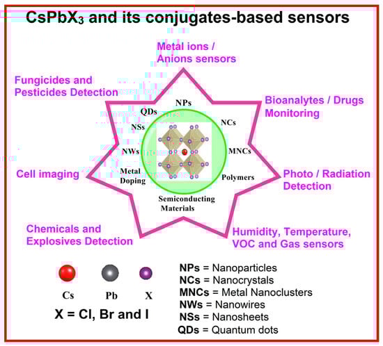
Figure 1.
Schematic representation of CsPbX3 (X = Cl, Br, and I) and composite-based sensors used in metal ion and anion sensors, bioanalyte and drug monitoring, pesticide detection, cell imaging, etc.
2. Role of Structural Stability and Optoelectronic Properties in Sensors
CsPbX3 crystals following the stoichiometry ABX3 have an undistorted cubic structure composed of Pb2+ surrounded octahedrally by ‘X’ anions (X = Cl, Br, and I) and a larger Cs+ cation with a 12-fold cuboctahedral coordination [65]. CsPbX3 in a cubic crystal structure can be stable only if the values of the Goldschmidt tolerance factor are between 0.9 and 1 [66]. The Goldschmidt tolerance factor of CsPbX3 is determined from t = (RA + RX)/√2 (RB + RX), where RA, RB, and RX are the ionic radii of Cs+, Pb2+, and halide (X) ions, respectively. When the values of the tolerance factor lie between 0.7 and 0.9, the CsPbX3 crystal may exist in distorted cubic structures with variations in symmetries/phases [65,66,67]. The structural distortion and instability of CsPbX3 may arise from external factors, such as temperature, quantum size moisture, etc. [68,69]. For example, phase transitions among cubic, monoclinic, tetragonal, or orthorhombic phases occur in the CsPbX3 crystal at a temperature range between 300 and 600 K [70]. Similar to the variations in particle sizes and compositions, the phase transformation in the CsPbX3 may also result in broad emission covering the entire visible range (from blue to red), which can be utilized to develop analyte sensors. For example, Protesescu et al. [71] demonstrated CsPbX3 NCs with tunable bandgap energies by controlling the quantum size and colloidal compositions of NCs, as shown in Figure 2. When exposed to moisture, humidity, gaseous environment, and the doping of foreign materials, structural distortions via surface-mediated absorption, oxidation, reduction, etc., could occur in CsPbX3, which can be regarded as sensor responses. The stability of hybrid halide perovskites also follows a similar trend as CsPbX3-based materials [9]. However, metal oxide perovskites show slightly better stability than that of CsPbX3 and hybrid halide perovskites [9,10,11,12].
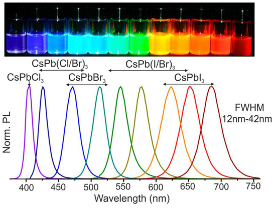
Figure 2.
Colloidal perovskite CsPbX3 NCs (X = Cl, Br, and I) exhibit size- and composition-tunable bandgap energies covering the entire visible spectral region with narrow and bright emission; colloidal solutions in toluene under UV lamp (λ = 365 nm) is shown in the upper corner of the figure (permission obtained from Ref. [71]).
Owing to its exceptional optoelectronic properties, CsPbX3 can be applied in analyte quantification by monitoring its responses in photoluminescence, absorbance, conductivity, temperature, etc. Moreover, CsPbX3 possesses bright blue-to-red emission, depending on the halide (X) concentration, and displays high PLQY (can reach over 90%). Therefore, CsPbX3 and composites can also be utilized in the quantification of halides [72]. CsPbX3 and composites can display unique absorbance and colorimetric responses in the presence/absence of specific analytes [73]. The density functional theory calculations conducted by Y. Kang and co-workers [74] showed that different halide (X) ions in CsPbX3 can lead to changes in intrinsic carrier mobility by a factor of 3 to 5, depending on the carrier concentration, which is between 1015 and 1018 cm−3. Their work also concluded that, in terms of carrier mobility, the preferred carrier type (electron or hole) also depends on halide (X). Kawano et al. investigated the halogen ion dependence on the low thermal conductivity of cesium halide perovskites using first-principle phonon calculations [75]. E. G. Ripka and co-workers reported variations in surface-ligand binding potential due to the halide ion exchange to afford diverse emissions and PLQY [76]. The changes in intrinsic carrier mobility, thermal conductivity, and surface-ligand binding potential can be adopted in designing various electrochemical, temperature, and colloidal sensors for the detection of specific analytes. However, some of the reported sensors were attributed to a combination of carrier mobility and surface-ligand binding. For instance, exposing CsPbX3 to gaseous or VOC analytes resulted in variations in conductivity, but the underlying mechanism was attributed to the efficacy of surface-ligand binding in the modification of oxidation or reduction [77,78,79].
3. CsPbX3 (X = Cl, Br, and I) and Composites toward Metal Ion Detection
Due to the importance of heavy metal ion detection in environmental protection, the use of CsPbX3 (X = Cl, Br, and I) and composites in metal ion sensors have been widely reported by many researchers, which are described in this section. Wu and co-workers demonstrated the PL-based detection of Cu2+ in the presence of ytterbium acetate (Yb(OAc)3) by engaging one-dimensional (1D)-CsPbCl3 NCs in the device [80]. The CsPbCl3 NCs were synthesized using the hot-injection method and characterized by transmission electron microscopy (TEM), X-ray diffraction (XRD), X-ray photoelectron spectroscopy (XPS), and Fourier transform infrared spectroscopy (FTIR). Yb(OAc)3 induces morphology changes from weakly emissive 1D CsPbCl3 NCs (PLQY = 2.1%) to highly luminescent 1D CsPbCl3 NWs (PLQY = 17.3%), which are low in defect density and high in conductivity. The PLQY depends on the size of nanostructures (due to bandgap variations), which can be adjusted with metal doping. The doping of Yb3+ in 1D-CsPbCl3 NWs resulted in a higher aspect ratio, uniformity, and lower number of defects compared with undoped ones; thus, Yb-doped 1D-CsPbCl3 NWs showed high PLQYs. The enhanced defective and rough surface morphologies of 1D-CsPbCl3 NCs effectively hindered light absorption and electron/charge transport, thereby lowering the PLQY. In the presence of acetate ion (AcO−; present in Yb(OAc)3), the steric hindrance was reduced by the copper-based counter-ion pair, which enhanced the adsorption of Cu2+ on the surface of the CsPbCl3 NCs and resulted in PL quenching. The linear regression of Cu2+ detection was observed between 0 and 1 µM (µM = micromole (10−6 M)) with an LOD of 0.06 nM (nM = nanomole (10−9 M)). This work is informative and reveals much detail on the role of Yb(OAc)3, underlying the dynamic quenching mechanism, surface-mediated analyte interaction, and feasible information of electron transfer/charge trap between CsPbCl3 and Cu2+. However, it lacks real-time applications, which should be demonstrated before commercialization.
Sheng et al. reported the use of CsPbBr3 QDs in the quantification of Yb3+ and Cu2+ [81]. CsPbBr3 QDs were synthesized using the hot-injection method, with a PLQY of 63%, and showed enhancement in PL with Yb3+ and PL quenching with Cu2+, as seen in Figure 3. The linear PL quenching range of Cu2+ is between 2 nM and 2 µM, with an estimated LOD of 2 nM. This work is impressive in reporting Cu2+ detection in edible oils but lacks detailed investigations on the underlying mechanisms. Liu et al. also described the use of CsPbBr3 QDs (PLQY = 90%) toward the luminescent detection of Cu2+ [82]. CsPbBr3 QDs were synthesized via the hot-injection method and displayed PL quenching between 0 and 100 nM, with an LOD of 0.1 nM. The detection of Cu2+ was carried out in organic media hexane following a dynamic quenching mechanism. This is a follow-up work of previous reports with additional validation on selectivity and time-resolved studies.
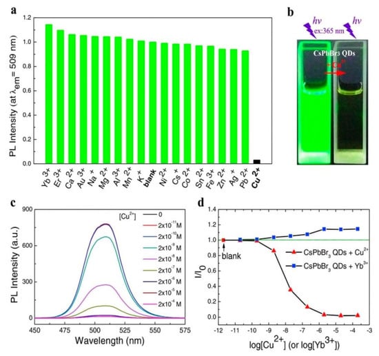
Figure 3.
(a) The effect of different metal ions on the PL intensity of CsPbBr3 QDs. The concentration of metal ions and CsPbBr3 QDs are 2.0 × 10⁻⁶ and ≈1.0 × 10⁻⁹ M, respectively. The PL peak intensity is normalized by CsPbBr3 QDs without adding metal ions (the “blank” column). (b) Photo of CsPbBr3 QDs in cyclohexane under ultraviolet light excitation with and without Cu2+. (c) PL spectra of CsPbBr3 QDs at different [Cu2+] concentrations and (d) PL intensity of CsPbBr3 QDs (λex = 365 nm) as a function of [Cu2+] and [Yb3+] (permission obtained from Ref. [81]).
To avoid using organic media in sensor investigations, Kar and co-workers reported that the use of poly(vinyl pyrrolidone), n-isopropylacrylamide-coated CsPbBr3 NCs (PVP-NIPAM-CsPbBr3 NCs) for aqueous media facilitated the discrimination of Cu2+ [83]. Firstly, SiO2-coated CsPbBr3 NCs and PVP-NIPAM-CsPbBr3 NCs were synthesized via ligand–ligand-assisted reprecipitation (LARP) method to achieve a maximum PLQY of 93%. The PVP-NIPAM-CsPbBr3 NCs showed improved stability and dispersity in water compared to silica-coated NCs, with redshifted emission peaks around 513–515 nm. The NCs displayed linear PL quenching between 0 and 412 µM, with a calculated LOD of 18.6 µM, as seen in Figure 4. This is an inspiring work that can be readily extended to biological studies. Li et al. reported the use of green-fluorescent CsPbBr3 (CPB) QDs for Cu2+ quantification using phase transfer [84]. A strong organic ligand (oleylamine, OAm) was added to selectively transfer Cu2+ from water to cyclohexane, which led to fluorescent quenching. The PL emission was quenched linearly between 1 µM and 10 mM. This is an innovative method that enables Cu2+ detection via phase transfer with a short response time (1 min). However, no real applications were reported in this work. Moreover, Song and co-workers developed long-wavelength-pass filters consisting of CsPbBr3 and CsPb(Cl/Br)3 QDs using a Cu2+-quenching strategy [85]. However, no clear real-time applications were demonstrated in this work.

Figure 4.
Images of the PbN-4 NC solution under a UV lamp (a) before and after adding different metal ions as marked in the figure. (b) Chart representing the comparison of the PL intensity of PbN-4 NCs that persisted after the addition of subsequent metal ions. (c) Emission spectra of PbN-4 NCs in the presence of different concentrations of Cu2+ solutions as shown in the legends. (d) The linear curve represents decreasing in the PL intensity of PbN-4 NCs after adding different concentrations of Cu2+ solution (permission obtained from Ref. [83]).
Li et al. proposed the utilization of macroporous CsPbBr3-(SH)polyHIPE NCs for the ultrasensitive detection of Cu2+ via PL quenching [86]. In this work, CsPbBr3 NCs were synthesized via the hot-injection method, with a PLQY of ~98%. The as-synthesized NCs were then composited with (SH)polyHIPE (generated from the monomers trimethylolpropane triacrylate (TMPTA) and trimethylolpropane tris(3-mercaptopropionate) (TMPTMP)). The linear regression of PL quenching was recorded between 10 fM and 10 mM, with an LOD of 10 fM. Although this work is supported by density functional theory (DFT) calculations, it lacks real-time applications. Gao and co-workers developed a CsPbBr3 QD-based fluorescence-enhanced microfluidic sensor for the in situ detection of Cu2+ in lubricating oil [87]. As displayed in Figure 5a, the polymethyl methacrylate opal photonic crystal (PMMA OPC) film is a microfluidic sensor substrate, which displays high sensitivity and low LOD with Cu2+. When coupling with OPCs, CsPbBr3 QDs synthesized through the hot-injection method showed a 26-fold enhancement in PL intensity (at 496–526 nm under 450 nm excitation). When adding Cu2+ in lubricating oil, the PL intensity of PMMA OPC/CsPbBr3 QD composites was quenched with Cu2+ concentrations of 1 nM–10 mM and an LOD of 0.4 nM, as shown in Figure 5b. The CsPbI3 QD/SiO2 IOPC (inverse opal photonic crystal) composite was further explored for the on-site rapid detection of Cu2+ [88]. CsPbI3 QDs synthesized with a hot-injection method displayed PL emission at 693 nm under 405 nm excitation. SiO2 IOPCs were introduced into the chip wells to couple with the CsPbI3 QDs, which further enhanced the PL emission by 22 folds. After the addition of Cu2+ in lubricating oil to the above composites, a linear PL quenching was observed in the ranges of 0–20 nM and 20–50 nM, with an LOD of 0.34 nM. Both reports [87,88] show distinct performance in terms of mechanical durability, operating temperatures (5 °C to 50 °C), reusability of chips, etc. Advancing in this research direction will require further demonstrations of practical applications.

Figure 5.
(a) The workstation setup for the detection system of microfluidic sensor. (b) The schematic of the formation of PMMA OPCs/c composites and Cu2+ detection (permission obtained from Ref. [87]).
Zhang and co-workers proposed the utilization of organic cross-linker hexamethylene diisocyanate (HDI)-reinforced small-sized CsPbBr3@SiO2-E NPs (size ≤ 50 nm) toward the fluorescent sequential detection of Cu2+ and S2− [89]. CsPbBr3 NCs (size = 6.8 nm; emission at 515 nm) were first synthesized using a hot-injection technique and then conjugated via a three-step synthetic path to afford CsPbBr3@SiO2-E NPs (emission peak at 508 nm; PLQY = 90%), where ‘E’ stands for enhanced performance. The linear PL quenching/recovery of CsPbBr3@SiO2-E NPs with Cu2+ and S2− were observed in ranges of 0–5 µM and 5–10 µM (for Cu2+) and 0–120 µM (for S2−), with corresponding LODs of 0.16 µM (for Cu2+) and 8.8 µM (for S2−). This work requires further support with real-time investigations. The CsPbBr3 QDs (PLQY = 88%) were encapsulated in polymethyl methacrylate (PMMA) fiber membrane (d ≈ 400 nm) to afford CPBQD/PMMA FM for detecting trypsin, Cu2+, and pH [90]. The CPBQD/PMMA FM and cyclam interacted effectively to capture Cu2+, with a linear range of 1 fM–1 M (fM = femtomole (10−15 M)) and an LOD of 1 fM. Moreover, fluorescence resonance energy transfer (FRET) between CPBQD and Cu2+ plays a vital role, resulting in PL quenching. Trypsin detection by CPBQD/PMMA FM via PL quenching was attained in the presence of peptide CF6 (Cys–Pro–Arg–Gly–R6G). Similarly, CPBQD/PMMA FM and R6G were combined to display pH-mediated fluorescent quenching. This is a unique work with exceptional sensor investigations. However, it can be improved further by supporting additional real-time applications. Ahmed et al. proposed a two-step surfactant-free procedure for producing a CsPbBr3 QD-embedded zinc(II) imidazole-4,5-dicarboxylate metal–organic framework (MOF) for the luminescent detection of Cu2+ [91]. The PL emission of CsPbBr3@MOF composites at 519 nm (under 360 nm excitation; PLQY = 39.2%) was quenched linearly between 100 and 600 nM, with an estimated LOD of 63 nM. PL quenching occurred through dynamic quenching and electron transfer with a Stern–Volmer quenching constant (KSV) of 1.55 × 10⁵ M−1. Although this report is an impressive work, it lacks real-sample investigation.
Wang et al. reported the use of a liquid–liquid extraction technique for the visual detection of Hg2+ in aqueous media [92]. In their study, the luminescent CsPbBr3 NCs (PL emission at 520 nm under 380 nm excitation) were synthesized using the hot-injection method to engage in the liquid–liquid-extraction-based visual detection of Hg2+. When adding Hg2+ dissolved in water into CsPbBr3 NCs in carbon tetrachloride (CCl4), the colorimetric PL emission quenching at 520 nm via liquid–liquid extraction was observed, as displayed in Figure 6. The linear regression of Hg2+ was recorded between 50 nM and 10 µM, with an estimated LOD of 35.65 nM. This work requires further investigations into the underlying mechanism and real-time applications.

Figure 6.
Illustration of liquid–liquid extraction and visual detection of Hg2+ using CsPbBr3 PNCs (permission obtained from Ref. [92]).
Jiang et al. proposed a two-step precipitation method to synthesize emissive CsPbBr3 crystals (PL emission at 525 nm under 395 nm excitation) toward Hg2+ detection [93]. When detecting Hg2+, both CsPbBr3 NCs and Hg2+ were co-precipitated in aqueous solution. The CsPbBr3 precursor was firstly dissolved in an aqueous solution containing Hg2+ (0–1000 nM) and then dropped onto a hydrophilic polydimethylsiloxane (PDMS) substrate with a microwell array. When the substrate was heated at 25 °C for 3 min, co-precipitation occurred, which resulted in PL quenching via Hg2+ doping into the CsPbBr3 lattice. The linear regression of Hg2+ was observed between 5 and 100 nM, with an estimated LOD of 0.1 nM. This work demonstrated an innovative technique and provided detailed studies on interference, pH effect, and underlying mechanisms toward Hg2+ detection. However, the cost-effectiveness and real-time applications of this method require more work. Through ligand engineering and silica encapsulation, a stable fluorescent CsPbBr3-mPEG@SiO2 composite (PL emission at 520 nm under 330 nm excitation; PLQY = 67.5%) was synthesized and adopted in the sequential detection of Hg2+ and glutathione (GSH) in aqueous solution via PL quenching and recovery, respectively [94]. Shu and co-workers demonstrated that the existence of 73% of the PL emission of NCs could last over 30 days in aqueous media. The PL quenching and recovery responses were attributed to the electron transfer process between NCs and Hg2+ and the effective interaction between Hg2+ and GSH. The linear PL responses of Hg2+ and GSH were observed in the ranges of 1–50 nM and 1–10 µM, with LODs of 0.08 nM and 0.19 µM, respectively. This work was successfully applied in tab and serum sample analysis; therefore, it can be regarded as a remarkable work in Hg2+ and GSH detection.
Guo et al. developed a nucleation growth method for producing CsPbBr3 NCs (PL emission at 518 nm under >360 nm excitation; PLQY > 89%) at a large scale and adopted as-synthesized NCs for detecting Zn2+ [95]. The PL emission of CsPbBr3 NCs was quenched linearly between 0 and 40 µM in the presence of Zn2+. The PL quenching was not due to the replacement of Pb2+ in the CsPbBr3 matrix but was caused by the Zn–oleic complex formation. The surface defects created led to the self-assembly of CsPbBr3 nanocubes into nanorods, thereby resulting in PL quenching. George and co-workers reported the use of alpha-amino butyric acid (A-ABA)-capped CsPbBr3 QDs (M PQDs) for developing Co2+ sensors [96]. The M PQDs were synthesized using the hot-injection method, which displayed PL emission at 489 nm. The PL emission of the M PQDs was quenched in Co2+ concentrations of 0–100 nM, with an LOD of 0.8 µM. This report uncovered that PL quenching was due to FRET-facilitated dynamic quenching and the inner filter effect (IFE). This is the only report on IFE-based PQD sensors using metal ions, but it lacks information in real-time applications. Halali et al. reported uranyl (UO22+) ion detection using green emissive CsPbBr3 PQDs (synthesized using the hot-injection method) [97]. When adding UO22+, the PL emission at 518 nm was quenched linearly in UO22+ concentrations of 0–3.3 µM, with a calculated LOD of 83.33 nM. Extensive mechanistic studies revealed that the PL quenching was due to the electrostatic interaction and adsorption of UO22+ over the surface of QDs. To support this work, further research on the interference and application studies is necessary.
Polyvinylpyrrolidone (PVP) polymer shell-grown silica-coated Zn-doped CsPbBr3 NCs (polymer-coated Zn-doped CsPbBr3/SiO2 core/shell NCs (PVP-0 NCs, PVP-2.5 NCs, PVP-5 NCs, PVP-7.5 NCs, and PVP-10 NCs)) were synthesized with the hot-injection method for detecting In3+ in water [98]. The double-coating method enhanced the water stability, dispersibility, and emission properties of the NCs. Among the various PVP shell-grown silica-coated Zn-doped CsPbBr3 NCs, PVP-5 NCs (PLQY = 88%) were more stable at higher temperatures and showed stronger luminescence and greater selectivity to In3+. When adding In3+, the PL emission of the PVP-5 NCs at 511 nm was quenched in In3+ concentrations of 0–104 µM, with an estimated LOD of 11 µM. The PL quenching was associated with the replacement of Pb2+ by In3+. This report is noteworthy but lacks evidence for the proposed mechanism and real-time applications. Pandey et al. employed the CsPbBr3−Ti3C2Tx MXene QD/QD heterojunction for the PL-based detection of Cd2+ [99]. CsPbBr3 QDs were synthesized using the hot-injection method and then were composited with Ti3C2Tx MXene in toluene. The PL emission of CsPbBr3−Ti3C2Tx MXene QDs at 505 nm (under 410 nm excitation) was quenched via charge transfer when adding Cd2+. The linear PL quenching of the QD composite was observed in Cd2+ concentrations of 99–590 µM with no information on the value of LOD. This work also demonstrated an on−off−on PL probe for cadmium ion detection, but more investigations are necessary to justify the underlying quenching (static/dynamic) mechanisms. Hsieh and co-workers proposed the use of (3-aminopropyl) triethoxysilane (APTES)0coated CsPbBr3–CsPb2Br5 QDs toward the PL-based detection of Fe3+, as illustrated in Figure 7 [100]. Through the ligand-assisted reprecipitation method, APTES-coated CsPbBr3–CsPb2Br5 QDs were synthesized. The PL emission of the QDs at 520 nm was quenched rapidly (response time = 8 s at 40 °C) in the presence of Fe3+. The linear PL responses of QDs to Fe3+ were observed in Fe3+ concentrations of 10 µM–10 mM with an LOD of 10 µM. This is a well-organized work with excellent results in response time and temperature, but it lacks supportive data on mechanistic investigations. Table 1 summarizes the synthetic route, PLQY, linear range, detection limit, and application of CsPbX3 (X = Cl, Br, and I) and composites for metal ion detection.

Figure 7.
(a) Emission spectra and (b) normalized emission intensity of AP-PQD in the presence of different Fe3+ concentrations. (c) Photographs of AP-PQD dispersed in ethanol containing Fe3+ under ambient light and 365 nm UV light. (d) Intensity ratio of AP-PQD in the presence of different Fe3+ concentrations (permission obtained from Ref. [100]).

Table 1.
The synthetic route, PLQY, linear range, detection limit, and application of CsPbX3 (X = Cl, Br, and I) and composites toward metal ion detection.
Critical Comments on CsPbX3 (X = Cl, Br, and I)-Based Metal Ion Detection
Based on the existing results, it is noted that as-synthesized CsPbX3 QDs, NCs, and NWs display high specific selectivity to Cu2+ through feasible energy transfer between the probes and Cu2+ [80,81,82]. Furthermore, it was clarified that Yb3+ doping enhanced the selectivity by reducing the surface defect [80,81], thereby suggesting the effectiveness of surface forces in sensors. Another critical issue in the use of CsPbX3 (X = Cl, Br, and I) probes for metal ion detection, which requires more attention, is their stability in aquatic environments. To solve the stability issues, using polymer and ligand capping/coating on CsPbX3 has been proposed [83,84,86,89,90], which may enhance the PLQY by avoiding surface exposure to environmental forces existing in water and air. However, whether this approach can be effective in exposure to Cu2+ in an aquatic environment remains an open question. The development of a pass filter consisting of CsPbBr3 and CsPb(Cl/Br)3 QDs was demonstrated for Cu2+ detection via PL quenching responses [85]. However, the development of such pass filters has not met commercial standards. It is a premature proposal and requires additional work. CsPbBr3 QD/CsPbI3 QDs were explored using the microfluidic technique, which facilitated the detection of Cu2+ [87,88]. However, the fabrication processes of such devices are rather complicated and require a well-equipped clean room environment, thereby restricting their advancement in most developing countries. Also, it is essential to determine whether this microfluidic method is effective in all environmental samples. The use of CsPbBr3 crystals, NCs, and QDs was also reported in the PL “turn-off” detection of Hg2+ and UO22+ [92,93,94,97]. Many of the available reports on CsPbX3 (X = Cl, Br, and I)-based metal ion sensors confirmed their selectivity to Cu2+; however, the underlying mechanisms of detecting Hg2+ and UO22+ by CsPbBr3 crystals, NCs, and QDs are still unclear. Likewise, the composites of CsPbX3 (X = Cl, Br, and I) with other emerging nanomaterials, such as MOFs, Mxene, APTES, etc., have been proved to be effective in discriminating diverse heavy metal ions [91,94,95,96,98,99,100]. However, most of those reports did not address the feasible surface-facilitated detection mechanisms, which restricted the development of analytical devices. These results also raise the question of the reliability of CsPbX3 (X = Cl, Br, and I)-based Cu2+ sensors. The reason behind the selective sensing of Cu2+ must be clarified by investigating the Pb2+ replacement mechanism, as well as the magnetic property (ferro-/ferri-electronic) changes. The crystalline and lattice features of the probes/compositions in the presence/absence of analytes are not considered from mechanistic aspects, which should be taken into account in future sensor designs. If the crystalline/lattice parameters of CsPbX3 (X = Cl, Br, and I)-based probes are taken into account, it is highly feasible to design chemoresistive and electrochemical sensors for heavy metal quantification in real samples.
4. Anion Detection by CsPbX3 (X = Cl, Br, and I) and Composites
Similar to metal ion quantification, the discrimination of anions was also demonstrated by perovskite nanomaterials, as described in this section. Jan et al. reported the synthesis of the CsPbBr3 nanoplatelets (PLQY = 83.7%) via the hot-injection method, which displayed PL emission at above 475 nm under 350 nm excitation [101]. The PL peak was blueshifted when the CsPbBr3 nanoplatelets were exposed to Cl− (from a HCl source). The sensor response showed a linear range from 0.2 to 0.4 nM, with an LOD of 28 pM. The observed response was attributed to the anion exchange mechanism. Moreover, CsPbBr3 nanoplatelets are also able to effectively detect the arsenate in the presence of hypochlorous acid (HOCl). The following reaction process (1) shows that As3+ is oxidized to produce Cl−:
AsO33− + OCl− → AsO43− + Cl−
A blueshifting phenomenon in PL emission from the reaction induced by Cl− species occurred via anion exchange, which showed a linear regression of 6.4–58 nM, with an LOD of 1 nM. This is an innovative work with dual-species recognition; however, it does not provide enough real-time applications. Thereafter, Huang et al. proposed the use of CsPbBr3 QDs for Cl− detection in water [102]. The CsPbBr3 QDs, with PL emission at 513 nm and a calculated PLQY value of 87%, were synthesized using the hot-injection method. When exposed to Cl−, the PL emission peak at 513 nm was blueshifted to 483 nm due to the anion exchange reaction with Br−. The shifting of the PL peak occurred in Cl− concentrations of 10–200 μM, with an estimated LOD of 4 µM. This report was well supported by real-time water analysis; therefore, it can be regarded as a unique work. Shu and co-workers developed highly stable CsPbBr3 NCs via amphiphilic polymer ligand-assisted synthesis [103]. Amphiphilic polymer octylamine-modified polyacrylic acid (OPA) was used as the capping agent to produce stable NCs, with PL emission at 520 nm and >40% PLQY. As displayed in Figure 8, the PL peak at 520 nm is blueshifted to 441 nm due to the anion exchange reaction in Cl− concentrations of 1–80 mM, with an LOD of 0.34 mM. This report demonstrated Cl− detection in sweat samples; thus, it is an innovative work. However, further research work is required for commercialization.

Figure 8.
(A) Fluorescence spectra and corresponding fluorescence photographs of the CsPbBr3/OPA + OAm NCs in the presence of different concentrations of chloride ions from 0 to 150 mM in an aqueous solution under 365 nm UV excitation. (B) The fitting curve of Δλ plotted as a function of Cl− concentration; inset: the corresponding calibration curve of Δλ and Cl− concentration from 1 to 80 mM. (C) Wavelength shift of different substances for the selectivity investigation of Cl− sensing. (D) Fluorescence spectra of actual samples and samples after being spiked with different concentrations of Cl−; inset: the corresponding fluorescence photos of the samples (permission obtained from Ref. [103]).
Shortly after, Li et al. adopted CsPbBr3 NCs for the luminescent colorimetric sensing of Cl− in n-hexane via a halide exchange reaction [104]. The CsPbBr3 NCs were synthesized using the hot-injection method, in which a rapid halide exchange reaction occurred at pH = 1. The green emissive peak of the CsPbBr3 NCs at 514 nm was blueshifted to a 452 nm peak (blue emission) in Cl− concentrations of 10–130 mM, with an estimated LOD of 3 mM. This work was applied in Cl− detection in sweat samples; hence, it is quite innovative. However, the values of the LOD must be further improved by combining other techniques. By taking advantage of the anion exchangeability of CsPbBr3 NCs, Dutt and co-workers proposed the construction of a glass plate-/paper-strip-based test kit for discriminating Cl− [105]. The PL emission peak of the kit at 509 nm was slowly blueshifted to 478 nm because of the anion exchange reaction. The linear regression of Cl− was observed between 100 µM and 10 mM with an LOD of 100 µM. This work requires more supportive evidence for possible commercialization.
Recently, Zhang and co-workers developed β-cyclodextrin (β-CD)-stabilized, arginine (Arg)-added CsPbBr3 NCs (ACD-PNCs; PLQY = 82%) via ligand-assisted synthesis and utilized them in discriminating Cl− and I− through a ligand exchange mechanism [106]. β-CD capping, together with the addition of Arg, helped to stabilize the PNCs. The green emission of ACD-PNCs was blue/redshifted from 508 nm to 424 nm (blue emission) and 511 nm to 637 nm (red emission), respectively, in the presence of Cl− and I−. The linear regression of Cl− and I− detection was observed in the ranges of 0.04–0.8 mM and 0.04–1.16 mM, with calculated LODs of 3.2 µM and 9 µM, respectively. This is a unique work that provides a comparative study with earlier reports. However, perspectives on further work are not mentioned. As discussed in many studies related to anion exchange reactions, CsPBBr3 can act as exceptional probes toward the quantification of Cl−/HCl, I−, and F− [107,108]. An alcohol-dispersed CsPbBr3@SiO2 PNCC nanocomposite was proposed for discriminating Cl− in an aqueous phase [109]. The green emissive peak at 506 nm was blueshifted to 447 nm through the homogeneous halide exchange between CsPbBr3@SiO2 PNCCs and Cl−. The recovery studies of Cl− in sea sand samples (with a linear range of 0–3%) attested to the reliability of this work, with an LOD of 0.05 mg/g. Moreover, the anion exchange between CsPbBr3@SiO2 PNCCs and Cl− occurred in the absence of magnetic stirring or pH regulation, which was a novel observation.
Fu et al. proposed the use of the NH2-functionalized CsPbBr3 NCs for detecting I− [110]. These NH2-functionalized NCs were synthesized in ethanol by using 3-aminopropyltriethoxysilane (APTES) as ligands. In contrast to the traditional halide exchange-based I− sensors of CsPbBr3, the luminescence of the NH2-PNCs in ethanol/water at 510 nm was quenched, as shown in Figure 9a. Linear regression is observed in I− concentrations of 4–28 µM, with an LOD of 1 µM, as seen in Figure 9b. I− showed higher selectivity among all other interferences, but this report lacks supporting evidence on the PL quenching response of the unshifted peak.
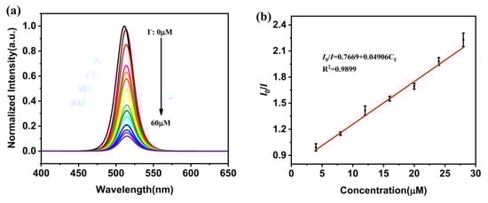
Figure 9.
(a) Fluorescence spectra of NH2-PNCs at various concentrations of I− (0–60 μM). (b) Linear fitting curve of I0/I of the fluorescence of NH2-PNCs and concentrations of I− (permission obtained from Ref. [110]).
Park and co-workers fabricated a CsPbBr3 QD/cellulose composite as an early diagnosis sensor for Cl− and I− [111]. The CsPbBr3 QD/cellulose composite was synthesized via a hot-injection method to form monodispersed CsPbBr3 QDs with high selectivity to Cl− and I−. The detection of Cl− and I− in aqueous media was confirmed by observing a color change from green to blue and from green to red, respectively. The color change occurred within 5 s because of the halide exchange reaction. The linear responses of the CsPbBr3 QD/cellulose composite to Cl− and I− were recorded at 0.1 mM–1 M, with calculated LODs of 2.56 mM (for Cl−) and 4.11 mM (for I−), respectively. In terms of real-time applications, this work can be regarded as a unique report on medical device fabrication. The halogen ion exchange reaction of CsPbBr3 and composites brings an additional advantage of the effective discrimination of edible oils. Zhang et al. demonstrated the discrimination of edible oils by using octadecylammonium iodide (ODAI) and ZnI2 as anion exchangers [112]. This colorimetric sensing strategy can be applied in detecting edible oil mixtures with 100% accuracy, but further research is required for commercialization. Wang et al. described the employment of tetraphenylporphyrin (TPPS)-modified CsPbBr3 NCs (CsPbBr3/TPPS nanocomposite) for the quantification of sulfide (S2−) [113]. The CsPbBr3/TPPS nanocomposite possessed good water stability and dual-emission properties. As shown in Figure 10, the CsPbBr3/TPPS nanocomposite displays strong green emission at 520 nm and moderate red emission from the TPPS at 650 nm. When adding S2−, the PL emission at 520 nm was quenched linearly in S2− concentrations of 0.2–15 nM, with an LOD of 0.05 nM. This was attributed to the destruction of CsPbBr3 NCs via the formation of more stable PbS. This work also reported the real-time water recovery study (>95%), which showed a relative standard deviation of <3%, thereby opening a new direction for future research. Table 2 summarizes the synthetic route, PLQY, linear range, LOD, and application of CsPbX3 (X = Cl, Br, and I) and composites toward the detection of anionic species.
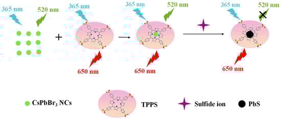
Figure 10.
Schematic illustration of CsPbBr3/TPPS nanocomposite-based ratiometric fluorescence detection of sulfide ion (permission obtained from Ref. [113]).
Table 2.
The synthetic route, PLQY, linear range, detection limit, and application of CsPbX3 (X = Cl, Br, and I) and composites toward the detection of anionic species.
Critical View on CsPbX3 (X = Cl, Br, and I)-Based Anion Sensors
CsPbX3 (X = Cl, Br, and I)-based probes/composites have been reported for discriminating Cl−, Br−, and I− to display red/blueshifted PL emissive peaks via anion exchange, as noted in Table 2 [101,102,103,104,105,106,107,108,109,110,111,112]. According to these sensing studies, the presence of anions in the aquatic environment may also lead to a rapid anion exchange due to the disturbed structural parameters. These issues should be addressed with in-depth investigations. Surface stabilization by using suitable capping agents may change lattice features, resulting in an enhanced PLQY; thus, the proposed anion-sensing performance by CsPbX3 (X = Cl, Br, and I)-based probes/composites is not yet confirmed and requires further research. The real question is how can the anion-sensing performance be confirmed if the sensing medium itself could affect the stability of the proposed CsPbX3 (X = Cl, Br, and I)-based probes/composites. The reaction-based sensing of S2− using tetraphenylporphyrin tetrasulfonic acid (TPPS)-modified CsPbBr3 NCs was also observed [113], which showed dependence on composition concentrations. Therefore, the optimization of composition concentrations is regarded as a high-priority task and requires detailed investigations. Due to the instability issues of CsPbX3 (X = Cl, Br, and I)-based probes, anion discrimination to distinct competing matrices in real water samples becomes more urgent.
5. CsPbX3 (X = Cl, Br, and I) and Composites for the Recognition of Chemicals and Explosives
Similar to the quantification of metal ions and anions, CsPbX3 (X = Cl, Br, and I) and composites have been widely applied in the discrimination of chemicals and explosives. Yin and co-workers reported the use of CsPbBr3 NCs (synthesized with the hot-injection method; PL at 510–520 nm) in the quantification of iodomethane (CH3I) via the halide exchange reaction in the presence of oleylamine (OLA) [114]. The presence of OLA induced the nucleophilic substitution of CH3I to release iodide species. As illustrated in Figure 11, the iodide exchange occurs rapidly within <5 s, resulting in a redshift of PL (nearly 150 nm; original PL at 660–670 nm). The selectivity of CsPbBr3 NCs toward CH3I was high with this portable approach. The linear range of CH3I detection is between 0.7 and 70 µM with an LOD of 0.2 ± 0.07 µM. Based on the results, this work can be regarded as innovative. Shortly after, Feng et al. described the use of yttrium single-atom-doped cesium lead bromide nanocrystals (Y-SA/CsPbBr3 NCs) for detecting CH3I [115]. The Y single-atom deposition was carried out using a photo-assisted method. In the presence of OLA, the PL peak of CH3I was redshifted due to the halide exchange reaction. The linear range of CH3I detection was between 5.6 and 157 µM, with an LOD of 0.3 µM. Except for the anchoring of the Y single atom, this is a follow-up work of an earlier report [114], hence requiring more research efforts to confirm its novelty.

Figure 11.
Reaction mechanisms and the spectroscopic response of CsPbBr3 perovskite nanocrystals (PNCs) to CH3I. (a) Oleylamine (OLA, 0.96 mM) or CH3I (20,000 ppbv solution) were introduced separately into PNC dispersions in toluene. The emission images under 365 nm UV light were recorded after 100 min, showing no change in emission. (b) OLA-pretreated CH3I solutions (CH3I concentration: 20,000 ppbv) were added to a PNC dispersion in toluene, with the emission color observed under 365 nm UV light before and 20 s after addition. The hypothesized reaction mechanism occurs when CH3I induces the alkylation of OLA via the SN2 mechanism and stops at dimethyl analog formation. (c) UV−visible absorption spectra of PNCs exposed to varying amounts of CH3I. (d) Emission spectra of CsPbBr3 PNCs as a function of the amount of added CH3I; inset: redshift of PNC PL emission as a function of CH3I concentration. Linear fitting of results from 100 to 10,000 ppbv is shown as a red line with R2 = 0.997. (e) CIE chart converted from the PL spectra of PNCs exposed to varying amounts of CH3I. Note: Spectra in c, d were recorded 20 s after CH3I addition at room temperature to ensure the reaction was complete (permission opted from Ref. [114]).
Xie et al. reported a paper-based microfluidic colorimetric assay for dichloromethane/dibromo methane (CH2Cl2/CH2Br2) in the presence of trialkyl phosphines (TOP) by using CsPbX3 (X = Cl, Br, or I) nanocrystals [116]. When adding TOP or through UV-photon-induced electron transfer, the homogeneous nucleophilic substitution could be enhanced to afford a colorimetric fluorescent response with corresponding peak shifts. In the presence of CH2Cl2, the CsPbBr3 NCs displayed a linear peak shift (from 510 nm to 460 nm) in CH2Cl2 concentrations of 0–0.9 M, with an LOD of 48 mM. Similarly, In the presence of CH2Br2, CsPbBr0.5I2.5 displayed a linear PL peak shift (from 660 nm to 560 nm) in CH2Br2 concentrations of 7.2–21.0 mM, with an LOD of 1.7 mM. Although the results of this work look appealing, improvement in LODs is required before commercialization. Saikia et al. used cetyltrimethylammonium bromide (CTAB)-passivated CsPbBr3 to effectively discriminate ethanol and methanol [117]. Due to the different interaction modes with CTAB, CsPbBr3 displayed diverse PL “turn-off/turn-on” responses with LODs down to 9.3 ppb. This technique has been validated in petrol and cough syrup samples; therefore, it is quite innovative.
Bahtiar et al. described the employment of CsPbBr3 NCs for quantifying the benzoyl peroxide (PBO) concentration in solutions [118]. When adding oleylammonium iodide (OLAM-I), the PL emission of CsPbBr3 NCs at 515 nm was redshifted to 660 nm via the halide exchange reaction. When BPO was added to the above solution, the original PL emission was blueshifted within 1–2 min. Zhang and co-workers also used luminescent CsPbBr3 NCs (synthesized with the hot-injection method; PLQY = 87%) to detect BPO [119]. The linear range of BPO detection was observed between 0 µM and 120 µM, with an LOD of 0.13 µM. The applicability of both reports was demonstrated in white flour and noodles. Thus, this is a unique method for BPO detection. Huangfu et al. confirmed the highly responsive photoluminescence sensing performance of CsPbBr3 quantum dots (QDs) for total polar material (TPM) identification in edible oils [120]. As seen in Figure 12, the CsPbBr3 QDs display diverse colorimetric and PL emissive responses to individual polar solvents (dimethyl sulfoxide (DMSO), dimethyl formamide (DMF), methanol, acetonitrile, ethanol, 1-propanol, acetone, ethyl acetate, chloroform, dichloromethane, and toluene). This TPM detection was effectively applied in edible oils, such as olive oil, soybean oil, and sunflower oil. Hence, it is regarded as an innovative method for the real-time quality assessment of edible oil.
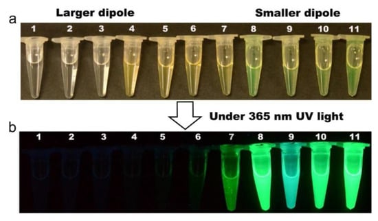
Figure 12.
Photos of the effect of different polar solvents on CsPbBr3 QD solution. The solvents are arranged basically in the order of increasing dipole moment: (1) DMSO, (2) DMF, (3) methanol, (4) acetonitrile, (5) ethanol, (6) 1-propanol, (7) acetone, (8) ethyl acetate, (9) chloroform, (10) dichloromethane, and (11) toluene; (a) under ambient light, (b) under 365 nm UV light (permission obtained from Ref. [120]).
Zhao and co-workers developed orange-emitting oil-soluble CsPbBr1.5I1.5 QDs for detecting excessive acid number (AN), 3-chloro-1,2-propanediol (3-MCPD), and moisture content (MC) for edible oil quality assessment [121]. The PL emission of the CsPbBr1.5I1.5 QDs at 609 nm was quenched when detecting excessive acid number (AN). The peak at 609 nm was blueshifted to 583 nm when 3-MCPD was detected. For MC detection, mesoporous silica-coated CsPbBr1.5I1.5 QDs were adopted as ratiometric sensors to develop water-stable green-emitting CsPbBr3 nanosheet (NS) probes. The LODs were determined for the detection of AN, 3-MCPD, and MC as 0.71 mg KOH/g, 39.8 μg/mL, and 0.45%, respectively. Based on these results, this work can be regarded as innovative. Aamir et al. demonstrated the use of CsPbBr3 microcrystals for the PL-based detection of nitrophenol [122]. The PL emission was quenched rapidly due to the π–π stacking interaction of the benzene ring with CsPbBr3 microcrystals. PL emission was quenched linearly in nitrophenol concentrations of 0.1–0.6 mM. This study reported a preliminary result, thereby requiring more research work.
Chen et al. reported the use of CsPbX3 QDs (Br/I; synthesized via the hot-injection method; PLQY = 52.88% and 46.18%, respectively) for the highly selective detection of explosive picric acid (PA) [123]. In which, the green/red fluorescence of CsPbX3 (Br/I) at 510 nm/675 nm was quenched in the presence of PA. The linear regression of CsPbX3 (Br/I) to PA was in the ranges of 0–180 nM and 0–270 nM, with estimated LODs of 0.8 nM and 1.9 nM, respectively. Based on the supported evidence, the authors speculated that the electrostatic-assisted energy transfer is the possible sensor mechanism. Figure 13 displays a schematic model of the CsPbBr3 QD-based quenching response to PA and its paper-strip application. This is outstanding work, but additional research is necessary for commercialization. Aznar-Gadea and co-workers described the consumption of molecularly imprinted CsPbBr3 nanocomposites for rapid explosive taggant detection at the gaseous stage [124].
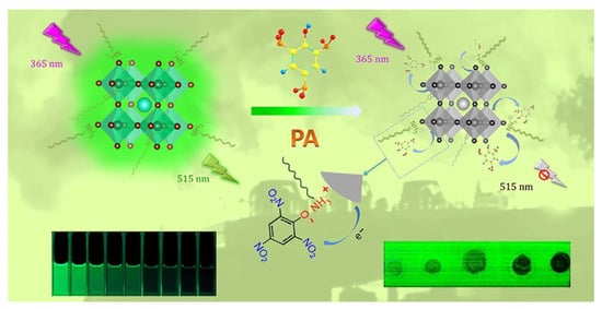
Figure 13.
Schematic for the sensitive fluorescence detection of PA based on perovskite quantum dots and its paper-strip applications (permission obtained from Ref. [123]).
A molecularly imprinted polymer (MIP) sensor was fabricated by embedding CsPbBr3 NCs in polycaprolactone (PCL). When exposed to template molecules, such as 3-nitrotoluene (3-NT) and nitromethane (NM), PL quenching responses (>75%) were observed. The MIP sensor showed high selectivity to NT within 5 s, with an LOD of 0.218 mg mL−1. This is a preliminary work; hence, it should be further extended for commercialization. Table 3 summarizes the synthetic route, PLQY, linear range, LOD, and application of CsPbX3 (X = Cl, Br, and I) and composites toward the detection of chemicals and explosives.
Table 3.
The synthetic route, PLQY, linear range, LOD, and application of CsPbX3 (X = Cl, Br, and I) and composites toward the detection of chemicals and explosives.
Critical View on CsPbX3 (X = Cl, Br, and I)-Based Chemical and Explosive Sensors
The detection/quantification of specialized chemicals, such as CH3I, CH2Cl2, CH2Br2, benzoyl peroxide, and excessive acid number (AN), via anion exchange mechanisms [114,115,116,117,118,119,120,121] cannot be regarded as a specific quantification procedure because of its similarity to anion detection. This should be critically investigated to pursue the “state-of-the-art” sensing procedure. Since the observed ratiometric PL responses are also similar to those in the anion sensing studies, critical investigations are required for commercialization. Furthermore, discriminating explosives was demonstrated via the PL quenching response resulting from surface interaction and charge transfer between nitro-containing explosives and CsPbX3 (X = Cl, Br, and I)-based probes or composites [122,123,124]. However, this also requires critical studies to justify the exact static/dynamic PL quenching responses.
6. CsPbX3 (X = Cl, Br, and I) and Composites for the Quantification of Gaseous Analytes and Volatile Organic Compounds (VOCs)
Many CsPbX3 (X = Cl, Br, and I) and composites have also been reported for the detection of gaseous analytes and VOCs, as described in this section. Huang et al. reported the oxygen-sensing performance of CsPbBr3 NCs [125]. NCs have a porous structure, which allows for the rapid diffusion of O2, resulting in PL quenching. The underlying sensor mechanism is that O2 molecules are directly involved in the extraction of photogenerated electrons from the conduction band of CsPbBr3 NCs. This work lacks in-depth sensor investigations, thereby requiring extensive research. Lin et al. developed Mn2+-doped cesium lead chloride nanocrystals (Mn:CsPbCl3 NCs) by using a heat-up strategy for sensing O2 via luminescent dopants and the host–dopant energy transfer mechanism [126]. As seen in Figure 14a–e, upon exposure to O2, the phosphorescence intensity of Mn:CsPbCl3 NCs decreased linearly between 0 and 12% of O2. High sensing reversibility, rapid signal response, and high photostability in air were also demonstrated. The estimated Stern–Volmer quenching constant (KSV) value following first-order kinetics was 0.0658% [O2]−1 (R2 = 0.9997). This work is innovative, but it lacks practical applications.
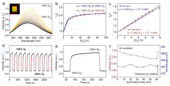
Figure 14.
Sensing responses of the Mn0.175:CsPb0.825Cl3 film to O2: (a) Phosphorescence spectra under different O2 fractions (%). (b) The first-order reaction kinetics curves of the maximum phosphorescence intensities under different O2 fractions (%). (c) Stern−Volmer plot under O2 fractions between 0 and 12%. (d) Reversibility test under the alternating exposure to 100% O2 or 100% N2 (detected at 586 nm). (e) The response time curve within one cycle test. (f) Photostability test in air condition (excited at 365 nm) (permission obtained from Ref. [126]).
Brintakis and co-workers described the use of CsPbBr3 nanocubes as self-powered ozone sensors, which showed higher sensitivity (54% in 187 ppb) and faster responses (between 100 s and 150 s) and recovery (between 250 s and 320 s) [127]. The sensor response of CsPbBr3 nanocubes to O3 was recorded between 4 and 2650 ppb (ppb = parts per billion). The as-synthesized CsPbBr3 nanocubes were semiconductors with a certain resistance. When exposed to O3, an accumulation layer of holes with lower resistance covering the whole surface of the nanocubes was formed resulting in an increase in electrical current. This is an innovative work, which has attracted many scientists to work in this direction. Park et al. fabricated cesium lead bromide nanofibers (CsPbBr3 NFs) by attaching CsPbBr3 NCs with cellulose nanofibers (CNFs) for N2-sensing investigations [128]. When exposed to N2 flow, the PL intensity of the CsPbBr3 NFs at 520 nm was quenched linearly between 1 and 20 ppm (R2 = 0.99433; ppm = parts per million) with an LOD of 1 ppm. The surface trapping of N2 was proposed as the underlying mechanism for N2 detection. This is a study on a conventional sensor, but further research is still necessary.
Nanocrystalline ZnO sensitized with CsPbBr3 NCs for the photoresistive sensing of NO2 gas was proposed by Chizhov and co-workers [129]. In a temperature range of 25–100 °C, the ZnO/CsPbBr3 nanocomposite showed a linear sensor response between 0.5 and 3.0 ppm to NO2. These sensing measurements were conducted under periodic blue LED illumination (light = tdark = 20 s; LED = light emitting diode). At 1 ppm of NO2 gas, it was found that the optimum temperature to provide the best reversibility of sensor measurements was 75 °C. Under periodic illumination, the increase in the electron concentration of ZnO led to the adsorption of oxidizing molecules over the surface. The photoexcited holes in CsPbBr3 were involved in the redox reaction, resulting in a change in the electrical signal. This research is an impressive report, and it could be further developed for commercialization. Yueyue et al. demonstrated the use of CsPbBr3 QD/ZnO MB nanocomposites (MBs = microballs) for NO2 detection at room temperature [130]. In the presence of diverse CsPbBr3 QDs (0.5 wt%, 1.0 wt%, and 1.5 wt%) with ZnO MBs, the sensing responses to NO2 were recorded. ZnO MB composited with 1.0 wt% QDs displayed a greater photoresistive response to NO2 (for 5 ppm NO2; Rgas/Rair = 53; response/recovery time = 63s/40s under 520 LED (1.2 W/m2) illumination). The adsorption and desorption of NO2 over the ZnO surface were proposed as the underlying mechanism, and the QDs had minimal effect on the sensor mechanism. This is a well-developed study on NO2-sensing studies of CsPbBr3-ZnO conjugates, but more studies on the interference and real-time applications are required.
The ultrafast sensing of NO2 was demonstrated using FA0.83Cs0.17PbI3 (FAC) prepared via one-step spin-coating in ambient conditions [131]. When exposed to NO2 (20, 10, 5, 2, 1, and 0.5 ppm), the photoresistive responses (Rgas/Rair) were measured as 2.64, 2, 1.52, 1.27, 1.17, 1.1, respectively. The optimum sensor response of NO2 was recorded at 10 ppm, with a response/recovery time of 2 s/22 s. The strong oxidizing ability of NO2 gas attracts electrons from FAC, which is a p-type semiconductor. The calculated values of the adsorption energy (Eads) of NO2 of FA+ and Cs+ in FACs are −0.37 and −0.60 eV, respectively, thereby allowing for the spontaneous adsorption of NO2. It should be noted that NO2 molecules attached to FACs may induce lattice distortion, which leads to a dipole moment and the migration of charge carriers. All these factors were involved in the sensing of NO2. The roles of FA+ and Cs+ in detecting NO2 were clearly justified. Further research in this direction is required to improve the sensitivity. Wang and co-workers combined ZnO nanorods with CsPbBr3/Cs4PbBr6 particles to afford the CsPbBr3/Cs4PbBr6/ZnO composite, which showed photoresistive sensor responses to NO (100 ppm), with the Rgas/Rair reaching 2296 (at 50 °C) and an LOD of 1 ppm [132]. The response and recovery time were determined as 1235 s and 173 s, respectively. NO gas attracts electrons from the surface of CsPbBr3/Cs4PbBr6/ZnO to release O2, which results in electrical signal changes. Although electrons were accumulated in the conduction band of ZnO, the diffusion of NO gas was hindered due to the covering layer of CsPbBr3/Cs4PbBr6 particles. Therefore, a long response time was recorded. This work requires further research for the optimization of the response/recovery time and interference studies.
Chen et al. described the use of luminescent CsPbBr3 QDs (synthesized via the sonication method) for the solution-mediated detection of hydrogen sulfide (H2S) [133]. As seen in Figure 15A,B, the luminescent intensity of QDs at 520 nm was quenched linearly between 0 and 100 µM with an LOD of 0.18 µM. Note that the QDs exhibit a greater selectivity to H2S among all the interfering species, as shown in Figure 15C. H2S penetrated the surface of QDs and reacted with Pb2+ to form PbS, which resulted in fluorescent quenching. The applicability of this work was demonstrated in rat brain samples; therefore, it can be considered a unique report. Luo and co-workers synthesized a CsPbBr3@CMO nanocomposite by encapsulating CsPbBr3 QDs with cetyltrimethylammonium bromide and mineral oil via sonication. The composite was stable in water and was applied for detecting H2S [134]. When adding H2S, the PL emission of CsPbBr3@CMO at 524 nm was quenched linearly in the range of 0.15–105 µM, with an estimated LOD of 53 nM. The sensor response was attributed to the formation of PbS originating from H2S and excessive Pb2+ present in CsPbBr3 QDs. The high selectivity and applicability of CsPbBr3@CMO to H2S were confirmed by interference studies and rat brain investigations.

Figure 15.
(A) Fluorescence spectra of CsPbBr3 QDs upon the addition of different concentrations of H2S. The concentration of H2S from top to bottom was 0−100 μM. (B) A linear relationship between the fluorescence intensity of CsPbBr3 QDs and H2S concentration. (C) Changes in fluorescence intensity of CsPbBr3 QDs in the presence of H2S (100 μM) and other interfering agents (1 mM) (permission obtained from Ref. [133]).
A water-soluble CsPbBr3@sulfobutylether-β-cyclodextrins nanocomposite was synthesized via sonication and employed as a photothermal sensor [135]. The H2S acted as a switch to trigger a photothermal response, which resulted in PL quenching (at 520 nm). The linear regression of H2S was observed between 0.5 µM and 6 mM with an LOD of 0.3 µM. This work involved zebra fish-based in vivo studies, but it lacks interference investigations; therefore, further research is mandatory. Shan et al. reported the utilization of tributyltin oxide (TBTO)-capped CsPbBr3 QDs (CsPbBr3-Sn QDs) for the chemoresistive sensing of H2S [136]. The sensor response (Rgas/Rair) at 100 ppm H2S reached 6.69, with a response/recovery time of 278 s/730 s. The sensitivity of H2S could reach 0.58 at 250 ppb. During the adsorption of H2S, the charge distribution of the internal CsPbBr3-Sn QDs was affected by a metalloorganic TBTO molecule, thereby leading to enhanced sensing performance. On the other hand, the sensor performance was affected by the interaction between H2S and CsPbBr3 QDs through PbS formation. This is an innovative work, but the response still needs to be improved with more interference studies.
Chen et al. described the fabrication of photoresistive sensors comprising a porous network of CsPbBr3, which can generate an open-circuit voltage of 0.87 V under visible light irradiation, to be employed in O2 and VOC (acetone and ethanol) detection [137]. The device showed 100% photocurrent enhancement for O2 with a corresponding response and recovery time of 17 s and 128 s under visible light irradiation. At 1 ppm of acetone/ethanol (at 30 °C; illumination density = 37.8 mW cm−2), the device displayed sensor responses (IVOC/Iair − 1) of 0.03 and 0.025 with a response/recovery time of >200 s/400 s, respectively. The sensor response was attributed to surface lattice changes. This work needs further optimization to improve the sensor response and response/recovery time before commercialization. Xuan et al. proposed the use of stable ZnO-coated CsPbBr3 NCs (CsPbBr3@ZnO NCs, synthesized using an in situ technique) for the photoresistive detection of heptanal (breath biomarker) at room temperature [138]. The sensor response of CsPbBr3@ZnO NCs at 200 ppm heptanal was measured as S = 0.36 (S = Ih − I0/I0, where Ih and I0 represent the current values in the presence and absence of heptanal gas, respectively) with response/recovery time of 36.5 s/5.3 s. Note that the LODs were down to 2 ppm in air and 3 ppm under artificial conditions. The heptanal-induced lattice distortion was attributed as the underlying mechanism for the sensor response. This method can facilitate the early detection of lung cancer and COVID-19. This is an innovative work and should be extensively studied with interfering species toward biomedical applications. CsPbBr2I was also reported as a self-powered sensor for reducing and oxidizing gas molecules via surface adsorption and desorption [139]. Because this device detects multiple gaseous analytes, the possibility of an interfering effect cannot be ruled out.
Liu and co-workers reported triethylamine (TEA) detection by using CsPbBr3-decorated ZnO polyhedrons derived from ZIF-8 [140]. ZnO-CsPbBr3 showed a higher photoresistive sensor response to TEA (~60 for 100 ppm at 180 °C) than pristine ZnO and ZnO NP-CsPbBr3. Note that the ZnO-CsPbBr3 also displayed shorter response and recovery times of 2 s and 18 s with an LOD of 5 ppb. The sensor response is due to the adsorption of oxygen molecules in the air onto the surface of ZnO-CsPbBr3, which generates oxygen anions to initiate a redox reaction when exposed to TEA. Based on the reported sensing performance, this work is innovative, but it requires more research to optimize interference studies. Xu et al. demonstrated the sensing performance of CsPbBr3 to ethanolamine (EA), in which a high response (Rgas/Rair = 29.87 for 100 ppm EA) with a response/recovery time of 62 s/782 s and an LOD of 21ppb were reported [141]. Figure 16 shows the EA sensing performance of CsPbBr3, reversibility, and linear ranges to EA. Following the reaction Formulas (2) and (3), the adsorbed O2 molecules on the CsPbBr3 surface generate O2− anions, which further reduce EA to generate sensor signals.
O2(gas) + e− → O2− (ads)
H2NCH2CH2OH + 3O2− → NH2OH + 2CO2 + 2H2O + 3e−

Figure 16.
Gas sensitivity test of the CsPbBr3 gas sensor at 13% RH: (a) Selective response to 100 ppm of different test gases. (b) Response curve to different concentrations of EA at RT. The error bars were taken from 5 sets of data. (c) Dynamic response curve; the inset shows that the response is linearly related to the low concentration of EA of less than 50 ppm. (d) Repeatable response curve of the CsPbBr3 sensor to 50 ppm EA at RT (permission obtained from Ref. [141]).
Shortly after, the same research group as in Ref. [141] demonstrated using the 3-mercaptopropionic acid (MPA)-regulated heterojunction of CsPbBr3 NPs/ZnO NPs for detecting EA [142]. The hydrophilic groups in MPA enhance the stable anchoring of ZnO over the CsPbBr3 surface via hydrogen bond-facilitated MPA network structures. The O2 molecules anchored on ZnO generated O2− species, which interacted with EA via a redox reaction to generate a sensor signal. At 100 ppm of EA, CsPbBr3-2MPA/ZnO displayed a chemoresistive sensor response of 13.23 with a response/recovery time of 50 s/698 s and an LOD of 31 ppb. This work is innovative, judging from its supportive evidence in interference studies and the justification of the underlying mechanism. Nevertheless, further optimization to maximize the sensor signal is still required. CsPbBr3 NC-anchored amine-functionalized graphene oxide (GO) was demonstrated in the electrochemiluminescence (ECL)-based detection of cupric oleate in acetonitrile containing 10 mM of tripropylamine (TPrA) [143]. The ECL response for the cupric oleate showed a decreasing trend in the range of 10−18−10−16 M with LODs down to the attomolar (10−18 M) level. This is a preliminary study, thereby requiring additional efforts.
Thiophene sulfides are one of the harmful contaminants of air pollutants; hence, their detection becomes vital. Feng and co-workers proposed the use of CsPbBr3 NCs and CsPbBr3/SiO2 NCs for detecting diverse thiophene sulfides [144]. Benzothiophene (BT), dibenzothiophene (DBT), 2-methylbenzothiophene (2-MeBT), 3-methylthiophene (3-MeBT), and thiophene (TP) were discriminated using the fluorescent quenching method, as seen in Figure 17. The linear regression of BT, DBT, 2-MeBT, and TP detection was observed between 10 and 50 ppm. As for t3-MTP, linearity was observed between 20 and 50 ppm. The fluorescence of perovskite NCs can be effectively weakened by thiophene sulfides to varying degrees due to the different interactions between thiophene sulfides and CsPbBr3 NCs and CsPbBr3/SiO2 NCs. Hence, this method can be adopted for both quantitative and qualitative detection of thiophene sulfides. This work is impressive in terms of its qualitative and quantitative measurements.
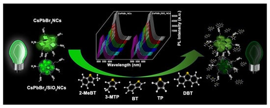
Figure 17.
Schematic illustration of the discrimination principle of the fluorescent sensor array for thiophene sulfides based on two perovskite NCs (permission obtained from Ref. [144]).
A method involving dynamic passivation over the surface of CsPbBr3 QDs (synthesized using the hot-injection method) was proposed by Huang et al. for the PL-enhanced detection of ammonia (NH3) [145]. In this study, the luminescence of purified QD film was enhanced when exposed to NH3. The linear range of PL enhancement at 610 nm was between 25 and 300 ppm with an LOD of 8.85 ppm. The photoluminescent response and recovery times were determined as 10 s and 30 s, respectively, at room temperature. In particular, this innovative work explains the analysis at room temperature. Following a similar approach, the employment of CsPbBr1.5Cl1.5 QDs, CsPbBr3 QDs, and CsPbBr1.5I1.5 QD films for the PL “turn-on” detection of ammonia (NH3) was demonstrated [146]. As illustrated in Figure 18, the QD films display exceptional selectivity among other interferences. All these films display good linear behavior (25–200 ppm) and LODs of ≈20 ppm. The uniqueness of this passivation method is well demonstrated by these reports, and hence it can be extended for developing commercialized devices for the detection of ammonia (NH3). Similar to the “turn-on” detection, a few CsPbX3-conjugated materials were also proposed for the discrimination and quantification of NH3 via PL quenching responses. Humidity-resistant CsPbBr3–SiO2 nanocomposites, porous nanofibers/nanocomposites (CsPbBr3 NFs and CsPbBr3/BNNF; BNNF = boron nitride nanofiber), and stable CsPbBr3 QDs grown within Fe-doped zeolite X were proposed for the PL-quenched detection of NH3 [147,148,149,150]. The adsorption and desorption of NH3 over the surface of these nanocomposites resulted in reversible cycles with given linear ranges and LODs. Table 4 summarizes the synthetic route, PLQY, linear ranges, LODs, and applications of CsPbX3 (X = Cl, Br, and I) and composites toward the detection of gas and VOCs.

Figure 18.
(a–c) The responses of CsPbBr1.5Cl1.5, CsPbBr3, and CsPbBr1.5I1.5 perovskite QD film sensors to various gases (permission obtained from Ref. [146]).
Table 4.
The synthetic route, PLQY, linear range, LOD, and application of CsPbX3 (X = Cl, Br, and I) and composites toward the detection of gas and VOCs.
Critical View on the Detection of CsPbX3 (X = Cl, Br, and I)-Based Gases and VOCs
Combining CsPbX3 (X = Cl, Br, and I) with different materials can result in composited materials with exceptional electro-optical properties and less defect, which can be adopted in the design of PL-based probes, electrochemiluminescence probes, chemoresistive sensors for discriminating N2, O2, H2S, tripropylamine (TPrA), thiophene sulfides, and NH3 [125,126,127,128,129,130,131,132,133,134,135,136,137,138,139,140,141,142,143,144,145,146,147,148,149,150]. To achieve the above goal, compositing ratios need to be optimized. Changes in the compositing ratios may significantly affect selectivity, thereby requiring careful/critical adjustments. The detection of H2S was demonstrated in rat brain and zebrafish studies [133,134,135], but there is no clear indication of how to overcome the toxicity induced by Pb2+ in CsPbX3. The film- or test-strip-based sensing of NH3 mostly displayed dependence on the crystalline and morphological features of CsPbX3 (X = Cl, Br, and I) and composites. Thus, optimizing the crystallinity/morphology of thin film is critical to attaining the best results.
7. Humidity, Temperature, and Radiation/Photodetection by CsPbX3 (X = Cl, Br, and I) and Composites
Humidity and moisture are the main causes resulting in perovskite material degradation, which affects the practical use and commercialization of perovskite-based energy devices [151]. The water molecules in the air react with the metal halide perovskite surface, which rapidly affects the morphology and uniformity, resulting in changes in optical properties and conductivity [152]. The above effect is the major mechanism of humidity-sensing responses. Doping with specified metal ions can improve the environmental stability of metal halide perovskites and reduce the humidity effect on the surface/morphology [153]. Due to the structural distortion (phase change) and instability of CsPbBr3, its composites can be effectively employed for the trace detection of water and humidity (%RH). For example, dimethyl aminoterephthalate-functionalized CsPbBr3 QDs (CsPbBr3@DMT-NH2 QDs) were synthesized with a low-temperature method and engaged in trace water detection in edible oils [154]. The PL emission at 530 nm was quenched in trace water (0.05–5%; v/v) with ratiometric enhancement at 445 nm. The LOD (3σ/slope) and limit of quantification (LOQ; 10σ/slope) were 0.01% and 0.04%, respectively. The involvement of the IFE, FRET, aggregation of QDs, and disintegration were proposed as the underlying mechanisms. This work is notable and can be extended to detect water traces in oils and chemicals. Thereafter, Xiang and co-workers described the luminescent quenching response of CsPbBr3 to trace water in herbal medicines [155]. The PL intensity at 503 nm was quenched linearly with water contents of 1–17%. The values of LODs in Seutellaria baicalensis and Astragalus flavone were 0.75% and 0.67%, respectively.
These materials also show a great response to varying humid conditions with a linear behavior between 33 and 98% RHs and an estimated LOD of 12% RH, as visualized in Figure 19. A phase change from CsPbBr3 to CsPb2Br5 was attributed as the mechanism for the observed PL quenching response to water and RH. The recovery of RH in the above herbals was in the range of 96.7–102.5%; therefore, this is noted as an inspiring work. The utilization of CsPbBr3 NPs in impedance-based humidity-sensing (under 20 mV) studies was also proposed [156]. The CsPbBr3 NPs were operative in the humidity range of 11–95% with a response/recovery time of 2.8 s/9.7 s and a sensitivity of 1.56% RH. This is also an inspiring work that can be further explored in commercial electrochemical device fabrication.
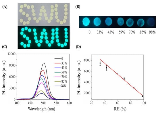
Figure 19.
(A) PL images of paper substrates loading CsPbBr3 perovskite. (B) PL images of paper-substrate-loading CsPbBr3 exposed to different RHs. (C) Humidity-dependent PL spectra of CsPbBr3 perovskite loaded on paper substrates. (D) Calibration curve for detecting different RHs, n = 3 (permission obtained from Ref. [155]).
Variations in temperature led to changes in phase and grain sizes [157], which can be adopted in temperature sensors via monitoring changes in I–V responses, PL intensity, absorbance, etc. On the other hand, alterations in temperature also affected the grain uniformity and morphologies of CsPbX3 and composites [158], resulting in changes in the PL intensity and current density. However, the doping of metal ions may also improve the temperature sensitivity of CsPbX3 and composites [159], as illustrated in this section. The doping of metal ions may enhance the sensitivity of CsPbX3 and composites. For example, Chang et al. described the temperature-sensing ability of Mn2+-doped CsPbCl3 QDs (CsPbCl3:0.1Mn2+ QDs; PLQY = 47.3%) via a dual-mode luminescent response at 298–353 K [160]. In this study, the stability of QDs was improved by replacing Pb2+ with Mn2+. For the 6% Mn2+-doped CsPbCl3 QDs, the PL intensity at 410 nm and 600 nm was quenched considerably compared with those of undoped CsPbCl3 QDs (quenching was observed at 410 nm only), as seen in Figure 20.

Figure 20.
Temperature-dependent emission spectra of (a) CsPbCl3 and (b) CsPbCl3:6% Mn2+ QDs in the range of 298 K–353 K. The integrated emission intensity centered at (c) approximately 410 nm and (d) approximately 600 nm as a function of temperature (permission opted from Ref. [160]).
The 6% Mn2+-doped CsPbCl3:QDs displayed 83% and 22% quenching at 410 nm and 600 nm, respectively. Using the fluorescence intensity ratio (FIR) and full width at half-maximum (FWHM), the maximum relative sensitivity (SR) of CsPbCl3:0% Mn2+ was 7.38% K−1 at 298 K and 2.13% K−1 at 353 K. Although this is a follow-up work of earlier reports [161,162], which also demonstrated the temperature-sensing ability through the dual-mode fluorescent response, its results are impressive. Moreover, Mn2+-doped CsPbCl3@glass [161] and Mn2+-doped CsPbCl3 NCs [162] were employed as dual-mode luminescent sensors in temperature ranges of 80–293 K and 80–30 K, respectively.
Similar to Mn2+ doping, Eu3+-doped cesium lead halide perovskite glasses (Eu3+:CsPbCl2Br1 QDs and Eu3+:CsPbBr3 QDs) were also used as effective materials in optical temperature-sensing studies [163,164]. With increasing temperature from 80 K to 440 K and 93 K to 383 K, both Eu3+:CsPbCl2Br1 QDs and Eu3+:CsPbBr3 QD glasses displayed blueshifted PL quenching at 458 nm (excitation at 395 nm) and 519 nm (excitation at 394 nm), respectively. For Eu3+:CsPbCl2Br1 QD glass, the absolute temperature sensitivity maxima (Sa) and the relative temperature sensitivity (Sr) were established as 0.0315 K−1 and 3.097%/K, respectively. Similarly, the Sa and Sr values of Eu3+:CsPbBr3 QD oxyhalide glass were 0.0224 K−1 and 2.25% K−1. Figure 21 shows the optical quenching response of one of the Eu3+:CsPbBr3 QD oxyhalide glass samples (S3) between 93 K and 383 K. These results also suggest that metal doping can enhance the temperature-sensing ability of cesium lead halide perovskites and composites for future device development.

Figure 21.
(a) Temperature-dependent PL spectra of Eu3+-doped PQDs@glass sample (S3). (b) Temperature-dependent PL spectra of CsPbBr3 PQD emission. (c) Integrated emission intensity of CsPbBr3 PQDs and Eu3+ as a function of temperature (permission obtained from Ref. [164]).
CsPbClxBr3−x NCs confined in hollow mesoporous silica (CsPbCl1.2Br1.8 NCs@h-SiO2) and integrated with K2SiF6:Mn4+ phosphor in the EVA polymer matrix (EVA = ethylene–vinyl acetate) were proposed by Huang and co-workers for reversible temperature sensing [165]. The high density of halide vacancies played a vital role in temperature sensing between 30.0 °C and 45.0 °C. The temperature sensitivity was established as 13.44% °C−1 at 37.0 °C. This is an inspiring work that investigates defects in sensor performance. Similarly, microencapsulated CsPbBr3 NCs with K2SiF6:Mn4+ phosphor (CsPbBr3-KSF-PS film) showed temperature sensitivity between 30 °C and 70 °C, with the Sr value reaching 0.31% °C−1 at 45 °C [166]. A highly stable film with its fluorescence visible to the naked eye (green-to-red fluorescence) was achieved by optimizing the CsPbBr3 and KSF ratio and applied for temperature sensing. Lu et al. developed dual-phase compounds containing CsPbBr3 QDs (emission at 529 nm) and NaYF4:Ho3+ NPs (emission at 647 nm and 751 nm) for conducting optical temperature sensing between 293 and 433 K at an excitation wavelength of 447 nm [167]. The nonradiative recombination between the energy band and the defective surface led to the thermal quenching of CsPbBr3. Sa and Sr values of 385 K−1 and 5.13% K−1 were reported in this work. This is an interesting report among all the studies on temperature sensors.
The use of cesium lead halides and composites in radiation/photo detection was proposed by numerous researchers [168,169,170,171,172,173,174,175,176,177,178,179,180]. Ti/Ni/CsPbBr3/Ni/Ti (with ER of 5.70% at 8250 V·cm−1; ER = energy resolution) [153], single crystals of CsPbBr3 (with diverse hole mobilities) [169,170,171,172,175], In/LiF/CPB/Au detectors [173], CsPbBr3 QDs embedded in P3HT:PC61BM (P3HT:PC61BM:QD; a bulk heterojunction photodiode) [174], CsPbBr3 microcrystals on ITO functional substrate [176], a single-crystalline thin film of CsPbBr3 (CsPbBr3 SCF; switching ratio = 3.2 × 10³, and response time = 200/300 ns) [177], CsPbBr3/RGO nanocomposites (RGO = reduced graphene oxide) [178], CsPbBr3 QD/ZnO NWs nanocomposites [179], and ZnONW/CsPbBr3 QD/graphene heterojunction were demonstrated for X-ray, radiation, and photodetection. Ion migration, photon–exciton coupling, electron–hole diffusion, and strain-based mechanisms were proposed as the underlying mechanisms for the radiation/photodetection. Most of the proposed cesium lead halide-based composites showed exceptional performance. For example, P3HT:PC61BM:QD [174] displayed good current density and X-ray photocurrent response by optimizing the weight ratio, as depicted in Figure 22. Since there are several reviews available on the radiation/photodetection performance of perovskites [14,15,16,17,18,19,32,61,63], they will not be discussed here.
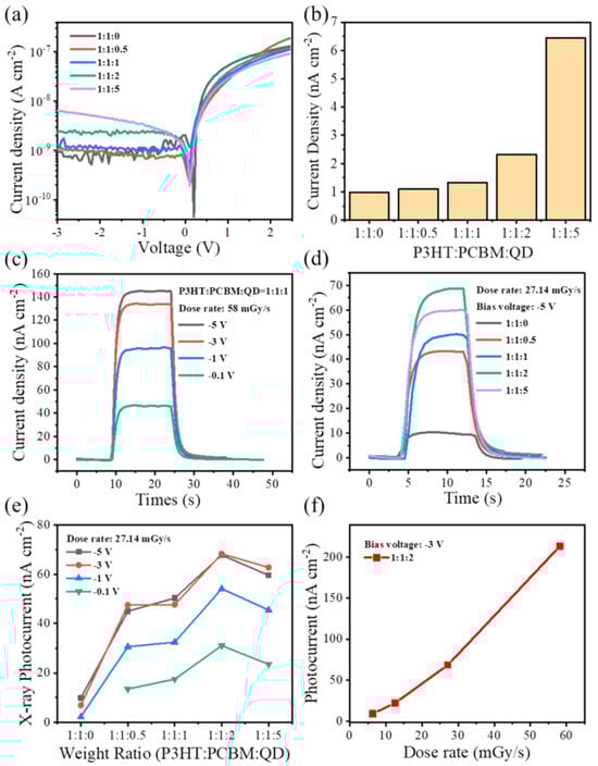
Figure 22.
(a) Dark J–V curves of the hybrid OPD with different weight ratios of P3HT:PC61BM:QD. (b) Dark current densities of hybrid OPDs under −3 V bias. (c) The X-ray response of the device with the weight ratio of 1:1:1 under various bias voltages. (d) The X-ray response of the hybrid OPDs with different QD weight ratios (dose rate: 27.14 mGy s−1). (e) X-ray photocurrent of devices with different QD weight ratios under a reversed bias varying from −0.1 to −5 V. (f) The linear response of the OPD device with a weight ratio of 1:1:2 at different dose rates (permission obtained from Ref. [174]).
Critical View on the Detection of CsPbX3 (X = Cl, Br, and I)-Based Humidity, Temperature, and Radiation/Photodetection
The low stability and degradation of CsPbX3 (X = Cl, Br, and I) and composites were adopted as sensor responses for humidity detection [151,152,153,154,155,156]. However, reports on = humidity sensors also discussed the recovery, which is problematic due to the environmental instability issue of the CsPbX3 (X = Cl, Br, and I) and composites. Thus, CsPbX3 (X = Cl, Br, and I) and composite-based recovery of humidity sensors must be carefully examined. Phase transitions may occur in CsPbX3 (X = Cl, Br, and I) and composites during temperature sensing [157,158,159,160,161,162,163,164,165,166,167]; thus, in-depth investigations are critical in many cases involving phase transitions. Likewise, doping of ions, such as Mn2+, may enhance the phase changes, which requires a more careful examination. When detecting photon/radiation, lattice defects could be generated in CsPbX3 (X = Cl, Br, and I). This problem can be alleviated by using composites with diverse materials [168,169,170,171,172,173,174,175,176,177,178,179,180]. However, the compositing ratios must be critically evaluated to achieve better sensing results.
8. CsPbX3 (X = Cl, Br, and I) and Composites in the Detection of Bioanalytes, Drugs, Fungicides, and Pesticides
Cesium lead halides and composites were applied in discriminating biologically significant analytes, as described in this section. Niu and co-workers constructed a dual-emitting nanoprobe consisting of CsPbBr3 NCs and red-emissive Cu NCs (CsPbBr3@Cu nanohybrid) for the luminescent detection of hydrogen peroxide (H2O2) and glucose [181]. CsPbBr3@Cu nanohybrid with PL emission centered at 517 nm and 645 nm displayed ratiometric responses to H2O2 and glucose. A linear ratiometric response at F645/F517 was observed in the ranges of 0.2–100 µM and 2.0–170.0 μM with LODs of 0.07 µM and 0.8 μM, respectively, when recognizing H2O2 and glucose. The electron-transfer-induced redox reaction was considered to be the underlying sensor mechanism. This method was also demonstrated for glucose detection in human serum samples with an impressive RSD value of < 4%. The TiO2/CsPbBr1.5I1.5 composite film and CsPbCl3/TiO2 served as inverse opal electrodes for the photoelectrochemical discrimination of dopamine (DA) and alpha-fetoprotein (AFP), respectively [182,183]. Slow volatilization was used to fabricate electrodes. The detection of DA showed a linear response between 0.1 and 250 μM, with an LOD of 12 nM, and the detection was also demonstrated in human serum samples [167]. Therefore, it is noted as an impressive report. On the contrary, the detection of AFP showed a linear regression between 0.08 and 980 ng/mL with an LOD of 30 pg/mL [183]. However, this report lacks information on real-time applications.
Saikia et al. reported the utilization of CsPbBr3 microcrystals (via one-pot synthesis; PLQY = 60%) as a sensing probe for the fluorometric detection of uric acid (UA) via hydrogen bonding interactions [184]. The PL of CsPbBr3 microcrystals at 520 nm (green to blue) was dynamically quenched within a response time of 30 s in the presence of UA. The linear regression of UA was observed between UA concentrations of 3.1 nM and 1.33 µM with an LOD of 0.063 ppm. This is a remarkable work with applications in human serum samples. Wang and co-workers described a ratiometric fluorescent approach for sensing acetylcholinesterase (AChE; 57 kDa protein; dispenses nerve impulse spread) by using the CsPbBr3 NC-TPPS nanocomposite (synthesized via self-assembly strategy; PLQY = 60%; TPPS = Tetraphenylporphyrin tetrasulfonic acid) [185]. The ratiometric response at F520/F650 to AChE showed a linear response between 0.05 and 1.0 U/L, with an LOD of 0.0042 U/L. Investigations of AChE quantification in human serum samples displayed >95% recovery, with an RSD of <4%. This is an interesting work with low interfering effects and real-time applicability. CsPbBr3 QDs and CsPbBr3 QD/MoS2 (MoS2 = molybdenum sulfide) nanoflakes were proposed as chemiluminescence biosensors for human hepatitis B, immunodeficiency virus, and AFP via the sandwich complex formation [186]. In this work, the CsPbBr3 QD/MoS2 nanoflakes were fabricated by including a parylene-C passivation layer. This is an interesting photosensor for multianalyte detection, which can be extended for commercialization.
Hu et al. reported the use of water-stable CsPbX3 (X = Br/I) as a probe for the sensitive detection of penicillamine (PA) [187]. Firstly, CsPbBr3 (PL maxima at 525 nm) interacted with iodide via an anion exchange reaction to afford CsPbX3 (X = Br/I), which displayed PL emission at 580 nm. As shown in Figure 23, the PL intensity increases accompanied by a blueshift when adding PA between 5.0 and 35.0 nM. The linear regression of PA quantification was observed between 5.0 and 35.0 nM, with LOD values of 1.19 nM (PL intensity vs. PA concentration) and 5.47 nM (PL peak shift vs. PA concentration). Based on the results from high-angle annular dark-field (HAADF) imaging and multiple investigations with organic S-containing substances, the interaction of the sulfhydryl group present in penicillamine with iodide is identified as the underlying sensor mechanism. This is an inspiring work in detecting PA in water. Through a one-pot synthetic method, stable carboxyl group-functionalized CsPbBr3–COOH QDs were developed by using amino-poly(ethylene glycol)-carboxyl and perfluorooctyltriethoxylsilane as ligands and were engaged in the PL “turn-on” detection of Mycobacterium tuberculosis (Mtb) [188].

Figure 23.
Detection of D-PA by CsPbX3 (Br/I) PNCs: (a) The 3D photoluminescence spectrum. (b) The 2D photoluminescence spectra and photos of corresponding samples at 365 nm (inset). The linear relationship between (c) relative intensity of photoluminescence and D-PA concentration, (d) photoluminescence wavelength shift, and D-PA concentration (permission obtained from Ref. [187]).
CsPbBr3–COOH QDs were composited with MoS2 and deoxy nucleic acid (DNA) to afford CsPbBr3 QD-DNA/MoS2 for the effective detection of Mtb via PL enhancement. Mtb showed a linear response between 0.2 and 4.0 nM, with an LOD of 51.9 pM. This work involved the clinical analysis of tuberculosis pathogens and thereby is noted as an innovative work. CsPbBr3 QDs composited with MoS2 and passivated with parylene (C, N, and F) were utilized in microbial detection [189] and photo-sensing of bioanalytes. An anodic electrochemiluminescence-based assay of alkaline phosphatase (LOD = 0.714 mU L−1; mU = milli units) was also proposed using CsPbBr3 QDs [190]. In both reports, charge/electron transport plays a vital role in the detection mechanism.
Li and co-workers used phospholipid-coated CsPbBr3 NCs for the effective detection of pore-forming biotoxins and prostate-specific antigens via dual-readout assays [191,192]. By using fluorometric and electrochemical assays, the pore-forming biotoxins were detected by CsPbBr3 NCs@PL, which showed a linear regression between 50 nM and 150 µM with an LOD of 50 nM [160]. Likewise, the detection of prostate-specific antigens by CsPbBr3 NCs@PL displayed a linear PL enhancement and colorimetric response in the ranges of 0.01–80 ng/mL and 0.1–15 ng/mL, with calculated LODs of 0.081 ng/mL and 0.29 ng/mL, respectively [192]. Both reports were applied in bacterial and clinical analyses, and they can be noted as innovative bioanalytical research. Qi et al. designed a composite consisting of of aptamer-functionalized CsPbBr3 NCs and magnetic nanoparticles of Fe3O4 (MNPs), namely the “Apt-PNCs@cDNA-MNPs” material, for detecting peanut allergen Ara h1 in food samples [193]. In their study, the CsPbBr3 NCs were employed as PL labeling probes for collecting fluorescent data. Also, the interaction of Ara h1 with aptamer resulted in PL recovery. The Apt-PNCs@cDNA-MNPs showed linear PL enhancement between 0.1 and 100 ng/mL, with an estimated LOD of 0.04 ng/mL. In terms of the reported real-time studies in food samples, this research can be regarded as inspiring work and should be extended toward food safety monitoring.
Similar to bioanalytes, cesium lead halide perovskites were also employed in the selective and sensitive quantification of drugs. 3-Aminopropyltriethoxysilane-functionalized CsPbBr3 QDs (APTES-IPQDs), mesoporous silica nanoparticle-composited CsPbBr3 QDs (LMSNs@IPQDs), perofluorooctyltriethyloxylsilane fluorocarbon-assembled Cs4PbBr6/CsPbBr3 NPs (CPB-PFOS), molecularly imprinted CsPbBr3 QDs (IPQDs@MIPs), and CsPbBr3 QD/BN composites (BN = boron nitride) were used in detecting tetracycline (TC; antibiotic drug) by means of photoinduced electron transfer (PET) or the inner-filter effect (IFE) mechanism [194,195,196,197,198]. All these materials displayed linear TC detection ranges in micro- to millimolar levels with estimated LODs at a nanomolar or micromolar concentration, as listed in Table 5. Moreover, these reports can further attest to the applicability of TC detection in food, water, and soil samples for possible commercialization. Figure 24 illustrates the PL quenching response of CPB-PFOS to TC and the related linear response [196].
Table 5.
The synthetic route, PLQY, linear range, detection limit, and application of CsPbX3 (X = Cl, Br, and I) and composites toward the detection of bioanalytes, drugs, fungicides, and pesticides.

Figure 24.
(a) Response of FL intensity of different concentrations of CPB-PFOS aqueous solution in the presence of 25 mM of TC, where F and F0 are FL intensity with and without TC, respectively. (b) The spectra of an aqueous solution of 5% (v/v) of CPB-PFOS samples with different concentrations of TC under an excitation of 355 nm wavelength. (c) Relationship between TC concentration and FL intensity log((F0 − F)/F0) = 3.281 + 0.7259 × log(CTC(M)) with R2 = 0.986 (permission opted from Ref. [196]).
Shi et al. described the sensing utility of CsPbBr3 NCs toward the quantification of ciprofloxacin hydrochloride (an antibiotic) via PL peak shifting induced by anion exchange [199]. In the presence of ciprofloxacin hydrochloride, the transformation from CsPbBr3 NCs to CsPbBr(3−x)Clx NCs occurred together with a corresponding peak shift from 513 nm to 442 nm via anion exchange. The PL peak shift was achieved between 0.8 and 50 mM, with an LOD of 0.1 mM. Though this work was applied in colorimetric paper-strip analysis, additional research is still required to lower the LODs. Through an in situ hot-injection method, CsPbBr3-loaded MIP nanogels were developed and employed for discriminating roxithromycin (ROX; an antibiotic) via PL quenching responses [200]. The nanogels showed linear PL quenching behavior between 100 pM and 100 nM, with an LOD of 20.6 pM in detecting ROX. Phase transformation and structural decomposition were proposed as the mechanisms underlying ROX detection. This work was applied in animal-derived food analysis, and hence it can be regarded as an inspiring work toward biomedical applications. Salari and co-workers adopted CsPbBr3 QDs (PLQY = 42%) in an organic phase, together with Fe(II) and K2S2O8, in an aqueous medium for the chemiluminescence-based detection of cefazolin (CFZ; an antibiotic) [201]. A chemiluminescence response of CsPbBr3 QDs in the presence of Fe(II) and K2S2O8 was observed in CFZ concentrations of 25–300 nM with an LOD of 9.6 nM at pH 7. This report was demonstrated with recoveries of >90% in multiple real samples, such as human plasma, urine, water, and milk samples. Although this is an interesting work, further research must be conducted to optimize experimental conditions.
Water-stable luminescent CsPbBr3/Cs4PbBr6 NCs were synthesized using a water emulsion technique and demonstrated for sensing folic acid (FA; vitamin B) [202]. The CsPbBr3/Cs4PbBr6 NCs showed a linear PL quenching response between 10 and 800 µM of FA, with an LOD of 1.695 µM. The quenching response was attributed to the electrostatic mechanism between NCs and FA. This work was demonstrated through a urine sample-based recovery (>99% with <0.5% RSD). It is a preliminary work on cesium lead halide-based FA detection. He et al. fabricated molecularly imprinted polymer-encoded CsPbX3 (X = Cl, Br, and I) microspheres for quantitatively detecting Sudan I (a food colorimetric enhancer) [203]. The PL emission of CsPbBr3, CsPbCl1.5Br1.5, and CsPbI2Br microspheres was recorded at 463 nm, 508 nm, and 644 nm, respectively. Interestingly, MIP-CsPbBr3 microspheres showed a greater response to Sudan I than nonimprinted ones (NIP-CsPbBr3), as shown in Figure 25. A linear range of between 2 and 604 nM with an estimated LOD of 1.21 nM was recorded for detecting Sudan I by MIP-CsPbBr3. This work was effectively applied in foodstuffs (egg and chili), which showed >95% recovery. Therefore, it can be utilized in commercial food safety monitoring. Thereafter, barium sulfate-coated cesium lead bromide nanocrystals (CsPbBr3 NCs@BaSO4) were proposed for the PL-enhanced quantitation of melamine [204]. The PL emission of CsPbBr3 NCs@BaSO4 was quenched via the IFE when adding Au NPs. When adding melamine to the above conjugate, the PL emission was restored to the original intensity via the weakening of the IFE. A linear detection range of melamine was recorded between 5 nM and 5 µM with an LOD of 0.42 nM. Through using this method, >95% recoveries of melamine in raw milk samples were achieved with <4% RSDs. This is an inspiring and innovative work for melamine monitoring. Su et al. fabricated a CsPbBr3/a-TiO2/FTO electrode for the photoelectrochemical immunoassay of aflatoxin B1 (AFB1; a carcinogen) by compositing CsPbBr3 NCs with amorphous titanium dioxide (TiO2) [205]. A linear regression of AFB1 detection by CsPbBr3/a-TiO2/FTO electrode was observed between 32 pM and 48 nM with an LOD of 9 pM. This work was confirmed by recoveries (ranging between 90.2% and 109.0%) in peanut and corn samples; therefore, it can be regarded as one of the most inspiring photoelectrochemical analytical methods.
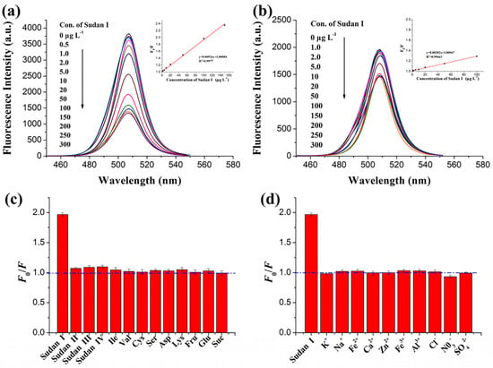
Figure 25.
Effect of different concentrations of Sudan I (0–300 µg L−1) on the fluorescence spectra of (a) MIP-CsPbBr3 and (b) NIP-CsPbBr3 probe; insets of (a,b) represent the corresponding calibration curve; fluorescence response of MIP-CsPbBr3 probe on (c) Sudan I, structural analogs, amino acids, and (d) some ions (permission obtained from Ref. [203]).
The employment of CsPbX3 (X = C, Br, and I) and composites toward the quantification of pesticides, insecticides, and fungicides has also been demonstrated by many researchers. CsPbBr3 QDs (PLQY = 96%), CsPbBr3 QD-coated MIPs (PLQY = 92%), CsPbI3 QDs (PLQY = 27%), MIP/CsPbBr3 QD composites, and MIP-mesoporous silica-embedded CsPbBr3 QDs were demonstrated for discriminating ziram (a fungicide), omethoate (an organophosphorus insecticide), clodinafop (an herbicide), phoxim (an organophosphate insecticide), and dichlorvos (2,2-dichlorovinyl dimethyl phosphate; an insecticide), respectively, via PL quenching responses [206,207,208,209,210]. These composites were developed using diverse methods, such as room-temperature-controlled synthesis, the slow hydrolysis of the capping agent, microwave synthesis, self-assembly, etc. It should be noted that cesium lead halide composites have displayed exceptional linear regression with excellent LODs and have been applied in real-time food stuff/soil analysis, as detailed in Table 5. Moreover, all these composites showed negligible interfering effects from competing species, which is rather unique. For example, the MIP/CsPbBr3 QD composite [209] displayed greater selectivity than other interferences, as seen in Figure 26. Table 5 summarizes the synthetic route, PLQY, linear range, detection limit, and application of CsPbX3 (X = Cl, Br, and I) and composites toward the detection of bioanalytes, drugs, fungicides, and pesticides.

Figure 26.
Response performance of the MIP/QDs and NIP/QDs to different OPPs (0.13 μM; OPPS = organophosphorus pesticides) (permission opted from Ref. [209]).
Critical View on the Detection of CsPbX3 (X = Cl, Br, and I)-Based Bioanalytes, Drugs, Fungicides, and Pesticides
The stability of CsPbX3 (X = Cl, Br, and I) in water is the main issue for discriminating bioanalytes, drugs, fungicides, and pesticides [181,182,183,184,185,186,187,188,189,190,191,192,193,194,195,196,197,198,199,200,201,202,203,204,205,206,207,208,209,210]. To avoid the above complications, compositing CsPbX3 (X = Cl, Br, and I) with other materials, such as APTES, BN, and MIPs has been proposed. However, there are still a few probes that require critical evaluation in terms of structural degradation in aqueous media. Water-stable CsPbBr3/Cs4PbBr6 NCs were proposed for detecting folic acid [202], but how long can the proposed structure remain stable is still under debate. Another controversial issue regarding CsPbX3 (X = Cl, Br, and I) and composite-based biomolecule sensing is how to avoid the toxicity of Pb2+ in food sample analysis. Consequently, the sensor’s selectivity to specific analytes requires careful investigation in many reports. Detecting pesticides and fungicides in real samples using CsPbX3 (X = Cl, Br, and I) and composites needs critical analysis to overcome the toxicity induced by Pb2+.
9. Cellular Imaging Applications of CsPbX3 (X = Cl, Br, and I) and Composites
Getachew et al. and Kar et al. described the cellular imaging utility of magnesium- and zinc-doped cesium lead halides, namely CsMgxPb1−xI3 QDs and zinc-doped CsPbBr3-Cs4PbBr6 nanocomposites [211,212]. The doping of metal ions in perovskite quantum dots could result in stability and biocompatibility improvement. In CsMgxPb1−xI3 QDs, Mg2+ partially substituted Pb2+ in the CsPbI3 framework [211]. When encapsulating CsMgxPb1−xI3 QDs with gadolinium-conjugated pluronic 127 (PF127-Gd), PQD@Gd nanoagents were achieved, which were applied in ROS detection in cancer cells and photocatalytic studies. The Zn2+ doping in CsPbBr3-Cs4PbBr6 NCs (emission at 494–506 nm; PLQY = 88%) improved the stability compared to bare NCs in highly polar solvents [212]. Moreover, the zinc-doped ensemble showed greater biocompatibility, thereby becoming effective in cellular imaging, as visualized in Figure 27. In this report, silica-coated CsPbBr3-Cs4PbBr6@(OA)2PbBr4 (core–shell NCs synthesized via the LARP method using (3-amino-propyl)trimethoxysilane [APTMS]) without zinc doping were labeled as NC-0. The NCs with 20%, 40%, 60%, and 80% zinc doping were labeled as NC-20, NC-40, NC-60, and NC-80, respectively. NC-40 was engaged in cellular imaging studies because of its exceptional biocompatibility and stability. Similarly, CsPbBr3@SiO2 core–shell, CsPbBr3/SiO2/mPEG-DSPE NCs (mPEG-DSPE = polyethylene glycol-grafted phospholipid), and phTEOS-TMOS@CsPbBr3 NCs (TMOS and phTEOS represent the alkoxysilanes) were also demonstrated in bioimaging studies [213,214,215]. SiO2-coated CsPbBr3 core–shell structures reduced the toxicity and made NCs more effective for in vitro cellular imaging [213]. Hydrophobic CsPbBr3/SiO2 encapsulated with mPEG-DSPE showed better water stability and photostability; thus, they can be applied in multiphoton bioimaging [214]. CsPbBr3 NCs coated with alkoxysilanes showed improved stability, water dispersibility, and lower toxicity and thus can be used in two-photon cellular imaging studies [215].
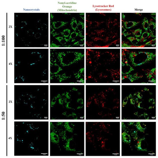
Figure 27.
Biocompatibility of NC-40 with mammalian cells. NCs (blue) are internalized by cells and do not associate with mitochondria (green) or lysosomes (red). Higher concentrations of NCs have a detrimental effect on cells as mitochondrial and lysosomal structures become atypical. NCs, nanocrystals (permission obtained from Ref. [212]).
Many research groups reported the preparation of encapsulated CsPbX3 (X = C, Br, and I) and composites with a polymer matrix to maintain the structural and emission stability of cesium lead halides. Methoxypolyethylene glycol amine-capped CsPbBr3 NCs (CsPbBr3/mPEG-NH2 NCs), polyvinylidene fluoride (PVDF)-encapsulated CsPbBr3, poly-vinyl pyrrolidone (PVP)-capped CsPbX3 NCs, poly(lactic-co-glycolic acid) (PLGA)-encapsulated CsPbBr3 QDs, and polystyrene-block-poly(acrylic acid) (PS-b-PAA) were developed with high water stability, greater biocompatibility, and low toxicity; thus, they can be applied in long-term cellular imaging studies [216,217,218,219,220]. For cellular imaging of cesium lead halide perovskites, Lou et al. synthesized insoluble CsPbBr3/CsPb2Br5-composited NCs (PLQY = 80%) via water-assisted chemical transformations and HeLa cellular imaging studies [221]. Judging from the synthetic simplicity and low toxicity, this work can be regarded as inspiring. However, more investigations on structural stability and PL emission are required.
Critical View on CsPbX3 (X = Cl, Br, and I)-Based Cellular Imaging
It has been argued that cellular imaging using CsPbX3 (X = Cl, Br, and I) could be hindered due to the unstable emission properties of CsPbX3 in aqueous media. To avoid the instability issue, capping CsPbX3 (X = Cl, Br, and I) with suitable ligands, such as 3-amino-propyl)trimethoxysilane (APTMS), polyethylene glycol-grafted phospholipid (mPEG-DSPE), and alkoxysilanes (TMOS and phTEOS) has been proposed. This capping could not only reduce toxicity due to Pb2+ but can also improve biocompatibility [211,212,213,214,215]. However, careful optimization is still required. Polymer capping, metal doping, and surface coating were also used to improve the biocompatibility of CsPbX3 (X = Cl, Br, and I) in cellular imaging studies [216,217,218,219,220,221]. Nevertheless, critical assessments of toxicity profiles when applying these composites in cellular imaging still require much attention.
10. Advantages and Limitations
The employment of CsPbX3 (X = Cl, Br, and I) and composites in sensing investigations has certain advantages and limitations, as noted below.
10.1. Advantages
- Due to the unique structural features, tuning the photophysical properties of CsPbX3 (X = Cl, Br, and I) and anion exchange can result in red, green, and blue emission with an enhanced PLQY (reaches up to 98%); therefore, PL-based sensors with relevant colorimetric responses can benefit from the above properties.
- The greater carrier mobility of CsPbX3 (X = Cl, Br, and I) can be adjusted by combining with other semiconducting materials (such as MoS2, graphene, mxenes, etc.), which is advantageous to the fabrication of heterojunction devices and electrodes for photo/chemoresistive and electrochemical detection of a specific analyte [222,223,224,225].
- The capping and encapsulation of the proposed CsPbX3 (X = Cl, Br, and I) system can enhance water stability, which allows for long-term tracking of bioanalytes.
- Core–shell/polymer encapsulation can produce a low-toxicity, biocompatible CsPbX3 (X = Cl, Br, and I) conjugate, which can be applied in bioimaging with comparable performance to other lead-free perovskites and luminescent organic probes [226,227,228,229].
- Advancing sensing studies in real samples is highly feasible with capped/functionalized CsPbX3 (X = Cl, Br, and I) nanostructures; therefore, their performance can be comparable to other nanoprobes (nanoparticles, nanoclusters, nanowires, nanocomposites, etc.) [230,231,232,233,234,235].
10.2. Limitations
- Many stable CsPbX3 (X = Cl, Br, and I) and composites are fabricated using multiple synthetic steps, which require precision optimization. However, optimizing synthesis processes is time-consuming, which restricts the use of CsPbX3 (X = Cl, Br, and I) as sensor probes.
- CsPbX3 (X = Cl, Br, and I) and composites can degrade rapidly when exposed to air moisture, elevated temperature, humid conditions, etc., thereby limiting their sensing utility in harsh conditions [236,237,238].
- The major contributing factor for the sensing performance of CsPbX3 (X = Cl, Br, and I) and composites has been attributed to structural/phase transformation [239,240]. Thus, it is questionable if reversible cycles in analyte detection can be realized.
- To investigate the precise underlying mechanism of the sensing response to a specific analyte, supporting lines of evidence through methods, such as TEM, XPS, dynamic light scattering spectra (DLS), Zeta potential, etc., are necessary. Thus, the cost-effectiveness of research is questionable, which limits such sensor development in developing or underdeveloped countries.
- Since real water samples may contain certain numbers of ionic species, the reliability of metal ion quantification by CsPbX3 (X = Cl, Br, and I) and composites in real samples is still questionable. This limits the application of CsPbX3 (X = Cl, Br, and I) probes toward the detection of metal ions and anions.
11. Conclusions and Perspectives
This article provides a detailed review of the sensing performance of CsPbX3 (X = Cl, Br, and I) and composites toward the detection of analytes, such as metal ions, anions, chemicals, explosives, gases, volatile organic compounds, bioanalytes, humidity, temperature, radiation, etc. In particular, the involved synthetic methods in developing CsPbX3 (X = Cl, Br, and I)-based sensor probes, the linear detection range, LODs, and real-time applicability were also tabulated for a broad audience. Discussions on the mechanistic aspects of analyte detection, novelty, deficiencies, and feasible directions were also extrapolated for readers. Finally, the bioimaging applications of CsPbX3 (X = Cl, Br, and I)-based composites were illustrated for future biomedical research.
The following questions/perspective points must be addressed or focused on in future research: (A) There are many reports available on the employment of CsPbX3 (X = Cl, Br, and I)-based probes toward Cu2+ quantification; hence, a state-of-the-art procedure for fabricating commercial sensors must be developed; (B) to date, the detection of Cu2+, Hg2+, Co2+, Zn2+, Cd2+, UO22+, In3+, and Fe3+ have been demonstrated using CsPbX3 (X = Cl, Br, and I)-based probes, but the research direction should be further extended toward the sensing of monovalent (M+) cations, divalent (M2+) cations, trivalent (M3+) cations, etc., by optimizing the functional/capping agents; (C) the method can be further fine-tuned to develop an effective procedure for the commercialization of anion-exchange-facilitated detection of Cl− and I− by CsPbBr3 toward real-time monitoring; (D) the optimization of functional/capping moieties can be carried out to extend the sensing capability of CsPbX3 (X = Cl, Br, and I)-based probes to existing anions, such as SO42−, PO42−, P2O72−, CN−, SCN−, etc.; (E) the anion-exchange-directed detection of alkyl halides, benzoyl peroxide, and solvent polarity by CsPbX3 (X = Cl, Br, and I) and composites requires in-depth investigation to confirm its novelty; (F) advancing CsPbX3 (X = Cl, Br, and I) and composites toward the detection of toxic gases, such as carbon monoxide (CO), carbon dioxide (CO2), phosgene (COCl2), etc., must be the focus in future research; (G) numerous reports on CsPbBr3-based NH3 sensors are available; therefore, fabricating commercial NH3-sensing devices must be the focus in future research; (H) research on humidity, temperature, radiation, and photodetection using CsPbX3 (X = Cl, Br, and I) and composites can be expanded to include other nanostructures, such as graphene, metal–organic frameworks, nanosheets, nanotubes, etc.; (I) few reports are available on detecting essential bioanalytes, drugs, fungicides, and pesticides using CsPbX3 (X = Cl, Br, and I) and composites; thus, more attention is required in this research direction; (J) in-depth (in vitro/in vivo) cellular imaging, tracking, and the underlying mechanisms of the imaging responses of CsPbX3 (X = Cl, Br, and I) and composites and the construction of low-toxicity probes must be carried out in future research; (K) the majority of CsPbX3 (X = Cl, Br, and I) and composite-based sensor reports lack theoretical support from density functional theory (DFT) calculations, which should be included in new sensor designs.
Apart from the above open questions and possible future research directions, sensing studies that involve composites of CsPbX3 (X = Cl, Br, and I) toward the detection of diverse analytes require more innovative approaches to realize real-time applications. Many scientists are currently working on developing a “state-of-the-art” sensor procedure for the detection of diverse analytes. Thus, innovative breakthroughs can be expected in the near future.
Author Contributions
Literature collection, organization, writing—original draft preparation, M.S.; writing—review and editing and project administration, K.W.S.; writing—review and editing, N.T.; writing—review and editing, M.B.; writing—review and editing, A.M. All authors have read and agreed to the published version of the manuscript.
Funding
This research was supported by the Ministry of Science and Technology of Taiwan under Contract No. MOST 111-2112-M-A49-031 and MOST 111-2811-M-A49-528.
Institutional Review Board Statement
Not applicable.
Informed Consent Statement
Not applicable.
Data Availability Statement
Not applicable.
Conflicts of Interest
The authors declare no conflicts of interest.
References
- Butt, M.A.; Voronkov, G.S.; Grakhova, E.P.; Kutluyarov, R.V.; Kazanskiy, N.L.; Khonina, S.N. Environmental Monitoring: A Comprehensive Review on Optical Waveguide and Fiber-Based Sensors. Biosensors 2022, 12, 1038. [Google Scholar] [CrossRef] [PubMed]
- Dhall, S.; Mehta, B.R.; Tyagi, A.K.; Sood, K. A Review on Environmental Gas Sensors: Materials and Technologies. Sens. Inter. 2021, 2, 100116. [Google Scholar] [CrossRef]
- Gavrilaș, S.; Ursachi, C.Ș.; Perța-Crișan, S.; Munteanu, F.-D. Recent Trends in Biosensors for Environmental Quality Monitoring. Sensors 2022, 22, 1513. [Google Scholar] [CrossRef] [PubMed]
- Shellaiah, M.; Sun, K.-W. Review on Anti-Aggregation-Enabled Colorimetric Sensing Applications of Gold and Silver Nanoparticles. Chemosensors 2022, 10, 536. [Google Scholar] [CrossRef]
- Kannan, P.; Maduraiveeran, G. Metal Oxides Nanomaterials and Nanocomposite-Based Electrochemical Sensors for Healthcare Applications. Biosensors 2023, 13, 542. [Google Scholar] [CrossRef]
- Shellaiah, M.; Sun, K.W. Review on Carbon Dot-Based Fluorescent Detection of Biothiols. Biosensors 2023, 13, 335. [Google Scholar] [CrossRef]
- Shakeel, A.; Rizwan, K.; Farooq, U.; Iqbal, S.; Iqbal, T.; Awwad, N.S.; Ibrahium, H.A. Polymer-based Nanocomposites: A Strategic Tool for Detection of Toxic Pollutants in Environmental Matrices. Chemosphere 2022, 303, 134923. [Google Scholar] [CrossRef]
- Cho, I.-H.; Kim, D.H.; Park, S. Electrochemical Biosensors: Perspective on Functional Nanomaterials for On-Site Analysis. Biomater. Res. 2020, 24, 6. [Google Scholar] [CrossRef] [PubMed]
- Shellaiah, M.; Sun, K.W. Review on Sensing Applications of Perovskite Nanomaterials. Chemosensors 2020, 8, 55. [Google Scholar] [CrossRef]
- George, K.J.; Halali, V.V.; Sanjayan, C.G.; Suvina, V.; Sakar, M.; Balakrishna, R.G. Perovskite Nanomaterials as Optical and Electrochemical Sensors. Inorg. Chem. Front. 2020, 7, 2702–2725. [Google Scholar] [CrossRef]
- Xiang, W.; Liu, S.; Tress, W. A Review on the Stability of Inorganic Metal Halide Perovskites: Challenges and Opportunities for Stable Solar Cells. Energy Environ. Sci. 2021, 14, 2090–2113. [Google Scholar] [CrossRef]
- Xun, J.; Deng, J.; Shen, W.; Li, M.; He, R. Rapid Synthesis of Highly Stable All-Inorganic Perovskite Nanocrystals Exhibiting Strong Blue Luminescence. J. Alloys Comp. 2021, 872, 159612. [Google Scholar] [CrossRef]
- Sanjayan, C.G.; Jyothi, M.S.; Balakrishna, R.G. Stabilization of CsPbBr3 Quantum Dots for Photocatalysis, Imaging and Optical Sensing in Water and Biological Medium: A Review. J. Mater. Chem. C 2022, 10, 6935–6956. [Google Scholar]
- Zhu, Z.; Sun, Q.; Zhang, Z.; Dai, J.; Xing, G.; Li, S.; Huang, X.; Huang, W. Metal halide perovskites: Stability and sensing-ability. J. Mater. Chem. C 2018, 6, 10121–10137. [Google Scholar] [CrossRef]
- Jia, D.; Xu, M.; Mu, S.; Ren, W.; Liu, C. Recent Progress of Perovskite Nanocrystals in Chem/Bio-Sensing. Biosensors 2022, 12, 754. [Google Scholar] [CrossRef] [PubMed]
- Casanova-Chafer, J.; Garcia-Aboal, R.; Atienzar, P.; Bittencourt, C.; Llobet, E. Perovskite@Graphene Nanohybrids for Breath Analysis: A Proof-of-Concept. Chemosensors 2021, 9, 215. [Google Scholar] [CrossRef]
- Aamir, M.; Shahiduzzaman, M.; Taima, T.; Akhtar, J.; Nunzi, J.-M. It is an All-Rounder! On the Development of Metal Halide Perovskite-Based Fluorescent Sensors and Radiation Detectors. Adv. Opt. Mater. 2021, 9, 2101276. [Google Scholar] [CrossRef]
- Bu, H.; He, C.; Xu, Y.; Xing, L.; Liu, X.; Ren, S.; Yi, S.; Chen, L.; Wu, H.; Zhang, G.; et al. Emerging New-Generation Detecting and Sensing of Metal Halide Perovskites. Adv. Electron. Mater. 2022, 8, 2101204. [Google Scholar] [CrossRef]
- Kailasa, S.K.; Vajubhai, G.N.; Koduru, J.R.; Park, T.J.; Hussain, C.M. Recent Progress on the Modifications of Ultra-Small Perovskite Nanomaterials for Sensing Applications. TrAC Trends Anal. Chem. 2021, 144, 116432. [Google Scholar] [CrossRef]
- Liu, P.; Yu, B.; Cai, W.; Yao, X.; Chang, K.; Zhao, X.; Si, Z.; Deng, W.; Zhou, Y.; Zhou, G.; et al. Air-Stable High-PLQY Cesium Lead Halide Perovskites for Laser-Patterned Displays. J. Mater. Chem. C 2023, 11, 2282–2290. [Google Scholar] [CrossRef]
- Ghosh, D.; Ali, M.Y.; Chaudhary, D.K.; Bhattacharyya, S. Dependence of Halide Composition on the Stability of Highly Efficient All-Inorganic Cesium Lead Halide Perovskite Quantum Dot Solar Cells. Solar Energy Mater. Solar Cells 2018, 185, 28–35. [Google Scholar] [CrossRef]
- Unger, E.L.; Kegelmann, L.; Suchan, K.; Sörell, D.; Korte, L.; Albrecht, S. Roadmap and Roadblocks For the Band Gap Tunability of Metal Halide Perovskites. J. Mater. Chem. A 2017, 5, 11401–11409. [Google Scholar] [CrossRef]
- Li, Y.; Lu, Y.; Huo, X.; Wei, D.; Meng, J.; Dong, J.; Qiao, B.; Zhao, S.; Xu, Z.; Song, D. Bandgap Tuning Strategy by Cations and Halide Ions of Lead Halide Perovskites Learned from Machine Learning. RSC Adv. 2021, 11, 15688–15694. [Google Scholar] [CrossRef] [PubMed]
- Jung, J.; Yun, Y.; Yang, S.W.; Oh, H.G.; Jeon, A.Y.; Nam, Y.; Heo, Y.-W.; Chae, W.-S.; Lee, S. Ternary Diagrams of Phase, Stability, and Optical Properties of Cesium Lead Mixed-Halide Perovskites. Acta Mater. 2023, 246, 118661. [Google Scholar] [CrossRef]
- Tai, M.; Lau, C.F.J.; Lin, H.; Wang, Z. Advances in Phase Stability of Cesium Lead Halide Perovskites. Sol. RRL 2020, 4, 2000495. [Google Scholar] [CrossRef]
- Yang, D.; Cao, M.; Zhong, Q.; Li, P.; Zhang, X.; Zhang, Q. All-Inorganic Cesium Lead Halide Perovskite Nanocrystals: Synthesis, Surface Engineering and Applications. J. Mater. Chem. C 2019, 7, 757–789. [Google Scholar] [CrossRef]
- Jung, H.-S.; Cho, J.; Neuman, K.C. Highly Stable Cesium Lead Bromide Perovskite Nanocrystals for Ultra-Sensitive and Selective Latent Fingerprint Detection. Anal. Chim. Acta 2021, 1181, 338850. [Google Scholar] [CrossRef] [PubMed]
- Yuan, G.; Feng, S.; Yang, Q.; Yi, F.; Li, X.; Yuan, Y.; Wang, C.; Yan, H. Promoting Charge Separation in A Composite of δ-CsPbI3 and Covalent Organic Frameworks. J. Mater. Chem. C 2023, 11, 7570–7574. [Google Scholar] [CrossRef]
- Cao, L.; Liu, X.; Li, Y.; Li, X.; Du, L.; Chen, S.; Zhao, S.; Wang, C. Recent Progress in All-Inorganic Metal Halide Nanostructured Perovskites: Materials Design, Optical Properties, and Application. Front. Phys. 2020, 16, 33201. [Google Scholar] [CrossRef]
- Skurlov, I.D.; Sokolova, A.V.; Tatarinov, D.A.; Parfenov, P.S.; Kurshanov, D.A.; Ismagilov, A.O.; Koroleva, A.V.; Danilov, D.V.; Zhizhin, E.V.; Mikushev, S.V.; et al. Engineering the Optical Properties of CsPbBr3 Nanoplatelets through Cd2+ Doping. Materials 2022, 15, 7676. [Google Scholar] [CrossRef]
- Akkerman, Q.A.; D’Innocenzo, V.; Accornero, S.; Scarpellini, A.; Petrozza, A.; Prato, M.; Manna, L. Tuning the Optical Properties of Cesium Lead Halide Perovskite Nanocrystals by Anion Exchange Reactions. J. Am. Chem. Soc. 2015, 137, 10276–10281. [Google Scholar] [CrossRef] [PubMed]
- Wu, Y.; Feng, J.; Yang, Z.; Liu, Y.; Liu, S. Halide Perovskite: A Promising Candidate for Next-Generation X-Ray Detectors. Adv. Sci. 2023, 10, 2205536. [Google Scholar] [CrossRef] [PubMed]
- Chen, Y.-C.; Chou, H.-L.; Lin, J.-C.; Lee, Y.-C.; Pao, C.-W.; Chen, J.-L.; Chang, C.-C.; Chi, R.-Y.; Kuo, T.-R.; Lu, C.-W.; et al. Enhanced Luminescence and Stability of Cesium Lead Halide Perovskite CsPbX3 Nanocrystals by Cu2+-Assisted Anion Exchange Reactions. J. Phys. Chem. C 2019, 123, 2353–2360. [Google Scholar] [CrossRef]
- Chistyakov, A.A.; Zvaigzne, M.A.; Nikitenko, V.R.; Tameev, A.R.; Martynov, I.L.; Prezhdo, O.V. Optoelectronic Properties of Semiconductor Quantum Dot Solids for Photovoltaic Applications. J. Phys. Chem. Lett. 2017, 8, 4129–4139. [Google Scholar] [CrossRef] [PubMed]
- Peltonen, L.; Strachan, C. Understanding Critical Quality Attributes for Nanocrystals from Preparation to Delivery. Molecules 2015, 20, 22286–22300. [Google Scholar] [CrossRef] [PubMed]
- Hu, F.; Zhang, H.; Sun, C.; Yin, C.; Lv, B.; Zhang, C.; Yu, W.W.; Wang, X.; Zhang, Y.; Xiao, M. Superior Optical Properties of Perovskite Nanocrystals as Single Photon Emitters. ACS Nano 2015, 9, 12410–12416. [Google Scholar] [CrossRef] [PubMed]
- Maity, S.; Bain, D.; Patra, A. An Overview on the Current Understanding of the Photophysical Properties of Metal Nanoclusters and Their Potential Applications. Nanoscale 2019, 11, 22685–22723. [Google Scholar] [CrossRef] [PubMed]
- Shellaiah, M.; Chen, T.H.; Simon, T.; Li, L.-C.; Sun, K.W.; Ko, F.-H. An Affordable Wet Chemical Route to Grow Conducting Hybrid Graphite-Diamond Nanowires: Demonstration by A Single Nanowire Device. Sci. Rep. 2017, 7, 11243. [Google Scholar] [CrossRef] [PubMed]
- Shellaiah, M.; Chen, Y.-C.; Simon, T.; Li, L.-C.; Sun, K.W.; Ko, F.-H. Effect of Metal Ions on Hybrid Graphite-Diamond Nanowire Growth: Conductivity Measurements from a Single Nanowire Device. Nanomaterials 2019, 9, 415. [Google Scholar] [CrossRef] [PubMed]
- Zhai, W.; Lin, J.; Li, C.; Hu, S.; Huang, Y.; Yu, C.; Wen, Z.; Liu, Z.; Fang, Y.; Tang, C. Solvothermal Synthesis of Cesium Lead Halide Perovskite Nanowires with Ultra-High Aspect Ratios for High-Performance Photodetectors. Nanoscale 2018, 10, 21451–21458. [Google Scholar] [CrossRef] [PubMed]
- Zhang, S.; Liu, C.; Zhang, G.; Chen, Y.; Shang, F.; Xia, Q.; Yang, W. Full review: The Progress and Developing Trends of Nanosheet-Based Sensing Applications. Coord. Chem. Rev. 2021, 433, 213742. [Google Scholar] [CrossRef]
- Lv, L.; Xu, Y.; Fang, H.; Luo, W.; Xu, F.; Liu, L.; Wang, B.; Zhang, X.; Yang, D.; Hu, W.; et al. Generalized Colloidal Synthesis of High-Quality, Two-Dimensional Cesium Lead Halide Perovskite Nanosheets and Their Applications in Photodetectors. Nanoscale 2016, 8, 13589–13596. [Google Scholar] [CrossRef]
- Khan, Y.; Sadia, H.; Ali Shah, S.Z.; Khan, M.N.; Shah, A.A.; Ullah, N.; Ullah, M.F.; Bibi, H.; Bafakeeh, O.T.; Khedher, N.B.; et al. Classification, Synthetic, and Characterization Approaches to Nanoparticles, and Their Applications in Various Fields of Nanotechnology: A Review. Catalysts 2022, 12, 1386. [Google Scholar] [CrossRef]
- Qasem, N.A.A.; Mohammed, R.H.; Lawal, D.U. Removal of Heavy Metal Ions from Wastewater: A Comprehensive and Critical Review. Npj Clean Water 2021, 4, 36. [Google Scholar] [CrossRef]
- Kumar, E.; Bhatnagar, A.; Hogland, W.; Marques, M.; Sillanpää, M. Interaction of Anionic Pollutants with Al-based Adsorbents in Aqueous media—A Review. Chem. Eng. J. 2014, 241, 443–456. [Google Scholar] [CrossRef]
- Zou, Y.; Wang, X.; Khan, A.; Wang, P.; Liu, Y.; Alsaedi, A.; Hayat, T.; Wang, X. Environmental Remediation and Application of Nanoscale Zero-Valent Iron and Its Composites for the Removal of Heavy Metal Ions: A Review. Environ. Sci. Technol. 2016, 50, 7290–7304. [Google Scholar] [CrossRef] [PubMed]
- Idris, A.O.; Orimolade, B.; Dennany, L.; Mamba, B.; Azizi, S.; Kaviyarasu, K.; Maaza, M. A Review on Monitoring of Organic Pollutants in Wastewater Using Electrochemical Approach. Electrocatalysis 2023, 14, 659–687. [Google Scholar] [CrossRef]
- Kim, C.; Eom, J.B.; Jung, S.; Ji, T. Detection of Organic Compounds in Water by an Optical Absorbance Method. Sensors 2016, 16, 61. [Google Scholar] [CrossRef]
- To, K.C.; Ben-Jaber, S.; Parkin, I.P. Recent Developments in the Field of Explosive Trace Detection. ACS Nano 2020, 14, 10804–10833. [Google Scholar] [CrossRef] [PubMed]
- Onyancha, R.B.; Ukhurebor, K.E.; Aigbe, U.O.; Osibote, O.A.; Kusuma, H.S.; Darmokoesoemo, H.; Balogun, V.A. A systematic review on the detection and monitoring of toxic gases using carbon nanotube-based biosensors. Sens. Bio-Sens. Res. 2021, 34, 100463. [Google Scholar] [CrossRef]
- Mirzaei, A.; Leonardi, S.G.; Neri, G. Detection of Hazardous Volatile Organic Compounds (VOCs) by Metal Oxide Nanostructures-based Gas Sensors: A Review. Ceram. Inter. 2016, 42, 15119–15141. [Google Scholar] [CrossRef]
- Wang, H.; Sun, Y.; Chen, J.; Wang, F.; Han, R.; Zhang, C.; Kong, J.; Li, L.; Yang, J. A Review of Perovskite-Based Photodetectors and Their Applications. Nanomaterials 2022, 12, 4390. [Google Scholar] [CrossRef] [PubMed]
- Arneodo, F.; Di Giovanni, A.; Marpu, P. A Review of Requirements for Gamma Radiation Detection in Space Using CubeSats. Appl. Sci. 2021, 11, 2659. [Google Scholar] [CrossRef]
- Arman Kuzubasoglu, B.; Kursun Bahadir, S. Flexible Temperature Sensors: A Review. Sens. Actuators A 2020, 315, 112282. [Google Scholar] [CrossRef]
- Wang, T.; Ramnarayanan, A.; Cheng, H. Real-Time Analysis of Bioanalytics in Healthcare, Food, Zoology and Botany. Sensors 2018, 18, 5. [Google Scholar] [CrossRef] [PubMed]
- Ahmed, S.R.; Chand, R.; Kumar, S.; Mittal, N.; Srinivasan, S.; Rajabzadeh, A.R. Recent Biosensing Advances in the Rapid Detection of Illicit Drugs. TrAC Trends Anal. Chem. 2020, 131, 116006. [Google Scholar] [CrossRef]
- Gikas, G.D.; Parlakidis, P.; Mavropoulos, T.; Vryzas, Z. Particularities of Fungicides and Factors Affecting Their Fate and Removal Efficacy: A Review. Sustainability 2022, 14, 4056. [Google Scholar] [CrossRef]
- Zamora-Sequeira, R.; Starbird-Pérez, R.; Rojas-Carillo, O.; Vargas-Villalobos, S. What are the Main Sensor Methods for Quantifying Pesticides in Agricultural Activities? A Review. Molecules 2019, 24, 2659. [Google Scholar] [CrossRef] [PubMed]
- Huang, Y.; Feng, Y.; Li, F.; Lin, F.; Wang, Y.; Chen, X.; Xie, R. Sensing Studies and Applications Based on Metal Halide Perovskite Materials: Current Advances and Future Perspectives. TrAC Trends Anal. Chem. 2021, 134, 116127. [Google Scholar] [CrossRef]
- Bui, T.H.; Shin, J.H. Perovskite Materials for Sensing Applications: Recent Advances and Challenges. Microchem. J. 2023, 191, 108924. [Google Scholar] [CrossRef]
- Wang, B.; Yang, X.; Chen, S.; Lu, S.; Zhao, S.; Qian, Q.; Cai, W.; Wang, S.; Zang, Z. Flexible Perovskite Scintillators and Detectors for X-Ray Detection. iScience 2022, 25, 105593. [Google Scholar] [CrossRef] [PubMed]
- Lian, H.; Li, Y.; Saravanakumar, S.; Jiang, H.; Li, Z.; Wang, J.; Xu, L.; Zhao, W.; Han, G. Metal Halide Perovskite Quantum Dots for Amphiprotic Bio-Imaging. Coord. Chem. Rev. 2022, 452, 214313. [Google Scholar] [CrossRef]
- Yang, T.; Li, F.; Zheng, R. Recent Progress on Cesium Lead Halide Perovskites for Photodetection Applications. ACS Appl. Electron. Mater. 2019, 1, 1348–1366. [Google Scholar] [CrossRef]
- Cheng, L.; Chi, J.; Su, M.; Song, Y. Interface Engineering of Perovskite Nanocrystals: Challenges and Opportunities for Biological Imaging and Detection. J. Mater. Chem. C 2023, 11, 7970–7981. [Google Scholar] [CrossRef]
- Plesko, S.; Kind, R.; Roos, J. Structural Phase Transitions in CsPbCl3 and RbCdCl3. J. Phys. Soc. Jpn. 1978, 45, 553–557. [Google Scholar] [CrossRef]
- Peña, M.A.; Fierro, J.L.G. Chemical Structures and Performance of Perovskite Oxides. Chem. Rev. 2001, 101, 1981–2018. [Google Scholar] [CrossRef] [PubMed]
- Rodová, M.; Brožek, J.; Knížek, K.; Nitsch, K. Phase Transitions in Ternary Cesium Lead Bromide. J. Therm. Anal. Calorim. 2003, 71, 667–673. [Google Scholar] [CrossRef]
- Tsvetkov, D.S.; Mazurin, M.O.; Sereda, V.V.; Ivanov, I.L.; Malyshkin, D.A.; Zuev, A.Y. Formation Thermodynamics, Stability, and Decomposition Pathways of CsPbX3 (X = Cl, Br, I) Photovoltaic Materials. J. Phys. Chem. C 2020, 124, 4252–4260. [Google Scholar] [CrossRef]
- Zhou, Y.; Zhao, Y. Chemical Stability and Instability of Inorganic Halide Perovskites. Energy Environ. Sci. 2019, 12, 1495–1511. [Google Scholar] [CrossRef]
- Haeger, T.; Heiderhoff, R.; Riedl, T. Thermal Properties of Metal-Halide Perovskites. J. Mater. Chem. C 2020, 8, 14289–14311. [Google Scholar] [CrossRef]
- Protesescu, L.; Yakunin, S.; Bodnarchuk, M.I.; Krieg, F.; Caputo, R.; Hendon, C.H.; Yang, R.X.; Walsh, A.; Kovalenko, M.V. Nanocrystals of Cesium Lead Halide Perovskites (CsPbX3, X = Cl, Br, and I): Novel Optoelectronic Materials Showing Bright Emission with Wide Color Gamut. Nano Lett. 2015, 15, 3692–3696. [Google Scholar] [CrossRef] [PubMed]
- Zhang, W.; Huang, S.; Zhang, Y.; Lv, H.; Ge, D.; Gu, Y.; Jiang, M. Cesium Lead Bromide Perovskite Nanocrystals for the Visual Detection of Chloride Ions: A Review. J. Solid State Chem. 2024, 329, 124418. [Google Scholar] [CrossRef]
- Shen, J.; Zhu, Q. Stability Strategies of Perovskite Quantum Dots and Their Extended Applications in Extreme Environment: A Review. Mater. Res. Bull. 2022, 156, 111987. [Google Scholar] [CrossRef]
- Kang, Y.; Han, S. Intrinsic Carrier Mobility of Cesium Lead Halide Perovskites. Phys. Rev. Appl. 2018, 10, 044013. [Google Scholar] [CrossRef]
- Kawano, S.; Tadano, T.; Iikubo, S. Effect of Halogen Ions on the Low Thermal Conductivity of Cesium Halide Perovskite. J. Phys. Chem. C 2021, 125, 91–97. [Google Scholar] [CrossRef]
- Ripka, E.G.; Deschene, C.R.; Franck, J.M.; Bae, I.-T.; Maye, M.M. Understanding the Surface Properties of Halide Exchanged Cesium Lead Halide Nanoparticles. Langmuir 2018, 34, 11139–11146. [Google Scholar] [CrossRef] [PubMed]
- Grisorio, R.; Di Clemente, M.E.; Fanizza, E.; Allegretta, I.; Altamura, D.; Striccoli, M.; Terzano, R.; Giannini, C.; Irimia-Vladu, M.; Suranna, G.P. Exploring the Surface Chemistry of Cesium Lead Halide Perovskite Nanocrystals. Nanoscale 2019, 11, 986–999. [Google Scholar] [CrossRef] [PubMed]
- Akkerman, Q.A.; Rainò, G.; Kovalenko, M.V.; Manna, L. Genesis, Challenges and Opportunities for Colloidal Lead Halide Perovskite Nanocrystals. Nat. Mater. 2018, 17, 394–405. [Google Scholar] [CrossRef] [PubMed]
- Kazes, M.; Udayabhaskararao, T.; Dey, S.; Oron, D. Effect of Surface Ligands in Perovskite Nanocrystals: Extending in and Reaching out. Acc. Chem. Res. 2021, 54, 1409–1418. [Google Scholar] [CrossRef] [PubMed]
- Wu, X.; Hu, S.; Shao, H.; Li, L.; Chen, W.; Dong, B.; Xu, L.; Xu, W.; Zhou, D.; Wu, Z.; et al. Introducing Ytterbium Acetate to Luminescent CsPbCl3 Nanocrystals for Enhanced Sensitivity of Cu2+ Detection. Inorg. Chem. Front. 2022, 9, 44–50. [Google Scholar] [CrossRef]
- Sheng, X.; Liu, Y.; Wang, Y.; Li, Y.; Wang, X.; Wang, X.; Dai, Z.; Bao, J.; Xu, X. Cesium Lead Halide Perovskite Quantum Dots as a Photoluminescence Probe for Metal Ions. Adv. Mater. 2017, 29, 1700150. [Google Scholar] [CrossRef] [PubMed]
- Liu, Y.; Tang, X.; Zhu, T.; Deng, M.; Ikechukwu, I.P.; Huang, W.; Yin, G.; Bai, Y.; Qu, D.; Huang, X.; et al. All-Inorganic CsPbBr3 Perovskite Quantum Dots as A Photoluminescent Probe for Ultrasensitive Cu2+ Detection. J. Mater. Chem. C 2018, 6, 4793–4799. [Google Scholar] [CrossRef]
- Kar, M.R.; Patel, U.; Bhaumik, S. Highly Stable and Water Dispersible Polymer-Coated CsPbBr3 Nanocrystals for Cu-Ion Detection in Water. Mater. Adv. 2022, 3, 8629–8638. [Google Scholar] [CrossRef]
- Li, Q.; Zhou, W.; Yu, L.; Lian, S.; Xie, Q. Perovskite Quantum Dots as a Fluorescent Probe for Metal Ion Detection in Aqueous Solution via Phase Transfer. Mater. Lett. 2021, 282, 128654. [Google Scholar] [CrossRef]
- Song, L.; Zhang, X.; Lin, M.; Guo, L.; Xu, S.; Sun, J.; Zhang, J.; Chen, B. Excellent Long-Wavelength-Pass Filters of CsPbBr3 and CsPb(Cl/Br)3 Quantum Dot Glasses by A Cu2+ Quenching Strategy. J. Opt. Soc. Am. B 2022, 39, 2120–2128. [Google Scholar] [CrossRef]
- Li, H.; Yin, W.; Ng, C.K.; Huang, R.; Du, S.; Sharma, M.; Li, B.; Yuan, G.; Michalska, M.; Matta, S.K.; et al. Macroporous Perovskite Nanocrystal Composites for Ultrasensitive Copper Ion Detection. Nanoscale 2022, 14, 11953–11962. [Google Scholar] [CrossRef] [PubMed]
- Gao, Y.; Pan, X.; Xu, S.; Liu, Z.; Wang, J.; Yu, K.; Wang, C.; Yuan, H.; Wu, S. Fluorescence-Enhanced Microfluidic Sensor for Highly Sensitive In-Situ Detection of Copper Ions in Lubricating Oil. Mater. Design 2020, 191, 108693. [Google Scholar] [CrossRef]
- Gao, Y.; Xu, S.; Liu, Z.; Yu, K.; Wang, C.; Wu, S.; Wang, J.; Pan, X. Fluorescence Enhanced Microfluidic Sensor with CsPbI3 Probe for Lubricant Copper Ions On-Site Rapid Detection Based on SiO2 Inverse Opal Photonic Crystals. J. Lumines. 2021, 238, 118276. [Google Scholar] [CrossRef]
- Zhang, P.; Li, S.; Xie, H.; Liu, Z.; Li, S. Organic Cross-Linker-Reinforced Small-Sized CsPbBr3@Silica Nanoparticles for Fluorescence Detection of Copper and Sulfide Ions. J. Mater. Sci. 2022, 57, 11871–11881. [Google Scholar] [CrossRef]
- Wang, Y.; Zhu, Y.; Huang, J.; Cai, J.; Zhu, J.; Yang, X.; Shen, J.; Li, C. Perovskite Quantum Dots Encapsulated in Electrospun Fiber Membranes as Multifunctional Supersensitive Sensors for Biomolecules, Metal Ions and pH. Nanoscale Horiz. 2017, 2, 225–232. [Google Scholar] [CrossRef] [PubMed]
- Ahmed, S.; Lahkar, S.; Saikia, P.; Mohanta, D.; Das, J.; Dolui, S.K. Stable and Highly Luminescent CsPbX3 (X = Br, Br/Cl) Perovskite Quantum Dot Embedded Into Zinc(II) Imidazole-4,5-dicarboxylate Metal-organic Framework as A Luminescent Probe for Metal Ion Detection. Mater. Chem. Phys. 2023, 295, 127093. [Google Scholar] [CrossRef]
- Wang, J.; Hu, Y.-L.; Zhao, R.-X.; Wen, Q.-L.; Wang, J.; Yang, N.; Ling, J.; Huang, C.Z.; Cao, Q. Liquid-Liquid Extraction and Visual Detection of Hg2+ in Aqueous Solution by Luminescent CsPbBr3 Perovskite Nanocrystals. Microchem. J. 2021, 170, 106769. [Google Scholar] [CrossRef]
- Jiang, W.; Xu, Y.; Wang, L.; Chen, L.; Li, S. Ultrasensitive Detection of Mercury(ii) in Aqueous Solutions Via the Spontaneous Precipitation of CsPbBr3 Crystallites. Dalton Trans. 2022, 51, 12996–13002. [Google Scholar] [CrossRef] [PubMed]
- Shu, Y.; Sun, L.; Wang, Y.; Jin, D.; Xu, Q.; Hu, X. Polymer Surface Ligand and Silica Coating Induced Highly Stable Perovskite Nanocrystals with Enhanced Aqueous Fluorescence for Efficient Hg2+ and Glutathione Detection. Analyst 2021, 146, 6798–6807. [Google Scholar] [CrossRef] [PubMed]
- Guo, J.; Ye, S.; Song, J.; Qu, J. Large-Scale Synthesis of Cesium Lead Halide Perovskite Nanocrystals for Zinc Ion Detection. J. Nanoparticle Res. 2020, 22, 153. [Google Scholar] [CrossRef]
- George, J.K.; Ramu, S.; Halali, V.V.; Balakrishna, R.G. Inner Filter Effect as a Boon in Perovskite Sensing Systems to Achieve Higher Sensitivity Levels. ACS Appl. Mater. Interfaces 2021, 13, 57264–57273. [Google Scholar] [CrossRef] [PubMed]
- Halali, V.V.; Shwetha Rani, R.; Geetha Balakrishna, R.; Budagumpi, S. Ultra-Trace Level Chemosensing of Uranyl Ions; Scuffle Between Electron and Energy Transfer from Perovskite Quantum Dots to Adsorbed Uranyl Ions. Microchem. J. 2020, 156, 104808. [Google Scholar] [CrossRef]
- Ray, S.; Mohapatra, A.; Bhaumik, S. Synthesis of Highly Stable Double-Coated Zn-Doped Cesium Lead Bromide Nanocrystals for Indium Ion Detection in Water. Mater. Adv. 2022, 3, 4684–4692. [Google Scholar] [CrossRef]
- Pandey, P.; Sengupta, A.; Parmar, S.; Bansode, U.; Gosavi, S.; Swarnkar, A.; Muduli, S.; Mohite, A.D.; Ogale, S. CsPbBr3–Ti3C2Tx MXene QD/QD Heterojunction: Photoluminescence Quenching, Charge Transfer, and Cd Ion Sensing Application. ACS Appl. Nano Mater. 2020, 3, 3305–3314. [Google Scholar] [CrossRef]
- Hsieh, C.-W.; Singh, R.K.; Som, S.; Lu, C.-H. Detection of Fe (III) Using APTES-Coated CsPbBr3–CsPb2Br5 Perovskite Quantum Dots. Chem. Eng. J. Adv. 2022, 12, 100358. [Google Scholar] [CrossRef]
- Jan, Q.; Nabi, S.; Ahmad Sofi, F.; Ahmad Bhat, M. CsPbBr3 Perovskite Nanoplatelets: Excellent Probes for Spectrofluorimetric Sensing of Chloride and Arsenite. Spectrochim. Acta A 2022, 270, 120749. [Google Scholar] [CrossRef] [PubMed]
- Huang, G.-B.; Guo, Z.-Y.; Ye, T.-X.; Zhang, C.; Zhou, Y.-M.; Yao, Q.-H.; Chen, X. Colorimetric Determination of Chloridion in Domestic Water Based on the Wavelength Shift of CsPbBr3 Perovskite Nanocrystals via Halide Exchange. J. Anal. Test. 2021, 5, 3–10. [Google Scholar] [CrossRef]
- Shu, Y.; Wang, Y.; Guan, J.; Ji, Z.; Xu, Q.; Hu, X. Amphiphilic Polymer Ligand-Assisted Synthesis of Highly Luminescent and Stable Perovskite Nanocrystals for Sweat Fluorescent Sensing. Anal. Chem. 2022, 94, 5415–5424. [Google Scholar] [CrossRef] [PubMed]
- Li, F.; Feng, Y.; Huang, Y.; Yao, Q.; Huang, G.; Zhu, Y.; Chen, X. Colorimetric Sensing of Chloride in Sweat Based on Fluorescence Wavelength Shift Via Halide Exchange of CsPbBr3 Perovskite Nanocrystals. Microchim. Acta 2021, 188, 2. [Google Scholar] [CrossRef] [PubMed]
- Vasavi Dutt, V.G.; Akhil, S.; Mishra, N. Cesium Lead Bromide Perovskite Nanocrystals as a Simple and Portable Spectrochemical Probe for Rapid Detection of Chlorides. ChemistrySelect 2021, 6, 8171–8176. [Google Scholar] [CrossRef]
- Zhang, P.; Xiong, C.; Liu, Z.; Chen, H.; Li, S. CsPbBr3 Nanocrystals as Luminescent Probe for In Situ Detection of Chloride and Iodide Ions in Water. Microchem. J. 2023, 190, 108651. [Google Scholar] [CrossRef]
- Chen, X.; Hu, H.; Xia, Z.; Gao, W.; Gou, W.; Qu, Y.; Ma, Y. CsPbBr3 Perovskite Nanocrystals as Highly Selective and Sensitive Spectrochemical Probes for Gaseous HCl Detection. J. Mater. Chem. C 2017, 5, 309–313. [Google Scholar] [CrossRef]
- Sandeep, K.; Hamida, K.T. CsPbBr3 Perovskite–Coated Paper Substrate for the Cost-Effective Detection of Fluoride, Chloride, and Iodide Ions in Water. Phys. Status Solidi A 2021, 218, 2100101. [Google Scholar] [CrossRef]
- Li, H.; Li, F.; Huang, Y.; Zhang, L.; Ye, M.; Jin, J.; Chen, X. Photoluminescence Sensing of Chloride Ions in Sea Sand Using Alcohol-Dispersed CsPbBr3@SiO2 Perovskite Nanocrystal Composites. Chemosensors 2022, 10, 170. [Google Scholar] [CrossRef]
- Fu, Y.-B.; Wen, Q.-L.; Ding, H.-T.; Yang, N.; Chai, X.-Y.; Zhang, Y.; Ling, J.; Shi, Y.-G.; Cao, Q. Green and simple synthesis of NH2-functionalized CsPbBr3 perovskite nanocrystals for detection of iodide ion. Microchem. J. 2022, 182, 107892. [Google Scholar] [CrossRef]
- Park, B.; Kang, S.-M.; Lee, G.-W.; Kwak, C.H.; Rethinasabapathy, M.; Huh, Y.S. Fabrication of CsPbBr3 Perovskite Quantum Dots/Cellulose-Based Colorimetric Sensor: Dual-Responsive On-Site Detection of Chloride and Iodide Ions. Ind. Eng. Chem. Res. 2020, 59, 793–801. [Google Scholar] [CrossRef]
- Zhang, X.; Feng, X.; Zhou, L.L.; Liu, B.; Chen, Z.; Zuo, X. A Colorimetric Sensor Array for Rapid Discrimination of Edible Oil Species Based on A Halogen Ion Exchange Reaction Between CsPbBr3 and Iodide. Analyst 2022, 147, 404–409. [Google Scholar] [CrossRef] [PubMed]
- Wang, H.; Li, Q.; Niu, X.; Du, J. Tetraphenylporphyrin-Modified Perovskite Nanocrystals Enable Ratiometric Fluorescent Determination of Sulfide Ion in Water Samples. J. Mater. Sci. 2021, 56, 15029–15039. [Google Scholar] [CrossRef]
- Yin, W.; Li, H.; Chesman, A.S.R.; Tadgell, B.; Scully, A.D.; Wang, M.; Huang, W.; McNeill, C.R.; Wong, W.W.H.; Medhekar, N.V.; et al. Detection of Halomethanes Using Cesium Lead Halide Perovskite Nanocrystals. ACS Nano 2021, 15, 1454–1464. [Google Scholar] [CrossRef] [PubMed]
- Feng, X.; Qin, Y.; Sui, R.; Wang, G.; Zhang, X.; Liu, X.; Pei, J.; Liu, D.; Chen, Z. CH3I Sensing Using Yttrium Single Atom-Doped Perovskite Nanocrystals. Nano Res. 2023, 16, 10429–10435. [Google Scholar] [CrossRef]
- Xie, L.; Zan, J.; Yang, Z.; Wu, Q.; Chen, X.; Ou, X.; Lin, C.; Chen, Q.; Yang, H. A Perovskite-Based Paper Microfluidic Sensor for Haloalkane Assays. Front. Chem. 2021, 9, 682006. [Google Scholar] [CrossRef] [PubMed]
- Saikia, P.; Kumar Dolui, S.; Pran Mahanta, S. CsPbBr3 Perovskites: A Dual Fluorescence Sensor to Distinguish Ethanol from Methanol. Spectrochim. Acta A 2023, 291, 122309. [Google Scholar] [CrossRef] [PubMed]
- Bahtiar, A.; Ginanjar, G.G. Colorimetric Sensor for Measuring Bleaching Benzoyl Peroxide (BPO) Content in Wheat Flour Based on Perovskite CsPbBr3 Nanocrystal: A Preliminary Study. J. Phys. Conf. Ser. 2022, 2376, 012012. [Google Scholar] [CrossRef]
- Zhang, L.; Zhu, Y.; Li, F.; Zhang, L.; You, L.; Guo, Z.; Huang, Y.; Zhao, L.; Chen, X. Colorimetric Sensing of Benzoyl Peroxide Based on the Emission Wavelength-Shift of CsPbBr3 Perovskite Nanocrystals. Chemosensors 2021, 9, 319. [Google Scholar] [CrossRef]
- Huangfu, C.; Feng, L. High-Performance Fluorescent Sensor Based on CsPbBr3 Quantum Dots for Rapid Analysis of Total Polar Materials in Edible Oils. Sens. Actuators B 2021, 344, 130193. [Google Scholar] [CrossRef]
- Zhao, Y.; Xu, Y.; Shi, L.; Fan, Y. Perovskite Nanomaterial-Engineered Multiplex-Mode Fluorescence Sensing of Edible Oil Quality. Anal. Chem. 2021, 93, 11033–11042. [Google Scholar] [CrossRef] [PubMed]
- Aamir, M.; Khan, M.D.; Sher, M.; Bhosale, S.V.; Malik, M.A.; Akhtar, J.; Revaprasadu, N. A Facile Route to Cesium Lead Bromoiodide Perovskite Microcrystals and Their Potential Application as Sensors for Nitrophenol Explosives. Eur. J. Inorg. Chem. 2017, 2017, 3755–3760. [Google Scholar] [CrossRef]
- Chen, X.; Sun, C.; Liu, Y.; Yu, L.; Zhang, K.; Asiri, A.M.; Marwani, H.M.; Tan, H.; Ai, Y.; Wang, X.; et al. All-Inorganic Perovskite Quantum Dots CsPbX3 (Br/I) for Highly Sensitive and Selective Detection of Explosive Picric Acid. Chem. Eng. J. 2020, 379, 122360. [Google Scholar] [CrossRef]
- Aznar-Gadea, E.; Sanchez-Alarcon, I.; Soosaimanickam, A.; Rodriguez-Canto, P.J.; Perez-Pla, F.; Martínez-Pastor, J.P.; Abargues, R. Molecularly Imprinted Nanocomposites of CsPbBr3 Nanocrystals: An Approach Towards Fast and Selective Gas Sensing of Explosive Taggants. J. Mater. Chem. C 2022, 10, 1754–1766. [Google Scholar] [CrossRef]
- Huang, Z.-H.; Layek, M.; Li, C.-F.; Lee, K.-M.; Huang, Y.-C. Cesium Lead Bromide Nanocrystals: Synthesis, Modification, and Application to O2 Sensing. Sensors 2022, 22, 8853. [Google Scholar] [CrossRef] [PubMed]
- Lin, F.; Li, F.; Lai, Z.; Cai, Z.; Wang, Y.; Wolfbeis, O.S.; Chen, X. MnII-Doped Cesium Lead Chloride Perovskite Nanocrystals: Demonstration of Oxygen Sensing Capability Based on Luminescent Dopants and Host-Dopant Energy Transfer. ACS Appl. Mater. Interfaces 2018, 10, 23335–23343. [Google Scholar] [CrossRef] [PubMed]
- Brintakis, K.; Gagaoudakis, E.; Kostopoulou, A.; Faka, V.; Argyrou, A.; Binas, V.; Kiriakidis, G.; Stratakis, E. Ligand-Free All-Inorganic Metal Halide Nanocubes for Fast, Ultra-Sensitive and Self-Powered Ozone Sensors. Nanoscale Adv. 2019, 1, 2699–2706. [Google Scholar] [CrossRef]
- Park, B.; Kang, H.; Han, S.; Kim, H.-U.; Cho, Y.; Huh, Y.S.; Kang, S.-M. The Fabrication of Cesium Lead Bromide-Coated Cellulose Nanocomposites and Their Effect on the Detection of Nitrogen Gas. Sensors 2022, 22, 7737. [Google Scholar] [CrossRef] [PubMed]
- Chizhov, A.S.; Rumyantseva, M.N.; Drozdov, K.A.; Krylov, I.V.; Batuk, M.; Hadermann, J.; Filatova, D.G.; Khmelevsky, N.O.; Kozlovsky, V.F.; Maltseva, L.N.; et al. Photoresistive Gas Sensor Based on Nanocrystalline ZnO Sensitized with Colloidal Perovskite CsPbBr3 Nanocrystals. Sens. Actuators B 2021, 329, 129035. [Google Scholar] [CrossRef]
- Yueyue, L.; Siqi, S.; Yilin, W.; Fengmin, L.; Hongtao, W.; Jihao, B.; Min, L.; Geyu, L. CsPbBr3 Quantum Dots Enhanced ZnO Sensing to NO2 at room temperature. Sens. Actuators B 2022, 368, 132189. [Google Scholar] [CrossRef]
- Lu, Z.; Lou, C.; Cheng, A.; Zhang, J.; Sun, J. A Sensitive and Ultrafast FA0.83Cs0.17PbI3 Perovskite Sensor for NO2 Detection at Room Temperature. J. Alloys Comp. 2022, 919, 165831. [Google Scholar] [CrossRef]
- Wang, Y.; Wang, Z.; Lin, P.; Wu, D.; Shi, Z.; Chen, X.; Xu, T.; Wang, X.; Tian, Y.; Li, X. All-Inorganic CsPbBr3/Cs4PbBr6 Perovskite/ZnO for Detection of NO with Enhanced Response and Low-Work Temperature. ChemistrySelect 2021, 6, 9657–9662. [Google Scholar] [CrossRef]
- Chen, C.; Cai, Q.; Luo, F.; Dong, N.; Guo, L.; Qiu, B.; Lin, Z. Sensitive Fluorescent Sensor for Hydrogen Sulfide in Rat Brain Microdialysis via CsPbBr3 Quantum Dots. Anal. Chem. 2019, 91, 15915–15921. [Google Scholar] [CrossRef] [PubMed]
- Luo, F.; Zhang, Y.; Zu, Y.; Li, S.; Chen, Y.; Chen, Z.; Huang, D.; Qiu, B.; Lin, Z. Quick Preparation of Water-Soluble Perovskite Nanocomposite Via Cetyltrimethylammonium Bromide and Its Application. Microchim. Acta 2022, 189, 68. [Google Scholar] [CrossRef] [PubMed]
- Li, S.; Zhang, Y.; Zu, Y.; Chen, Y.; Luo, F.; Huang, D.; Weng, Z.; Qiu, B.; Lin, Z. Photothermal Sensor Based on Water-Soluble CsPbBr3@Sulfobutylether-β-Cyclodextrins Nanocomposite Using A Thermometer as Readout. Sens. Actuators B 2022, 355, 131301. [Google Scholar] [CrossRef]
- Shan, H.; Xuan, W.; Li, Z.; Hu, D.; Gu, X.; Huang, S. Room-Temperature Hydrogen Sulfide Sensor Based on Tributyltin Oxide Functionalized Perovskite CsPbBr3 Quantum Dots. ACS Appl. Nano Mater. 2022, 5, 6801–6809. [Google Scholar] [CrossRef]
- Chen, H.; Zhang, M.; Bo, R.; Barugkin, C.; Zheng, J.; Ma, Q.; Huang, S.; Ho-Baillie, A.W.Y.; Catchpole, K.R.; Tricoli, A. Superior Self-Powered Room-Temperature Chemical Sensing with Light-Activated Inorganic Halides Perovskites. Small 2018, 14, 1702571. [Google Scholar] [CrossRef] [PubMed]
- Xuan, W.; Shan, H.; Hu, D.; Zhu, L.; Guan, T.; Zhao, Y.; Qiang, Y.; Song, J.; Zhang, J.; Sui, M.; et al. In-Situ Synthesis of Stable ZnO-Coated CsPbBr3 Nanocrystals for Room-Temperature Heptanal Sensors. Mater. Today Chem. 2022, 26, 101155. [Google Scholar] [CrossRef]
- Chen, H.; Zhang, M.; Fu, X.; Fusco, Z.; Bo, R.; Xing, B.; Nguyen, H.T.; Barugkin, C.; Zheng, J.; Lau, C.F.J.; et al. Light-Activated Inorganic CsPbBr2I Perovskite for Room-Temperature Self-powered Chemical Sensing. Phys. Chem. Chem. Phys. 2019, 21, 24187–24193. [Google Scholar] [CrossRef]
- Liu, J.; Peng, Y.; Zhu, B.; Li, Y.; Zhang, L.; Yu, J. ZIF-8 derived ZnO-CsPbBr3 Polyhedrons for Efficient Triethylamine Detection. Sens. Actuators B 2022, 357, 131366. [Google Scholar] [CrossRef]
- Xu, X.; Wang, S.; Chen, Y.; Liu, W.; Wang, X.; Jiang, H.; Ma, S.; Yun, P. CsPbBr3-Based Nanostructures for Room-Temperature Sensing of Volatile Organic Compounds. ACS Appl. Mater. Interfaces 2022, 14, 39524–39534. [Google Scholar] [CrossRef] [PubMed]
- Xu, X.; Wang, X.; Liu, W.; Wang, S.; Jiang, H.; Ma, S.; Yuan, F.; Ma, N. Ambient Stable CsPbBr3/ZnO Nanostructures for Ethanolamine Sensing. ACS Appl. Nano Mater. 2022, 5, 15030–15041. [Google Scholar] [CrossRef]
- Lin, L.; Qiu, L.; Li, F.; You, C.; Zhu, Y.; Yao, Q.; Zeng, Y.; Wang, Y.; Li, J.; Chen, X. Stable Electrochemiluminescence of CsPbBr3 Perovskite Nanocrystals Assisted by Graphene Oxide for Ultrasensitive Sensing. ACS Appl. Nano Mater. 2021, 4, 8823–8833. [Google Scholar] [CrossRef]
- Feng, X.; Zhang, X.; Huang, J.; Wu, R.; Leng, Y.; Chen, Z. CsPbBr3 and CsPbBr3/SiO2 Nanocrystals as a Fluorescence Sensing Platform for High-Throughput Identification of Multiple Thiophene Sulfides. Anal. Chem. 2022, 94, 5946–5952. [Google Scholar] [CrossRef] [PubMed]
- Huang, H.; Hao, M.; Song, Y.; Dang, S.; Liu, X.; Dong, Q. Dynamic Passivation in Perovskite Quantum Dots for Specific Ammonia Detection at Room Temperature. Small 2020, 16, 1904462. [Google Scholar] [CrossRef] [PubMed]
- Huang, Y.; Zhang, J.; Zhang, X.; Jian, J.; Zou, J.; Jin, Q.; Zhang, X. The Ammonia Detection of Cesium Lead Halide Perovskite Quantum Dots in Different Halogen Ratios at Room Temperature. Opt. Mater. 2022, 134, 113155. [Google Scholar] [CrossRef]
- Huangfu, C.; Wang, Y.; Wang, Z.; Hu, Q.; Feng, L. A Stable and Humidity Resistant NH3 Sensor Based on Luminous CsPbBr3 Perovskite Nanocrystals. Talanta 2023, 253, 124070. [Google Scholar] [CrossRef]
- Park, B.; Kim, S.; Kwak, C.H.; Shanmugam, K.R.; Han, Y.-K.; Cho, Y.; Huh, Y.S. Visual Colorimetric Detection of Ammonia Under Gaseous and Aqueous State: Approach on Cesium Lead Bromide Perovskite-Loaded Porous Electrospun Nanofibers. J. Ind. Eng. Chem. 2021, 97, 515–522. [Google Scholar] [CrossRef]
- He, X.; Yu, C.; Yu, M.; Lin, J.; Li, Q.; Fang, Y.; Liu, Z.; Xue, Y.; Huang, Y.; Tang, C. Synthesis of Perovskite CsPbBr3 Quantum Dots/Porous Boron Nitride Nanofiber Composites with Improved Stability and Their Reversible Optical Response to Ammonia. Inorg. Chem. 2020, 59, 1234–1241. [Google Scholar] [CrossRef] [PubMed]
- Wu, W.; Zhao, C.; Hu, M.; Pan, A.; Xiong, W.; Chen, Y. CsPbBr3 Perovskite Quantum Dots Grown Within Fe-Doped Zeolite X with Improved Stability for Sensitive NH3 Detection. Nanoscale 2023, 15, 5705–5711. [Google Scholar] [CrossRef] [PubMed]
- Fu, Y.; Zhu, H.; Chen, J.; Hautzinger, M.P.; Zhu, X.Y.; Jin, S. Metal Halide Perovskite Nanostructures for Optoelectronic Applications and the Study of Physical Properties. Nat. Rev. Mater. 2019, 4, 169–188. [Google Scholar] [CrossRef]
- Haque, M.A.; Syed, A.; Akhtar, F.H.; Shevate, R.; Singh, S.; Peinemann, K.-V.; Baran, D.; Wu, T. Giant Humidity Effect on Hybrid Halide Perovskite Microstripes: Reversibility and Sensing Mechanism. ACS Appl. Mater. Interfaces 2019, 11, 29821–29829. [Google Scholar] [CrossRef] [PubMed]
- Petrila, I.; Tudorache, F. Influence of Sb3+ Cations on the Structural, Magnetic and Electrical Properties of AlFeO3 Multiferroic Perovskite with Humidity Sensors Applicative Characteristics. Materials 2022, 15, 8369. [Google Scholar] [CrossRef] [PubMed]
- Wang, S.; Yu, L.; Wei, Z.; Xu, Q.; Zhou, W.; Xiao, Y. Dual-Response Ratiometric Fluorescence-Based Ligand-Functionalized CsPbBr3 Perovskite Quantum Dots for Sensitive Detection of Trace Water in Edible Oils. Sens. Actuators B 2022, 366, 132010. [Google Scholar] [CrossRef]
- Xiang, X.; Ouyang, H.; Li, J.; Fu, Z. Humidity-Sensitive CsPbBr3 Perovskite-Based Photoluminescent Sensor for Detecting Water Content in Herbal Medicines. Sens. Actuators B 2021, 346, 130547. [Google Scholar] [CrossRef]
- Wu, Z.; Yang, J.; Sun, X.; Wu, Y.; Wang, L.; Meng, G.; Kuang, D.; Guo, X.; Qu, W.; Du, B.; et al. An Excellent Impedance-Type Humidity Sensor Based on Halide Perovskite CsPbBr3 Nanoparticles for Human Respiration Monitoring. Sens. Actuators B 2021, 337, 129772. [Google Scholar] [CrossRef]
- Liu, D.; Yang, P.; Zhang, Y.; Chen, Y.; Dai, H.; Li, T.; Xue, R.; Chen, J.; Su, Y.; Chen, Z. Effects of Microdefects and Grain Size on the Phase Transition Properties of Nano-VO2(M). J. Solid State Chem. 2020, 288, 121450. [Google Scholar] [CrossRef]
- Huang, S.; Li, Z.; Wang, B.; Zhu, N.; Zhang, C.; Kong, L.; Zhang, Q.; Shan, A.; Li, L. Morphology Evolution and Degradation of CsPbBr3 Nanocrystals under Blue Light-Emitting Diode Illumination. ACS Appl. Mater. Interfaces 2017, 9, 7249–7258. [Google Scholar] [CrossRef]
- Lu, C.-H.; Biesold-McGee, G.V.; Liu, Y.; Kang, Z.; Lin, Z. Doping and Ion Substitution in Colloidal Metal Halide Perovskite Nanocrystals. Chem. Soc. Rev. 2020, 49, 4953–5007. [Google Scholar] [CrossRef] [PubMed]
- Chang, Q.; Zhou, X.; Jiang, S.; Xiang, G.; Li, L.; Li, Y.; Jing, C.; Ling, F.; Wang, Y.; Xiao, P. Dual-Mode Luminescence Temperature Sensing Performance of Manganese (II) Doped CsPbCl3 Perovskite Quantum Dots. Ceram. Inter. 2022, 48, 33645–33652. [Google Scholar] [CrossRef]
- Zhuang, B.; Liu, Y.; Yuan, S.; Huang, H.; Chen, J.; Chen, D. Glass Stabilized Ultra-Stable Dual-Emitting Mn-Doped Cesium Lead Halide Perovskite Quantum Dots for Cryogenic Temperature Sensing. Nanoscale 2019, 11, 15010–15016. [Google Scholar] [CrossRef] [PubMed]
- Wu, W.; Liu, W.; Wang, Q.; Han, Q.; Yang, Q. Temperature-Dependent Photoluminescence of Pure and Mn-Doped CsPbCl3 Nanocrystals. J. Alloys Comp. 2019, 787, 165–172. [Google Scholar] [CrossRef]
- Yu, Y.; Shao, G.; Ding, L.; Zhang, H.; Liang, X.; Liu, J.; Xiang, W. Ultra-Stable Eu3+-Doped CsPbCl2Br1 Perovskite Quantum Dots Glass for Optical Temperature Sensing. J. Rare Earths 2021, 39, 1497–1505. [Google Scholar] [CrossRef]
- Li, X.; Yu, Y.; Hong, J.; Feng, Z.; Guan, X.; Chen, D.; Zheng, Z. Optical Temperature Sensing of Eu3+-Doped Oxyhalide Glasses Containing CsPbBr3 Perovskite Quantum Dots. J. Lumines. 2020, 219, 116897. [Google Scholar] [CrossRef]
- Huang, Y.; Lai, Z.; Jin, J.; Lin, F.; Li, F.; Lin, L.; Tian, D.; Wang, Y.; Xie, R.; Chen, X. Ultrasensitive Temperature Sensing Based on Ligand-Free Alloyed CsPbClxBr3−x Perovskite Nanocrystals Confined in Hollow Mesoporous Silica with High Density of Halide Vacancies. Small 2021, 17, 2103425. [Google Scholar] [CrossRef] [PubMed]
- Jin, J.; Lin, J.; Huang, Y.; Zhang, L.; Jiang, Y.; Tian, D.; Lin, F.; Wang, Y.; Chen, X. High Sensitivity Ratiometric Fluorescence Temperature Sensing Using the Microencapsulation of CsPbBr3 and K2SiF6:Mn4+ Phosphor. Chin. Chem. Lett. 2022, 33, 4798–4802. [Google Scholar] [CrossRef]
- Lu, P.; Lin, L.; Li, Z.; Feng, Z.; Yang, Y.; Cai, J.; Wang, Z.; Zheng, Z. Highly Sensitive Temperature Sensing of Compound of CsPbBr3 Perovskite Quantum Dots and NaYF4:Ho3+ Nanoparticles. Optik 2021, 246, 167794. [Google Scholar] [CrossRef]
- Zhang, M.; Yang, Y.; Xia, G.; Zou, J.; Deng, W.; Tian, F.; Deng, J. Metal–Semiconductor–Metal-Nanostructured CsPbBr3 Crystal Detector for Long-Term Stable α-Particle Detection. ACS Appl. Nano Mater. 2022, 5, 16039–16044. [Google Scholar] [CrossRef]
- Toufanian, R.; Swain, S.; Becla, P.; Motakef, S.; Datta, A. Cesium Lead Bromide Semiconductor Radiation Detectors: Crystal Growth, Detector Performance and Polarization. J. Mater. Chem. C 2022, 10, 12708–12714. [Google Scholar] [CrossRef]
- Feng, Y.; Pan, L.; Wei, H.; Liu, Y.; Ni, Z.; Zhao, J.; Rudd, P.N.; Cao, L.R.; Huang, J. Low Defects Density CsPbBr3 Single Crystals Grown by An Additive Assisted Method for Gamma-Ray Detection. J. Mater. Chem. C 2020, 8, 11360–11368. [Google Scholar] [CrossRef]
- Zhang, H.; Wang, F.; Lu, Y.; Sun, Q.; Xu, Y.; Zhang, B.-B.; Jie, W.; Kanatzidis, M.G. High-Sensitivity X-Ray Detectors Based on Solution-Grown Caesium Lead Bromide Single Crystals. J. Mater. Chem. C 2020, 8, 1248–1256. [Google Scholar] [CrossRef]
- Peng, J.; Xia, C.Q.; Xu, Y.; Li, R.; Cui, L.; Clegg, J.K.; Herz, L.M.; Johnston, M.B.; Lin, Q. Crystallization of CsPbBr3 Single Crystals in Water for X-Ray Detection. Nat. Commun. 2021, 12, 1531. [Google Scholar] [CrossRef] [PubMed]
- Hao, Y.; Li, F.; Bai, R.; Zhang, X.; Sun, Q.; Jie, W.; Xu, Y. Investigation of LiF Interlayer on Charge Collection Efficiency and Leakage Current in CsPbBr3 Radiation Detector. IEEE Trans. Electron. Devices 2022, 69, 6837–6842. [Google Scholar] [CrossRef]
- Xiang, L.; Huang, X.; Wang, Y.; Xin, Z.; Chai, G.; Xu, Y.; Wang, K.; Chen, J.; Liu, C.; Wang, X.; et al. X-ray Sensitive Hybrid Organic Photodetectors with Embedded CsPbBr3 Perovskite Quantum Dots. Org. Electron. 2021, 98, 106306. [Google Scholar] [CrossRef]
- Jiang, J.; Xiang, M.; Fan, K.; Bao, C.; Xin, D.; Pan, Z.; Fei, L.; Huang, H.; Zhou, L.; Yao, K.; et al. Synergistic Strain Engineering of Perovskite Single Crystals for Highly Stable and Sensitive X-Ray Detectors with Low-Bias Imaging and Monitoring. Nat. Photonics 2022, 16, 575–581. [Google Scholar] [CrossRef]
- Abiedh, K.; Dhanabalan, B.; Kutkan, S.; Lauciello, S.; Pasquale, L.; Toma, A.; Salerno, M.; Arciniegas, M.P.; Hassen, F.; Krahne, R. Surface-Dependent Properties and Tunable Photodetection of CsPbBr3 Microcrystals Grown on Functional Substrates. Adv. Optical Mater. 2022, 10, 2101807. [Google Scholar] [CrossRef]
- Chen, F.; Li, C.; Shang, C.; Wang, K.; Huang, Q.; Zhao, Q.; Zhu, H.; Ding, J. Ultrafast Response of Centimeter Scale Thin CsPbBr3 Single Crystal Film Photodetector for Optical Communication. Small 2022, 18, 2203565. [Google Scholar] [CrossRef] [PubMed]
- Tang, X.; Zu, Z.; Zang, Z.; Hu, Z.; Hu, W.; Yao, Z.; Chen, W.; Li, S.; Han, S.; Zhou, M. CsPbBr3/Reduced Graphene Oxide Nanocomposites and Their Enhanced Photoelectric Detection Application. Sens. Actuators B 2017, 245, 435–440. [Google Scholar] [CrossRef]
- Tang, J.-F.; Sie, Y.-D.; Tseng, Z.-L.; Lin, J.-H.; Chen, L.-C.; Hsu, C.-L. Perovskite Quantum Dot–ZnO Nanowire Composites for Ultraviolet–Visible Photodetectors. ACS Appl. Nano Mater. 2022, 5, 7237–7245. [Google Scholar] [CrossRef]
- Liu, S.; Liu, X.; Zhu, Z.; Wang, S.; Gu, Y.; Shan, F.; Zou, Y. Improved Flexible ZnO/CsPbBr3/Graphene UV Photodetectors with Interface Optimization by Solution Process. Mater. Res. Bull. 2020, 130, 110956. [Google Scholar] [CrossRef]
- Niu, X.; Gao, H.; Du, J. CsPbBr3 Perovskite Nanocrystals Decorated with Cu Nanoclusters for Ratiometric Detection of Glucose. ACS Appl. Nano Mater. 2022, 5, 2350–2357. [Google Scholar] [CrossRef]
- Chen, X.; Li, D.; Pan, G.; Zhou, D.; Xu, W.; Zhu, J.; Wang, H.; Chen, C.; Song, H. All-Inorganic Perovskite Quantum Dot/TiO2 Inverse Opal Electrode Platform: Stable and Efficient Photoelectrochemical Sensing of Dopamine Under Visible Irradiation. Nanoscale 2018, 10, 10505–10513. [Google Scholar] [CrossRef] [PubMed]
- Qin, J.; Cui, S.; Yang, X.; Yang, G.; Zhu, Y.; Wang, Y.; Qiu, D. CsPbCl3 Perovskite Quantum Dots/TiO2 Inverse Opal Photonic Crystals for Efficient Photoelectrochemical Detection of alpha-fetoprotein. J. Phys. D Appl. Phys. 2019, 52, 415101. [Google Scholar] [CrossRef]
- Saikia, P.; Nath, J.; Dolui, S.K.; Mahanta, S.P. Cesium Lead Bromide as A Colorimetric and Fluorometric Sensing Platform for the Selective Detection of Uric Acid. New J. Chem. 2023, 47, 7425–7431. [Google Scholar] [CrossRef]
- Wang, H.; Wang, J.; Li, Q.; Du, J. Substrate-Free Fluorescence Ratiometric Detection of Serum Acetylcholinesterase Activity with A Self-Assembled CsPbBr3 Perovskite Nanocrystals/Tetraphenylporphyrin Tetrasulfonic Acid Nanocomposite. Talanta 2022, 250, 123746. [Google Scholar] [CrossRef]
- Kim, H.-R.; Bong, J.-H.; Park, J.-H.; Song, Z.; Kang, M.-J.; Son, D.H.; Pyun, J.-C. Cesium Lead Bromide (CsPbBr3) Perovskite Quantum Dot-Based Photosensor for Chemiluminescence Immunoassays. ACS Appl. Mater. Interfaces 2021, 13, 29392–29405. [Google Scholar] [CrossRef] [PubMed]
- Hu, Y.-L.; Yang, N.; Zhao, R.-X.; Fu, Y.-B.; Ling, J.; Xie, X.-G.; Cao, Q. A Water-Soluble Luminescent Cesium-Lead Perovskite Nanocrystal Probe for Sensitive Detection of Penicillamine. Dyes Pig. 2022, 205, 110537. [Google Scholar] [CrossRef]
- Jiang, X.; Zeng, H.; Duan, C.; Hu, Q.; Wu, Q.; Yu, Y.; Yang, X. One-Pot Synthesis of Stable and Functional Hydrophilic CsPbBr3 Perovskite Quantum Dots for “Turn-On” Fluorescence Detection of Mycobacterium Tuberculosis. Dalton Trans. 2022, 51, 3581–3589. [Google Scholar] [CrossRef] [PubMed]
- Park, J.-H.; Kim, H.-R.; Kim, M.-J.; Song, Z.; Kang, M.-J.; Son, D.H.; Pyun, J.-C. Defect-Passivated Photosensor Based on Cesium Lead Bromide (CsPbBr3) Perovskite Quantum Dots for Microbial Detection. ACS Appl. Mater. Interfaces 2023, 15, 56702–56716. [Google Scholar] [CrossRef]
- Wang, X.-Y.; Wu, M.-X.; Ding, S.-N. Anodic Electrochemiluminescence From CsPbBr3 Perovskite Quantum Dots for An Alkaline Phosphatase Assay. Chem. Commun. 2020, 56, 8099–8102. [Google Scholar] [CrossRef] [PubMed]
- Li, M.; Tian, T.; Zeng, Y.; Zhu, S.; Li, C.; Yin, Y.; Li, G. One-Step Assay of Pore-Forming Biotoxins Based on Biomimetic Perovskite Nanocrystals. Sens. Actuators B 2021, 338, 129839. [Google Scholar] [CrossRef]
- Li, M.; Wang, Y.; Hu, H.; Feng, Y.; Zhu, S.; Li, C.; Feng, N. A Dual-Readout Sandwich Immunoassay Based on Biocatalytic Perovskite Nanocrystals for Detection of Prostate Specific Antigen. Biosens. Bioelectron. 2022, 203, 113979. [Google Scholar] [CrossRef] [PubMed]
- Qi, S.; Sun, A.; Dong, X.; Sun, Y.; Qin, M.; Ding, N.; Zhang, Y.; Wang, Z. High-Stable Perovskite Nanocrystal Fluorescent Probe-Based Aptasensor for Ultrasensitive Detection of Peanut Allergen Ara h1. Sens. Actuators B 2023, 379, 133232. [Google Scholar] [CrossRef]
- Wang, T.; Wei, X.; Zong, Y.; Zhang, S.; Guan, W. An Efficient and Stable Fluorescent Sensor Based on APTES-Functionalized CsPbBr3 Perovskite Quantum Dots for Ultrasensitive Tetracycline Detection in Ethanol. J. Mater. Chem. C 2020, 8, 12196–12203. [Google Scholar] [CrossRef]
- Wei, X.; Li, Y.; Wang, T.; Guo, J.; Zhang, S.; Wang, X.; Liu, X.; Wang, F. Design and Implementation of Fluorescence Sensor Based on All-Inorganic Perovskite Quantum Dots for Rapid Detection of Tetracycline in Water. J. Lumines. 2022, 252, 119344. [Google Scholar] [CrossRef]
- Thuy, T.T.; Huy, B.T.; Kumar, A.P.; Lee, Y.-I. Highly Stable Cs4PbBr6/CsPbBr3 Perovskite Nanoparticles as A New Fluorescence Nanosensor for Selective Detection of Trace Tetracycline in Food Samples. J. Ind. Eng. Chem. 2021, 104, 437–444. [Google Scholar] [CrossRef]
- Wei, X.; Zhang, S.; Wang, T.; Chen, A.; Guo, J.; Bai, B.; Yang, S.; Li, Y.; Wang, X.; Liu, X. Molecularly Imprinted CsPbBr3 Quantum Dot-Based Fluorescent Sensor for Trace Tetracycline Detection in Aqueous Environments. J. Mater. Chem. C 2022, 10, 8432–8440. [Google Scholar] [CrossRef]
- Wang, W.; Deng, P.; Liu, X.; Ma, Y.; Yan, Y. A CsPbBr3 Quantum Dots/Ultra-Thin BN Fluorescence Sensor for Stability and Highly Sensitive Detection of Tetracycline. Microchem. J. 2021, 162, 105876. [Google Scholar] [CrossRef]
- Shi, X.; Kralj, M.; Zhang, Y. Colorimetric Paper Test Strips Based on Cesium Lead Bromide Perovskite Nanocrystals for Rapid Detection of Ciprofloxacin Hydrochloride. J. Phys. Condens. Matter. 2022, 34, 304002. [Google Scholar] [CrossRef]
- Han, J.; Sharipov, M.; Hwang, S.; Lee, Y.; Huy, B.T.; Lee, Y.-I. Water-Stable Perovskite-Loaded Nanogels Containing Antioxidant Property for Highly Sensitive and Selective Detection of Roxithromycin in Animal-Derived Food Products. Sci. Rep. 2022, 12, 3147. [Google Scholar] [CrossRef]
- Salari, R.; Amjadi, M.; Hallaj, T. Perovskite Quantum Dots as A Chemiluminescence Platform for Highly Sensitive Assay of Cefazolin. Spectrochim. Acta A 2023, 285, 121845. [Google Scholar] [CrossRef] [PubMed]
- She, W.-Z.; Zhao, R.-X.; Liu, J.-Z.; Zhang, H.-C.; Li, R.S.; Liu, M.-T.; Zhou, C.-H.; Ling, J.; Cao, Q. Selective Detection of Folic Acid Using a Water-Stable Fluorescent CsPbBr3/Cs4PbBr6 Perovskite Nanocrystal Probe. Chemosensors 2023, 11, 19. [Google Scholar] [CrossRef]
- He, J.; Yu, L.; Jiang, Y.; Lü, L.; Han, Z.; Zhao, X.; Xu, Z. Encoding CsPbX3 Perovskite Quantum Dots with Different Colors in Molecularly Imprinted Polymers as Fluorescent Probes for the Quantitative Detection of Sudan I in Food Matrices. Food Chem. 2023, 402, 134499. [Google Scholar] [CrossRef] [PubMed]
- Li, Q.; Wang, H.; Yue, X.; Du, J. Perovskite Nanocrystals Fluorescence Nanosensor for Ultrasensitive Detection of Trace Melamine in Dairy Products by the Manipulation of Inner Filter Effect of Gold Nanoparticles. Talanta 2020, 211, 120705. [Google Scholar] [CrossRef] [PubMed]
- Su, L.; Tong, P.; Zhang, L.; Luo, Z.; Fu, C.; Tang, D.; Zhang, Y. Photoelectrochemical Immunoassay of Aflatoxin B1 in Foodstuff Based on Amorphous TiO2 and CsPbBr3 Perovskite Nanocrystals. Analyst 2019, 144, 4880–4886. [Google Scholar] [CrossRef]
- Chen, S.; Huang, M.; Huang, M.; Feng, L. Fluorometric Determination of Ziram Using CsPbBr3 Quantum Dots. Microchim. Acta 2021, 188, 390. [Google Scholar] [CrossRef] [PubMed]
- Huang, S.; Guo, M.; Tan, J.; Geng, Y.; Wu, J.; Tang, Y.; Su, C.; Lin, C.C.; Liang, Y. Novel Fluorescence Sensor Based on All-Inorganic Perovskite Quantum Dots Coated with Molecularly Imprinted Polymers for Highly Selective and Sensitive Detection of Omethoate. ACS Appl. Mater. Interfaces 2018, 10, 39056–39063. [Google Scholar] [CrossRef] [PubMed]
- Vajubhai, G.N.; Chetti, P.; Kailasa, S.K. Perovskite Quantum Dots for Fluorescence Turn-Off Detection of the Clodinafop Pesticide in Food Samples via Liquid–Liquid Microextraction. ACS Appl. Nano Mater. 2022, 5, 18220–18228. [Google Scholar] [CrossRef]
- Tan, L.; Guo, M.; Tan, J.; Geng, Y.; Huang, S.; Tang, Y.; Su, C.; Lin, C.; Liang, Y. Development of High-Luminescence Perovskite Quantum Dots Coated with Molecularly Imprinted Polymers for Pesticide Detection by Slowly Hydrolysing the Organosilicon Monomers In-Situ. Sens. Actuators B 2019, 291, 226–234. [Google Scholar] [CrossRef]
- Huang, S.; Tan, L.; Zhang, L.; Wu, J.; Zhang, L.; Tang, Y.; Wang, H.; Liang, Y. Molecularly Imprinted Mesoporous Silica Embedded with Perovskite CsPbBr3 Quantum Dots for the Fluorescence Sensing of 2,2-Dichlorovinyl Dimethyl Phosphate. Sens. Actuators B 2020, 325, 128751. [Google Scholar] [CrossRef]
- Getachew, G.; Korupalli, C.; Rasal, A.S.; Dirersa, W.B.; Fahmi, M.Z.; Chang, J.-Y. Highly Luminescent, Stable, and Red-Emitting CsMgxPb1−xI3 Quantum Dots for Dual-Modal Imaging-Guided Photodynamic Therapy and Photocatalytic Activity. ACS Appl. Mater. Interfaces 2022, 14, 278–296. [Google Scholar] [CrossRef] [PubMed]
- Kar, M.R.; Chakraborty, R.; Patel, U.; Chakraborty, R.; Ray, S.; Acharya, T.K.; Goswami, C.; Bhaumik, S. Impact of Zn-Doping on the Composition, Stability, Luminescence Properties of Silica-Coated All-Inorganic Cesium Lead Bromide Nanocrystals and Their Biocompatibility. Mater. Today Chem. 2022, 23, 100753. [Google Scholar] [CrossRef]
- Kumar, P.; Patel, M.; Park, C.; Han, H.; Jeong, B.; Kang, H.; Patel, R.; Koh, W.-G.; Park, C. Highly Luminescent Biocompatible CsPbBr3@SiO2 Core–Shell Nanoprobes for Bioimaging and Drug Delivery. J. Mater. Chem. B 2020, 8, 10337–10345. [Google Scholar] [CrossRef] [PubMed]
- Chan, K.K.; Giovanni, D.; He, H.; Sum, T.C.; Yong, K.-T. Water-Stable All-Inorganic Perovskite Nanocrystals with Nonlinear Optical Properties for Targeted Multiphoton Bioimaging. ACS Appl. Nano Mater. 2021, 4, 9022–9033. [Google Scholar] [CrossRef]
- Talianov, P.M.; Peltek, O.O.; Masharin, M.; Khubezhov, S.; Baranov, M.A.; Drabavičius, A.; Timin, A.S.; Zelenkov, L.E.; Pushkarev, A.P.; Makarov, S.V.; et al. Halide Perovskite Nanocrystals with Enhanced Water Stability for Upconversion Imaging in a Living Cell. J. Phys. Chem. Lett. 2021, 12, 8991–8998. [Google Scholar] [CrossRef]
- Yan, Q.-B.; Bao, N.; Ding, S.-N. Thermally Stable and Hydrophilic CsPbBr3/mPEG-NH2 Nanocrystals with Enhanced Aqueous Fluorescence for Cell Imaging. J. Mater. Chem. B 2019, 7, 4153–4160. [Google Scholar] [CrossRef]
- Wang, F.; Wang, H.; Ali, A.; Zhang, Y.; Cui, X.; Liu, Y. In-Situ One-Step Electrospray Fabrication of Polyvinylidene Fluoride Encapsulated CsPbBr3 Spheres with High Stability and Cell Imaging Application. Inorg. Chem. Commun. 2019, 106, 99–103. [Google Scholar] [CrossRef]
- Zhang, H.; Wang, X.; Liao, Q.; Xu, Z.; Li, H.; Zheng, L.; Fu, H. Embedding Perovskite Nanocrystals into a Polymer Matrix for Tunable Luminescence Probes in Cell Imaging. Adv. Funct. Mater. 2017, 27, 1604382. [Google Scholar] [CrossRef]
- Wu, D.; Chi, J.; Zhang, M.; Cheng, L.; Wang, X.; Fan, J.; Huang, Z.; Wang, H.; Xie, H.; Pan, Q.; et al. Water-Dispersing Perovskite Probes for the Rapid Imaging of Glioma Cells. Adv. Opt. Mater. 2022, 10, 2101835. [Google Scholar] [CrossRef]
- Avugadda, S.K.; Castelli, A.; Dhanabalan, B.; Fernandez, T.; Silvestri, N.; Collantes, C.; Baranov, D.; Imran, M.; Manna, L.; Pellegrino, T.; et al. Highly Emitting Perovskite Nanocrystals with 2-Year Stability in Water through an Automated Polymer Encapsulation for Bioimaging. ACS Nano 2022, 16, 13657–13666. [Google Scholar] [CrossRef]
- Lou, S.; Zhou, Z.; Xuan, T.; Li, H.; Jiao, J.; Zhang, H.; Gautier, R.; Wang, J. Chemical Transformation of Lead Halide Perovskite into Insoluble, Less Cytotoxic, and Brightly Luminescent CsPbBr3/CsPb2Br5 Composite Nanocrystals for Cell Imaging. ACS Appl. Mater. Interfaces 2019, 11, 24241–24246. [Google Scholar] [CrossRef] [PubMed]
- Shellaiah, M.; Sun, K.W. Inorganic-Diverse Nanostructured Materials for Volatile Organic Compound Sensing. Sensors 2021, 21, 633. [Google Scholar] [CrossRef] [PubMed]
- Mahajan, S.; Jagtap, S. Nanomaterials-Based Resistive Sensors for Detection of Environmentally Hazardous H2S Gas. J. Electron. Mater. 2021, 50, 2531–2555. [Google Scholar] [CrossRef]
- Liu, X.; Huang, L.; Qian, K. Nanomaterial-Based Electrochemical Sensors: Mechanism, Preparation, and Application in Biomedicine. Adv. NanoBiomed. Res. 2021, 1, 2000104. [Google Scholar] [CrossRef]
- Shellaiah, M.; Sun, K.W. Diamond-Based Electrodes for Detection of Metal Ions and Anions. Nanomaterials 2022, 12, 64. [Google Scholar] [CrossRef]
- Xiao, L.; Huang, L.; Su, W.; Wang, T.; Liu, H.; Wei, Z.; Fan, H. Efficiency Enhancement Strategies for Stable Bismuth-Based Perovskite and Its Bioimaging Applications. Int. J. Mol. Sci. 2023, 24, 4711. [Google Scholar] [CrossRef] [PubMed]
- Shellaiah, M.; Awasthi, K.; Chandran, S.; Aazaad, B.; Sun, K.W.; Ohta, N.; Wu, S.-P.; Lin, M.-C. Methylammonium Tin Tribromide Quantum Dots for Heavy Metal Ion Detection and Cellular Imaging. ACS Appl. Nano Mater. 2022, 5, 2859–2874. [Google Scholar] [CrossRef]
- Yang, Y.; Gao, F.; Wang, Y.; Li, H.; Zhang, J.; Sun, Z.; Jiang, Y. Fluorescent Organic Small Molecule Probes for Bioimaging and Detection Applications. Molecules 2022, 27, 8421. [Google Scholar] [CrossRef] [PubMed]
- Shellaiah, M.; Chen, H.-C.; Awasthi, K.; Aazaad, B.; Sun, K.W.; Ohta, N.; Lin, M.-C. An AIE Active Anthracene-Based Schiff Base Probe for “Turn-On” Detection of Cu2+ Ions: Demonstrations with Nanostructural Investigations, DFT, Cellular Imaging, and Real Water Analysis. J. Mol. Struct. 2024, 1301, 137347. [Google Scholar] [CrossRef]
- Shellaiah, M.; Sun, K.W. Conjugation of Cysteamine Functionalized Nanodiamond to Gold Nanoparticles for pH Enhanced Colorimetric Detection of Cr3+ Ions Demonstrated by Real Water Sample Analysis. Spectrochim. Acta A 2023, 286, 121962. [Google Scholar] [CrossRef] [PubMed]
- Shellaiah, M.; Simon, T.; Thirumalaivasan, N.; Sun, K.W.; Ko, F.-H.; Wu, S.-P. Cysteamine-Capped Gold-Copper Nanoclusters for Fluorometric Determination and Imaging of Chromium(VI) and Dopamine. Microchim. Acta 2019, 186, 788. [Google Scholar] [CrossRef] [PubMed]
- Li, C.-Y.; Shellaiah, M.; Sun, K.W. Structural, Photophysical, and Transport Properties of Porous MAPbBr3 Single Nanowire and Nanodevice for Electrochemical Sensing. J. Electrochem. Soc. 2023, 170, 077505. [Google Scholar] [CrossRef]
- Rahman, M.M.; Ahmed, J.; Asiri, A.M. A Glassy Carbon Electrode Modified with γ-Ce2S3-Decorated CNT Nanocomposites for Uric Acid Sensor Development: A Real Sample Analysis. RSC Adv. 2017, 7, 14649–14659. [Google Scholar] [CrossRef]
- Wen, Y.; Zhang, P.; Sharma, V.K.; Ma, X.; Zhou, H.-C. Metal-Organic Frameworks for Environmental Applications. Cell Rep. Phys. Sci. 2021, 2, 100348. [Google Scholar] [CrossRef]
- Li, J.; Zhang, L.; Li, P.; Zhang, Y.; Dong, C. One Step Hydrothermal Synthesis of Carbon Nanodots to Realize the Fluorescence Detection of Picric Acid in Real Samples. Sens. Actuators B 2018, 258, 580–588. [Google Scholar] [CrossRef]
- Galisteo-López, J.F.; Anaya, M.; Calvo, M.E.; Míguez, H. Environmental Effects on the Photophysics of Organic–Inorganic Halide Perovskites. J. Phys. Chem. Lett. 2015, 6, 2200–2205. [Google Scholar] [CrossRef] [PubMed]
- Kachhap, S.; Singh, S.; Singh, A.K.; Singh, S.K. Lanthanide-Doped Inorganic Halide Perovskites (CsPbX3): Novel Properties and Emerging Applications. J. Mater. Chem. C 2022, 10, 3647–3676. [Google Scholar] [CrossRef]
- De Giorgi, M.L.; Milanese, S.; Klini, A.; Anni, M. Environment-Induced Reversible Modulation of Optical and Electronic Properties of Lead Halide Perovskites and Possible Applications to Sensor Development: A Review. Molecules 2021, 26, 705. [Google Scholar] [CrossRef] [PubMed]
- Fransson, E.; Wiktor, J.; Erhart, P. Phase Transitions in Inorganic Halide Perovskites from Machine-Learned Potentials. J. Phys. Chem. C 2023, 127, 13773–13781. [Google Scholar] [CrossRef]
- Steele, J.A.; Lai, M.; Zhang, Y.; Lin, Z.; Hofkens, J.; Roeffaers, M.B.J.; Yang, P. Phase Transitions and Anion Exchange in All-Inorganic Halide Perovskites. Acc. Mater. Res. 2020, 1, 3–15. [Google Scholar] [CrossRef]
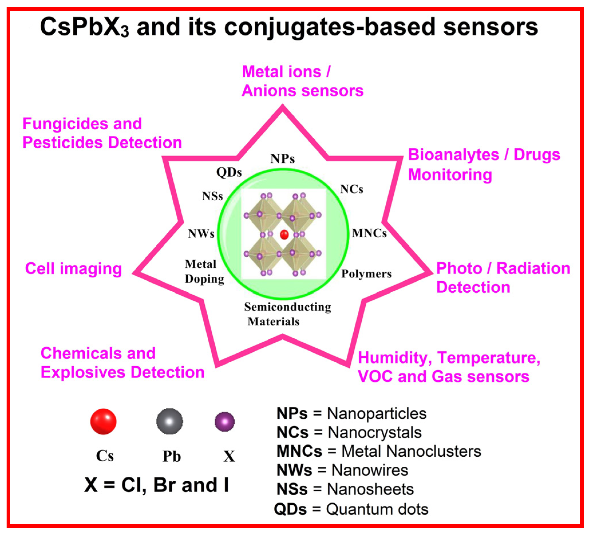


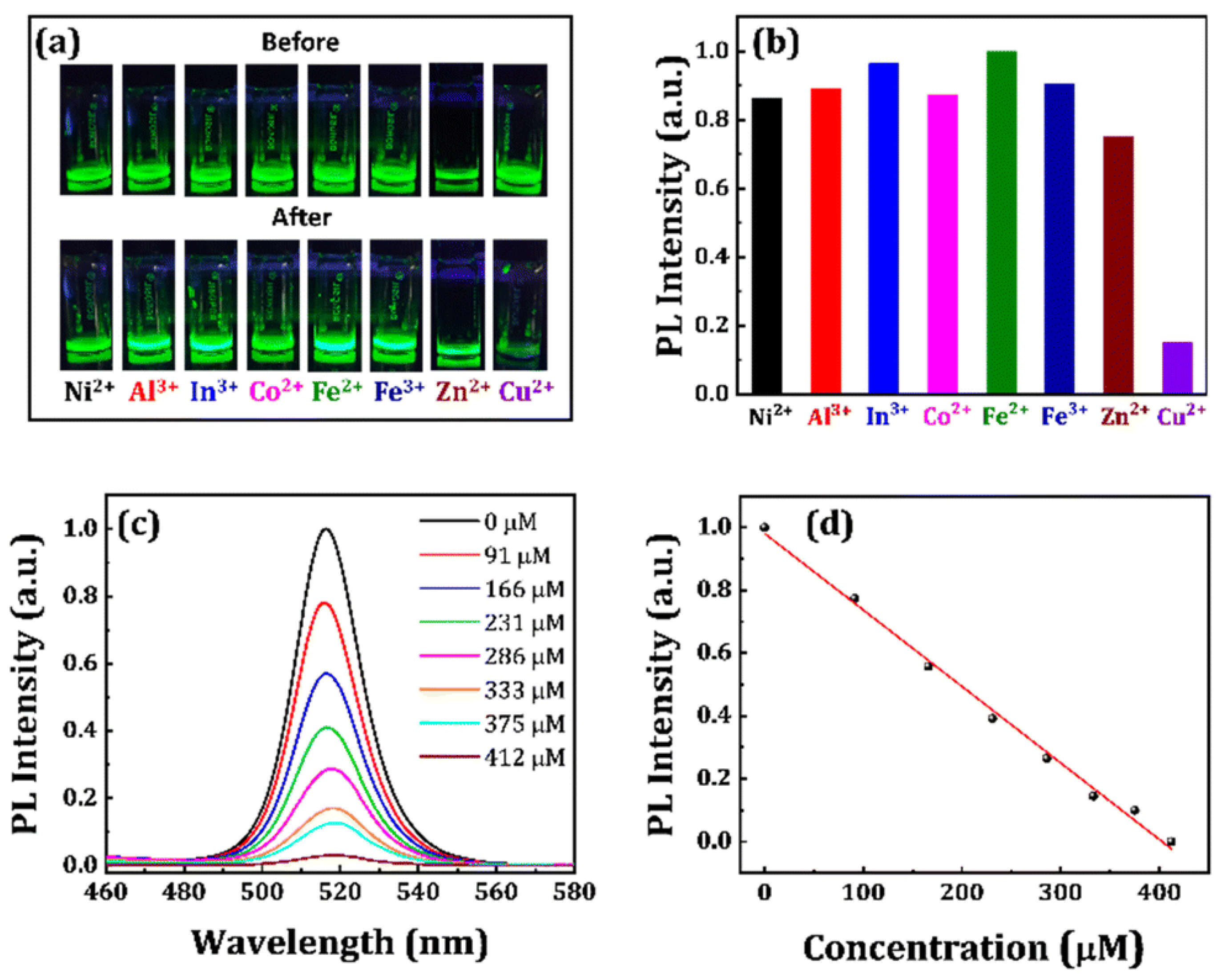
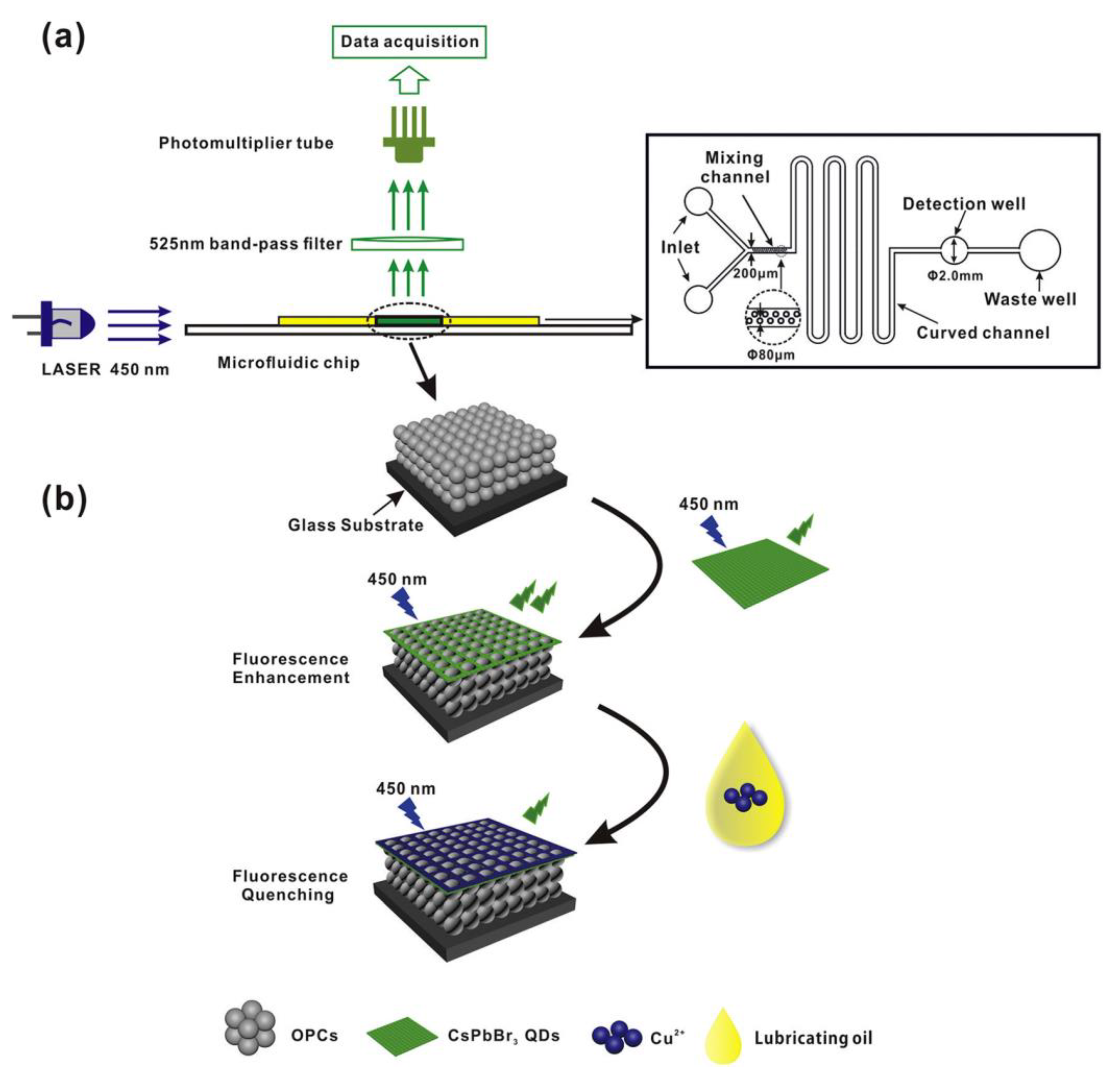
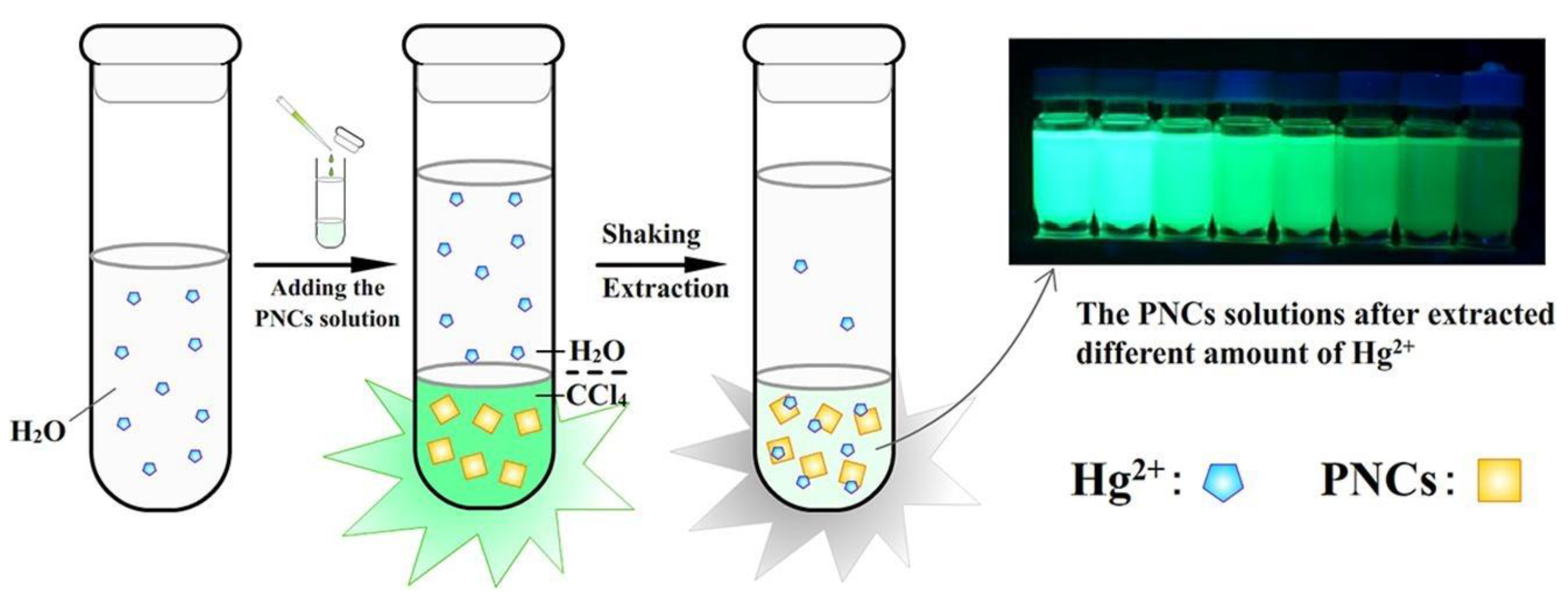
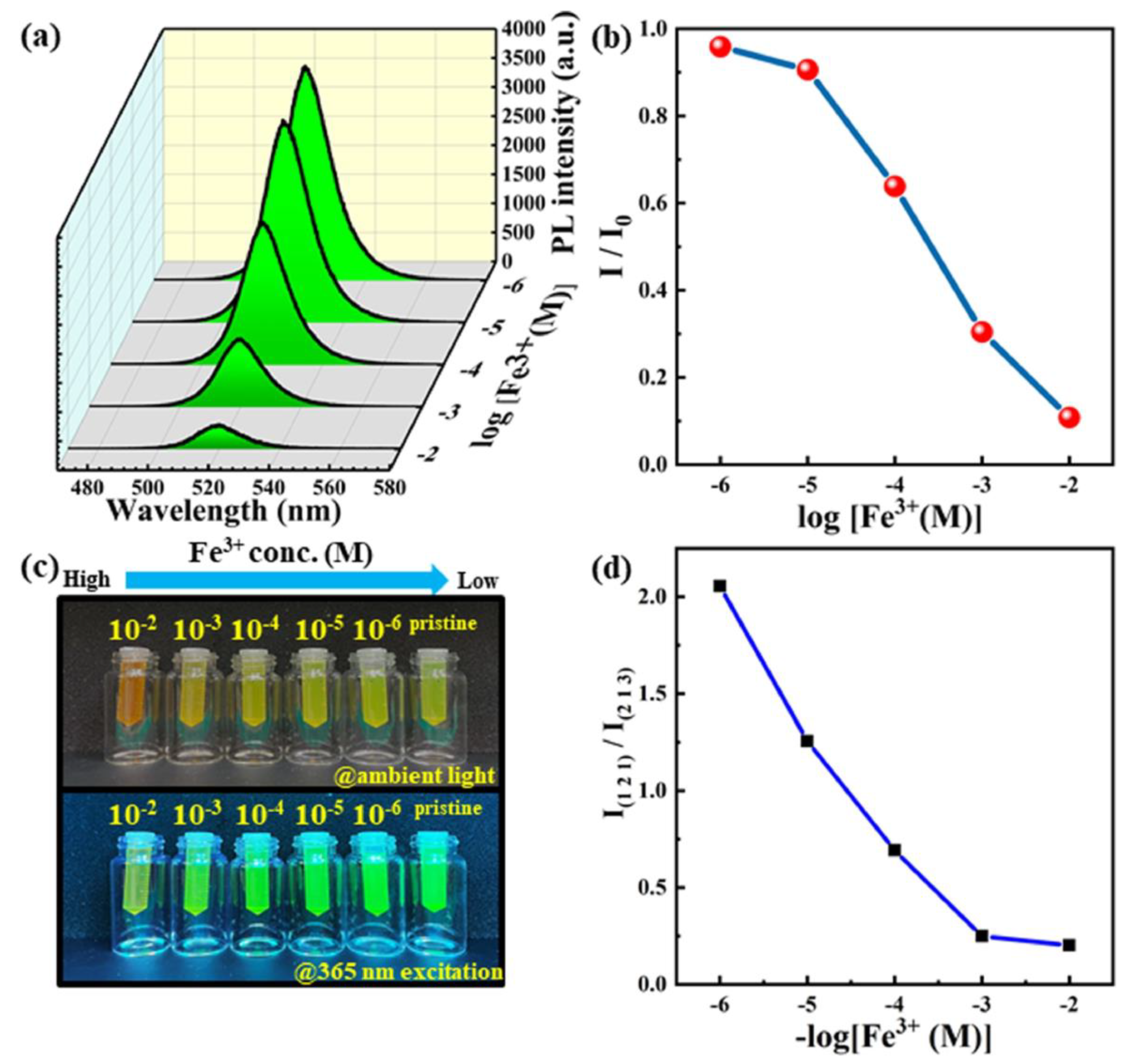
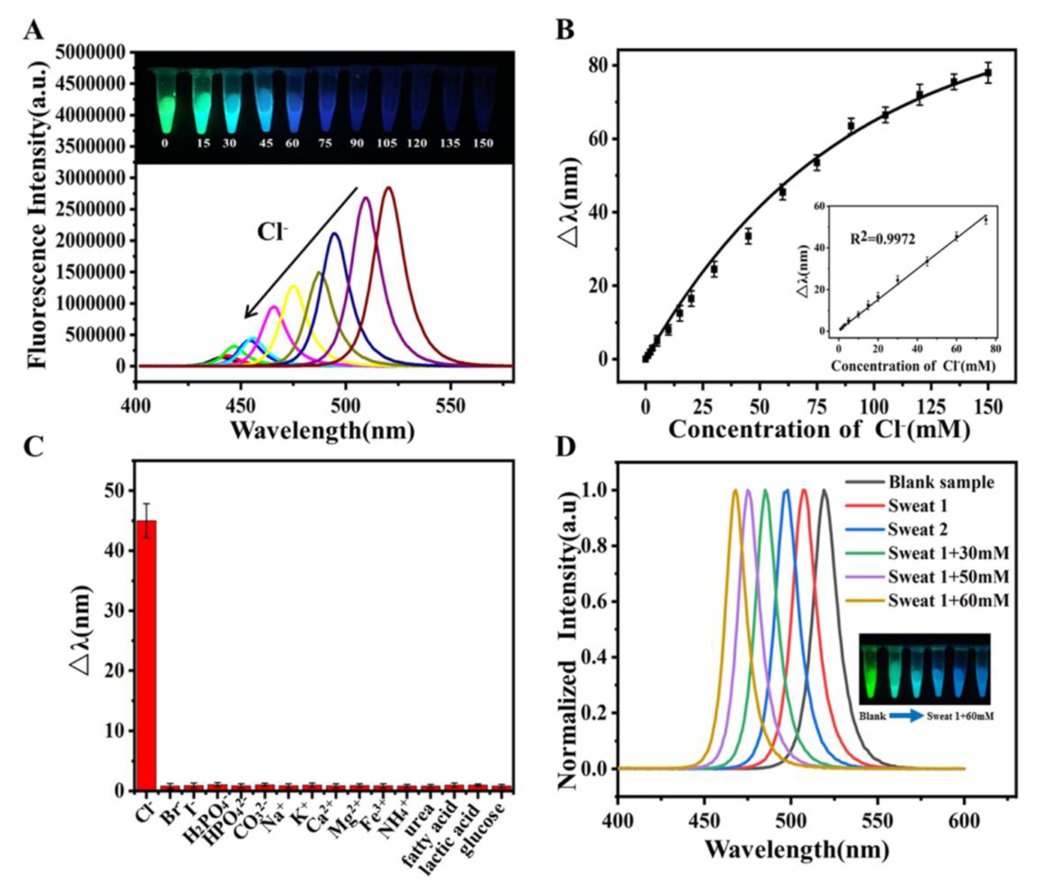
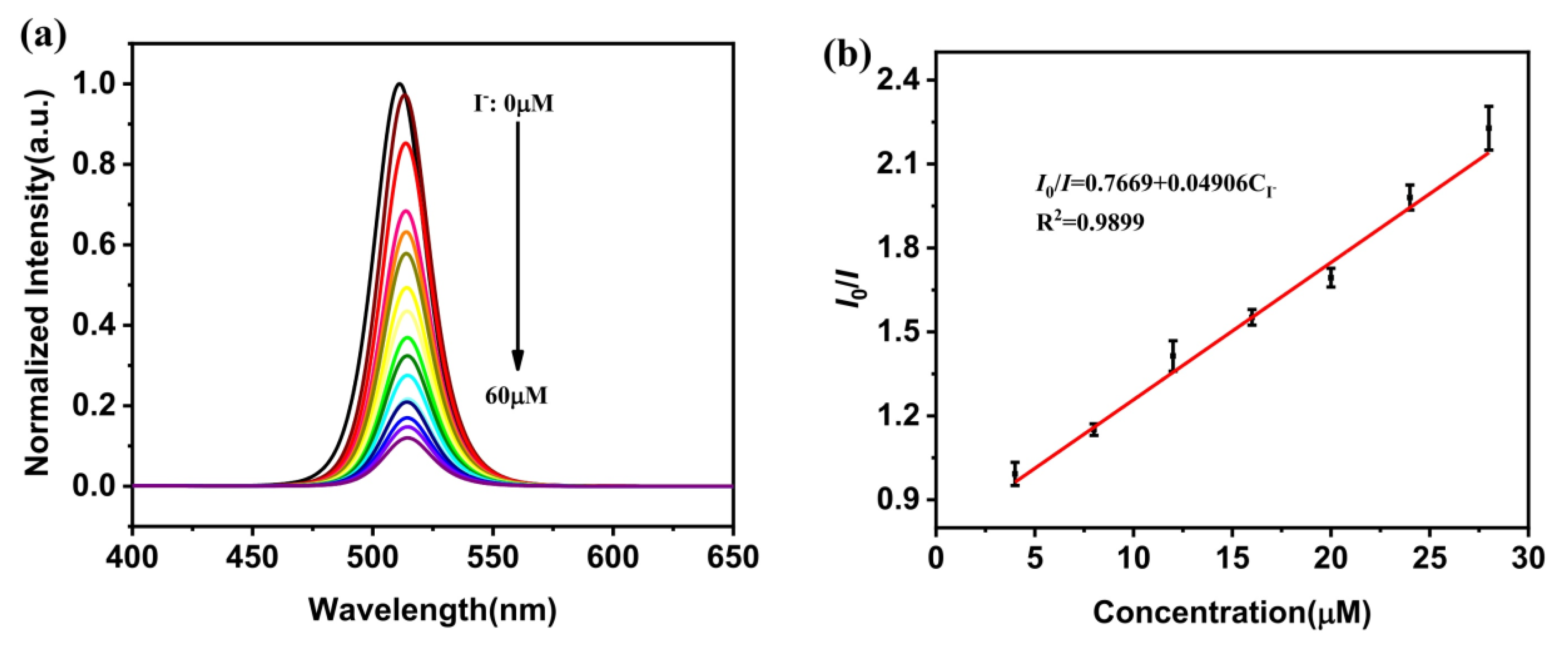
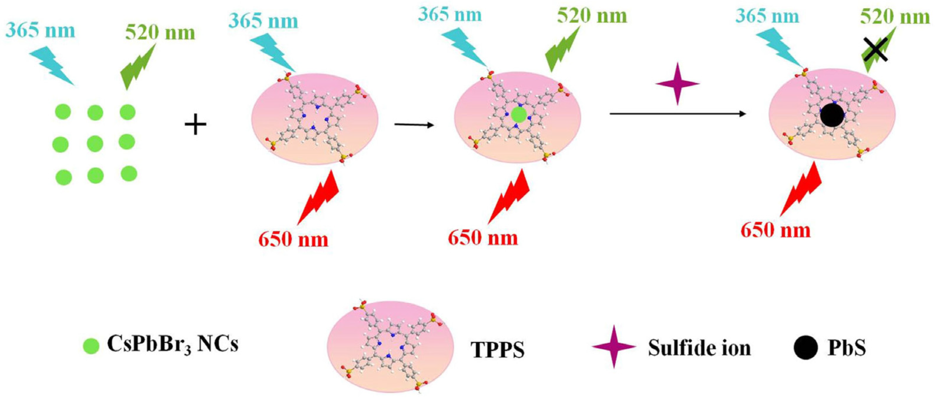


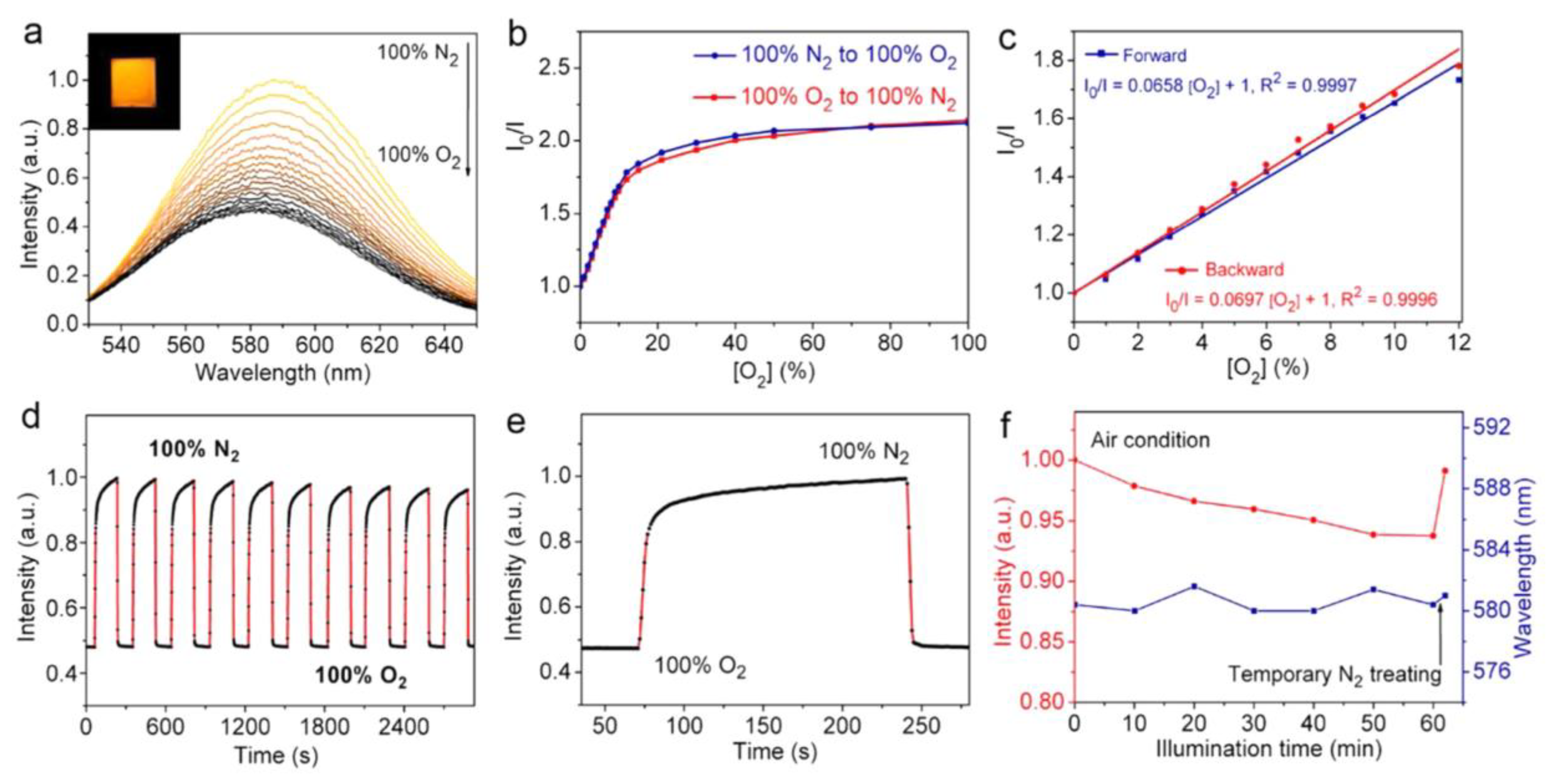
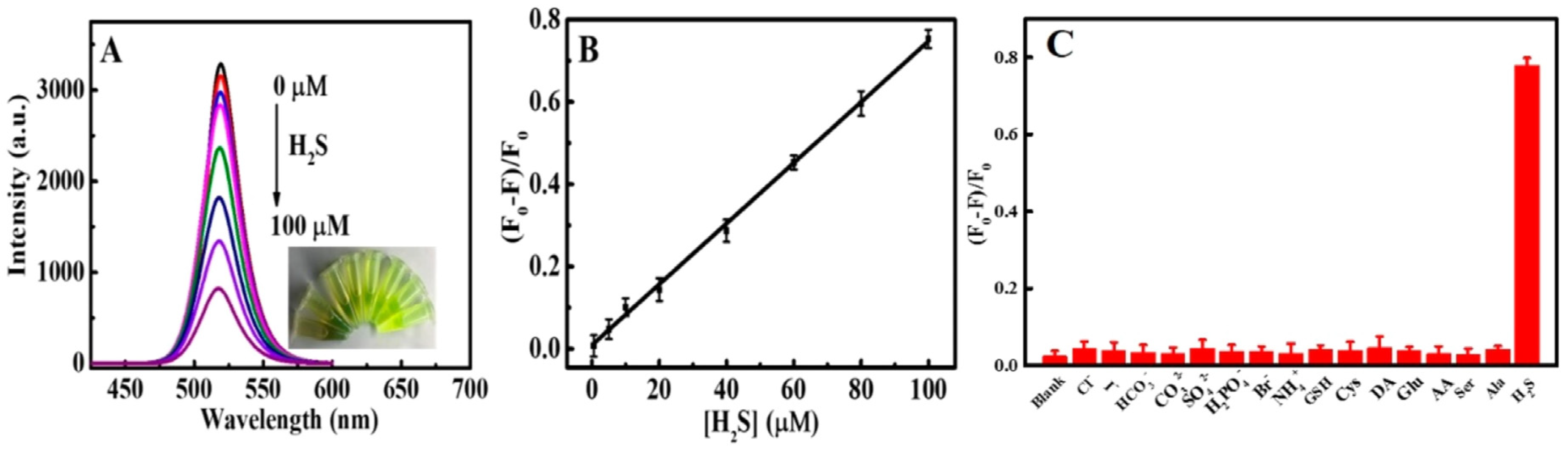
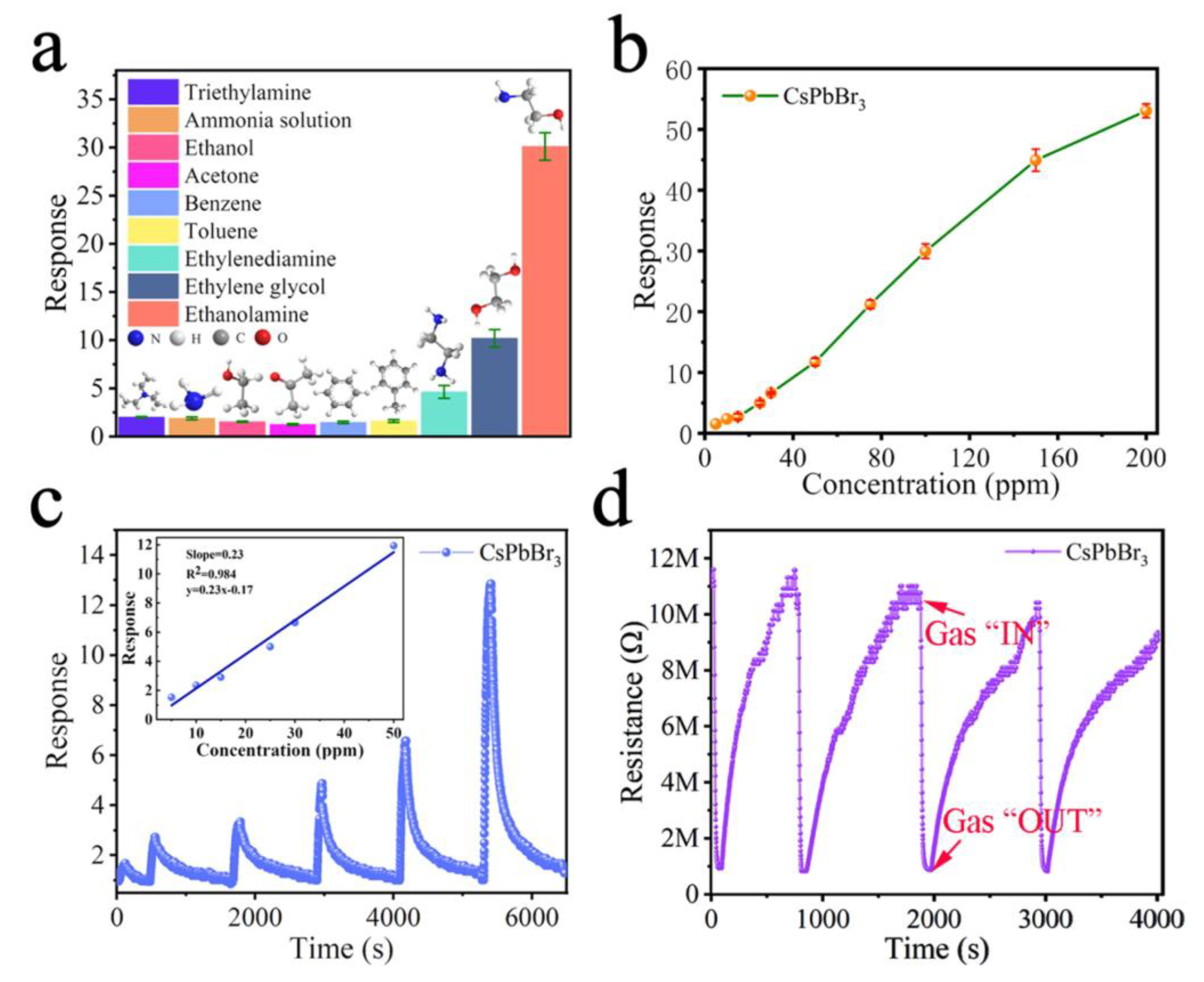
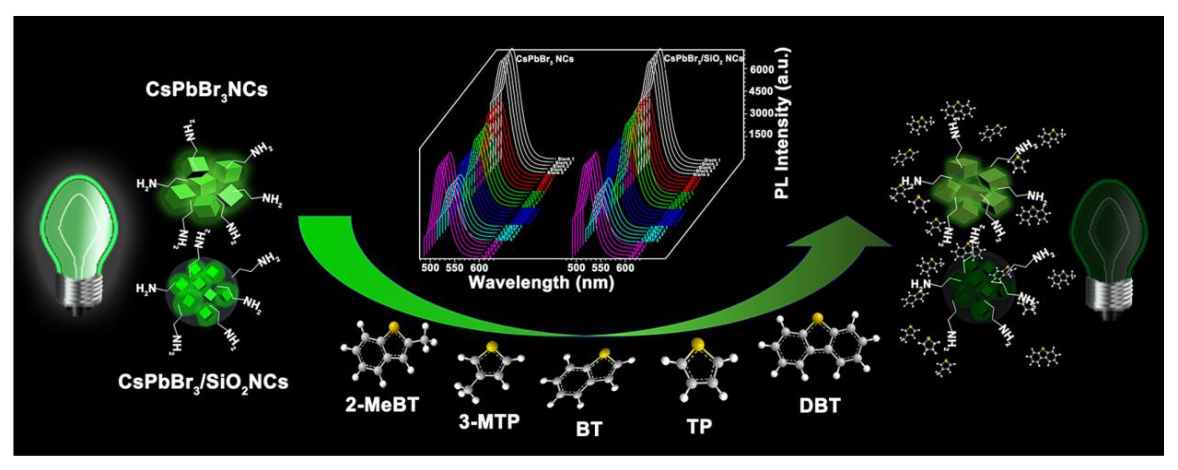
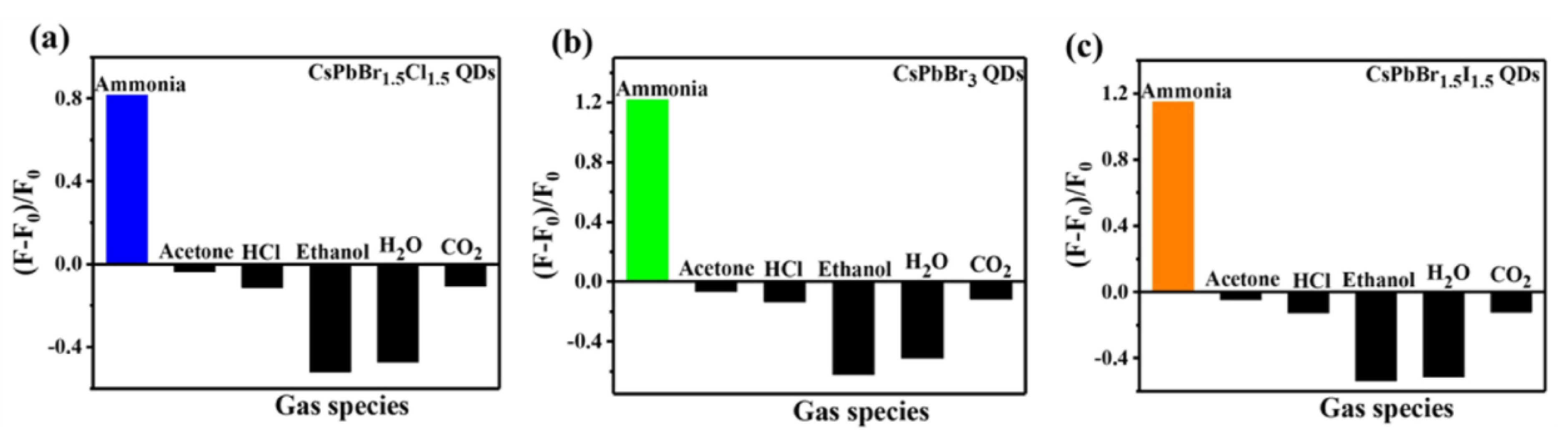
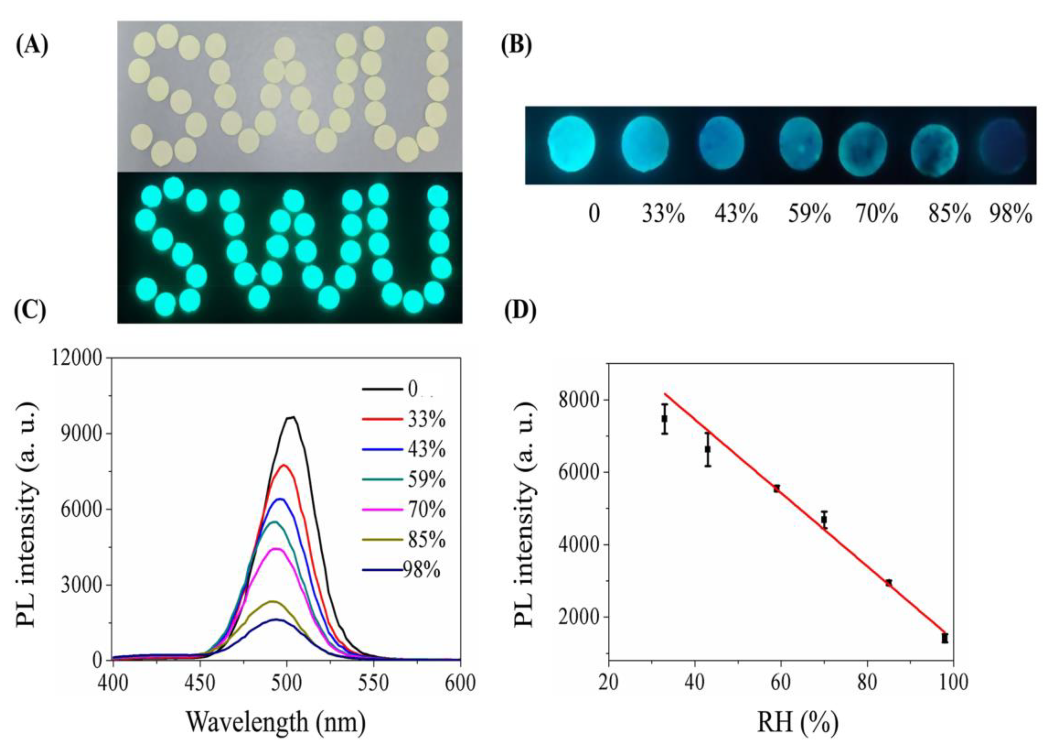
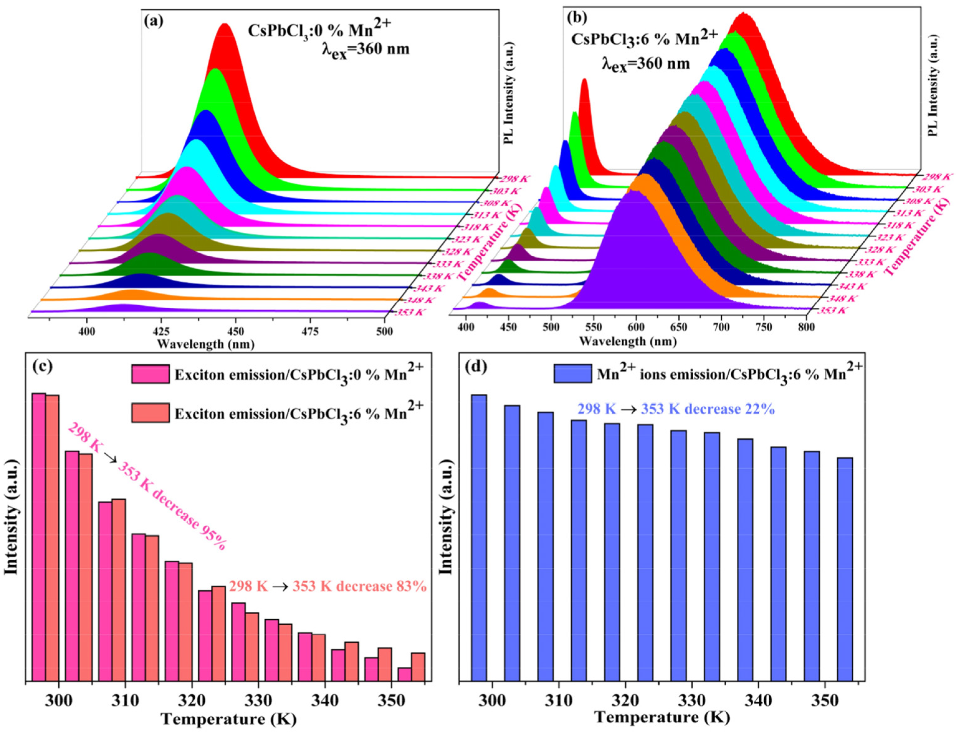
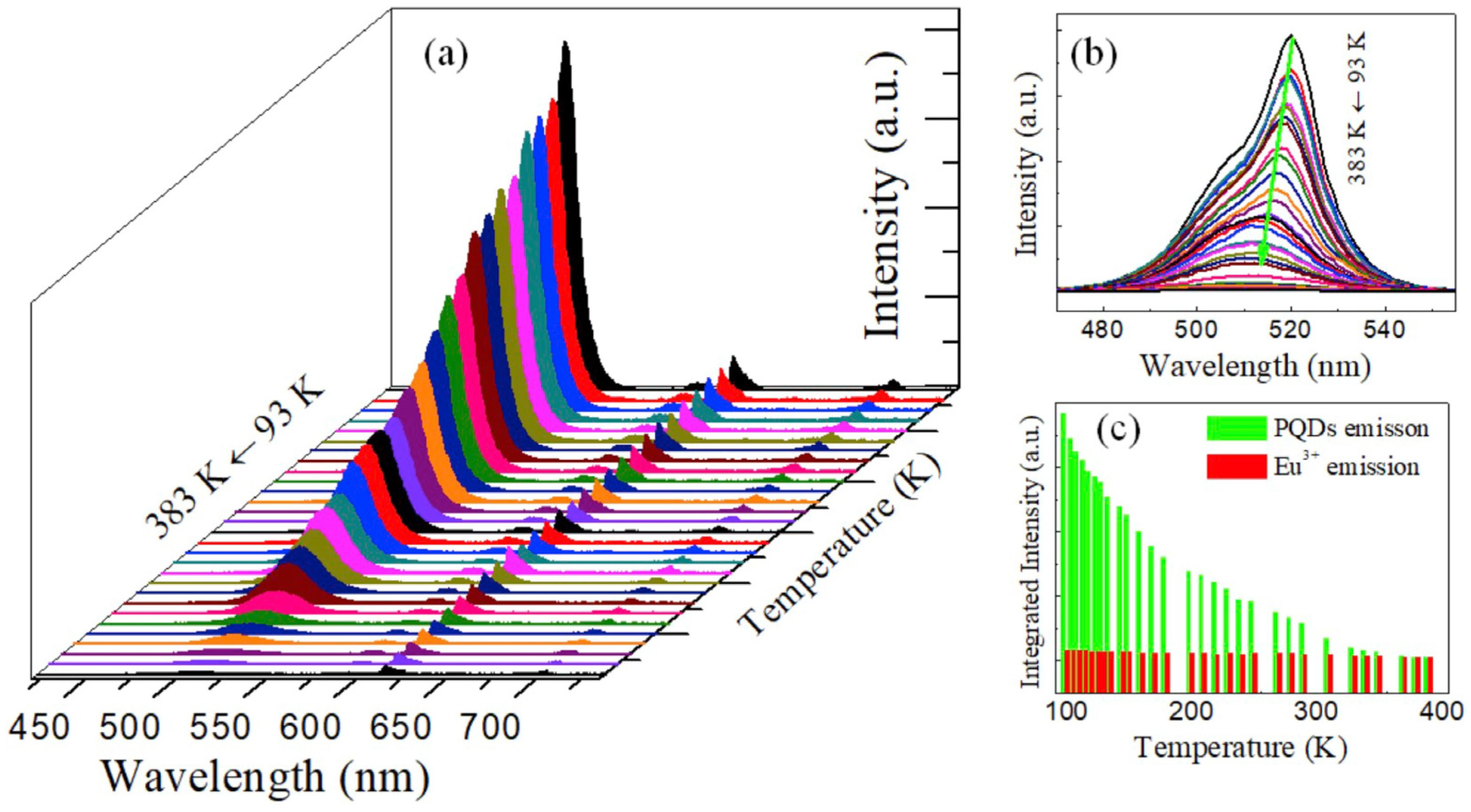

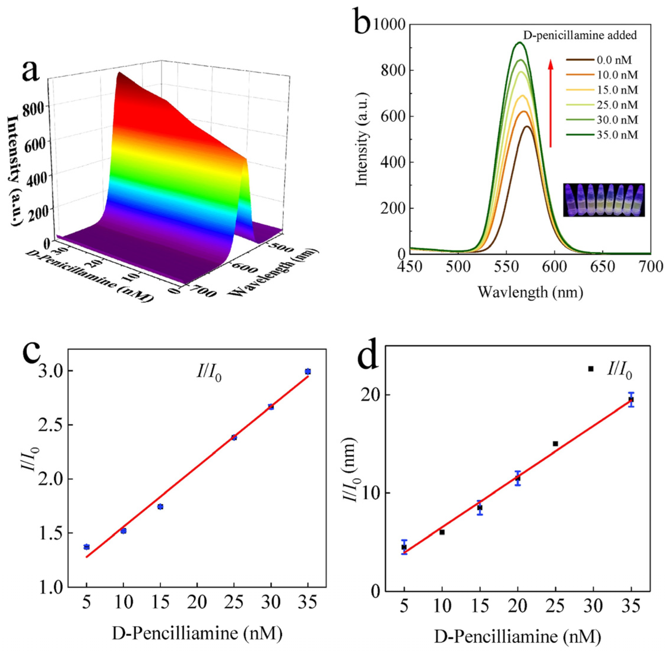
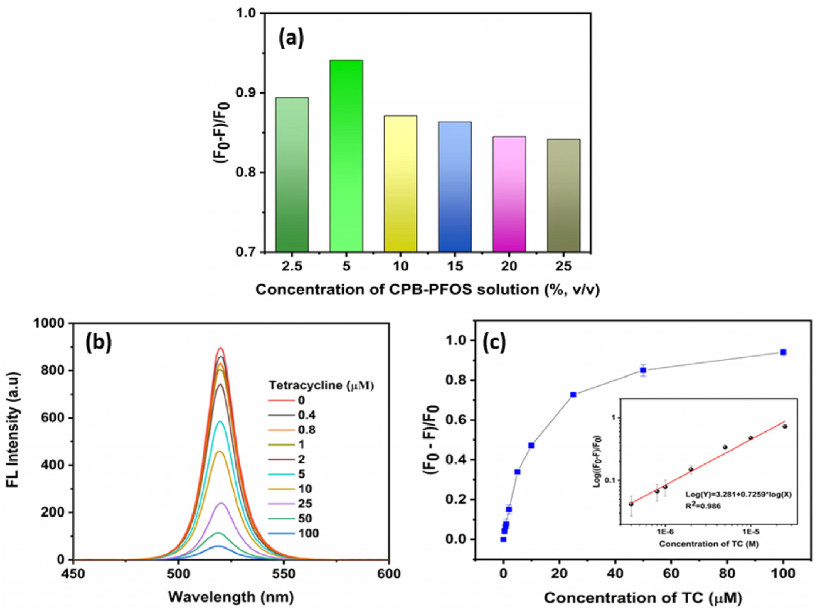
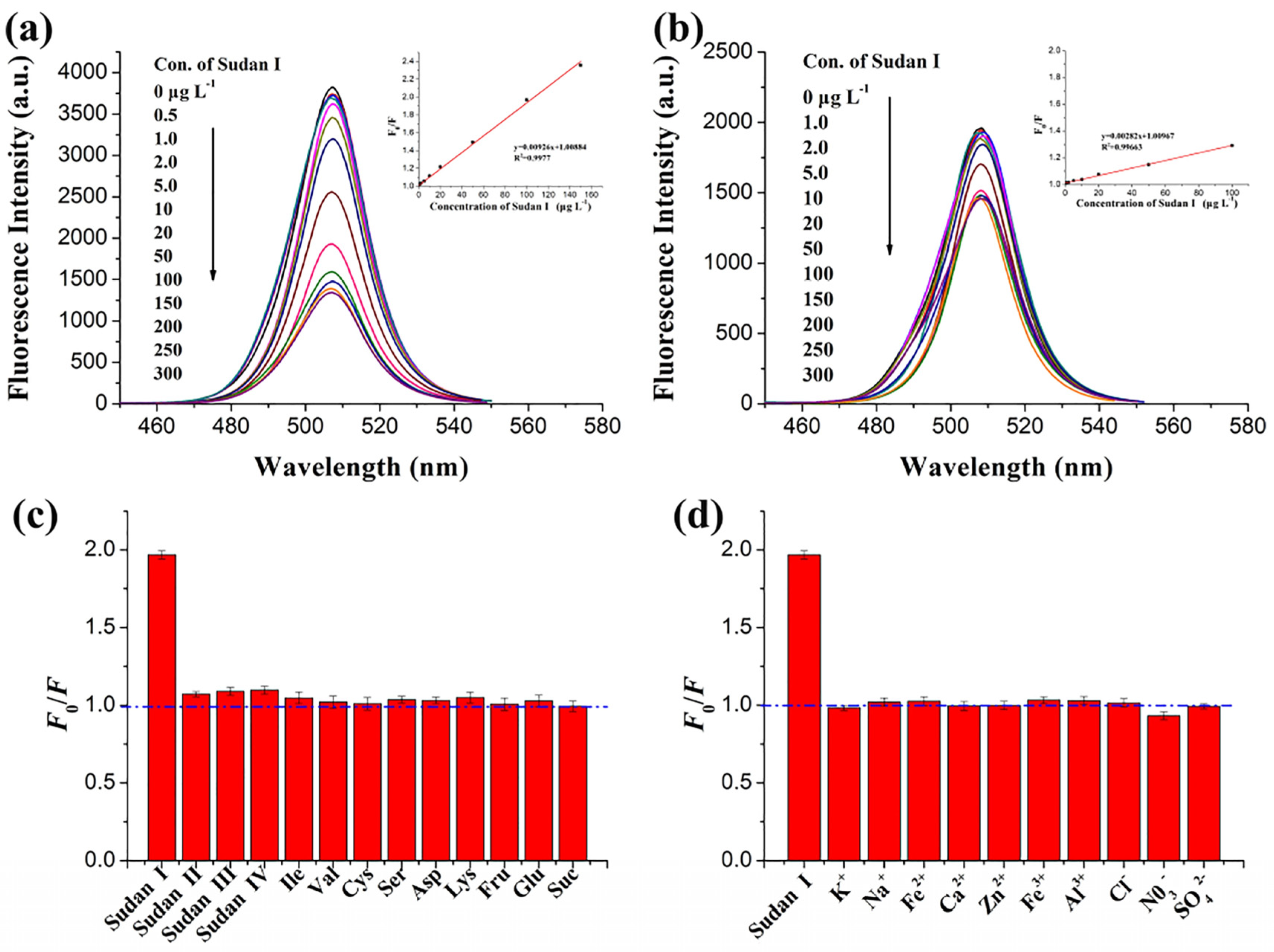
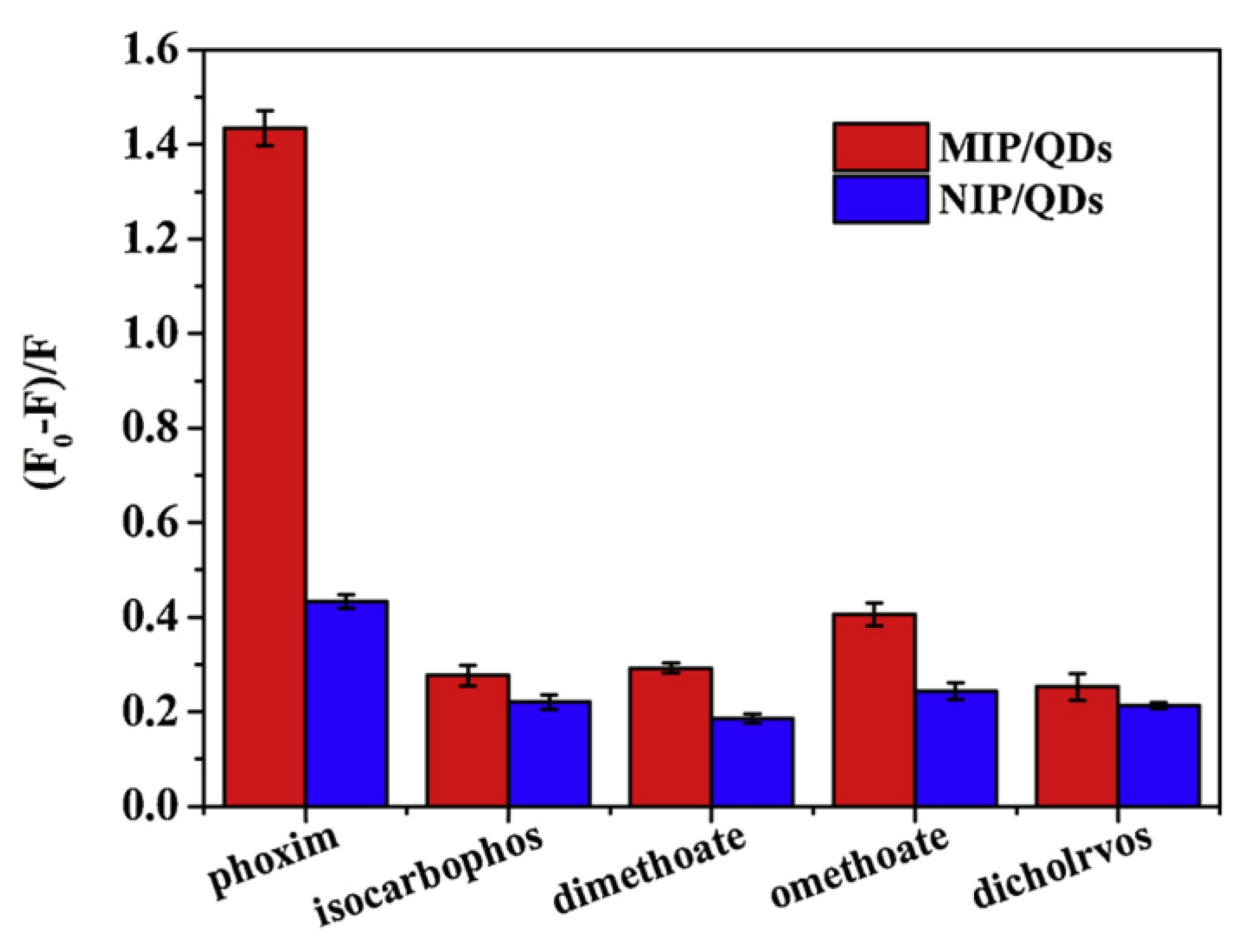
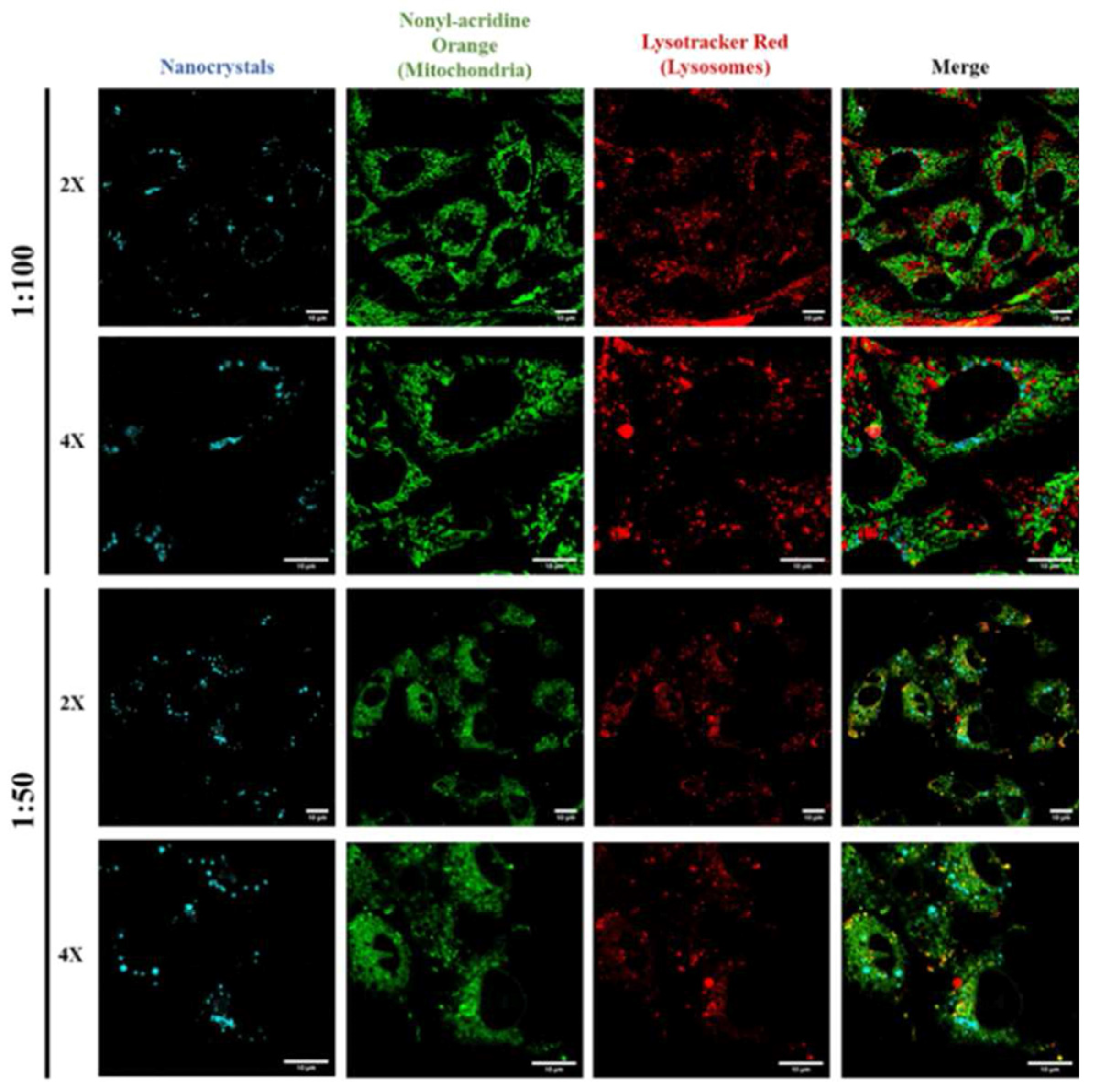
Disclaimer/Publisher’s Note: The statements, opinions and data contained in all publications are solely those of the individual author(s) and contributor(s) and not of MDPI and/or the editor(s). MDPI and/or the editor(s) disclaim responsibility for any injury to people or property resulting from any ideas, methods, instructions or products referred to in the content. |
© 2024 by the authors. Licensee MDPI, Basel, Switzerland. This article is an open access article distributed under the terms and conditions of the Creative Commons Attribution (CC BY) license (https://creativecommons.org/licenses/by/4.0/).