Abstract
Wound healing can result in complex problems, and discovering an effective method to improve the healing process is essential. Polymeric biomaterials have structures similar to those identified in the extracellular matrix of the tissue to be regenerated and also avoid chronic inflammation, and immunological reactions. To obtain smart and effective dressings, bioactive agents, such as essential oils, are also used to promote a wide range of biological properties, which can accelerate the healing process. Therefore, we intend to explore advances in the potential for applying hybrid materials in wound healing. For this, fifty scientific articles dated from 2010 to 2023 were investigated using the Web of Science, Scopus, Science Direct, and PubMed databases. The principles of the healing process, use of polymers, type and properties of essential oils and processing techniques, and characteristics of dressings were identified. Thus, the plants Syzygium romanticum or Eugenia caryophyllata, Origanum vulgare, and Cinnamomum zeylanicum present prospects for application in clinical trials due to their proven effects on wound healing and reducing the incidence of inflammatory cells in the site of injury. The antimicrobial effect of essential oils is mainly due to polyphenols and terpenes such as eugenol, cinnamaldehyde, carvacrol, and thymol.
1. Introduction
The convergence of tissue engineering, biomaterials science, and wound healing has led to significant advances in developing novel therapeutic strategies for skin injuries [1,2]. Traditional pharmaceutical treatments are no longer viable when tissues or organs are severely diseased or lost due to trauma [3]. In such cases, the option of artificial organs (including tissues) or organ transplantation arises to reconstruct these compromised tissues or organs [4,5]. Chronic wounds and tissue defects present significant clinical challenges, requiring ongoing efforts to promote effective healing and tissue regeneration [6,7,8]. Tissue engineering aims to repair, replace, maintain, or enhance the function of a specific tissue or organ [9,10].
The process of wound healing is a fascinating and intricate mechanism encompassing four distinct phases: hemostasis, inflammation, proliferation, and remodeling [11]. Coagulation factors are activated, forming a clot of platelets to minimize blood loss at the wound site (hemostasis). This is followed by an inflammatory response, characterized by the release of proteolytic enzymes and pro-inflammatory cytokines (inflammation) [12,13]. Subsequently, angiogenesis is stimulated, leading to scar formation (proliferation). Finally, the newly formed capillaries regress, and the majority of macrophages and fibroblasts undergo apoptosis (remodeling) [14,15]. In addition to these processes, a suitable sterile covering (dressing) is also crucial, providing the characteristics of skin tissue regeneration and a natural barrier to the external environment, mimicking the epithelium [16,17].
Biomaterials can be used to create wound dressings. Both natural and synthetic polymers have beneficial characteristics such as adjustable biodegradation rates, mechanical properties, high porosity with varying pore sizes, and a high surface-to-volume ratio based on the synthesis technique [18,19,20]. Chitosan is a polymeric, antimicrobial, antioxidant, biocompatible, and biodegradable material with low toxicity and the ability to accelerate dermal regeneration [21,22,23] usually used in biomedical areas such as wound healing and tissue engineering.
Antibacterial properties aim to reduce inflammation caused by infections, slowing the healing process [24,25,26]. One strategy to improve biological properties is to produce smart or modern dressings interspersed with essential oils (EOs), which act as bioactive agents.
Essential oils (EOs) are volatile secondary aromatic compounds characterized by the presence of phenylpropanoids and terpenoids [27]. They have antioxidant and antibacterial effects, as well as antiviral, insecticidal, analgesic, and anti-inflammatory properties [28]. In the healing process, EOs can accelerate wound closure, improve collagen deposition, and increase fibroblast proliferation [29,30].
This review, therefore, elucidates recent advances in the potential application of hybrid materials (biomaterials/EO) in the process of healing skin wounds, based on the investigation of fifty scientific articles evaluated in the following databases: Web of Science, Scopus, Science Direct, and PubMed. This study covers the principles of the healing process, the use of natural and synthetic polymers, the type and properties of essential oils and processing techniques, and the characteristics of dressings, emphasizing the chitosan biomaterial and its properties. The aim here is to provide insights into new wound treatment and tissue regeneration approaches.
2. Polymeric Biomaterials
Biomaterials can be understood as material designed to assume a form that can guide any therapeutic or diagnostic procedure through interactions with living systems [31] and represent a significant fraction of the products used in healthcare [32]. Among examples are biomedical devices such as biosensors [33] and blood circulation tubes [34], implantable materials (such as sutures, plates, bone substitutes, tendons, mesh, heart valves, lenses, and teeth) [35], drug delivery vehicles [36], artificial organs (such as the heart, kidney, liver, pancreas, lungs, and skin) [37], and dressings [38], among many others.
Biomaterials are classified into three main types of materials: metals, polymers, and ceramics [39]. As scaffolds, biomaterials must allow tissue growth and maturation and support cells’ physiological activities. The particularities of each depend on the type of cell in the tissue or organ to which they will be applied [40,41,42].
2.1. Synthetic Polymers
Polymers are macromolecules comprising the union of small repeating units called mers. They are formed by small molecules (monomers) through chemical reactions, polyaddition, or polycondensation, for example, and can be of natural or synthetic origin [43,44,45]. In biological terms, many polymers present structures similar to the extracellular matrix of the tissue to be regenerated, avoiding chronic inflammation, immunological reactions, and toxicity [46,47].
Synthetic polymers can be reproduced in various forms with desirable volume and surface properties. These materials’ specific advantages include the ability to tailor their mechanical properties and degradation kinetics to suit diverse applications [48,49]. For applications such as implants or controlled drug release systems, synthetic polymers that degrade hydrolytically are preferred, as their degradation is invariant from patient to patient and the implantation site [50,51]. In opposition, biopolymers have enzymatic degradation, and this mode is investigated for tissue engineering and as a substitute for the extracellular matrix [52,53].
The most used synthetic polymers as biomaterials include poly(lactic acid) (PLA), polyvinylpyrrolidone (PVP), polyvinyl alcohol (PVA), poly(ε-caprolactone) (PCL), polyurethane (PU), polyethylene glycol (PEG), polyethylene oxide (PEO), poly(L-lactide-co-caprolactone) (PLCL), carboxymethyl cellulose (CMC), and poly(vinylidene fluoride) (PVDF), among others.
PLA is a polymer derived from renewable resources approved by the Food and Drug Administration (FDA). It has become a candidate for medical applications due to its biodegradability, biocompatibility, good mechanical properties, low cost, and potential to integrate antimicrobial agents [54,55,56,57,58]. PLA also has some limitations, such as a low degradation rate, low tenacity, low crystallization rate, low heat distortion temperature, and low reactivity between hydrophobic interactions [59,60].
PVP is water-soluble, biocompatible, and biodegradable. It has low toxicity, high surface activity, strong adsorption capacity, and good water vapor transmission, and is impermeable to bacteria [61,62]. It is commonly used in artificial limb manufacturing, controlled drug delivery, tissue engineering, cardiovascular devices, artificial skin, blood plasma expanders, and wound dressings [63,64,65,66].
PVA is a hydrophilic synthetic polymer soluble in water, whose structure is mainly composed of C-C bonds, with hydroxyl and acetate groups on the sides [67,68]. It presents properties such as soft consistency when in membrane form, inherent non-toxicity, non-carcinogenicity, biodegradability, excellent biocompatibility, high surface activity, good mechanical properties, great swelling capacity in aqueous solutions, and excellent transparency and chemical resistance [69,70,71,72], being a strong candidate for the controlled release of drugs, such as dressings, artificial organs, and contact lenses [62,73,74,75,76,77,78,79,80,81,82,83,84,85,86,87,88,89,90,91].
PCL, a polyester bioabsorbable polymer with a semi-crystalline structure, is used for various medical and pharmaceutical applications, such as delivery devices and tissue regeneration [92,93,94,95,96,97,98]. PCL has good biocompatibility, low cytotoxicity in human cells, cost benefit, and ease of manufacturing. However, it has a hydrophobic surface without functional groups, making it an ineffective cell adhesion substrate. Also, due to its low stiffness, PCL as a matrix has limited biomedical applications [99]. To overcome these limitations, the combination of bioactive materials with PCL to manufacture nanofibers has emerged as a solution, leading to the creation of new membranes with superior cell proliferation properties and wound healing [100,101,102]. The introduction of functional groups to the polymer further augments its adhesive, hydrophilic, or biocompatible properties, thereby eliciting improved cellular responses and opening up intriguing potential applications [103,104].
PU is another example of a synthetic polymer also widely used in the medical field, in the form of bioadhesive fabrics, vascular stents, artificial organs, drug administration, tissue regeneration, and dressings, linked to its excellent mechanical properties and superior hydrophobicity [105,106,107,108,109,110,111,112,113].
When used as a dressing material, PU presents itself as an excellent option due to the external barrier it forms against bacteria on the surface of the wound and oxygen permeability, in addition to demonstrating that it is highly compatible with living organisms, not causing rejection or irritation to the wound [114]. It also promotes a moist microenvironment that favors epithelialization. Because it is transparent, it brings greater comfort to the patient, as it is unnecessary to remove it to view the wound [115,116]. However, the hydrophobic nature of this polymer is a crucial factor in wound care, as it results in poor wound contact, leading to insufficient exudate adsorption and the inability to release embedded antibacterial agents [111].
PEG is a viscous, synthetic, water-soluble amphiphilic polymer that exhibits biological properties such as non-toxicity, biocompatibility, biodegradability, elastomeric nature, anti-fouling nature, transparency, and economy [117,118], making it suitable for various medical applications [119,120,121,122,123]. PEO is characterized by high water solubility, rapid hydration, non-toxicity, insensitivity to the pH of physiological fluids, and ease of production. It is non-linear, non-crosslinked, hydrophilic, biodegradable, biocompatible, non-toxic, and FDA-approved [124,125], thus making it suitable for biomedical applications [86,126,127,128,129,130,131,132]. PLCL is also a synthetic polymer with excellent mechanical properties, biocompatibility, and biodegradability [133]. The FDA has approved it for clinical applications. However, its poor wettability may make it slightly disadvantageous as a wound dressing material [134,135]. PVDF, together with the other polymers mentioned above, is a non-reactive thermoplastic synthetic polymer formed by the polymerization of vinylidene difluoride. Due to its excellent mechanical and biocompatibility properties, good processability, and long-term stability, PVDF can be used to encapsulate active substances in the biomedical field [136,137], as well as in electronic skins, wearable tactile sensors, microfluidic cell-based assays and self-powered cardiac devices, sensors, and tissue engineering [138,139,140,141].
2.2. Natural Polymers
Natural polymers or biopolymers are obtained from animals, plants, microorganisms (algae and fungi), and bacteria. Chemically, they have structures with monomers of amino acids, nucleotides, esters, or monosaccharides, which are covalently linked to form polysaccharides, peptides, polyesters, or polyphenols. Widely used natural polymers include sodium alginate, gelatin, collagen, hyaluronic acid, κ-carrageenan, cellulose, gum arabic, and chitosan [142].
Alginate is a low-cost biopolymer used in many biological applications due to its biocompatible, non-toxic, biodegradable, and gel-forming nature [71,143,144]. It is an anionic, water-soluble polysaccharide found in the cell walls of marine brown algae of the class Phaeophyceae [145,146].
Biological applications of alginate are in controlled drug release systems, extracellular matrix material for biological studies, scaffolds for wound healing, and tissue engineering [147]. For example, alginate films associated with rifampicin accelerated the healing process, achieving total wound healing in 14 days [148]. Alginate dressings are designed to maintain a moist environment in the wound bed, absorb exudate, and stop bleeding [149]. It should also control pain, reduce microbial contamination, and wound odor, and absorb proteinases [150,151].
Gelatin is also a water-soluble natural polymer extracted from animal by-products obtained by hydrolysis of collagen [152,153,154,155]. It has good biocompatibility, low antigenicity, biodegradability, and non-toxicity [156,157,158]. It has been widely used in the food and pharmaceutical industries as emulsifiers, hydrogels, microencapsulating agents for antioxidant and antibacterial essential oils, and films for wound dressings and food packaging [68,159,160,161,162,163,164].
Pure gelatin films, though brittle and easily degradable, are transformed into reliable tools when plasticizers are added. This composition increases flexibility and influences cellular activities such as proliferation, migration, and differentiation [165,166,167]. More importantly, it serves as a formulation stabilizer, ensuring the reliability of medical products, and as a plasma enhancer, further enhancing its value in the medical market [64,168].
Collagen, one of the main components of the extracellular matrix, is a diverse protein family, with each type having its unique location and function. It constitutes approximately 80% of the dry weight of human skin and is also found in connective tissues, such as bones, cartilage, and tendons. This protein is considered the most abundant in the human body [169]. Type I, for instance, is found in bone, tendons, skin, teeth, and ligaments, while type II is located in cartilage and intervertebral discs. Type III, on the other hand, is found in blood vessels, skin, and muscles, and type IV in the basement membrane and basal lamina [170,171,172].
Collagen is responsible for several essential functions, such as cell migration and differentiation, cellular behavior through the regulation of the extracellular matrix (ECM), synthesis of various proteins, shear resistance to absorb energy, and ductility [169,173,174]. It can also be successfully used for tissue regeneration engineering in vitro and in vivo [175,176,177,178,179].
Collagen dressings belong to bioactive dressings that are composed of collagen obtained from bovine, porcine, marine, and avian sources, among others, and are available in the form of particles, gels, pads, ropes, sheets, or solutions [180,181]. It is a dressing impermeable to bacteria. It maintains the healing process by maintaining a moist environment around the wound, absorbs exudate, and promotes the derivation of numerous cells, such as keratinocytes and fibroblasts [182].
Hyaluronic acid is a polymer present in the extracellular matrix, with viscoelastic and hygroscopic properties, and controls cell proliferation and migration [183,184,185]. Hyaluronic acid possesses biocompatibility, biodegradability, non-immunogenicity, and wound healing properties [186,187]. Hyaluronic acid’s sizable supramolecular structures can trap large amounts of water and ions to provide hydration and turgidity to tissues, thus being considered an attractive anti-fouling material [188,189].
Carrageenin, a linear polysaccharide derived from the sea, is highly adaptable in the field of wound healing. Its ability to mimic the microarchitecture of the extracellular matrix of living tissues, coupled with its high water absorption capacity, hemostatic nature, good biocompatibility, and hydrogel-forming ability, makes it a promising candidate [190,191]. However, the challenge of controlling its gelling properties, mechanical stability, and high degradation rate in the biological environment has limited its application as a wound dressing [192,193]. To overcome these limitations and further enhance its properties, carrageenin can be combined with other polymers or pharmacologically active agents that can stimulate wound healing at each stage [123,194,195,196,197,198].
Chitosan and gum arabic, as biopolymers, are chosen as encapsulating materials for their biocompatibility and low toxicity [80,199]. Gum arabic, a negatively charged polysaccharide–protein complex with excellent emulsifying properties, is obtained from the stems and branches of acacia trees. The hydrophilic parts of these polymers play a crucial role in stabilizing the emulsion against droplet aggregation, while the hydrophobic branched proteins are rapidly adsorbed on the emulsion droplet surface [200,201]. In addition to emulsifying properties, gum arabic’s high solubility, lower viscosity, good film formation, and non-toxicity further reinforce its biocompatibility [202,203].
Chitosan is a natural, semi-crystalline cationic polymer derived from chitin in fungi’s cell walls or the exoskeleton of arthropods [204]. Chitosan-based films, with their moderate oxygen barrier and good carbon dioxide barrier properties [205,206], are instrumental in maintaining a moist environment around the lesion, a critical factor in wound healing [207,208].
Chitosan
Chitosan, discovered and named in 1859 by Roget [209], is obtained by the partial deacetylation of chitin poly(β-(1→4)-N-acetyl-D-glucosamine), under alkaline conditions, being the most crucial derivative in terms of applications [210,211,212]. Chemically, it is a copolymer of various proportions of N-acetyl-d-glucosamine and d-glucosamine (Figure 1). Its properties are highly dependent on its degree of deacetylation, average molecular weight, polydispersity, and structure [213,214].
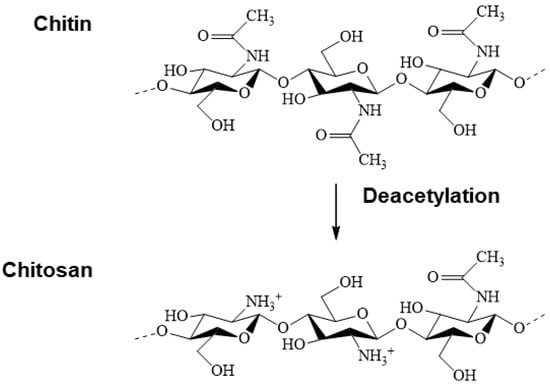
Figure 1.
Chemical structure of chitin and chitosan.
Changes in charge density affect the dissolution and binding properties of chitosan [215,216,217]. The variation in charge density gives rise to a considerable change in the swelling index, pore size, and permeability of the chitosan drug membrane [218]. The molecular weight alters the content of N-acetylglucosamine units in chitosan, which will have both an intramolecular and intermolecular influence, resulting in different conformations [216].
Intrinsic properties of chitosan such as biocompatibility, biodegradability, non-toxicity, biological adhesiveness, antimicrobial activity, and hemostatic effect [199,216,219,220,221,222,223,224] make it a versatile material. With its reactive amino groups, chitosan is the only natural cationic polymer with numerous commercial applications: it accelerates wound healing and is an anticoagulant, antifungal, and antitumor [216,220,221,222,225]. The cationic nature of chitosan is the key to most of its biological properties, with the degree of deacetylation (DG) being the parameter with the most significant impact [226].
The effect of the constant molecular weight (around 810 kDa) and variable degree of deacetylation (75%, 87%, and 96%) of chitosan membranes influences tensile strength and results in more excellent elongation at break [227]. The degree of deacetylation can also affect the antimicrobial activity of chitosan [228,229], where the high DG increases the electrostatic binding to the cell membrane and the permeabilizing effect. On the other hand, the high molecular weight generates high permeation in the cell nucleus [230,231,232,233].
The various studies on the toxicity of chitosan [82,234,235,236,237,238,239] have reported its potential use as a biocompatible biomaterial. Chitosan/gelatin scaffolds loaded with an ethanolic extract of Jatropha mollissima (EEJM) and chitosan/bioglass composites tested with mouse fibroblasts and L929 fibroblasts by MTT, respectively, were shown to be non-cytotoxic. Although not present in mammals, chitosan is subject to in vivo degradation by many enzymes such as pepsin, lysozyme, and papain [240,241].
The biodegradation kinetics of chitosan is closely linked to the degree of crystallinity, primarily influenced by the degree of deacetylation [224,232]. As the degree of deacetylation decreases, biodegradation increases [232,242,243,244]. The impact of chain length (molecular weight) on the biodegradation rate has also been well documented [245,246]. Controlling the degradation rate of chitin- and chitosan-based devices is highly desirable since biodegradation is crucial for small and large molecule release applications and regeneration functional tissue applications [232].
The ease of modifying chitosan with other bioactive molecules can provide additional properties for tissue construction [247]. Chitosan membranes/films, hydrogels, sponges, scaffolds, and fibers have been investigated for wound healing or tissue engineering applications [247,248,249], as excipients for drug administration [250], and in gene delivery [251,252].
In tissue engineering, chitosan plays a pivotal role in positively influencing the different phases of the wound healing process [209,253,254]. Chitosan’s ability to modulate the activation of platelets, promote blood clotting in vivo, and regulate the activity of inflammatory cells creates a promising microenvironment for healing. It also provides non-protein matrix support for tissue growth [255]. Furthermore, chitosan’s gradual depolymerization to release N-acetyl-β-D-glucosamine stimulates fibroblast proliferation, angiogenesis, and orderly collagen deposition at the wound site [209,253,256]. On top of these benefits, chitosan can also prevent skin infections, a significant complication associated with wound healing [26].
Despite its numerous favorable properties, chitosan, on the other hand, has poor mechanical properties. To this end, to improve these properties, blends and mixtures of chitosan with other synthetic or natural polymers have been used [78,79,83,130,257]. Furthermore, studies have demonstrated the bacteriostatic and bactericidal effects of chitosan, its possible mechanisms of action against pathogenic microorganisms [258], and the influence of molecular mass, source of origin of this biopolymer, degree of deacetylation, type of microorganism, and cultivation, in biological properties. During the wound healing process, chitosan and its derivatives have also acted efficiently in the hemostasis, inflammation, and proliferation phases, helping to stop hemorrhage, eliminate bacteria, and accelerate skin proliferation, with the growth of granulation tissue, respectively [259].
By adjusting the physicochemical properties of chitosan and its biological properties, with the addition of bioactives, it is possible to develop more advanced and efficient approaches for treating and controlling skin wounds. From this perspective, active compounds are incorporated into chitosan and other polymers to enhance antibacterial properties and overcome toxicity [168,260,261,262,263,264]. These antibacterial agents are biodegradable and come from renewable sources, where essential oils are one of the most promising herbal medicines to promote the wound healing process, minimizing bacterial infections [131,265,266,267,268].
3. Wound Healing
The skin, a multifunctional organ, protects the body from the invasion of microorganisms, provides sensory functions, and plays a crucial role in regulating body temperature [269,270,271,272,273]. However, when the skin is damaged, such as in the case of thermal burns, cuts, lacerations, surgical incisions, or chronic wounds like pressure ulcers or diabetic foot ulcers, the structure and functions of the skin can be compromised, presenting a biological burden, and psychological, social, and financial challenges for both individual patients and healthcare systems globally [274,275,276,277,278].
When wounds occur, there is susceptibility to invasion by microorganisms [279], and subsequent wound infection complicates the healing process [280]. Wound healing is a dynamic physiological process involving a multiplicity of cellular, humoral, and molecular events, aiming to restore the integrity and functionality of injured skin [281,282]. The breakdown of this complex pathway can result in a delayed or impaired healing process, resulting in acute wounds (burns, trauma) or chronic wounds (diabetes, tumor, physiological contamination) [122,283,284,285]. Wounds can further be classified according to the damaged layers of the skin, defined as (i) superficial wounds (only the epidermal layer is damaged); (ii) partial thickness wounds (damaged layers of the skin involve epidermal layers and deep layers, including blood vessels, hair follicles, and sweat glands); and (iii) full-thickness wounds (the skin down to the depth of the subcutaneous tissues is damaged) [283].
Wounds heal within 4 to 12 weeks in healthy people [223,286]. However, chronic wounds, a more severe condition, fail to make headway through the normal healing stages and are not repaired in an orderly and timely manner. Studies have shown that around 70% of chronic wounds are prone to developing an infection. These infections lead to biofilm formation, posing the threat of antibiotic resistance [258,287]. The severity of chronic wounds significantly impacts the healing process and requires careful management.
The wound healing process can be divided into four phases: hemostasis, inflammation, proliferation maturation, and remodeling [281,286,288,289,290]. Several cellular activities occur during these phases, such as activating keratinocytes, fibroblasts, endothelial cells, macrophages, and platelets. In patients with diabetes, however, the healing process is significantly impacted due to uncontrolled blood glucose levels. This condition leads to slowed blood circulation, decreased efficiency of white blood cells, collagen synthesis, growth factor formation, inflammatory cells, fibroblasts, and proliferation and migration of keratinocytes. As a result, the formation of new tissues or vessels is delayed [291].
In the healing process, the initial response to injury is hemostasis. Clotting factors are activated and form a knot of platelets to reduce blood loss at the wound site [11,12]. The second phase includes inflammation that lasts between 24 h and 4 to 6 days. This phase begins with the emission of proteolytic enzymes and pro-inflammatory cytokines in immune cells that invade the wound area [13]. Inflammatory cells generate reactive oxygen species, and their quantity correlates with the type of wound but is generally more significant in burns and chronic wounds [292].
Still, in the inflammatory phase, neutrophils and macrophages remove all foreign particles and tissue debris from the wound bed, thus preventing infections. At this stage, the release of cytokines and enzymes stimulates fibroblasts and myofibroblasts, and wound exudate guarantees the moisture essential for healing [293,294,295].
The third stage is proliferation, in which platelets and leukocytes release cytokines, and stimulate angiogenesis, fibroblast proliferation, collagen, and elastin synthesis to restore the dermis, leading to scar formation [13,14]. At this stage, the intact epidermis is restored over the newly formed tissue, forming a new extracellular matrix [296]. Finally, the last stage of the healing process is remodeling, where during this phase, the newly formed capillaries regress, and most macrophages and fibroblasts undergo apoptosis [15]. Thus, the matrix composition changes, and type III collagen, initially abundant in the early stages of wound healing, is replaced by type I. This transition from type III to type I collagen increases the new tissue’s tensile strength [11,293,297,298].
Wounds are treated by debriding the infected area (eliminating dead tissue, foreign particles, and microbial biofilms), reducing pressure at the wound site, and preventing bacterial attack [299,300,301]. In addition to these procedures, a biomaterial is necessary, acting as a dressing (sterile covering) to provide the skin’s tissue regeneration characteristics and a natural barrier to the external environment, mimicking the epithelium [16,17].
4. Essential Oils and Biological Properties
When the skin is compromised due to injury or damage, it becomes more vulnerable to microbial infections [302]. These infections are often caused by different types of bacteria, including Gram-positive bacteria such as Staphylococcus aureus and Staphylococcus epidermidis, as well as Gram-negative bacteria like Escherichia coli and Pseudomonas aeruginosa [303,304,305]. Gram-negative bacteria have two lipid membranes and a thin layer of peptidoglycan, while Gram-positive bacteria have a single bilayer membrane surrounded by a thick layer of peptidoglycans [306,307,308,309].
Staphylococcus aureus, commonly found on healthy and damaged skin [310], poses a significant challenge due to its antibiotic resistance, which can impede the natural healing processes [311,312,313,314]. On the other hand, Staphylococcus epidermidis is beneficial in preserving the skin’s integrity and producing antimicrobial molecules that hinder the formation of biofilms by S. aureus [315,316,317,318]. Escherichia coli is known for its ability to form biofilms on various surfaces [319]. At the same time, Pseudomonas aeruginosa is notorious for causing persistent infections and delayed wound healing, mainly due to its high antibiotic resistance and strong biofilm-forming capacity [320,321].
Antibacterial properties are fundamental in biomedical applications, aiming to reduce inflammation caused by infections, which delay healing [24,25,26]. In this context, the emergence of resistant bacteria has spurred studies on therapeutic alternatives and the incorporation of antimicrobial agents that can enhance the efficiency of dressings [265,281,322,323]. Among these alternatives, essential oils (EOs) stand out due to their high capacity to control microbial infections [267,268,278,324,325,326]. EOs have shown promising potential in the eradication of multi-drug-resistant pathogens, as they inhibit the growth of microorganisms, creating disturbances in the cytoplasmic membrane; interrupting the proton motive force, the flow of electrons, and active transport; and hindering protein synthesis [327,328].
EOs are volatile compounds of low molar mass extracted from aromatic plants from different tissues (roots, flowers, stems, leaves, seeds, fruits, or the entire plant) [329]. They present excellent antimicrobial, antifungal, antioxidant, and anti-inflammatory properties. In addition to being biodegradable and lipophilic, they are also sedatives and analgesics with a low degree of toxicity [330,331,332,333,334,335,336].
Chemically, EOs are characterized by terpenes and phenylpropanoids [27,337]. Terpene compounds can be divided into two main categories: terpenes with a hydrocarbon structure (mono-, sesqui-, and diterpenes) and their oxygenated derivatives (alcohols, oxides, aldehydes, ketones, phenols, acids, esters, and lactones) [27]. The antimicrobial properties of EOs are attributed to active constituents, mainly related isoprenes, along with other hydrocarbons and phenols [338]. Therefore, the presence of phenolic compounds (carvacrol, eugenol, and thymol, among others) generates a rupture of the cytoplasmic membrane by the proton motive force, by the flow of electrons, by active transport, and also by the coagulation of cellular contents. Therefore, essential oils characterized by a high level of phenolic compounds, such as carvacrol, eugenol, and thymol, have important antibacterial activities [339], resulting in essential antibacterial properties [309,340,341,342,343,344]. Given these characteristics, between 2020 and 2023, recent studies reported using essential oils (clove, tea tree, and oregano) in wound healing [83,89,345,346].
The EO composition also influences its antioxidant potential. Phenolics and secondary metabolites with conjugated double bonds generally exhibit considerable antioxidant properties [347]. These phenolic compounds possess redox properties and are essential in neutralizing free radicals and decomposing peroxides [327]. The antioxidant activity of essential oils is also associated with some alcohols, ethers, ketones, aldehydes, and monoterpenes: linalool, 1,8-cineole, geranial/neral, citronellal, isomenthone, and menthone [348,349,350]. EOs of α-terpineol, linalool, linalyl acetate, limonene, δ-3-carene, α-pinene, and 1,8-cineole have important anti-inflammatory activity [351,352,353]. These compounds act by inhibiting histamine release or reducing the production of inflammatory mediators. Therefore, the anti-inflammatory activity of EOs can be attributed to their antioxidant activities and their interactions involving cytokines, regulatory transcription factors, and the expression of pro-inflammatory genes [341].
EOs can be obtained by various extraction techniques such as pressing, hydrodistillation, and steam distillation [309,341,343,354,355]. In the hydrodistillation technique, the sample is immersed in water in a distillation system with heating. The volatiles in the sample are boiled, forming a heterogeneous mixture at the end of the process. In extraction by steam distillation, water vapors pass through the sample, which entrains plant volatiles, as the plant sample is suspended and there is no direct contact with water [356,357].
The main disadvantages of applying EO are its volatility, low stability, high sensitivity [337,358], and degradation under the processing temperature [359,360,361]. Therefore, to overcome these deficiencies, EOs can be encapsulated and incorporated into polymeric matrices to increase their activity and stability, improve water solubility, and facilitate their delivery in healing applications and tissue engineering [362,363,364,365,366,367].
4.1. Wound Dressings
The dressing is essential for wound treatment in the medical and pharmaceutical sectors. The global market was valued at USD 12.4 billion in 2021 and is projected to grow annually by 5.3% between 2022 and 2030 [368]. The ideal dressing should promote quick healing with minimal inconvenience for the patient. Therefore, developing an advanced biomaterial for wound treatment is relevant [369,370,371].
The ideal wound dressing should also focus on the following characteristics: (i) provide or maintain a moist environment at the wound/dressing interface; (ii) allow gas exchange (water vapor, oxygen) between the injured tissue and the environment, aiming to maintain adequate tissue temperature, in order to improve blood flow to the wound bed; (iii) have suitable mechanical properties; (iv) provide a barrier to microorganisms; and finally, (v) remove excess exudates and toxic components from the wound surface. In addition, the dressing must be non-toxic, non-allergenic, economical, and easy to remove [248,281,289,372,373,374].
Potential dressings can be classified into traditional and modern dressings [281,375]. Traditional dressings, which are still widely used to treat wounds and burns, are applied to stop bleeding and prevent the wound from coming into contact with the environment [376]. They include dry dressings (gauze and bandages) and topical pharmaceutical formulations (solutions, suspensions, emulsions, creams, and ointments). Dry dressings are recommended for open, dry wounds, or secondary dressings due to their low wound coverage capacity. Topical pharmaceutical formulations, on the other hand, have the disadvantage of a short lifespan in the wound bed, especially when there is excess exudate, as they absorb fluid, lose their rheological characteristics, and become mobile [281,283].
While traditional dressings have limitations, modern dressings have advanced features and benefits [370]. They were developed with better biocompatibility, degradability, pain relief, and moisture retention. These modern wound dressings can be synthesized through films, membranes, scaffolds, hydrogels, nanocapsules, sponges, and nanofibers, offering a promising solution to the drawbacks of traditional dressings [273,373,377,378,379,380]. These structures comprise multilayers (absorbent and self-adhesive layers) [381,382]. The inner layers protect the wound from bacteria, cleanse infections, and heal the wound through direct contact with wound cells [383,384,385,386]. Additionally, the outer layer protects the wound from mechanical stress [387]. Incorporating essential oils is generally carried out in an external or wet layer, which provides superior healing control, eliminating contaminants such as bacteria, proteins, viruses, dyes, and/or metallic ions [388,389]. The synthesis of multilayer structures is an ideal strategy for preparing efficient multifunctional dressings [390,391]. Many studies have been carried out to investigate the potential application of EOs in wound healing through their incorporation into biomaterials such as dressings. Regarding this research, 50 studies were collected from 2010 to 2023 in the Web of Science, Scopus, Science Direct, and PubMed databases, demonstrating the synergistic use of polymeric biomaterials incorporated with EO for wound healing. According to the survey, most studies focused on clove, oregano, cinnamon, tea tree, thyme, and copaiba oil in manufacturing biomaterials such as bioactive dressings. Table 1 compiles data on essential oils and their major compounds, their biological results, systems, and the form of dressings developed.

Table 1.
Summary of the primary information and results found in the studies of EO that have shown curative activity in the past thirteen years.
4.1.1. Films and Membranes
Films and membranes function as temporary skin substitutes, acting as a physical and mechanical barrier, mainly in managing wound infections [248,376,411]. The film can be defined as a film with restricted dimensions, that is, whose thickness is much smaller than the other dimensions [412,413]. Its production process generally involves inter- and intramolecular associations or crosslinks of polymer chains, forming a semi-rigid three-dimensional network in which the solvent is immobilized [414]. Membranes, in turn, have inherent transport properties, being a discontinuous interface between two distinct environments (permeated and retained flow). The microstructure, selectivity, and permeability of the membrane are crucial factors in the diffusion, separation, or transport of nutrients and substances. Polymeric films and membranes, therefore, tend to promote improvements in the regenerative process, including some advantages, such as the ability to absorb fluids and exudates from the wound without leaks, not requiring frequent changing and cleaning; appropriate gas exchange, providing a humid microenvironment; and wound protection against microorganisms and other toxic agents, increasing the quality of the regeneration process and reducing the risk of complications [415,416]. They also exhibit high flexibility, resistance, and transparency. Moreover, they can incorporate active ingredients, which are substances that have a therapeutic effect on the wound, such as antimicrobial agents or growth factors, and release them in a controlled or prolonged manner. This allows for a more comfortable and functional treatment, with less need to change dressings, and, consequently, reduces the dose administered, avoiding pain [417,418,419].
The main methods of processing films and membranes to obtain dense structures are the “casting” system (drying a film-forming solution, where the solvent evaporates slowly at a controlled temperature), coating, layer by layer, and extrusion, and of porous structures, they are particulate leaching, thermally induced phase separation, and electrospinning [420]. These methods allow for a wide range of film thickness, from μm to mm, depending on the methods used and the number of polymers [370].
Chitosan dressings obtained by the “casting” technique stimulate the immune system, accelerating healing due to their hemostatic properties, low immunogenicity, and high absorption. This way, they protect against microorganisms and fungi and are thermally stable [421,422]. PVA is also considered an excellent option for film/dressing production, as it has high biocompatibility, surface tension that promotes good elasticity, and good vapor transmission properties, promoting a moist microenvironment that favors epithelialization [423].
Several studies demonstrate the efficiency of dressings incorporated with clove, tea tree, and rosemary EOs due to meaningful biological interactions in the healing process [28,78,85,131,147,393,401].
Membranes with rosemary and tea tree EO incorporated into chitosan using the casting method were tested in vitro (simulating in vivo conditions through a cellular model) and in vivo (in rats) and demonstrated a significant increase in the percentage of wound contracting. Results revealed an excellent protective effect on human erythrocytes (>63%) and efficiently promoted different stages of wound healing, in addition to decreasing oxidative stress in the wound area [78,393].
The chitosan-based film incorporated with clove and tea tree essential oils was obtained by the casting method. It showed good transparency to visible light, flexibility, mechanical resistance to touch, thicknesses more minor than the dermis, and excellent wettability in distilled water and a phosphate-buffered saline solution. In in vitro tests, films obtained with 1% (v/v) of tea tree and 3% (v/v) of clove revealed activity against Staphylococcus aureus (halo of 6.0 mm and 9.0 mm, respectively), Escherichia coli (9.0 mm and 8.0 mm halo, respectively), and Candida albicans (7.0 mm halo in both) [28].
The dressing formulation, incorporating silver nanoparticles, sodium alginate, and essential oils of tangerine, niaouli, and clove, proved to be a prospective asset in wound care. It exhibited potential antimicrobial and antibiofilm properties, effectively treating wounds and preventing infection without antibiotics and topical antiseptic products [147]. Zein/clove EO electrospun fibrous membranes exhibited higher gas permeability with superhydrophilicity to absorb wound exudate, good biocompatibility, and antibacterial effects [131].
Chitosan and PVA films loaded with cinnamon and clove essential oil also showed bactericidal effects against Staphylococcus aureus and Pseudomonas aeruginosa after two hours of direct contact within the infected microenvironments [85]. Gelatin films loaded with clove essential oil and hydrotalcite nanoplates obtained by the casting method demonstrated antimicrobial activity against Staphylococcus aureus and Escherichia coli and in vitro biocompatibility. The films were non-toxic for HeLa cell lines, with cell viability above 70% [401].
The EO of Hypericum perforatum also demonstrated excellent potential for incorporation into different materials with healing action [122,223]. The chitosan films incorporated with H. perforatum oil, analyzed by the agar diffusion method, demonstrated an antimicrobial effect on Escherichia coli (2.9 ± 0.1 mm) and Staphylococcus aureus (1.97 ± 0.05 mm). Results showed that Escherichia coli is more sensitive to the films obtained than the bacterium Staphylococcus aureus. Furthermore, the films had no cytotoxic effects on NIH3T3 mouse fibroblast cells and provided a good surface for cell adhesion and proliferation [223].
H. perforatum oil incorporated into two-layer membranes formed from electrospun nanofibers of PCL and PEG exhibited controlled release and antimicrobial action against Staphylococcus aureus (12.9 mm) and Escherichia coli (11.9 mm) by the disk diffusion method. The biomaterial evaluated in vitro showed no risk of adhesion to the wound and did not demonstrate apoptotic/necrotic effects, defining it as a biocompatible material. Furthermore, the membranes had a proliferative effect on L929 fibroblast cells [122].
Lemongrass essential oil has also been studied to synthesize wound dressing materials due to its antimicrobial, antioxidant, cytotoxic, and insecticidal effects [403,404]. Chitosan films incorporated with lemongrass essential oil inhibited microbiological growth, demonstrating antimicrobial activity against Escherichia coli and Staphylococcus aureus and protection against environmental oxidative stress caused by free radicals [403]. Likewise, bioactive collagen/chitosan membranes loaded with 0.7% lemongrass essential oil showed biocompatibility and antimicrobial activity greater than 99.60%, in addition to thermal resistance to oxidation and the suppression of radicals generated by radiation gamma [404].
Other essential oils were also loaded into films and membranes with healing action [20,80,88,91,374,407]. Thyme essential oil (1.2% v/v) incorporated into chitosan generated films with antimicrobial activities against Escherichia coli, Klebsiella pneumoniae, Pseudomonas aeruginosa, and Staphylococcus aureus, mainly due to the majority constituent, the monoterpene carvacrol. The films also promoted increased water vapor and oxygen transmission rates by adding oil [374].
Zataria multiflora EO added to PVA/gelatin formed films with antioxidant and antibacterial actions against Pseudomonas aeruginosa. The zeta potential, particle size, and viscosity were significantly altered by adding Zataria multiflora to the polymer matrix [91]. Black pepper and ginger essential oils incorporated into PVA/gum arabic and chitosan generated break-resistant and flexible films with improved thermal stability, as well as significantly inhibiting the growth of Bacillus cereus, Staphylococcus aureus, Escherichia coli, and Salmonella typhimurium [80].
Loading Ruta graveolens essential oil into chitosan generated a film with lower permeability, water solubility, and thermal resistance, comparable to control chitosan films, without demonstrating allergic or cytotoxic reactions in erythrocytes. However, the more significant amount of EO resulted in a greater reabsorption of the implanted material in vivo in Wistar rats, with an abundant inflammatory infiltrate (typical of the healing process). This statement demonstrates that high concentrations of EO can produce greater porosity and cracking of the films and, thus, can be applied to improve cell adhesion and proliferation. However, the optimal percentage must be <1.0% [20].
Silica nanoparticles and patchouli essential oil incorporated into PVA/chitosan (matrix) resulted in nanocomposite films with good hygroscopicity and controlled release of the oil for more than five days. The films also exhibited a good long-term (>48 h) antibacterial effect on Staphylococcus aureus and low toxicity on mouse fibroblasts (L929 cells) [88]. Finally, frankincense essential oil added to gelatin/Persian gum/nanocellulose generated more stable films with constant degradation under a PBS medium. Such films showed a non-homolytic nature in vitro. Results showed that incorporating EO improved the anti-inflammatory and antibacterial activity against Staphylococcus aureus (standard and hospital), which can be attributed to the compound α-pinene [407].
4.1.2. Hydrogels
Hydrogels are three-dimensional network structures comprising biopolymers and hydrophilic synthetic polymers with high water absorption and transport capacity [375,424,425,426,427]. Hybrid hydrogels, in turn, exploit natural polymers’ high porosity and biocompatibility and the adjustability of synthetic polymers [428,429].
Hydrogel-based wound dressings, with their promising new properties and treatment options, are poised to become leading candidates for wound treatment and repair [429]. Their unique hydration capabilities, driven by a capillary effect; the presence of carboxylic, hydroxyl, and hydrophilic compounds; and osmotic pressure [430,431], offer a refreshing and calming effect when in contact with wounds, tissues, or similar structures [69,432,433,434,435]. These properties not only make hydrogels effective as irritation-reducing agents but also equip them to prevent microbial invasion, providing a secure shield against infections while facilitating the efficient transport of bioactive molecules, such as antimicrobial agents and pharmaceuticals, even in their hydrated state [119,436,437].
Another significant advantage of hydrogels is their non-adhesive property, allowing painless dressing changes. This property enhances the patient’s comfort and promotes the healing process. Hydrogels effectively absorb wound exudates, reduce chronic infections, and promote healing without scar formation, and re-epithelialization [119,438,439]. The hydrophilic groups in the structure of hydrogel membranes enable them to be easily removed from the wound surface without breaking [345]. However, it is crucial for the resistance of hydrogel membranes to be greater than that of human skin (11.5 MPa), with the strength of this membrane primarily depending on the polymer used [440].
Hydrogels can be physically crosslinked through non-covalent bonds, such as ionic, hydrophobic, van der Waals, stereo complexation, and polyelectrolyte complexation, making them reversible and weak, or via chemical crosslinking through covalent bonds, or by photopolymerization (use of ultraviolet light), generating hydrogels with high stability and permanent crosslinks. Glutaraldehyde, genipin, and epichlorohydrin, among others, are some crosslinking agents used in the process [441].
Chitosan, alginate, starch, agarose, gelatin, cyclodextrin, carrageenan, fibrin, collagen, dextran, and hyaluronic acid are natural polymers that prepare hydrogels. On the other hand, we can mention synthetic polymers such as PVA, PVP, and PEG. DNA and peptides can also be used as raw materials [442]. Furthermore, in some formulations, plasticizers such as glycerol, sorbitol, and polyethylene glycol are used, which are added to increase mechanical stability and barrier capacity [443].
Nanoparticulate systems for controlled release of clove oil through a hydrogel matrix (chitosan, guar gum, and acacia gum) and PVA nanofibers demonstrated high anti-inflammatory activities in vivo. Essential oil ex vivo skin permeation results showed that nanofibers with oil nanoemulsion can sustain oil penetration through the skin [83].
Clove, oregano, and tea tree essential oils were incorporated into PVA/starch-based hydrogel membranes, resulting in good mechanical and physical properties. Furthermore, the antimicrobial activity test using the disk diffusion method revealed that the diameter of the inhibition zone was smaller against Escherichia coli (37.0 ± 0.29 mm) than against Staphylococcus aureus (39.0 ± 0.57 mm) by 0.1 mL of clove oil. The hydrogels showed high swelling capacity against water and blood, as did MgCl2 and NaCl solutions, as they can exude wound fluids, reducing humidity in the injured area [345].
The synergistic combination between PVA and chitosan functionalized with tea tree essential oil presented broad spectrums of biological action related to repairing and healing injured tissues such as burns. Results showed that the hydrogel obtained has a superhydrophilic character and a reasonable degree of swelling in the presence of fluids, distilled water, and PBS, inducing the regeneration of injured tissues [89]. Similarly, in rats, a nanoemulgel (chitosan, gelatin, and PVP/oregano essential oil) was used in vivo as a combined therapy with low-level laser therapy. Results demonstrated a maximum healing rate of 97.5%, minimal scar formation, increased granulation, enhanced re-epithelialization, and a dramatic decrease in inflammation and neutrophil infiltration within the period (days 3, 7, and 14) of treatment compared with monotherapy [346].
Other essential oils have also been loaded into a hydrogel matrix with healing action [123,398,399,400]. Innovative bioactive hydrogel dressings based on Psyllium and Carbopol supported with frankincense essential oil revealed that incorporating the oil up to 5% improves the porosity of the dressing, increases the water vapor transmission rate, and has antioxidant activity. The dressing attenuated the microbial growth of Staphylococcus aureus, Escherichia coli, and Candida albicans after 18 h and presented faster healing in vivo, with better biochemical parameters [398].
The nanogel obtained from the encapsulation of Satureja khuzistanica jamzad essential oil in chitosan nanoparticles promoted an increase in the thermal stability of the oil. The product demonstrated antibacterial effects against Gram-positive and Gram-negative bacteria (MIC between 7.8 and 500 mg/mL) and also acceptable anticancer activities against the KB and A549 tumor cell lines (IC50 of 5.59 µg/mL and 7.78 µg/mL, respectively) [399].
The antibacterial hydrogel obtained based on polysaccharides (carboxymethyl chitosan and carbomer 940) associated with essential oils (eucalyptus, ginger, and cumin) was applied to repair burned skin. The hydrogels incorporated with eucalyptus oil exhibited high antibacterial activity against Staphylococcus aureus (inhibition rate of 46.26 ± 2.22%) and Escherichia coli (inhibition rate of 63.05 ± 0.99%), along with cell viability of 92.37%, by the MTT assay using L929 cells. Results also showed that this hydrogel induced a significant migration of L929 cells (59.83% after 24 h and 100% after 48 h) and accelerated wound healing in vivo, promoting recovery of the dermis and epidermis [400]. Thyme essential oil incorporated (15%) into κ-carrageenan and PEG hydrogel membranes also showed in vitro antimicrobial activity (>95%) against S. aureus and E. coli with a slow-release profile. Cytocompatibility, determined with HEK293 cells (human embryonic kidney cell line 293) using the MTT assay, demonstrated that the membranes exhibited ~89% cell viability [123].
4.1.3. Nanofibers
Nanofibers, with their unique properties of a large specific surface area, high porosity, excellent pore interconnectivity, and good mechanical properties [75,444,445], offer distinct advantages for wound healing. These properties aid in cell adhesion and proliferation, facilitate permeability to moisture and gasses, and promote cell growth. They also allow the absorption of additional exudates containing nutrients for bacterial growth [446,447,448,449,450,451,452]. This unique combination of properties makes nanofibers promising candidates for dressing materials, providing an ideal environment for wound healing. Among the techniques used to obtain nanofibers, electrospinning is a simple, efficient, and economical method to produce nanoscale fibers by applying an electric field to polymer-based solutions [453,454,455]. The diameter of the nanofibers obtained by this technology can reach several hundred nanometers or even less than one hundred nanometers [456]. The internal pore structure of electrospun nanofibers can be controlled, as well as their composition and structure [457,458]. During the electrospinning process, fiber morphology is affected by multiple factors, such as polymer properties, electrospinning process parameters, and environmental parameters, which regulate electrospun fibers’ shape, diameter, and quality [19].
The high porosity of nanofibers provides a contact surface more suitable for gas exchange and liquid absorption, which generates excellent permeability. This, in turn, keeps the wound moist and acts as a strong barrier against microbial invasion, ensuring the safety of the wound [459,460,461,462]. Simultaneously, the high flexibility of the nanofiber makes the dressing suitable for different parts and shapes of the wound [463,464].
The polymeric materials used to synthesize nanofibrous dressings include natural and synthetic polymers. Natural polymers easily form strong hydrogen bonds with the aqueous solution, not allowing spinning, as they form a highly viscous solution [456]. To improve spinnability, chitosan, for example, is usually mixed with other polymers, such as PVA, PLA, PEO, and PCL, to obtain nanofibers for wound dressings [24,160,465,466,467,468,469,470].
Solution blow spinning can produce PLA and PVP nanofibers loaded with copaiba oil (Copaifera sp.). Adding PVP to PLA enabled a higher rate of controlled oil release in vitro. The nanofibers obtained from blends containing more significant amounts of PVP revealed greater antimicrobial action against Staphylococcus aureus [392].
Zataria multiflora EO was incorporated into electrospun nanofibers based on chitosan/PVA/gelatin. The dressing generated demonstrated the total inhibition of the growth of Staphylococcus aureus, Pseudomonas aeruginosa, and Candida albicans after 24 h of incubation at a concentration of 10% EO. In vitro biocompatibility tests on mouse fibroblast cells (L929) have considered the product as non-toxic [79].
The electrospun nanofibers based on concentrated collagen hydrolysate loaded with oregano and thyme essential oils showed antimicrobial properties against Staphylococcus aureus, Escherichia coli, Pseudomonas aeruginosa, and Candida albicans. They do not present cytotoxicity in in vitro cultivation tests with NCTC 929 clones of fibroblast cells (oil concentrations below 500 μg/mL) [395].
PLCL and silk fibroin, also loaded with oregano essential oil, produced biocompatible electrospun nanofibers active against Gram-positive and Gram-negative bacteria. By a histological analysis, the incorporation of 5% of the oil improved the quality of wound healing, indicating neoepithelialization, granulation tissue formation, angiogenesis, and collagen deposition [396]. Likewise, materials obtained by electrospun PLCL nanofibers loaded with ZnO nanoparticles and oregano essential oil revealed intense antibacterial and antioxidant activities in vitro. In wound healing in vivo, bioactive membranes acted in epithelialization, granulation tissue formation, and angiogenesis. In the inflammatory cycle, membranes showed an anti-inflammatory effect by downregulating pro-inflammatory cytokines [402].
The PLGA/gelatin nanofibrous dressings added with bioactive glass and oregano essential oil showed rapid hemostasis, improved chemotactic response, more significant angiogenesis, decreased bacterial colonization, and anti-inflammatory response in vitro. Preliminary biocompatibility testing in a subcutaneous implantation model revealed membrane arrangement with endogenous cellular components and neo-tissue formation, promoting membrane remodeling in vivo. In in vivo tests, the synthesized dressings substantially improved wound re-epithelialization and neo-vessel formation, inducing macrophage polarization, suppressing inflammation, and promoting scar-free healing [409].
The sodium alginate solution, PVA, and essential oils (cinnamon, clove, and lavender) generated electrospun nanofibers with good antibacterial properties against Staphylococcus aureus, which makes them a suitable replacement for antibiotics. Nanofibers loaded with cinnamon oil showed better antibacterial properties compared to other oils. Results demonstrated that cotton gauze coated with nanofibers showed more excellent liquid absorption than plain cotton gauze [81]. Additionally, PCL/gelatin and PCL nanofibers containing clove essential oil and peppermint essential oil, respectively, produced by electrospinning, showed antibacterial activity against Staphylococcus aureus and Escherichia coli and had no cytotoxic effects on normal human dermal fibroblast cells [98,394].
The electrospun nanofibers formed from PU loaded with lavender oil and silver nanoparticles showed improved hydrophilicity, guaranteed fibroblast proliferation in their natural form, and excellent bactericidal properties against E. coli and S. aureus [111]. Clove oil encapsulated with chitosan/PEO under the electrospinning technique generated nanofibers with good antibacterial activity against Staphylococcus aureus and Escherichia coli and non-cytotoxic behavior against human fibroblast cell lines, with good wound healing potential [130]. In the study with combinations of polymers (hyaluronic acid/PVA/PEO) and antimicrobial agents (cinnamon oil and zinc oxide nanoparticles), it was observed that electrospun nanofibers inhibited the growth of Staphylococcus aureus in vivo, being an innovative trend to avoid the use of an antibiotic [86].
Lemon balm and dill essential oils encapsulated in collagen/chitosan hydrolysates generated electrospun nanofibers with good in vivo biocompatibility and improved antimicrobial activity against Staphylococcus aureus, Enterococcus faecalis, Candida albicans, and Candida glabrata [359]. Black pepper essential oil encapsulated in PLA coated with chitosan demonstrated that electrospun porous biocompatible fibers have a more significant antimicrobial effect than uncoated fibers. Chitosan coating stopped bacterial growth and improved hydrophilicity, promoting cell adhesion and proliferation [282]. Zingiber cassumunar Roxb essential oil (Plai) incorporated into a polymeric mixture (PLA and PEO) showed good antibacterial properties against S. aureus and E. coli and was not toxic to human fibroblasts and keratinocyte cells [132].
4.1.4. Scaffolds
Scaffolds are made of biomaterials and have a three-dimensional structure that supports and promotes cell development, an essential step in tissue regeneration [2,471,472]. Furthermore, they serve as cell carriers and provide a growth environment for cellular communication and functional maintenance [473,474,475,476,477]. Scaffolds must simulate the cellular microenvironment in vivo and maintain cell viability and function, aiding cell proliferation, differentiation, and biosynthesis [478,479,480,481].
The chemical structure of the source biomaterial and its processing determine the functional properties and the interaction of cells with the scaffold [482]. Therefore, scaffolds can be designed in two ways: porous or dense. Porosity is a fundamental characteristic of adequate cell housing. It provides scaffold–cell interaction (microporosity) and optimizes the transport of nutrients and gasses through the three-dimensional matrix (macroporosity) and its vascularization. Porosity should not be excessive to not compromise the scaffolds’ mechanical stability [483,484,485,486]. In addition, the scaffold must be biocompatible, biodegradable, and non-toxic [2,450,478,487,488,489].
Scaffolds can be synthesized from natural polymeric materials (e.g., collagen, alginate, chitosan, decellularized matrices) or synthetic materials (e.g., PEG, PU, PLA, PCL, and poly(ethylene glycol) diacrylate (PEGDA)), intended to replicate the natural three-dimensional (3D) environment (extracellular matrix (ECM)) so that cells can proliferate and organize into tissues or organs while maintaining their specialized configurations and morphologies [490,491,492,493,494,495,496,497,498,499,500,501,502]. Furthermore, they have significant clinical relevance for repairing or regenerating diseased or damaged tissues [479], and adding essential oils to their structure tends to overcome biocompatibility and biological activity challenges.
Satureja mutica and Oliveira decumbens essential oils were encapsulated in the core of nanofibrous scaffolds synthesized based on chitosan/PVA and PVP/maltodextrin (core and shell, respectively). The addition of such essential oils increased the antioxidant and antimicrobial activity of the scaffolds, completely inhibiting the growth of standard strains of Pseudomonas aeruginosa, Escherichia coli, Staphylococcus aureus, Candida dubliniensis, and Candida albicans. The wide range of antimicrobial effects protects the wound site from probable infection. It is related to its phenolic compounds (carvacrol and thymol) as they disturb the cell cytoplasm’s balance of ions and pH [84].
The encapsulation of oregano essential oil in PVDF through electrospinning demonstrated, by an in vitro analysis, that the obtained scaffold was biocompatible in normal human cells and had significant antioxidant and antitumor activities against the liver cancer cell line (Huh7) and triple-negative breast cancer (MDA-MB 231), even after six months of storage at room temperature [406].
The antibacterial properties of cinnamon essential oil and eugenol combined with PCL have also been studied for medical applications. The highly porous scaffolds with 30% eugenol and cinnamon oil showed a small inhibition halo for Gram-positive microorganisms (between 21.07 ± 0.30 and 28.63 ± 0.17 mm). Adhesion experiments, on the other hand, revealed a significant decrease in adherent and planktonic bacteria recorded for Staphylococcus aureus, S. epidermidis, and Escherichia coli, showing an anti-adhesive characteristic. Results also confirmed the absence of a cytotoxic effect in an in vitro assay with the human osteosarcoma cell line Saos-2 [93].
Cellulose acetate was also electrospun to encapsulate retinyl palmitate and clove essential oil to obtain a wound dressing. The effect of incorporating clove oil on the scaffolds’ antioxidant activity, antibacterial activity, cell viability, and release behavior was investigated. The results showed that the 5% w/w ratio of the oil resulted in a biocompatible scaffold with L929 fibroblast cells, in addition to antioxidant and antibacterial properties against E. coli and S. aureus [410].
Another technique to obtain scaffolds with a macroporous profile is the “cryogelation” technology (gelation at cryogenic or freezing temperatures) [503]. Cryogels are synthesized at subzero temperatures where most of the solvent phase is frozen while a small portion remains in the liquid phase, called the thawed liquid microphase. Dissolved solutes concentrate and can undergo necessary chemical reactions, leading to the formation of cryogels. Ice crystals are porogenic elements that leave behind interconnected pores when thawing [478,504,505]. Thus, some parameters must control the physical properties of these materials, such as the degree and type of crosslinking, composition, temperature and incubation time, and freezing rate [506].
Scaffolds manufactured by cryogelation are soft, spongy, highly porous, physically stable, and elastic, and can come in various shapes [505]. Chitosan cryogel scaffolds loaded with Hypericum perforatum oil demonstrated excellent antimicrobial effects against E. coli and L. pneumophila and can be applied in tissue engineering as dressings for exuding and long-term healing wounds [300].
Similarly, gelatin and chitosan cryogels with incorporated clove oil were macroporous, were biodegradable, had mechanical properties similar to commercial skin substitutes, were cytocompatible, were antibacterial, and allowed prolonged oil release for up to at least 14 days. The results also showed that clove essential oil, being antimicrobial, is a scaffold formulator that acts to close scratch wounds more quickly in vitro, improving the migration of fibroblasts [405].
Aerogels are also functional materials of fiber and air scaffolds without water or any other solvent. They have been prepared from various organic and inorganic materials and used in numerous medical and non-medical applications [507,508]. Cinnamon essential oil and chitosan incorporated into cellulose nanofiber bioaerogel were obtained by high-pressure homogenization and freeze-drying. Results showed improved mechanical properties and in vitro stability in water for more than four weeks at room temperature. They also demonstrated better biocompatibility, intense antibacterial activity against S. aureus and E. coli, and the proliferation of L929 fibroblast cells, and they can be used as surgical sutures and in tissue regeneration [408].
4.1.5. Other Modern Wound Dressings
Polymeric nanocapsules hold immense potential as healing materials. They are vesicular nanostructures comprising an oily core enveloped by a polymeric wall. Their unique properties, such as controlled drug release, high encapsulation of lipophilic components, and drug protection [365,509,510,511], pave the way for promising advancements in wound healing and antibacterial treatments.
The effectiveness of chitosan films, when incorporated with copaiba oil nanocapsules, is undeniable. These films have demonstrated potent in vitro antibacterial effects against Staphylococcus aureus and Pseudomonas aeruginosa. This antibacterial activity is likely attributed to the presence of lipophilic terpenes (β-caryophyllene), which can disrupt the bacterial cell’s integrity, leading to the leakage of intracellular contents and subsequent cell death. The presence of copaiba oil nanocapsules in the films also enhances their homogeneity and surface characteristics [257], further bolstering their efficacy.
Matrices composed of chitosan-based emulsions functionalized with cabreuva essential oil (Myrocarpus fastigiatus) and PVA were also synthesized to obtain smooth, flexible, and thin dressings capable of acting as a delivery vehicle. The dressings were effective against S. aureus and S. epidermidis, promoting cell regeneration after 24 h of contact and the absence of cytotoxicity in HaCaT cells [82].
Sponges are also potential dressings for treating wounds with high exudate [512,513]. In addition to their good ability to absorb wound exudates, sponges are also suitable for treating bleeding wounds. Therefore, developing porous sponges with hemostatic, antibacterial, and antioxidant performance is advantageous due to their distinct three-dimensional structures, which allow them mechanical stability. It is worth highlighting that the three-dimensional structure strengthens the adhesion and proliferation of cells recruited for wound healing [514].
The synergistic effect of the PVA-based sponge compound, marjoram essential oil, and kaolin effectively controlled hemorrhage due to the great water absorption. Results demonstrate excellent antibacterial performance against Escherichia coli and Bacillus cereus, and good antioxidant properties and thrombogenicity, developing high thrombus mass and hemocompatibility, in addition to its notable safety regarding fibroblast cells, making it promising for wound healing [87].
Another type of modern dressing is nanocomposites, which can be formed from clay and a polymer. The presence of clay prevents the evaporation of the antimicrobial essential oil as it enters the clay galleries due to interaction with the organomodified clay. The ability of tea tree essential oil to eliminate resistant microorganisms in new bionanocomposites, with montmorillonite clay and PLA, was analyzed. The results showed the effectiveness of tea tree oil against Escherichia coli and Staphylococcus aureus bacteria [397].
5. Materials and Methods
The present study was based on scientific publications on EOs from plants for dressing materials with antimicrobial and anti-inflammatory activity between 2010 and 2023. Figure 2 emphasizes the gradual growth in publications on this topic from 2019 to 2021. As a result, the production of this review highlighted aromatic plants with antimicrobial and anti-inflammatory activity, as well as the main chemical components of EOs, systems, and forms of dressings and their biological results, which are listed in Table 1.
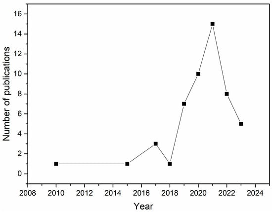
Figure 2.
A graphical representation of the number of publications on EOs with antimicrobial and anti-inflammatory activity in the past 13 years.
5.1. Search Strategy and Inclusion and Exclusion Criteria
The search for information on the chemical composition of EOs and tests performed was implemented considering all articles published in the past thirteen years (2010–2023) in the literature databases Web of Science (https://www.webofscience.com, accessed on 17 February 2023), Scopus (https://www.scopus.com, accessed on 17 February 2023), Science Direct (https://www.sciencedirect.com/, accessed on 15 February 2023), and PubMed (https://pubmed.ncbi.nlm.nih.gov/, accessed on 17 February 2023). The primary keyword “essential oil”, “tissue engineering”, “wound healing”, “antimicrobial”, “anti-inflammatory”, and “polymers” activities were searched for and combined in the titles and abstracts. Inclusion criteria for sections of this study were accessed for systems (polymers—essential oil) and assays to support antimicrobial and anti-inflammatory activity. Searches performed with review articles, books, book chapters, and non-medical applications were excluded. Figure 3 summarizes the general methodology, highlighting the articles collected in each database, duplicated, and excluded, and finally, how many were selected for writing this review.
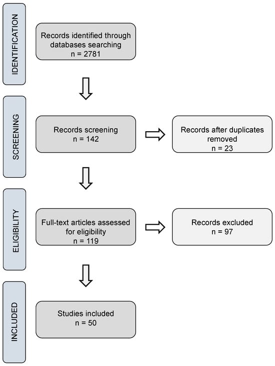
Figure 3.
Search strategy flowchart.
5.2. Study Records: Data Management
Table 1 describes the mechanism used for data management, uniformly naming the plant species, main components, and antimicrobial and anti-inflammatory effects of the EO.
6. Conclusions and Future Perspectives
The understanding that the application of polymers with biocompatible and biodegradable characteristics, such as chitosan, can offer practical alternatives for the treatment of severe wounds and burns, as it presents excellent biocompatibility, biodegradability, and non-toxicity, has been proven through numerous studies. Ideally, the scaffold/membrane/film/hydrogel material should induce as little pain as possible, allow rapid healing, and direct the growth in defect-free epidermal cells. To this end, various bioactive compounds have also been incorporated into dressings to provide these desired characteristics. EOs act as secondary metabolites, defending the host from microbial invasion. The antimicrobial effect comes mainly from known polyphenols and terpenes: monoterpenes, eugenol, cinnamaldehyde, carvacrol, and thymol.
Many plant species that contain EOs with antioxidant, anti-inflammatory, and antimicrobial activities widely studied have shown promise for studies in the biomedical area; among them are Syzygium aromaticum or Eugenia caryophyllata (clove), Organum vulgare (oregano), and Cinnamomum zeylanicum (cinnamon). These plants are good candidates for clinical trials due to their proven effects on wound healing and reducing the incidence of inflammatory cells at the wound site. They are also of low toxicity and are widely available and used in the food and cosmetics industry.
Studies with polymeric membranes loaded with essential oils on the formation of new blood vessels in the wound area, formation of epithelial tissue, and evaluation of the reduction in oxidative stress are also approaches that should be considered in the future. These approaches aim to improve the local oxygenation process of the affected area and reduce scars and tissue regeneration. Furthermore, these materials can be suitable substitutes for conventional antibiotics due to their potent antimicrobial activities against various pathogens.
The integration of engineering techniques, such as electrospinning, rotospinning, and 3D bioprinting, could also represent, shortly, a positive impact on the generation of hybrid materials from polymers and EOs by integrating cells, growth factors, and biomaterials with personalized structures, mimicking natural characteristics of organs and tissues. From this perspective, the continued need for research into EOs for bioactives is clear, valuing traditional knowledge and biodiversity. Therefore, this review offers an overview of the evidence found in the past thirteen years of using EOs in wound healing, as well as the dressing form and biological outcomes.
Author Contributions
Conceptualization, B.M.A.d.B.B., J.K.R.d.S. and M.F.P.; methodology, B.M.A.d.B.B., J.K.R.d.S. and M.F.P.; writing—original draft preparation, B.M.A.d.B.B.; writing—review and editing, B.M.A.d.B.B., J.K.R.d.S. and M.F.P.; visualization, P.L.B.F., J.K.R.d.S. and M.F.P.; supervision, J.K.R.d.S. and M.F.P. All authors have read and agreed to the published version of the manuscript.
Funding
This research was funded by Conselho Nacional de Desenvolvimento Científico e Tecnológico (CNPq), grant number 168387/2022-0, and by Fundação da Amazônia de Amparo a Estudos e Pesquisas (FAPESPA).
Data Availability Statement
The data presented in this study are available on request from the corresponding author.
Conflicts of Interest
The authors declare no conflicts of interest.
References
- Harrison, R.H.; St-Pierre, J.-P.; Stevens, M.M. Tissue Engineering and Regenerative Medicine: A Year in Review. Tissue Eng. Part B Rev. 2014, 20, 1–16. [Google Scholar] [CrossRef] [PubMed]
- Ikada, Y. Challenges in tissue engineering. J. R. Soc. Interface 2006, 3, 589–601. [Google Scholar] [CrossRef] [PubMed]
- Mota, C.; Camarero-Espinosa, S.; Baker, M.B.; Wieringa, P.; Moroni, L. Bioprinting: From Tissue and Organ Development to in Vitro Models. Chem. Rev. 2020, 120, 10547–10607. [Google Scholar] [CrossRef]
- De Bartolo, L.; Leindlein, A.; Hofmann, D.; Bader, A.; de Grey, A.; Curcio, E.; Drioli, E. Bio-hybrid organs and tissues for patient therapy: A future vision for 2030. Chem. Eng. Process. Process Intensif. 2012, 51, 79–87. [Google Scholar] [CrossRef]
- Berthiaume, F.; Maguire, T.J.; Yarmush, M.L. Tissue Engineering and Regenerative Medicine: History, Progress, and Challenges. Annu. Rev. Chem. Biomol. Eng. 2011, 2, 403–430. [Google Scholar] [CrossRef]
- Jayakumar, R.; Tamura, H.; Nair, S.; Furuike, T. Perspectives of chitin and chitosan nanofibrous scaffolds in tissue engineering. In Tissue Engineering; IntechOpen: London, UK, 2010; p. 205. [Google Scholar]
- Lee, K.Y.; Mooney, D.J. Hydrogels for tissue engineering. Chem. Rev. 2001, 101, 1869–1880. [Google Scholar] [CrossRef] [PubMed]
- Zhang, X.; Li, Z.; Yang, P.; Duan, G.; Liu, X.; Gu, Z.; Li, Y. Polyphenol scaffolds in tissue engineering. Mater. Horiz. 2021, 8, 145–167. [Google Scholar] [CrossRef]
- Boccaccini, A.R.; Ma, P.X.; Liverani, L. Tissue Engineering Using Ceramics and Polymers; Woodhead Publishing: Sawston, UK, 2021. [Google Scholar]
- Jorgensen, A.M.; Yoo, J.J.; Atala, A. Solid organ bioprinting: Strategies to achieve organ function. Chem. Rev. 2020, 120, 11093–11127. [Google Scholar] [CrossRef]
- Sinno, H.; Prakash, S. Complements and the wound healing cascade: An updated review. Plast. Surg. Int. 2013, 2013, 146764. [Google Scholar] [CrossRef]
- Phillipson, M.; Kubes, P. The healing power of neutrophils. Trends Immunol. 2019, 40, 635–647. [Google Scholar] [CrossRef]
- Rodrigues, M.; Kosaric, N.; Bonham, C.A.; Gurtner, G.C. Wound healing: A cellular perspective. Physiol. Rev. 2019, 99, 665–706. [Google Scholar] [CrossRef] [PubMed]
- Werner, S.; Krieg, T.; Smola, H. Keratinocyte–fibroblast interactions in wound healing. J. Investig. Dermatol. 2007, 127, 998–1008. [Google Scholar] [CrossRef] [PubMed]
- Reinke, J.; Sorg, H. Wound repair and regeneration. Eur. Surg. Res. 2012, 49, 35–43. [Google Scholar] [CrossRef] [PubMed]
- Nho, Y.; Park, K. Preparation and properties of PVA/PVP hydrogels containing chitosan by radiation. J. Appl. Polym. Sci. 2002, 85, 1787–1794. [Google Scholar] [CrossRef]
- Li, X.; Sun, Q.; Li, Q.; Kawazoe, N.; Chen, G. Functional hydrogels with tunable structures and properties for tissue engineering applications. Front. Chem. 2018, 6, 499. [Google Scholar] [CrossRef] [PubMed]
- Bolívar-Monsalve, E.J.; Alvarez, M.M.; Hosseini, S.; Espinosa-Hernandez, M.A.; Ceballos-González, C.F.; Sanchez-Dominguez, M.; Shin, S.R.; Cecen, B.; Hassan, S.; Di Maio, E. Engineering bioactive synthetic polymers for biomedical applications: A review with emphasis on tissue engineering and controlled release. Mater. Adv. 2021, 2, 4447–4478. [Google Scholar] [CrossRef]
- Zhao, K.; Lu, Z.-H.; Zhao, P.; Kang, S.-X.; Yang, Y.-Y.; Yu, D.-G. Modified tri–axial electrospun functional core–shell nanofibrous membranes for natural photodegradation of antibiotics. Chem. Eng. J. 2021, 425, 131455. [Google Scholar] [CrossRef]
- Tovar, C.D.G.; Castro, J.I.; Valencia Llano, C.H.; Navia Porras, D.P.; Delgado Ospina, J.; Valencia Zapata, M.E.; Herminsul Mina Hernandez, J.; Chaur, M.N. Synthesis, characterization, and histological evaluation of chitosan-Ruta graveolens essential oil films. Molecules 2020, 25, 1688. [Google Scholar] [CrossRef] [PubMed]
- Lu, J.; Chen, Y.; Ding, M.; Fan, X.; Hu, J.; Chen, Y.; Li, J.; Li, Z.; Liu, W. A 4arm-PEG macromolecule crosslinked chitosan hydrogels as antibacterial wound dressing. Carbohydr. Polym. 2022, 277, 118871. [Google Scholar] [CrossRef]
- López-Mata, M.A.; Ruiz-Cruz, S.; de Jesús Ornelas-Paz, J.; Del Toro-Sánchez, C.L.; Márquez-Ríos, E.; Silva-Beltrán, N.P.; Cira-Chávez, L.A.; Burruel-Ibarra, S.E. Mechanical, barrier and antioxidant properties of chitosan films incorporating cinnamaldehyde. J. Polym. Environ. 2018, 26, 452–461. [Google Scholar] [CrossRef]
- Cruz Sánchez, E.; García, M.T.; Pereira, J.; Oliveira, F.; Craveiro, R.; Paiva, A.; Gracia, I.; García-Vargas, J.M.; Duarte, A.R.C. Alginate–Chitosan Membranes for the Encapsulation of Lavender Essential Oil and Development of Biomedical Applications Related to Wound Healing. Molecules 2023, 28, 3689. [Google Scholar] [CrossRef] [PubMed]
- Unnithan, A.R.; Gnanasekaran, G.; Sathishkumar, Y.; Lee, Y.S.; Kim, C.S. Electrospun antibacterial polyurethane–cellulose acetate–zein composite mats for wound dressing. Carbohydr. Polym. 2014, 102, 884–892. [Google Scholar] [CrossRef]
- Gao, Y.; Bach Truong, Y.; Zhu, Y.; Louis Kyratzis, I. Electrospun antibacterial nanofibers: Production, activity, and in vivo applications. J. Appl. Polym. Sci. 2014, 131. [Google Scholar] [CrossRef]
- Simões, D.; Miguel, S.P.; Ribeiro, M.P.; Coutinho, P.; Mendonça, A.G.; Correia, I.J. Recent advances on antimicrobial wound dressing: A review. Eur. J. Pharm. Biopharm. 2018, 127, 130–141. [Google Scholar] [CrossRef] [PubMed]
- Moghaddam, M.; Mehdizadeh, L. Chapter 13—Chemistry of Essential Oils and Factors Influencing Their Constituents. In Soft Chemistry and Food Fermentation; Academic Press: Cambridge, MA, USA, 2017; pp. 379–419. [Google Scholar] [CrossRef]
- Pereira dos Santos, E.; Nicácio, P.H.M.; Coêlho Barbosa, F.; Nunes da Silva, H.; Andrade, A.L.S.; Lia Fook, M.V.; de Lima Silva, S.M.; Farias Leite, I. Chitosan/Essential Oils Formulations for Potential Use as Wound Dressing: Physical and Antimicrobial Properties. Materials 2019, 12, 2223. [Google Scholar] [CrossRef] [PubMed]
- Pérez-Recalde, M.; Arias, I.E.R.; Hermida, É.B. Could essential oils enhance biopolymers performance for wound healing? A systematic review. Phytomedicine 2018, 38, 57–65. [Google Scholar] [CrossRef]
- Saporito, F.; Sandri, G.; Bonferoni, M.C.; Rossi, S.; Boselli, C.; Cornaglia, A.I.; Mannucci, B.; Grisoli, P.; Vigani, B.; Ferrari, F. Essential oil-loaded lipid nanoparticles for wound healing. Int. J. Nanomed. 2018, 13, 175. [Google Scholar] [CrossRef]
- Zhang, X.; Williams, D. Definitions of Biomaterials for the Twenty-First Century; Elsevier: Amsterdam, The Netherlands, 2019. [Google Scholar]
- Pires, A.L.R.; Bierhalz, A.C.; Moraes, A.M. Biomaterials: Types, applications, and market. Química Nova 2015, 38, 957–971. [Google Scholar] [CrossRef]
- Gonçalves, I.M.; Rodrigues, R.O.; Moita, A.S.; Hori, T.; Kaji, H.; Lima, R.A.; Minas, G. Recent trends of biomaterials and biosensors for organ-on-chip platforms. Bioprinting 2022, 26, e00202. [Google Scholar] [CrossRef]
- Melvin, A.C.; Wick, T.V.; Zang, Y.; Harea, G.T.; Cancio, L.C.; Reynolds, M.M.; Batchinsky, A.I.; Roberts, T.R. Development and Blood Compatibility of a Stable and Bioactive Metal–Organic Framework Composite Coating for Blood-Circulation Tubing. ACS Biomater. Sci. Eng. 2022, 8, 3438–3449. [Google Scholar] [CrossRef]
- Zhang, D.; Chen, Q.; Shi, C.; Chen, M.; Ma, K.; Wan, J.; Liu, R. Dealing with the foreign-body response to implanted biomaterials: Strategies and applications of new materials. Adv. Funct. Mater. 2021, 31, 2007226. [Google Scholar] [CrossRef]
- Wen, P.; Wu, W.; Wang, F.; Zheng, H.; Liao, Z.; Shi, J.; Zhu, C.; Zhao, P.; Cheng, H.; Li, H. Cell delivery devices for cancer immunotherapy. J. Control. Release 2023, 353, 875–888. [Google Scholar] [CrossRef]
- Lee, S.; Choi, G.; Yang, Y.J.; Joo, K.I.; Cha, H.J. Visible light-crosslinkable tyramine-conjugated alginate-based microgel bioink for multiple cell-laden 3D artificial organ. Carbohydr. Polym. 2023, 313, 120895. [Google Scholar] [CrossRef] [PubMed]
- Wang, Q.; Luo, Z.; Wu, Y.-L.; Li, Z. Recent Advances in Enzyme-Based Biomaterials Toward Diabetic Wound Healing. Adv. NanoBiomed Res. 2023, 3, 2200110. [Google Scholar] [CrossRef]
- Huang, Z.; Shao, G.; Li, L. Micro/nano functional devices fabricated by additive manufacturing. Prog. Mater. Sci. 2023, 131, 101020. [Google Scholar] [CrossRef]
- Bakhshayesh, A.R.D.; Annabi, N.; Khalilov, R.; Akbarzadeh, A.; Samiei, M.; Alizadeh, E.; Alizadeh-Ghodsi, M.; Davaran, S.; Montaseri, A. Recent advances on biomedical applications of scaffolds in wound healing and dermal tissue engineering. Artif. Cells Nanomed. Biotechnol. 2018, 46, 691–705. [Google Scholar] [CrossRef] [PubMed]
- Naghieh, S.; Sarker, M.; Izadifar, M.; Chen, X. Dispensing-based bioprinting of mechanically-functional hybrid scaffolds with vessel-like channels for tissue engineering applications—A brief review. J. Mech. Behav. Biomed. Mater. 2018, 78, 298–314. [Google Scholar] [CrossRef]
- Rider, P.; Kačarević, Ž.P.; Alkildani, S.; Retnasingh, S.; Barbeck, M. Bioprinting of tissue engineering scaffolds. J. Tissue Eng. 2018, 9, 2041731418802090. [Google Scholar] [CrossRef] [PubMed]
- Budnyak, T.; Yanovska, E.; Kołodyńska, D.; Sternik, D.; Pylypchuk, I.V.; Ischenko, M.; Tertykh, V. Preparation and properties of organomineral adsorbent obtained by sol–gel technology. J. Therm. Anal. Calorim. 2016, 125, 1335–1351. [Google Scholar] [CrossRef]
- HPS, A.K.; Saurabh, C.K.; Adnan, A.; Fazita, M.N.; Syakir, M.; Davoudpour, Y.; Rafatullah, M.; Abdullah, C.; Haafiz, M.; Dungani, R. A review on chitosan-cellulose blends and nanocellulose reinforced chitosan biocomposites: Properties and their applications. Carbohydr. Polym. 2016, 150, 216–226. [Google Scholar] [CrossRef]
- Ziegler-Borowska, M.; Chełminiak, D.; Kaczmarek, H.; Kaczmarek-Kędziera, A. Effect of side substituents on thermal stability of the modified chitosan and its nanocomposites with magnetite. J. Therm. Anal. Calorim. 2016, 124, 1267–1280. [Google Scholar] [CrossRef]
- Shaari, N.; Kamarudin, S. Chitosan and alginate types of bio-membrane in fuel cell application: An overview. J. Power Sources 2015, 289, 71–80. [Google Scholar] [CrossRef]
- Olaru, A.-M.; Marin, L.; Morariu, S.; Pricope, G.; Pinteala, M.; Tartau-Mititelu, L. Biocompatible chitosan based hydrogels for potential application in local tumour therapy. Carbohydr. Polym. 2018, 179, 59–70. [Google Scholar] [CrossRef] [PubMed]
- Armiñán, A.; Sepúlveda, P.; Vicent, M.J. Polymer Therapeutics as Nano-Sized Medicines for Tissue Regeneration and Repair. Polym. Regen. Med. Biomed. Appl. Nano-Macro-Struct. 2014, 249–284. [Google Scholar] [CrossRef]
- Tehrani, S.F.; Bharadwaj, P.; Leblond Chain, J.; Roullin, V.G. Purification processes of polymeric nanoparticles: How to improve their clinical translation? J. Control. Release 2023, 360, 591–612. [Google Scholar] [CrossRef] [PubMed]
- Nair, L.S.; Laurencin, C.T. Biodegradable polymers as biomaterials. Prog. Polym. Sci. 2007, 32, 762–798. [Google Scholar] [CrossRef]
- Seo, S.-J.; Mahapatra, C.; Singh, R.K.; Knowles, J.C.; Kim, H.-W. Strategies for osteochondral repair: Focus on scaffolds. J. Tissue Eng. 2014, 5, 2041731414541850. [Google Scholar] [CrossRef]
- Keane, T.J.; Badylak, S.F. Biomaterials for tissue engineering applications. Semin. Pediatr. Surg. 2014, 23, 112–118. [Google Scholar] [CrossRef] [PubMed]
- Lutolf, M.P.; Lauer-Fields, J.L.; Schmoekel, H.G.; Metters, A.T.; Weber, F.E.; Fields, G.B.; Hubbell, J.A. Synthetic matrix metalloproteinase-sensitive hydrogels for the conduction of tissue regeneration: Engineering cell-invasion characteristics. Proc. Natl. Acad. Sci. USA 2003, 100, 5413–5418. [Google Scholar] [CrossRef]
- Li, G.; Zhao, M.; Xu, F.; Yang, B.; Li, X.; Meng, X.; Teng, L.; Sun, F.; Li, Y. Synthesis and biological application of polylactic acid. Molecules 2020, 25, 5023. [Google Scholar] [CrossRef]
- Xu, F.; Cui, F.-Z.; Jiao, Y.-P.; Meng, Q.-Y.; Wang, X.-P.; Cui, X.-Y. Improvement of cytocompatibility of electrospinning PLLA microfibers by blending PVP. J. Mater. Sci. Mater. Med. 2009, 20, 1331–1338. [Google Scholar] [CrossRef] [PubMed]
- Pan, P.; Inoue, Y. Polymorphism and isomorphism in biodegradable polyesters. Prog. Polym. Sci. 2009, 34, 605–640. [Google Scholar] [CrossRef]
- Im, S.H.; Im, D.H.; Park, S.J.; Chung, J.J.; Jung, Y.; Kim, S.H. Stereocomplex polylactide for drug delivery and biomedical applications: A review. Molecules 2021, 26, 2846. [Google Scholar] [CrossRef] [PubMed]
- Boey, J.Y.; Mohamad, L.; Khok, Y.S.; Tay, G.S.; Baidurah, S. A review of the applications and biodegradation of polyhydroxyalkanoates and poly (lactic acid) and its composites. Polymers 2021, 13, 1544. [Google Scholar] [CrossRef] [PubMed]
- Sun, J.; Shen, J.; Chen, S.; Cooper, M.A.; Fu, H.; Wu, D.; Yang, Z. Nanofiller reinforced biodegradable PLA/PHA composites: Current status and future trends. Polymers 2018, 10, 505. [Google Scholar] [CrossRef] [PubMed]
- Su, S.; Kopitzky, R.; Tolga, S.; Kabasci, S. Polylactide (PLA) and its blends with poly (butylene succinate) (PBS): A brief review. Polymers 2019, 11, 1193. [Google Scholar] [CrossRef] [PubMed]
- Poonguzhali, R.; Basha, S.K.; Kumari, V.S. Nanostarch reinforced with chitosan/poly (vinyl pyrrolidone) blend for in vitro wound healing application. Polym. Plast. Technol. Eng. 2018, 57, 1400–1410. [Google Scholar] [CrossRef]
- Wang, B.-L.; Liu, X.-S.; Ji, Y.; Ren, K.-F.; Ji, J. Fast and long-acting antibacterial properties of chitosan-Ag/polyvinylpyrrolidone nanocomposite films. Carbohydr. Polym. 2012, 90, 8–15. [Google Scholar] [CrossRef] [PubMed]
- Razzak, M.; Dewi, S.; Lely, H.; Taty, E. The characterization of dressing component materials and radiation formation of PVA–PVP hydrogel. Radiat. Phys. Chem. 1999, 55, 153–165. [Google Scholar] [CrossRef]
- Poonguzhali, R.; Basha, S.K.; Kumari, V.S. Synthesis and characterization of chitosan-PVP-nanocellulose composites for in-vitro wound dressing application. Int. J. Biol. Macromol. 2017, 105, 111–120. [Google Scholar] [CrossRef]
- Román-Doval, R.; Tellez-Cruz, M.; Rojas-Chávez, H.; Cruz-Martínez, H.; Carrasco-Torres, G.; Vásquez-Garzón, V. Enhancing electrospun scaffolds of PVP with polypyrrole/iodine for tissue engineering of skin regeneration by coating via a plasma process. J. Mater. Sci. 2019, 54, 3342–3353. [Google Scholar] [CrossRef]
- Yu, D.G.; Zhang, X.F.; Shen, X.X.; Brandford-White, C.; Zhu, L.M. Ultrafine ibuprofen-loaded polyvinylpyrrolidone fiber mats using electrospinning. Polym. Int. 2009, 58, 1010–1013. [Google Scholar] [CrossRef]
- Kim, J.H.; Lee, H.; Jatoi, A.W.; Im, S.S.; Lee, J.S.; Kim, I.-S. Juniperus chinensis extracts loaded PVA nanofiber: Enhanced antibacterial activity. Mater. Lett. 2016, 181, 367–370. [Google Scholar] [CrossRef]
- Dadfar, S.M.M.; Kavoosi, G.; Mohammad Ali Dadfar, S. Investigation of mechanical properties, antibacterial features, and water vapor permeability of polyvinyl alcohol thin films reinforced by glutaraldehyde and multiwalled carbon nanotube. Polymer Compos. 2014, 35, 1736–1743. [Google Scholar] [CrossRef]
- Zhang, D.; Zhou, W.; Wei, B.; Wang, X.; Tang, R.; Nie, J.; Wang, J. Carboxyl-modified poly(vinyl alcohol)-crosslinked chitosan hydrogel films for potential wound dressing. Carbohydr. Polym. 2015, 125, 189–199. [Google Scholar] [CrossRef] [PubMed]
- Jia, Y.-T.; Gong, J.; Gu, X.-H.; Kim, H.-Y.; Dong, J.; Shen, X.-Y. Fabrication and characterization of poly (vinyl alcohol)/chitosan blend nanofibers produced by electrospinning method. Carbohydr. Polym. 2007, 67, 403–409. [Google Scholar] [CrossRef]
- Kamoun, E.A.; Chen, X.; Eldin, M.S.M.; Kenawy, E.-R.S. Crosslinked poly (vinyl alcohol) hydrogels for wound dressing applications: A review of remarkably blended polymers. Arab. J. Chem. 2015, 8, 1–14. [Google Scholar] [CrossRef]
- Venkatesan, J.; Bhatnagar, I.; Manivasagan, P.; Kang, K.-H.; Kim, S.-K. Alginate composites for bone tissue engineering: A review. Int. J. Biol. Macromol. 2015, 72, 269–281. [Google Scholar] [CrossRef]
- Aranha, I.B.; Lucas, E.F. Poli (álcool vinílico) modificado com cadeias hidrocarbônicas: Avaliação do balanço hidrófilo/lipófilo. Polímeros 2001, 11, 174–181. [Google Scholar] [CrossRef]
- Jacob, J.; Peter, G.; Thomas, S.; Haponiuk, J.T.; Gopi, S. Chitosan and polyvinyl alcohol nanocomposites with cellulose nanofibers from ginger rhizomes and its antimicrobial activities. Int. J. Biol. Macromol. 2019, 129, 370–376. [Google Scholar] [CrossRef]
- Wang, X.; Ding, B.; Sun, G.; Wang, M.; Yu, J. Electro-spinning/netting: A strategy for the fabrication of three-dimensional polymer nano-fiber/nets. Prog. Mater. Sci. 2013, 58, 1173–1243. [Google Scholar] [CrossRef] [PubMed]
- Tarique, J.; Sapuan, S.M.; Khalina, A. Effect of glycerol plasticizer loading on the physical, mechanical, thermal, and barrier properties of arrowroot (Maranta arundinacea) starch biopolymers. Sci. Rep. 2021, 11, 13900. [Google Scholar] [CrossRef]
- Rasool, A.; Ata, S.; Islam, A. Stimuli responsive biopolymer (chitosan) based blend hydrogels for wound healing application. Carbohydr. Polym. 2019, 203, 423–429. [Google Scholar] [CrossRef]
- Labib, R.M.; Ayoub, I.M.; Michel, H.E.; Mehanny, M.; Kamil, V.; Hany, M.; Magdy, M.; Moataz, A.; Maged, B.; Mohamed, A. Appraisal on the wound healing potential of Melaleuca alternifolia and Rosmarinus officinalis L. essential oil-loaded chitosan topical preparations. PLoS ONE 2019, 14, e0219561. [Google Scholar] [CrossRef] [PubMed]
- Ardekani, N.T.; Khorram, M.; Zomorodian, K.; Yazdanpanah, S.; Veisi, H.; Veisi, H. Evaluation of electrospun poly (vinyl alcohol)-based nanofiber mats incorporated with Zataria multiflora essential oil as potential wound dressing. Int. J. Biol. Macromol. 2019, 125, 743–750. [Google Scholar] [CrossRef] [PubMed]
- Amalraj, A.; Haponiuk, J.T.; Thomas, S.; Gopi, S. Preparation, characterization and antimicrobial activity of polyvinyl alcohol/gum arabic/chitosan composite films incorporated with black pepper essential oil and ginger essential oil. Int. J. Biol. Macromol. 2020, 151, 366–375. [Google Scholar] [CrossRef] [PubMed]
- Rafiq, M.; Hussain, T.; Abid, S.; Nazir, A.; Masood, R. Development of sodium alginate/PVA antibacterial nanofibers by the incorporation of essential oils. Mater. Res. Express 2018, 5, 035007. [Google Scholar] [CrossRef]
- Lamarra, J.; Bucci, P.; Giannuzzi, L.; Montanari, J.; Rivero, S.; Pinotti, A. Biomaterial-based dressings as vehicle for chitosan-encapsulated cabreuva essential oil: Cytotoxicity and regenerative activity. React. Funct. Polym. 2020, 156, 104728. [Google Scholar] [CrossRef]
- Aman, R.M.; Abu Hashim, I.I.; Meshali, M.M. Novel Clove essential oil nanoemulgel tailored by Taguchi’s model and scaffold-based nanofibers: Phytopharmaceuticals with promising potential as cyclooxygenase-2 inhibitors in external inflammation. Int. J. Nanomed. 2020, 15, 2171–2195. [Google Scholar] [CrossRef]
- Barzegar, S.; Zare, M.R.; Shojaei, F.; Zareshahrabadi, Z.; Koohi-Hosseinabadi, O.; Saharkhiz, M.J.; Iraji, A.; Zomorodian, K.; Khorram, M. Core-shell chitosan/PVA-based nanofibrous scaffolds loaded with Satureja mutica or Oliveria decumbens essential oils as enhanced antimicrobial wound dressing. Int. J. Pharm. 2021, 597, 120288. [Google Scholar] [CrossRef]
- Antunes, J.C.; Tavares, T.D.; Teixeira, M.A.; Teixeira, M.O.; Homem, N.C.; Amorim, M.T.P.; Felgueiras, H.P. Eugenol-Containing Essential Oils Loaded onto Chitosan/Polyvinyl Alcohol Blended Films and Their Ability to Eradicate Staphylococcus aureus or Pseudomonas aeruginosa from Infected Microenvironments. Pharmaceutics 2021, 13, 195. [Google Scholar] [CrossRef] [PubMed]
- El-Aassar, M.R.; El-Beheri, N.G.; Agwa, M.M.; Eltaher, H.M.; Alseqely, M.; Sadik, W.S.; El-Khordagui, L. Antibiotic-free combinational hyaluronic acid blend nanofibers for wound healing enhancement. Int. J. Biol. Macromol. 2021, 167, 1552–1563. [Google Scholar] [CrossRef] [PubMed]
- Tamer, T.M.; Alsehli, M.H.; Omer, A.M.; Afifi, T.H.; Sabet, M.M.; Mohy-Eldin, M.S.; Hassan, M.A. Development of Polyvinyl Alcohol/Kaolin Sponges Stimulated by Marjoram as Hemostatic, Antibacterial, and Antioxidant Dressings for Wound Healing Promotion. Int. J. Mol. Sci. 2021, 22, 13050. [Google Scholar] [CrossRef] [PubMed]
- Jia, J.; Duan, S.; Zhou, X.; Sun, L.; Qin, C.; Li, M.; Ge, F. Long-Term Antibacterial Film Nanocomposite Incorporated with Patchouli Essential Oil Prepared by Supercritical CO2 Cyclic Impregnation for Wound Dressing. Molecules 2021, 26, 5005. [Google Scholar] [CrossRef] [PubMed]
- Severo, A.M.C.; Fook, M.V.L.; Leite, I.F. PVA/chitosan hydrogels funcionalized with tea tree oil for application as dressings. Rev. Matéria 2022, 27, e13149. [Google Scholar] [CrossRef]
- Doostan, M.; Doostan, M.; Mohammadi, P.; Khoshnevisan, K.; Maleki, H. Wound healing promotion by flaxseed extract-loaded polyvinyl alcohol/chitosan nanofibrous scaffolds. Int. J. Biol. Macromol. 2023, 228, 506–516. [Google Scholar] [CrossRef]
- Kavoosi, G.; Bordbar, Z.; Dadfar, S.M.; Dadfar, S.M.M. Preparation and characterization of a novel gelatin–poly (vinyl alcohol) hydrogel film loaded with Zataria multiflora essential oil for antibacterial–antioxidant wound-dressing applications. J. Appl. Polym. Sci. 2017, 134, 45351. [Google Scholar] [CrossRef]
- Allizond, V.; Comini, S.; Cuffini, A.M.; Banche, G. Current knowledge on biomaterials for orthopedic applications modified to reduce bacterial adhesive ability. Antibiotics 2022, 11, 529. [Google Scholar] [CrossRef] [PubMed]
- Comini, S.; Scutera, S.; Sparti, R.; Banche, G.; Coppola, B.; Bertea, C.M.; Bianco, G.; Gatti, N.; Cuffini, A.M.; Palmero, P.; et al. Combination of Poly(ε-Caprolactone) Biomaterials and Essential Oils to Achieve Anti-Bacterial and Osteo-Proliferative Properties for 3D-Scaffolds in Regenerative Medicine. Pharmaceutics 2022, 14, 1873. [Google Scholar] [CrossRef] [PubMed]
- Comini, S.; Sparti, R.; Coppola, B.; Mohammadi, M.; Scutera, S.; Menotti, F.; Banche, G.; Cuffini, A.M.; Palmero, P.; Allizond, V. Novel silver-functionalized poly (ε-caprolactone)/biphasic calcium phosphate scaffolds designed to counteract post-surgical infections in orthopedic applications. Int. J. Mol. Sci. 2021, 22, 10176. [Google Scholar] [CrossRef]
- García-Salinas, S.; Evangelopoulos, M.; Gámez-Herrera, E.; Arruebo, M.; Irusta, S.; Taraballi, F.; Mendoza, G.; Tasciotti, E. Electrospun anti-inflammatory patch loaded with essential oils for wound healing. Int. J. Pharm. 2020, 577, 119067. [Google Scholar] [CrossRef] [PubMed]
- Kapustová, M.; Puškárová, A.; Bučková, M.; Granata, G.; Napoli, E.; Annušová, A.; Mesárošová, M.; Kozics, K.; Pangallo, D.; Geraci, C. Biofilm inhibition by biocompatible poly (ε-caprolactone) nanocapsules loaded with essential oils and their cyto/genotoxicity to human keratinocyte cell line. Int. J. Pharm. 2021, 606, 120846. [Google Scholar] [CrossRef] [PubMed]
- Sahal, G.; Nasseri, B.; Ebrahimi, A.; Bilkay, I.S. Electrospun essential oil-polycaprolactone nanofibers as antibiofilm surfaces against clinical Candida tropicalis isolates. Biotechnol. Lett. 2019, 41, 511–522. [Google Scholar] [CrossRef] [PubMed]
- Unalan, I.; Endlein, S.J.; Slavik, B.; Buettner, A.; Goldmann, W.H.; Detsch, R.; Boccaccini, A.R. Evaluation of Electrospun Poly(ε-Caprolactone)/Gelatin Nanofiber Mats Containing Clove Essential Oil for Antibacterial Wound Dressing. Pharmaceutics 2019, 11, 570. [Google Scholar] [CrossRef] [PubMed]
- Abedalwafa, M.; Wang, F.; Wang, L.; Li, C. Biodegradable poly-epsilon-caprolactone (PCL) for tissue engineering applications: A review. Rev. Adv. Mater. Sci. 2013, 34, 123–140. [Google Scholar]
- Quirós, J.; Boltes, K.; Rosal, R. Bioactive applications for electrospun fibers. Polym. Rev. 2016, 56, 631–667. [Google Scholar] [CrossRef]
- Ko, Y.-M.; Choi, D.-Y.; Jung, S.-C.; Kim, B.-H. Characteristics of plasma treated electrospun polycaprolactone (PCL) nanofiber scaffold for bone tissue engineering. J. Nanosci. Nanotechnol. 2015, 15, 192–195. [Google Scholar] [CrossRef] [PubMed]
- Mochane, M.J.; Motsoeneng, T.S.; Sadiku, E.R.; Mokhena, T.C.; Sefadi, J.S. Morphology and properties of electrospun PCL and its composites for medical applications: A mini review. Appl. Sci. 2019, 9, 2205. [Google Scholar] [CrossRef]
- Mohamed, R.M.; Yusoh, K. A review on the recent research of polycaprolactone (PCL). Adv. Mater. Res. 2016, 1134, 249–255. [Google Scholar] [CrossRef]
- Li, Z.; Tan, B.H. Towards the development of polycaprolactone based amphiphilic block copolymers: Molecular design, self-assembly and biomedical applications. Mater. Sci. Eng. C 2014, 45, 620–634. [Google Scholar] [CrossRef]
- Ulery, B.D.; Nair, L.S.; Laurencin, C.T. Biomedical applications of biodegradable polymers. J. Polym. Sci. Part B Polym. Phys. 2011, 49, 832–864. [Google Scholar] [CrossRef] [PubMed]
- Sheikh, F.A.; Kanjwal, M.A.; Saran, S.; Chung, W.-J.; Kim, H. Polyurethane nanofibers containing copper nanoparticles as future materials. Appl. Surf. Sci. 2011, 257, 3020–3026. [Google Scholar] [CrossRef]
- Unnithan, A.R.; Sasikala, A.R.K.; Murugesan, P.; Gurusamy, M.; Wu, D.; Park, C.H.; Kim, C.S. Electrospun polyurethane-dextran nanofiber mats loaded with Estradiol for post-menopausal wound dressing. Int. J. Biol. Macromol. 2015, 77, 1–8. [Google Scholar] [CrossRef] [PubMed]
- Akduman, C.; Özgüney, I.; Kumbasar, E.P.A. Preparation and characterization of naproxen-loaded electrospun thermoplastic polyurethane nanofibers as a drug delivery system. Mater. Sci. Eng. C 2016, 64, 383–390. [Google Scholar] [CrossRef] [PubMed]
- Fang, L.; Liang, B.; Yang, G.; Hu, Y.; Zhu, Q.; Ye, X. A needle-type glucose biosensor based on PANI nanofibers and PU/E-PU membrane for long-term invasive continuous monitoring. Biosens. Bioelectron. 2017, 97, 196–202. [Google Scholar] [CrossRef] [PubMed]
- Burke, A.; Hasirci, N. Polyurethanes in biomedical applications. In Biomaterials: From Molecules to Engineered Tissue; Springer: Boston, MA, USA, 2004; pp. 83–101. [Google Scholar]
- Sofi, H.S.; Akram, T.; Tamboli, A.H.; Majeed, A.; Shabir, N.; Sheikh, F.A. Novel lavender oil and silver nanoparticles simultaneously loaded onto polyurethane nanofibers for wound-healing applications. Int. J. Pharm. 2019, 569, 118590. [Google Scholar] [CrossRef]
- Mi, H.Y.; Palumbo, S.; Jing, X.; Turng, L.S.; Li, W.J.; Peng, X.F. Thermoplastic polyurethane/hydroxyapatite electrospun scaffolds for bone tissue engineering: Effects of polymer properties and particle size. J. Biomed. Mater. Res. Part B Appl. Biomater. 2014, 102, 1434–1444. [Google Scholar] [CrossRef]
- Sheikh, F.A.; Barakat, N.A.; Kanjwal, M.A.; Chaudhari, A.A.; Jung, I.-H.; Lee, J.H.; Kim, H.Y. Electrospun antimicrobial polyurethane nanofibers containing silver nanoparticles for biotechnological applications. Macromol. Res. 2009, 17, 688–696. [Google Scholar] [CrossRef]
- Abrigo, M.; McArthur, S.L.; Kingshott, P. Electrospun nanofibers as dressings for chronic wound care: Advances, challenges, and future prospects. Macromol. Biosci. 2014, 14, 772–792. [Google Scholar] [CrossRef]
- Jafari, A.; Hassanajili, S.; Karimi, M.B.; Emami, A.; Ghaffari, F.; Azarpira, N. Effect of organic/inorganic nanoparticles on performance of polyurethane nanocomposites for potential wound dressing applications. J. Mech. Behav. Biomed. Mater. 2018, 88, 395–405. [Google Scholar] [CrossRef]
- Khil, M.S.; Cha, D.I.; Kim, H.Y.; Kim, I.S.; Bhattarai, N. Electrospun nanofibrous polyurethane membrane as wound dressing. J. Biomed. Mater. Res. Part B Appl. Biomater. 2003, 67, 675–679. [Google Scholar] [CrossRef] [PubMed]
- Pandey, S.; Banik, R. Extractive fermentation for enhanced production of alkaline phosphatase from Bacillus licheniformis MTCC 1483 using aqueous two-phase systems. Bioresour. Technol. 2011, 102, 4226–4231. [Google Scholar] [CrossRef] [PubMed]
- Chen, S.-L.; Fu, R.-H.; Liao, S.-F.; Liu, S.-P.; Lin, S.-Z.; Wang, Y.-C. A PEG-based hydrogel for effective wound care management. Cell Transplant. 2018, 27, 275–284. [Google Scholar] [CrossRef] [PubMed]
- Kamoun, E.A.; Kenawy, E.-R.S.; Chen, X. A review on polymeric hydrogel membranes for wound dressing applications: PVA-based hydrogel dressings. J. Adv. Res. 2017, 8, 217–233. [Google Scholar] [CrossRef] [PubMed]
- Shingel, K.I.; Di Stabile, L.; Marty, J.-P.; Faure, M.-P. Inflammatory inert poly(ethylene glycol)–protein wound dressing improves healing responses in partial- and full-thickness wounds. Int. Wound J. 2006, 3, 332–342. [Google Scholar] [CrossRef] [PubMed]
- Sinha, M.; Banik, R.M.; Haldar, C.; Maiti, P. Development of ciprofloxacin hydrochloride loaded poly(ethylene glycol)/chitosan scaffold as wound dressing. J. Porous Mater. 2013, 20, 799–807. [Google Scholar] [CrossRef]
- Eğri, Ö.; Erdemir, N. Production of Hypericum perforatum oil-loaded membranes for wound dressing material and in vitro tests. Artif. Cells Nanomed. Biotechnol. 2019, 47, 1404–1415. [Google Scholar] [CrossRef]
- Singh, P.; Verma, C.; Mukhopadhyay, S.; Gupta, A.; Gupta, B. Preparation of thyme oil loaded κ-carrageenan-polyethylene glycol hydrogel membranes as wound care system. Int. J. Pharm. 2022, 618, 121661. [Google Scholar] [CrossRef]
- Wu, N.; Wang, L.-S.; Tan, D.C.-W.; Moochhala, S.M.; Yang, Y.-Y. Mathematical modeling and in vitro study of controlled drug release via a highly swellable and dissoluble polymer matrix: Polyethylene oxide with high molecular weights. J. Control. Release 2005, 102, 569–581. [Google Scholar] [CrossRef]
- Ma, L.; Deng, L.; Chen, J. Applications of poly (ethylene oxide) in controlled release tablet systems: A review. Drug Dev. Ind. Pharm. 2014, 40, 845–851. [Google Scholar] [CrossRef]
- Eskitoros-Togay, Ş.M.; Bulbul, Y.E.; Tort, S.; Demirtaş Korkmaz, F.; Acartürk, F.; Dilsiz, N. Fabrication of doxycycline-loaded electrospun PCL/PEO membranes for a potential drug delivery system. Int. J. Pharm. 2019, 565, 83–94. [Google Scholar] [CrossRef] [PubMed]
- Locilento, D.A.; Mercante, L.A.; Andre, R.S.; Mattoso, L.H.; Luna, G.L.; Brassolatti, P.; Anibal, F.d.F.; Correa, D.S. Biocompatible and biodegradable electrospun nanofibrous membranes loaded with grape seed extract for wound dressing application. J. Nanomater. 2019, 2019, 2472964. [Google Scholar] [CrossRef]
- Wang, B.; Li, H.; Yao, Q.; Zhang, Y.; Zhu, X.; Xia, T.; Wang, J.; Li, G.; Li, X.; Ni, S. Local in vitro delivery of rapamycin from electrospun PEO/PDLLA nanofibers for glioblastoma treatment. Biomed. Pharmacother. 2016, 83, 1345–1352. [Google Scholar] [CrossRef] [PubMed]
- Honarbakhsh, S.; Pourdeyhimi, B. Scaffolds for drug delivery, part I: Electrospun porous poly(lactic acid) and poly(lactic acid)/poly(ethylene oxide) hybrid scaffolds. J. Mater. Sci. 2011, 46, 2874–2881. [Google Scholar] [CrossRef]
- Hameed, M.; Rasul, A.; Waqas, M.K.; Saadullah, M.; Aslam, N.; Abbas, G.; Latif, S.; Afzal, H.; Inam, S.; Akhtar Shah, P. Formulation and Evaluation of a Clove Oil-Encapsulated Nanofiber Formulation for Effective Wound-Healing. Molecules 2021, 26, 2491. [Google Scholar] [CrossRef] [PubMed]
- Qin, M.; Mou, X.J.; Dong, W.H.; Liu, J.X.; Liu, H.; Dai, Z.; Huang, X.W.; Wang, N.; Yan, X. In situ electrospinning wound healing films composed of zein and clove essential oil. Macromol. Mater. Eng. 2020, 305, 1900790. [Google Scholar] [CrossRef]
- Sinsup, P.; Teeranachaideekul, V.; Makarasen, A.; Chuenchom, L.; Prajongtat, P.; Techasakul, S.; Yingyuad, P.; Dechtrirat, D. Zingiber cassumunar Roxb. Essential Oil-Loaded Electrospun Poly(lactic acid)/Poly(ethylene oxide) Fiber Blend Membrane for Antibacterial Wound Dressing Application. Membranes 2021, 11, 648. [Google Scholar] [CrossRef] [PubMed]
- Kuang, H.; Wang, Y.; Shi, Y.; Yao, W.; He, X.; Liu, X.; Mo, X.; Lu, S.; Zhang, P. Construction and performance evaluation of Hep/silk-PLCL composite nanofiber small-caliber artificial blood vessel graft. Biomaterials 2020, 259, 120288. [Google Scholar] [CrossRef] [PubMed]
- Kim, S.H.; Kim, S.H.; Jung, Y. TGF-β3 encapsulated PLCL scaffold by a supercritical CO2–HFIP co-solvent system for cartilage tissue engineering. J. Control. Release 2015, 206, 101–107. [Google Scholar] [CrossRef]
- Li, F.; Li, X.; He, R.; Cheng, J.; Ni, Z.; Zhao, G. Preparation and evaluation of poly (D, L-lactic acid)/poly (L-lactide-co-ε-caprolactone) blends for tunable sirolimus release. Colloids Surf. A Physicochem. Eng. Asp. 2020, 590, 124518. [Google Scholar] [CrossRef]
- Chinya, I.; Pal, A.; Sen, S. Polyglycolated zinc ferrite incorporated poly (vinylidene fluoride) (PVDF) composites with enhanced piezoelectric response. J. Alloys Compd. 2017, 722, 829–838. [Google Scholar] [CrossRef]
- Costa, C.M.; Cardoso, V.F.; Brito-Pereira, R.; Martins, P.; Correia, D.M.; Correia, V.; Ribeiro, C.; Martins, P.M.; Lanceros-Méndez, S. Chapter 1—Electroactive poly(vinylidene fluoride)-based materials: Recent progress, challenges, and opportunities. In Fascinating Fluoropolymers and Their Applications; Elsevier: Amsterdam, The Netherlands, 2020; pp. 1–43. [Google Scholar] [CrossRef]
- Chorsi, M.T.; Curry, E.J.; Chorsi, H.T.; Das, R.; Baroody, J.; Purohit, P.K.; Ilies, H.; Nguyen, T.D. Piezoelectric biomaterials for sensors and actuators. Adv. Mater. 2019, 31, 1802084. [Google Scholar] [CrossRef] [PubMed]
- El Fawal, G.F. Polymer nanofibers electrospinning: A review. Egypt. J. Chem. 2020, 63, 1279–1303. [Google Scholar] [CrossRef]
- Ribeiro, C.; Correia, D.M.; Ribeiro, S.; Sencadas, V.; Botelho, G.; Lanceros-Méndez, S. Piezoelectric poly (vinylidene fluoride) microstructure and poling state in active tissue engineering. Eng. Life Sci. 2015, 15, 351–356. [Google Scholar] [CrossRef]
- Yuan, H.; Lei, T.; Qin, Y.; He, J.-H.; Yang, R. Design and application of piezoelectric biomaterials. J. Phys. D Appl. Phys. 2019, 52, 194002. [Google Scholar] [CrossRef]
- Sahana, T.; Rekha, P. Biopolymers: Applications in wound healing and skin tissue engineering. Mol. Biol. Rep. 2018, 45, 2857–2867. [Google Scholar] [CrossRef] [PubMed]
- Shen, W.; Hsieh, Y.-L. Biocompatible sodium alginate fibers by aqueous processing and physical crosslinking. Carbohydr. Polym. 2014, 102, 893–900. [Google Scholar] [CrossRef] [PubMed]
- Holzapfel, B.M.; Reichert, J.C.; Schantz, J.-T.; Gbureck, U.; Rackwitz, L.; Nöth, U.; Jakob, F.; Rudert, M.; Groll, J.; Hutmacher, D.W. How smart do biomaterials need to be? A translational science and clinical point of view. Adv. Drug Deliv. Rev. 2013, 65, 581–603. [Google Scholar] [CrossRef] [PubMed]
- Nesic, A.R.; Seslija, S.I. The influence of nanofillers on physical–chemical properties of polysaccharide-based film intended for food packaging. Food Packag. 2017, 637–697. [Google Scholar] [CrossRef]
- Kai, D.; Liow, S.S.; Loh, X.J. Biodegradable polymers for electrospinning: Towards biomedical applications. Mater. Sci. Eng. C 2014, 45, 659–670. [Google Scholar] [CrossRef]
- Vasile, B.S.; Birca, A.C.; Musat, M.C.; Holban, A.M. Wound dressings coated with silver nanoparticles and essential oils for the management of wound infections. Materials 2020, 13, 1682. [Google Scholar] [CrossRef] [PubMed]
- Sharma, A.; Puri, V.; Kumar, P.; Singh, I. Rifampicin-loaded alginate-gelatin fibers incorporated within transdermal films as a fiber-in-film system for wound healing applications. Membranes 2020, 11, 7. [Google Scholar] [CrossRef] [PubMed]
- Thomas, S. Surgical Dressings and Wound Management; Medetec Publications: Cardiff, South Wales, 2010. [Google Scholar]
- Chrisman, C.A. Care of chronic wounds in palliative care and end-of-life patients. Int. Wound J. 2010, 7, 214–235. [Google Scholar] [CrossRef] [PubMed]
- Sweeney, I.R.; Miraftab, M.; Collyer, G. A critical review of modern and emerging absorbent dressings used to treat exuding wounds. Int. Wound J. 2012, 9, 601–612. [Google Scholar] [CrossRef]
- Schmidt, M.; Dornelles, R.; Mello, R.; Kubota, E.; Mazutti, M.; Kempka, A.; Demiate, I. Collagen extraction process. Int. Food Res. J. 2016, 23, 913. [Google Scholar]
- Cardoso, G.P.; Dutra, M.P.; Fontes, P.R.; Ramos, A.d.L.S.; de Miranda Gomide, L.A.; Ramos, E.M. Selection of a chitosan gelatin-based edible coating for color preservation of beef in retail display. Meat Sci. 2016, 114, 85–94. [Google Scholar] [CrossRef] [PubMed]
- Djagny, K.B.; Wang, Z.; Xu, S. Gelatin: A valuable protein for food and pharmaceutical industries. Crit. Rev. Food Sci. Nutr. 2001, 41, 481–492. [Google Scholar] [CrossRef] [PubMed]
- Etxabide, A.; Uranga, J.; Guerrero, P.; De la Caba, K. Development of active gelatin films by means of valorisation of food processing waste: A review. Food Hydrocoll. 2017, 68, 192–198. [Google Scholar] [CrossRef]
- Han, F.; Dong, Y.; Su, Z.; Yin, R.; Song, A.; Li, S. Preparation, characteristics and assessment of a novel gelatin–chitosan sponge scaffold as skin tissue engineering material. Int. J. Pharm. 2014, 476, 124–133. [Google Scholar] [CrossRef]
- Sghayyar, H.N.; Lim, S.S.; Ahmed, I.; Lai, J.Y.; Cheong, X.Y.; Chong, Z.W.; Lim, A.F.X.; Loh, H.-S. Fish biowaste gelatin coated phosphate-glass fibres for wound-healing application. Eur. Polym. J. 2020, 122, 109386. [Google Scholar] [CrossRef]
- Laffleur, F.; Strasdat, B. Gelatin-based formulations for dermal application. Eur. Polym. J. 2019, 118, 542–550. [Google Scholar] [CrossRef]
- Moussa, D.G.; Aparicio, C. Present and future of tissue engineering scaffolds for dentin-pulp complex regeneration. J. Tissue Eng. Regen. Med. 2019, 13, 58–75. [Google Scholar] [CrossRef] [PubMed]
- Tsai, R.-Y.; Hung, S.-C.; Lai, J.-Y.; Wang, D.-M.; Hsieh, H.-J. Electrospun chitosan–gelatin–polyvinyl alcohol hybrid nanofibrous mats: Production and characterization. J. Taiwan Inst. Chem. Eng. 2014, 45, 1975–1981. [Google Scholar] [CrossRef]
- Rawdkuen, S.; Sai-Ut, S.; Benjakul, S. Properties of gelatin films from giant catfish skin and bovine bone: A comparative study. Eur. Food Res. Technol. 2010, 231, 907–916. [Google Scholar] [CrossRef]
- Shutava, T.G.; Balkundi, S.S.; Vangala, P.; Steffan, J.J.; Bigelow, R.L.; Cardelli, J.A.; O’Neal, D.P.; Lvov, Y.M. Layer-by-layer-coated gelatin nanoparticles as a vehicle for delivery of natural polyphenols. ACS Nano 2009, 3, 1877–1885. [Google Scholar] [CrossRef] [PubMed]
- Gómez-Guillén, M.; Giménez, B.; López-Caballero, M.A.; Montero, M. Functional and bioactive properties of collagen and gelatin from alternative sources: A review. Food Hydrocoll. 2011, 25, 1813–1827. [Google Scholar] [CrossRef]
- Kavoosi, G.; Dadfar, S.M.M.; Mohammadi Purfard, A.; Mehrabi, R. Antioxidant and antibacterial properties of gelatin films incorporated with carvacrol. J. Food Saf. 2013, 33, 423–432. [Google Scholar] [CrossRef]
- Schiefer, J.L.; Rath, R.; Held, M.; Petersen, W.; Werner, J.-O.; Schaller, H.-E.; Rahmanian-Schwarz, A. Frequent application of the new gelatin-collagen nonwoven accelerates wound healing. Adv. Ski. Wound Care 2016, 29, 73–78. [Google Scholar] [CrossRef] [PubMed]
- Martucci, J.F.; Ruseckaite, R.A. Biodegradable bovine gelatin/Na+-montmorillonite nanocomposite films. Structure, barrier and dynamic mechanical properties. Polym. Plast. Technol. Eng. 2010, 49, 581–588. [Google Scholar] [CrossRef]
- Yao, R.; He, J.; Meng, G.; Jiang, B.; Wu, F. Electrospun PCL/Gelatin composite fibrous scaffolds: Mechanical properties and cellular responses. J. Biomater. Sci. Polym. Ed. 2016, 27, 824–838. [Google Scholar] [CrossRef]
- Gaspar-Pintiliescu, A.; Stanciuc, A.-M.; Craciunescu, O. Natural composite dressings based on collagen, gelatin and plant bioactive compounds for wound healing: A review. Int. J. Biol. Macromol. 2019, 138, 854–865. [Google Scholar] [CrossRef] [PubMed]
- Brett, D. A Review of Collagen and Collagen-based Wound Dressings. Wounds A Compend. Clin. Res. Pract. 2008, 20, 347–356. [Google Scholar]
- Souza, M.V.d.; Silva, M.B.; Pinto, J.d.O.; Lima, M.B.d.S.; Crepaldi, J.; Lopes, G.F.M.; Santos, H.B.d.; Ribeiro, R.I.M.d.A.; Thomé, R.G. Immunohistochemical expression of collagens in the skin of horses treated with leukocyte-poor platelet-rich plasma. BioMed Res. Int. 2015, 2015, 893485. [Google Scholar] [CrossRef] [PubMed]
- Sun, L.; Li, L.; Wang, Y.; Li, M.; Xu, S.; Zhang, C. A collagen-based bi-layered composite dressing for accelerated wound healing. J. Tissue Viability 2022, 31, 180–189. [Google Scholar] [CrossRef] [PubMed]
- Hajikhani, M.; Emam-Djomeh, Z.; Askari, G. Fabrication and characterization of mucoadhesive bioplastic patch via coaxial polylactic acid (PLA) based electrospun nanofibers with antimicrobial and wound healing application. Int. J. Biol. Macromol. 2021, 172, 143–153. [Google Scholar] [CrossRef] [PubMed]
- Rozario, T.; DeSimone, D.W. The extracellular matrix in development and morphogenesis: A dynamic view. Dev. Biol. 2010, 341, 126–140. [Google Scholar] [CrossRef] [PubMed]
- Viguet-Carrin, S.; Garnero, P.; Delmas, P. The role of collagen in bone strength. Osteoporos. Int. 2006, 17, 319–336. [Google Scholar] [CrossRef]
- Horbert, V.; Xin, L.; Foehr, P.; Brinkmann, O.; Bungartz, M.; Burgkart, R.H.; Graeve, T.; Kinne, R.W. In vitro analysis of cartilage regeneration using a collagen type I hydrogel (CaReS) in the bovine cartilage punch model. Cartilage 2019, 10, 346–363. [Google Scholar] [CrossRef]
- Iejima, D.; Saito, T.; Uemura, T. A collagen–phosphophoryn sponge as a scaffold for bone tissue engineering. J. Biomater. Sci. Polym. Ed. 2003, 14, 1097–1103. [Google Scholar] [CrossRef]
- Sumita, Y.; Honda, M.J.; Ohara, T.; Tsuchiya, S.; Sagara, H.; Kagami, H.; Ueda, M. Performance of collagen sponge as a 3-D scaffold for tooth-tissue engineering. Biomaterials 2006, 27, 3238–3248. [Google Scholar] [CrossRef]
- Boyce, S.T. Fabrication, quality assurance, and assessment of cultured skin substitutes for treatment of skin wounds. Biochem. Eng. J. 2004, 20, 107–112. [Google Scholar] [CrossRef]
- Hosseini, Y.; Verbridge, S.S.; Agah, M. Bio-inspired microstructures in collagen type I hydrogel. J. Biomed. Mater. Res. Part A 2015, 103, 2193–2197. [Google Scholar] [CrossRef]
- Bohn, G.; Liden, B.; Schultz, G.; Yang, Q.; Gibson, D.J. Ovine-based collagen matrix dressing: Next-generation collagen dressing for wound care. Adv. Wound Care 2016, 5, 1–10. [Google Scholar] [CrossRef]
- Fleck, C.A.; Simman, R. Modern collagen wound dressings: Function and purpose. J. Am. Coll. Certif. Wound Spec. 2010, 2, 50–54. [Google Scholar] [CrossRef] [PubMed]
- Santhanam, R.; Rameli, M.A.P.; Jeffri, A.A.; Ismail, W.I.W. Bovine based collagen dressings in wound care management. J. Pharm. Res. Int. 2020, 32, 48–63. [Google Scholar] [CrossRef]
- Lee, S.; Kim, S.; Park, J.; Lee, J.Y. Universal surface modification using dopamine-hyaluronic acid conjugates for anti-biofouling. Int. J. Biol. Macromol. 2020, 151, 1314–1321. [Google Scholar] [CrossRef] [PubMed]
- Nusgens, B. Hyaluronic acid and extracellular matrix: A primitive molecule? Ann. Dermatol. Vénéréologie 2010, 137, S3–S8. [Google Scholar] [CrossRef] [PubMed]
- Voigt, J.; Driver, V.R. Hyaluronic acid derivatives and their healing effect on burns, epithelial surgical wounds, and chronic wounds: A systematic review and meta-analysis of randomized controlled trials. Wound Repair Regen. 2012, 20, 317–331. [Google Scholar] [CrossRef]
- Hu, H.; Xu, F.-J. Rational design and latest advances of polysaccharide-based hydrogels for wound healing. Biomater. Sci. 2020, 8, 2084–2101. [Google Scholar] [CrossRef]
- Zamboni, F.; Vieira, S.; Reis, R.L.; Oliveira, J.M.; Collins, M.N. The potential of hyaluronic acid in immunoprotection and immunomodulation: Chemistry, processing and function. Prog. Mater. Sci. 2018, 97, 97–122. [Google Scholar] [CrossRef]
- Toole, B.P. Hyaluronan in morphogenesis. Semin. Cell Dev. Biol. 2001, 12, 79–87. [Google Scholar] [CrossRef] [PubMed]
- Liu, X.; Huang, R.; Su, R.; Qi, W.; Wang, L.; He, Z. Grafting hyaluronic acid onto gold surface to achieve low protein fouling in surface plasmon resonance biosensors. ACS Appl. Mater. Interfaces 2014, 6, 13034–13042. [Google Scholar] [CrossRef]
- Kuznetsova, T.A.; Andryukov, B.G.; Besednova, N.N.; Zaporozhets, T.S.; Kalinin, A.V. Marine algae polysaccharides as basis for wound dressings, drug delivery, and tissue engineering: A review. J. Mar. Sci. Eng. 2020, 8, 481. [Google Scholar] [CrossRef]
- Yegappan, R.; Selvaprithiviraj, V.; Amirthalingam, S.; Jayakumar, R. Carrageenan based hydrogels for drug delivery, tissue engineering and wound healing. Carbohydr. Polym. 2018, 198, 385–400. [Google Scholar] [CrossRef]
- Mihaila, S.M.; Gaharwar, A.K.; Reis, R.L.; Marques, A.P.; Gomes, M.E.; Khademhosseini, A. Photocrosslinkable kappa-carrageenan hydrogels for tissue engineering applications. Adv. Healthc. Mater. 2013, 2, 895–907. [Google Scholar] [CrossRef] [PubMed]
- Pettinelli, N.; Rodriguez-Llamazares, S.; Bouza, R.; Barral, L.; Feijoo-Bandin, S.; Lago, F. Carrageenan-based physically crosslinked injectable hydrogel for wound healing and tissue repairing applications. Int. J. Pharm. 2020, 589, 119828. [Google Scholar] [CrossRef] [PubMed]
- Gupta, B.; Agarwal, R.; Sarwar Alam, M. Preparation and characterization of polyvinyl alcohol-polyethylene oxide-carboxymethyl cellulose blend membranes. J. Appl. Polym. Sci. 2013, 127, 1301–1308. [Google Scholar] [CrossRef]
- Nair, A.V.; Raman, M.; Doble, M. Cyclic β-(1→3)(1→6) glucan/carrageenan hydrogels for wound healing applications. RSC Adv. 2016, 6, 98545–98553. [Google Scholar] [CrossRef]
- Dev, A.; Mohanbhai, S.J.; Kushwaha, A.C.; Sood, A.; Sardoiwala, M.N.; Choudhury, S.R.; Karmakar, S. κ-carrageenan-C-phycocyanin based smart injectable hydrogels for accelerated wound recovery and real-time monitoring. Acta Biomater. 2020, 109, 121–131. [Google Scholar] [CrossRef]
- Yu, H.C.; Zhang, H.; Ren, K.; Ying, Z.; Zhu, F.; Qian, J.; Ji, J.; Wu, Z.L.; Zheng, Q. Ultrathin κ-carrageenan/chitosan hydrogel films with high toughness and antiadhesion property. ACS Appl. Mater. Interfaces 2018, 10, 9002–9009. [Google Scholar] [CrossRef]
- Singh, S.; Gupta, B. Development and characterization of nanosoy-reinforced dextran nanocomposite membranes. J. Appl. Polym. Sci. 2017, 134. [Google Scholar] [CrossRef]
- Dash, M.; Chiellini, F.; Ottenbrite, R.M.; Chiellini, E. Chitosan—A versatile semi-synthetic polymer in biomedical applications. Prog. Polym. Sci. 2011, 36, 981–1014. [Google Scholar] [CrossRef]
- Campelo, P.H.; Junqueira, L.A.; Resende, J.V.d.; Zacarias, R.D.; Fernandes, R.V.d.B.; Botrel, D.A.; Borges, S.V. Stability of lime essential oil emulsion prepared using biopolymers and ultrasound treatment. Int. J. Food Prop. 2017, 20, S564–S579. [Google Scholar] [CrossRef]
- Ozturk, B.; Argin, S.; Ozilgen, M.; McClements, D.J. Formation and stabilization of nanoemulsion-based vitamin E delivery systems using natural biopolymers: Whey protein isolate and gum arabic. Food Chem. 2015, 188, 256–263. [Google Scholar] [CrossRef] [PubMed]
- Paladugu, K.; Gunasekaran, K. Development of gum arabic edible coating formulation through Nanotechnological approaches and their effect on physico-chemical change in tomato (Solanum lycopersicum L.) fruit during storage. Int. J. Agric. Sci. 2017, 9, 3866–3870. Available online: http://www.bioinfopublication.org/jouarchive.php?opt=&jouid=BPJ0000217 (accessed on 26 May 2024).
- Mousa, R.M.A. Simultaneous inhibition of acrylamide and oil uptake in deep fat fried potato strips using gum Arabic-based coating incorporated with antioxidants extracted from spices. Food Hydrocoll. 2018, 83, 265–274. [Google Scholar] [CrossRef]
- Gasti, T.; Dixit, S.; Hiremani, V.D.; Chougale, R.B.; Masti, S.P.; Vootla, S.K.; Mudigoudra, B.S. Chitosan/pullulan based films incorporated with clove essential oil loaded chitosan-ZnO hybrid nanoparticles for active food packaging. Carbohydr. Polym. 2022, 277, 118866. [Google Scholar] [CrossRef]
- Sabbah, M.; Di Pierro, P.; Cammarota, M.; Dell’Olmo, E.; Arciello, A.; Porta, R. Development and properties of new chitosan-based films plasticized with spermidine and/or glycerol. Food Hydrocoll. 2019, 87, 245–252. [Google Scholar] [CrossRef]
- Di Filippo, M.F.; Panzavolta, S.; Albertini, B.; Bonvicini, F.; Gentilomi, G.A.; Orlacchio, R.; Passerini, N.; Bigi, A.; Dolci, L.S. Functional properties of chitosan films modified by snail mucus extract. Int. J. Biol. Macromol. 2020, 143, 126–135. [Google Scholar] [CrossRef]
- Shikhi-Abadi, P.G.; Irani, M. A review on the applications of electrospun chitosan nanofibers for the cancer treatment. Int. J. Biol. Macromol. 2021, 183, 790–810. [Google Scholar] [CrossRef]
- Smaniotto, P.H.d.S.; Ferreira, M.C.; Isaac, C.; Galli, R. Systematization of dressings for clinical treatment of wounds. Rev. Bras. Cir. Plástica 2012, 27, 623–626. [Google Scholar] [CrossRef]
- Patrulea, V.; Ostafe, V.; Borchard, G.; Jordan, O. Chitosan as a starting material for wound healing applications. Eur. J. Pharm. Biopharm. 2015, 97, 417–426. [Google Scholar] [CrossRef] [PubMed]
- Rinaudo, M. Chitin and chitosan: Properties and applications. Prog. Polym. Sci. 2006, 31, 603–632. [Google Scholar] [CrossRef]
- Huang, K.-X.; Zhou, L.-Y.; Chen, J.-Q.; Peng, N.; Chen, H.-X.; Gu, H.-Z.; Zou, T. Applications and perspectives of quaternized cellulose, chitin and chitosan: A review. Int. J. Biol. Macromol. 2023, 242, 124990. [Google Scholar] [CrossRef] [PubMed]
- Triunfo, M.; Tafi, E.; Guarnieri, A.; Salvia, R.; Scieuzo, C.; Hahn, T.; Zibek, S.; Gagliardini, A.; Panariello, L.; Coltelli, M.B. Characterization of chitin and chitosan derived from Hermetia illucens, a further step in a circular economy process. Sci. Rep. 2022, 12, 6613. [Google Scholar] [CrossRef] [PubMed]
- Vedula, S.S.; Yadav, G.D. Chitosan-based membranes preparation and applications: Challenges and opportunities. J. Indian Chem. Soc. 2021, 98, 100017. [Google Scholar] [CrossRef]
- Hu, X.; Ricci, S.; Naranjo, S.; Hill, Z.; Gawason, P. Protein and Polysaccharide-Based Electroactive and Conductive Materials for Biomedical Applications. Molecules 2021, 26, 4499. [Google Scholar] [CrossRef] [PubMed]
- Domard, A. Some physicochemical and structural basis for applicability of chitin and chitosan. In Proceedings of the 2nd Asia Pacific Chitin Symposium, Asian, Bangkok, Thailand, 21–23 November 1996; pp. 1–12. [Google Scholar]
- Hamedi, H.; Moradi, S.; Hudson, S.M.; Tonelli, A.E. Chitosan based hydrogels and their applications for drug delivery in wound dressings: A review. Carbohydr. Polym. 2018, 199, 445–460. [Google Scholar] [CrossRef]
- Ng, C.; Chandrkrachang, S.; Stevens, W. Effect of the deacetylation conditions on the distribution pattern of the N-acetamide groups in partially deacetylated chitin. In Chitin and Chitosan in Life Science; Uragami, T., Kurita, K., Fukamizo, T., Eds.; Kodansha Scientific: Tokyo, Japan, 2001; pp. 336–339. [Google Scholar]
- Thein-Han, W.W.; Stevens, W.F. Transdermal delivery controlled by a chitosan membrane. Drug Dev. Ind. Pharm. 2004, 30, 397–404. [Google Scholar] [CrossRef]
- Miguel, S.P.; Moreira, A.F.; Correia, I.J. Chitosan based-asymmetric membranes for wound healing: A review. Int. J. Biol. Macromol. 2019, 127, 460–475. [Google Scholar] [CrossRef]
- He, Y.; Jin, Y.; Wang, X.; Yao, S.; Li, Y.; Wu, Q.; Ma, G.; Cui, F.; Liu, H. An antimicrobial peptide-loaded gelatin/chitosan nanofibrous membrane fabricated by sequential layer-by-layer electrospinning and electrospraying techniques. Nanomaterials 2018, 8, 327. [Google Scholar] [CrossRef]
- Faralli, A.; Shekarforoush, E.; Ajalloueian, F.; Mendes, A.C.; Chronakis, I.S. In vitro permeability enhancement of curcumin across Caco-2 cells monolayers using electrospun xanthan-chitosan nanofibers. Carbohydr. Polym. 2019, 206, 38–47. [Google Scholar] [CrossRef]
- Ali, A.; Ahmed, S. A review on chitosan and its nanocomposites in drug delivery. Int. J. Biol. Macromol. 2018, 109, 273–286. [Google Scholar] [CrossRef]
- Güneş, S.; Tıhmınlıoğlu, F. Hypericum perforatum incorporated chitosan films as potential bioactive wound dressing material. Int. J. Biol. Macromol. 2017, 102, 933–943. [Google Scholar] [CrossRef] [PubMed]
- Suh, J.-K.F.; Matthew, H.W. Application of chitosan-based polysaccharide biomaterials in cartilage tissue engineering: A review. Biomaterials 2000, 21, 2589–2598. [Google Scholar] [CrossRef]
- Kim, S. Competitive biological activities of chitosan and its derivatives: Antimicrobial, antioxidant, anticancer, and anti-inflammatory activities. Int. J. Polym. Sci. 2018, 2018, 1708172. [Google Scholar] [CrossRef]
- Stasińska-Jakubas, M.; Hawrylak-Nowak, B. Protective, Biostimulating, and Eliciting Effects of Chitosan and Its Derivatives on Crop Plants. Molecules 2022, 27, 2801. [Google Scholar] [CrossRef] [PubMed]
- Trung, T.S.; Thein-Han, W.W.; Qui, N.T.; Ng, C.-H.; Stevens, W.F. Functional characteristics of shrimp chitosan and its membranes as affected by the degree of deacetylation. Bioresour. Technol. 2006, 97, 659–663. [Google Scholar] [CrossRef] [PubMed]
- Liu, X.F.; Lin Guan, Y.; Zhi Yang, D.; Li, Z.; De Yao, K. Antibacterial action of chitosan and carboxymethylated chitosan. J. Appl. Polym. Sci. 2001, 79, 1324–1335. [Google Scholar] [CrossRef]
- Chung, Y.-C.; Su, Y.P.; Chen, C.-C.; Jia, G.; Wang, H.L.; Wu, J.G.; Lin, J.G. Relationship between antibacterial activity of chitosan and surface characteristics of cell wall. Acta Pharmacol. Sin. 2004, 25, 932–936. [Google Scholar]
- Jeon, Y.-J.; Park, P.-J.; Kim, S.-K. Antimicrobial effect of chitooligosaccharides produced by bioreactor. Carbohydr. Polym. 2001, 44, 71–76. [Google Scholar] [CrossRef]
- Aranaz, I.; Alcántara, A.R.; Civera, M.C.; Arias, C.; Elorza, B.; Heras Caballero, A.; Acosta, N. Chitosan: An Overview of Its Properties and Applications. Polymers 2021, 13, 3256. [Google Scholar] [CrossRef] [PubMed]
- Aranaz, I.; Mengíbar, M.; Harris, R.; Paños, I.; Miralles, B.; Acosta, N.; Galed, G.; Heras, Á. Functional characterization of chitin and chitosan. Curr. Chem. Biol. 2009, 3, 203–230. [Google Scholar] [CrossRef]
- No, H.K.; Park, N.Y.; Lee, S.H.; Meyers, S.P. Antibacterial activity of chitosans and chitosan oligomers with different molecular weights. Int. J. Food Microbiol. 2002, 74, 65–72. [Google Scholar] [CrossRef]
- Xu, J.; Fang, H.; Zheng, S.; Li, L.; Jiao, Z.; Wang, H.; Nie, Y.; Liu, T.; Song, K. A biological functional hybrid scaffold based on decellularized extracellular matrix/gelatin/chitosan with high biocompatibility and antibacterial activity for skin tissue engineering. Int. J. Biol. Macromol. 2021, 187, 840–849. [Google Scholar] [CrossRef] [PubMed]
- Lima, D.B.; Almeida, R.D.; Pasquali, M.; Borges, S.P.; Fook, M.L.; Lisboa, H.M. Physical characterization and modeling of chitosan/peg blends for injectable scaffolds. Carbohydr. Polym. 2018, 189, 238–249. [Google Scholar] [CrossRef]
- Santos, K.O.; Barbosa, R.C.; da Silva Buriti, J.; Bezerra Junior, A.G.; de Sousa, W.J.B.; de Barros, S.M.C.; de Oliveira, R.J.; Fook, M.V.L. Thermal, chemical, biological and mechanical properties of chitosan films with powder of eggshell membrane for biomedical applications. J. Therm. Anal. Calorim. 2018, 136, 725–735. [Google Scholar] [CrossRef]
- Tomaz, A.F.; Carvalho, S.M.S.d.; Barbosa, R.C.; Silva, S.M.L.; Gutierrez, M.A.S.; Lima, A.G.B.d.; Fook, M.V.L. Ionically Crosslinked Chitosan Membranes Used as Drug Carriers for Cancer Therapy Application. Materials 2018, 11, 2051. [Google Scholar] [CrossRef] [PubMed]
- Mohonta, S.K.; Maria, K.H.; Rahman, S.; Das, H.; Hoque, S.M. Synthesis of hydroxyapatite nanoparticle and role of its size in hydroxyapatite/chitosan–gelatin biocomposite for bone grafting. Int. Nano Lett. 2021, 11, 381–393. [Google Scholar] [CrossRef]
- Badhe, R.V.; Bijukumar, D.; Chejara, D.R.; Mabrouk, M.; Choonara, Y.E.; Kumar, P.; du Toit, L.C.; Kondiah, P.P.; Pillay, V. A composite chitosan-gelatin bi-layered, biomimetic macroporous scaffold for blood vessel tissue engineering. Carbohydr. Polym. 2017, 157, 1215–1225. [Google Scholar] [CrossRef]
- de Souza, M.F.; da Silva, H.N.; Rodrigues, J.F.B.; Macêdo, M.D.M.; de Sousa, W.J.B.; Barbosa, R.C.; Fook, M.V.L. Chitosan/Gelatin Scaffolds Loaded with Jatropha mollissima Extract as Potential Skin Tissue Engineering Materials. Polymers 2023, 15, 603. [Google Scholar] [CrossRef]
- Buriti, J.S.; Barreto, M.E.V.; Barbosa, F.C.; Brito Buriti, B.M.A.; Lima Souza, J.W.; Vasconcelos Pina, H.; Luna Rodrigues, P.; Fook, M.V.L. Synthesis and characterization of Ag-doped 45S5 bioglass and chitosan/45S5-Ag biocomposites for biomedical applications. J. Therm. Anal. Calorim. 2020, 145, 39–50. [Google Scholar] [CrossRef]
- Sashiwa, H.; Saimoto, H.; Shigemasa, Y.; Ogawa, R.; Tokura, S. Distribution of the acetamide group in partially deacetylated chitins. Carbohydr. Polym. 1991, 16, 291–296. [Google Scholar] [CrossRef]
- Kurita, K.; Kaji, Y.; Mori, T.; Nishiyama, Y. Enzymatic degradation of β-chitin: Susceptibility and the influence of deacetylation. Carbohydr. Polym. 2000, 42, 19–21. [Google Scholar] [CrossRef]
- Hirano, S.; Tsuchida, H.; Nagao, N. N-acetylation in chitosan and the rate of its enzymic hydrolysis. Biomaterials 1989, 10, 574–576. [Google Scholar] [CrossRef] [PubMed]
- Huang, M.; Khor, E.; Lim, L.-Y. Uptake and cytotoxicity of chitosan molecules and nanoparticles: Effects of molecular weight and degree of deacetylation. Pharm. Res. 2004, 21, 344–353. [Google Scholar] [CrossRef]
- Zhang, H.; Neau, S.H. In vitro degradation of chitosan by a commercial enzyme preparation: Effect of molecular weight and degree of deacetylation. Biomaterials 2001, 22, 1653–1658. [Google Scholar] [CrossRef]
- Sharma, S.; Batra, S. Chapter 5—Recent advances of chitosan composites in artificial skin: The next era for potential biomedical application. In Materials for Biomedical Engineering; Holban, A.-M., Grumezescu, A.M., Eds.; Elsevier: Amsterdam, The Netherlands, 2019; pp. 97–119. [Google Scholar]
- Jayakumar, R.; Prabaharan, M.; Sudheesh Kumar, P.T.; Nair, S.V.; Tamura, H. Biomaterials based on chitin and chitosan in wound dressing applications. Biotechnol. Adv. 2011, 29, 322–337. [Google Scholar] [CrossRef]
- Croisier, F.; Jérôme, C. Chitosan-based biomaterials for tissue engineering. Eur. Polym. J. 2013, 49, 780–792. [Google Scholar] [CrossRef]
- Jayakumar, R.; Nwe, N.; Tokura, S.; Tamura, H. Sulfated chitin and chitosan as novel biomaterials. Int. J. Biol. Macromol. 2007, 40, 175–181. [Google Scholar] [CrossRef]
- Borchard, G. Chitosans for gene delivery. Adv. Drug Deliv. Rev. 2001, 52, 145–150. [Google Scholar] [CrossRef]
- Jayakumar, R.; Chennazhi, K.; Muzzarelli, R.; Tamura, H.; Nair, S.; Selvamurugan, N. Chitosan conjugated DNA nanoparticles in gene therapy. Carbohydr. Polym. 2010, 79, 1–8. [Google Scholar] [CrossRef]
- Ahmed, S.; Ikram, S. Chitosan Based Scaffolds and Their Applications in Wound Healing. Achiev. Life Sci. 2016, 10, 27–37. [Google Scholar] [CrossRef]
- Al-Rooqi, M.M.; Hassan, M.M.; Moussa, Z.; Obaid, R.J.; Suman, N.H.; Wagner, M.H.; Natto, S.S.A.; Ahmed, S.A. Advancement of chitin and chitosan as promising biomaterials. J. Saudi Chem. Soc. 2022, 26, 101561. [Google Scholar] [CrossRef]
- Muzzarelli, R.A.A.; Morganti, P.; Morganti, G.; Palombo, P.; Palombo, M.; Biagini, G.; Mattioli Belmonte, M.; Giantomassi, F.; Orlandi, F.; Muzzarelli, C. Chitin nanofibrils/chitosan glycolate composites as wound medicaments. Carbohydr. Polym. 2007, 70, 274–284. [Google Scholar] [CrossRef]
- Liu, H.; Wang, C.; Li, C.; Qin, Y.; Wang, Z.; Yang, F.; Li, Z.; Wang, J. A functional chitosan-based hydrogel as a wound dressing and drug delivery system in the treatment of wound healing. RSC Adv. 2018, 8, 7533–7549. [Google Scholar] [CrossRef] [PubMed]
- Pinto, E.P.; Menezes, R.P.; Tavares, W.d.S.; Ferreira, A.M.; de Sousa, F.F.O.; da Silva, G.A.; Zamora, R.R.; Araújo, R.S.; de Souza, T.M. Copaiba essential oil loaded-nanocapsules film as a potential candidate for treating skin disorders: Preparation, characterization, and antibacterial properties. Int. J. Pharm. 2023, 633, 122608. [Google Scholar] [CrossRef] [PubMed]
- Matica, M.A.; Aachmann, F.L.; Tøndervik, A.; Sletta, H.; Ostafe, V. Chitosan as a wound dressing starting material: Antimicrobial properties and mode of action. Int. J. Mol. Sci. 2019, 20, 5889. [Google Scholar] [CrossRef]
- Feng, P.; Luo, Y.; Ke, C.; Qiu, H.; Wang, W.; Zhu, Y.; Hou, R.; Xu, L.; Wu, S. Chitosan-based functional materials for skin wound repair: Mechanisms and applications. Front. Bioeng. Biotechnol. 2021, 9, 650598. [Google Scholar] [CrossRef]
- Ilyas, R.A.; Sapuan, S.M.; Harussani, M.M.; Hakimi, M.Y.A.Y.; Haziq, M.Z.M.; Atikah, M.S.N.; Asyraf, M.R.M.; Ishak, M.R.; Razman, M.R.; Nurazzi, N.M.; et al. Polylactic Acid (PLA) Biocomposite: Processing, Additive Manufacturing and Advanced Applications. Polymers 2021, 13, 1326. [Google Scholar] [CrossRef]
- Ilyas, R.A.; Aisyah, H.A.; Nordin, A.H.; Ngadi, N.; Zuhri, M.Y.M.; Asyraf, M.R.M.; Sapuan, S.M.; Zainudin, E.S.; Sharma, S.; Abral, H. Natural-fiber-reinforced chitosan, chitosan blends and their nanocomposites for various advanced applications. Polymers 2022, 14, 874. [Google Scholar] [CrossRef]
- Tarique, J.; Sapuan, S.M.; Khalina, A.; Sherwani, S.F.K.; Yusuf, J.; Ilyas, R.A. Recent developments in sustainable arrowroot (Maranta arundinacea Linn) starch biopolymers, fibres, biopolymer composites and their potential industrial applications: A review. J. Mater. Res. Technol. 2021, 13, 1191–1219. [Google Scholar] [CrossRef]
- La Mantia, F.P.; Morreale, M. Green composites: A brief review. Compos. Part A Appl. Sci. Manuf. 2011, 42, 579–588. [Google Scholar] [CrossRef]
- Ramamoorthy, S.K.; Skrifvars, M.; Persson, A. A Review of Natural Fibers Used in Biocomposites: Plant, Animal and Regenerated Cellulose Fibers. Polym. Rev. 2015, 55, 107–162. [Google Scholar] [CrossRef]
- Silva, N.; Fernandes Júnior, A. Biological properties of medicinal plants: A review of their antimicrobial activity. J. Venom. Anim. Toxins Incl. Trop. Dis. 2010, 16, 402–413. [Google Scholar] [CrossRef]
- Khoshnevisan, K.; Maleki, H.; Samadian, H.; Shahsavari, S.; Sarrafzadeh, M.H.; Larijani, B.; Dorkoosh, F.A.; Haghpanah, V.; Khorramizadeh, M.R. Cellulose acetate electrospun nanofibers for drug delivery systems: Applications and recent advances. Carbohydr. Polym. 2018, 198, 131–141. [Google Scholar] [CrossRef]
- Sadri, M.; Arab-Sorkhi, S.; Vatani, H.; Bagheri-Pebdeni, A. New wound dressing polymeric nanofiber containing green tea extract prepared by electrospinning method. Fibers Polym. 2015, 16, 1742–1750. [Google Scholar] [CrossRef]
- Jaganathan, S.K.; Mani, M.P.; Khudzari, A.Z.M. Electrospun combination of peppermint oil and copper sulphate with conducive physico-chemical properties for wound dressing applications. Polymers 2019, 11, 586. [Google Scholar] [CrossRef]
- Shi, R.; Geng, H.; Gong, M.; Ye, J.; Wu, C.; Hu, X.; Zhang, L. Long-acting and broad-spectrum antimicrobial electrospun poly (ε-caprolactone)/gelatin micro/nanofibers for wound dressing. J. Colloid Interface Sci. 2018, 509, 275–284. [Google Scholar] [CrossRef] [PubMed]
- Hussein, Y.; El-Fakharany, E.M.; Kamoun, E.A.; Loutfy, S.A.; Amin, R.; Taha, T.H.; Salim, S.A.; Amer, M. Electrospun PVA/hyaluronic acid/L-arginine nanofibers for wound healing applications: Nanofibers optimization and in vitro bioevaluation. Int. J. Biol. Macromol. 2020, 164, 667–676. [Google Scholar] [CrossRef]
- Movahedi, M.; Asefnejad, A.; Rafienia, M.; Khorasani, M.T. Potential of novel electrospun core-shell structured polyurethane/starch (hyaluronic acid) nanofibers for skin tissue engineering: In vitro and in vivo evaluation. Int. J. Biol. Macromol. 2020, 146, 627–637. [Google Scholar] [CrossRef]
- Angulo, D.E.L.; Sobral, P.J.d.A. Characterization of gelatin/chitosan scaffold blended with aloe vera and snail mucus for biomedical purpose. Int. J. Biol. Macromol. 2016, 92, 645–653. [Google Scholar] [CrossRef] [PubMed]
- Memic, A.; Abdullah, T.; Mohammed, H.S.; Joshi Navare, K.; Colombani, T.; Bencherif, S.A. Latest progress in electrospun nanofibers for wound healing applications. ACS Appl. Bio Mater. 2019, 2, 952–969. [Google Scholar] [CrossRef] [PubMed]
- Järbrink, K.; Ni, G.; Sönnergren, H.; Schmidtchen, A.; Pang, C.; Bajpai, R.; Car, J. The humanistic and economic burden of chronic wounds: A protocol for a systematic review. Syst. Rev. 2017, 6, 1–7. [Google Scholar] [CrossRef]
- Kalan, L.R.; Brennan, M.B. The role of the microbiome in nonhealing diabetic wounds. Ann. N. Y. Acad. Sci. 2019, 1435, 79–92. [Google Scholar] [CrossRef]
- Pereira, R.F.; Barrias, C.C.; Granja, P.L.; Bartolo, P.J. Advanced biofabrication strategies for skin regeneration and repair. Nanomedicine 2013, 8, 603–621. [Google Scholar] [CrossRef] [PubMed]
- Groeber, F.; Holeiter, M.; Hampel, M.; Hinderer, S.; Schenke-Layland, K. Skin tissue engineering—In vivo and in vitro applications. Adv. Drug Deliv. Rev. 2011, 63, 352–366. [Google Scholar] [CrossRef]
- Yoshida, C.M.; Pacheco, M.S.; de Moraes, M.A.; Lopes, P.S.; Severino, P.; Souto, E.B.; da Silva, C.F. Effect of Chitosan and Aloe Vera Extract Concentrations on the Physicochemical Properties of Chitosan Biofilms. Polymers 2021, 13, 1187. [Google Scholar] [CrossRef] [PubMed]
- Yang, X.; Yang, J.; Wang, L.; Ran, B.; Jia, Y.; Zhang, L.; Yang, G.; Shao, H.; Jiang, X. Pharmaceutical intermediate-modified gold nanoparticles: Against multidrug-resistant bacteria and wound-healing application via an electrospun scaffold. Acs Nano 2017, 11, 5737–5745. [Google Scholar] [CrossRef]
- Kandhasamy, S.; Perumal, S.; Madhan, B.; Umamaheswari, N.; Banday, J.A.; Perumal, P.T.; Santhanakrishnan, V.P. Synthesis and fabrication of collagen-coated ostholamide electrospun nanofiber scaffold for wound healing. ACS Appl. Mater. Interfaces 2017, 9, 8556–8568. [Google Scholar] [CrossRef]
- Boateng, J.S.; Matthews, K.H.; Stevens, H.N.; Eccleston, G.M. Wound healing dressings and drug delivery systems: A review. J. Pharm. Sci. 2008, 97, 2892–2923. [Google Scholar] [CrossRef]
- Milanesi, G.; Vigani, B.; Rossi, S.; Sandri, G.; Mele, E. Chitosan-Coated Poly(lactic acid) Nanofibres Loaded with Essential Oils for Wound Healing. Polymers 2021, 13, 2582. [Google Scholar] [CrossRef]
- Boateng, J.; Catanzano, O. Advanced therapeutic dressings for effective wound healing—A review. J. Pharm. Sci. 2015, 104, 3653–3680. [Google Scholar] [CrossRef] [PubMed]
- Jung, E.J.; Youn, D.K.; Lee, S.H.; No, H.K.; Ha, J.G.; Prinyawiwatkul, W. Antibacterial activity of chitosans with different degrees of deacetylation and viscosities. Int. J. Food Sci. Technol. 2010, 45, 676–682. [Google Scholar] [CrossRef]
- Rahim, K.; Saleha, S.; Zhu, X.; Huo, L.; Basit, A.; Franco, O.L. Bacterial contribution in chronicity of wounds. Microb. Ecol. 2017, 73, 710–721. [Google Scholar] [CrossRef] [PubMed]
- Zahedi, P.; Rezaeian, I.; Ranaei-Siadat, S.O.; Jafari, S.H.; Supaphol, P. A review on wound dressings with an emphasis on electrospun nanofibrous polymeric bandages. Polym. Adv. Technol. 2010, 21, 77–95. [Google Scholar] [CrossRef]
- Guest, J.F.; Ayoub, N.; McIlwraith, T.; Uchegbu, I.; Gerrish, A.; Weidlich, D.; Vowden, K.; Vowden, P. Health economic burden that wounds impose on the National Health Service in the UK. BMJ Open 2015, 5, e009283. [Google Scholar] [CrossRef] [PubMed]
- El-Aassar, M.; Ibrahim, O.M.; Fouda, M.M.; El-Beheri, N.G.; Agwa, M.M. Wound healing of nanofiber comprising Polygalacturonic/Hyaluronic acid embedded silver nanoparticles: In-vitro and in-vivo studies. Carbohydr. Polym. 2020, 238, 116175. [Google Scholar] [CrossRef] [PubMed]
- Thu, H.-E.; Zulfakar, M.H.; Ng, S.-F. Alginate based bilayer hydrocolloid films as potential slow-release modern wound dressing. Int. J. Pharm. 2012, 434, 375–383. [Google Scholar] [CrossRef] [PubMed]
- Paul, W.; Sharma, C.P. Chitosan and alginate wound dressings: A short review. Trends Biomater Artif Organs 2004, 18, 18–23. [Google Scholar]
- Shin, D.Y.; Park, J.-U.; Choi, M.-H.; Kim, S.; Kim, H.-E.; Jeong, S.-H. Polydeoxyribonucleotide-delivering therapeutic hydrogel for diabetic wound healing. Sci. Rep. 2020, 10, 16811. [Google Scholar] [CrossRef]
- Latha, B.; Babu, M. The involvement of free radicals in burn injury: A review. Burns 2001, 27, 309–317. [Google Scholar] [CrossRef] [PubMed]
- Das, S.; Baker, A.B. Biomaterials and nanotherapeutics for enhancing skin wound healing. Front. Bioeng. Biotechnol. 2016, 4, 82. [Google Scholar] [CrossRef] [PubMed]
- Broughton, I.I.G.; Janis, J.E.; Attinger, C.E. Wound healing: An overview. Plast. Reconstr. Surg. 2006, 117, 1e-S–32e-S. [Google Scholar] [CrossRef] [PubMed]
- Suarato, G.; Bertorelli, R.; Athanassiou, A. Borrowing from nature: Biopolymers and biocomposites as smart wound care materials. Bioeng. Biotechnol. 2018, 6, 137. [Google Scholar] [CrossRef] [PubMed]
- Tomasek, J.J.; Gabbiani, G.; Hinz, B.; Chaponnier, C.; Brown, R.A. Myofibroblasts and mechano-regulation of connective tissue remodelling. Nat. Rev. Mol. Cell Biol. 2002, 3, 349–363. [Google Scholar] [CrossRef] [PubMed]
- Bielefeld, K.A.; Amini-Nik, S.; Alman, B.A. Cutaneous wound healing: Recruiting developmental pathways for regeneration. Cell. Mol. Life Sci. 2013, 70, 2059–2081. [Google Scholar] [CrossRef] [PubMed]
- Gurtner, G.C.; Werner, S.; Barrandon, Y.; Longaker, M.T. Wound repair and regeneration. Nature 2008, 453, 314–321. [Google Scholar] [CrossRef] [PubMed]
- Yang, Y.; Liang, Y.; Chen, J.; Duan, X.; Guo, B. Mussel-inspired adhesive antioxidant antibacterial hemostatic composite hydrogel wound dressing via photo-polymerization for infected skin wound healing. Bioact. Mater. 2022, 8, 341–354. [Google Scholar] [CrossRef]
- Bölgen, N.; Demir, D.; Yalçın, M.S.; Özdemir, S. Development of Hypericum perforatum oil incorporated antimicrobial and antioxidant chitosan cryogel as a wound dressing material. Int. J. Biol. Macromol. 2020, 161, 1581–1590. [Google Scholar] [CrossRef]
- Rayala, B.Z. Skin Ulcers: Wound Management. FP Essent. 2020, 499, 19–24. [Google Scholar]
- Zhao, Y.; Li, Z.; Song, S.; Yang, K.; Liu, H.; Yang, Z.; Wang, J.; Yang, B.; Lin, Q. Skin-inspired antibacterial conductive hydrogels for epidermal sensors and diabetic foot wound dressings. Adv. Funct. Mater. 2019, 29, 1901474. [Google Scholar] [CrossRef]
- Aubert-Viard, F.; Mogrovejo-Valdivia, A.; Tabary, N.; Maton, M.; Chai, F.; Neut, C.; Martel, B.; Blanchemain, N. Evaluation of antibacterial textile covered by layer-by-layer coating and loaded with chlorhexidine for wound dressing application. Mater. Sci. Eng. C 2019, 100, 554–563. [Google Scholar] [CrossRef] [PubMed]
- Mihai, M.M.; Preda, M.; Lungu, I.; Gestal, M.C.; Popa, M.I.; Holban, A.M. Nanocoatings for chronic wound repair—Modulation of microbial colonization and biofilm formation. Int. J. Mol. Sci. 2018, 19, 1179. [Google Scholar] [CrossRef] [PubMed]
- Epand, R.M.; Walker, C.; Epand, R.F.; Magarvey, N.A. Molecular mechanisms of membrane targeting antibiotics. Biochim. Biophys. Acta (BBA)-Biomembr. 2016, 1858, 980–987. [Google Scholar] [CrossRef] [PubMed]
- Epand, R.M.; Epand, R.F. Domains in bacterial membranes and the action of antimicrobial agents. Mol. BioSystems 2009, 5, 580–587. [Google Scholar] [CrossRef] [PubMed]
- Mingeot-Leclercq, M.-P.; Décout, J.-L. Bacterial lipid membranes as promising targets to fight antimicrobial resistance, molecular foundations and illustration through the renewal of aminoglycoside antibiotics and emergence of amphiphilic aminoglycosides. MedChemComm 2016, 7, 586–611. [Google Scholar] [CrossRef]
- Quintiliani, R.; Sahm, D.; Courvalin, P. Mechanisms of resistance to antimicrobial agents. In Manual of Clinical Microbiology, 7th ed.; Murray, P.R., Baron, E.J., Pfaller, M.A., Tenover, F.C., Yolken, R.H., Eds.; ASM Press: Washington, DC, USA, 1999; pp. 1505–1525. [Google Scholar]
- Tavares, T.D.; Antunes, J.C.; Padrão, J.; Ribeiro, A.I.; Zille, A.; Amorim, M.T.P.; Ferreira, F.; Felgueiras, H.P. Activity of specialized biomolecules against gram-positive and gram-negative bacteria. Antibiotics 2020, 9, 314. [Google Scholar] [CrossRef] [PubMed]
- Chen, P.; He, G.; Qian, J.; Zhan, Y.; Xiao, R. Potential role of the skin microbiota in Inflammatory skin diseases. J. Cosmet. Dermatol. 2021, 20, 400–409. [Google Scholar] [CrossRef] [PubMed]
- Alavi, S.M.; Khosravi, A.Z. Bacteriologic Study of Diabetic Foot Ulcer. Int. J. Infect. Dis. 2008, 12, e209. [Google Scholar] [CrossRef]
- Sloan, T.J.; Turton, J.C.; Tyson, J.; Musgrove, A.; Fleming, V.M.; Lister, M.M.; Loose, M.W.; Sockett, R.E.; Diggle, M.; Game, F.L. Examining diabetic heel ulcers through an ecological lens: Microbial community dynamics associated with healing and infection. J. Med. Microbiol. 2019, 68, 230–240. [Google Scholar] [CrossRef]
- Kalan, L.R.; Meisel, J.S.; Loesche, M.A.; Horwinski, J.; Soaita, I.; Chen, X.; Uberoi, A.; Gardner, S.E.; Grice, E.A. Strain- and Species-Level Variation in the Microbiome of Diabetic Wounds Is Associated with Clinical Outcomes and Therapeutic Efficacy. Cell Host Microbe 2019, 25, 641–655.e645. [Google Scholar] [CrossRef] [PubMed]
- Schierle, C.F.; De la Garza, M.; Mustoe, T.A.; Galiano, R.D. Staphylococcal biofilms impair wound healing by delaying reepithelialization in a murine cutaneous wound model. Wound Repair Regen. 2009, 17, 354–359. [Google Scholar] [CrossRef] [PubMed]
- Iwase, T.; Uehara, Y.; Shinji, H.; Tajima, A.; Seo, H.; Takada, K.; Agata, T.; Mizunoe, Y. Staphylococcus epidermidis Esp inhibits Staphylococcus aureus biofilm formation and nasal colonization. Nature 2010, 465, 346–349. [Google Scholar] [CrossRef] [PubMed]
- Laborel-Préneron, E.; Bianchi, P.; Boralevi, F.; Lehours, P.; Fraysse, F.; Morice-Picard, F.; Sugai, M.; Sato’o, Y.; Badiou, C.; Lina, G. Effects of the Staphylococcus aureus and Staphylococcus epidermidis secretomes isolated from the skin microbiota of atopic children on CD4+ T cell activation. PLoS ONE 2015, 10, e0141067. [Google Scholar] [CrossRef]
- Maheswary, T.; Nurul, A.A.; Fauzi, M.B. The insights of microbes’ roles in wound healing: A comprehensive review. Pharmaceutics 2021, 13, 981. [Google Scholar] [CrossRef] [PubMed]
- Zheng, Y.; Hunt, R.L.; Villaruz, A.E.; Fisher, E.L.; Liu, R.; Liu, Q.; Cheung, G.Y.; Li, M.; Otto, M. Commensal Staphylococcus epidermidis contributes to skin barrier homeostasis by generating protective ceramides. Cell Host Microbe 2022, 30, 301–313.e309. [Google Scholar] [CrossRef] [PubMed]
- Pratt, L.A.; Kolter, R. Genetic analysis of Escherichia coli biofilm formation: Roles of flagella, motility, chemotaxis and type I pili. Mol. Microbiol. 1998, 30, 285–293. [Google Scholar] [CrossRef]
- Buhl, M.; Peter, S.; Willmann, M. Prevalence and risk factors associated with colonization and infection of extensively drug-resistant Pseudomonas aeruginosa: A systematic review. Expert Rev. Anti-Infect. Ther. 2015, 13, 1159–1170. [Google Scholar] [CrossRef] [PubMed]
- Sader, H.S.; Jones, R.N. Antimicrobial susceptibility of uncommonly isolated non-enteric Gram-negative bacilli. Int. J. Antimicrob. Agents 2005, 25, 95–109. [Google Scholar] [CrossRef]
- Halcón, L.; Milkus, K. Staphylococcus aureus and wounds: A review of tea tree oil as a promising antimicrobial. Am. J. Infect. Control 2004, 32, 402–408. [Google Scholar] [CrossRef]
- Jin, S.G.; Kim, K.S.; Yousaf, A.M.; Kim, D.W.; Jang, S.W.; Son, M.-W.; Kim, Y.H.; Yong, C.S.; Kim, J.O.; Choi, H.-G. Mechanical properties and in vivo healing evaluation of a novel Centella asiatica-loaded hydrocolloid wound dressing. Int. J. Pharm. 2015, 490, 240–247. [Google Scholar] [CrossRef] [PubMed]
- Rieger, K.A.; Birch, N.P.; Schiffman, J.D. Electrospinning chitosan/poly (ethylene oxide) solutions with essential oils: Correlating solution rheology to nanofiber formation. Carbohydr. Polym. 2016, 139, 131–138. [Google Scholar] [CrossRef] [PubMed]
- Genesi, B.P.; de Melo Barbosa, R.; Severino, P.; Rodas, A.C.; Yoshida, C.M.; Mathor, M.B.; Lopes, P.S.; Viseras, C.; Souto, E.B.; da Silva, C.F. Aloe vera and copaiba oleoresin-loaded chitosan films for wound dressings: Microbial permeation, cytotoxicity, and in vivo proof of concept. Int. J. Pharm. 2023, 634, 122648. [Google Scholar] [CrossRef] [PubMed]
- Debone, H.S.; Lopes, P.S.; Severino, P.; Yoshida, C.M.P.; Souto, E.B.; da Silva, C.F. Chitosan/Copaiba oleoresin films for would dressing application. Int. J. Pharm. 2019, 555, 146–152. [Google Scholar] [CrossRef] [PubMed]
- Burt, S. Essential oils: Their antibacterial properties and potential applications in foods—A review. Int. J. Food Microbiol. 2004, 94, 223–253. [Google Scholar] [CrossRef] [PubMed]
- Gholami-Ahangaran, M.; Ahmadi-Dastgerdi, A.; Azizi, S.; Basiratpour, A.; Zokaei, M.; Derakhshan, M. Thymol and carvacrol supplementation in poultry health and performance. Vet. Med. Sci. 2022, 8, 267–288. [Google Scholar] [CrossRef] [PubMed]
- Silva, L.C.d.M.A.; de Oliveira Mendes, F.d.C.; de Castro Teixeira, F.; de Lima Fernandes, T.E.; Ribeiro, K.R.B.; da Silva Leal, K.C.; Dantas, D.V.; Dantas, R.A.N. Use of Lavandula angustifolia essential oil as a complementary therapy in adult health care: A scoping review. Heliyon 2023. [Google Scholar] [CrossRef]
- Figueiredo, A.C.; Barroso, J.G.; Pedro, L.G.; Scheffer, J.J. Factors affecting secondary metabolite production in plants: Volatile components and essential oils. Flavour Fragr. J. 2008, 23, 213–226. [Google Scholar] [CrossRef]
- Calo, J.R.; Crandall, P.G.; O’Bryan, C.A.; Ricke, S.C. Essential oils as antimicrobials in food systems—A review. Food Control 2015, 54, 111–119. [Google Scholar] [CrossRef]
- Pesavento, G.; Calonico, C.; Bilia, A.; Barnabei, M.; Calesini, F.; Addona, R.; Mencarelli, L.; Carmagnini, L.; Di Martino, M.; Nostro, A.L. Antibacterial activity of Oregano, Rosmarinus and Thymus essential oils against Staphylococcus aureus and Listeria monocytogenes in beef meatballs. Food Control 2015, 54, 188–199. [Google Scholar] [CrossRef]
- Raut, J.S.; Karuppayil, S.M. A status review on the medicinal properties of essential oils. Ind. Crops Prod. 2014, 62, 250–264. [Google Scholar] [CrossRef]
- Chouhan, S.; Sharma, K.; Guleria, S. Antimicrobial Activity of Some Essential Oils—Present Status and Future Perspectives. Medicines 2017, 4, 58. [Google Scholar] [CrossRef] [PubMed]
- Elshafie, H.S.; Camele, I. An overview of the biological effects of some mediterranean essential oils on human health. BioMed Res. Int. 2017, 2017, 9268468. [Google Scholar] [CrossRef] [PubMed]
- Bakkali, F.; Averbeck, S.; Averbeck, D.; Idaomar, M. Biological effects of essential oils—A review. Food Chem. Toxicol. 2008, 46, 446–475. [Google Scholar] [CrossRef] [PubMed]
- Shetta, A.; Kegere, J.; Mamdouh, W. Comparative study of encapsulated peppermint and green tea essential oils in chitosan nanoparticles: Encapsulation, thermal stability, in-vitro release, antioxidant and antibacterial activities. Int. J. Biol. Macromol. 2019, 126, 731–742. [Google Scholar] [CrossRef] [PubMed]
- Petretto, G.L.; Chessa, M.; Piana, A.; Masia, M.D.; Foddai, M.; Mangano, G.; Culeddu, N.; Afifi, F.U.; Pintore, G. Chemical and biological study on the essential oil of Artemisiacaerulescens L. ssp. densiflora (Viv.). Nat. Prod. Res. 2013, 27, 1709–1715. [Google Scholar] [CrossRef] [PubMed]
- Pauli, A. Antimicrobial properties of essential oil constituents. Int. J. Aromather. 2001, 11, 126–133. [Google Scholar] [CrossRef]
- Velluti, A.; Sanchis, V.; Ramos, A.; Egido, J.; Marın, S. Inhibitory effect of cinnamon, clove, lemongrass, oregano and palmarose essential oils on growth and fumonisin B1 production by Fusarium proliferatum in maize grain. Int. J. Food Microbiol. 2003, 89, 145–154. [Google Scholar] [CrossRef] [PubMed]
- Dhifi, W.; Bellili, S.; Jazi, S.; Bahloul, N.; Mnif, W. Essential Oils’ Chemical Characterization and Investigation of Some Biological Activities: A Critical Review. Medicines 2016, 3, 25. [Google Scholar] [CrossRef]
- Marchese, A.; Barbieri, R.; Coppo, E.; Orhan, I.E.; Daglia, M.; Nabavi, S.F.; Izadi, M.; Abdollahi, M.; Nabavi, S.M.; Ajami, M. Antimicrobial activity of eugenol and essential oils containing eugenol: A mechanistic viewpoint. Crit. Rev. Microbiol. 2017, 43, 668–689. [Google Scholar] [CrossRef]
- Felgueiras, H.P.; Teixeira, M.A.; Tavares, T.D.; Homem, N.C.; Zille, A.; Amorim, M.T.P. Antimicrobial action and clotting time of thin, hydrated poly (vinyl alcohol)/cellulose acetate films functionalized with LL37 for prospective wound-healing applications. J. Appl. Polym. Sci. 2020, 137, 48626. [Google Scholar] [CrossRef]
- Woollard, A.; Tatham, K.; Barker, S. The influence of essential oils on the process of wound healing: A review of the current evidence. J. Wound Care 2007, 16, 255–257. [Google Scholar] [CrossRef] [PubMed]
- Altaf, F.; Niazi, M.B.K.; Jahan, Z.; Ahmad, T.; Akram, M.A.; Safdar, A.; Butt, M.S.; Noor, T.; Sher, F. Synthesis and characterization of PVA/starch hydrogel membranes incorporating essential oils aimed to be used in wound dressing applications. J. Polym. Environ. 2021, 29, 156–174. [Google Scholar] [CrossRef]
- Razack, S.A.; Lee, Y.; Shin, H.; Duraiarasan, S.; Chun, B.-S.; Kang, H.W. Cellulose nanofibrils reinforced chitosan-gelatin based hydrogel loaded with nanoemulsion of oregano essential oil for diabetic wound healing assisted by low level laser therapy. Int. J. Biol. Macromol. 2023, 226, 220–239. [Google Scholar] [CrossRef] [PubMed]
- Koh, K.; Pearce, A.; Marshman, G.; Finlay-Jones, J.; Hart, P.H. Tea tree oil reduces histamine-induced skin inflammation. Br. J. Dermatol. 2002, 147, 1212–1217. [Google Scholar] [CrossRef] [PubMed]
- Zielińska, A.; Ferreira, N.R.; Feliczak-Guzik, A.; Nowak, I.; Souto, E.B. Loading, release profile and accelerated stability assessment of monoterpenes-loaded solid lipid nanoparticles (SLN). Pharm. Dev. Technol. 2020, 25, 832–844. [Google Scholar] [CrossRef] [PubMed]
- Liao, P.-C.; Yang, T.-S.; Chou, J.-C.; Chen, J.; Lee, S.-C.; Kuo, Y.-H.; Ho, C.-L.; Chao, L.K.-P. Anti-inflammatory activity of neral and geranial isolated from fruits of Litsea cubeba Lour. J. Funct. Foods 2015, 19, 248–258. [Google Scholar] [CrossRef]
- Zielińska, A.; Martins-Gomes, C.; Ferreira, N.R.; Silva, A.M.; Nowak, I.; Souto, E.B. Anti-inflammatory and anti-cancer activity of citral: Optimization of citral-loaded solid lipid nanoparticles (SLN) using experimental factorial design and LUMiSizer®. Int. J. Pharm. 2018, 553, 428–440. [Google Scholar] [CrossRef]
- Hart, P.; Brand, C.; Carson, C.; Riley, T.; Prager, R.; Finlay-Jones, J. Terpinen-4-ol, the main component of the essential oil of Melaleuca alternifolia (tea tree oil), suppresses inflammatory mediator production by activated human monocytes. Inflamm. Res. 2000, 49, 619–626. [Google Scholar] [CrossRef]
- Sharififar, F.; Mirtajadini, M.; Azampour, M.J.; Zamani, E. Essential oil and methanolic extract of Zataria multiflora Boiss with anticholinesterase effect. Pak. J. Biol. Sci. PJBS 2012, 15, 49–53. [Google Scholar] [CrossRef]
- Yoon, H.S.; Moon, S.C.; Kim, N.D.; Park, B.S.; Jeong, M.H.; Yoo, Y.H. Genistein induces apoptosis of RPE-J cells by opening mitochondrial PTP. Biochem. Biophys. Res. Commun. 2000, 276, 151–156. [Google Scholar] [CrossRef] [PubMed]
- Aumeeruddy-Elalfi, Z.; Gurib-Fakim, A.; Mahomoodally, M.F. Chemical composition, antimicrobial and antibiotic potentiating activity of essential oils from 10 tropical medicinal plants from Mauritius. J. Herb. Med. 2016, 6, 88–95. [Google Scholar] [CrossRef]
- Dobros, N.; Zawada, K.D.; Paradowska, K. Phytochemical Profiling, Antioxidant and Anti-Inflammatory Activity of Plants Belonging to the Lavandula Genus. Molecules 2022, 28, 256. [Google Scholar] [CrossRef] [PubMed]
- Jerônimo, L.B.; Lima Santos, P.V.; Pinto, L.C.; da Costa, J.S.; Andrade, E.H.d.A.; Setzer, W.N.; da Silva, J.K.d.R.; de Araújo, J.A.C.; Figueiredo, P.L.B. Acmella oleracea (L.) R.K. Jansen essential oils: Chemical composition, antioxidant, and cytotoxic activities. Biochem. Syst. Ecol. 2024, 112, 104775. [Google Scholar] [CrossRef]
- El Asbahani, A.; Miladi, K.; Badri, W.; Sala, M.; Addi, E.A.; Casabianca, H.; El Mousadik, A.; Hartmann, D.; Jilale, A.; Renaud, F. Essential oils: From extraction to encapsulation. Int. J. Pharm. 2015, 483, 220–243. [Google Scholar] [CrossRef] [PubMed]
- Faidi, A.; Lassoued, M.A.; Becheikh, M.E.H.; Touati, M.; Stumbé, J.-F.; Farhat, F. Application of sodium alginate extracted from a Tunisian brown algae Padina pavonica for essential oil encapsulation: Microspheres preparation, characterization and in vitro release study. Int. J. Biol. Macromol. 2019, 136, 386–394. [Google Scholar] [CrossRef]
- Râpă, M.; Gaidau, C.; Mititelu-Tartau, L.; Berechet, M.-D.; Berbecaru, A.C.; Rosca, I.; Chiriac, A.P.; Matei, E.; Predescu, A.-M.; Predescu, C. Bioactive Collagen Hydrolysate-Chitosan/Essential Oil Electrospun Nanofibers Designed for Medical Wound Dressings. Pharmaceutics 2021, 13, 1939. [Google Scholar] [CrossRef] [PubMed]
- Nazzaro, F.; Fratianni, F.; De Martino, L.; Coppola, R.; De Feo, V. Effect of essential oils on pathogenic bacteria. Pharmaceuticals 2013, 6, 1451–1474. [Google Scholar] [CrossRef] [PubMed]
- Guimarães, L.G.d.L.; Cardoso, M.d.G.; Zacaroni, L.M.; Lima, R.K.d.; Pimentel, F.A.; Morais, A.R.d. Influência da luz e da temperatura sobre a oxidação do óleo essencial de capim-limão (Cymbopogon citratus (D.C.) Stapf). Química Nova 2008, 31. [Google Scholar] [CrossRef]
- Okamoto, M.; John, B. Synthetic biopolymer nanocomposites for tissue engineering scaffolds. Prog. Polym. Sci. 2013, 38, 1487–1503. [Google Scholar] [CrossRef]
- Lammari, N.; Louaer, O.; Meniai, A.H.; Elaissari, A. Encapsulation of essential oils via nanoprecipitation process: Overview, progress, challenges and prospects. Pharmaceutics 2020, 12, 431. [Google Scholar] [CrossRef]
- Pimentel-Moral, S.; Verardo, V.; Robert, P.; Segura-Carretero, A.; Martínez-Férez, A. Nanoencapsulation strategies applied to maximize target delivery of intact polyphenols. In Encapsulations; Elsevier: Amsterdam, The Netherlands, 2016; pp. 559–595. [Google Scholar]
- Flores, F.C.; Ribeiro, R.F.; Ourique, A.F.; Rolim, C.M.B.; Silva, C.d.B.d.; Pohlmann, A.R.; Beck, R.C.R.; Guterres, S.S. Nanostructured systems containing an essential oil: Protection against volatilization. Química Nova 2011, 34. [Google Scholar] [CrossRef]
- Fraj, A.; Jaâfar, F.; Marti, M.; Coderch, L.; Ladhari, N. A comparative study of oregano (Origanum vulgare L.) essential oil-based polycaprolactone nanocapsules/microspheres: Preparation, physicochemical characterization, and storage stability. Ind. Crops Prod. 2019, 140, 111669. [Google Scholar] [CrossRef]
- Sánchez-Gómez, S.; Pagán, R.; Pavela, R.; Mazzara, E.; Spinozzi, E.; Marinelli, O.; Zeppa, L.; Morshedloo, M.R.; Maggi, F.; Canale, A.; et al. Lethal and sublethal effects of essential oil-loaded zein nanocapsules on a zoonotic disease vector mosquito, and their non-target impact. Ind. Crops Prod. 2022, 176, 114413. [Google Scholar] [CrossRef]
- Shi, S.; Wu, H.; Zhi, C.; Yang, J.; Si, Y.; Ming, Y.; Fei, B.; Hu, J. A skin-like nanostructured membrane for advanced wound dressing. Compos. Part B Eng. 2023, 250, 110438. [Google Scholar] [CrossRef]
- Pawar, H.; Tetteh, J.; Boateng, J. Preparation, optimisation and characterisation of novel wound healing film dressings loaded with streptomycin and diclofenac. Colloids Surf. B Biointerfaces 2013, 102, 102–110. [Google Scholar] [CrossRef]
- Moeini, A.; Pedram, P.; Makvandi, P.; Malinconico, M.; Gomez d’Ayala, G. Wound healing and antimicrobial effect of active secondary metabolites in chitosan-based wound dressings: A review. Carbohydr. Polym. 2020, 233, 115839. [Google Scholar] [CrossRef]
- Saghazadeh, S.; Rinoldi, C.; Schot, M.; Kashaf, S.S.; Sharifi, F.; Jalilian, E.; Nuutila, K.; Giatsidis, G.; Mostafalu, P.; Derakhshandeh, H. Drug delivery systems and materials for wound healing applications. Adv. Drug Deliv. Rev. 2018, 127, 138–166. [Google Scholar] [CrossRef]
- Dabiri, G.; Damstetter, E.; Phillips, T. Choosing a wound dressing based on common wound characteristics. Adv. Wound Care 2016, 5, 32–41. [Google Scholar] [CrossRef]
- Dhivya, S.; Padma, V.V.; Santhini, E. Wound dressings—A review. BioMedicine 2015, 5, 22. [Google Scholar] [CrossRef]
- Altiok, D.; Altiok, E.; Tihminlioglu, F. Physical, antibacterial and antioxidant properties of chitosan films incorporated with thyme oil for potential wound healing applications. J. Mater. Sci. Mater. Med. 2010, 21, 2227–2236. [Google Scholar] [CrossRef]
- Sezer, A.D.; Cevher, E. 19 Biopolymers as wound healing materials: Challenges and new strategies. In Biomaterials Applications for Nanomedicine; Pignatello, R., Ed.; IntechOpen: Rijeka, Croatia, 2011; pp. 383–414. [Google Scholar]
- Stashak, T.S.; Farstvedt, E.; Othic, A. Update on wound dressings: Indications and best use. Clin. Tech. Equine Pract. 2004, 3, 148–163. [Google Scholar] [CrossRef]
- Ovington, L.G. Advances in wound dressings. Clin. Dermatol. 2007, 25, 33–38. [Google Scholar] [CrossRef]
- Ahn, S.; Ardona, H.A.M.; Campbell, P.H.; Gonzalez, G.M.; Parker, K.K. Alfalfa nanofibers for dermal wound healing. ACS Appl. Mater. Interfaces 2019, 11, 33535–33547. [Google Scholar] [CrossRef]
- Güldiken, Ç.G.; Karaosmanoğlu, O.; Sivas, H.; Gerçel, H.F. ZnO microparticle-loaded chitosan/poly(vinyl alcohol)/acacia gum nanosphere-based nanocomposite thin film wound dressings for accelerated wound healing. J. Appl. Polym. Sci. 2020, 137, 48445. [Google Scholar] [CrossRef]
- Yudanova, T.; Reshetov, I. Modern wound dressings: Manufacturing and properties. Pharm. Chem. J. 2006, 40, 85–92. [Google Scholar] [CrossRef]
- Huang, S.-M.; Liu, S.-M.; Tseng, H.-Y.; Chen, W.-C. Development and In Vitro Analysis of Layer-by-Layer Assembled Membranes for Potential Wound Dressing: Electrospun Curcumin/Gelatin as Middle Layer and Gentamicin/Polyvinyl Alcohol as Outer Layers. Membranes 2023, 13, 564. [Google Scholar] [CrossRef]
- Türkoğlu, G.C.; Sarıışık, M.; Karavana, S.Y.; Aydın Köse, F. Production of wheat germ oil containing multilayer hydrogel dressing. Carbohydr. Polym. 2021, 269, 118287. [Google Scholar] [CrossRef] [PubMed]
- Eskandarinia, A.; Kefayat, A.; Agheb, M.; Rafienia, M.; Amini Baghbadorani, M.; Navid, S.; Ebrahimpour, K.; Khodabakhshi, D.; Ghahremani, F. A Novel Bilayer Wound Dressing Composed of a Dense Polyurethane/Propolis Membrane and a Biodegradable Polycaprolactone/Gelatin Nanofibrous Scaffold. Sci. Rep. 2020, 10, 3063. [Google Scholar] [CrossRef] [PubMed]
- Haldar, S.; Sharma, A.; Gupta, S.; Chauhan, S.; Roy, P.; Lahiri, D. Bioengineered smart trilayer skin tissue substitute for efficient deep wound healing. Mater. Sci. Eng. C 2019, 105, 110140. [Google Scholar] [CrossRef]
- Qiu, Y.; Wang, Q.; Chen, Y.; Xia, S.; Huang, W.; Wei, Q. A Novel Multilayer Composite Membrane for Wound Healing in Mice Skin Defect Model. Polymers 2020, 12, 573. [Google Scholar] [CrossRef]
- Rahmati, M.; Blaker, J.J.; Lyngstadaas, S.P.; Mano, J.F.; Haugen, H.J. Designing multigradient biomaterials for skin regeneration. Mater. Today Adv. 2020, 5, 100051. [Google Scholar] [CrossRef]
- Soleiman-Dehkordi, E.; Reisi-Vanani, V.; Hosseini, S.; Lorigooini, Z.; Zvareh, V.A.; Farzan, M.; Khorasgani, E.M.; Lozano, K.; Abolhassanzadeh, Z. Multilayer PVA/gelatin nanofibrous scaffolds incorporated with Tanacetum polycephalum essential oil and amoxicillin for skin tissue engineering application. Int. J. Biol. Macromol. 2024, 262, 129931. [Google Scholar] [CrossRef] [PubMed]
- Araga, R.; Sharma, C.S. Amine Functionalized Electrospun Cellulose Nanofibers for Fluoride Adsorption from Drinking Water. J. Polym. Environ. 2019, 27, 816–826. [Google Scholar] [CrossRef]
- Karim, Z.; Hakalahti, M.; Tammelin, T.; Mathew, A.P. In situ TEMPO surface functionalization of nanocellulose membranes for enhanced adsorption of metal ions from aqueous medium. RSC Adv. 2017, 7, 5232–5241. [Google Scholar] [CrossRef]
- Derakhshan, M.A.; Nazeri, N.; Khoshnevisan, K.; Heshmat, R.; Omidfar, K. Three-layered PCL-collagen nanofibers containing melilotus officinalis extract for diabetic ulcer healing in a rat model. J. Diabetes Metab. Disord. 2022, 21, 313–321. [Google Scholar] [CrossRef] [PubMed]
- Jeckson, T.A.; Neo, Y.P.; Sisinthy, S.P.; Gorain, B. Delivery of Therapeutics from Layer-by-Layer Electrospun Nanofiber Matrix for Wound Healing: An Update. J. Pharm. Sci. 2021, 110, 635–653. [Google Scholar] [CrossRef]
- Bonan, R.F.; Bonan, P.R.F.; Batista, A.U.D.; Sampaio, F.C.; Albuquerque, A.J.R.; Moraes, M.C.B.; Mattoso, L.H.C.; Glenn, G.M.; Medeiros, E.S.; Oliveira, J.E. In vitro antimicrobial activity of solution blow spun poly(lactic acid)/polyvinylpyrrolidone nanofibers loaded with Copaiba (Copaifera sp.) oil. Mater. Sci. Eng. C 2015, 48, 372–377. [Google Scholar] [CrossRef] [PubMed]
- Quihui-Cota, L.; Morales-Figueroa, G.; Valbuena-Gregorio, E.; Campos-García, J.; Silva-Beltrán, N.; López-Mata, M. Membrana de Quitosano con Aceites Esenciales de Romero y Árbol de Té: Potencial como Biomaterial. Rev. Mex. Ing. Biomédica 2017, 38, 255–264. [Google Scholar] [CrossRef]
- Unalan, I.; Slavik, B.; Buettner, A.; Goldmann, W.H.; Frank, G.; Boccaccini, A.R. Physical and Antibacterial Properties of Peppermint Essential Oil Loaded Poly (ε-caprolactone) (PCL) Electrospun Fiber Mats for Wound Healing. Front. Bioeng. Biotechnol. 2019, 7, 346. [Google Scholar] [CrossRef]
- Berechet, M.D.; Gaidau, C.; Miletic, A.; Pilic, B.; Râpă, M.; Stanca, M.; Ditu, L.-M.; Constantinescu, R.; Lazea-Stoyanova, A. Bioactive Properties of Nanofibres Based on Concentrated Collagen Hydrolysate Loaded with Thyme and Oregano Essential Oils. Materials 2020, 13, 1618. [Google Scholar] [CrossRef] [PubMed]
- Khan, A.u.R.; Huang, K.; Jinzhong, Z.; Zhu, T.; Morsi, Y.; Aldalbahi, A.; El-Newehy, M.; Yan, X.; Mo, X. PLCL/Silk fibroin based antibacterial nano wound dressing encapsulating oregano essential oil: Fabrication, characterization and biological evaluation. Colloids Surf. B Biointerfaces 2020, 196, 111352. [Google Scholar] [CrossRef] [PubMed]
- Proença, L.B.; Pena, C.A.P.; da Silva, G.V.; Camargo, I.L.B.d.C.; Branciforti, M.C. Study of the Antibacterial Property of Tea Tree Oil and Its Incorporation Into Poly (Lactic Acid)-montmorillonite Clay Bionanocomposites. Macromol. Symp. 2020, 394, 2000073. [Google Scholar] [CrossRef]
- El-Araby, E.; Raafat, A.; Elsonbaty, S.M. Radiation Synthesis of Psyllium/Frankincense Essential Oil-Based Wound Dressing Hydrogels: Antimicrobial, Antioxidant and Wound Healing Performance. Arab. J. Nucl. Sci. Appl. 2021, 54, 19–36. [Google Scholar] [CrossRef]
- Rashidipour, M.; Ashrafi, B.; Nikbakht, M.R.; Veiskarami, S.; Taherikalani, M.; Soroush, S. Encapsulation of Satureja khuzistanica jamzad essential oil in chitosan nanoparticles with enhanced antibacterial and anticancer activities. Prep. Biochem. Biotechnol. 2021, 51, 971–978. [Google Scholar] [CrossRef] [PubMed]
- Wang, H.; Liu, Y.; Cai, K.; Zhang, B.; Tang, S.; Zhang, W.; Liu, W. Antibacterial polysaccharide-based hydrogel dressing containing plant essential oil for burn wound healing. Burn. Trauma 2021, 9, tkab041. [Google Scholar] [CrossRef] [PubMed]
- Blanco, G.E.d.O.; Souza, C.W.O.; Bernardo, M.P.; Zenke, M.; Mattoso, L.H.C.; Moreira, F.K.V. Antimicrobially active gelatin/[Mg-Al-CO3]-LDH composite films based on clove essential oil for skin wound healing. Mater. Today Commun. 2021, 27, 102169. [Google Scholar] [CrossRef]
- Khan, A.u.R.; Huang, K.; Khalaji, M.S.; Yu, F.; Xie, X.; Zhu, T.; Morsi, Y.; Jinzhong, Z.; Mo, X. Multifunctional bioactive core-shell electrospun membrane capable to terminate inflammatory cycle and promote angiogenesis in diabetic wound. Bioact. Mater. 2021, 6, 2783–2800. [Google Scholar] [CrossRef]
- Gaspar, A.L.; Gaspar, A.B.; Contini, L.R.F.; Silva, M.F.; Chagas, E.G.L.; Bahú, J.O.; Concha, V.O.C.; Carvalho, R.A.; Severino, P.; Souto, E.B.; et al. Lemongrass (Cymbopogon citratus)-incorporated chitosan bioactive films for potential skincare applications. Int. J. Pharm. 2022, 628, 122301. [Google Scholar] [CrossRef]
- Râpă, M.; Zaharescu, T.; Stefan, L.M.; Gaidău, C.; Stănculescu, I.; Constantinescu, R.R.; Stanca, M. Bioactivity and Thermal Stability of Collagen–Chitosan Containing Lemongrass Essential Oil for Potential Medical Applications. Polymers 2022, 14, 3884. [Google Scholar] [CrossRef]
- Saha, R.; Tayalia, P. Clove Oil-Incorporated Antibacterial Gelatin–Chitosan Cryogels for Tissue Engineering: An In Vitro Study. ACS Biomater. Sci. Eng. 2022, 8, 3557–3567. [Google Scholar] [CrossRef] [PubMed]
- El Fawal, G.; Abu-Serie, M.M. Bioactive properties of nanofibers based on poly(vinylidene fluoride) loaded with oregano essential oil: Fabrication, characterization and biological evaluation. J. Drug Deliv. Sci. Technol. 2022, 69, 103133. [Google Scholar] [CrossRef]
- Abdollahi, S.; Raoufi, Z. Gelatin/Persian gum/bacterial nanocellulose composite films containing Frankincense essential oil and Teucrium polium extract as a novel and bactericidal wound dressing. J. Drug Deliv. Sci. Technol. 2022, 72, 103423. [Google Scholar] [CrossRef]
- Yahya, E.B.; Abdul Khalil, H.P.S.; Ahmad, M.I.; Rizal, S.; Muhammad, S. Cleaner approach of preparing antibacterial bioaerogel scaffolds using oil palm waste nanocellulose. Ind. Crops Prod. 2023, 191, 115897. [Google Scholar] [CrossRef]
- Yuan, Z.; Zhang, L.; Jiang, S.; Shafiq, M.; Cai, Y.; Chen, Y.; Song, J.; Yu, X.; Ijima, H.; Xu, Y.; et al. Anti-inflammatory, antibacterial, and antioxidative bioactive glass-based nanofibrous dressing enables scarless wound healing. Smart Mater. Med. 2023, 4, 407–426. [Google Scholar] [CrossRef]
- Akturk, A. Enrichment of Cellulose Acetate Nanofibrous Scaffolds with Retinyl Palmitate and Clove Essential Oil for Wound Healing Applications. ACS Omega 2023, 8, 5553–5560. [Google Scholar] [CrossRef] [PubMed]
- Lin, W.-C.; Lien, C.-C.; Yeh, H.-J.; Yu, C.-M.; Hsu, S.-h. Bacterial cellulose and bacterial cellulose–chitosan membranes for wound dressing applications. Carbohydr. Polym. 2013, 94, 603–611. [Google Scholar] [CrossRef] [PubMed]
- Minassian-Saraga, L.T. Thin films including layers: Terminology in relation to their preparation and characterization (IUPAC Recommendations 1994). Pure Appl. Chem. 1994, 66, 1667–1738. [Google Scholar] [CrossRef]
- Sanfelice, R.C.; Pavinatto, A.; Correa, D.; Correa, D.S. Nanotecnologia Aplicada a Polímeros; Edgard Blücher Ltda: São Paulo, Brazil, 2022; p. 614. [Google Scholar]
- Rindlav-Westling, Å.; Stading, M.; Gatenholm, P. Crystallinity and morphology in films of starch, amylose and amylopectin blends. Biomacromolecules 2002, 3, 84–91. [Google Scholar] [CrossRef]
- Hasatsri, S.; Pitiratanaworanat, A.; Swangwit, S.; Boochakul, C.; Tragoonsupachai, C. Comparison of the morphological and physical properties of different absorbent wound dressings. Dermatol. Res. Pract. 2018, 2018, 9367034. [Google Scholar] [CrossRef]
- Ali, M.; Khan, N.R.; Basit, H.M.; Mahmood, S. Physico-chemical based mechanistic insight into surfactant modulated sodium Carboxymethylcellulose film for skin tissue regeneration applications. J. Polym. Res. 2020, 27, 20. [Google Scholar] [CrossRef]
- Souza, L.; Bruno, C.; Lopes, L.; Pulcinelli, S.H.; Santilli, C.V.; Chiavacci, L. Ureasil–polyether hybrid film-forming materials. Colloids Surf. B Biointerfaces 2013, 101, 156–161. [Google Scholar] [CrossRef] [PubMed]
- Junior, J.A.O.; Shiota, L.M.; Chiavacci, L.A. Desenvolvimento de formadores de filmes poliméricos orgânico-inorgânico para liberação controlada de fármacos e tratamento de feridas. Matéria 2014, 19, 24–32. [Google Scholar] [CrossRef]
- Schroeder, I.Z.; Franke, P.; Schaefer, U.F.; Lehr, C.-M. Development and characterization of film forming polymeric solutions for skin drug delivery. Eur. J. Pharm. Biopharm. 2007, 65, 111–121. [Google Scholar] [CrossRef]
- Bierhalz, A.C.K.; da Silva, M.A.; Kieckbusch, T.G. Chapter 2—Fundamentals of two-dimensional films and membranes. In Biopolymer Membranes and Films; de Moraes, M.A., da Silva, C.F., Vieira, R.S., Eds.; Elsevier: Amsterdam, The Netherlands, 2020; pp. 35–66. [Google Scholar]
- Saber-Samandari, S.; Yekta, H.; Saber-Samandari, S. Effect of iron substitution in hydroxyapatite matrix on swelling properties of composite bead. JOM 2015, 9, 19–25. [Google Scholar]
- Dennis, G.; Harrison, W.; Agnes, K.; Erastus, G. Effect of biological control antagonists adsorbed on chitosan immobilized silica nanocomposite on Ralstonia solanacearum and growth of tomato seedlings. Adv. Res. 2016, 6, 1–23. [Google Scholar] [CrossRef]
- Tarun, K.; Gobi, N. Calcium Alginate/PVA Blended Nano Fibre Matrix for Wound Dressing; NISCAIR-CSIR: Delhi, India, 2012. [Google Scholar]
- Makvandi, P.; Gu, J.T.; Zare, E.N.; Ashtari, B.; Moeini, A.; Tay, F.R.; Niu, L.-n. Polymeric and inorganic nanoscopical antimicrobial fillers in dentistry. Acta Biomater. 2020, 101, 69–101. [Google Scholar] [CrossRef]
- Oyarce, E.; Pizarro, G.D.C.; Oyarzún, D.P.; Zúñiga, C.; Sánchez, J. Hydrogels based on 2-hydroxyethyl methacrylate: Synthesis, characterization and hydration capacity. J. Chil. Chem. Soc. 2020, 65, 4682–4685. [Google Scholar] [CrossRef]
- Aswathy, S.; Narendrakumar, U.; Manjubala, I. Commercial hydrogels for biomedical applications. Heliyon 2020, 6, e03719. [Google Scholar] [CrossRef]
- Monjezi, J.; Jamaledin, R.; Ghaemy, M.; Moeini, A.; Makvandi, P. A performance comparison of graft copolymer hydrogels based on functionalized-tragacanth gum/polyacrylic acid and polyacrylamide as antibacterial and antifungal drug release vehicles. Am. J. Nanotechnol. Nanomed. Res. 2018, 1, 10–15. [Google Scholar]
- Aly, H.M.; El-Mohdy, H.A. Functional modification of poly vinyl alcohol/acrylic acid hydrogels prepared by γ-radiation through some amine compounds. Arab. J. Sci. Eng. 2016, 41, 2199–2209. [Google Scholar] [CrossRef]
- Wang, P.; Cai, F.; Li, Y.; Yang, X.; Feng, R.; Lu, H.; Bai, X.; Han, J. Emerging trends in the application of hydrogel-based biomaterials for enhanced wound healing: A literature review. Int. J. Biol. Macromol. 2024, 261, 129300. [Google Scholar] [CrossRef] [PubMed]
- Vasile, C.; Pamfil, D.; Stoleru, E.; Baican, M. New developments in medical applications of hybrid hydrogels containing natural polymers. Molecules 2020, 25, 1539. [Google Scholar] [CrossRef] [PubMed]
- Pal, K.; Banthia, A.; Majumdar, D.K. Polymeric hydrogels: Characterization and biomedical applications. Des. Monomers Polym. 2009, 12, 197–220. [Google Scholar] [CrossRef]
- Chen, H.; Cheng, R.; Zhao, X.; Zhang, Y.; Tam, A.; Yan, Y.; Shen, H.; Zhang, Y.S.; Qi, J.; Feng, Y. An injectable self-healing coordinative hydrogel with antibacterial and angiogenic properties for diabetic skin wound repair. NPG Asia Mater. 2019, 11, 3. [Google Scholar] [CrossRef]
- Xue, Z.; Wang, S.; Lin, L.; Chen, L.; Liu, M.; Feng, L.; Jiang, L. A novel superhydrophilic and underwater superoleophobic hydrogel-coated mesh for oil/water separation. Adv. Mater. 2011, 23, 4270–4273. [Google Scholar] [CrossRef] [PubMed]
- Namazi, H.; Rakhshaei, R.; Hamishehkar, H.; Kafil, H.S. Antibiotic loaded carboxymethylcellulose/MCM-41 nanocomposite hydrogel films as potential wound dressing. Int. J. Biol. Macromol. 2016, 85, 327–334. [Google Scholar] [CrossRef] [PubMed]
- Harrison, I.P.; Spada, F. Hydrogels for atopic dermatitis and wound management: A superior drug delivery vehicle. Pharmaceutics 2018, 10, 71. [Google Scholar] [CrossRef] [PubMed]
- Song, A.; Rane, A.A.; Christman, K.L. Antibacterial and cell-adhesive polypeptide and poly (ethylene glycol) hydrogel as a potential scaffold for wound healing. Acta Biomater. 2012, 8, 41–50. [Google Scholar] [CrossRef]
- Koehler, J.; Brandl, F.P.; Goepferich, A.M. Hydrogel wound dressings for bioactive treatment of acute and chronic wounds. Eur. Polym. J. 2018, 100, 1–11. [Google Scholar] [CrossRef]
- Chirani, N.; Yahia, L.; Gritsch, L.; Motta, F.L.; Chirani, S.; Farè, S. History and applications of hydrogels. J. Biomed. Sci. 2015, 4, 1–23. [Google Scholar] [CrossRef]
- Singh, R.; Singh, D. Radiation synthesis of PVP/alginate hydrogel containing nanosilver as wound dressing. J. Mater. Sci. Mater. Med. 2012, 23, 2649–2658. [Google Scholar] [CrossRef] [PubMed]
- Hassan, A.; Niazi, M.B.K.; Hussain, A.; Farrukh, S.; Ahmad, T. Development of anti-bacterial PVA/starch based hydrogel membrane for wound dressing. J. Polym. Environ. 2018, 26, 235–243. [Google Scholar] [CrossRef]
- Zhang, Y.; Huang, Y. Rational design of smart hydrogels for biomedical applications. Front. Chem. 2021, 8, 615665. [Google Scholar] [CrossRef] [PubMed]
- Peers, S.; Montembault, A.; Ladaviere, C. Chitosan hydrogels incorporating colloids for sustained drug delivery. Carbohydr. Polym. 2022, 275, 118689. [Google Scholar] [CrossRef] [PubMed]
- Solmaz, H.; Gülsoy, M.; Ülgen, Y. 635nm diode laser biostimulation on cutaneous wounds. In Proceedings of the Biophotonics: Photonic Solutions for Better Health Care IV, Brussels, Belgium, 14–17 April 2014; pp. 499–505. [Google Scholar]
- Song, X.; Jiang, Y.; Zhang, W.; Elfawal, G.; Wang, K.; Jiang, D.; Hong, H.; Wu, J.; He, C.; Mo, X. Transcutaneous tumor vaccination combined with anti-programmed death-1 monoclonal antibody treatment produces a synergistic antitumor effect. Acta Biomater. 2022, 140, 247–260. [Google Scholar] [CrossRef] [PubMed]
- Brimo, N.; Serdaroğlu, D.Ç.; Uysal, B. Comparing antibiotic pastes with electrospun nanofibers as modern drug delivery systems for regenerative endodontics. Curr. Drug Deliv. 2022, 19, 904–917. [Google Scholar] [CrossRef] [PubMed]
- Keshvardoostchokami, M.; Majidi, S.S.; Huo, P.; Ramachandran, R.; Chen, M.; Liu, B. Electrospun nanofibers of natural and synthetic polymers as artificial extracellular matrix for tissue engineering. Nanomaterials 2020, 11, 21. [Google Scholar] [CrossRef] [PubMed]
- Liu, M.; Duan, X.-P.; Li, Y.-M.; Yang, D.-P.; Long, Y.-Z. Electrospun nanofibers for wound healing. Mater. Sci. Eng. C 2017, 76, 1413–1423. [Google Scholar] [CrossRef]
- Wang, J.; Planz, V.; Vukosavljevic, B.; Windbergs, M. Multifunctional electrospun nanofibers for wound application—Novel insights into the control of drug release and antimicrobial activity. Eur. J. Pharm. Biopharm. 2018, 129, 175–183. [Google Scholar] [CrossRef]
- Nazarnezhad, S.; Baino, F.; Kim, H.-W.; Webster, T.J.; Kargozar, S. Electrospun nanofibers for improved angiogenesis: Promises for tissue engineering applications. Nanomaterials 2020, 10, 1609. [Google Scholar] [CrossRef]
- Kataria, K.; Gupta, A.; Rath, G.; Mathur, R.; Dhakate, S. In vivo wound healing performance of drug loaded electrospun composite nanofibers transdermal patch. Int. J. Pharm. 2014, 469, 102–110. [Google Scholar] [CrossRef]
- Politi, S.; Carotenuto, F.; Rinaldi, A.; Di Nardo, P.; Manzari, V.; Albertini, M.C.; Araneo, R.; Ramakrishna, S.; Teodori, L. Smart ECM-based electrospun biomaterials for skeletal muscle regeneration. Nanomaterials 2020, 10, 1781. [Google Scholar] [CrossRef] [PubMed]
- Tanzli, E.; Ehrmann, A. Electrospun nanofibrous membranes for tissue engineering and cell growth. Appl. Sci. 2021, 11, 6929. [Google Scholar] [CrossRef]
- Hu, X.; Liu, S.; Zhou, G.; Huang, Y.; Xie, Z.; Jing, X. Electrospinning of polymeric nanofibers for drug delivery applications. J. Control. Release 2014, 185, 12–21. [Google Scholar] [CrossRef]
- Xue, J.; Wu, T.; Dai, Y.; Xia, Y. Electrospinning and electrospun nanofibers: Methods, materials, and applications. Chem. Rev. 2019, 119, 5298–5415. [Google Scholar] [CrossRef] [PubMed]
- Rogina, A. Electrospinning process: Versatile preparation method for biodegradable and natural polymers and biocomposite systems applied in tissue engineering and drug delivery. Appl. Surf. Sci. 2014, 296, 221–230. [Google Scholar] [CrossRef]
- Huang, C.; Xu, X.; Fu, J.; Yu, D.-G.; Liu, Y. Recent Progress in Electrospun Polyacrylonitrile Nanofiber-Based Wound Dressing. Polymers 2022, 14, 3266. [Google Scholar] [CrossRef]
- Gruppuso, M.; Turco, G.; Marsich, E.; Porrelli, D. Polymeric wound dressings, an insight into polysaccharide-based electrospun membranes. Appl. Mater. Today 2021, 24, 101148. [Google Scholar] [CrossRef]
- Bhardwaj, N.; Kundu, S.C. Electrospinning: A fascinating fiber fabrication technique. Biotechnol. Adv. 2010, 28, 325–347. [Google Scholar] [CrossRef]
- Yue, Y.; Gong, X.; Jiao, W.; Li, Y.; Yin, X.; Si, Y.; Yu, J.; Ding, B. In-situ electrospinning of thymol-loaded polyurethane fibrous membranes for waterproof, breathable, and antibacterial wound dressing application. J. Colloid Interface Sci. 2021, 592, 310–318. [Google Scholar] [CrossRef] [PubMed]
- Séon-Lutz, M.; Couffin, A.-C.; Vignoud, S.; Schlatter, G.; Hébraud, A. Electrospinning in water and in situ crosslinking of hyaluronic acid/cyclodextrin nanofibers: Towards wound dressing with controlled drug release. Carbohydr. Polym. 2019, 207, 276–287. [Google Scholar] [CrossRef] [PubMed]
- Gul, A.; Gallus, I.; Tegginamath, A.; Maryska, J.; Yalcinkaya, F. Electrospun antibacterial nanomaterials for wound dressings applications. Membranes 2021, 11, 908. [Google Scholar] [CrossRef] [PubMed]
- Yeniay, E.; Öcal, L.; Altun, E.; Giray, B.; Nuzhet Oktar, F.; Talat Inan, A.; Ekren, N.; Kilic, O.; Gunduz, O. Nanofibrous wound dressing material by electrospinning method. Int. J. Polym. Mater. Polym. Biomater. 2019, 68, 11–18. [Google Scholar] [CrossRef]
- Ambekar, R.S.; Kandasubramanian, B. Advancements in nanofibers for wound dressing: A review. Eur. Polym. J. 2019, 117, 304–336. [Google Scholar] [CrossRef]
- Liu, G.-S.; Yan, X.; Yan, F.-F.; Chen, F.-X.; Hao, L.-Y.; Chen, S.-J.; Lou, T.; Ning, X.; Long, Y.-Z. In situ electrospinning iodine-based fibrous meshes for antibacterial wound dressing. Nanoscale Res. Lett. 2018, 13, 309. [Google Scholar] [CrossRef]
- Cui, Z.; Zheng, Z.; Lin, L.; Si, J.; Wang, Q.; Peng, X.; Chen, W. Electrospinning and crosslinking of polyvinyl alcohol/chitosan composite nanofiber for transdermal drug delivery. Adv. Polym. Technol. 2018, 37, 1917–1928. [Google Scholar] [CrossRef]
- Li, C.; Luo, X.; Li, L.; Cai, Y.; Kang, X.; Li, P. Carboxymethyl chitosan-based electrospun nanofibers with high citral-loading for potential anti-infection wound dressings. Int. J. Biol. Macromol. 2022, 209, 344–355. [Google Scholar] [CrossRef] [PubMed]
- Yin, J.; Xu, L.; Ahmed, A. Batch Preparation and Characterization of Electrospun Porous Polylactic Acid-Based Nanofiber Membranes for Antibacterial Wound Dressing. Adv. Fiber Mater. 2022, 4, 832–844. [Google Scholar] [CrossRef]
- Wang, S.; Yan, F.; Ren, P.; Li, Y.; Wu, Q.; Fang, X.; Chen, F.; Wang, C. Incorporation of metal-organic frameworks into electrospun chitosan/poly (vinyl alcohol) nanofibrous membrane with enhanced antibacterial activity for wound dressing application. Int. J. Biol. Macromol. 2020, 158, 9–17. [Google Scholar] [CrossRef]
- Sadri, M.; Arab Sorkhi, S. Preparation and characterization of CS/PEO/cefazolin nanofibers with in vitro and in vivo testing. Nanomed. Res. J. 2017, 2, 100–110. [Google Scholar] [CrossRef]
- Kriegel, C.; Kit, K.; McClements, D.; Weiss, J. Nanofibers as carrier systems for antimicrobial microemulsions. II. Release characteristics and antimicrobial activity. J. Appl. Polym. Sci. 2010, 118, 2859–2868. [Google Scholar] [CrossRef]
- Malda, J.; Visser, J.; Melchels, F.P.; Jüngst, T.; Hennink, W.E.; Dhert, W.J.A.; Groll, J.; Hutmacher, D.W. 25th Anniversary Article: Engineering Hydrogels for Biofabrication. Adv. Mater. 2013, 25, 5011–5028. [Google Scholar] [CrossRef] [PubMed]
- Huang, K.; Li, Q.; Li, Y.; Yao, Z.; Luo, D.; Rao, P.; Xiao, J. Cartilage tissue regeneration: The roles of cells, stimulating factors and scaffolds. Curr. Stem Cell Res. Ther. 2018, 13, 547–567. [Google Scholar] [CrossRef] [PubMed]
- Mardinoglu, A.; Boren, J.; Smith, U.; Uhlen, M.; Nielsen, J. Systems biology in hepatology: Approaches and applications. Nat. Rev. Gastroenterol. Hepatol. 2018, 15, 365–377. [Google Scholar] [CrossRef] [PubMed]
- Asadi, N.; Del Bakhshayesh, A.R.; Davaran, S.; Akbarzadeh, A. Common biocompatible polymeric materials for tissue engineering and regenerative medicine. Mater. Chem. Phys. 2020, 242, 122528. [Google Scholar] [CrossRef]
- Ashammakhi, N.; Ahadian, S.; Darabi, M.A.; El Tahchi, M.; Lee, J.; Suthiwanich, K.; Sheikhi, A.; Dokmeci, M.R.; Oklu, R.; Khademhosseini, A. Minimally invasive and regenerative therapeutics. Adv. Mater. 2019, 31, 1804041. [Google Scholar] [CrossRef] [PubMed]
- Shafiee, A.; Atala, A. Tissue engineering: Toward a new era of medicine. Annu. Rev. Med. 2017, 68, 29–40. [Google Scholar] [CrossRef] [PubMed]
- Morais, A.d.S.; Vieira, S.; Zhao, X.; Mao, Z.; Gao, C.; Oliveira, J.M.; Reis, R.L. Advanced biomaterials and processing methods for liver regeneration: State-of-the-art and future trends. Adv. Healthc. Mater. 2020, 9, 1901435. [Google Scholar] [CrossRef]
- Kirsebom, H.; Elowsson, L.; Berillo, D.; Cozzi, S.; Inci, I.; Piskin, E.; Galaev, I.Y.; Mattiasson, B. Enzyme-Catalyzed Crosslinking in a Partly Frozen State: A New Way to Produce Supermacroporous Protein Structures. Macromol. Biosci. 2013, 13, 67–76. [Google Scholar] [CrossRef]
- Wang, J.; Huang, D.; Yu, H.; Cheng, Y.; Ren, H.; Zhao, Y. Developing tissue engineering strategies for liver regeneration. Eng. Regen. 2022, 3, 80–91. [Google Scholar] [CrossRef]
- Gainza, G.; Villullas, S.; Pedraz, J.L.; Hernandez, R.M.; Igartua, M. Advances in drug delivery systems (DDSs) to release growth factors for wound healing and skin regeneration. Nanomed. Nanotechnol. Biol. Med. 2015, 11, 1551–1573. [Google Scholar] [CrossRef]
- Thein-Han, W.; Saikhun, J.; Pholpramoo, C.; Misra, R.; Kitiyanant, Y. Chitosan–gelatin scaffolds for tissue engineering: Physico-chemical properties and biological response of buffalo embryonic stem cells and transfectant of GFP–buffalo embryonic stem cells. Acta Biomater. 2009, 5, 3453–3466. [Google Scholar] [CrossRef] [PubMed]
- Hollister, S.J. Porous scaffold design for tissue engineering. Nat. Mater. 2005, 4, 518–524. [Google Scholar] [CrossRef]
- Andrews, D.; Scholes, G.; Wiederrecht, G. Comprehensive Nanoscience and Technology; Academic Press: Cambridge, MA, USA, 2010. [Google Scholar]
- Carletti, E.; Motta, A.; Migliaresi, C. Scaffolds for tissue engineering and 3D cell culture. In 3D Cell Culture: Methods and Protocols; Humana Press: Totowa, NJ, USA, 2011; pp. 17–39. [Google Scholar] [CrossRef]
- Cunha, C.; Panseri, S.; Antonini, S. Emerging nanotechnology approaches in tissue engineering for peripheral nerve regeneration. Nanomed. Nanotechnol. Biol. Med. 2011, 7, 50–59. [Google Scholar] [CrossRef]
- Hench, L.; Jones, J. Biomaterials, Artificial Organs and Tissue Engineering; Elsevier: Amsterdam, The Netherlands, 2005. [Google Scholar]
- Kalirajan, C.; Dukle, A.; Nathanael, A.J.; Oh, T.-H.; Manivasagam, G. A Critical Review on Polymeric Biomaterials for Biomedical Applications. Polymers 2021, 13, 3015. [Google Scholar] [CrossRef]
- Freed, L.E.; Vunjak-Novakovic, G. Culture of organized cell communities. Adv. Drug Deliv. Rev. 1998, 33, 15–30. [Google Scholar] [CrossRef]
- Saha, R.; Patkar, S.; Maniar, D.; Pillai, M.M.; Tayalia, P. A bilayered skin substitute developed using an eggshell membrane crosslinked gelatin–chitosan cryogel. Biomater. Sci. 2021, 9, 7921–7933. [Google Scholar] [CrossRef] [PubMed]
- Jiang, J.; Tan, Y.; Liu, A.; Yan, R.; Ma, Y.; Guo, L.; Sun, J.; Guo, Z.; Fan, H. Tissue engineered artificial liver model based on viscoelastic hyaluronan-collagen hydrogel and the effect of EGCG intervention on ALD. Colloids Surf. B Biointerfaces 2021, 206, 111980. [Google Scholar] [CrossRef]
- Lanza, R.; Langer, R.; Vacanti, J.P.; Atala, A. Principles of Tissue Engineering; Academic Press: Cambridge, MA, USA, 2020. [Google Scholar]
- Wang, Y.; Guo, J.; Sun, L.; Chen, H.; Zhao, Y. Dual-responsive graphene hybrid structural color hydrogels as visually electrical skins. Chem. Eng. J. 2021, 415, 128978. [Google Scholar] [CrossRef]
- Wang, Y.; Shang, L.; Chen, G.; Sun, L.; Zhang, X.; Zhao, Y. Bioinspired structural color patch with anisotropic surface adhesion. Sci. Adv. 2020, 6, eaax8258. [Google Scholar] [CrossRef] [PubMed]
- Shao, C.; Liu, Y.; Chi, J.; Chen, Z.; Wang, J.; Zhao, Y. Droplet microarray on patterned butterfly wing surfaces for cell spheroid culture. Langmuir 2019, 35, 3832–3839. [Google Scholar] [CrossRef] [PubMed]
- Nadi, A.; Moradi, L.; Ai, J.; Asadpour, S. Stem cells and hydrogels for liver tissue engineering: Synergistic cure for liver regeneration. Stem Cell Rev. Rep. 2020, 16, 1092–1104. [Google Scholar] [CrossRef] [PubMed]
- Matai, I.; Kaur, G.; Seyedsalehi, A.; McClinton, A.; Laurencin, C.T. Progress in 3D bioprinting technology for tissue/organ regenerative engineering. Biomaterials 2020, 226, 119536. [Google Scholar] [CrossRef] [PubMed]
- Su, T.; Zhang, M.; Zeng, Q.; Pan, W.; Huang, Y.; Qian, Y.; Dong, W.; Qi, X.; Shen, J. Mussel-inspired agarose hydrogel scaffolds for skin tissue engineering. Bioact. Mater. 2021, 6, 579–588. [Google Scholar] [CrossRef] [PubMed]
- Garakani, S.S.; Khanmohammadi, M.; Atoufi, Z.; Kamrava, S.K.; Setayeshmehr, M.; Alizadeh, R.; Faghihi, F.; Bagher, Z.; Davachi, S.M.; Abbaspourrad, A. Fabrication of chitosan/agarose scaffolds containing extracellular matrix for tissue engineering applications. Int. J. Biol. Macromol. 2020, 143, 533–545. [Google Scholar] [CrossRef] [PubMed]
- Cui, Z.-K.; Kim, S.; Baljon, J.J.; Wu, B.M.; Aghaloo, T.; Lee, M. Microporous methacrylated glycol chitosan-montmorillonite nanocomposite hydrogel for bone tissue engineering. Nat. Commun. 2019, 10, 3523. [Google Scholar] [CrossRef] [PubMed]
- Tong, X.F.; Zhao, F.Q.; Ren, Y.Z.; Zhang, Y.; Cui, Y.L.; Wang, Q.S. Injectable hydrogels based on glycyrrhizin, alginate, and calcium for three-dimensional cell culture in liver tissue engineering. J. Biomed. Mater. Res. Part A 2018, 106, 3292–3302. [Google Scholar] [CrossRef] [PubMed]
- Mahmoud, E.; Sayed, M.; El-Kady, A.M.; Elsayed, H.; Naga, S. In vitro and in vivo study of naturally derived alginate/hydroxyapatite bio composite scaffolds. Int. J. Biol. Macromol. 2020, 165, 1346–1360. [Google Scholar] [CrossRef]
- Das, P.; DiVito, M.D.; Wertheim, J.A.; Tan, L.P. Collagen-I and fibronectin modified three-dimensional electrospun PLGA scaffolds for long-term in vitro maintenance of functional hepatocytes. Mater. Sci. Eng. C 2020, 111, 110723. [Google Scholar] [CrossRef]
- Udenni Gunathilake, T.; Ching, Y.C.; Ching, K.Y.; Chuah, C.H.; Abdullah, L.C. Biomedical and microbiological applications of bio-based porous materials: A review. Polymers 2017, 9, 160. [Google Scholar] [CrossRef] [PubMed]
- Lozinsky, V.I.; Okay, O. Basic principles of cryotropic gelation. In Polymeric Cryogels: Macroporous Gels with Remarkable Properties; Springer: Cham, Switzerland, 2014; pp. 49–101. [Google Scholar] [CrossRef]
- Kathuria, N.; Tripathi, A.; Kar, K.K.; Kumar, A. Synthesis and characterization of elastic and macroporous chitosan–gelatin cryogels for tissue engineering. Acta Biomater. 2009, 5, 406–418. [Google Scholar] [CrossRef] [PubMed]
- Henderson, T.M.; Ladewig, K.; Haylock, D.N.; McLean, K.M.; O’Connor, A.J. Cryogels for biomedical applications. J. Mater. Chem. B 2013, 1, 2682–2695. [Google Scholar] [CrossRef] [PubMed]
- Chen, B.; Zheng, Q.; Zhu, J.; Li, J.; Cai, Z.; Chen, L.; Gong, S. Mechanically strong fully biobased anisotropic cellulose aerogels. RSC Adv. 2016, 6, 96518–96526. [Google Scholar] [CrossRef]
- Jose, J.; Pai, A.R.; Gopakumar, D.A.; Dalvi, Y.; Ruby, V.; Bhat, S.G.; Pasquini, D.; Kalarikkal, N.; Thomas, S. Novel 3D porous aerogels engineered at nano scale from cellulose nano fibers and curcumin: An effective treatment for chronic wounds. Carbohydr. Polym. 2022, 287, 119338. [Google Scholar] [CrossRef] [PubMed]
- Rata, D.M.; Cadinoiu, A.N.; Atanase, L.I.; Popa, M.; Mihai, C.-T.; Solcan, C.; Ochiuz, L.; Vochita, G. Topical formulations containing aptamer-functionalized nanocapsules loaded with 5-fluorouracil-An innovative concept for the skin cancer therapy. Mater. Sci. Eng. C 2021, 119, 111591. [Google Scholar] [CrossRef] [PubMed]
- Ferreira, R.R.; Souza, A.G.; Quispe, Y.M.; Rosa, D.S. Essential oils loaded-chitosan nanocapsules incorporation in biodegradable starch films: A strategy to improve fruits shelf life. Int. J. Biol. Macromol. 2021, 188, 628–638. [Google Scholar] [CrossRef]
- Araújo, R.S.; Garcia, G.M.; Vilela, J.M.C.; Andrade, M.S.; Oliveira, L.A.M.; Kano, E.K.; Lange, C.C.; e Brito, M.A.V.P.; de Mello Brandão, H.; Mosqueira, V.C.F. Cloxacillin benzathine-loaded polymeric nanocapsules: Physicochemical characterization, cell uptake, and intramammary antimicrobial effect. Mater. Sci. Eng. C 2019, 104, 110006. [Google Scholar] [CrossRef] [PubMed]
- Vowden, P.; Bond, E.; Meuleneire, F. Managing high viscosity exudate. Wounds UK 2015, 11, 56–60. [Google Scholar]
- Wiegand, C.; Tittelbach, J.; Hipler, U.-C.; Elsner, P. Clinical efficacy of dressings for treatment of heavily exuding chronic wounds. Chronic Wound Care Manag. Res. 2015, 2015, 101–111. [Google Scholar] [CrossRef]
- Shefa, A.A.; Amirian, J.; Kang, H.J.; Bae, S.H.; Jung, H.-I.; Choi, H.-j.; Lee, S.Y.; Lee, B.-T. In vitro and in vivo evaluation of effectiveness of a novel TEMPO-oxidized cellulose nanofiber-silk fibroin scaffold in wound healing. Carbohydr. Polym. 2017, 177, 284–296. [Google Scholar] [CrossRef] [PubMed]

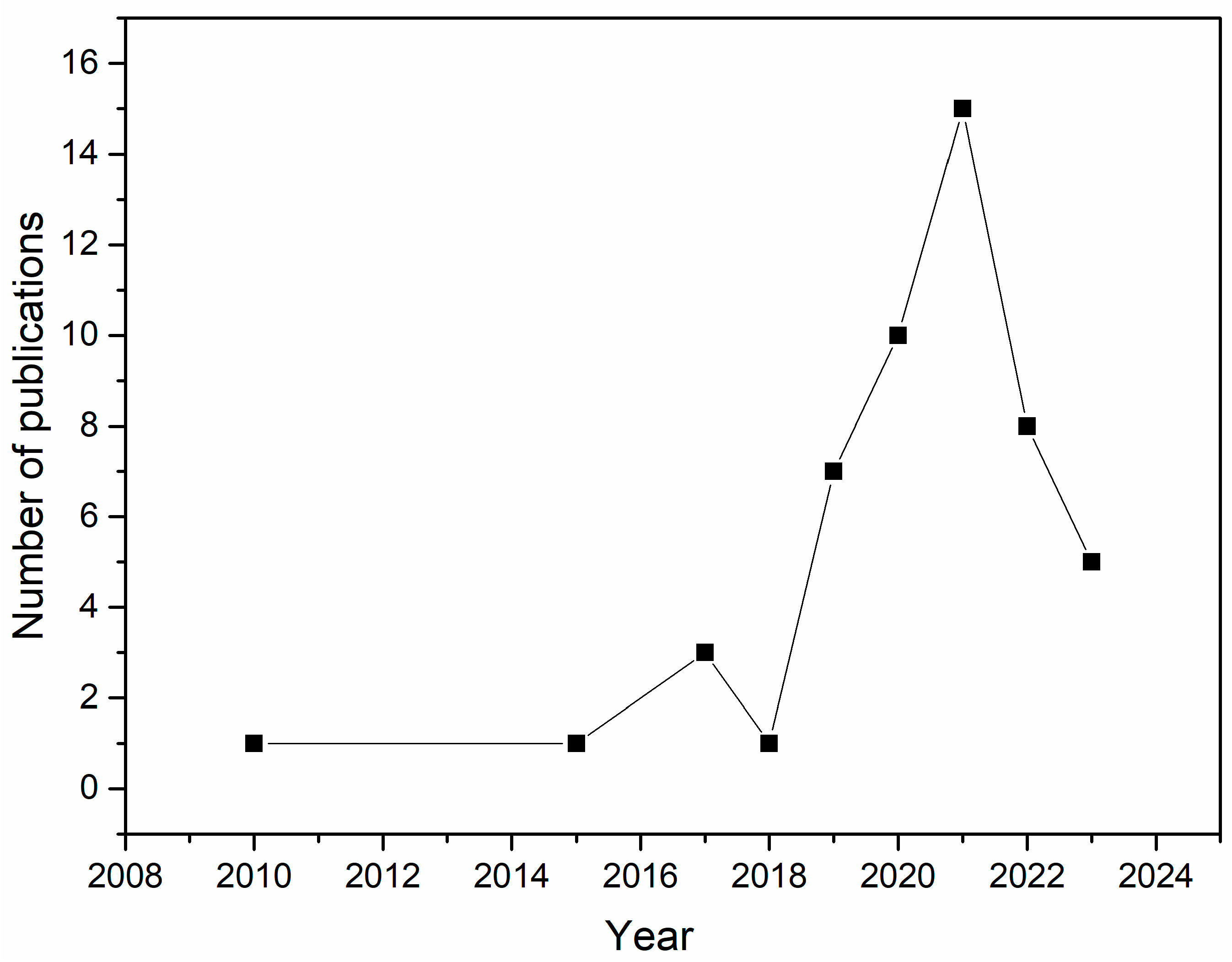
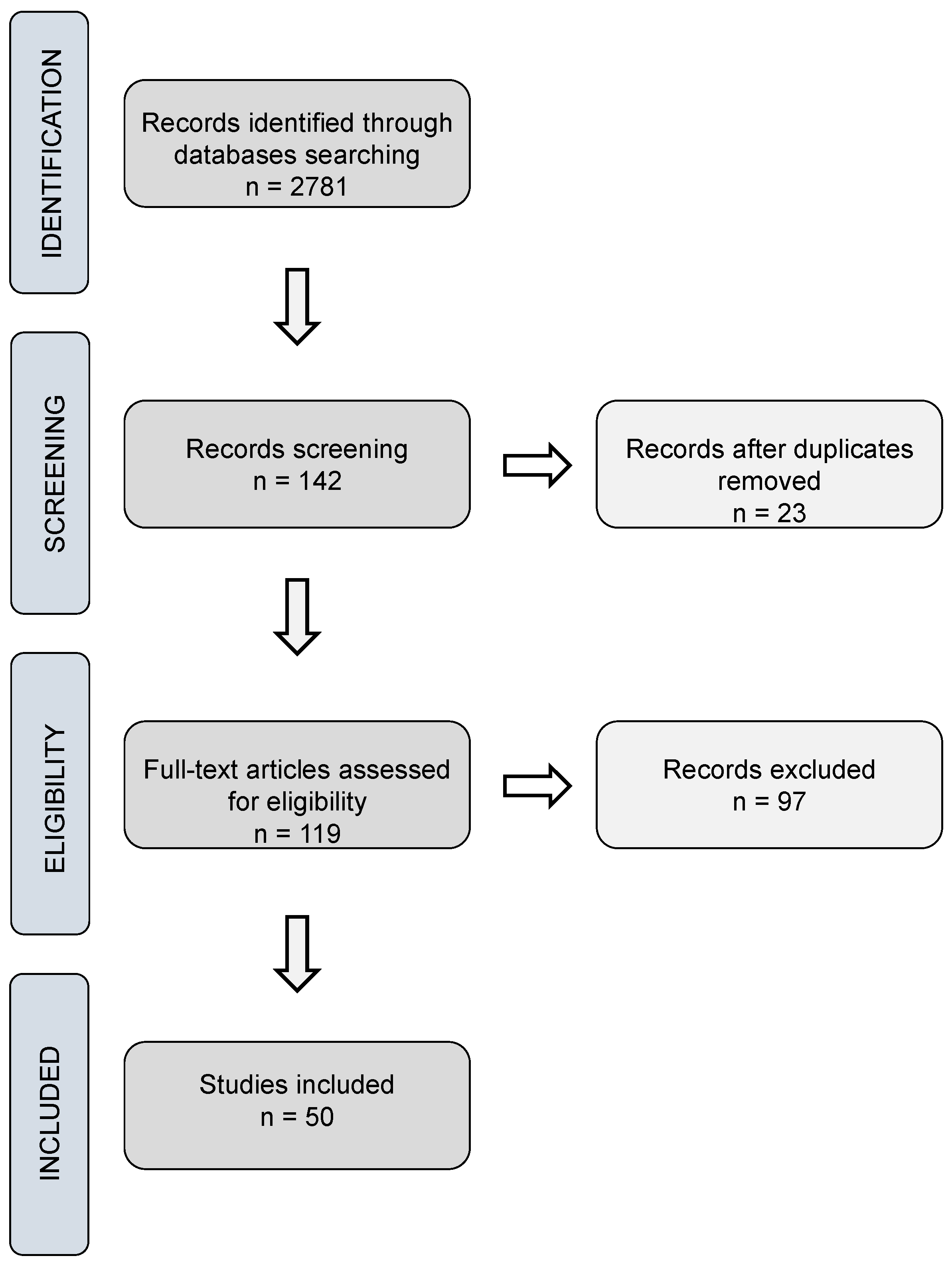
Disclaimer/Publisher’s Note: The statements, opinions and data contained in all publications are solely those of the individual author(s) and contributor(s) and not of MDPI and/or the editor(s). MDPI and/or the editor(s) disclaim responsibility for any injury to people or property resulting from any ideas, methods, instructions or products referred to in the content. |
© 2024 by the authors. Licensee MDPI, Basel, Switzerland. This article is an open access article distributed under the terms and conditions of the Creative Commons Attribution (CC BY) license (https://creativecommons.org/licenses/by/4.0/).