Article

Synthesis and Characterization of Chemically and Green-Synthesized Silver Oxide Particles for Evaluation of Antiviral and Anticancer Activity

by
Muhammad Asif
1,
Wajeeha Iqbal
1,
Muhammad Fakhar-e-Alam
1,*,
Zahid Hussain
1,
Malik Saadullah
2,
Mudassir Hassan
3,
Javed Rehman
4,5,
Kholood A. Dahlous
6,* and
Noora H. Al-Qahtani
7
1
Department of Physics, Government College University Faisalabad, Faisalabad 38000, Pakistan
2
Department of Pharmaceutical Chemistry, Government College University Faisalabad, Faisalabad 38000, Pakistan
3
Department of Zoology, Government College University Faisalabad, Faisalabad 38000, Pakistan
4
State Key Laboratory of Metastable Materials Science and Technology, School of Materials Science and Engineering, Yanshan University, Qinhuangdao 066004, China
5
MEU Research Unit, Middle East University, Amman 11831, Jordan
6
Department of Chemistry, College of Science, King Saud University, Riyadh 11451, Saudi Arabia
7
Center for Advanced Materials, Qatar University, Doha P.O. Box 2713, Qatar
*
Authors to whom correspondence should be addressed.
Pharmaceuticals 2024, 17(7), 908; https://doi.org/10.3390/ph17070908
Submission received: 4 May 2024 / Revised: 1 July 2024 / Accepted: 3 July 2024 / Published: 8 July 2024
(This article belongs to the Special Issue Therapeutic Potential of Silver Nanoparticles (AgNPs))

Abstract

:
Silver oxide (Ag2O) particles are wonderful candidates due to their unique properties, and their use in a wide range of research, industrial and biomedical applications is rapidly increasing. This makes it fundamental to develop simple, environmentally friendly methods with possible scaling. Herein, sodium borohydride and Datura innoxia leaf extract were applied as chemical and biological stabilizing and reducing agents to develop Ag2O particles. The primary aim was to evaluate the anticancer and antiviral activity of Ag2O particles prepared via two methods. XRD, UV-visible and SEM analyses were used to examine the crystallite structure, optical properties and morphology, respectively. The resulting green-synthesized Ag2O particles exhibited small size, spherically agglomerated shape, and high anticancer and antiviral activities compared to chemically synthesized Ag2O particles. The MTT (3-(4,5-dimethylthiazol-2-yl)-2,5 diphenyltetrazolium-bromide) assay of green-synthesized Ag2O particles showed high anticancer activity against MCF-7 cells with IC50 = 17.908 µg/mL compared to chemically synthesized Ag2O particles with IC50 = 23.856 µg/mL. The antiviral activity of green-synthesized Ag2O particles and chemically synthesized Ag2O particles was also evaluated by a plaque-forming assay, and green-synthesized Ag2O particles showed higher antiviral ability with IC50 = 0.618 µg/mL as compared to chemically synthesized Ag2O particles with IC50 = 6.129 µg/mL. We propose the use of green-synthesized Ag2O particles in cancer treatment and drug delivery.

1. Introduction

The global cancer problem is progressively increasing every year [1]. According to the World Health Organization (WHO) 2020 reports, there were 9.9 million deaths globally, with breast cancer mortality (2.26 million) being particularly high in developing countries, especially in Pakistan [2,3,4].
Metal oxide particles have been leading the way in materials research for industrial, energy, and biomedical applications over the last few decades.
A number of benefits, including ease of use, environmental friendliness, and ease of scaling, have generated considerable interest in green nanotechnology. Herbals are ingredients (extracts) that are derived from plants, either in combination or alone, and are used to treat and prevent many diseases. Many plant extracts have shown anticancer activity against several cancer cells without harming the growth of healthy cells. Plants are widely considered the optimal choice of material due to their stabilizing capabilities and bio-reducing properties [5,6].
Silver oxide particles have attracted significant interest among metal oxide particles, including gold, silver, manganese, zinc, and copper particles, because of their diverse range of uses in sectors such as textiles, cancer treatment, antiviral medications, electronics, food production, optical instruments, and rechargeable batteries and especially in biomedical applications [7,8,9,10]. Ag2O particles have antifungal [10], anticancer [11], antiviral [12], anticancer [13], anti-inflammatory [5], and antioxidant properties [14]. Ag2O particles can be prepared using a variety of techniques, including chemical, physical, and green-synthesis techniques [15,16,17]. Ag2O particles have high efficiency due to their outstanding antimicrobial activity towards viruses, bacteria, and other eukaryotic microorganisms [18,19]. Some important viruses are influenza virus [20], human immunodeficiency virus (HIV), human parainfluenza virus [21], dengue virus [22], and herpes simplex virus 1 (HSV1) [23]. These viral infections are causing current epidemics and threaten the lives of humans on this planet [24,25].
In the current study, Ag2O particles were prepared via a green synthesis process by using Datura innoxia leaf extract. The extract effectively decreased and stabilized the ions present in the silver nitrate solution, resulting in Ag2O particle synthesis without requiring any chemical reducing or stabilizing agent. However, in the chemical synthesis process, sodium borohydride was used to prepare the Ag2O particles, which is a very strong stabilizing/reducing agent. The green and chemically prepared Ag2O particles were characterized. Finally, the anticancer and antiviral efficacy of green and chemically prepared Ag2O particles were explored. We suggest that further in vivo anticancer and antiviral studies of Datura innoxia Ag2O particles should be performed.

2. Results and Discussions

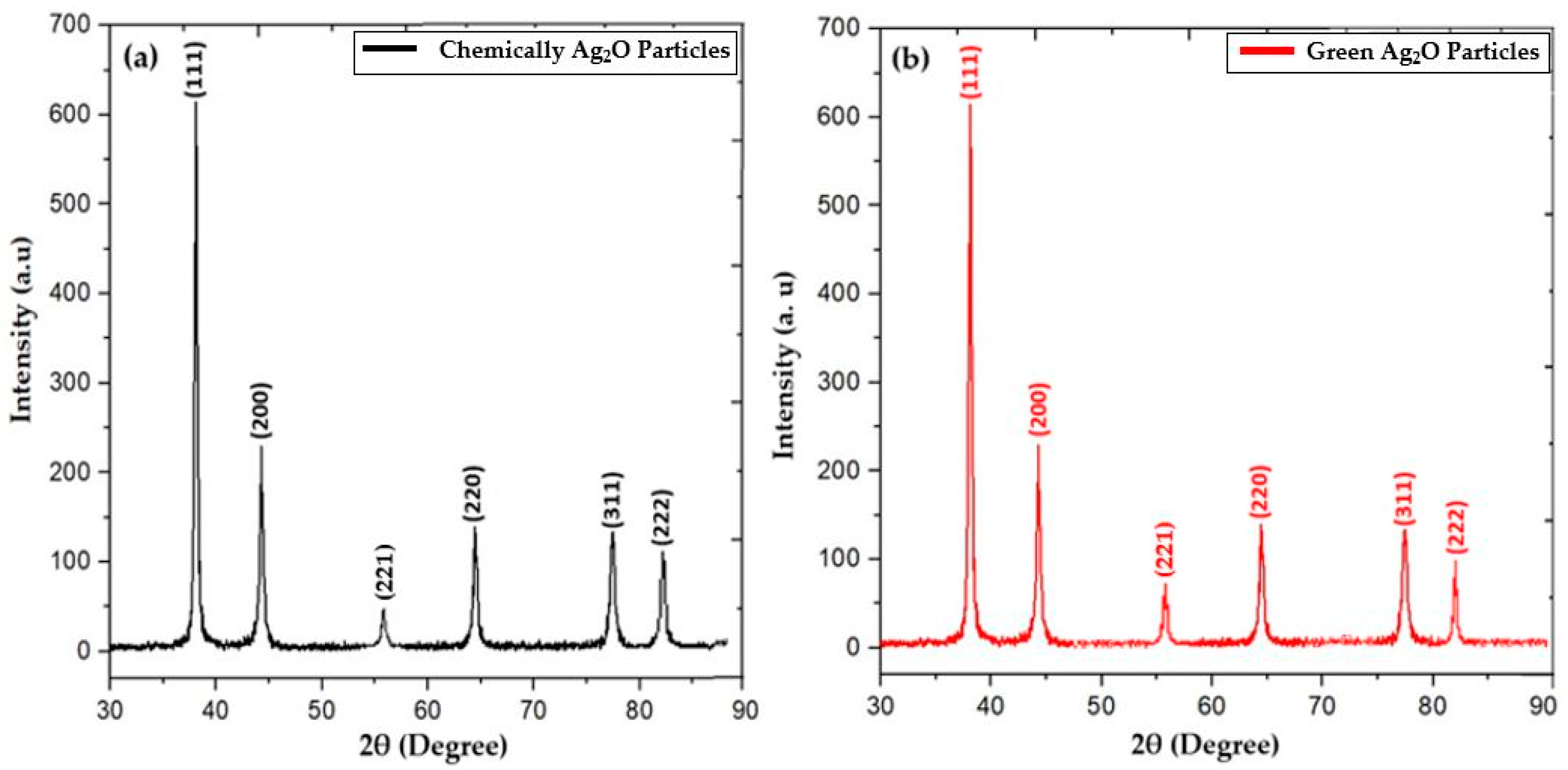
2.1. X-ray Diffraction Analysis
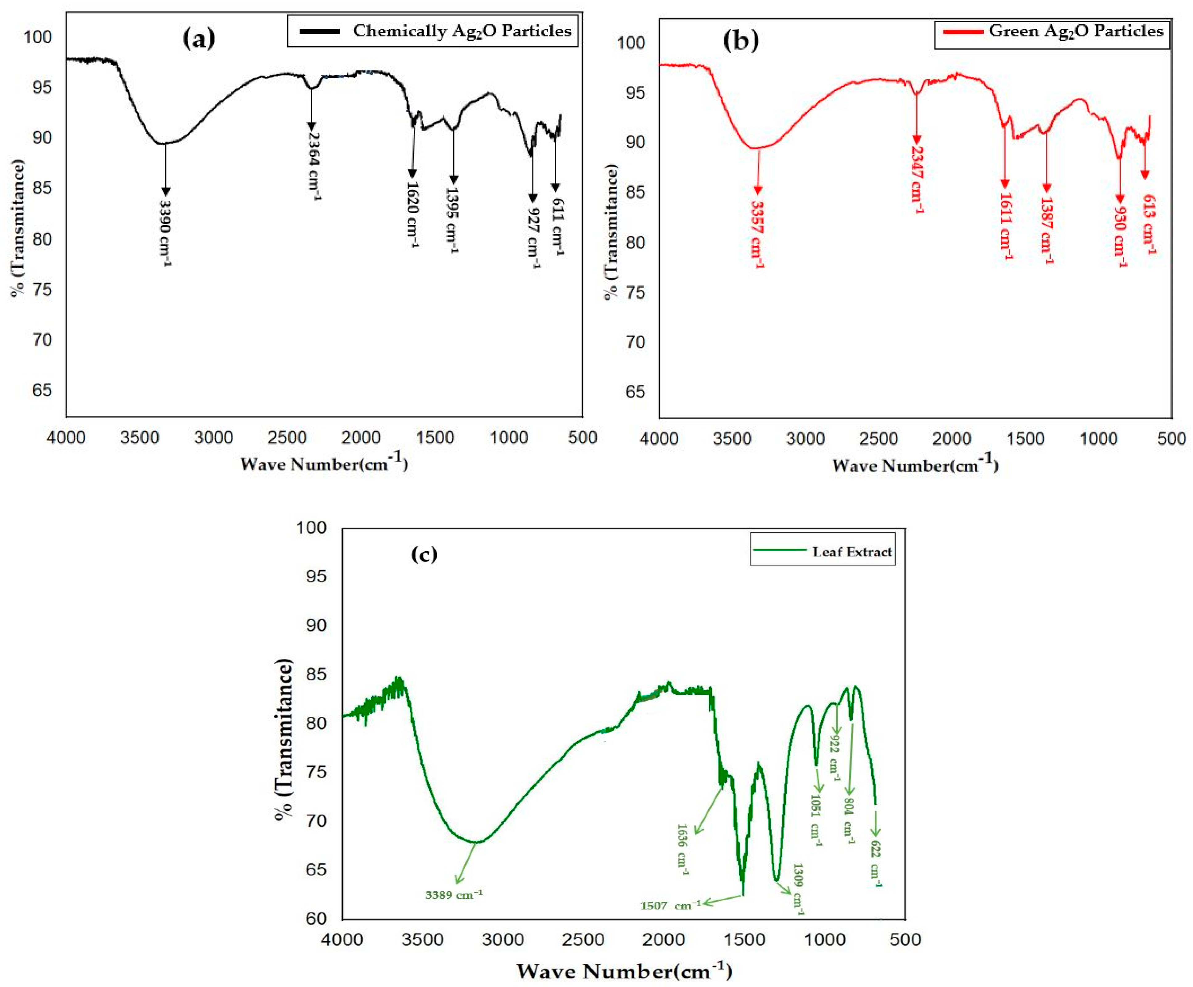
An X-ray diffraction (XRD) study was used to determine the crystal structure and calculate the crystallite size of Ag2O particles prepared using chemical and green synthesis processes. Figure 1a,b present the crystal structure patterns of the chemical and green synthesis of Ag2O particles prepared by coprecipitation processes and annealed at 400 °C for 12 h for refinement. The crystal structure of chemically and green-prepared Ag2O particles was investigated and calculated as cubic crystal structure and justified while comparing JCPDS No. 01-087-0719.The miller indices of maximum intensity peak are labeled with the (111) reflection/lattice plane at a diffraction angle of 38.4°, while the remaining lattice planes appeared in the following order: peaks at two thetas (2θ) = 44.5°, 64.6°, 77.6° and 82.3° labeled, and with respective indices of (200), (220), (311) and (222) in both chemical and green processes, respectively. The XRD pattern was similar in both conditions. In addition, it was observed that the maximum intensity peak had a broad size, which confirmed the growth of the particles. The average crystallite sizes of chemically and green-synthesized Ag2O particles were ~63 and ~54 nm, respectively, as shown in Table 1. Moreover, the crystallite size was calculated from X-ray diffraction (XRD) data using the given formulas [26,27,28,29].
C . S = k λ β c o s θ
Here, the crystallite size is (C.S), Scherrer constant (k), wavelength (λ), full width half maximum (β), and angle (θ).

2.2. FTIR Analysis

The chemical bond and functional groups of both chemically and green-synthesized Ag2O particles were investigated using Fourier transform infrared spectroscopy (FTIR) in the wavelength range of 500–4000 cm−1. In Figure 2a,b, it can be observed that the FTIR spectra of chemically synthesized Ag2O particles exhibited a shift in the bands compared to those synthesized using Datura innoxia plant extract. The addition of Datura innoxia plant extract to silver nitrate resulted in a change in the functional groups of Ag2O particles. The green-synthesized Ag2O particles showed several peaks corresponding to major functional groups at 3357, 2347, and 1631 cm−1, while the chemically synthesized Ag2O particles showed a shift in the bands at 3390, 2364, and 1620 cm−1. These peaks are associated with the stretching vibration of the hydroxyl group (H-bonded O-H stretch) [30,31,32]. Additionally, the O-H bending vibration of water molecules absorbed on the surface of Ag2O particles, which may be crucial for biomedical assays such as anticancer and antimicrobial activities [30,31], and a weak stretching vibration of C=C [17], were observed. The weak peaks at 1395 and 1387 cm−1 of Ag2O correspond to NO3− ions (nitrates). Furthermore, the presence of an O-Ag-O bending mode of vibration at 611 cm−1 confirmed the formation of metal–oxygen bonding [9,30,31]. Moreover, the FTIR spectra of Datura innoxia leaf extract are shown in Figure 2c, which are quite similar to those in the previously published article [17].

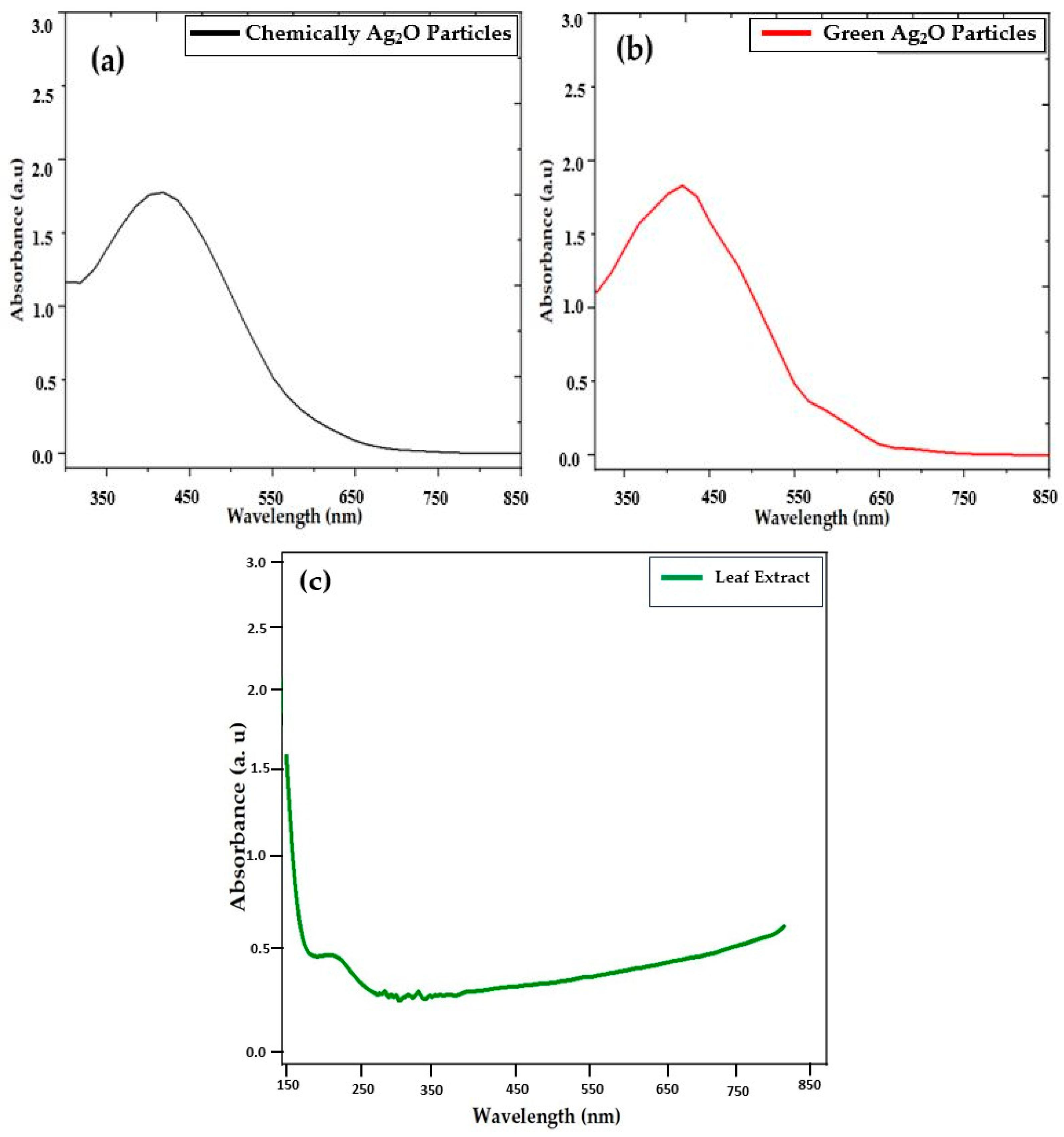
2.3. UV–Visible Analysis

UV–visible spectroscopy was employed to analyze the optical density or absorption spectra of both chemically and green-synthesized particles. In the current experimental strategy, chemically and green-synthesized Ag2O particles were carefully examined. When chemically and green-synthesized Ag2O particle samples were dispersed in deionized water to find the absorption spectra in the range of 350 nm to 850 nm, we revealed a sharp absorption band at 410 nm in the absorption spectra of chemically synthesized Ag2O particles, as depicted in Figure 3a. This might indicate the presence of chemically synthesized Ag2O particles such as in [17]. The absorption spectra of green-synthesized Ag2O particles are depicted in Figure 3b, where the presence of Ag2O particles is confirmed from the green-synthesized Ag2O sample using these spectra. Generally, green-synthesized Ag2O particles in deionized water absorbed radiation near the wavelength of 414.7 nm. It was observed from the UV–visible graph that the confirmation of the surface plasmon resonance peak near 425 nm indicates the presence of green-synthesized Ag2O particles, as shown in Figure 3b. On the metallic surface of green-synthesized Ag2O particles, there are resonant oscillations of free electrons through interbond transitions and surface plasmon resonance (SPR) bands [33]. In the graph, the surface plasmon peak indicates the presence of Ag2O particles with different sizes, and the spherical shape is confirmed in SEM. The absorption spectra of Datura innoxia leaf extract is depicted in Figure 3c, which shows a peak at ~220 nm. For more details about the absorbance of the Datura innoxia leaf extract, see the previously published paper [34].

2.4. SEM and EDX Analysis

The surface morphology of both chemically and green-synthesized Ag2O particles was analyzed using scanning electron microscopy (SEM). The SEM images of chemically and green-synthesized Ag2O particles are shown in Figure 4a,b at a scale of 3 µm scale, respectively. Spherical grains of the chemically and green-synthesized Ag2O particles can be seen in Figure 4, with some non-uniformly distributed aggregates present. The SEM micrographs confirmed that the chemically and green-synthesized Ag2O particles mostly have spherical shapes of various sizes. In Figure 4b, the bioagents of Datura innoxia leaf extract strongly interact with Ag ions to produce uniform spherical shapes with small sizes. The green-synthesized Ag2O particles had spherical shapes of dissimilar sizes but were smaller than the chemically synthesized Ag2O particles. The spherical grains were circulated with red circles. Hence, the SEM results of green-synthesized Ag2O particles were much better than those of the chemical Ag2O particles. The energy-dispersive X-ray spectroscopy (EDX) technique was employed to conduct an elemental compositional analysis of both chemically and green-synthesized Ag2O particles. The results from the EDX analysis verified the existence of Ag as a main element and O and C as the primary elements in the samples, as depicted in Figure 4c,d. The negligible percentage of carbon is due to lab environment or lab equipment. Kasthuri et al. [17] and Gajendaran et al. [35] also reported similar results, where elemental silver was detected in the graph obtained by EDX analysis, confirming the reduction of silver ions to elemental silver.

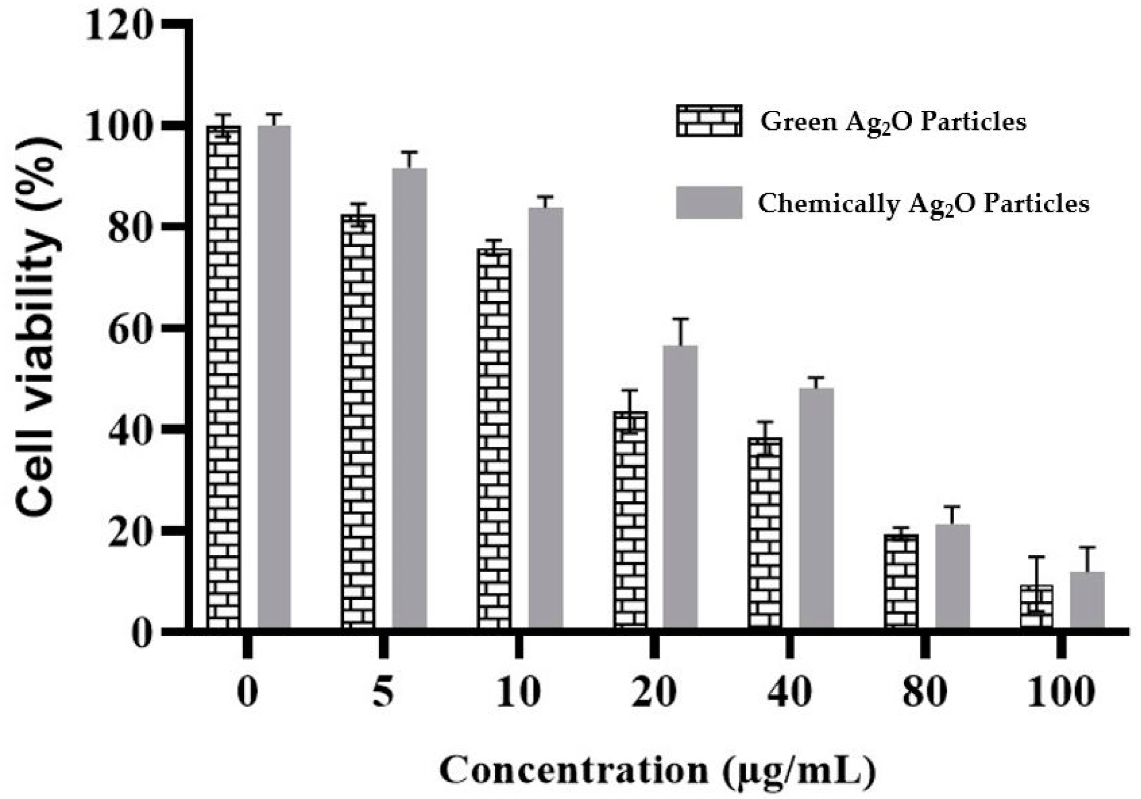
2.5. Anticancer Activity against MCF-7 Cells

The results of the cytotoxicity investigation of both types of Ag2O particles on the breast cancer (MCF-7) cell line revealed that both particles were cytotoxic against the cancer cell line and were less toxic towards the Vero cell line. Maximum cell death was detected for the green-synthesized Ag2O particles with an IC50 of 17.908 µg/mL, compared to chemically synthesized Ag2O particles with an IC50 of 23.856 µg/mL against MCF-7 cells after 48 h of exposure. As can be seen in Figure 5, the cytotoxicity of both Ag2O particles is concentration-dependent [36].

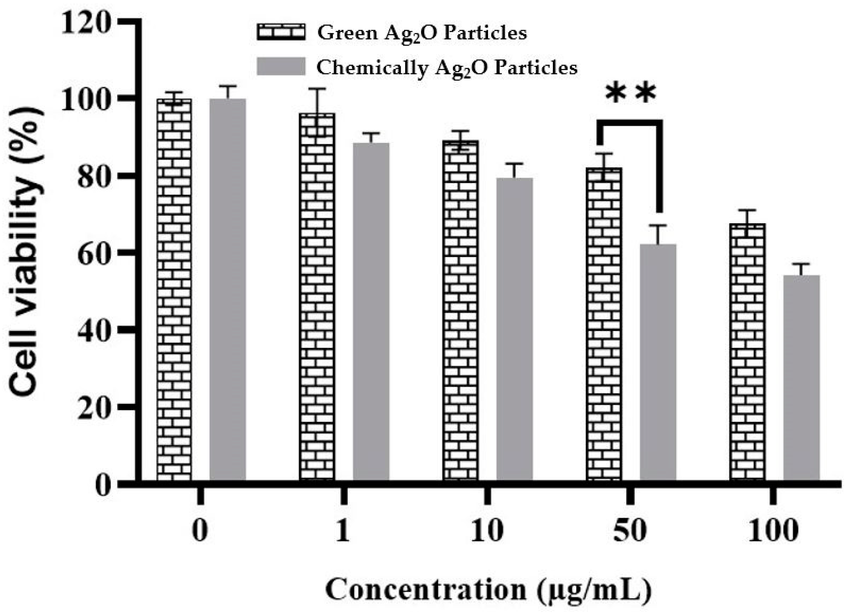
2.6. Cytotoxicity Activity against Vero Cells

To rule out the possibility that cellular toxicity may be the cause of the decline in infectivity, a range of concentrations (1, 5, 10, 50, and 100 µg/mL) were incubated with Vero cell monolayers, as shown in Figure 6. Each concentration of Ag2O particles was incubated for various time periods (48 h), and cell viability was measured using the 3-(4,5-dimethyllthiazol-2-yl)-2,5 diphenyl-tetrazolium-bromide (MTT) assay. Chemically prepared Ag2O particles had toxic effects in the concentration range of 10–50 µg/mL. However, green-synthesized Ag2O particles within the range were shown/found to have low cytotoxicity in Vero cells. These statistics were valuable for determining concentrations for testing in antiviral assays. We used concentrations that were significantly below the cytotoxic threshold, ranging from 0.5 to 10 µg/mL.

2.7. Antiviral Activity

A stock solution of silver oxide particles was prepared via a chemical-synthesis process as well as a green-synthesis process with a concentration solution of 10 mg/L by dispersing the working solution of silver oxide particles (0.1, 0.5, 1, 2, 4, 8 and 10 µg/mL) into Newcastle disease virus (NDV)-incorporated Vero cells. The solution was then removed, washed and inoculated with 0.5% methyl cellulose for 72 h. The medium was aspirated, and the cells were fixed with 10% formaldehyde and stained with 1% crystal violet; then, the plaques were counted. The plaque analysis formation was calculated. Green-synthesized Ag2O particles showed significant toxicity against NDV virus compared to chemically synthesized Ag2O particles (Figure 7).

2.8. Properties of Green-Synthesized Ag2O Particles

The Ag2O particles prepared using the green-synthesis process, which utilizes plant extracts, showed distinct properties compared to those prepared through traditional chemical methods. Some of the new properties of green-synthesized Ag2O particles are mentioned, such as smaller crystallite size, higher surface area, improved stability, increased antimicrobial and antioxidant potential, anticancer and antiviral properties, reduced toxicity and environmental impact, unique crystal structures and morphologies, potential for large-scale production, and cost-effectiveness. The green-synthesis method offers a sustainable and eco-friendly approach to producing Ag2O particles with advanced properties for various applications [6,12,13,16,17]. For more information on the properties of green-synthesized particles, see Table 2.

3. Materials and Methods

3.1. Materials

Silver nitrate (AgNO3), sodium borohydride (NaBH4), ethanol, polyvinylpyrrolidone, deionized water, dimethyl sulfoxide (DMSO), fetal bovine serum (FBS), Dulbecco’s modified eagle medium (DMEM), phosphate-buffered saline (PBS), and antibiotics (penicillin + streptomycin) were purchased from Sigma-Aldrich (St. Louis, MO, USA). In addition, the Datura innoxia plant was collected from the garden of the Department of Botany, Government College University, Faisalabad).

3.2. Preparation of Extract

Fresh leaves of Datura innoxia were cut into small pieces, washed twice with distilled water and dried [17]. In the next step, 200 g of powdered Datura innoxia was mixed with 750 mL of distilled water and heated at 80 °C for 3 h. Then, the obtained solution was filtered and scraped to obtain a dark yellow colored extract. Next, 0.407 g of extract was dissolved in 50 mL of deionized water and stirred continuously for 10 min for homogeneity.

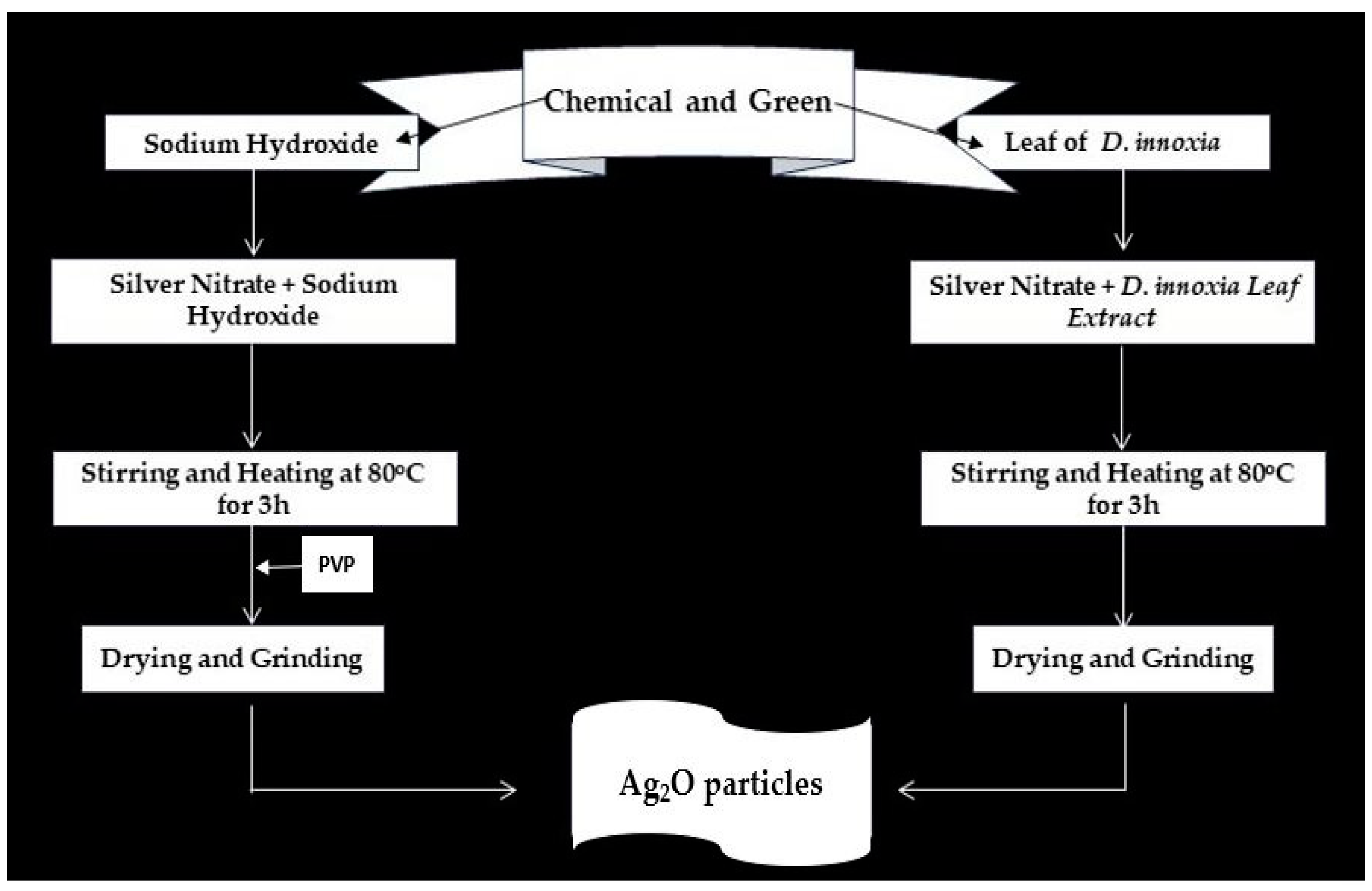
3.3. Green Synthesis Method

An amount of 3.5 g of AgNO3 was dissolved in 200 mL of distilled water. The prepared extract was added dropwise into the AgNO3 solution until the color of the solution turned dark brown and maintained its pH (9–11), and finally, we obtained the precipitates. The solution was centrifuged at 7000 rpm for 10 min and dried. Then, it was annealed at 400 °C for 12 h. Finally, the prepared precipitate was ground to obtain a fine powder [17,18,45]. A schematic flow chart is depicted in Figure 8.

3.4. Chemical Synthesis Method

An amount of 0.407 g of NaBH4 was dissolved in 50 mL of deionized water and kept in an ice bath for 3 h. In the next step, 3.5 g of AgNO3 was taken and dissolved in 200 mL of distilled water and kept under continuous stirring on a magnetic stirrer for 40 min to obtain a homogeneous solution [46]. Next, the NaBH4 solution was removed from the ice bath and placed in a magnetic stirrer, and then the AgNO3 solution was added dropwise. The solution was constantly stirred for 3 h to obtain dark-colored precipitates; then, 0.3 g Polyvinylpyrrolidone was mixed in 10 mL of water, and precipitates were prepared. The solution was centrifuged at 7000 rpm for 10 min and dried. Then, it was annealed at 400 °C for 12 h, and finally, the dark precipitate was ground to obtain a fine powder [27,47]. A schematic flow chart is depicted in Figure 8.

3.5. Characterization of the Synthesized Nanomaterial

X-ray diffraction (D8 advance Bruker, Karlsruhe, Germany) was used to confirm the crystal structure of the prepared particles. UV–visible spectroscopy (BK-UV1800PC DB, Bio-base, Jinan, China) was employed to compute the optical density, and FTIR (Spectrum 2, Perkin Elmer, Waltham, MA, USA) was used to study the functional groups (stretching and vibrational). The surface morphology of both chemically and green-synthesized Ag2O particles was analyzed using scanning electron microscopy (Cube 10 Emcraft, Hanam, Republic of Korea) analysis. An energy-dispersive X-ray (EDX) spectrometer was used to confirm the elemental composition of the synthesized materials.

3.6. Bioassay

3.6.1. Cell Lines and Viruses

MCF-7 and green African monkey kidney (Vero) cell lines were gifted from the National Institute of Biotechnology and Genetic Engineering (NIBGE) in Faisalabad, Pakistan. MCF-7 and Vero cells were cultivated in DMEM/F-12 medium containing 1% (v/v) antibiotics and 10% (v/v) FBS. The flasks were kept at 37 °C in a humidified atmosphere with 5% CO2. NDV was grown in 9-day-old chicken embryo eggs. Before use, stock viruses were collected, titrated, and stored [46].

3.6.2. Cytotoxicity Assay

MTT assays were used to measure the cytotoxicity of the chemically and green-synthesized Ag2O particles. MCF-7 cells were seeded in 96-well plates. Cells were incubated with increasing concentrations of particles at 37 °C and 5% CO2 for 48 h. The cells were treated with a 5 mg/mL MTT solution and then incubated for 4 h. After removing the MTT, 150 μL of DMSO was added and left for 5 min. The optical density was calculated using a microplate reader. Each experiment was performed in triplicate. The cytotoxicity level was quantified by determining the IC50 value, representing the concentration of particles that suppressed the growth of MCF-7 cells by half (50%) in comparison to untreated cells. Due to chemically synthesized Ag2O particles, the basicity of the solution may increase and hence become toxic towards the cell line. On the other hand, green-synthesized Ag2O particles were synthesized in a water-based extract, so they exhibited low toxicity. The percentage viability of Ag2O particles was calculated using the following formula [48,49].
% a g e   C e l l   V i a b i l i t y = A b s   o f   c o n t r o l A b s   o f   t r e a t e d   c e l l s A b s   o f   c o n t r o l × 100 %

3.6.3. Antiviral Activity

Chemically and green-synthesized Ag2O particles were dissolved in DMEM without serum and used at concentrations of 0.5, 1, 2, 4, 8 and 10 µg/mL. The percentage (%) inhibition was calculated by dividing the number of plaques acquired in the positive control, which had 0% inhibition (no antiviral compounds added to the cell monolayers). A plaque-forming assay was performed to check the antiviral activity of both chemically and green-synthesized Ag2O particles. For this, confluent Vero cell monolayers (12-well plates) were cleaned with phosphate-buffered saline and infected with NDV for 1 h at 37 °C, with an infection diversity of 0.02 plaque-forming units per cell. The virus inoculum was mixed with Ag2O particles to perform the test. Unencapsulated virus was inactivated using citrate buffer at pH 3. After being washed in PBS, the samples were incubated for 48 h in fresh growing medium with carboxymethylcellulose. Monolayers infected with NDV were fixed and stained with 10% formaldehyde. The plates were then stained with crystal violet after fixation. Microscopically, the plaques were counted. The mean plaque count for each drug concentration was expressed as a percentage of the mean plaque count of the control virus, using the formula shown below. The plaques were quantified under a microscope. The mean plaque count for every drug concentration was calculated as a ratio to the mean plaque count of the control virus, using the formula below. A graph was plotted to show the relationship between the number of plaques and the drug concentration [29,49].
I n h i b i t i o n   % = M e a n   n o .   o f   P l a q u e s   i n   c o n t r o l M e a n   n o .   o f   P l a q u e s   i n   s a m p l e s M e a n   n o .   o f   P l a q u e s   i n   c o n t r o l × 100 %

3.7. Statistical Analysis (S.A)

Statistical analysis (S.A) was performed using Student’s t-test. All cytotoxic results on MCF-7 and Vero cell line experiments were reported as mean ± SD (standard deviation). A value of p ≤ 0.05 was statistically significant (indicated by *), and a value of p ≤ 0.01 was considered highly significant (indicated by **).

4. Conclusions

Silver oxide (Ag2O) particles were synthesized via a chemical- and green-synthesis approach. The XRD results revealed a cubic crystal structure. UV–visible spectroscopy results gave maximum absorption spectra at 410 nm and 414.7 nm, respectively, confirming the growth of chemically and green-synthesized Ag2O particles. An in vitro MTT assay of the green and chemically Ag2O particles revealed that both possess significant anticancer activity against MCF-7 cells with IC50 17.908 µg/mL and IC50 23.856 µg/mL, respectively. The antiviral activity of both particles was also evaluated by plaque-forming assay, and the green-synthesized Ag2O particles exhibited a higher antiviral ability with IC50 0.618 µg/mL compared to chemically synthesized Ag2O particles with IC50 6.129 µg/mL. These results revealed that green-synthesized Ag2O particles proved to be better than chemically synthesized Ag2O particles against cancer and viral infections. In the case of chemically synthesized Ag2O particles, toxicity (toxic behavior) was observed against normal (Vero) cells, unlike green-synthesized Ag2O particles. This observation further supports the idea that green-synthesized Ag2O particles are safer than chemically synthesized Ag2O particles. This research could potentially play a role in biomedical applications, particularly in the field of nanomedicine in the near future. We also suggest that in vivo anticancer and antiviral studies of green-synthesized Ag2O particles should be performed.

Author Contributions

Conceptualization, M.F.-e.-A., M.A. and W.I.; methodology, M.A. and W.I.; software, M.A.; validation, M.F.-e.-A., M.H. and J.R.; formal analysis, Z.H.; investigation, M.A. and M.S.; resources, M.S.; data curation, M.A. and M.H.; writing—original draft preparation, M.A. and W.I.; writing—review and editing, M.F.-e.-A., J.R. and K.A.D.; visualization, M.F.-e.-A., M.S. and N.H.A.-Q.; supervision, M.F.-e.-A.; project administration, M.F.-e.-A. and K.A.D.; funding acquisition, K.A.D. All authors have read and agreed to the published version of the manuscript.

Funding

This work was funded by researchers Supporting Project Number (RSP2024R388) King Saud University, Riyadh, Saudi Arabia.

Institutional Review Board Statement

Not applicable.

Informed Consent Statement

Not applicable.

Data Availability Statement

Data is contained within the article.

Acknowledgments

This work was funded by researchers Supporting Project Number (RSP2024R388) King Saud University, Riyadh, Saudi Arabia.

Conflicts of Interest

The authors declare no conflict of interest.

References

  1. Bray, F.; Parkin, D.M. Cancer in sub-Saharan Africa in 2020: A review of current estimates of the national burden, data gaps, and future needs. Lancet Oncol. 2022, 23, 719–728. [Google Scholar] [CrossRef] [PubMed]
  2. Sung, H.; Ferlay, J.; Siegel, R.L.; Laversanne, M.; Soerjomataram, I.; Jemal, A.; Bray, F. Global Cancer Statistics 2020: GLOBOCAN Estimates of Incidence and Mortality Worldwide for 36 Cancers in 185 Countries. CA Cancer J. Clin. 2021, 71, 209–249. [Google Scholar] [CrossRef]
  3. Mettlin, C. Global breast cancer mortality statistics. CA Cancer J. Clin. 1999, 49, 138–144. [Google Scholar] [CrossRef] [PubMed]
  4. Khan, N.H.; Duan, S.F.; Wu, D.D.; Ji, X.Y. Better reporting and awareness campaigns needed for breast cancer in Pakistani women. Cancer Manag. Res. 2021, 13, 2125–2129. [Google Scholar] [CrossRef] [PubMed]
  5. Iqbal, S.; Fakhar-e-Alam, M.; Akbar, F.; Shafiq, M.; Atif, M.; Amin, N.; Ismail, M.; Hanif, A.; Farooq, W.A. Application of silver oxide nanoparticles for the treatment of cancer. J. Mol. Struct. 2019, 1189, 203–209. [Google Scholar] [CrossRef]
  6. Bonam, S.R.; Wu, Y.S.; Tunki, L.; Chellian, R.; Halmuthur, M.S.K.; Muller, S.; Pandy, V. What has come out from phytomedicines and herbal edibles for the treatment of cancer? ChemMedChem 2018, 13, 1854–1872. [Google Scholar] [CrossRef]
  7. Zargar, M.; Shameli, K.; Najafi, G.R.; Farahani, F. Plant mediated green biosynthesis of silver nanoparticles using Vitex negundo L. extract. J. Ind. Eng. Chem. 2014, 20, 4169–4175. [Google Scholar] [CrossRef]
  8. Wiley, B.J.; Im, S.H.; McLellan, J.; Siekkinen, A.; Xia, Y. Maneuvering the surface plasmon resonance of silver nanostructures through shape-controlled synthesis. J. Phys. Chem. B 2006, 110, 15666–15675. [Google Scholar] [CrossRef]
  9. Sondi, I.; Salopek-Sondi, B. Silver nanoparticles as antimicrobial agent: A case study on E. coli as a model for Gram-negative bacteria. J. Colloid Interface Sci. 2004, 275, 177–182. [Google Scholar] [CrossRef]
  10. Yao, J.; Yang, M.; Duan, Y. Chemistry, biology, and medicine of fluorescent nanomaterials and related systems: New insights into biosensing, bioimaging, genomics, diagnostics, and therapy. Chem. Rev. 2014, 114, 6130–6178. [Google Scholar] [CrossRef]
  11. Pryshchepa, O.; Pomastowski, P.; Buszewski, B. Silver nanoparticles: Synthesis, investigation techniques, and properties. Adv. Colloid Interface Sci. 2020, 284, 102246. [Google Scholar] [CrossRef]
  12. Danish, M.S.S.; Estrella-Pajulas, L.L.; Alemaida, I.M.; Grilli, M.L.; Mikhaylov, A.; Senjyu, T. Green synthesis of silver oxide nanoparticles for photocatalytic environmental remediation and biomedical applications. Metals 2022, 12, 769. [Google Scholar] [CrossRef]
  13. Ismail, G.A.; El-Sheekh, M.M.; Samy, R.M.; Gheda, S.F. Antimicrobial, antioxidant, and antiviral activities of biosynthesized silver nanoparticles by phycobiliprotein crude extract of the cyanobacteria Spirulina platensis and Nostoc linckia. Bionanoscience 2021, 11, 355–370. [Google Scholar] [CrossRef]
  14. Islam, T.; Ara, I.; Islam, T.; Sah, P.K.; de Almeida, R.S.; Matias, E.F.F.; Ramalho, C.L.G.; Coutinho, H.D.M.; Islam, M.T. Ethnobotanical uses and phytochemical, biological, and toxicological profiles of Datura metel L.: A review. Curr. Res. Toxicol. 2023, 4, 100106. [Google Scholar] [CrossRef] [PubMed]
  15. Khan, Z.; Al-Thabaiti, S.A.; Obaid, A.Y.; Al-Youbi, A. Preparation and characterization of silver nanoparticles by chemical reduction method. Colloids Surf. B Biointerfaces 2011, 82, 513–517. [Google Scholar] [CrossRef]
  16. Moond, M.; Singh, S.; Sangwan, S.; Devi, R.; Beniwal, R. Green synthesis and applications of silver nanoparticles: A systematic review. AATCC J. Res. 2022, 9, 272–285. [Google Scholar] [CrossRef]
  17. Mousavi-Khattat, M.; Keyhanfar, M.; Razmjou, A. A comparative study of stability, antioxidant, DNA cleavage and antibacterial activities of green and chemically synthesized silver nanoparticles. Artif. Cells Nanomed. Biotechnol. 2018, 46 (Suppl. 3), 1022–1031. [Google Scholar] [CrossRef]
  18. Flieger, J.; Franus, W.; Panek, R.; Szymańska-Chargot, M.; Flieger, W.; Flieger, M.; Kołodziej, P. Green synthesis of silver nanoparticles using natural extracts with proven antioxidant activity. Molecules 2021, 26, 4986. [Google Scholar] [CrossRef] [PubMed]
  19. Martina, B.E.E.; Koraka, P.; Osterhaus, A.D.M.E. Dengue virus pathogenesis: An integrated view. Clin. Microbiol. Rev. 2009, 22, 564–581. [Google Scholar] [CrossRef]
  20. Gaikwad, S.; Ingle, A.; Gade, A.; Rai, M.; Falanga, A.; Incoronato, N.; Russo, L.; Galdiero, S.; Galdiero, M. Antiviral activity of mycosynthesized silver nanoparticles against herpes simplex virus and human parainfluenza virus type 3. Int. J. Nanomed. 2013, 8, 4303–4314. [Google Scholar]
  21. Scaturro, P.; Cortese, M.; Chatel-Chaix, L.; Fischl, W.; Bartenschlager, R. Dengue virus non-structural protein 1 modulates infectious particle production via interaction with the structural proteins. PLoS Pathog. 2015, 11, e1005277. [Google Scholar] [CrossRef] [PubMed]
  22. Shen, Y.; Nemunaitis, J. Herpes simplex virus 1 (HSV-1) for cancer treatment. Cancer Gene Ther. 2006, 13, 975–992. [Google Scholar] [CrossRef] [PubMed]
  23. Pierson, T.C.; Diamond, M.S. The continued threat of emerging flaviviruses. Nat. Microbiol. 2020, 5, 796–812. [Google Scholar] [CrossRef] [PubMed]
  24. Wasik, B.R.; de Wit, E.; Munster, V.; Lloyd-Smith, J.O.; Martinez-Sobrido, L.; Parrish, C.R. Onward transmission of viruses: How do viruses emerge to cause epidemics after spillover? Philos. Trans. R. Soc. B 2019, 374, 20190017. [Google Scholar] [CrossRef] [PubMed]
  25. Panáček, A.; Kolář, M.; Večeřová, R.; Prucek, R.; Soukupová, J.; Kryštof, V.; Hamal, P.; Zbořil, R.; Kvítek, L. Antifungal activity of silver nanoparticles against Candida spp. Biomaterials 2009, 30, 6333–6340. [Google Scholar] [CrossRef] [PubMed]
  26. Shankar, S.; Rhim, J.W. Amino acid mediated synthesis of silver nanoparticles and preparation of antimicrobial agar/silver nanoparticles composite films. Carbohydr. Polym. 2015, 130, 353–363. [Google Scholar] [CrossRef] [PubMed]
  27. Mani, M.; Harikrishnan, R.; Purushothaman, P.; Pavithra, S.; Rajkumar, P.; Kumaresan, S.; Al Farraj, D.A.; Elshikh, M.S.; Balasubramanian, B.; Kaviyarasu, K. Systematic green synthesis of silver oxide nanoparticles for antimicrobial activity. Environ. Res. 2021, 202, 111627. [Google Scholar] [CrossRef] [PubMed]
  28. Gajendran, B.; Chinnasamy, A.; Durai, P.; Raman, J.; Ramar, M. Biosynthesis and characterization of silver nanoparticles from Datura inoxia and its apoptotic effect on human breast cancer cell line MCF7. Mater. Lett. 2014, 122, 98–102. [Google Scholar] [CrossRef]
  29. Al-Khedhairy, A.A.; Wahab, R. Silver nanoparticles: An instantaneous solution for anticancer activity against human liver (HepG2) and breast (MCF-7) cancer cells. Metals 2022, 12, 148. [Google Scholar] [CrossRef]
  30. Allahverdiyev, A.M.; Abamor, E.S.; Bagirova, M.; Rafailovich, M. Antimicrobial effects of TiO2 and Ag2O nanoparticles against drug-resistant bacteria and leishmania parasites. Future Microbiol. 2011, 6, 933–940. [Google Scholar] [CrossRef]
  31. Fayyadh, A.A.; Jaduaa Alzubaidy, M.H. Green-synthesis of Ag2O nanoparticles for antimicrobial assays. J. Mech. Behav. Mater. 2021, 30, 228–236. [Google Scholar] [CrossRef]
  32. Stoimenov, P.K.; Klinger, R.L.; Marchin, G.L.; Klabunde, K.J. Metal oxide nanoparticles as bactericidal agents. Langmuir 2002, 18, 6679–6686. [Google Scholar] [CrossRef]
  33. Chandirika, J.U.; Annadurai, G. Biosynthesis and characterization of silver nanoparticles using leaf extract Abutilon indicum. Glob. J. Biotechnol. Biochem 2018, 13, 7–11. [Google Scholar]
  34. Mardare, G.; Lazar, L.; Malutan, T. Potential Application of Invasive Plant Species Datura innoxia for the Scopolamine Extracts of the Plant Organs and Analysis Using UV–VIS Spectrophotometry. Forests 2022, 13, 1555. [Google Scholar] [CrossRef]
  35. Kasthuri, J.; Veerapandian, S.; Rajendiran, N. Biological synthesis of silver and gold nanoparticles using apiin as reducing agent. Colloids Surf. B Biointerfaces 2009, 68, 55–60. [Google Scholar] [CrossRef]
  36. Rogers, J.V.; Parkinson, C.V.; Choi, Y.W.; Speshock, J.L.; Hussain, S.M. A preliminary assessment of silver nanoparticle inhibition of monkeypox virus plaque formation. Nanoscale Res. Lett. 2008, 3, 129–133. [Google Scholar] [CrossRef]
  37. Patil, S.; Chandrasekaran, R. Biogenic nanoparticles: A comprehensive perspective in synthesis, characterization, application and its challenges. J. Genet. Eng. Biotechnol. 2020, 18, 67. [Google Scholar] [CrossRef]
  38. Banerjee, S.; Islam, S.; Chattopadhyay, A.; Sen, A.; Kar, P. Synthesis of silver nanoparticles using underutilized fruit Baccaurea ramiflora (Latka) juice and its biological and cytotoxic efficacy against MCF-7 and MDA-MB 231 cancer cell lines. S. Afr. J. Bot. 2022, 145, 228–235. [Google Scholar] [CrossRef]
  39. Venugopal, K.; Rather, H.A.; Rajagopal, K.; Shanthi, M.P.; Sheriff, K.; Illiyas, M.; Rather, R.A.; Manikandan, E.; Uvarajan, S.; Bhaskar, M.; et al. Synthesis of silver nanoparticles (Ag NPs) for anticancer activities (MCF 7 breast and A549 lung cell lines) of the crude extract of Syzygium aromaticum. J. Photochem. Photobiol. B Biol. 2017, 167, 282–289. [Google Scholar] [CrossRef]
  40. Narayanan, M.; Divya, S.; Natarajan, D.; Senthil-Nathan, S.; Kandasamy, S.; Chinnathambi, A.; Alahmadi, T.A.; Pugazhendhi, A. Green synthesis of silver nanoparticles from aqueous extract of Ctenolepis garcini L. and assess their possible biological applications. Process Biochem. 2021, 107, 91–99. [Google Scholar] [CrossRef]
  41. Gupta, A.; Pandey, S.; Variya, B.; Shah, S.; Yadav, J.S. Green synthesis of gold nanoparticles using different leaf extracts of Ocimum gratissimum Linn for anti-tubercular activity. Curr. Nanomed. 2019, 9, 146–157. [Google Scholar] [CrossRef]
  42. Akintelu, S.A.; Oyebamiji, A.K.; Olugbeko, S.C.; Latona, D.F. Green chemistry approach towards the synthesis of copper nanoparticles and its potential applications as therapeutic agents and environmental control. Curr. Res. Green Sustain. Chem. 2021, 4, 100176. [Google Scholar] [CrossRef]
  43. Lara, H.H.; Ayala-Nuñez, N.V.; Ixtepan-Turrent, L.; Rodriguez-Padilla, C. Mode of antiviral action of silver nanoparticles against HIV-1. J. Nanobiotechnology 2010, 8, 1. [Google Scholar] [CrossRef]
  44. Chamani, E.; Ebrahimi, R.; Khorsandi, K.; Meshkini, A.; Zarban, A.; Sharifzadeh, G. In vitro cytotoxicity of polyphenols from Datura innoxia aqueous leaf-extract on human leukemia K562 cells: DNA and nuclear proteins as targets. Drug Chem. Toxicol. 2020, 43, 138–148. [Google Scholar] [CrossRef]
  45. Tahir, M.; Fakhar-e-Alam, M.; Asif, M.; Iqbal, M.J.; Abbas, A.; Hassan, M.; Rehman, J.; Bhatti, Q.A.; Mustafa, G.; Alothman, A.A.; et al. Investigation of gadolinium doped manganese nano spinel ferrites via magnetic hypothermia therapy effect towards MCF-7 breast cancer. Heliyon 2024, 10, E24792. [Google Scholar] [CrossRef]
  46. Rhim, J.W.; Wang, L.F.; Hong, S.I. Preparation and characterization of agar/silver nanoparticles composite films with antimicrobial activity. Food Hydrocoll. 2013, 33, 327–335. [Google Scholar] [CrossRef]
  47. Sadiqa, A.; Rasul, A.; Hassan, M.; Sultana, S.; Jabeen, F. Identification of Novel Natural Inhibitors to Human 3-Phosphoglycerate Dehydrogenase (PHGDH) for Cancer Treatment. Molecules 2022, 27, 6108. [Google Scholar] [CrossRef]
  48. Fakhar-e-Alam, M.; Amjad, I.; Saadullah, M.; Tahir, M.; Jawad, M.; Asif, M.; Atif, M.; Zara, S.; Rashad, M. Antitumor activity of Zinc oxide nanoparticles fused with Green Extract of Nigella Sativa. J. Saudi Chem. Soc. 2024, 28, 101814. [Google Scholar] [CrossRef]
  49. Shetti, N.P.; Mishra, A.; Bukkitgar, S.D.; Basu, S.; Narang, J.; Raghava Reddy, K.; Aminabhavi, T.M. Conventional and nanotechnology-based sensing methods for SARS coronavirus (2019-nCoV). ACS Appl. Bio Mater. 2021, 4, 1178–1190. [Google Scholar] [CrossRef] [PubMed]
Figure 1. XRD graph of (a) chemically synthesized Ag2O particles and (b) green-synthesized Ag2O particles.
Pharmaceuticals 17 00908 g001
Figure 2. FTIR graph of (a) chemically synthesized Ag2O particles, (b) green-synthesized Ag2O particles, and (c) Datura innoxia leaf extract.
Pharmaceuticals 17 00908 g002
Figure 3. UV–visible graph of (a) chemically synthesized Ag2O particles, (b) green-synthesized Ag2O particles, and (c) Datura innoxia leaf extract.
Pharmaceuticals 17 00908 g003
Figure 4. SEM images and EDX spectrum of (a) chemically synthesized Ag2O particles, (b) green-synthesized Ag2O particles at 3 µm, (c) chemically synthesized Ag2O particles, and (d) green-synthesized Ag2O particles.
Pharmaceuticals 17 00908 g004
Figure 5. Cytotoxicity evaluation of both green- and chemically synthesized Ag2O particles against MCF-7 cell lines.
Pharmaceuticals 17 00908 g005
Figure 6. Cytotoxicity evaluation of both green and chemical Ag2O particles against normal (Vero) cell. ** p ≤ 0.01.
Pharmaceuticals 17 00908 g006
Figure 7. Green-synthesized Ag2O particles showed higher antiviral activity as compared to chemical-based Ag2O particles in a dose-dependent manner, where * is p ≤ 0.05, ** p ≤ 0.01.
Pharmaceuticals 17 00908 g007
Figure 8. Schematic flow chart of chemically and green-synthesized Ag2O particles.
Pharmaceuticals 17 00908 g008
Table 1. (a) shows the parameters, miller indices, diffraction angle, and crystallite size of chemically synthesized Ag2O particles. (b) shows the parameters, miller indices, diffraction angle, and crystallite size of the green-synthesized Ag2O particles.
Miller Indices
(h,k,l)
Diffraction Angle
2θ (Theta)
Crystallite Size “C.S” (nm)
(a)
(111)38.141.3
(200)44.355.7
(220)64.575.6
(311)77.481.2
(b)
(111)38.132.2
(200)44.346.6
(220)64.866.6
(311)77.769.9
Table 2. Previous reported study of different plant extracts, nanoparticles, and their applications.
Sr#Materials and Plants NameApplicationsIC50 (µg/mL)References
1Green-synthesized AgNPs (Datura innoxia leaf extract)anticancer15.2 and 8.5[37]
2Biosynthesis of AgNPs with Datura innoxiaanticancer20[28]
3Green-synthesized AgNPs (Baccaurea ramiflora)anticancer110 and 140[38]
4AgNPs with Syzygium aromaicumanticancer60 and 50[39]
5Green-synthesized AgNPs (Ctenolepis garcini)anticancer33.78[40]
6Green-synthesized gold nanoparticles (AuNPs) with (leaf extracts of Ocimum gratissimum Linn)anticancer10 and 25[41]
7Green copper nanoparticles (CuNPs) with Eclipta prostrataanticancer1.71 and 1.81[42]
8PVP-coated AgNPsantiviral0.44[43]
9Datura innoxia leaf extractanticancer0.6[44]
10Green (Datura innoxia extract) and chemically synthesized Ag NPsAnticancer, antibacterial and antioxidant--[17]
Disclaimer/Publisher’s Note: The statements, opinions and data contained in all publications are solely those of the individual author(s) and contributor(s) and not of MDPI and/or the editor(s). MDPI and/or the editor(s) disclaim responsibility for any injury to people or property resulting from any ideas, methods, instructions or products referred to in the content.

