Review

Hereditary Gastrointestinal Tumor Syndromes: When Risk Comes with Your Genes

by
María Jesús Fernández Aceñero
1,2,* and
Cristina Díaz del Arco
1,2,*
1
Department of Legal Medicine, Psychiatry and Pathology, School of Medicine, Complutense University of Madrid, 28040 Madrid, Spain
2
Department of Pathology, Hospital Clínico San Carlos, Health Research Institute of the Hospital Clínico San Carlos (IdISSC), 28040 Madrid, Spain
*
Authors to whom correspondence should be addressed.
Curr. Issues Mol. Biol. 2024, 46(7), 6440-6471; https://doi.org/10.3390/cimb46070385
Submission received: 27 May 2024 / Revised: 19 June 2024 / Accepted: 25 June 2024 / Published: 26 June 2024

Abstract

:
Despite recent campaigns for screening and the latest advances in cancer therapy and molecular biology, gastrointestinal (GI) neoplasms remain among the most frequent and lethal human tumors. Most GI neoplasms are sporadic, but there are some well-known familial syndromes associated with a significant risk of developing both benign and malignant GI tumors. Although some of these entities were described more than a century ago based on clinical grounds, the increasing molecular information obtained with high-throughput techniques has shed light on the pathogenesis of several of them. The vast amount of information gained from next-generation sequencing has led to the identification of some high-risk genetic variants, although others remain to be discovered. The opportunity for genetic assessment and counseling in these families has dramatically changed the management of these syndromes, though it has also resulted in significant psychological distress for the affected patients, especially those with indeterminate variants. Herein, we aim to summarize the most relevant hereditary cancer syndromes involving the stomach and colon, with an emphasis on new molecular findings, novel entities, and recent changes in the management of these patients.

1. Introduction

Despite recent campaigns for screening and the latest advances in therapy and molecular biology, gastrointestinal (GI) neoplasms remain among the most frequent and lethal human tumors [1]. Most GI neoplasms are sporadic, but there are some well-known familial syndromes associated with a predisposition to the development of GI cancer. The increasing use of next-generation sequencing (NGS) and revolutionary genetic technologies has led to the discovery of many new pathogenic or likely pathogenic variants of genes associated with GI and other human cancers [2]. The aim of this report is to review the genetic basis of both well-known syndromes and some of the most novel ones associated with gastric cancer (GC) and colorectal cancer (CRC), highlighting how this knowledge influences disease prevention and management.

2. Gastric Cancer

GC is the fifth most common cancer and the fourth leading cause of cancer-related deaths worldwide [3]. Its incidence varies geographically, being higher in Asian countries and lower in Western countries, where it is often diagnosed at advanced stages and has a five-year survival rate under 30% [4,5]. GC is a heterogeneous disease that can be classified according to various criteria. Microscopically, most GCs are adenocarcinomas. The most commonly used histological classifications are those published by Laurén and the World Health Organization [6]. Laurén’s classification identifies intestinal and diffuse types of GC. This classification will be used throughout this article due to its prevalence in the literature and its association with clinicopathological and prognostic factors. Laurén’s intestinal type forms tubular or glandular structures, whereas the diffuse type has poorly cohesive cells forming small nests or rows with little or no gland formation. In some cases, these cells exhibit a signet-ring morphology, with abundant cytoplasmic mucin and eccentrically displaced nuclei [7].

Familial and Hereditary Gastric Cancer

Most GC cases are sporadic, associated with somatic molecular alterations, while a small subset shows familial aggregation and is linked to hereditary syndromes caused by germline mutations. The familial aggregation of GC is estimated at 5–10%, with hereditary GC accounting for 1–3% of cases [8,9].
Notable syndromes predominantly associated with GC include hereditary diffuse GC, gastric adenocarcinoma and proximal polyposis of the stomach, and familial intestinal GC [10,11]. Additionally, GC can occur in other syndromes such as Li–Fraumeni, familial adenomatous polyposis, Lynch syndrome, Peutz–Jeghers syndrome, juvenile polyposis syndrome, MUTYH-associated polyposis, hereditary breast and ovarian cancer, or PTEN hamartoma tumor syndrome [9]. Figure 1 presents the main genes associated with GC hereditary syndromes. The majority of these syndromes are most commonly associated with intestinal GC, except for hereditary diffuse GC (Figure 2). However, diffuse GC may be found in small proportions across most hereditary syndromes. Lynch syndrome, familial adenomatous polyposis, Peutz–Jeghers syndrome, juvenile polyposis syndrome, MUTYH-associated polyposis, and PTEN hamartoma tumor syndrome will be discussed in the colorectal section (Section 12 and Section 21).

3. Hereditary Diffuse Gastric Cancer

Hereditary diffuse GC is an autosomal dominant syndrome characterized by the early onset of diffuse GC and invasive lobular breast cancer [12]. Familial aggregation of diffuse GC was first noted in 1964 [13], and in 1998, the genetic basis of hereditary diffuse GC was discovered in Māori families from New Zealand [14]. Over time, similar findings have been observed in families worldwide from various ethnic backgrounds [15,16].
Currently, hereditary diffuse GC is defined by the presence of a pathogenic germline mutation in either CDH1 or CTNNA1 in an individual with diffuse GC or in a family with one or more cases of diffuse GC among first- or second-degree relatives [12]. CDH1 mutations also predispose individuals to lobular breast cancer and non-syndromic cleft lip and palate [17]. Families that meet the criteria for hereditary diffuse GC genetic testing but do not have pathogenic variants in CDH1 or CTNNA1 are classified as hereditary diffuse GC-like, as seen in other syndromes [9].

3.1. Clinical Presentation

Initially described in 1999, hereditary diffuse GC criteria included (1) two or more cases of diffuse GC in first- or second-degree relatives, with at least one diagnosed before age 50, or (2) three or more cases of diffuse GC in first- and second-degree relatives regardless of age [18]. The latest guidelines update from 2020 incorporates both familial and individual criteria. Familial criteria for genetic testing include (1) two or more cases of familial GC regardless of age, with at least one being diffuse; (2) one or more cases of diffuse GC at any age and one or more cases of lobular breast cancer before age 70; or (3) two or more cases of lobular breast cancer in relatives before age 50 [12]. GC and breast cancer cases must be histologically confirmed, and the family members must be first- or second-degree blood relatives [17]. Indications for genetic testing in an individual depend on the age at diagnosis of cancer, presence or absence of bilateral breast cancer, ethnicity, associated phenotypic features, and other factors [12]. Additionally, the 2020 update recommends testing for CTNNA1 in addition to CDH1.
In index cases, GC is generally diagnosed at an advanced stage, potentially presenting as linitis plastica [19]. Prophylactic gastrectomies often reveal precursor lesions such as in situ signet ring cell carcinoma or pagetoid spread of signet ring cells [20]. Intramucosal diffuse GC foci are also commonly found. In 95% of prophylactic gastrectomies, intramucosal carcinoma foci have been described, but the detection rate for in situ and pagetoid lesions seems to be lower (30%), suggesting intramucosal carcinoma might develop without a preceding preinvasive phase [21]. These lesions are associated with decreased E-cadherin expression on immunohistochemistry (IHC), which can appear as complete loss, mild membrane staining, or aberrant cytoplasmic or granular staining [22]. However, this staining is not sufficiently sensitive or specific to be used for screening patients for CDH1 testing [23].

3.2. Gastric Cancer Risk

3.2.1. Individuals with Pathogenic CDH1 Variants

Penetrance is incomplete but high, with a significant risk of GC, estimated between 37 and 70% for men and between 25 and 83% for women [24,25,26,27]. Women with CDH1 mutations also face a 42–55% risk of lobular breast cancer, leading to a cumulative cancer risk of 90% [28,29].
The age of GC onset varies widely, from 14 to 85 years, even within the same family [12]. The median age varies by study, with the most common range being between 30 and 50 years [30,31]. Recent reports indicate most cases are diagnosed before age 40 [8].

3.2.2. Individuals with Pathogenic CTNNA1 Variants

The causal relationship between CTNNA1 and hereditary diffuse GC has been more recently recognized, so evidence on the associated risks of GC and breast cancer is still emerging. A recent study indicated a cumulative GC risk of 49–57% by age 80, similar to CDH1 mutations [32]. The risk of breast cancer in these patients remains under investigation [9].

3.3. Molecular Basis

3.3.1. CDH1

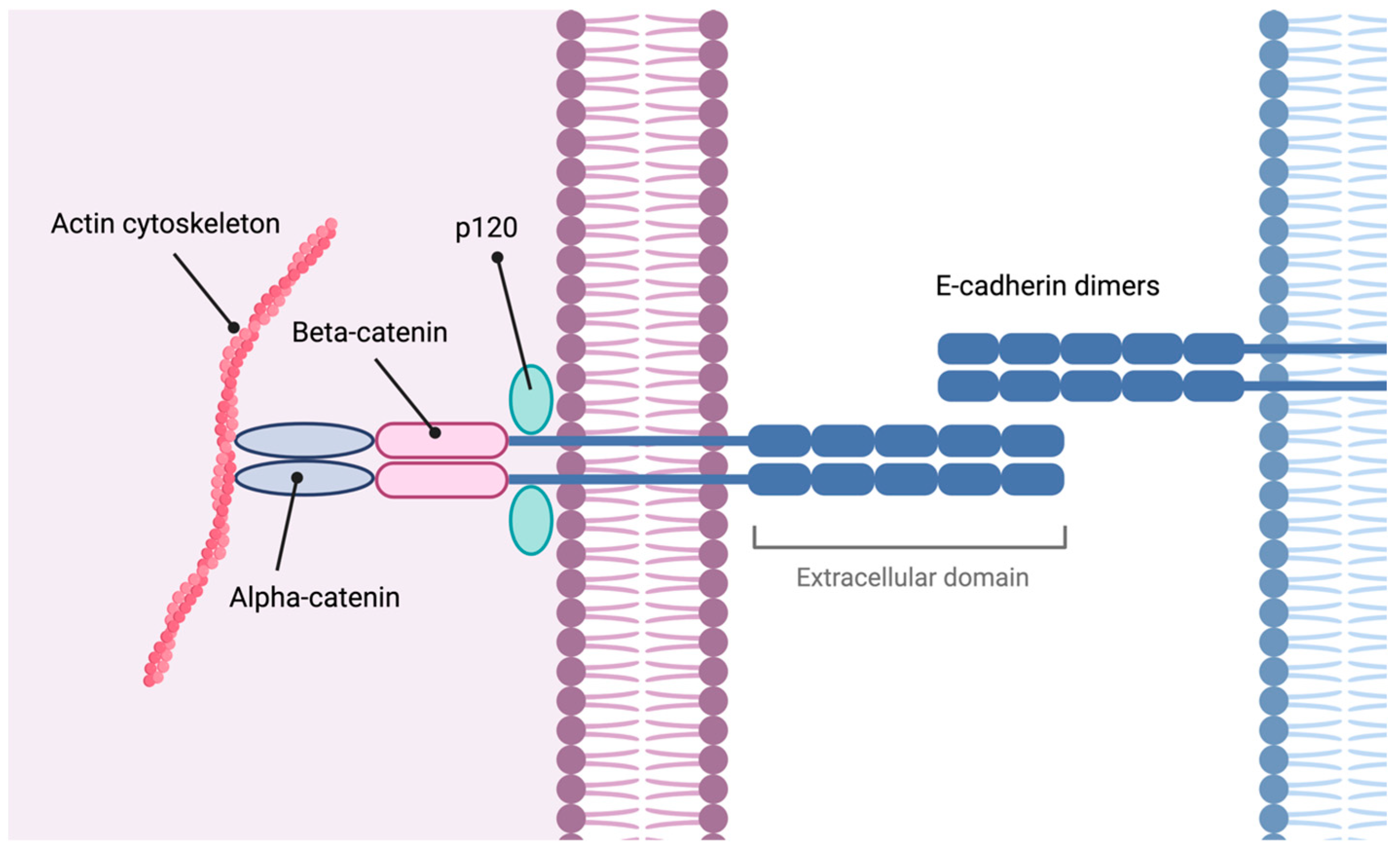
The CDH1 gene is a tumor suppressor that encodes E-cadherin, a transmembrane protein involved in adherens junctions of epithelial cells. It plays a critical role in cell–cell adhesion, tension sensing, and signal transduction [33]. Figure 3 illustrates the composition of these cell junctions.
In hereditary diffuse GC, alterations in the CDH1 gene result in a loss of function [17]. CDH1 variants are estimated to have a frequency of 5–10 per 100,000 births [12]. Mutations can be found throughout the coding sequence, with no identified hot spots [9,34,35]. According to the two-hit hypothesis, a somatic second-hit event causes biallelic inactivation, leading to the deregulation of E-cadherin and the development of diffuse GC or invasive lobular breast cancer [17].
E-cadherin plays a crucial role in maintaining cellular differentiation, polarity, and epithelial architecture [9]. Disruption of adherens junction components leads to loss of cell cohesion, promoting epithelial–mesenchymal transition (EMT), cell invasiveness, carcinogenesis, and development of metastasis [36].
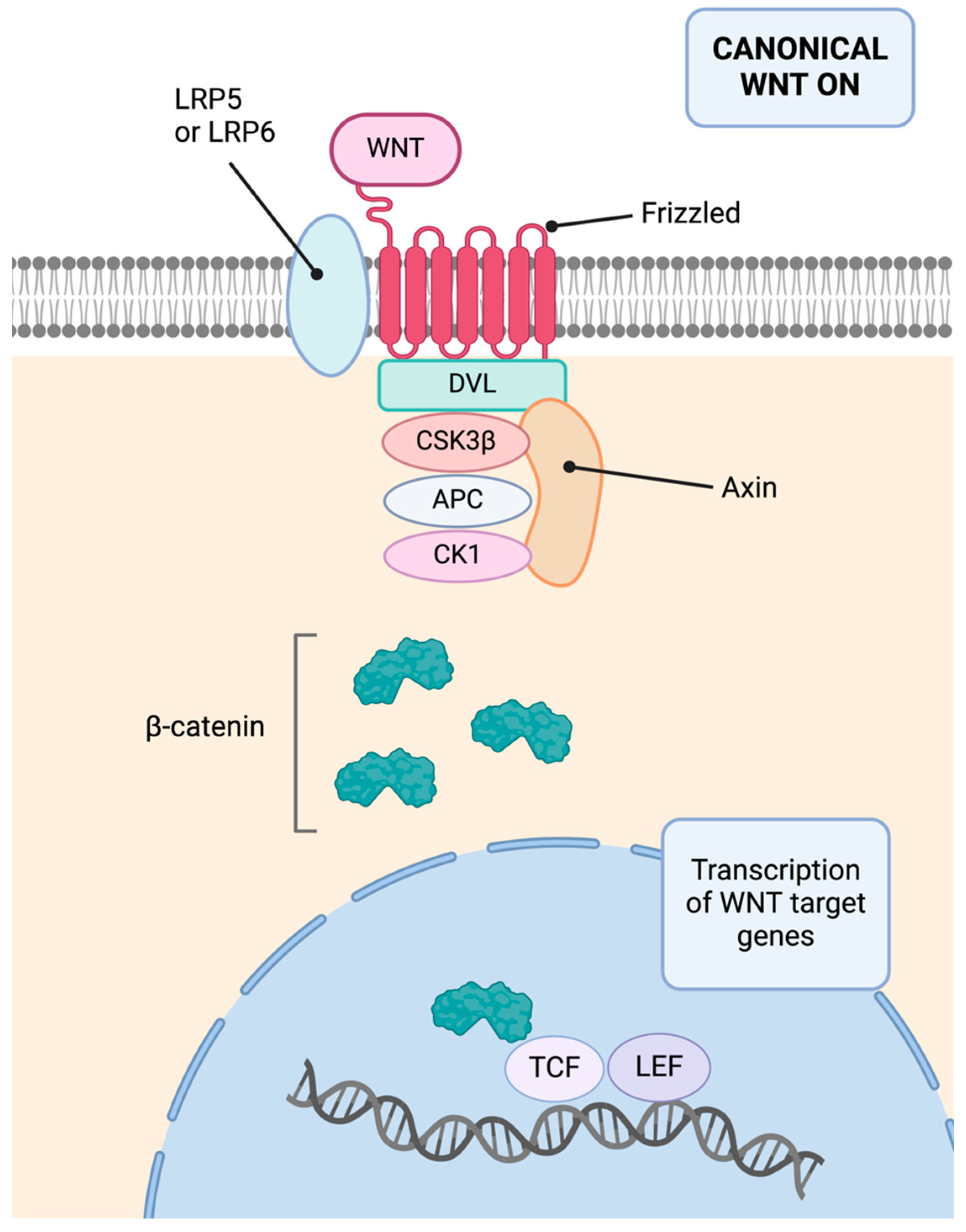
In addition, E-cadherin sequesters β-catenin at the cell membrane, preventing its nuclear entry and cell proliferation promotion [37]. This inhibition of the Wnt/β-catenin pathway has a protective effect, as this pathway is also linked to carcinogenesis, cellular invasiveness, EMT, metastasis, and chemotherapy resistance [38,39]. Additionally, E-cadherin’s extracellular domain can interact with growth factors, inhibiting ligand-dependent activation of receptor tyrosine kinases and suppressing proliferative signals, as well as inhibiting the Hippo-YAP pathway [40]. Figure 4 presents a schematic of the activated canonical Wnt pathway.

3.3.2. CTNNA1

CTNNA1 encodes α-catenin, a protein also involved in adherens junctions. Mutations in this gene account for less than 2% of hereditary diffuse GC cases [41,42,43,44]. It is hypothesized that α-catenin alterations also occur after a second-hit event [43]. Previous studies have identified at least 25 mutations in CTNNA1 in GC [42].
In respect of the structure and functions of CTNNA1 and α-catenin, CTNNA1 is located on chromosome 5q31 and is widely expressed in human tissues [45]. The α-catenin protein family includes three members: αE-catenin (epithelial), αN-catenin (neuronal), and αT-catenin (testis and heart) [9]. This family maintains the integrity of adherens junctions by anchoring the cadherin-β-catenin complex to the actin cytoskeleton, strengthening intercellular adhesion [46,47].
α-catenin has additional tumor suppressor functions beyond those related to intercellular junctions, as it inhibits critical pathways such as NF-κB, Hedgehog, and Wnt/β-catenin and regulates the Hippo-YAP pathway [48]. The NF-κB pathway is involved in cell proliferation, survival, differentiation, EMT, angiogenesis, metastasis, and immune response [49]. The Hedgehog pathway is associated with GI tumor development, maintaining tumor stem cells in a proliferative state or establishing a microenvironment essential for tumor growth [50]. Finally, the Hippo-YAP pathway has opposing roles in carcinogenesis, exhibiting both pro- and anti-oncogenic effects [51]. α-catenin sequesters YAP in the cytoplasm, inhibiting its function, and previous studies have shown that loss of α-catenin function can have pro-tumorigenic effects [48].

3.3.3. Other Molecular Alterations

Previous studies have suggested that hereditary diffuse GC may be associated with alterations in other tumor suppressor genes [42,52]. For example, a relationship has been identified between alterations in genes involved in double-stranded DNA repair and diffuse GC (PALB2, BRCA2, possibly RAD51C, and ATM) [53]. However, most of these studies are at the research level, and the association of these mutations with GC risk and their pathogenicity needs to be demonstrated for clinical application [44]. Therefore, some authors emphasize the need for future studies to investigate the specific mechanisms of diverse genes implicated in diffuse GC [45].

3.4. Follow-Up and Treatment

For patients with CDH1 alterations, endoscopic surveillance and prophylactic total gastrectomy are recommended. Gastrectomy is advised between the ages of 20 and 30, or 5 years before the youngest family member was diagnosed with GC, and is generally not recommended for those over 70 years old [12,54]. Post-surgery, in an ideal resource setting, examination of the entirety of the resected specimen is recommended. However, due to limitations in some laboratories, guidelines offer various options depending on the context [12].
When patients meet certain criteria, such as lacking a family history of GC, having variants of unknown pathogenicity, being under 20 years old, or declining or having contraindications for prophylactic gastrectomy, endoscopic surveillance may be performed [55]. Endoscopic examination should be conducted every 6–12 months, and any visible lesions should be biopsied along with random areas. A minimum of 30 biopsies from the antrum, transitional zone, body, fundus, and cardia is recommended, following the Cambridge protocol, a consensus approach [56]. To increase sensitivity, methods such as chromoendoscopy, endoscopic ultrasonography, or confocal endoscopic microscopy can be used [57,58]. The application of artificial intelligence techniques to endoscopic studies could enhance sensitivity, but more work is needed in this field to reach clinical application [59,60].
Some authors suggest that for asymptomatic carriers of CTNNA1 mutations, annual endoscopic surveillance is recommended. However, due to the recently described risk of GC associated with pathogenic mutations of this gene, these alterations should probably be managed similarly to CDH1 alterations [12].

4. Gastric Adenocarcinoma and Proximal Polyposis of the Stomach

Gastric adenocarcinoma and proximal polyposis of the stomach is a recently described autosomal dominant gastric polyposis syndrome [53]. It was first identified in 2012 in a large Australian family, along with two smaller families in the US and Canada [61]. The syndrome is characterized by the presence of over 100 polyps (though some family members have fewer) in the gastric body and fundus. These polyps are typically sessile and small (<1 cm), although they can grow larger [62,63]. The youngest patient diagnosed with this syndrome was 10 years old [61]. Gastric adenocarcinoma and proximal polyposis of the stomach exhibits phenotypic variability, with differences in age of onset, penetrance, and degree of dysplasia. Diagnosis is based on clinical and endoscopic criteria [64]. A colonoscopy is necessary to exclude attenuated familial adenomatous polyposis, and proton pump inhibitor use must be ruled out [65].

4.1. Gastric Cancer Risk

Gastric adenocarcinoma and proximal polyposis of the stomach carries a significant risk of GC, estimated between 12 and 25%, with incomplete penetrance [61,62,66,67]. It is commonly associated with intestinal-type GC, which tends to occur in areas with high-grade dysplastic fundic gland polyps and adenomatous polyps. Some authors suggest the risk may be lower due to the limited quality of the literature, as most cases are described in case reports rather than series [55]. The age of GC onset varies between 25 and 70 years [67,68].

4.2. Molecular Basis

The molecular alteration associated with gastric adenocarcinoma and proximal polyposis of the stomach was described in 2016, identifying a germline point mutation in the promoter 1B of the APC gene [66]. Subsequent research has detected various mutations in this promoter [69]. These mutations reduce the binding of the transcription factor Yin Yang 1, decreasing the transcriptional activity of the APC promoter [66]. In most cases, a second hit occurs via loss of the wild-type allele or truncating mutations [70]. Interestingly, similar mutations are found in rare families with familial adenomatous polyposis [66]. The functions of the APC gene are further discussed in the sections on familial adenomatous polyposis and MUTYH-associated polyposis (see below).

4.3. Follow-Up and Treatment

A personalized approach is recommended due to the interindividual heterogeneity in gastric adenocarcinoma and proximal polyposis of the stomach and the limited available data [70]. Recommendations are primarily based on expert opinion, and follow-up criteria vary by guidelines [64,71]. Some investigators suggest performing an upper endoscopy every 6–12 months, while others recommend follow-up based on the degree of polyposis: every 5 years if no polyps are present, every 3 years if fundic gland polyps without dysplasia are present, and individualized decisions for patients with over 100 fundic gland polyps [67,71]. Endoscopic screening should begin at age 15 or with the onset of dyspeptic symptoms, including a detailed examination of the polyps and surrounding mucosa with multiple biopsies of suspicious areas [71].
Diagnosing GC in the context of this syndrome is challenging because of the numerous polyps, some with adenomatous or dysplastic changes, which are indistinguishable endoscopically. In some cases, GC has only been diagnosed after prophylactic gastrectomy despite endoscopic surveillance [68]. Prophylactic gastrectomy is recommended at least for patients with dysplastic polyps [71]. However, given the limited correlation between endoscopic and histopathological findings, some authors advocate for performing gastrectomy on all gastric adenocarcinoma and proximal polyposis of the stomach patients without contraindications for surgery [72].

5. Familial Intestinal Gastric Cancer

Familial intestinal GC is an autosomal dominant syndrome associated with an increased risk of intestinal-type GC and other tumors, such as colorectal and breast cancer [11,73], in patients who do not present gastric polyposis or a genetic alteration associated with another syndrome [74]. It is important to note that data on this entity are very scarce, resulting in suboptimal quality of available evidence.

5.1. Clinical Presentation

The diagnostic criteria for familial intestinal GC were established in 1999 [18]. In countries with high incidence, familial intestinal GC is suspected when a family has three or more relatives with intestinal GC, with at least one being a first-degree relative, GC diagnosis in at least two generations, and at least one GC patient diagnosed before the age of 50. In countries with low incidence, the criteria include two scenarios: families with at least two first- or second-degree relatives with intestinal GC, one diagnosed before age 50, and families with three cases of intestinal GC regardless of the age of diagnosis [75]. Recent recommendations suggest relaxing these criteria, considering familial intestinal GC in any family with two cases of GC (one intestinal), regardless of the age of diagnosis [11].

5.2. Gastric Cancer Risk

The clinical spectrum of familial intestinal GC is not well defined and has not significantly changed in recent years [75]. A recent study suggested that GC appears later in familial intestinal GC than in hereditary diffuse GC and about 10 years earlier than sporadic intestinal GC [11]. The risk of GC in these families is not well established; however, a 2024 analysis in a Western country with a low incidence of GC observed a 4.9% incidence of GC and a 43.9% incidence of precursor lesions in patients undergoing endoscopic follow-up after familial intestinal GC diagnosis [74]. Another study found a 66% prevalence of GC in a cohort of 50 families meeting familial intestinal GC criteria [11].

5.3. Molecular Basis

The genetic cause of familial intestinal GC remains unclear [76,77]. In one family, heterozygous mutations in the IL12RB1 gene were identified [76]. NGS studies have identified other potential genes such as TP53, BRCA2, BRCA1, MSH2, and BRIP1 [75]. Microsatellite instability (MSI) has been observed in 38% of cases [11]. However, the relationship between these genes and the disease is not clear. Carvalho et al. suggested that familial intestinal GC might be a polygenic disease, as their 2021 study on this disease did not find alterations in high-penetrance genes [11].

5.4. Follow-Up and Treatment

Endoscopic surveillance is recommended; however, due to the limited information on this entity, standardized follow-up protocols do not exist and recommendations vary. Some authors suggest starting endoscopic surveillance at age 60, while others recommend starting at age 40, five years before the youngest GC diagnosis in the family, or ten years before said diagnosis [78,79]. The recommended interval between endoscopies also varies, ranging from annually to every three years or every five years [74].
As with all syndromes related to intestinal GC, preventive eradication of Helicobacter pylori is recommended. This recommendation is supported by results from Llach et al., who observed that H. pylori infection was the only factor independently associated with the development of precursor lesions in this syndrome [74].

6. Li–Fraumeni Syndrome

Li–Fraumeni syndrome is a rare autosomal dominant syndrome caused by germline mutations in the TP53 gene [53]. It is characterized by familial aggregation of tumors in individuals under 45 years of age, predominantly including sarcomas, breast cancer, brain tumors, and adrenal cancer, which account for 80% of cases [80]. First described in 1969 within four families exhibiting early-onset tumors, Li–Fraumeni syndrome has since been identified in 24 families comprising 151 relatives with cancer [81,82]. In 1990, the molecular cause was discovered through the identification of germline missense mutations in TP53 in five families, with typical findings [83].

6.1. Clinical Presentation and Gastric Cancer Risk

The French Li–Fraumeni syndrome working group has revised the criteria for Li–Fraumeni syndrome to encompass various clinical presentations linked to germline TP53 mutations and to improve clinical identification. The new “Chompret criteria” outline three primary scenarios indicative of Li–Fraumeni syndrome: familial presentation, where a proband has an Li–Fraumeni syndrome-associated tumor under 46 years old and a first- or second-degree relative has an Li–Fraumeni syndrome-associated tumor under 56 years old or has multiple tumors; multiple primary tumors, with at least two belonging to the Li–Fraumeni syndrome spectrum, the first of which developed before 46 years old; and rare cancers such as adrenocortical carcinoma or choroid plexus carcinoma, regardless of family history [84]. The risk of GC in individuals with Li–Fraumeni syndrome is relatively low. The most extensive series identified this tumor in 5% of patients and 22.6% of families with Li–Fraumeni syndrome, with an average age of onset at 43 years [85]. According to the International Agency for Research on Cancer database, the individual and familial risk of GC in this syndrome is 3.3% and 5.9%, respectively [86]. Li–Fraumeni syndrome-associated GC can be either intestinal or diffuse, although intestinal GC appears to be more common [85].

6.2. Molecular Basis

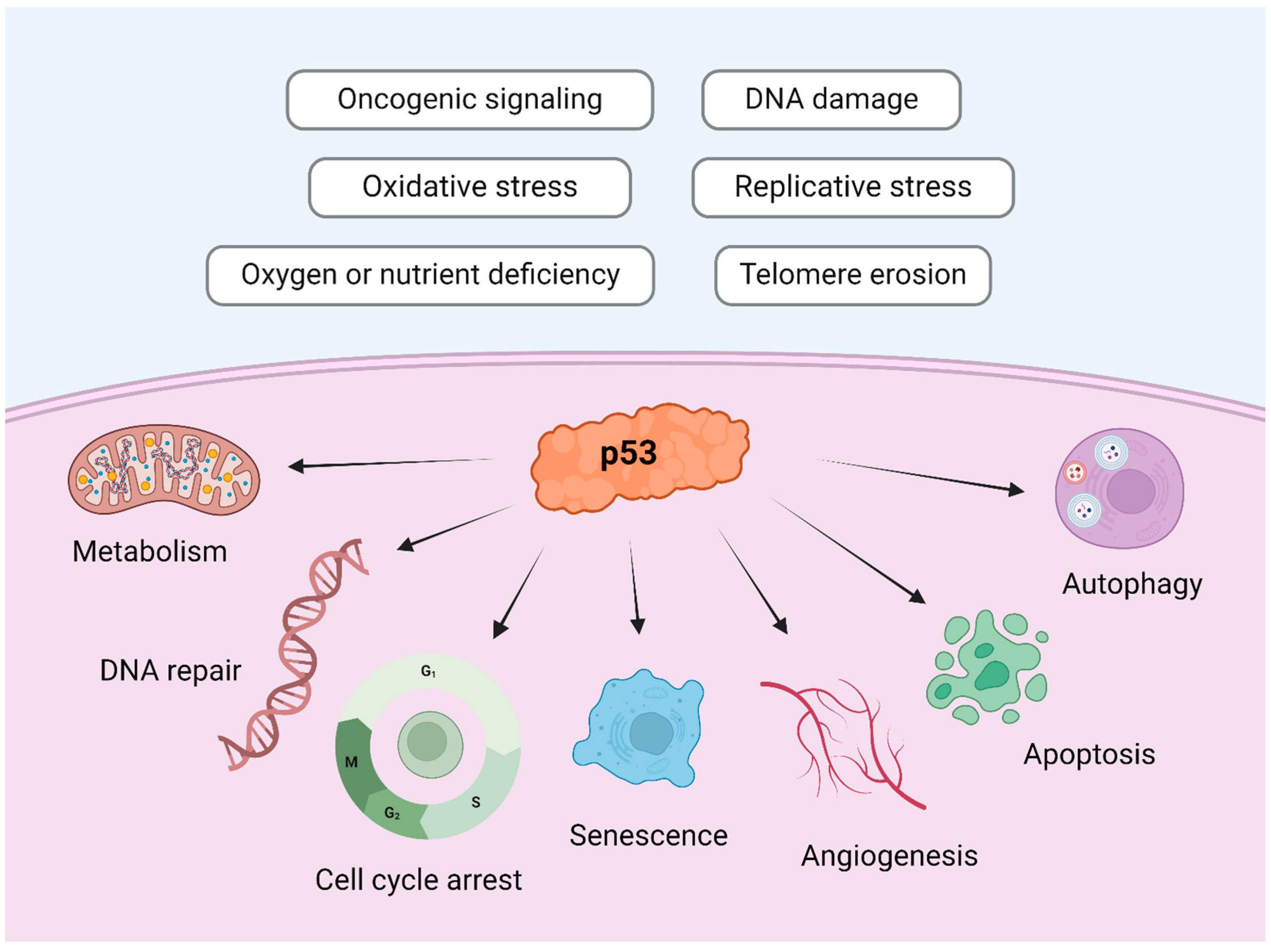
Loss-of-function mutations in TP53 lead to an accumulation of genetic alterations, resulting in tumor formation [87]. TP53 is located on the short arm of chromosome 17, spanning 20 kb with an extensive intronic region [88]. It is a tumor suppressor gene, also known as “the guardian of the genome”, and it is involved in DNA repair, genomic stability, senescence, autophagy, cell-cycle control, cell differentiation, angiogenesis, and apoptosis [89]. Figure 5 illustrates the principal physiological functions of the p53 protein.

6.3. Follow-Up and Treatment

In this syndrome, surveillance with physical exams, lab tests, and frequent imaging using a combination of whole-body, brain and breast magnetic resonance imaging, mammography, abdominal and pelvic ultrasound, and colonoscopy is recommended, as it has been shown to improve patient survival [90].
On the other hand, the management of upper GI tumors in Li–Fraumeni syndrome is not standardized due to limited available data [91,92]. Generally, upper endoscopy is recommended from the age of 25 or 10 years before the first case of GC in the family, but the cost–benefit of endoscopic screening is debated [53,93]. Interestingly, Tjandra et al. reported an excess of gastro-esophageal junction tumors in TP53 mutation carriers, emphasizing a detailed endoscopic examination of the esophagus and gastro-esophageal junction in these patients [93].

7. Hereditary Breast and Ovarian Cancer

Recent studies suggest that GC may be part of the spectrum of this syndrome, with both BRCA1 and BRCA2 mutations [94]. However, the risk of GC for each gene, as well as its pathogenesis and the need and protocol for GC screening are still under investigation [95].

8. Colon

CRC ranks as the second leading cause of cancer-related mortality worldwide. Approximately one-third of CRC cases exhibit familial clustering, but less than 20% are associated with germline mutations in cancer predisposition genes [96]. Non-syndromic CRC is defined as the clustering of CRC cases in a family without any apparent hereditary molecular abnormality. This clustering may be due to yet unidentified genetic alterations or to somatic tumors that aggregate in families because of a combination of environmental and common genetic factors [97].
In a recent review of nearly 12,000 patients with malignant tumors by Stadler et al., both tumor and germline mutation analyses were performed. Among these patients, nearly 2000 (10.5%) had CRC, with 72.4% presenting with metastatic disease. A germline mutation predisposing to cancer was found in 15.4% of the cases, many of which were potentially targetable [98].
Another study by You et al., focused on CRC patients under 50 years of age—a rapidly growing demographic—found that 67% of cases lacked a family history of cancer. Only 8% had deficiencies in mismatch repair (MMR) proteins, but a germline mutation analysis in 70% of the cohort (130 patients) revealed pathogenic variants in 19% of cases and variants of uncertain significance (VUS) in 18%. Notably, 12% of patients without a family history of disease and 13% of cases with intact MMR proteins carried pathogenic variants [99]. These findings suggest that germline mutation analysis can inform the management of both advanced-metastatic tumors and early-stage patients, aiding in risk reduction strategies.
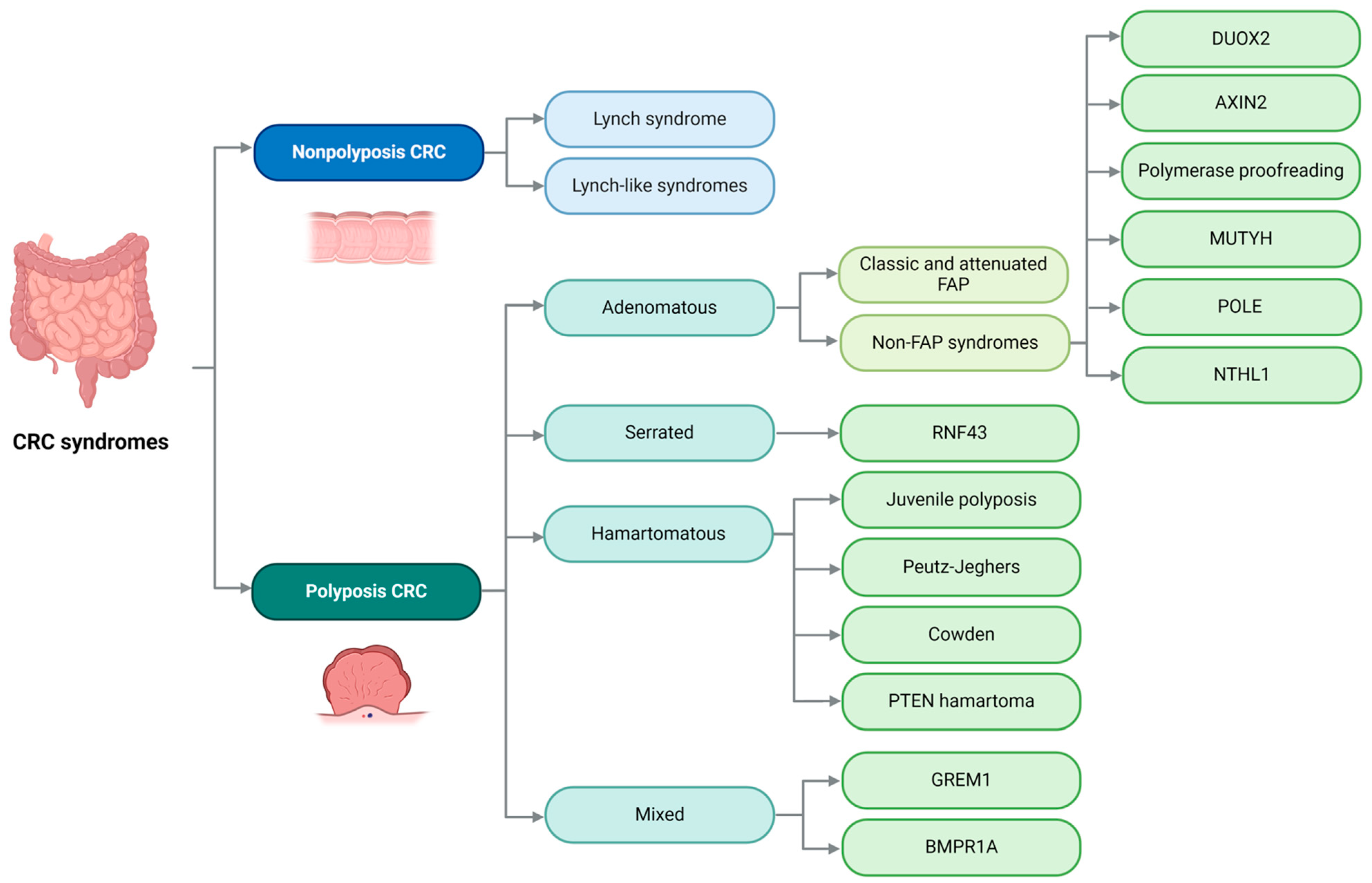
Historically, our understanding of the genetic basis of hereditary non-polyposis CRC, specifically Lynch syndrome, and hereditary polyposis CRC, including adenomatous, serrated, and hamartomatous polyposis, accounted for less than 7% of familial CRC clustering. However, NGS is expanding our knowledge of the mutations that can lead to CRC, adding complexity to this issue [100]. Figure 6 illustrates the various entities that will be discussed in this review.

9. Lynch Syndrome

Aldred Scott Warthin described the first case of what we now call Lynch syndrome in 1895 in a large family with a high incidence of carcinomas (the so-called “family G”) [101]. In 1971, Henry Lynch reported the results of genetic studies he performed on members of the original family G described by Warthin, confirming an autosomal pattern of inheritance with a high prevalence of GI and endometrial tumors, coining the term “Cancer Family Syndrome”. However, Lynch could not describe the genetic basis of this disease due to the technical limitations of the time. It was soon noted that some families had only CRC while others also had extraintestinal neoplasms, leading to the terms Lynch syndrome I and II, which later fell into disuse. In 1985, Lynch used the term hereditary non-polyposis CRC for this disease. In 2005, Douglas et al., working again with members of family G, found a specific mutation in the MSH2 gene [102].

9.1. Clinical Presentation

Lynch syndrome occurs due to inherited genetic mutations that impair DNA MMR. It is associated with a wide variety of neoplasms, depending on the gene affected, which usually appear at a young age [103]. Lynch syndrome is associated with a high risk of CRC; endometrial cancer; and cancers of the ovary, stomach, small intestine, hepatobiliary tract, upper urinary tract, brain, and skin [104]. However, not all patients with mutations develop neoplasms. Both personal and familial history are important to define the risk of disease and the need for surveillance and screening [105]. In 1991, an international collaborative group established the Amsterdam criteria to suspect Lynch syndrome, emphasizing the importance of familial clustering of CRC [106]. According to these criteria, suspicion of Lynch syndrome arises when three relatives are affected by CRC, one of whom is a first-degree relative of the other two; CRC appears in at least two generations; and at least one member is diagnosed with CRC before the age of 50. Additionally, it is crucial that the family history shows no features of familial adenomatous polyposis. These criteria were revised in 1999 to include not only CRC but also other cancers associated with this disease. Later, the Bethesda criteria were proposed as an alternative for diagnosing Lynch syndrome [107].

9.2. Colorectal Cancer Risk

Although Lynch syndrome is the most frequent hereditary CRC syndrome, it represents less than 5% of all CRC cases. However, the risk of developing CRC for patients with Lynch syndrome is significantly increased compared to the general population (82% vs. 5%) [108]. The lifetime risk of developing CRC is highest for MLH1 pathogenic variants (50% for men and 45% for women), followed by MSH2 (45% for men and 40% for women), PMS2 (20% for men and 15% for women), and MSH6 (10% for men and 15% for women) [105].

9.3. Gastric Cancer Risk

The risk of GC in Lynch syndrome ranges from 6% to 13%, with variations depending on ethnicity and geographic location [109]. Approximately two-thirds of the reported cases correspond to intestinal GC.

9.4. Molecular Basis

Lynch syndrome is an autosomal dominant syndrome caused by pathogenic variants in one of the DNA MMR genes, namely MSH2, MSH6, MLH1, and PMS2 [110]. Less frequently, it can be due to deletion of the EPCAM gene with subsequent MSH2 silencing or epigenetic changes, leading to MLH1 promoter methylation [111,112]. As extensively reviewed in previous studies, loss of MMR gene function leads to the accumulation of mutations and accelerates tumor development. The molecular abnormalities typical of Lynch syndrome are also related to other syndromes, such as Muir–Torre syndrome (which includes sebaceous neoplasms) and Turcot syndrome (which includes brain tumors) [113,114].
Lynch syndrome can be detected either through polymerase chain reaction or NGS-based analysis of microsatellite loci or MMR genes. IHC examination of the proteins encoded by MMR genes may serve as a screening method for germline testing. For a long time, IHC was performed only in patients fulfilling Amsterdam or Bethesda clinical criteria, but nowadays, IHC is indicated for all newly diagnosed CRC cases, regardless of age or history [106]. If loss of MLH1 and/or PMS2 is found, it is necessary to analyze BRAF mutations and MLH1 promoter methylation, as many sporadic cases also lose MMR protein expression without germline mutations. There are many pathogenic variants of the MMR genes associated with LS, but also many VUS that can be found in multiple genomic databases for patient counseling [115]. Likely, pathogenic variants are managed clinically as pathogenic ones. Multigene analysis panels are indicated in cases suspicious of LS, as they are more cost-efficient than isolated gene detection tests.

9.5. Follow-Up and Treatment

Periodic follow-up of Lynch syndrome patients with colonoscopy is indicated every 2 years beginning at 20–25 years for MLH1- or MSH2-associated cases, or at 35 years for MSH6 and PMS2 cases. This screening can allow for the prevention and earlier detection of CRC, consequently improving prognosis [116]. The risk of developing both synchronous and metachronous CRC has led some groups to recommend total abdominal colectomy as an alternative for Lynch syndrome patients who have already developed CRC, as it improves quality of life and saves lives [117]. This recommendation is only for carriers of MSH2 or MLH1 abnormalities, with segmental colectomy preferred for MSH6 and PMS2 mutation carriers. Prophylactic colectomy is not recommended in MMR carriers, as the risk of disease is not 100%. Decisions on surgical therapy should always be based on mutation analysis, not on the results of IHC or MLH1 promoter methylation [118].
Management of patients with VUS is more complex and still under study. It is necessary to develop individualized management strategies, as VUS can appear in almost 30% of suspected familial cases [119], and they add significant stress on families faced with a situation that neither their doctors nor they can adequately manage at this moment [120].
Evidence suggests that patients with Lynch syndrome can reduce the risk of CRC with the regular use of daily aspirin, adjusting the dose to the risks associated with long-term consumption of this drug [121].
Regarding GC screening, there is no consensus on the use of upper endoscopy. A recent review recommends performing it from the age of 30, and in younger patients if they have gastric symptoms or if they have young family members diagnosed with GC [122].

10. Lynch-like Syndromes

Lynch-like syndromes comprise a group of patients who exhibit deficiencies in the DNA MMR system but lack MMR mutations associated with Lynch syndrome. This classification represents a heterogeneous spectrum of cases that evolves continuously with advancements in molecular understanding [100]. Up to 70% of Lynch-like syndrome patients demonstrate loss of IHC expression of one of the MMR proteins, without identifiable germline mutations. Instead, they often exhibit either somatic methylation of the MLH1 promoter or dual somatic pathogenic changes in MMR genes. Notably, patients with Lynch-like syndromes typically have a lower familial burden of disease and tend to manifest at an older age [100]. However, they require vigilant monitoring for early disease detection and prompt treatment. Thus, in cases where hypermethylation or BRAF mutations are absent, it is recommended to conduct colonoscopies biennially from ages 25 to 75 years to mitigate the risk of CRC [121].
Several syndromes share similarities with Lynch syndrome but possess distinct genetic characteristics, including constitutional mismatch repair deficiency syndrome and familial CRC type x syndrome.

10.1. Constitutional Mismatch Repair Deficiency Syndrome

This autosomal recessive disorder is marked by homozygous or compound heterozygous germline pathogenic variants of MMR genes [123]. Constitutional mismatch repair deficiency syndrome ranks among the most aggressive cancer predisposition syndromes, with patients developing CRC and other malignancies, including hematological and solid tumors, at a remarkably young age. Screening protocols typically start at age 8, and surveillance includes brain magnetic resonance imaging due to the heightened susceptibility to brain tumors. Despite challenges in genetic testing interpretation, an international consortium of experts has recently established clinical, genetic, and ancillary criteria to facilitate diagnosis, as surveillance and early intervention are critical for prognosis [124].

10.2. Familial Colorectal Cancer Type X Syndrome

This autosomal dominant hereditary syndrome meets the clinical criteria for Lynch syndrome but lacks germline mutations in MMR genes or MSI [125]. The genetic basis of familial CRC type x syndrome remains poorly elucidated, with only the RSP40 gene definitively linked to the disease, though numerous potential candidates are under investigation [126,127]. While ongoing studies aim to uncover more cases of familial CRC type x syndrome, evidence suggests that the progression rate from adenomas to carcinomas is slower, allowing for surveillance every 3–5 years commencing at the age of the youngest affected relative [125].

11. Classic and Attenuated Familial Adenomatous Polyposis

This group of hereditary polyposis CRC syndromes with adenomatous polyps is characterized by the development of colorectal adenomas that progress to CRC. While the molecular basis of familial adenomatous polyposis has been known for some time, it accounts for only a small percentage of cases. The widespread use of NGS has led to an explosion of knowledge about other pathogenic variants that cause similar clinical findings, resulting in what is termed APC-negative adenomatous polyposis [128].
The first reported case of familial adenomatous polyposis dates back to 1881 by Sklifasowski, with Cripps proposing hereditary risk for the development of multiple polyps in 1882. Over subsequent years, numerous authors contributed cases and families to this emerging entity, establishing a high risk of malignant transformation of these polyps and their association with similar polyps throughout the GI tract. In 1939, Dukes published data from 10 families followed in the first Polyposis Registry at St. Mark’s Hospital [129]. The genetic basis of the disease was elucidated in 1986 when Herrera et al. described an abnormality in chromosome 5q, and in 1987, two independent groups identified the APC gene mapped to 5q21-22 as the molecular abnormality underlying familial adenomatous polyposis [130].

11.1. Clinical Presentation

This syndrome is characterized by the presence of a large number of polyps in the large bowel, with rectal bleeding being the most common symptom leading to diagnosis. However, some cases are identified through genetic screening. Polyps typically develop in childhood, and at the time of diagnosis, 60% of patients have more than 50 polyps along the colon. Other symptoms associated with APC mutations include hypertrophy of the retinal pigment epithelium; desmoid tumors (Gardner syndrome); osteomas; epidermal cysts; supernumerary teeth; and GI carcinomas, including CRC, small bowel cancer, and pancreatic cancer [131]. The phenotype appears to be influenced by the location of APC mutations, leading to two distinct clinical scenarios: classical familial adenomatous polyposis (including profuse familial adenomatous polyposis with over 1000 adenomas along the colonic mucosa and sparse familial adenomatous polyposis with 100–1000 polyps) and attenuated familial adenomatous polyposis with 10–99 adenomas, predominating in the right colon [132].

11.2. Risk of Colorectal Cancer

Although familial adenomatous polyposis is the most common cause of polyposis, it explains less than 1% of all CRC cases. Early reports showed that malignancy develops in classical familial adenomatous polyposis before the ages of 40–50 in almost 100% of cases, but more recent data suggest lower rates of cancer due to the adoption of preventive measures [133,134]. This risk may also vary depending on the location of APC mutations [135].

11.3. Molecular Basis

The APC gene, located on chromosome 5q, acts as a tumor suppressor gene, and its loss of function leads to the activation of the Wnt pathway, loss of cell adhesion, defects in cell cycle control, deregulation of base excision repair (BER), and impairment of chromosomal segregation [136]. The BER mechanism will be discussed in more detail in the section on MUTYH-associated polyposis. Mutations involving codons 1250–1464 of the APC gene are associated with classical familial adenomatous polyposis. Attenuated forms result from pathogenic or likely pathogenic variants in the 5′ or 3′ ends of the gene and selective splicing of exon 9 [137]. The location of the APC mutation also influences extracolonic manifestations of the disease. However, APC pathogenic variants are found in only 60% of clinically suspected cases (termed APC-negative polyposis), some of which are explained by other molecular abnormalities [132]. Multipanel testing is recommended, but single-site tests can be used if a pathogenic variant has already been confirmed in a given family.

11.4. Follow-Up and Treatment

Traditionally, patients with familial adenomatous polyposis were recommended proctocolectomy in their 20s to prevent CRC. However, this approach has evolved with the understanding of genotype–phenotype correlations, allowing for postponing surgery or using less invasive and aggressive surgical options. Nevertheless, surgery remains the best therapeutic option for these patients [121]. From the age of 12, familial adenomatous polyposis patients should undergo colonoscopies every 1–3 years according to the phenotype. After colectomy, pouchoscopy is recommended every 1–3 years, depending on the phenotype [132].

12. MUTYH-Associated Polyposis

MUTYH-associated polyposis is an autosomal recessive syndrome characterized by biallelic pathogenic variants in the MUTYH gene. Patients with MUTYH-associated polyposis typically develop numerous colorectal polyps, although rarely exceeding 100. Unlike some other syndromes, extraintestinal manifestations are infrequent in this entity, and primarily involve the proximal small bowel [138]. The risk of CRC can be as high as 90% in biallelic carriers, while monoallelic carriers exhibit only a slightly increased risk compared to the general population and often lack a polyposis phenotype. Therefore, biallelic carriers require regular annual colonoscopies from a young age (18–20 years). Interestingly, previous reports have found CRC even in patients with a low number of polyps [136]. Recent findings have suggested an increased risk of other malignant tumors, such as breast, pancreas, ovary, and bladder cancers, in monoallelic MUTYH mutation carriers, prompting the proposal to rename the condition MUTYH-associated tumor syndrome [139].
Patients under 60 years with 10 or more adenomas, and those over 60 with 20 or more adenomas or 10 or more adenomas with a family history of adenomas or CRC but without pathogenic variants in APC or MUTYH are categorized as having “multiple colorectal adenomas” [140]. In these cases, guidelines recommend 1–2 colonoscopies per year from the time of diagnosis until 75 years of age to reduce CRC risk, along with germline panel testing to exclude potential genetic abnormalities.

Molecular Basis

The MUTYH gene, located on chromosome 1, plays a crucial role in DNA repair, specifically in the BER mechanism. With the advent of NGS analysis, our understanding of the BER process, in which DNA glycosylases like MUTYH initiate DNA repair upon detection of base abnormalities, has significantly improved. This system repairs various forms of oxidative, deamination, alkylation, and abasic single-base damage, which may have minimal effects on the helix but are associated with significant processes such as carcinogenesis, inflammation, aging, or the development of neurodegenerative disorders [141].
The BER mechanism aids in repairing damaged DNA through two primary pathways: short-patch and long-patch. In the short-patch BER pathway, a single nucleotide is replaced in the repair process, while the long-patch BER pathway involves the replacement of at least two nucleotides. As previously mentioned, initiation of the BER pathway is facilitated by DNA glycosylases, responsible for recognizing and removing damaged bases. Subsequently, the completion of the BER pathway requires the involvement of at least three additional enzymes, which carry out tasks such as strand incision, gap-filling, and ligation [142].

13. NTHL1-Associated Tumor Syndrome

The NTHL1 (Nth-like DNA glycosylase 1) gene encodes another DNA glycosylase that, similarly to MUTYH, plays a role in the BER process. This syndrome follows a recessive mode of inheritance [143]. Biallelic mutations in NTHL1 predispose individuals to polyposis, CRC, and numerous other tumor types, earning it the name NTHL1-associated tumor syndrome [144]. An initial description of a polyposis syndrome in 7 cases with adenomatous polyps identified a germline mutation in p.Q90* [145]. Subsequent studies revealed either homozygous mutations in p.Q90* or a heterozygous combination of p.Q90*/p.287* [146].
A recent review showed that the risk of CRC in patients with this syndrome is high, occurring in 49% of cases, with a mean age at diagnosis of 55 years [147]. Among heterozygous carriers, 18% were diagnosed with CRC. Tumors most frequently associated with this syndrome, in addition to CRC, include those of the breast and endometrium, suggesting that along with colonoscopic surveillance, periodic breast and gynecological examinations would be advisable in these patients [148].

Molecular Basis

The NTHL1 gene is located on the short arm of chromosome 16 and encodes the protein of the same name [149]. In contrast to MUTYH, NTHL1 is bifunctional, as it can perform both glycosylase and lyase functions, whereas MUTYH only exhibits glycosylase activity [143]. During the BER process, bifunctional glycosylases remove the abnormal nucleotide base, cleave the DNA strand, and create a single-stranded gap harboring modified 3′ ends, which is remodeled by other enzymes. Thus, the dysfunctional NTHL1 protein leads to genomic instability and increases the risk of cell transformation.
Interestingly, research is ongoing to delineate the pathogenic role of single nucleotide polymorphisms and other genomic alterations of the NTHL1 gene that could influence cancer development and aging, likely through telomere length control [143].

14. Polymerase Proofreading-Associated Polyposis

Polymerase proofreading-associated polyposis is an autosomal dominant syndrome associated with a high risk of CRC and other malignancies, including brain, duodenal, and breast carcinomas [150]. It is caused by germline pathogenic variants of the POLD1 gene (chromosome 19) and POLE gene (chromosome 12). These genes encode the catalytic subunits of polymerases delta and epsilon, respectively, which are involved in DNA proofreading to ensure replication fidelity. Pathogenic variants in these genes lead to the accumulation of thousands of mutations, resulting in an ultramutated phenotype [151]. Patients develop GI polyps and have an increased risk of malignant transformation, with CRC risk approaching 90% for POLD1 variant carriers and nearly 30% for POLE variant carriers. Despite the better prognosis of ultramutated tumors and their favorable response to immune checkpoint inhibition, affected individuals should undergo stringent surveillance from a young age [137].

15. Axis Inhibition Protein 2 (AXIN2)-Associated Polyposis

AXIN2-associated polyposis is an autosomal dominant syndrome characterized by colorectal polyps and oligodontia, defined as a congenital absence of at least seven teeth. However, a recent French series involving 11 unrelated individuals with AXIN2 variants revealed that oligodontia was present in only 60% of cases. Nonetheless, this report confirmed that loss-of-function variants of this gene increase the risk of CRC and other GI neoplasms, with 40% of patients developing malignant disease and 91% exhibiting colorectal polyposis [152]. The prevalence of these variants in the report was notably low, at 0.24% [153]. Despite its rarity, AXIN2 presents an intriguing potential target for boosting immunotherapy [154].
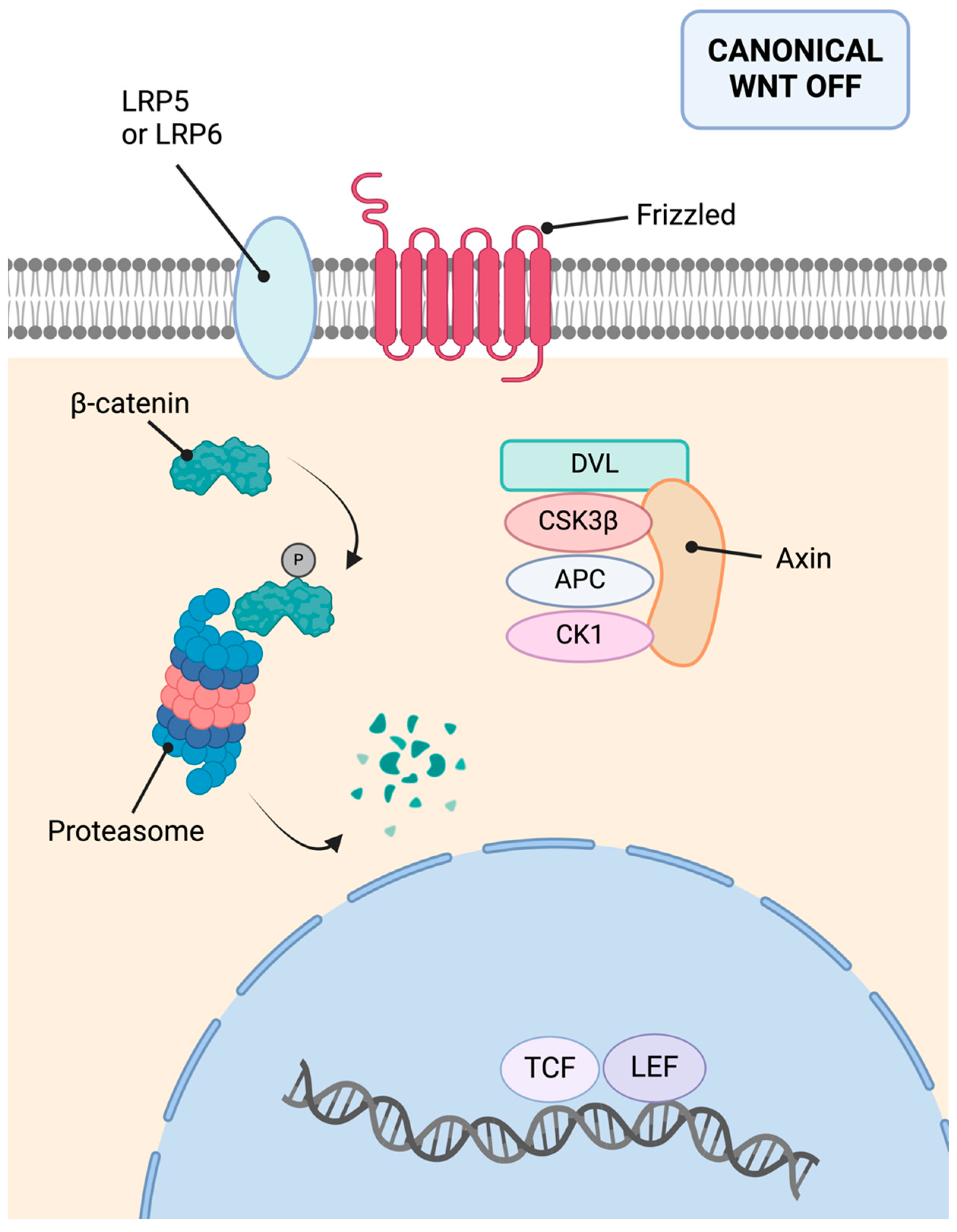
Regarding its molecular basis, the AXIN2 gene is a tumor suppressor gene located on chromosome 17q24. It encodes the Axin-2 protein, also known as conductin [155]. This protein is crucial as a negative regulator of the Wnt pathway. It acts as a scaffold for the β-catenin destruction complex, facilitating β-catenin degradation [153]. Consequently, it inhibits the pro-proliferative function of the Wnt pathway. Figure 7 summarizes the inactive canonical Wnt pathway.

16. Dual Oxidase 2 (DUOX2)-Associated Polyposis

DUOX2 has traditionally been linked to thyroid function, as it has been shown to contribute to congenital hypothyroidism due to thyroid dysgenesis [156]. However, it also appears to play a role in maintaining normal GI microbiota and has recently been suggested as a candidate gene for APC-negative familial polyposis [157]. In the GI tract, DUOX2 alterations are rare but have been associated with the occurrence of CRC and increased tumor aggressiveness [158]. These alterations seem to cause endoplasmic reticulum retention with an unfolded protein response, which promotes adenomatous changes and carcinogenesis. Both missense and nonsense mutations of the DUOX gene lead to the overexpression of a truncated pathogenic protein [159]. The clinical relevance and management strategies for these variants are still not well defined.

17. Other Candidate Genes in Adenomatous Polyposis Syndromes

NGS studies of different cohorts of APC-negative polyposis syndrome patients are leading to the identification of many gene variants associated with these conditions [160,161]. Studies are ongoing to evaluate the potential pathogenic role of genes such as MLH3, MSH3, GALNT12, ATM, and SDHA, among others [162]. In the near future, expanding knowledge about these and other candidate genes will enable the early management of more patients.

18. Hereditary Polyposis Colorectal Carcinoma with Serrated Polyps

Serrated polyps, which include hyperplastic polyps (usually left-sided), sessile serrated polyps (usually right-sided), and serrated adenomas, are confirmed through histopathological analysis. Though once considered rare, serrated polyposis syndrome is now recognized as the most prevalent polyposis syndrome leading to CRC. It is characterized by the development of multiple serrated polyps throughout the colon and rectum. The inheritance pattern of serrated polyposis syndrome remains unclear, yet first-degree relatives of patients seem to face a significantly increased risk of CRC and pancreatic cancer [163].

18.1. Clinical Presentation

The World Health Organization defines serrated polyposis syndrome by two possible clinical criteria. A diagnosis can be established if a patient has at least five serrated polyps, each measuring 5mm or larger, located proximal to the rectum with at least two of these polyps being 10mm or larger. Alternatively, a diagnosis may be made when a patient presents with more than 20 serrated polyps anywhere along the bowel, with at least five of these being proximal to the rectum [164]. The average age at diagnosis is around 50 years, and less than half of the patients report a familial history of the disease, highlighting the ongoing debate regarding its hereditary nature.
The incidence of CRC in patients with serrated polyposis syndrome varies but is noted to be over 20% according to various studies. The highest risk occurs at the time of diagnosis (10–20%) and tends to decrease during follow-up (2–4%) [165].

18.2. Molecular Basis

Serrated polyposis syndrome appears to be a heterogeneous condition, characterized by an activating mutation in the BRAF gene, which may or may not be associated with MSI [164]. Although the progression risk is well documented, less than 5% of serrated polyposis syndrome cases have been linked to germline pathogenic variants. A review by Murphy et al. indicated significant overlap with other polyposis syndromes; some patients with pathogenic variants in genes like MUTYH, PTEN, SMAD4, BMPR1A, POLE, and GREM1 presented features of both serrated polyposis syndrome and other syndromes [166]. Therefore, there is controversy regarding its molecular basis, and some authors emphasize that many cases have no family history of polyposis and a strong association with environmental factors has also been observed [167].

18.3. Follow-Up and Treatment

Genetic testing is recommended for younger patients, those with multiple affected relatives, or those who have dysplasia in any polyps. Management primarily involves regular, high-quality colonoscopies. The recommended colonoscopy interval is every 1–2 years, with removal of all polyps if feasible, or at least those 5 mm or larger or those suspected of having dysplastic changes [168]. In cases where endoscopic control is impractical due to numerous lesions, radical surgery (colectomy) may be necessary. First-degree relatives should undergo colonoscopy every 5 years, starting from the age of 45 years [169].

18.4. RNF43-Associated Polyposis

Recent studies underscore the tumor suppressor role of the RNF43 gene [170,171,172]. RNF43 is a U3 ubiquitin ligase with multiple anti-carcinogenic functions: it is responsible for ubiquitinating and stabilizing p53, regulates the cell cycle, and inhibits the Wnt and Hippo pathways [173]. Germline variants in RNF43, considered likely pathogenic, have been identified in families characterized by BRAF-mutated CRC linked to the serrated pathway and exhibit an autosomal dominant pattern of inheritance [170]. RNF43 is emerging as a promising candidate gene for explaining familial predisposition to serrated polyposis syndrome and is also being explored as a potential marker for targeted drug development [174]. However, the precise role of RNF43 in CRC tumorigenesis remains to be fully determined.

19. Juvenile Polyposis Syndrome

Juvenile polyposis syndrome is an autosomal dominant cancer syndrome characterized primarily by the development of numerous juvenile polyps in the GI tract, particularly in the colon and rectum, where they are present in 98% of cases [175]. Despite its name, “juvenile” refers to the histological appearance of the polyps rather than the age of onset. The average age at diagnosis is 18 years.
The number of polyps in juvenile polyposis syndrome patients can range from a few to over 100 [176]. The syndrome is suspected when a patient has five or more juvenile polyps in the colon (especially in the proximal section), a juvenile polyp in the proximal GI tract, or any number of juvenile polyps along with a family history of juvenile polyposis syndrome. Extraintestinal manifestations include pigmented nevi, telangiectasias on the skin, and various skeletal abnormalities [177]. Patients with juvenile polyposis syndrome face a high lifetime risk of developing colorectal cancer, which can reach up to 70% [175].
More than one-third of juvenile polyposis syndrome cases arise from de novo mutations and frequently mutated genes include Bone Morphogenetic Protein Receptor 1A (BMPR1A) or Suppressor Mothers Against Decapentaplegic Homolog 4 (SMAD4 or DPC4) [178]. BMPR1A, a serine-threonine kinase type I receptor, regulates the Transforming Growth Factor β (TGF-β) pathway, crucial for cell growth inhibition and apoptosis [179]. Juvenile polyposis syndrome-associated abnormalities typically include large deletions and point mutations, occasionally involving the gene promoter, and affected patients tend to show a large bowel-predominant phenotype. SMAD4, identified as a tumor suppressor gene in 1996, is an essential mediator of TGF-β pathway [180]. Mutations in SMAD4 are linked to both colonic and gastric polyps and to hereditary hemorrhagic telangiectasia, with high penetrance, leading to the development of colonic polyps in 97% of affected individuals before the age of 50 and increasing risks for gastric and pancreatic cancers.
For patients with a confirmed diagnosis of juvenile polyposis syndrome who lack the typical pathogenic variants, extensive genomic evaluations may uncover other significant genetic mutations [181,182]. Surveillance recommendations include annual colonoscopy starting at 12 years old, though the starting age and frequency may be adjusted based on polyp burden and symptoms [183]. All polyps larger than 5 mm should undergo removal and histological examination. Colectomy is contemplated only for patients with numerous polyps that are unmanageable through endoscopic approaches.

Juvenile Polyposis of Infancy

Juvenile polyposis of infancy, also known as chromosome 10q22.2-q23.2 deletion syndrome, represents a severe subtype of juvenile polyposis syndrome [184]. Linked to microdeletions in the BMPR1A gene, juvenile polyposis of infancy is characterized by early onset before age 2, severe enteropathy, and high mortality [185]. The 10q22.2-q23.2 deletion often involves the PTEN gene, which is also associated with Cowden disease. Advances in genomic technology have started to uncover small copy number variations that contribute to the molecular basis of juvenile polyposis of infancy.

20. Peutz–Jeghers Syndrome

Peutz–Jeghers syndrome is an autosomal dominant disorder characterized by hamartomatous GI polyposis, mucocutaneous pigmentation, and a predisposition to cancer, caused by germline pathogenic variants in the STK11 gene [186]. Hamartomatous polyps can occur throughout the GI tract but are most commonly found in the small intestine, particularly the jejunum [187]. The most frequent types of cancer in Peutz–Jeghers syndrome patients include breast and GI cancers, specially CRC. Peutz–Jeghers syndrome is also associated with tumors of the liver, lungs, ovaries, uterus, and testicles [188]. The syndrome is rare, with an estimated incidence of 1 in 50,000 to 200,000 births [189].
The diagnostic criteria for Peutz–Jeghers syndrome are (1) two or more histologically confirmed Peutz–Jeghers-type polyps, (2) any number of Peutz–Jeghers-type polyps in an individual with a first-degree relative with Peutz–Jeghers syndrome, (3) characteristic mucocutaneous pigmentation in a person with a family history of Peutz–Jeghers syndrome, or (4) any number of Peutz–Jeghers-type polyps and characteristic mucocutaneous pigmentation [53].

20.1. Clinical Presentation

The average age at diagnosis is between 18 and 23 years [190,191]. Characteristic pigmentation appears in 95% of patients as small dark blue, black, or brown mucocutaneous macules, visible during childhood from around 5 years of age and potentially fading during puberty, although perioral lesions often persist [189]. These macules typically appear around the eyes, nostrils, and perioral area, as well as on the palms, soles, fingers, and perianal area [192]. They are not usually associated with the development of malignant melanotic tumors.
Peutz–Jeghers intestinal polyps are hamartomatous and characterized by an arborizing pattern of smooth muscle cells between mucosal epithelial compartments. Gastric polyps may exhibit less specific histological features, though classic cases have been documented [193,194]. GI polyps can lead to chronic bleeding, anemia, and recurrent bowel obstruction and intussusception [195].

20.2. Colorectal Cancer Risk

Patients with Peutz–Jeghers syndrome have a significantly increased risk of cancer (15–18 times higher than the general population), with a mean onset age of 41 years and a cumulative probability of 55% by age 60 [189,196]. The lifetime risk for CRC is estimated at 39% [197]. Regarding GC, data on its occurrence in Peutz–Jeghers syndrome are limited. A previous study showed a high risk of GC, with a cumulative probability of 29% between the ages of 15 and 64 years [198].

20.3. Molecular Basis

Peutz–Jeghers syndrome is caused by germline mutations in the STK11 gene, combined with an acquired second-hit defect [199]. This gene is located on chromosome 19 (19p13.3) and consists of nine coding exons (1302 base pairs) and one non-coding exon [200]. It encodes the LKB1 protein, which has 433 amino acids and a molecular weight of 50 kDa [201]. The protein comprises an amine-terminal domain, a kinase domain, and a carboxy-terminal regulatory domain [202].
The STK11 gene acts as a tumor suppressor, and its defect can make cells particularly vulnerable to energetic stress mediated by the AMP-activated protein kinase [203]. This kinase also regulates cell cycle, cell differentiation, apoptosis, and DNA damage repair [204]. Additionally, LKB1 inhibits the mTOR pathway [205]. This pathway is crucial in cancer development, because it promotes tumor growth, cell proliferation, metabolism, and immune tolerance, while inhibiting autophagy and apoptosis [206]. Activation of the mTOR pathway has pro-oncogenic effects, and STK11 dysfunction can trigger these effects. Indeed, previous studies have shown that loss-of-function mutations in STK11 reduce response to immunotherapy in patients with non-small cell lung cancer [207]. In GI cancer, the association of STK11 mutations with treatment resistance is less clear [208].
Apart from these roles, LKB1 is involved in chromosomal segregation, inhibits the TGF-β and Wnt signaling pathways, and regulates p53 activity [209]. By inhibiting these pathways and mediating p53 activity, it further fulfills its tumor suppressor functions [210,211]. The roles of the Wnt pathway and p53 protein have been discussed in more detail in the sections on hereditary diffuse GC and Li–Fraumeni syndrome (see above). The TGF-β pathway has protumorigenic effects, promoting EMT and enhancing cellular plasticity and survival [212]. TGF-β has a dual effect, inducing apoptosis in early stages and promoting tumor growth in later stages, increasing aggressiveness, invasiveness, and metastasis [213]. TGF-β acts not only at the tumor cell level but also within the tumor microenvironment, including immune and stromal components [214].

20.4. Follow-Up and Treatment

According to the European Society of Gastrointestinal Endoscopy, the first endoscopic examination (oesophagogastroduodenoscopy and colonoscopy) is recommended at 8 years of age, with follow-up intervals depending on the presence of symptoms and identification of polyps [215]. Polyps larger than 15–20 mm, or smaller polyps causing symptoms, should be resected.

21. Cowden Syndrome

Cowden syndrome was first described in 1963 as an autosomal dominant disorder. It is characterized by multiple hamartomas affecting various organs and an increased risk of several tumors, including breast, thyroid, or uterine cancer [216]. The diagnostic criteria for Cowden syndrome were established in 1996 [217]. Most patients begin to exhibit cutaneous symptoms during adolescence, and approximately 50–70% of cases develop thyroid abnormalities [218]. Additionally, up to 93% of patients undergoing colonoscopy reveal polyps, predominantly hamartomatous, although hyperplastic polyps, ganglioneuromas, and leiomyomas are also common [219]. It is typical for these patients to present with several different types of polyps synchronously. Cowden syndrome may also be associated with dysplastic gangliocytoma (Lhermitte–Duclos disease) and can overlap with Bannayan–Riley–Ruvalcaba syndrome [220,221]. The clinical diagnosis can be challenging, with genetic studies playing a crucial role in the diagnostic process [185].

21.1. Colorectal Cancer Risk

Traditionally, it was believed that patients with Cowden syndrome were prone to developing thyroid and endometrial cancers but not at an increased risk for CRC. However, recent studies suggest otherwise, estimating that the risk of CRC could be as high as 13–15%, with all cases occurring before the age of 50 [222,223].

21.2. Molecular Basis

Cowden syndrome is linked to mutations in the phosphatase and tensin homolog (PTEN) gene located on chromosome 10, which encodes a protein tyrosine phosphatase. PTEN is considered a tumor suppressor gene, as it inhibits the mTOR pathway and plays important roles in cell growth, proliferation, survival, DNA repair, and cell motility [224]. Various genetic alterations, including nonsense and missense mutations, deletions–insertions, and splice site mutations, have been described in up to 81% of Cowden syndrome patients, affecting the entire length of the gene, predominantly spanning exon 1 [225]. Notably, about 47% of Cowden syndrome cases arise from de novo mutations [226].

21.3. Follow-Up and Treatment

For Cowden syndrome patients, colonoscopy is recommended to begin at age 35. If polyps are detected, subsequent colonoscopies should be scheduled annually; if none are found, every five years is sufficient. If a family member develops CRC, surveillance should start 5–10 years prior to the age at which the relative was diagnosed. Colectomy is indicated only in select cases, based on clinical findings [226].

21.4. PTEN Hamartoma Tumor Syndrome

PTEN hamartoma tumor syndrome encompasses conditions related to PTEN germline mutations, such as Proteus syndrome and Bannayan–Riley–Ruvalcaba syndrome, and is often discussed interchangeably with Cowden syndrome [227]. However, no clear genotype–phenotype correlation has been established. The revised criteria for diagnosing this syndrome include major and minor criteria [228]. Among the major criteria are the detection of breast, endometrial, or thyroid cancers; GI hamartomas; and other lesions such as macrocephaly, acral keratoses, mucocutaneous neuromas, and oral papillomas.

22. Hereditary Polyposis with Mixed Polyps Syndrome

Hereditary polyposis with mixed polyps syndrome is characterized by the development of polyps with histologically mixed features. This can involve polyps showing histologically different areas or the presence of multiple polyps of different types in the same patient, though a strict quantitative definition is still lacking. At least two distinct forms of the disease have been described [229].

22.1. GREM1-Associated Mixed Polyposis

Gremlin 1 (GREM1) encodes a member of the bone morphogenetic protein antagonist family. It exerts its antagonistic effect by directly binding to bone morphogenetic proteins, influencing body patterning and tissue differentiation. Its role in cancer is not clear, as it exerts both anti-tumorigenic and pro-tumorigenic effects, favoring stemness, activation of cancer-associated fibroblasts, angiogenesis, and cell proliferation [230].
Cenani–Lenz syndrome, characterized by oligosyndactyly, metacarpal synostosis, and phalangeal disorganization with additional facial features, is mostly associated with LRP4 mutations [231]. However, some cases have been linked to truncating mutations in the APC gene, mutations in formin 1 (FMN1), or rearrangements of the GREM1-FMN1 loci. It is worth noting that this last molecular alteration has been associated with an increased risk of CRC through mechanisms yet to be fully elucidated. Additionally, this occurs more frequently in diabetic and obese patients, highlighting the interplay between environmental and genetic factors in cancer development [232].

22.2. BMPR1A-Associated Mixed Polyposis

Bone morphogenetic protein receptor type 1A (BMPR1A) has been linked to juvenile polyposis syndrome and, more recently, to a syndrome with overlapping features known as hereditary mixed polyposis syndrome 2. Patients with hereditary mixed polyposis syndrome 2 exhibit germline mutations, including frame-shift, nonsense, or missense mutations in the BMPR1A gene. They display a variable phenotype characterized by polyps with mixed histological features and, in some cases, the development of CRC without prior polyps [233].

Author Contributions

M.J.F.A.: study design, data acquisition analysis and interpretation, manuscript draft, approval, and agreement. C.D.d.A.: study design, data acquisition analysis and interpretation, manuscript draft, approval, and agreement. All authors have read and agreed to the published version of the manuscript.

Funding

This research received no external funding.

Conflicts of Interest

The authors declare no conflicts of interest.

References

  1. Sung, H.; Ferlay, J.; Siegel, R.L.; Laversanne, M.; Soerjomataram, I.; Jemal, A.; Bray, F. Global Cancer Statistics 2020: GLOBOCAN Estimates of Incidence and Mortality Worldwide for 36 Cancers in 185 Countries. CA Cancer J. Clin. 2021, 71, 209–249. [Google Scholar] [CrossRef] [PubMed]
  2. Sabour, L.; Sabour, M.; Ghorbian, S. Clinical Applications of Next-Generation Sequencing in Cancer Diagnosis. Pathol. Oncol. Res. 2017, 23, 225–234. [Google Scholar] [CrossRef]
  3. Ilic, M.; Ilic, I. Epidemiology of stomach cancer. World J. Gastroenterol. 2022, 28, 1187–1203. [Google Scholar] [CrossRef] [PubMed]
  4. Jin, H.; Pinheiro, P.S.; Callahan, K.E.; Altekruse, S.F. Examining the gastric cancer survival gap between Asians and whites in the United States. Gastric Cancer 2017, 20, 573–582. [Google Scholar] [CrossRef] [PubMed]
  5. El Halabi, M.; Horanieh, R.; Tamim, H.; Mukherji, D.; Jdiaa, S.; Temraz, S.; Shamseddine, A.; Barada, K. The impact of age on prognosis in patients with gastric cancer: Experience in a tertiary care centre. J. Gastrointest. Oncol. 2020, 11, 1233–1241. [Google Scholar] [CrossRef] [PubMed]
  6. Del Arco, C.D.; Medina, L.O.; Muñoz, L.E.; de las Heras, S.G.G.; Aceñero, M.J.F. Is there still a place for conventional histopathology in the age of molecular medicine? Laurén classification, inflammatory infiltration and other current topics in gastric cancer diagnosis and prognosis. Histol. Histopathol. 2021, 36, 587–613. [Google Scholar] [CrossRef]
  7. Mariette, C.; Carneiro, F.; Grabsch, H.I.; van der Post, R.S.; Allum, W.; de Manzoni, G.; Luca, B.G.; Maria, B.; Jean-Francois, F.; Uberto, F.; et al. Consensus on the pathological definition and classification of poorly cohesive gastric carcinoma. Gastric Cancer 2019, 22, 1–9. [Google Scholar] [CrossRef] [PubMed]
  8. Kole, C.; Charalampakis, N.; Sakellariou, S.; Papaxoinis, G.; Apostolou, K.G.; Machairas, N.; Papanikolaou, I.S.; Schizas, D. Hereditary Diffuse Gastric Cancer: A 2022 Update. J. Pers. Med. 2022, 12, 2032. [Google Scholar] [CrossRef]
  9. Taja-Chayeb, L.; Vidal-Millán, S.; Trejo-Becerril, C.; Pérez-Cárdenas, E.; Chávez-Blanco, A.; Domínguez-Gómez, G.; González-Fierro, A.; Romo-Pérez, A.; Dueñas-González, A. Hereditary diffuse gastric cancer (HDGC). An overview. Clin. Res. Hepatol. Gastroenterol. 2022, 46, 101820. [Google Scholar] [CrossRef] [PubMed]
  10. Lerner, B.A.; Llor, X. Genetic Gastric Cancer Risk Syndromes. Curr. Treat. Options Gastroenterol. 2020, 18, 604–615. [Google Scholar] [CrossRef]
  11. Carvalho, J.; Oliveira, P.; Senz, J.; São José, C.; Hansford, S.; Teles, S.P.; Ferreira, M.; Corso, G.; Pinheiro, H.; Lemos, D.; et al. Redefinition of familial intestinal gastric cancer: Clinical and genetic perspectives. J. Med. Genet. 2021, 58, 1–11. [Google Scholar] [CrossRef]
  12. Blair, V.R.; McLeod, M.; Carneiro, F.; Coit, D.G.; D’Addario, J.L.; van Dieren, J.M.; Harris, K.L.; Hoogerbrugge, N.; Oliveira, C.; van der Post, R.S.; et al. Hereditary diffuse gastric cancer: Updated clinical practice guidelines. Lancet. Oncol. 2020, 21, e386–e397. [Google Scholar] [CrossRef]
  13. Jones, E. Familial gastric cancer. N. Z. Med. J. 1964, 63, 287–296. [Google Scholar]
  14. Guilford, P.; Hopkins, J.; Harraway, J.; McLeod, M.; McLeod, N.; Harawira, P.; Taite, H.; Scoular, R.; Miller, A.; Reeve, A.E. E-cadherin germline mutations in familial gastric cancer. Nature 1998, 392, 402–405. [Google Scholar] [CrossRef]
  15. Gayther, S.; Gorringe, K.; Ramus, S.; Huntsman, D.; Roviello, F.; Grehan, N.; Machado, J.; Pinto, E.; Seruca, R.; Halling, K.; et al. Identification of germ-line E-cadherin mutations in gastric cancer families of European origin. Cancer Res. 1998, 15, 4086–4089. [Google Scholar]
  16. Richards, F.M.; McKee, S.A.; Rajpar, M.H.; Cole, T.R.P.; Evans, D.G.R.; Jankowski, J.A.; McKeown, C.; Sanders, D.S.A.; Maher, E.R. Germline E-cadherin gene (CDH1) mutations predispose to familial gastric cancer and colorectal cancer. Hum. Mol. Genet. 1999, 8, 607–610. [Google Scholar] [CrossRef]
  17. Gregory, S.N.; Davis, J.L. CDH1 and hereditary diffuse gastric cancer: A narrative review. Chin. Clin. Oncol. 2023, 12, 25. [Google Scholar] [CrossRef]
  18. Caldas, C.; Carneiro, F.; Lynch, H.T.; Yokota, J.; Wiesner, G.L.; Powell, S.M.; Lewis, F.R.; Huntsman, D.G.; Pharoah, P.D.P.; Jankowski, J.A.; et al. Familial gastric cancer: Overview and guidelines for management. J. Med. Genet. 1999, 36, 873–880. [Google Scholar]
  19. Lynch, H.T.; Grady, W.; Suriano, G.; Huntsman, D. Gastric cancer: New genetic developments. J. Surg. Oncol. 2005, 90, 114–133. [Google Scholar] [CrossRef]
  20. Shepard, B.; Yoder, L.; Holmes, C. Prophylactic Total Gastrectomy for Hereditary Diffuse Gastric Cancer. ACG Case Rep. J. 2016, 3, e179. [Google Scholar] [CrossRef]
  21. Rocha, J.P.; Gullo, I.; Wen, X.; Devezas, V.; Baptista, M.; Oliveira, C.; Carneiro, F. Pathological features of total gastrectomy specimens from asymptomatic hereditary diffuse gastric cancer patients and implications for clinical management. Histopathology 2018, 73, 878–886. [Google Scholar] [CrossRef] [PubMed]
  22. Tsugeno, Y.; Nakano, K.; Nakajima, T.; Namikawa, K.; Takamatsu, M.; Yamamoto, N.; Fujisaki, J.; Nunobe, S.; Kitagawa, M.; Takeuchi, K.; et al. Histopathologic Analysis of Signet-ring Cell Carcinoma In Situ in Patients with Hereditary Diffuse Gastric Cancer. Am. J. Surg. Pathol. 2020, 44, 1204–1212. [Google Scholar] [CrossRef] [PubMed]
  23. Corso, G.; Carvalho, J.; Marrelli, D.; Vindigni, C.; Carvalho, B.; Seruca, R.; Roviello, F.; Oliveira, C. Somatic mutations and deletions of the E-cadherin gene predict poor survival of patients with gastric cancer. J. Clin. Oncol. 2013, 31, 868–875. [Google Scholar] [CrossRef] [PubMed]
  24. Roberts, M.E.; Ranola, J.M.O.; Marshall, M.L.; Susswein, L.R.; Graceffo, S.; Bohnert, K.; Tsai, G.; Klein, R.T.; Hruska, K.S.; Shirts, B.H. Comparison of CDH1 Penetrance Estimates in Clinically Ascertained Families vs Families Ascertained for Multiple Gastric Cancers. JAMA Oncol. 2019, 5, 1325–1331. [Google Scholar] [CrossRef] [PubMed]
  25. Xicola, R.M.; Li, S.; Rodriguez, N.; Reinecke, P.; Karam, R.; Speare, V.; Black, M.H.; Laduca, H.; Llor, X. Clinical features and cancer risk in families with pathogenic CDH1 variants irrespective of clinical criteria. J. Med. Genet. 2019, 56, 838–843. [Google Scholar] [CrossRef] [PubMed]
  26. Cosma, L.S.; Schlosser, S.; Tews, H.C.; Müller, M.; Kandulski, A. Hereditary Diffuse Gastric Cancer: Molecular Genetics, Biological Mechanisms and Current Therapeutic Approaches. Int. J. Mol. Sci. 2022, 23, 7281. [Google Scholar] [CrossRef] [PubMed]
  27. Blair, V.; Martin, I.; Shaw, D.; Winship, I.; Kerr, D.; Arnold, J.; Harawira, P.; McLeod, M.; Parry, S.; Charlton, A.; et al. Hereditary diffuse gastric cancer: Diagnosis and management. Clin. Gastroenterol. Hepatol. 2006, 4, 262–275. [Google Scholar] [CrossRef] [PubMed]
  28. Benusiglio, P.R.; Malka, D.; Rouleau, E.; de Pauw, A.; Buecher, B.; Noguès, C.; Fourme, E.; Colas, C.; Coulet, F.; Warcoin, M.; et al. CDH1 germline mutations and the hereditary diffuse gastric and lobular breast cancer syndrome: A multicentre study. J. Med. Genet. 2013, 50, 486–489. [Google Scholar] [CrossRef] [PubMed]
  29. Gurzu, S.; Jung, I.; Orlowska, J.; Sugimura, H.; Kadar, Z.; Turdean, S.; Bara, T. Hereditary diffuse gastric cancer—An overview. Pathol. Res. Pract. 2015, 211, 629–632. [Google Scholar] [CrossRef] [PubMed]
  30. Pharoah, P.D.P.; Guilford, P.; Caldas, C. Incidence of gastric cancer and breast cancer in CDH1 (E-cadherin) mutation carriers from hereditary diffuse gastric cancer families. Gastroenterology 2001, 121, 1348–1353. [Google Scholar] [CrossRef]
  31. Cisco, R.M.; Ford, J.M.; Norton, J.A. Hereditary diffuse gastric cancer. Cancer 2008, 113, 1850–1856. [Google Scholar] [CrossRef] [PubMed]
  32. Coudert, M.; Drouet, Y.; Delhomelle, H.; Svrcek, M.; Benusiglio, P.R.; Coulet, F.; Clark, D.F.; Katona, B.W.; Van Hest, L.P.; Van Der Kolk, L.E.; et al. First estimates of diffuse gastric cancer risks for carriers of CTNNA1 germline pathogenic variants. J. Med. Genet. 2022, 59, 1189–1195. [Google Scholar] [CrossRef] [PubMed]
  33. Lecuit, T.; Yap, A.S. E-cadherin junctions as active mechanical integrators in tissue dynamics. Nat. Cell Biol. 2015, 17, 533–539. [Google Scholar] [CrossRef] [PubMed]
  34. Oliveira, C.; De Bruin, J.; Nabais, S.; Ligtenberg, M.; Moutinho, C.; Nagengast, F.M.; Seruca, R.; Van Krieken, H.; Carneiro, F. Intragenic deletion of CDH1 as the inactivating mechanism of the wild-type allele in an HDGC tumour. Oncogene 2004, 23, 2236–2240. [Google Scholar] [CrossRef] [PubMed]
  35. Corso, G.; Corso, F.; Bellerba, F.; Carneiro, P.; Seixas, S.; Cioffi, A.; La Vecchia, C.; Magnoni, F.; Bonanni, B.; Veronesi, P.; et al. Geographical Distribution of E-cadherin Germline Mutations in the Context of Diffuse Gastric Cancer: A Systematic Review. Cancers 2021, 13, 1269. [Google Scholar] [CrossRef] [PubMed]
  36. Na, T.Y.; Schecterson, L.; Mendonsa, A.M.; Gumbiner, B.M. The functional activity of E-cadherin controls tumor cell metastasis at multiple steps. Proc. Natl. Acad. Sci. USA 2020, 117, 5931–5937. [Google Scholar] [CrossRef] [PubMed]
  37. Park, S.Y.; Shin, J.H.; Kee, S.H. E-cadherin expression increases cell proliferation by regulating energy metabolism through nuclear factor-κB in AGS cells. Cancer Sci. 2017, 108, 1769–1777. [Google Scholar] [CrossRef] [PubMed]
  38. Tian, X.; Liu, Z.; Niu, B.; Zhang, J.; Tan, T.K.; Lee, S.R.; Zhao, Y.; Harris, D.C.H.; Zheng, G. E-cadherin/β-catenin complex and the epithelial barrier. J. Biomed. Biotechnol. 2011, 2011, 567305. [Google Scholar] [CrossRef] [PubMed]
  39. Koni, M.; Pinnarò, V.; Brizzi, M.F. The Wnt Signalling Pathway: A Tailored Target in Cancer. Int. J. Mol. Sci. 2020, 21, 7697. [Google Scholar] [CrossRef] [PubMed]
  40. Rubtsova, S.N.; Zhitnyak, I.Y.; Gloushankova, N.A. Dual role of E-cadherin in cancer cells. Tissue Barriers 2022, 10, 2005420. [Google Scholar] [CrossRef]
  41. Majewski, I.J.; Kluijt, I.; Cats, A.; Scerri, T.S.; De Jong, D.; Kluin, R.J.C.; Hansford, S.; Hogervorst, F.B.L.; Bosma, A.J.; Hofland, I.; et al. An α-E-catenin (CTNNA1) mutation in hereditary diffuse gastric cancer. J. Pathol. 2013, 229, 621–629. [Google Scholar] [CrossRef] [PubMed]
  42. Lobo, S.; Benusiglio, P.R.; Coulet, F.; Boussemart, L.; Golmard, L.; Spier, I.; Hüneburg, R.; Aretz, S.; Colas, C.; Oliveira, C. Cancer predisposition and germline CTNNA1 variants. Eur. J. Med. Genet. 2021, 64, 104316. [Google Scholar] [CrossRef] [PubMed]
  43. Benusiglio, P.R.; Colas, C.; Guillerm, E.; Canard, A.; Delhomelle, H.; Warcoin, M.; Bellanger, J.; Eyries, M.; Zizi, M.; Netter, J.; et al. Clinical implications of CTNNA1 germline mutations in asymptomatic carriers. Gastric Cancer 2019, 22, 899–903. [Google Scholar] [CrossRef] [PubMed]
  44. Clark, D.F.; Michalski, S.T.; Tondon, R.; Nehoray, B.; Ebrahimzadeh, J.; Hughes, S.K.; Soper, E.R.; Domchek, S.M.; Rustgi, A.K.; Pineda-Alvarez, D.; et al. Loss-of-function variants in CTNNA1 detected on multigene panel testing in individuals with gastric or breast cancer. Genet. Med. 2020, 22, 840–846. [Google Scholar] [CrossRef]
  45. Huang, J.; Wang, H.; Xu, Y.; Li, C.; Lv, X.; Han, X.; Chen, X.; Chen, Y.; Yu, Z. The Role of CTNNA1 in Malignancies: An Updated Review. J. Cancer 2023, 14, 219–230. [Google Scholar] [CrossRef]
  46. Pokutta, S.; Choi, H.J.; Ahlsen, G.; Hansen, S.D.; Weis, W.I. Structural and thermodynamic characterization of cadherin·β-catenin·α-catenin complex formation. J. Biol. Chem. 2014, 289, 13589–13601. [Google Scholar] [CrossRef] [PubMed]
  47. Pokutta, S.; Weis, W.I. Structure and mechanism of cadherins and catenins in cell-cell contacts. Annu. Rev. Cell Dev. Biol. 2007, 23, 237–261. [Google Scholar] [CrossRef]
  48. Sun, Y.; Zhang, J.; Ma, L. α-catenin. A tumor suppressor beyond adherens junctions. Cell Cycle 2014, 13, 2334–2339. [Google Scholar] [CrossRef]
  49. Zhang, T.; Ma, C.; Zhang, Z.; Zhang, H.; Hu, H. NF-κB signaling in inflammation and cancer. MedComm 2021, 2, 618–653. [Google Scholar] [CrossRef]
  50. Jiang, J. Hedgehog signaling mechanism and role in cancer. Semin. Cancer Biol. 2022, 85, 107–122. [Google Scholar] [CrossRef]
  51. Cunningham, R.; Hansen, C.G. The Hippo pathway in cancer: YAP/TAZ and TEAD as therapeutic targets in cancer. Clin. Sci. 2022, 136, 197–222. [Google Scholar] [CrossRef] [PubMed]
  52. Hansford, S.; Kaurah, P.; Li-Chang, H.; Woo, M.; Senz, J.; Pinheiro, H.; Schrader, K.A.; Schaeffer, D.F.; Shumansky, K.; Zogopoulos, G.; et al. Hereditary Diffuse Gastric Cancer Syndrome: CDH1 Mutations and Beyond. JAMA Oncol. 2015, 1, 23–32. [Google Scholar] [CrossRef] [PubMed]
  53. Kim, W.; Kidambi, T.; Lin, J.; Idos, G. Genetic Syndromes Associated with Gastric Cancer. Gastrointest. Endosc. Clin. N. Am. 2022, 32, 147–162. [Google Scholar] [CrossRef] [PubMed]
  54. van der Post, R.S.; Vogelaar, I.P.; Carneiro, F.; Guilford, P.; Huntsman, D.; Hoogerbrugge, N.; Caldas, C.; Chelcun Schreiber, K.E.; Hardwick, R.H.; Ausems, M.G.E.M.; et al. Hereditary diffuse gastric cancer: Updated clinical guidelines with an emphasis on germline CDH1 mutation carriers. J. Med. Genet. 2015, 52, 361–374. [Google Scholar] [CrossRef] [PubMed]
  55. Carneiro, F. Familial and hereditary gastric cancer, an overview. Best Pract. Res. Clin. Gastroenterol. 2022, 58–59, 101800. [Google Scholar] [CrossRef] [PubMed]
  56. Lee, C.Y.C.; Olivier, A.; Honing, J.; Lydon, A.M.; Richardson, S.; O’Donovan, M.; Tischkowitz, M.; Fitzgerald, R.C.; di Pietro, M. Endoscopic surveillance with systematic random biopsy for the early diagnosis of hereditary diffuse gastric cancer: A prospective 16-year longitudinal cohort study. Lancet Oncol. 2023, 24, 107–116. [Google Scholar] [CrossRef] [PubMed]
  57. Kumar, S.; Long, J.M.; Ginsberg, G.G.; Katona, B.W. The role of endoscopy in the management of hereditary diffuse gastric cancer syndrome. World J. Gastroenterol. 2019, 25, 2878–2886. [Google Scholar] [CrossRef] [PubMed]
  58. Ruff, S.; Curtin, B.; Quezado, M.; Heller, T.; Koh, C.; Steinberg, S.M.; Connolly, M.; Hernandez, J.M.; Davis, J.L. Evaluation of confocal endoscopic microscopy for detection of early-stage gastric cancer in hereditary diffuse gastric cancer (HDGC) syndrome. J. Gastrointest. Oncol. 2019, 10, 407–411. [Google Scholar] [CrossRef] [PubMed]
  59. Ali, H.; Muzammil, M.A.; Dahiya, D.S.; Ali, F.; Yasin, S.; Hanif, W.; Gangwani, M.K.; Aziz, M.; Khalaf, M.; Basuli, D.; et al. Artificial intelligence in gastrointestinal endoscopy: A comprehensive review. Ann. Gastroenterol. 2024, 37, 133–141. [Google Scholar] [CrossRef]
  60. Chen, P.C.; Lu, Y.R.; Kang, Y.N.; Chang, C.C. The Accuracy of Artificial Intelligence in the Endoscopic Diagnosis of Early Gastric Cancer: Pooled Analysis Study. J. Med. Internet Res. 2022, 24, e27694. [Google Scholar] [CrossRef]
  61. Worthley, D.L.; Phillips, K.D.; Wayte, N.; Schrader, K.A.; Healey, S.; Kaurah, P.; Shulkes, A.; Grimpen, F.; Clouston, A.; Moore, D.; et al. Gastric adenocarcinoma and proximal polyposis of the stomach (GAPPS): A new autosomal dominant syndrome. Gut 2012, 61, 774–779. [Google Scholar] [CrossRef] [PubMed]
  62. De Boer, W.B.; Ee, H.; Kumarasinghe, M.P. Neoplastic Lesions of Gastric Adenocarcinoma and Proximal Polyposis Syndrome (GAPPS) Are Gastric Phenotype. Am. J. Surg. Pathol. 2018, 42, 1–8. [Google Scholar] [CrossRef] [PubMed]
  63. Beer, A.; Streubel, B.; Asari, R.; Dejaco, C.; Oberhuber, G. Gastric adenocarcinoma and proximal polyposis of the stomach (GAPPS)—A rare recently described gastric polyposis syndrome—Report of a case. Z. Gastroenterol. 2017, 55, 1131–1134. [Google Scholar] [CrossRef] [PubMed]
  64. Rudloff, U. Gastric adenocarcinoma and proximal polyposis of the stomach: Diagnosis and clinical perspectives. Clin. Exp. Gastroenterol. 2018, 11, 447–459. [Google Scholar] [CrossRef] [PubMed]
  65. Freeman, H.J. Proton pump inhibitors and an emerging epidemic of gastric fundic gland polyposis. World J. Gastroenterol. 2008, 14, 1318–1320. [Google Scholar] [CrossRef] [PubMed]
  66. Li, J.; Woods, S.L.; Healey, S.; Beesley, J.; Chen, X.; Lee, J.S.; Sivakumaran, H.; Wayte, N.; Nones, K.; Waterfall, J.J.; et al. Point Mutations in Exon 1B of APC Reveal Gastric Adenocarcinoma and Proximal Polyposis of the Stomach as a Familial Adenomatous Polyposis Variant. Am. J. Hum. Genet. 2016, 98, 830–842. [Google Scholar] [CrossRef] [PubMed]
  67. Foretova, L.; Navratilova, M.; Svoboda, M.; Grell, P.; Nemec, L.; Sirotek, L.; Obermannova, R.; Novotny, I.; Sachlova, M.; Fabian, P.; et al. GAPPS—Gastric Adenocarcinoma and Proximal Polyposis of the Stomach Syndrome in 8 Families Tested at Masaryk Memorial Cancer Institute—Prevention and Prophylactic Gastrectomies. Klin. Onkol. 2019, 32, S109–S117. [Google Scholar] [CrossRef] [PubMed]
  68. Repak, R.; Kohoutova, D.; Podhola, M.; Rejchrt, S.; Minarik, M.; Benesova, L.; Lesko, M.; Bures, J. The first European family with gastric adenocarcinoma and proximal polyposis of the stomach: Case report and review of the literature. Gastrointest. Endosc. 2016, 84, 718–725. [Google Scholar] [CrossRef] [PubMed]
  69. Sakuma, T.; Sera, T.; Aoyama, R.; Sawada, A.; Kasashima, H.; Ogisawa, K.; Bamba, H.; Yashiro, M. Two families with gastric adenocarcinoma and proximal polyposis of the stomach (GAPPS): Case reports and literature review. J. Gastrointest. Oncol. 2023, 14, 2650–2657. [Google Scholar] [CrossRef]
  70. Iwatsuki, M.; Matsumoto, C.; Mimori, K.; Baba, H. The comprehensive review of gastric adenocarcinoma and proximal polyposis of the stomach (GAPPS) from diagnosis and treatment. Ann. Gastroenterol. Surg. 2023, 7, 725. [Google Scholar] [CrossRef]
  71. Tacheci, I.; Repak, R.; Podhola, M.; Benesova, L.; Cyrany, J.; Bures, J.; Kohoutova, D. Gastric adenocarcinoma and proximal polyposis of the stomach (GAPPS)—A Helicobacter-opposite point. Best Pract. Res. Clin. Gastroenterol. 2021, 50–51, 101728. [Google Scholar] [CrossRef]
  72. Salami, A.; Stone, J.; Greenberg, R.; Leighton, J.; Miick, R.; Zavala, S.; Zeitzer, K.; Bakhos, C. Early Prophylactic Gastrectomy for the Management of Gastric Adenomatous Proximal Polyposis Syndrome (GAPPS). ACS Case Rev. Surg. 2022, 3, 62–68. [Google Scholar]
  73. Oliveira, C.; Pinheiro, H.; Figueiredo, J.; Seruca, R.; Carneiro, F. Familial gastric cancer: Genetic susceptibility, pathology, and implications for management. Lancet. Oncol. 2015, 16, e60–e70. [Google Scholar] [CrossRef]
  74. Llach, J.; Salces, I.; Guerra, A.; Peñas, B.; Rodriguez-Alcalde, D.; Redondo, P.D.; Cubiella, J.; Murcia, Ó.; Escalante, M.; Gratacós-Ginès, J.; et al. Endoscopic surveillance for familial intestinal gastric cancer in low-incidence areas: An effective strategy. Int. J. Cancer 2024, 154, 124–132. [Google Scholar] [CrossRef]
  75. Garcia-Pelaez, J.; Barbosa-Matos, R.; São José, C.; Sousa, S.; Gullo, I.; Hoogerbrugge, N.; Carneiro, F.; Oliveira, C. Gastric cancer genetic predisposition and clinical presentations: Established heritable causes and potential candidate genes. Eur. J. Med. Genet. 2022, 65, 104401. [Google Scholar] [CrossRef]
  76. Vogelaar, I.P.; van der Post, R.S.; van de Vosse, E.; van Krieken, J.H.J.M.; Hoogerbrugge, N.; Ligtenberg, M.J.L.; Gómez García, E. Gastric cancer in three relatives of a patient with a biallelic IL12RB1 mutation. Fam. Cancer 2015, 14, 89–94. [Google Scholar] [CrossRef] [PubMed]
  77. Chen, H.; Wang, J.; Zhuang, Y.; Wu, H. Identification of the potential molecular mechanism and driving mutations in the pathogenesis of familial intestinal gastric cancer by whole exome sequencing. Oncol. Rep. 2018, 40, 2316–2324. [Google Scholar] [CrossRef] [PubMed]
  78. Corso, G.; Roncalli, F.; Marrelli, D.; Carneiro, F.; Roviello, F. History, pathogenesis, and management of familial gastric cancer: Original study of John XXIII’s family. Biomed. Res. Int. 2013, 2013, 385132. [Google Scholar] [CrossRef]
  79. Sereno, M.; Aguayo, C.; Guillén Ponce, C.; Gómez-Raposo, C.; Zambrana, F.; Gómez-López, M.; Casado, E. Gastric tumours in hereditary cancer syndromes: Clinical features, molecular biology and strategies for prevention. Clin. Transl. Oncol. 2011, 13, 599–610. [Google Scholar] [CrossRef] [PubMed]
  80. Corso, G.; Pedrazzani, C.; Marrelli, D.; Pinto, E.; Roviello, F. Familial gastric cancer and Li-Fraumeni syndrome. Eur. J. Cancer Care 2010, 19, 377–381. [Google Scholar] [CrossRef]
  81. Li, F.P.; Fraumeni, J.F. Soft-tissue sarcomas, breast cancer, and other neoplasms. A familial syndrome? Ann. Intern. Med. 1969, 71, 747–752. [Google Scholar] [CrossRef] [PubMed]
  82. Li, F.; Fraumeni, J.; Mulvihill, J.; Blattner, W.; Dreyfus, M.; Tucker, M.; Miller, R. A cancer family syndrome in twenty-four kindreds. Cancer Res. 1988, 48, 5358–5362. [Google Scholar] [PubMed]
  83. Malkin, D.; Li, F.P.; Strong, L.C.; Fraumeni, J.F.; Nelson, C.E.; Kim, D.H.; Kassel, J.; Gryka, M.A.; Bischoff, F.Z.; Tainsky, M.A.; et al. Germ line p53 mutations in a familial syndrome of breast cancer, sarcomas, and other neoplasms. Science 1990, 250, 1233–1238. [Google Scholar] [CrossRef] [PubMed]
  84. Bougeard, G.; Renaux-Petel, M.; Flaman, J.M.; Charbonnier, C.; Fermey, P.; Belotti, M.; Gauthier-Villars, M.; Stoppa-Lyonnet, D.; Consolino, E.; Brugières, L.; et al. Revisiting Li-Fraumeni syndrome from TP53 mutation carriers. J. Clin. Oncol. 2015, 33, 2345–2352. [Google Scholar] [CrossRef] [PubMed]
  85. Masciari, S.; Dewanwala, A.; Stoffel, E.M.; Lauwers, G.Y.; Zheng, H.; Achatz, M.I.; Riegert-Johnson, D.; Foretova, L.; Silva, E.M.; Digianni, L.; et al. Gastric cancer in individuals with Li-Fraumeni syndrome. Genet. Med. 2011, 13, 651–657. [Google Scholar] [CrossRef] [PubMed]
  86. Katona, B.W.; Powers, J.; McKenna, D.B.; Long, J.M.; Le, A.N.; Hausler, R.; Zelley, K.; Jennings, S.; Domchek, S.M.; Nathanson, K.L.; et al. Upper Gastrointestinal Cancer Risk and Surveillance Outcomes in Li-Fraumeni Syndrome. Am. J. Gastroenterol. 2020, 115, 2095–2097. [Google Scholar] [CrossRef] [PubMed]
  87. Olivier, M.; Goldgar, D.; Sodha, N.; Ohgaki, H.; Kleihues, P.; Hainaut, P.; Eeles, R. Li-Fraumeni and related syndromes: Correlation between tumor type, family structure, and TP53 genotype. Cancer Res. 2003, 63, 6643–6650. [Google Scholar] [PubMed]
  88. Hamzehloie, T.; Mojarrad, M.; Hasanzadeh-Nazarabadi, M.; Shekouhi, S. The role of tumor protein 53 mutations in common human cancers and targeting the murine double minute 2-p53 interaction for cancer therapy. Iran. J. Med. Sci. 2012, 37, 3–8. [Google Scholar] [PubMed]
  89. Voskarides, K.; Giannopoulou, N. The Role of TP53 in Adaptation and Evolution. Cells 2023, 12, 512. [Google Scholar] [CrossRef]
  90. Villani, A.; Shore, A.; Wasserman, J.D.; Stephens, D.; Kim, R.H.; Druker, H.; Gallinger, B.; Naumer, A.; Kohlmann, W.; Novokmet, A.; et al. Biochemical and imaging surveillance in germline TP53 mutation carriers with Li-Fraumeni syndrome: 11 year follow-up of a prospective observational study. Lancet. Oncol. 2016, 17, 1295–1305. [Google Scholar] [CrossRef]
  91. Kratz, C.P.; Achatz, M.I.; Brugieres, L.; Frebourg, T.; Garber, J.E.; Greer, M.L.C.; Hansford, J.R.; Janeway, K.A.; Kohlmann, W.K.; McGee, R.; et al. Cancer Screening Recommendations for Individuals with Li-Fraumeni Syndrome. Clin. Cancer Res. 2017, 23, e38–e45. [Google Scholar] [CrossRef]
  92. Daly, M.B.; Pilarski, R.; Yurgelun, M.B.; Berry, M.P.; Buys, S.S.; Dickson, P.; Domchek, S.M.; Elkhanany, A.; Friedman, S.; Garber, J.E.; et al. NCCN Guidelines Insights: Genetic/Familial High-Risk Assessment: Breast, Ovarian, and Pancreatic, Version 1.2020. J. Natl. Compr. Cancer Netw. 2020, 18, 380–391. [Google Scholar] [CrossRef]
  93. Tjandra, D.; Boussioutas, A. Li Fraumeni Syndrome predisposes to gastro-esophageal junction tumours. Fam. Cancer 2024, 23, 29–33. [Google Scholar] [CrossRef]
  94. Momozawa, Y.; Sasai, R.; Usui, Y.; Shiraishi, K.; Iwasaki, Y.; Taniyama, Y.; Parsons, M.T.; Mizukami, K.; Sekine, Y.; Hirata, M.; et al. Expansion of Cancer Risk Profile for BRCA1 and BRCA2 Pathogenic Variants. JAMA Oncol. 2022, 8, 871–878. [Google Scholar] [CrossRef]
  95. Buckley, K.H.; Niccum, B.A.; Maxwell, K.N.; Katona, B.W. Gastric Cancer Risk and Pathogenesis in BRCA1 and BRCA2 Carriers. Cancers 2022, 14, 5953. [Google Scholar] [CrossRef]
  96. Uson, P.L.S.; Riegert-Johnson, D.; Boardman, L.; Kisiel, J.; Mountjoy, L.; Patel, N.; Lizaola-Mayo, B.; Borad, M.J.; Ahn, D.; Sonbol, M.B.; et al. Germline Cancer Susceptibility Gene Testing in Unselected Patients with Colorectal Adenocarcinoma: A Multicenter Prospective Study. Clin. Gastroenterol. Hepatol. 2022, 20, e508–e528. [Google Scholar] [CrossRef]
  97. Armelao, F.; De Pretis, G. Familial colorectal cancer: A review. World J. Gastroenterol. 2014, 20, 9292–9298. [Google Scholar] [CrossRef]
  98. Stadler, Z.K.; Maio, A.; Chakravarty, D.; Kemel, Y.; Sheehan, M.; Salo-Mullen, E.; Tkachuk, K.; Fong, C.J.; Nguyen, B.; Erakky, A.; et al. Therapeutic Implications of Germline Testing in Patients with Advanced Cancers. J. Clin. Oncol. 2021, 39, 2698–2709. [Google Scholar] [CrossRef]
  99. You, Y.N.; Moskowitz, J.B.; Chang, G.J.; Mork, M.E.; Rodriguez-Bigas, M.A.; Bednarski, B.K.; Messick, C.A.; Tillman, M.M.; Skibber, J.M.; Nguyen, S.T.; et al. Germline Cancer Risk Profiles of Patients with Young-Onset Colorectal Cancer: Findings from a Prospective Universal Germline Testing and Telegenetics Program. Dis. Colon Rectum 2023, 66, 531–542. [Google Scholar] [CrossRef]
  100. Rebuzzi, F.; Ulivi, P.; Tedaldi, G. Genetic Predisposition to Colorectal Cancer: How Many and Which Genes to Test? Int. J. Mol. Sci. 2023, 24, 2137. [Google Scholar] [CrossRef]
  101. Boland, C.R.; Lynch, H.T. The history of Lynch syndrome. Fam. Cancer 2013, 12, 145–157. [Google Scholar] [CrossRef]
  102. Douglas, J.A.; Gruber, S.B.; Meister, K.A.; Bonner, J.; Watson, P.; Krush, A.J.; Lynch, H.T. History and molecular genetics of Lynch syndrome in family G: A century later. JAMA 2005, 294, 2195–2202. [Google Scholar] [CrossRef]
  103. Picó, M.D.; Castillejo, A.; Murcia, Ó.; Giner-Calabuig, M.; Alustiza, M.; Sánchez, A.; Moreira, L.; Pellise, M.; Castells, A.; Carrillo-Palau, M.; et al. Clinical and Pathological Characterization of Lynch-Like Syndrome. Clin. Gastroenterol. Hepatol. 2020, 18, 368–374.e1. [Google Scholar] [CrossRef]
  104. Kastrinos, F.; Mukherjee, B.; Tayob, N.; Wang, F.; Sparr, J.; Raymond, V.M.; Bandipalliam, P.; Stoffel, E.M.; Gruber, S.B.; Syngal, S. Risk of pancreatic cancer in families with Lynch syndrome. JAMA 2009, 302, 1790–1795. [Google Scholar] [CrossRef]
  105. Edwards, P.; Monahan, K.J. Diagnosis and management of Lynch syndrome. Frontline Gastroenterol. 2022, 13, e80–e87. [Google Scholar] [CrossRef]
  106. Muller, C.; Matthews, L.; Kupfer, S.S.; Weiss, J.M. Effective Identification of Lynch Syndrome in Gastroenterology Practice. Curr. Treat. Options Gastroenterol. 2019, 17, 666. [Google Scholar] [CrossRef]
  107. Umar, A.; Boland, C.R.; Terdiman, J.P.; Syngal, S.; de la Chapelle, A.; Rüschoff, J.; Fishel, R.; Lindor, N.M.; Burgart, L.J.; Hamelin, R.; et al. Revised Bethesda Guidelines for Hereditary Nonpolyposis Colorectal Cancer (Lynch Syndrome) and Microsatellite Instability. J. Natl. Cancer Inst. 2004, 96, 261. [Google Scholar] [CrossRef]
  108. Morris, B.; Hughes, E.; Rosenthal, E.; Gutin, A.; Bowles, K.R. Classification of genetic variants in genes associated with Lynch syndrome using a clinical history weighting algorithm. BMC Genet. 2016, 17, 99. [Google Scholar] [CrossRef]
  109. Boland, C.R.; Yurgelun, M.B.; Mraz, K.A.; Boland, P.M. Managing gastric cancer risk in lynch syndrome: Controversies and recommendations. Fam. Cancer 2022, 21, 75. [Google Scholar] [CrossRef]
  110. Sobocińska, J.; Kolenda, T.; Teresiak, A.; Badziąg-Leśniak, N.; Kopczyńska, M.; Guglas, K.; Przybyła, A.; Filas, V.; Bogajewska-Ryłko, E.; Lamperska, K.; et al. Diagnostics of Mutations in MMR/ EPCAM Genes and Their Role in the Treatment and Care of Patients with Lynch Syndrome. Diagnostics 2020, 10, 786. [Google Scholar] [CrossRef]
  111. Maratt, J.K.; Stoffel, E. Identification of Lynch Syndrome. Gastrointest. Endosc. Clin. N. Am. 2022, 32, 45–58. [Google Scholar] [CrossRef] [PubMed]
  112. Hitchins, M.P.; Dámaso, E.; Alvarez, R.; Zhou, L.; Hu, Y.; Diniz, M.A.; Pineda, M.; Capella, G.; Pearlman, R.; Hampel, H. Constitutional MLH1 Methylation Is a Major Contributor to Mismatch Repair-Deficient, MLH1-Methylated Colorectal Cancer in Patients Aged 55 Years and Younger. J. Natl. Compr. Cancer Netw. 2023, 21, 743–752.e11. [Google Scholar] [CrossRef]
  113. Peltomäki, P. Lynch syndrome genes. Fam. Cancer 2005, 4, 227–232. [Google Scholar] [CrossRef] [PubMed]
  114. Bhaijee, F.; Brown, A.S. Muir-Torre syndrome. Arch. Pathol. Lab. Med. 2014, 138, 1685–1689. [Google Scholar] [CrossRef]
  115. Sijmons, R.H.; Greenblatt, M.S.; Genuardi, M. Gene variants of unknown clinical significance in Lynch syndrome. Introd. Clinicians. Fam. Cancer 2013, 12, 181–187. [Google Scholar] [CrossRef]
  116. Suerink, M.; Rodríguez-Girondo, M.; van der Klift, H.M.; Colas, C.; Brugieres, L.; Lavoine, N.; Jongmans, M.; Munar, G.C.; Evans, D.G.; Farrell, M.P.; et al. An alternative approach to establishing unbiased colorectal cancer risk estimation in Lynch syndrome. Genet. Med. 2019, 21, 2706–2712. [Google Scholar] [CrossRef]
  117. Jiang, B.; Ofshteyn, A.; Idrees, J.J.; Giglia, M.; Gallego, C.; Stein, S.L.; Steinhagen, E. Total abdominal colectomy is cost-effective in treating colorectal cancer in patients with genetically diagnosed Lynch Syndrome. Am. J. Surg. 2019, 218, 928–933. [Google Scholar] [CrossRef] [PubMed]
  118. Seppälä, T.T.; Latchford, A.; Negoi, I.; Sampaio Soares, A.; Jimenez-Rodriguez, R.; Evans, D.G.; Ryan, N.; Crosbie, E.J.; Dominguez-Valentin, M.; Burn, J.; et al. European guidelines from the EHTG and ESCP for Lynch syndrome: An updated third edition of the Mallorca guidelines based on gene and gender. Br. J. Surg. 2021, 108, 484–498. [Google Scholar] [CrossRef]
  119. Chrysafi, P.; Jani, C.T.; Lotz, M.; Al Omari, O.; Singh, H.; Stafford, K.; Agarwal, L.; Rupal, A.; Dar, A.Q.; Dangelo, A.; et al. Prevalence of Variants of Uncertain Significance in Patients Undergoing Genetic Testing for Hereditary Breast and Ovarian Cancer and Lynch Syndrome. Cancers 2023, 15, 5762. [Google Scholar] [CrossRef]
  120. Clift, K.; Macklin, S.; Halverson, C.; McCormick, J.B.; Abu Dabrh, A.M.; Hines, S. Patients’ views on variants of uncertain significance across indications. J. Community Genet. 2020, 11, 139–145. [Google Scholar] [CrossRef]
  121. Monahan, K.J.; Bradshaw, N.; Dolwani, S.; Desouza, B.; Dunlop, M.G.; East, J.E.; Ilyas, M.; Kaur, A.; Lalloo, F.; Latchford, A.; et al. Guidelines for the management of hereditary colorectal cancer from the British Society of Gastroenterology (BSG)/Association of Coloproctology of Great Britain and Ireland (ACPGBI)/United Kingdom Cancer Genetics Group (UKCGG). Gut 2020, 69, 411–444. [Google Scholar] [CrossRef] [PubMed]
  122. Kumar, S.; Farha, N.; Burke, C.A.; Katona, B.W. Upper Gastrointestinal Cancer Surveillance in Lynch Syndrome. Cancers 2022, 14, 1000. [Google Scholar] [CrossRef] [PubMed]
  123. Bin Naeem, S.; Ullah, N.; Jhatial, M.A.; Muzaffar, S.; Abbas, M.; Iftikhar, I.; Jameel, A.; Masood Sheikh, R. Constitutional Mismatch Repair Deficiency (CMMRD) Syndrome: A Case Report of a Patient with Multiple Metachronous Malignancies. Cureus 2023, 15, e41870. [Google Scholar] [CrossRef] [PubMed]
  124. Aronson, M.; Colas, C.; Shuen, A.; Hampel, H.; Foulkes, W.D.; Feldman, H.B.; Goldberg, Y.; Muleris, M.; Schneider, K.W.; McGee, R.B.; et al. Diagnostic criteria for constitutional mismatch repair deficiency (CMMRD): Recommendations from the international consensus working group. J. Med. Genet. 2022, 59, 318–327. [Google Scholar] [CrossRef]
  125. Nejadtaghi, M.; Jafari, H.; Farrokhi, E.; Samani, K.G. Familial Colorectal Cancer Type X (FCCTX) and the correlation with various genes-A systematic review. Curr. Probl. Cancer 2017, 41, 388–397. [Google Scholar] [CrossRef] [PubMed]
  126. Garcia, F.A.d.O.; de Andrade, E.S.; de Campos Reis Galvão, H.; da Silva Sábato, C.; Campacci, N.; de Paula, A.E.; Evangelista, A.F.; Santana, I.V.V.; Melendez, M.E.; Reis, R.M.; et al. New insights on familial colorectal cancer type X syndrome. Sci. Rep. 2022, 12, 2846. [Google Scholar] [CrossRef]
  127. Martín-Morales, L.; Garre, P.; Lorca, V.; Cazorla, M.; Llovet, P.; Bando, I.; García-Barberan, V.; González-Morales, M.L.; Esteban-Jurado, C.; De La Hoya, M.; et al. BRIP1, a Gene Potentially Implicated in Familial Colorectal Cancer Type X. Cancer Prev. Res. 2021, 14, 185–193. [Google Scholar] [CrossRef]
  128. Testa, U.; Pelosi, E.; Castelli, G. Colorectal cancer: Genetic abnormalities, tumor progression, tumor heterogeneity, clonal evolution and tumor-initiating cells. Med. Sci. 2018, 6, 31. [Google Scholar] [CrossRef]
  129. Bülow, S.; Berk, T.; Neale, K. The history of familial adenomatous polyposis. Fam. Cancer 2006, 5, 213–220. [Google Scholar] [CrossRef]
  130. Meera Khan, P.; Tops, C.M.J.; Broek, M.V.D.; Breukel, C.; Wijnen, J.T.; Oldenburg, M.; Bos, J.V.D.; van Leeuwen-Cornelisse, I.S.J.; Vasen, H.F.A.; Griffioen, G.; et al. Close linkage of a highly polymorphic marker (D5S37) to familial adenomatous polyposis (FAP) and confirmation of FAP localization on chromosome 5q21-q22. Hum. Genet. 1988, 79, 183–185. [Google Scholar] [CrossRef]
  131. Kennedy, R.D.; Potter, D.D.; Moir, C.R.; El-Youssef, M. The natural history of familial adenomatous polyposis syndrome: A 24 year review of a single center experience in screening, diagnosis, and outcomes. J. Pediatr. Surg. 2014, 49, 82–86. [Google Scholar] [CrossRef] [PubMed]
  132. Zhang, T.; Xu, Y. Application of Molecular Profiling in Colorectal Cancer Surgery: Update on Surgical Management of FAP. Clin. Colon Rectal Surg. 2023, 36, 385. [Google Scholar] [CrossRef] [PubMed]
  133. Karstensen, J.G.; Bülow, S.; Højen, H.; Jelsig, A.M.; Jespersen, N.; Andersen, K.K.; Wewer, M.D.; Burisch, J.; Pommergaard, H.C. Cancer in Patients with Familial Adenomatous Polyposis: A Nationwide Danish Cohort Study with Matched Controls. Gastroenterology 2023, 165, 573–581.e3. [Google Scholar] [CrossRef] [PubMed]
  134. Stec, R.; Pławski, A.; Synowiec, A.; Mączewski, M.; Szczylik, C. Colorectal cancer in the course of familial adenomatous polyposis syndrome (“de novo” pathogenic mutation of APC gene): Case report, review of the literature and genetic commentary. Arch. Med. Sci. 2010, 6, 283–287. [Google Scholar] [CrossRef] [PubMed]
  135. Ghorbanoghli, Z.; Nieuwenhuis, M.H.; Houwing-Duistermaat, J.J.; Jagmohan-Changur, S.; Hes, F.J.; Tops, C.M.; Wagner, A.; Aalfs, C.M.; Verhoef, S.; Gómez García, E.B.; et al. Colorectal cancer risk variants at 8q23.3 and 11q23.1 are associated with disease phenotype in APC mutation carriers. Fam. Cancer 2016, 15, 563–570. [Google Scholar] [CrossRef] [PubMed]
  136. Patel, R.; McGinty, P.; Cuthill, V.; Hawkins, M.; Moorghen, M.; Clark, S.K.; Latchford, A. MUTYH-associated polyposis-colorectal phenotype and management. Color. Dis. 2020, 22, 1271–1278. [Google Scholar] [CrossRef]
  137. Weiss, J.M.; Gupta, S.; Burke, C.A.; Axell, L.; Chen, L.M.; Chung, D.C.; Clayback, K.M.; Dallas, S.; Felder, S.; Gbolahan, O.; et al. NCCN Guidelines® Insights: Genetic/Familial High-Risk Assessment: Colorectal, Version 1.2021. J. Natl. Compr. Cancer Netw. 2021, 19, 1122–1132. [Google Scholar] [CrossRef]
  138. Colas, C.; Bonadona, V.; Baert-Desurmont, S.; Bonnet, D.; Coulet, F.; Dhooge, M.; Saurin, J.C.; Remenieras, A.; Bignon, Y.J.; Caron, O.; et al. MUTYH-associated polyposis: Review and update of the French recommendations established in 2012 under the auspices of the National Cancer institute (INCa). Eur. J. Med. Genet. 2020, 63, 104078. [Google Scholar] [CrossRef] [PubMed]
  139. Magrin, L.; Fanale, D.; Brando, C.; Corsini, L.R.; Randazzo, U.; Di Piazza, M.; Gurrera, V.; Pedone, E.; Bazan Russo, T.D.; Vieni, S.; et al. MUTYH-associated tumor syndrome: The other face of MAP. Oncogene 2022, 41, 2531–2539. [Google Scholar] [CrossRef]
  140. Tieu, A.H.; Edelstein, D.; Axilbund, J.; Romans, K.E.; Brosens, L.A.; Wiley, E.; Hylind, L.; Giardiello, F.M. Clinical Characteristics of Multiple Colorectal Adenoma Patients without Germline APC or MYH Mutations. J. Clin. Gastroenterol. 2016, 50, 584. [Google Scholar] [CrossRef]
  141. Gohil, D.; Sarker, A.H.; Roy, R. Base Excision Repair: Mechanisms and Impact in Biology, Disease, and Medicine. Int. J. Mol. Sci. 2023, 24, 14186. [Google Scholar] [CrossRef] [PubMed]
  142. Robertson, A.B.; Klungland, A.; Rognes, T.; Leiros, I. DNA repair in mammalian cells: Base excision repair: The long and short of it. Cell. Mol. Life Sci. 2009, 66, 981–993. [Google Scholar] [CrossRef] [PubMed]
  143. Das, L.; Quintana, V.G.; Sweasy, J.B. NTHL1 in genomic integrity, aging and cancer. DNA Repair. 2020, 93, 102920. [Google Scholar] [CrossRef] [PubMed]
  144. Belhadj, S.; Quintana, I.; Mur, P.; Munoz-Torres, P.M.; Alonso, M.H.; Navarro, M.; Terradas, M.; Piñol, V.; Brunet, J.; Moreno, V.; et al. NTHL1 biallelic mutations seldom cause colorectal cancer, serrated polyposis or a multi-tumor phenotype, in absence of colorectal adenomas. Sci. Rep. 2019, 9, 9020. [Google Scholar] [CrossRef] [PubMed]
  145. Grot, N.; Kaczmarek-Ryś, M.; Lis-Tanaś, E.; Kryszczyńska, A.; Nowakowska, D.; Jakubiuk-Tomaszuk, A.; Paszkowski, J.; Banasiewicz, T.; Hryhorowicz, S.; Pławski, A. NTHL1 Gene Mutations in Polish Polyposis Patients-Weighty Player or Vague Background? Int. J. Mol. Sci. 2023, 24, 14548. [Google Scholar] [CrossRef] [PubMed]
  146. Weren, R.D.A.; Ligtenberg, M.J.L.; Geurts van Kessel, A.; De Voer, R.M.; Hoogerbrugge, N.; Kuiper, R.P. NTHL1 and MUTYH polyposis syndromes: Two sides of the same coin? J. Pathol. 2018, 244, 135–142. [Google Scholar] [CrossRef] [PubMed]
  147. Beck, S.H.; Jelsig, A.M.; Yassin, H.M.; Lindberg, L.J.; Wadt, K.A.W.; Karstensen, J.G. Intestinal and extraintestinal neoplasms in patients with NTHL1 tumor syndrome: A systematic review. Fam. Cancer 2022, 21, 453–462. [Google Scholar] [CrossRef] [PubMed]
  148. Weatherill, C.B.; Burke, S.A.; Haskins, C.G.; Berry, D.K.; Homer, J.P.; Demeure, M.J.; Darabi, S. Six case reports of NTHL1-associated tumor syndrome further support it as a multi-tumor predisposition syndrome. Clin. Genet. 2023, 103, 231–235. [Google Scholar] [CrossRef] [PubMed]
  149. Boulouard, F.; Kasper, E.; Buisine, M.P.; Lienard, G.; Vasseur, S.; Manase, S.; Bahuau, M.; Barouk Simonet, E.; Bubien, V.; Coulet, F.; et al. Further delineation of the NTHL1 associated syndrome: A report from the French Oncogenetic Consortium. Clin. Genet. 2021, 99, 662–672. [Google Scholar] [CrossRef]
  150. Palles, C.; Martin, L.; Domingo, E.; Chegwidden, L.; McGuire, J.; Cuthill, V.; Heitzer, E.; Kerr, R.; Kerr, D.; Kearsey, S.; et al. The clinical features of polymerase proof-reading associated polyposis (PPAP) and recommendations for patient management. Fam. Cancer 2022, 21, 197–209. [Google Scholar] [CrossRef]
  151. Magrin, L.; Fanale, D.; Brando, C.; Fiorino, A.; Corsini, L.R.; Sciacchitano, R.; Filorizzo, C.; Dimino, A.; Russo, A.; Bazan, V. POLE, POLD1, and NTHL1: The last but not the least hereditary cancer-predisposing genes. Oncogene 2021, 40, 5893–5901. [Google Scholar] [CrossRef]
  152. Leclerc, J.; Beaumont, M.; Vibert, R.; Pinson, S.; Vermaut, C.; Flament, C.; Lovecchio, T.; Delattre, L.; Demay, C.; Coulet, F.; et al. AXIN2 germline testing in a French cohort validates pathogenic variants as a rare cause of predisposition to colorectal polyposis and cancer. Genes. Chromosomes Cancer 2023, 62, 210–222. [Google Scholar] [CrossRef]
  153. Miete, C.; Solis, G.P.; Koval, A.; Brückner, M.; Katanaev, V.L.; Behrens, J.; Bernkopf, D.B. Gαi2-induced conductin/axin2 condensates inhibit Wnt/β-catenin signaling and suppress cancer growth. Nat. Commun. 2022, 13, 674. [Google Scholar] [CrossRef] [PubMed]
  154. Zhai, J.; Chen, H.; Wong, C.C.; Peng, Y.; Gou, H.; Zhang, J.; Pan, Y.; Chen, D.; Lin, Y.; Wang, S.; et al. ALKBH5 Drives Immune Suppression Via Targeting AXIN2 to Promote Colorectal Cancer and Is a Target for Boosting Immunotherapy. Gastroenterology 2023, 165, 445–462. [Google Scholar] [CrossRef] [PubMed]
  155. Rosales-Reynoso, M.A.; Rosas-Enríquez, V.; Saucedo-Sariñana, A.M.; Pérez-Coria, M.; Gallegos-Arreola, M.P.; Salas-González, E.; Barros-Núñez, P.; Juárez-Vázquez, C.I.; Flores-Martínez, S.E.; Sánchez-Corona, J. Genotypes and Haplotypes in the AXIN2 and TCF7L2 Genes are Associated with Susceptibility and with Clinicopathological Characteristics in Breast Cancer Patients. Br. J. Biomed. Sci. 2022, 79, 10211. [Google Scholar] [CrossRef]
  156. Kizys, M.M.L.; Louzada, R.A.; Mitne-Neto, M.; Jara, J.R.; Furuzawa, G.K.; De Carvalho, D.P.; Dias-Da-Silva, M.R.; Nesi-França, S.; Dupuy, C.; Maciel, R.M.B. DUOX2 Mutations Are Associated with Congenital Hypothyroidism with Ectopic Thyroid Gland. J. Clin. Endocrinol. Metab. 2017, 102, 4060–4071. [Google Scholar] [CrossRef]
  157. Burgueño, J.F.; Fritsch, J.; González, E.E.; Landau, K.S.; Santander, A.M.; Fernández, I.; Hazime, H.; Davies, J.M.; Santaolalla, R.; Phillips, M.C.; et al. Epithelial TLR4 Signaling Activates DUOX2 to Induce Microbiota-Driven Tumorigenesis. Gastroenterology 2021, 160, 797–808.e6. [Google Scholar] [CrossRef]
  158. Zhang, X.; Han, J.; Feng, L.; Zhi, L.; Jiang, D.; Yu, B.; Zhang, Z.; Gao, B.; Zhang, C.; Li, M.; et al. DUOX2 promotes the progression of colorectal cancer cells by regulating the AKT pathway and interacting with RPL3. Carcinogenesis 2021, 42, 105–117. [Google Scholar] [CrossRef]
  159. Yang, M.; Zhao, Y.; Ding, Y.; Wang, J.; Tan, Y.; Xu, D.; Yuan, Y. A truncated protein product of the germline variant of the DUOX2 gene leads to adenomatous polyposis. Cancer Biol. Med. 2021, 18, 215. [Google Scholar] [CrossRef] [PubMed]
  160. Te Paske, I.B.A.W.; Ligtenberg, M.J.L.; Hoogerbrugge, N.; de Voer, R.M. Candidate Gene Discovery in Hereditary Colorectal Cancer and Polyposis Syndromes–Considerations for Future Studies. Int. J. Mol. Sci. 2020, 21, 8757. [Google Scholar] [CrossRef] [PubMed]
  161. Valle, L.; de Voer, R.M.; Goldberg, Y.; Sjursen, W.; Försti, A.; Ruiz-Ponte, C.; Caldés, T.; Garré, P.; Olsen, M.F.; Nordling, M.; et al. Update on genetic predisposition to colorectal cancer and polyposis. Mol. Asp. Med. 2019, 69, 10–26. [Google Scholar] [CrossRef] [PubMed]
  162. Jelsig, A.M.; Byrjalsen, A.; Madsen, M.B.; Kuhlmann, T.P.; Hansen, T.v.O.; Wadt, K.A.W.; Karstensen, J.G. Novel Genetic Causes of Gastrointestinal Polyposis Syndromes. Appl. Clin. Genet. 2021, 14, 455. [Google Scholar] [CrossRef] [PubMed]
  163. Win, A.K.; Walters, R.J.; Buchanan, D.D.; Jenkins, M.A.; Sweet, K.; Frankel, W.L.; De La Chapelle, A.; McKeone, D.M.; Walsh, M.D.; Clendenning, M.; et al. Cancer risks for relatives of patients with serrated polyposis. Am. J. Gastroenterol. 2012, 107, 770–778. [Google Scholar] [CrossRef] [PubMed]
  164. Guarinos, C.; Sánchez-Fortún, C.; Rodríguez-Soler, M.; Alenda, C.; Payá, A.; Jover, R. Serrated polyposis syndrome: Molecular, pathological and clinical aspects. World J. Gastroenterol. 2012, 18, 2452. [Google Scholar] [CrossRef] [PubMed]
  165. Muller, C.; Yamada, A.; Ikegami, S.; Haider, H.; Komaki, Y.; Komaki, F.; Micic, D.; Sakuraba, A. Risk of Colorectal Cancer in Serrated Polyposis Syndrome: A Systematic Review and Meta-analysis. Clin. Gastroenterol. Hepatol. 2022, 20, 622–630.e7. [Google Scholar] [CrossRef] [PubMed]
  166. Murphy, A.; Solomons, J.; Risby, P.; Gabriel, J.; Bedenham, T.; Johnson, M.; Atkinson, N.; Bailey, A.A.; Bird-Lieberman, E.; Leedham, S.J.; et al. Germline variant testing in serrated polyposis syndrome. J. Gastroenterol. Hepatol. 2022, 37, 861–869. [Google Scholar] [CrossRef]
  167. Carballal, S.; Balaguer, F.; IJspeert, J.E.G. Serrated polyposis syndrome; epidemiology and management. Best Pract. Res. Clin. Gastroenterol. 2022, 58–59, 101791. [Google Scholar] [CrossRef]
  168. East, J.E.; Atkin, W.S.; Bateman, A.C.; Clark, S.K.; Dolwani, S.; Ket, S.N.; Leedham, S.J.; Phull, P.S.; Rutter, M.D.; Shepherd, N.A.; et al. British Society of Gastroenterology position statement on serrated polyps in the colon and rectum. Gut 2017, 66, 1181–1196. [Google Scholar] [CrossRef] [PubMed]
  169. Van Leerdam, M.E.; Roos, V.H.; Van Hooft, J.E.; Dekker, E.; Jover, R.; Kaminski, M.F.; Latchford, A.; Neumann, H.; Pellisé, M.; Saurin, J.C.; et al. Endoscopic management of polyposis syndromes: European Society of Gastrointestinal Endoscopy (ESGE) Guideline. Endoscopy 2019, 51, 877–895. [Google Scholar] [CrossRef]
  170. Yan, H.H.N.; Lai, J.C.W.; Ho, S.L.; Leung, W.K.; Law, W.L.; Lee, J.F.Y.; Chan, A.K.W.A.S.Y.; Tsui, W.Y.; Chan, A.K.W.A.S.Y.; Lee, B.C.H.; et al. RNF43 germline and somatic mutation in serrated neoplasia pathway and its association with BRAF mutation. Gut 2017, 66, 1645–1656. [Google Scholar] [CrossRef]
  171. Chan, J.M.; Clendenning, M.; Joseland, S.; Georgeson, P.; Mahmood, K.; Joo, J.E.; Walker, R.; Como, J.; Preston, S.; Chai, S.M.; et al. Inherited BRCA1 and RNF43 pathogenic variants in a familial colorectal cancer type X family. Fam. Cancer 2024, 23, 9–21. [Google Scholar] [CrossRef] [PubMed]
  172. Mikaeel, R.R.; Young, J.P.; Li, Y.; Poplawski, N.K.; Smith, E.; Horsnell, M.; Uylaki, W.; Tomita, Y.; Townsend, A.R.; Feng, J.; et al. RNF43 pathogenic Germline variant in a family with colorectal cancer. Clin. Genet. 2022, 101, 122–126. [Google Scholar] [CrossRef] [PubMed]
  173. Zhu, D.; Zhang, L.; Shi, X.; Gao, S.; Yue, C.; Zhang, L.; Bai, Y.; Wang, Q.; Okada, A.; Yasui, T.; et al. RNF43 is a novel tumor-suppressor and prognostic indicator in clear cell renal cell carcinoma. Oncol. Res. 2021, 29, 159. [Google Scholar] [CrossRef] [PubMed]
  174. Radaszkiewicz, T.; Nosková, M.; Gömöryová, K.; Blanářová, O.V.; Radaszkiewicz, K.A.; Picková, M.; Víchová, R.; Gybeľ, T.; Kaiser, K.; Demková, L.; et al. RNF43 inhibits WNT5A-driven signaling and suppresses melanoma invasion and resistance to the targeted therapy. Elife 2021, 10, e65759. [Google Scholar] [CrossRef] [PubMed]
  175. Dal Buono, A.; Gaiani, F.; Poliani, L.; Laghi, L. Juvenile polyposis syndrome: An overview. Best Pract. Res. Clin. Gastroenterol. 2022, 58–59, 101799. [Google Scholar] [CrossRef] [PubMed]
  176. Gao, X.H.; Li, J.; Zhao, Z.Y.; Xu, X.D.; Du, Y.Q.; Yan, H.L.; Liu, L.J.; Bai, C.G.; Zhang, W. Juvenile polyposis syndrome might be misdiagnosed as familial adenomatous polyposis: A case report and literature review. BMC Gastroenterol. 2020, 20, 167. [Google Scholar] [CrossRef] [PubMed]
  177. Latchford, A.R.; Neale, K.; Phillips, R.K.S.; Clark, S.K. Juvenile polyposis syndrome: A study of genotype, phenotype, and long-term outcome. Dis. Colon Rectum 2012, 55, 1038–1043. [Google Scholar] [CrossRef]
  178. MacFarland, S.P.; Ebrahimzadeh, J.E.; Zelley, K.; Begum, L.; Bass, L.M.; Brand, R.E.; Dudley, B.; Fishman, D.S.; Ganzak, A.; Karloski, E.; et al. Phenotypic Differences in Juvenile Polyposis Syndrome with or without a Disease-causing SMAD4/ BMPR1A Variant. Cancer Prev. Res. 2021, 14, 215–222. [Google Scholar] [CrossRef] [PubMed]
  179. Sayed, M.G.; Ahmed, A.F.; Ringold, J.R.; Anderson, M.E.; Bair, J.L.; Mitros, F.A.; Lynch, H.T.; Tinley, S.T.; Petersen, G.M.; Giardiello, F.M.; et al. Germline SMAD4 or BMPR1A mutations and phenotype of juvenile polyposis. Ann. Surg. Oncol. 2002, 9, 901–906. [Google Scholar] [CrossRef]
  180. Zhao, M.; Mishra, L.; Deng, C.X. The role of TGF-β/SMAD4 signaling in cancer. Int. J. Biol. Sci. 2018, 14, 111. [Google Scholar] [CrossRef]
  181. Katz, L.H.; Gingold-Belfer, R.; Vainer, E.; Hegger, S.; Laish, I.; Derazne, E.; Weintraub, I.; Reznick-Levi, G.; Goldberg, Y.; Levi, Z.; et al. Phenotypic diversity among juvenile polyposis syndrome patients from different ethnic background. Hered. Cancer Clin. Pract. 2022, 20, 2. [Google Scholar] [CrossRef] [PubMed]
  182. Micolonghi, C.; Piane, M.; Germani, A.; Sadeghi, S.; Libi, F.; Savio, C.; Fabiani, M.; Mancini, R.; Ranieri, D.; Pizzuti, A.; et al. A New SMAD4 Splice Site Variant in a Three-Generation Italian Family with Juvenile Polyposis Syndrome. Diagnostics 2022, 12, 2684. [Google Scholar] [CrossRef] [PubMed]
  183. Matsumoto, T.; Umeno, J.; Jimbo, K.; Arai, M.; Iwama, I.; Kashida, H.; Kudo, T.; Koizumi, K.; Sato, Y.; Sekine, S.; et al. Clinical Guidelines for Diagnosis and Management of Juvenile Polyposis Syndrome in Children and Adults-Secondary Publication. J. Anus Rectum Colon 2023, 7, 115–125. [Google Scholar] [CrossRef] [PubMed]
  184. Busoni, V.B.; Orsi, M.; Lobos, P.A.; D’Agostino, D.; Wagener, M.; De La Iglesia, P.; Fox, V.L. Successful Treatment of Juvenile Polyposis of Infancy with Sirolimus. Pediatrics 2019, 144, e20182922. [Google Scholar] [CrossRef] [PubMed]
  185. Dahdaleh, F.S.; Carr, J.C.; Calva, D.; Howe, J.R. Juvenile polyposis and other intestinal polyposis syndromes with microdeletions of chromosome 10q22-23. Clin. Genet. 2012, 81, 110–116. [Google Scholar] [CrossRef] [PubMed]
  186. Beggs, A.D.; Latchford, A.R.; Vasen, H.F.A.; Moslein, G.; Alonso, A.; Aretz, S.; Bertario, L.; Blanco, I.; Bülow, S.; Burn, J.; et al. Peutz-Jeghers syndrome: A systematic review and recommendations for management. Gut 2010, 59, 975–986. [Google Scholar] [CrossRef] [PubMed]
  187. McGarrity, T.J.; Amos, C.I.; Baker, M.J. Peutz-Jeghers Syndrome. In GeneReviews®; University of Washington: Seattle, WA, USA, 1993; 1993–2024. Available online: https://www.ncbi.nlm.nih.gov/books/NBK1266/ (accessed on 1 May 2024).
  188. Tavusbay, C.; Acar, T.; Kar, H.; Atahan, K.; Kamer, E. The patients with Peutz-Jeghers syndrome have a high risk of developing cancer. Turk. J. Surg. 2018, 34, 162–164. [Google Scholar] [CrossRef] [PubMed]
  189. Klimkowski, S.; Ibrahim, M.; Rovira, J.J.I.; Elshikh, M.; Javadi, S.; Klekers, A.R.; Abusaif, A.A.; Moawad, A.W.; Ali, K.; Elsayes, K.M. Peutz-Jeghers Syndrome and the Role of Imaging: Pathophysiology, Diagnosis, and Associated Cancers. Cancers 2021, 13, 5121. [Google Scholar] [CrossRef] [PubMed]
  190. Tchekmedyian, A.; Amos, C.I.; Bale, S.J.; Zhu, D.; Arold, S.; Berrueta, J.; Nabon, N.; McGarrity, T. Findings from the Peutz-Jeghers syndrome registry of uruguay. PLoS ONE 2013, 8, e79639. [Google Scholar] [CrossRef]
  191. Kim, S.H.; Kim, E.R.; Park, J.J.; Kim, E.S.; Goong, H.J.; Kim, K.O.; Nam, J.H.; Park, Y.; Lee, S.P.; Jang, H.J. Cancer risk in patients with Peutz-Jeghers syndrome in Korea: A retrospective multi-center study. Korean J. Intern. Med. 2023, 38, 176–185. [Google Scholar] [CrossRef]
  192. Shukla, R.; Tiwari, P.; Dariya, S.; Jain, S.; Sharma, S.; Laddha, A.; Lahoti, B.; Thanna, H. Peutz-Jeghers Syndrome: Lessons to be Learned in the Clinical Diagnosis. J. Indian.. Assoc. Pediatr. Surg. 2023, 28, 218. [Google Scholar] [CrossRef] [PubMed]
  193. Tse, J.Y.; Wu, S.; Shinagare, S.A.; Lauwers, G.Y.; Yilmaz, O.; Wu, C.L.; Deshpande, V. Peutz-Jeghers syndrome: A critical look at colonic Peutz-Jeghers polyps. Mod. Pathol. 2013, 26, 1235–1240. [Google Scholar] [CrossRef] [PubMed]
  194. Yoshizawa, N.; Yamaguchi, H.; Kaminishi, M. Differential diagnosis of solitary gastric Peutz-Jeghers-type polyp with stomach cancer: A case report. Int. J. Surg. Case Rep. 2018, 51, 261–264. [Google Scholar] [CrossRef] [PubMed]
  195. Shakil, S.; Aldaher, Z.; DiValentin, L. Peutz-Jeghers Syndrome Presenting with Anemia: A Case Report. Cureus 2022, 14, e26481. [Google Scholar] [CrossRef] [PubMed]
  196. Chen, H.Y.; Jin, X.W.; Li, B.R.; Zhu, M.; Li, J.; Mao, G.P.; Zhang, Y.F.; Ning, S. Bin Cancer risk in patients with Peutz-Jeghers syndrome: A retrospective cohort study of 336 cases. Tumour Biol. 2017, 39, 1010428317705131. [Google Scholar] [CrossRef] [PubMed]
  197. Park, S.J.; Kim, J.M. Peutz-Jeghers Syndrome. In Small Intestine Disease: A Comprehensive Guide to Diagnosis and Management; Springer Nature: Berlin/Heidelberg, Germany, 2024; pp. 231–233. [Google Scholar] [CrossRef]
  198. Giardiello, F.M.; Brensinger, J.D.; Tersmette, A.C.; Goodman, S.N.; Petersen, G.M.; Booker, S.V.; Cruz-Correa, M.; Offerhaus, J.A. Very high risk of cancer in familial Peutz-Jeghers syndrome. Gastroenterology 2000, 119, 1447–1453. [Google Scholar] [CrossRef] [PubMed]
  199. Tacheci, I.; Kopacova, M.; Bures, J. Peutz-Jeghers syndrome. Curr. Opin. Gastroenterol. 2021, 37, 245–254. [Google Scholar] [CrossRef] [PubMed]
  200. Altamish, M.; Dahiya, R.; Singh, A.K.; Mishra, A.; Aljabali, A.A.A.; Satija, S.; Mehta, M.; Dureja, H.; Prasher, P.; Negi, P.; et al. Role of the Serine/Threonine Kinase 11 (STK11) or Liver Kinase B1 (LKB1) Gene in Peutz-Jeghers Syndrome. Crit. Rev. Eukaryot. Gene Expr. 2020, 30, 245–252. [Google Scholar] [CrossRef] [PubMed]
  201. Imai, K.; Inukai, K.; Ikegami, Y.; Awata, T.; Katayama, S. LKB1, an upstream AMPK kinase, regulates glucose and lipid metabolism in cultured liver and muscle cells. Biochem. Biophys. Res. Commun. 2006, 351, 595–601. [Google Scholar] [CrossRef]
  202. Konen, J.; Wilkinson, S.; Lee, B.; Fu, H.; Zhou, W.; Jiang, Y.; Marcus, A.I. LKB1 kinase-dependent and -independent defects disrupt polarity and adhesion signaling to drive collagen remodeling during invasion. Mol. Biol. Cell 2016, 27, 1069–1084. [Google Scholar] [CrossRef]
  203. Gowans, G.J.; Hawley, S.A.; Ross, F.A.; Hardie, D.G. AMP is a true physiological regulator of AMP-activated protein kinase by both allosteric activation and enhancing net phosphorylation. Cell Metab. 2013, 18, 556–566. [Google Scholar] [CrossRef] [PubMed]
  204. Yamamoto, H.; Sakamoto, H.; Kumagai, H.; Abe, T.; Ishiguro, S.; Uchida, K.; Kawasaki, Y.; Saida, Y.; Sano, Y.; Takeuchi, Y.; et al. Clinical Guidelines for Diagnosis and Management of Peutz-Jeghers Syndrome in Children and Adults. Digestion 2023, 104, 335–347. [Google Scholar] [CrossRef] [PubMed]
  205. Zhou, W.; Marcus, A.I.; Vertino, P.M. Dysregulation of mTOR activity through LKB1 inactivation. Chin. J. Cancer 2013, 32, 427–433. [Google Scholar] [CrossRef] [PubMed]
  206. Zou, Z.; Tao, T.; Li, H.; Zhu, X. mTOR signaling pathway and mTOR inhibitors in cancer: Progress and challenges. Cell Biosci. 2020, 10, 31. [Google Scholar] [CrossRef] [PubMed]
  207. Malhotra, J.; Ryan, B.; Patel, M.; Chan, N.; Guo, Y.; Aisner, J.; Jabbour, S.K.; Pine, S. Clinical outcomes and immune phenotypes associated with STK11 co-occurring mutations in non-small cell lung cancer. J. Thorac. Dis. 2022, 14, 1772–1783. [Google Scholar] [CrossRef] [PubMed]
  208. Laderian, B.; Mundi, P.; Fojo, T.; Bates, S.E. Emerging Therapeutic Implications of STK11 Mutation: Case Series. Oncologist 2020, 25, 733–737. [Google Scholar] [CrossRef] [PubMed]
  209. Hezel, A.F.; Bardeesy, N. LKB1; linking cell structure and tumor suppression. Oncogene 2008, 27, 6908–6919. [Google Scholar] [CrossRef] [PubMed]
  210. Karuman, P.; Gozani, O.; Odze, R.D.; Zhou, X.C.; Zhu, H.; Shaw, R.; Brien, T.P.; Bozzuto, C.D.; Ooi, D.; Cantley, L.C.; et al. The Peutz-Jegher gene product LKB1 is a mediator of p53-dependent cell death. Mol. Cell 2001, 7, 1307–1319. [Google Scholar] [CrossRef] [PubMed]
  211. Liu, K.; Luo, Y.; Tian, H.; Yu, K.Z.; He, J.X.; Shen, W.Y. The tumor suppressor LKB1 antagonizes WNT signaling pathway through modulating GSK3β activity in cell growth of esophageal carcinoma. Tumour Biol. 2014, 35, 995–1002. [Google Scholar] [CrossRef]
  212. Kuburich, N.A.; Sabapathy, T.; Demestichas, B.R.; Maddela, J.J.; den Hollander, P.; Mani, S.A. Proactive and reactive roles of TGF-β in cancer. Semin. Cancer Biol. 2023, 95, 120–139. [Google Scholar] [CrossRef]
  213. Syed, V. TGF-β Signaling in Cancer. J. Cell. Biochem. 2016, 117, 1279–1287. [Google Scholar] [CrossRef] [PubMed]
  214. Baba, A.B.; Rah, B.; Bhat, G.R.; Mushtaq, I.; Parveen, S.; Hassan, R.; Hameed Zargar, M.; Afroze, D. Transforming Growth Factor-Beta (TGF-β) Signaling in Cancer-A Betrayal within. Front. Pharmacol. 2022, 13, 791272. [Google Scholar] [CrossRef]
  215. Wagner, A.; Aretz, S.; Auranen, A.; Bruno, M.J.; Cavestro, G.M.; Crosbie, E.J.; Goverde, A.; Jelsig, A.M.; Latchford, A.; van Leerdam, M.E.; et al. The Management of Peutz-Jeghers Syndrome: European Hereditary Tumour Group (EHTG) Guideline. J. Clin. Med. 2021, 10, 473. [Google Scholar] [CrossRef] [PubMed]
  216. Mester, J.; Eng, C. Cowden syndrome: Recognizing and managing a not-so-rare hereditary cancer syndrome. J. Surg. Oncol. 2015, 111, 125–130. [Google Scholar] [CrossRef] [PubMed]
  217. Gammon, A.; Jasperson, K.; Champine, M. Genetic basis of Cowden syndrome and its implications for clinical practice and risk management. Appl. Clin. Genet. 2016, 9, 83. [Google Scholar] [CrossRef] [PubMed]
  218. Patraquim, C.; Fernandes, V.; Martins, S.; Antunes, A.; Marques, O.; Carvalho, J.L.; Correia-Pinto, J.; Meireles, C.; Ferreira, A.M. A Pediatric Case of Cowden Syndrome with Graves’ Disease. Case Rep. Pediatr. 2017, 2017, 2750523. [Google Scholar] [CrossRef]
  219. Stanich, P.P.; Owens, V.L.; Sweetser, S.; Khambatta, S.; Smyrk, T.C.; Richardson, R.L.; Goetz, M.P.; Patnaik, M.M. Colonic Polyposis and Neoplasia in Cowden Syndrome. Mayo Clin. Proc. 2011, 86, 489. [Google Scholar] [CrossRef] [PubMed]
  220. Khandpur, U.; Huntoon, K.; Smith-Cohn, M.; Shaw, A.; Elder, J.B. Bilateral Recurrent Dysplastic Cerebellar Gangliocytoma (Lhermitte-Duclos Disease) in Cowden Syndrome: A Case Report and Literature Review. World Neurosurg. 2019, 127, 319–325. [Google Scholar] [CrossRef] [PubMed]
  221. Perriard, J.; Saurat, J.H.; Harms, M. An overlap of Cowden’s disease and Bannayan-Riley-Ruvalcaba syndrome in the same family. J. Am. Acad. Dermatol. 2000, 42, 348–350. [Google Scholar] [CrossRef]
  222. Heald, B.; Mester, J.; Rybicki, L.; Orloff, M.S.; Burke, C.A.; Eng, C. Frequent gastrointestinal polyps and colorectal adenocarcinomas in a prospective series of PTEN mutation carriers. Gastroenterology 2010, 139, 1927–1933. [Google Scholar] [CrossRef]
  223. Pilarski, R.; Burt, R.; Kohlman, W.; Pho, L.; Shannon, K.M.; Swisher, E. Cowden syndrome and the PTEN hamartoma tumor syndrome: Systematic review and revised diagnostic criteria. J. Natl. Cancer Inst. 2013, 105, 1607–1616. [Google Scholar] [CrossRef] [PubMed]
  224. Luongo, F.; Colonna, F.; Calapà, F.; Vitale, S.; Fiori, M.E.; De Maria, R. PTEN Tumor-Suppressor: The Dam of Stemness in Cancer. Cancers 2019, 11, 1076. [Google Scholar] [CrossRef] [PubMed]
  225. Marsh, D.J.; Zori, R.T. Genetic insights into familial cancers—Update and recent discoveries. Cancer Lett. 2002, 181, 125–164. [Google Scholar] [CrossRef] [PubMed]
  226. Smerdel, M.P.; Skytte, A.B.; Jelsig, A.M.; Ebbehøj, E.; Stochholm, K. Revised Danish guidelines for the cancer surveillance of patients with Cowden Syndrome. Eur. J. Med. Genet. 2020, 63, 103873. [Google Scholar] [CrossRef] [PubMed]
  227. Blumenthal, G.M.; Dennis, P.A. PTEN hamartoma tumor syndromes. Eur. J. Hum. Genet. 2008, 16, 1289–1300. [Google Scholar] [CrossRef] [PubMed]
  228. Dragoo, D.D.; Taher, A.; Wong, V.K.; Elsaiey, A.; Consul, N.; Mahmoud, H.S.; Mujtaba, B.; Stanietzky, N.; Elsayes, K.M. PTEN Hamartoma Tumor Syndrome/Cowden Syndrome: Genomics, Oncogenesis, and Imaging Review for Associated Lesions and Malignancy. Cancers 2021, 13, 3120. [Google Scholar] [CrossRef] [PubMed]
  229. Cao, X.; Eu, K.W.; Kumarasinghe, M.P.; Li, H.H.; Loi, C.; Cheah, P.Y. Mapping of hereditary mixed polyposis syndrome (HMPS) to chromosome 10q23 by genomewide high-density single nucleotide polymorphism (SNP) scan and identification of BMPR1A loss of function. J. Med. Genet. 2006, 43, e13. [Google Scholar] [CrossRef]
  230. Gao, Z.; Houthuijzen, J.M.; ten Dijke, P.; Brazil, D.P. GREM1 signaling in cancer: Tumor promotor and suppressor? J. Cell Commun. Signal. 2023, 17, 1517–1526. [Google Scholar] [CrossRef]
  231. Al-Qattan, M.M.; Alkuraya, F.S. Cenani-Lenz syndrome and other related syndactyly disorders due to variants in LRP4, GREM1/FMN1, and APC: Insight into the pathogenesis and the relationship to polyposis through the WNT and BMP antagonistic pathways. Am. J. Med. Genet. A 2019, 179, 266–279. [Google Scholar] [CrossRef]
  232. Aglago, E.K.; Kim, A.; Lin, Y.; Qu, C.; Evangelou, M.; Ren, Y.; Morrison, J.; Albanes, D.; Arndt, V.; Barry, E.L.; et al. A Genetic Locus within the FMN1/GREM1 Gene Region Interacts with Body Mass Index in Colorectal Cancer Risk. Cancer Res. 2023, 83, 2572–2583. [Google Scholar] [CrossRef]
  233. Zhao, Z.Y.; Lei, Y.; Wang, Z.M.; Han, H.; Xing, J.J.; Xu, X.D.; Gao, X.H.; Zhang, W.; Yu, E. Da Re-recognition of BMPR1A-related polyposis: Beyond juvenile polyposis and hereditary mixed polyposis syndrome. Gastroenterol. Rep. 2023, 11, goac082. [Google Scholar] [CrossRef] [PubMed]
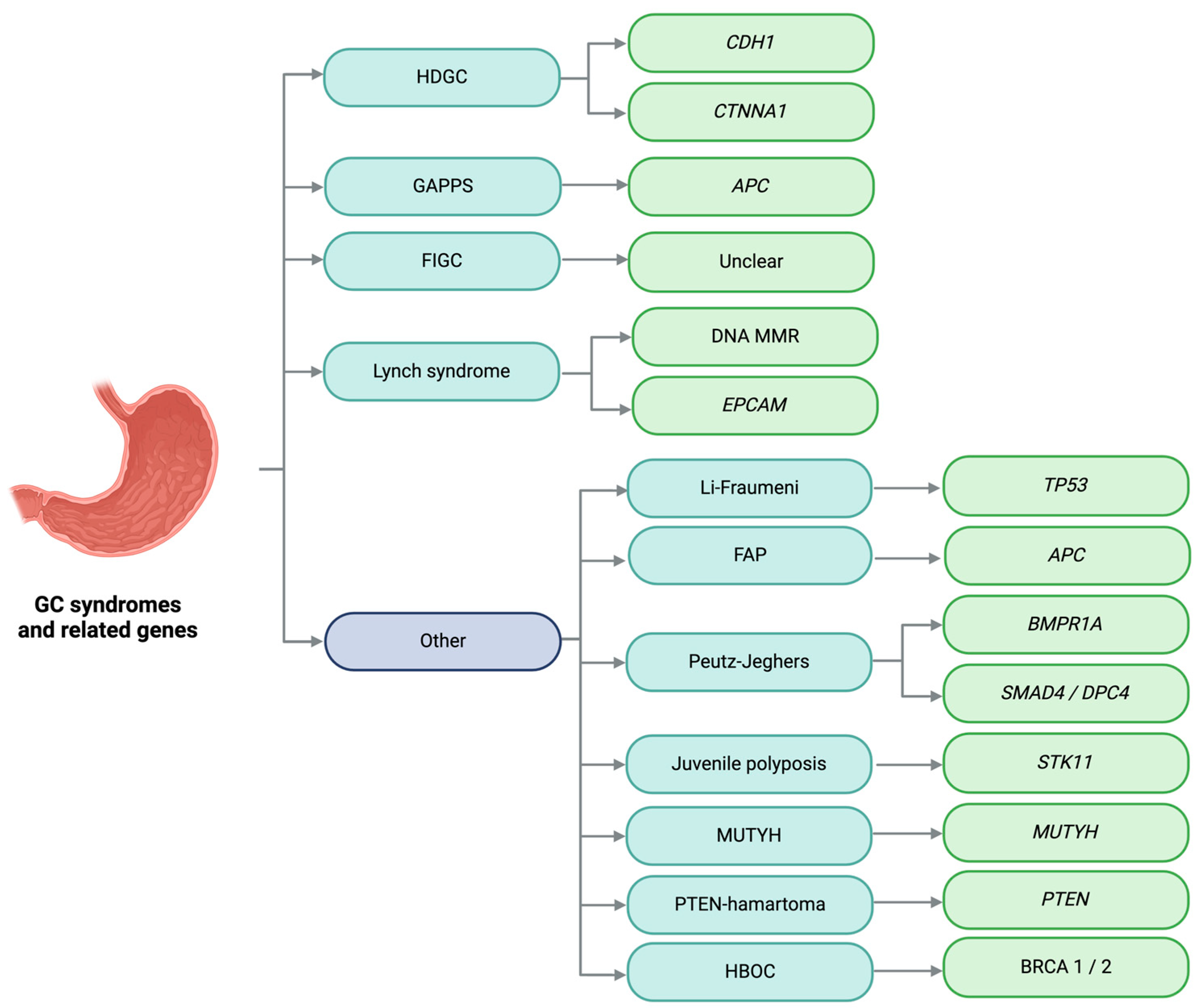
Figure 1. Hereditary gastric cancer syndromes and related genes. GC: gastric cancer; HDGC: hereditary diffuse gastric cancer; GAPPS: gastric adenocarcinoma and proximal polyposis of the stomach; FIGC: familial intestinal gastric cancer; FAP: familial adenomatous polyposis; MUTYH: MUTYH-associated polyposis, PTEN-hamartoma: PTEN hamartoma tumor syndrome; HBOC: hereditary breast and ovarian cancer; DNA MMR: DNA mismatch repair genes. Image created with Biorender.com (https://www.biorender.com/, accessed on 1 May 2024) under an individual license (Dr. Díaz del Arco).
Cimb 46 00385 g001
Figure 2. Laurén subtypes predominantly linked to hereditary gastric cancer syndromes. GC: gastric cancer; GAPPS: gastric adenocarcinoma and proximal polyposis of the stomach; FIGC: familial intestinal gastric cancer, HDGC: hereditary diffuse gastric cancer. Image created with Biorender.com (https://www.biorender.com/, accessed on 1 May 2024) under an individual license (Dr. Díaz del Arco).
Cimb 46 00385 g002
Figure 3. Adherens junction. E-cadherins have extracellular, transmembrane, and intracellular domains. They form homodimers, and the intracellular domain interacts with adaptor proteins such as p120 and catenins to connect with the actin cytoskeleton. Image created with Biorender.com (https://www.biorender.com/, accessed on 1 May 2024) under an individual license (Dr. Díaz del Arco).
Cimb 46 00385 g003
Figure 4. Activated canonical Wnt pathway. The Wnt protein binds to the extracellular domain of a Frizzled family receptor, alongside various co-receptors. Upon activation, it triggers a cytoplasmic cascade, leading to the accumulation of β-catenin, its translocation to the nucleus, and the transcription of target genes. Image created with Biorender.com (https://www.biorender.com/, accessed on 1 May 2024) under an individual license (Dr. Díaz del Arco).
Cimb 46 00385 g004
Figure 5. Physiological roles of p53. The p53 protein is a transcription factor that responds to various stimuli and, as a result, can induce cell cycle arrest, DNA repair, senescence, or programmed cell death, among other processes. Image created with Biorender.com (https://www.biorender.com/, accessed on 1 May 2024) under an individual license (Dr. Díaz del Arco).
Cimb 46 00385 g005
Figure 6. Main familial/hereditary syndromes of colorectal cancer. CRC: colorectal cancer; FAP: familial adenomatous polyposis. Image created with Biorender.com (https://www.biorender.com/, accessed on 1 May 2024) under an individual license (Dr. Díaz del Arco).
Cimb 46 00385 g006
Figure 7. Inactive canonical Wnt pathway. The absence of Wnt ligands leads to the phosphorylation of β-catenin by the destruction complex, comprising Axin, APC, and several kinases. This phosphorylated β-catenin is subsequently degraded by the proteasome. Additionally, without nuclear β-catenin, a repressive complex (TCF-TLE) inhibits gene transcription. Image created with Biorender.com (https://www.biorender.com/, accessed on 1 May 2024) under an individual license (Dr. Díaz del Arco).
Cimb 46 00385 g007
Disclaimer/Publisher’s Note: The statements, opinions and data contained in all publications are solely those of the individual author(s) and contributor(s) and not of MDPI and/or the editor(s). MDPI and/or the editor(s) disclaim responsibility for any injury to people or property resulting from any ideas, methods, instructions or products referred to in the content.