Article

A Novel Dhillonvirus Phage against Escherichia coli Bearing a Unique Gene of Intergeneric Origin

by
Anastasios Vasileiadis
1,2,
Petros Bozidis
2,
Konstantinos Konstantinidis
1,
Nikolaos Kesesidis
1,
Louiza Potamiti
3,
Anna Kolliopoulou
4,
Apostolos Beloukas
4,
Mihalis I. Panayiotidis
3,
Sophia Havaki
5,
Vassilis G. Gorgoulis
5,6,7,8,9,
Konstantina Gartzonika
2 and
Ioannis Karakasiliotis
1,*
1
Laboratory of Biology, Department of Medicine, Democritus University of Thrace, 68100 Alexandroupolis, Greece
2
Department of Microbiology, Faculty of Medicine, School of Health Sciences, University of Ioannina, 45332 Ioannina, Greece
3
Department of Cancer Genetics, Therapeutics & Ultrastructural Pathology, The Cyprus Institute of Neurology & Genetics, Nicosia 2371, Cyprus
4
Molecular Microbiology and Immunology Laboratory, Department of Biomedical Sciences, University of West Attica, 12243 Athens, Greece
5
Molecular Carcinogenesis Group, Department of Histology and Embryology, Medical School, National and Kapodistrian University of Athens, 11527 Athens, Greece
6
Biomedical Research Foundation, Academy of Athens, 11527 Athens, Greece
7
Ninewells Hospital and Medical School, University of Dundee, Dundee DD1 9SY, UK
8
Faculty Institute for Cancer Sciences, Manchester Academic Health Sciences Centre, University of Manchester, Manchester M20 4GJ, UK
9
Faculty of Health and Medical Sciences, University of Surrey, Surrey GU2 7YH, UK
*
Author to whom correspondence should be addressed.
Curr. Issues Mol. Biol. 2024, 46(9), 9312-9329; https://doi.org/10.3390/cimb46090551
Submission received: 24 July 2024 / Revised: 19 August 2024 / Accepted: 21 August 2024 / Published: 23 August 2024
(This article belongs to the Section Molecular Microbiology)

Abstract

:
Antibiotics resistance is expanding amongst pathogenic bacteria. Phage therapy is a revived concept for targeting bacteria with multiple antibiotics resistances. In the present study, we isolated and characterized a novel phage from hospital treatment plant input, using Escherichia coli (E. coli) as host bacterium. Phage lytic activity was detected by using soft agar assay. Whole-genome sequencing of the phage was performed by using Next-Generation Sequencing (NGS). Host range was determined using other species of bacteria and representative genogroups of E. coli. Whole-genome sequencing of the phage revealed that Escherichia phage Ioannina is a novel phage within the Dhillonvirus genus, but significantly diverged from other Dhillonviruses. Its genome is a 45,270 bp linear double-stranded DNA molecule that encodes 61 coding sequences (CDSs). The coding sequence of CDS28, a putative tail fiber protein, presented higher similarity to representatives of other phage families, signifying a possible recombination event. Escherichia phage Ioannina lytic activity was broad amongst the E. coli genogroups of clinical and environmental origin with multiple resistances. This phage may present in the future an important therapeutic tool against bacterial strains with multiple antibiotic resistances.

1. Introduction

Phages are viruses that can infect bacteria [1]. Bacteriophages are found everywhere throughout the environment (e.g., in oceans, drinking water and food we consume). They are found in large numbers, estimated to be approximately 1031 in total [2]. In addition, bacteriophages have a very important role in the regulation of the microbial balance in each ecosystem studied [3].
Due to their great diversity, bacteriophages have many niche applications in the food industry [4,5], biotechnology [6] and medicine [7]. In recent years, it has been shown that the use of bacteriophages in combination with antibiotics and disinfectants can break down biofilms and dramatically enhance the reduction of the bacteria load [8]. As the prevalence of antibiotic resistance is increasing worldwide, phage therapy is a promising alternative treatment modality [9,10]. Τhe importance of the identification of new phages is highlighted by the recent advances on the phage virome (phageome) and its balance with the host microbiome in patients and healthy individuals [11]. The dissection of the vast phage ecosystem showcased phages that infect not only pathogenic bacteria but also symbiotic commensal flora [12]. Such phages may regulate the abundance and function of important bacteria for mucosal homeostasis and metabolism [13].
Escherichia coli (E. coli), one of the most prevalent bacteria in the human gut, is a Gram-negative bacterium which belongs to the family of Enterobacteriaceae and plays an important role in the formation of the intestinal microbiome. E. coli strains are classified into more than 180 O-antigen serotypes [14]. Each serotype has distinct attributes and may differentially affect mucosal homeostasis [15]. ATCC 25922 is a strain of E. coli representative of serotype O6 and biotype 1 and has been characterized as part of the non-pathogenic symbiotic commensal flora. ATCC 25922 and other symbiotic bacteria may exert immunomodulatory effects on inflammatory conditions such as the allergic airway inflammation [16]. In addition, ATCC 25922 has been widely used as a reference strain in a plethora of quality control and for antibiotic susceptibility testing [17].
In the present study, we sought to isolate and characterize lytic bacteriophages against E. coli from raw sewage of a tertiary hospital, before biological treatment. A novel lytic bacteriophage belonging to a genetically distinct branch of Dhillonviruses was isolated and characterized both physically and genetically.

2. Materials and Methods

2.1. Bacterial Strains and Growth Conditions

Mueller–Hinton broth (MHB) (2 g/L of beef infusion solids, 1.5 g/L of starch and 17.5 g/L of casein hydrosylate) was used for the bacterial culture. E. coli 25922 (Becton Dickinson, France S.A.S.) strain was stored in MHB supplemented with 50% glycerol at −80 °C. E. coli 25922 was grown in MHB at 37 °C with vigorous rotary shaking at 250 rpm.

2.2. Sample Collections

Sewage wastewater samples were collected from the input of the wastewater treatment plant of the University Hospital of Ioannina, Epirus, Greece. Specifically, 16 samples were collected for a time span of 4 months (February–May) in 2018.

2.3. Phage Isolation and Enrichment

Totally, 60 mL of 6 different sewage samples were centrifuged at 1100× g for 15 min at room temperature, and the supernatant was filtered through a 0.2 μm membrane filter to remove bacterial debris. Phages were concentrated using the PEG method. Final concentrations of 10% PEG 8000 and 1 M of NaCl were added, and the filtered sample was incubated overnight at 4 °C for phage precipitation. The following day, the filtered sample was centrifuged at 3000× g for 1 h and 30 min at room temperature, and the pellet was resuspended in 2 mL of SM buffer (50 mM of Tris-Cl pH 7.5, MgSO4. 7 H2O 8 mM, NaCl 100 mM). Bacteriophage stocks were then stored at 4 °C. For phage enrichment, 100 μL of the bacteriophage stock was mixed with 1 mL of overnight-grown E. coli 25922 strain, 4 mL of medium culture (MHB) and 50 μL of 1 M CaCl2.

2.4. Bacteriophage Plaque Assay

A total of 100 μL of bacteriophage enrichment stock was diluted into the MHB with serial 10-fold dilutions from 10−1 up to 10−6. Briefly, 100 μL of each dilution were mixed with 400 μL of the E. coli 25922 strain culture in the logarithmic growth phase (OD600nm = 0.4) and 35 μL of 1 Μ CaCl2. The mix was incubated at 37 °C for 8 min. Then, 3 mL of molten soft agar (0.8% agar and 1% MHB) was added and poured onto 1.5% Mueller–Hinton agar plates, and the plates were incubated overnight at 37 °C. The next day, plaques were observed and counted.

2.5. Phage DNA Extraction

Single plaques were picked with a tip and inoculated in a mix of 1 mL of E. coli 25922 strain culture in the logarithmic growth phase, 2 mL of ΜHΒ and 30 μL of 1 Μ CaCl2. The enrichment was incubated overnight at 37 °C. Subsequently, chloroform was added on the enrichment culture at a final concentration of 0.2% (v/v). The sample was then centrifuged at 2000× g for 20 min at room temperature. Supernatant (1.8 mL) was treated with DNase I (1 μg/mL) and RNase A (12.5 μg/mL) at 37 °C for 30 min. Then, the phage enrichment was treated with 46 μL of 20% SDS and 18 μL of Proteinase K (10 mg/mL) and incubated at 56 °C for 30 min. Phenol-chloroform extraction and isopropanol precipitation using sodium acetate (3 M, pH 5.2) were used for DNA purification. Finally, DNA was resuspended in 50 μL 1% TE (Tris 10 mM, EDTA 1 mM, pH 7.4) and stored at −20 °C.

2.6. Genome Sequencing

Genomic DNA libraries were prepared by using an Ion SingleseqTM 96 Kit (#A34763, ThermoFischer Scientific, CA, USA) according to the manufacturer’s instructions. The concentration of the library was measured using a QubitTM 4 Fluorometer (ThermoFischer Scientific, Eugene, OR, USA) and was loaded on an Ion 540TM chip. Sequencing was performed on an Ion GeneStudio S5 System (ThermoFischer Scientific, CA, USA). The raw sequencing datasets for the current study are available in the NCBI Sequence Read Archive repository, under the Bioproject with accession number PRJNA941078 (NCBI BioProject database, https://www.ncbi.nlm.nih.gov/bioproject/PRJNA941078—last accessed date 13 March 2023).

2.7. Phage Genome Assembly and Characterization

Following the NGS procedure, quality trimmed reads were used as input for a de novo assembly using Trinity (v2.8.5) [18]. The generated contigs were aligned against the non-redundant (nr) nucleotide and protein databases by using BLASTn and BLASTx [19], respectively, in order to be annotated. Nucleotide sequences corresponding to the same BLAST hit were fed into the CAP3 tool [20], using default parameters, in order to generate assembly scaffolds. The assembled genomic sequence of the phage identified in this study is available in the NCBI Nucleotide repository (NCBI Nucleotide database, https://www.ncbi.nlm.nih.gov/nuccore—last, accessed date 13 March 2023) and can be accessed online using the GenBank accession number OQ589852. CDS analysis was carried out by using the SnapGene 6.0.2 tool (SnapGene, GSL Biotech LLC, Boston, USA). CDSs were individually annotated using the BLASTp tool [19]. The tRNAscan-SE v. 2.0 tool was used to find possible tRNA genes in the whole genome of the phage [21]. Genome organization analysis of the identified phage was performed via EasyFig v. 2.2.5 [22], including representative highly similar phages from the same taxon after BLASTn of the assembled genomic sequence.

2.8. Transmission Electron Microscopy (TEM) Methodology

The negative staining technique was applied for examining phages by TEM. Specifically, 5 μL of sample was allowed to be absorbed for 2 min to the surface of a Formvar/Carbon-coated copper grid. The used grids were placed previously in a glow discharge unit to render them hydrophilic. After absorption, each grid was blotted with a filter paper, washed thrice on drops of ultrapure water and placed on a drop of 2% aqueous uranyl acetate (UA) solution for 1 min. The excess UA was removed and the grids were left to air dry. Phages were then examined under a JEM 2100 Plus Transmission Electron Microscope (Jeol, Tokyo, Japan) operating at 120 kV and photographed with a Gatan OneView digital camera (Gatan, Inc., Pleasanton, CA, USA).

2.9. One-Step Growth Curve and Adsorption Assay

The latency period and burst size of isolated phage were determined by observing changes in the number of phage particles during a lytic cycle as described [23]. Briefly, host strain E. coli 25922 was grown at 37 °C until log phase (OD600 = 0.5, 108 CFU/mL). Then, 990 μL of bacterial grown culture was mixed with 10 μL of phage suspension (108 PFU/mL) to achieve a multiplicity of infection of 0.01. The mixture was incubated for 10 min at 37 °C and then centrifuged at 16,200× g for 5 min. The pellet was washed with 1 mL of MHB to remove the unabsorbed phages. This process was repeated once more, and the pellet was resuspended in 10 mL of MHB and incubated at 37 °C with shaking. Aliquots of 500 μL were collected at 10 min intervals for 2 h, and then in every sample was added 1% chloroform, followed by centrifugation at 13,800× g for 2 min. The supernatant was immediately diluted and plated by using the soft agar assay method for phage titers’ determination. The latent period was determined as the time between infection and the shortest incubation time, allowing a phage particle to reproduce inside an infected host cell. The burst size was calculated as the ratio between the number of phage particles produced during a lytic cycle and the initial infected bacterial cells. Experiments for one-step phage growth curve were carried out in triplicates.
Adsorption assay was performed to evaluate the efficiency of isolated phage to ab-sorb the host strain E. coli 25922. Briefly, 990 μL of log-phase culture (108 CFU/mL) was mixed with 10 μL of diluted phage suspension (107 PFU/mL), and the mixture was incubated at 37 °C for 10 min. Subsequently, the mixture was centrifuged at 16,200× g for 5 min, and the titer of the supernatant was estimated by using the soft agar assay method. The phage adsorption efficiency was determined with the equation (initial phage titer—residual phage titer in the supernatant/initial phage titer).

2.10. Thermal and pH Stability

To test the thermotolerance of the phage, 100 μL of a 10−6 dilution of the enrichment was tested using the soft agar assay method at 37, 40, 45, 50, 55 and 60 °C for 1 h in a thermal cycler. Τo test the stability of the phage, in different pH values (2, 4, 6, 8 and 10), sodium acetate (1 M) was prepared. A 100 μL quantity from the phage enrichment was mixed with 900 μL of sodium acetate (1 M) at each different pH value and incubated at 37 °C for 1 h. Afterwards, serial 10-fold dilutions were carried out, and 100 μL from dilution 10−6 was tested for phage titer determination using the soft agar assay method. Experiments for thermal and pH stability were carried out in triplicates.

2.11. Host Range Determination

For the host range determination, spot test was used to evaluate the lytic ability of the bacteriophage to form lytic plaques. The Escherichia phage Ioannina was tested against 47 E. coli isolates deriving from different environments (hospital waste water, wastewater treatment plant, river water), as well as clinical strains. Additionally, the phage isolate was tested against 9 reference strains and a clinical strain (E. coli O157:H7). Forty-seven E. coli isolates were representatives of all 4 phylogenetic groups (i.e., A, B1, B2 and D) according to Clermont’s schema and with a variety of AMR profile (from WT to MDR) [24,25]. Briefly, bacteria maintained as glycerol stocks at −80 °C were subcultured directly in Nutrient Agar (for pure isolates), or first in MacConkey Agar and subsequently in Nutrient Agar (to verify purity of the isolate). Bacterial colonies were used to prepare suspensions in 0.9% NaCl according to the MacFarland scale of 1–5. Then, suspensions were used for the spot assay, where bacteria were first spread on separate Nutrient Agar plates, and then 1 μL of the phage preparation (in LB, maintained at 4 °C) was added at the center of each plate. Spot assay results were evaluated on the next day. Phage lytic activity was shown by the appearance of visible lytic plaques at the site of enrichment application [26].

2.12. Phylogenetic Analysis

The phylogenetic analysis of the assembled phage was based on the amino acid sequences of three proteins, namely the major capsid protein, the large terminase subunit protein and the portal protein, which were used for the construction of the corresponding phylogenetic trees as in recently published literature [27,28,29,30]. Each amino acid sequence was used as input to BLASTp [19] against the non-redundant (nr) protein sequence database of NCBI with the “Max target sequences” parameter set to 1000. BLASTp hits with not less than 30% coverage and 30% identity were selected in order to download their corresponding and complete protein sequences in FASTA format. Taxonomy data of the phages that encoded the retrieved protein sequences were also fetched utilizing the NCBI tool Entrez-direct [31], and subsequently, the protein sequences were taxonomically filtered to include only a few phage entries as representatives from different taxa. All sequences were deposited to a FASTA formatted file and were successively input to MEGA11 software (v11.0.11) [32], for the elucidation of phylogenetic relationships. Lastly, the phylogenetic reconstruction was performed using the Maximum Likelihood (ML) method [33] for 100 bootstrap replications [34], and the inferred trees were exported via MEGA11. The same procedure, as described above, as well as alignments by MUSCLE (v3.8) [35] were performed for the phylogenetic analysis of a unique CDS28 (putative tail fiber protein). The VIRIDIC tool was used to calculate and visualize the isolated phage intergenomic relatedness using default parameters [36].

3. Results

In order to isolate and characterize lytic bacteriophages against E. coli ATCC 25922, sewage samples were collected from the Ioannina hospital wastewater treatment plant. Multiple passages of the inoculum were used for the enrichment of bacteriophages against Ε. coli ATCC 25922. Only samples that showed marked reduction in culture medium turbidity were further processed. Soft agar assays were carried out in order to assess the efficiency in plaque formation of bacteriophages, and single-plaque isolates were subsequently cultured on E. coli ATCC 25922.

3.1. Genome Characterization

Full-genome sequencing of the isolated bacteriophages was carried out using the Ion Torrent Next-Generation Sequencing (NGS) technology. The output NGS reads were assembled using the Trinity assembly (v2.8.5) software. The analysis showed that all samples yielded sequences of the same bacteriophage, named Escherichia phage Ioannina, which was a 45,270 bp linear double-stranded DNA molecule (Figure 1). The GC content of the identified bacteriophage was 53.62%. Using the SnapGene 6.0.2 tool, we predicted the presence of 61 putative coding sequences (CDSs), while no tRNA genes were found. Annotation of all predicted CDSs was performed by BLASTp tool, which indicated that 38 genes were on the forward and 23 were on the reverse strand (Figure 1, Supplementary Table S1). The nucleotide identity of the full-length genome compared to other closely related Dhillonvirus genomes reached up to 72.7% (Supplementary Table S2). Notably, the nucleotide identity of the full-length genome was closer (77.75%) to a phage metagenome (CtFRY1, NCBI GenBank accession number BK032676.1); however, no other details, besides the nucleotide sequence, could be retrieved for this GenBank entry.

3.2. Genome Organization

The CDS analysis (SnapGene 6.0.2) of the complete Escherichia phage Ioannina genome showed six distinct functional clusters, namely: (i) DNA replication, modification and transcriptional regulations: Replicative DNA helicase (CDS48), DNA polymerases (CDS39 and CDS60), DNA N-6-adenine methyltransferases (CDS30 and CDS32), Nucleotide modification-associated domain 5 (CDS6), Cytosine specific methyltransferase (CDS35), (ii) Head structure: Portal protein (CDS3), minor capsid protein (CDS4), minor structural protein (CDS7) and major capsid protein (CDS8), (iii) Packaging: DNA terminases (small subunit = CDS1 and large terminase = CDS2), mature oligodendrocyte transmembrane protein (CDS42), Putative Head Tail Connector Protein (CDS9) and Head Tail Attachment (CDS10), (iv) host lysis: Putative holin-like class II protein (CDS56), Putative holin-like class I protein (CDS57) and Lysozyme (CDS58), (v) tail structure: tail fiber proteins (CDS26 and CDS28), tail protein (CDS23), tail assembly proteins (CDS14 and CDS22), tail component (CDS11), tail completion protein (CDS12), tail tube protein (CDS13), major tail protein (CDS15), minor tail proteins (CDS19 and CDS20) and tail length tape measure protein (CDS18), (vi) hypothetical or unknown functions. A synteny plot of the Escherichia phage Ioannina genome against representative highly similar Dhillonvirus phages after BLASTn (Supplementary Table S2) showed that all these phages have nearly the same genome organization with the exception of some minor differences (Supplementary File S1). While all five phages seem to have the same genes encoding tail fiber proteins, one major difference is the presence of an additional putative tail protein (CDS28) on Escherichia phage Ioannina genome in contrast with the other representative Dhillonvirus phages.

3.3. Bacteriophage Plaque Formation and Morphology

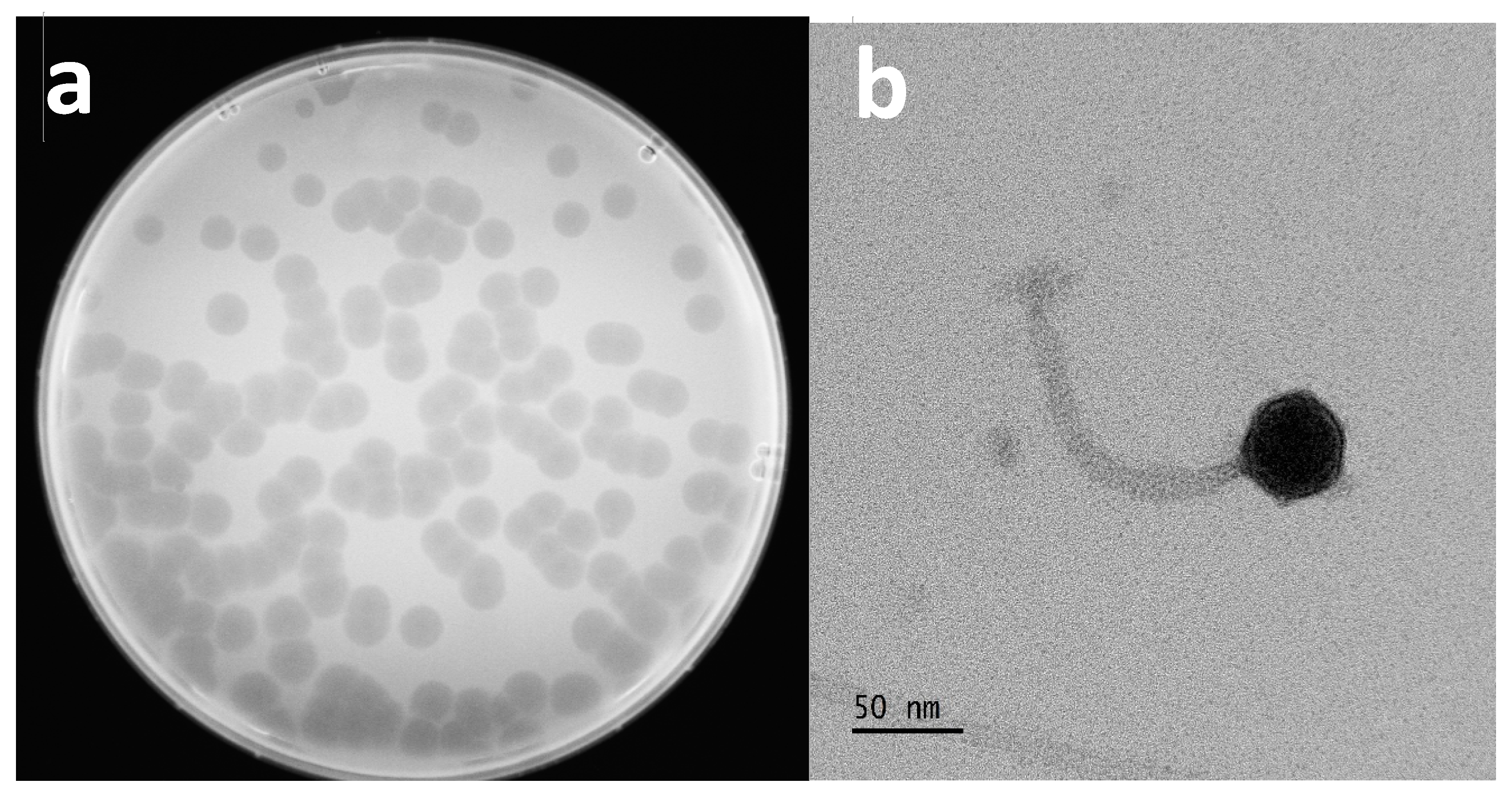
Following genomic characterization, we further analyzed the physical properties of the Escherichia phage Ioannina. By using the soft agar assay method, clear plaques of the bacteriophage were visible and uniform, showing a particularly potent lytic activity. The average size of the plaques was 5.3 ± 0.5 mm 16 h post infection (Figure 2a).
Transmission electron microscopy (TEM) analysis showed that the Escherichia phage Ioannina consisted of a 37 ± 3 nm diameter icosahedral head and a long non-contractile tail 122 ± 8 nm long (Figure 2b). Based on the morphological characteristics, the phage could be classified as a siphovirus according to the International Committee on Taxonomy of Viruses (ICTV).

3.4. One-Step Growth Curve and Adsorption Assay

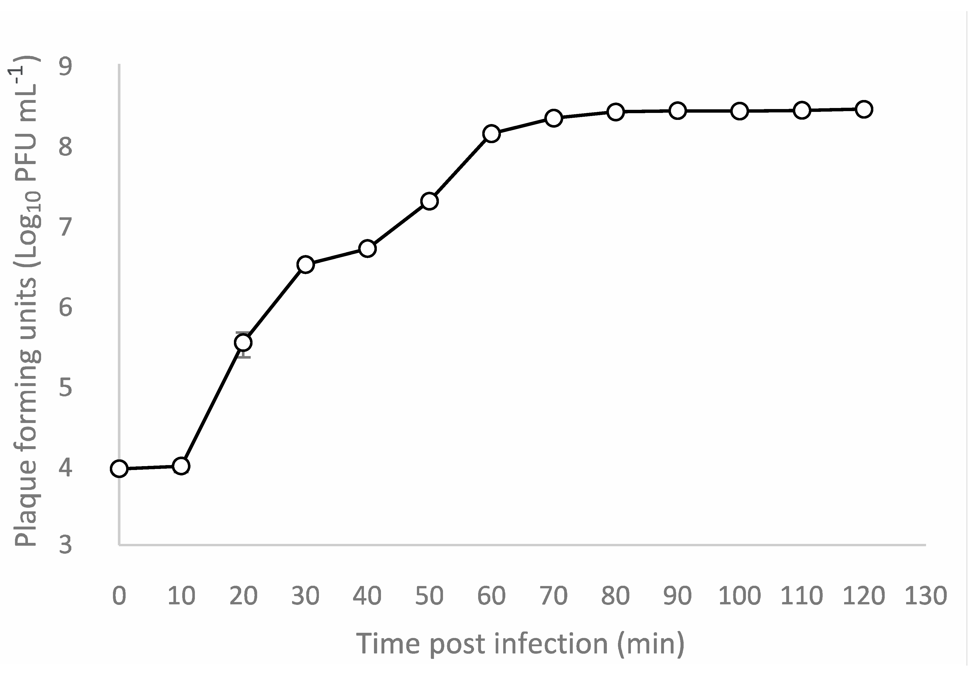
One-step growth experiment was performed to determine the latent period and the burst size of Escherichia phage Ioannina on host strain E. coli ATCC 25922. The latent period was 10 min, and the burst size was about 316 plaque-forming units (pfu) per infected cell (Figure 3). Adsorption efficiency of Escherichia phage Ioannina on host strain E. coli ATCC 25922 was approximately 99.83%.

3.5. Thermal and PH Stability of the Phage

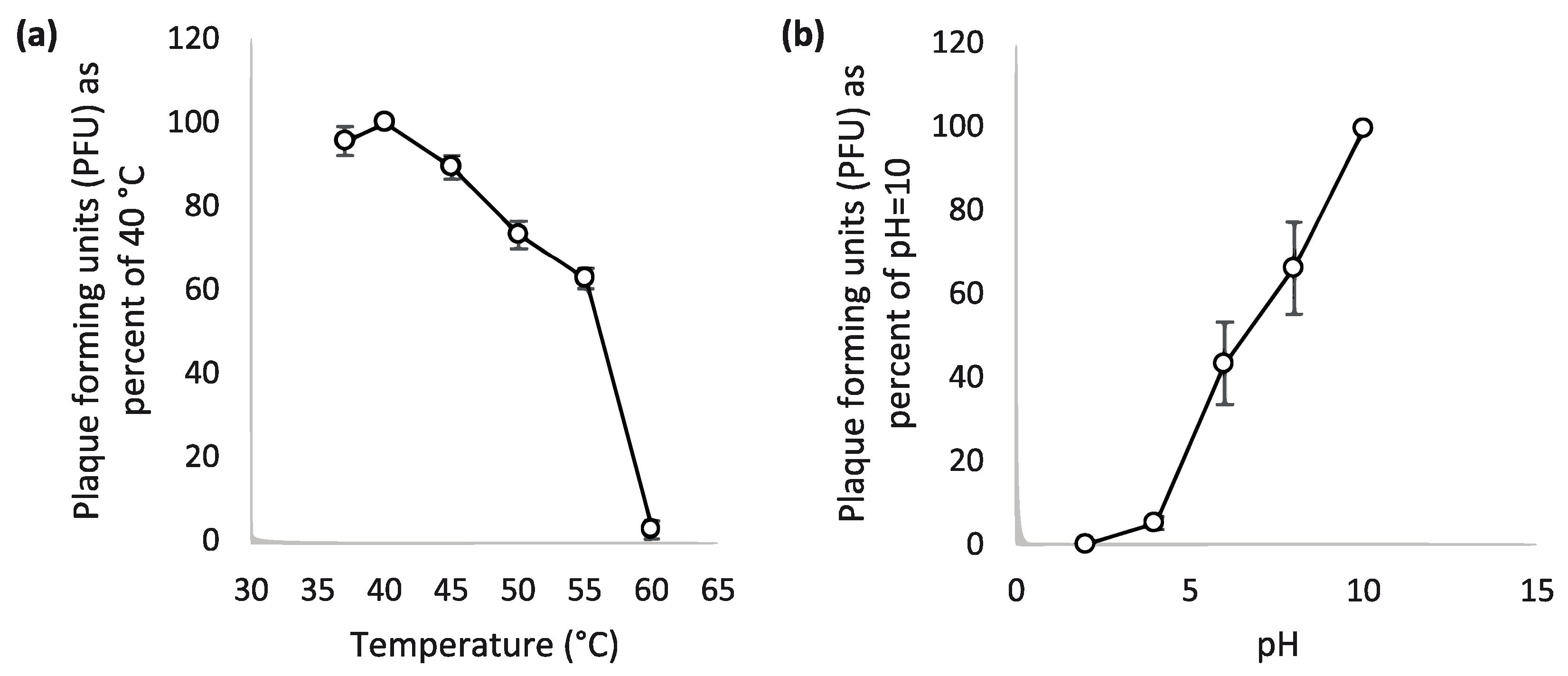
Temperature and pH are two important factors for the survival of a bacteriophage. For this reason, the stability of Escherichia phage Ioannina at different pH values (2, 4, 6, 8, 10) and temperatures (37, 40, 45, 50, 55, 60 °C) was determined (Figure 4a). Heat treatment for 1 h at 45–55 °C presented a gradual reduction of about 40% in phage viability as measured by its plaque-forming ability. Heat treatment at all temperatures greater than 40 °C diminished phage viability. Phage viability is eliminated at 60 °C. Τhe phage also showed highly lytic activity in alkaline pH. More specifically, the phage viability showed a constant reduction towards the acidic end of the pH spectrum (Figure 4b).

3.6. Host Range Determination

Host range determination was carried out by testing the identified phage against 47 E. coli isolates, nine reference strains and a clinical strain E. coli O157:H7. Host range testing revealed that Escherichia phage Ioannina was highly specific. The Escherichia phage Ioannina lysed the reference strain E. coli ATCC 25922, and 11 E. coli isolates derived from both environmental and clinical samples. Regarding the genotypes of E. coli strains susceptible to infection, they were found in all of four phylogenetic groups (A, B1, B2 and D). Escherichia phage Ioannina successfully infected several MDR E. coli clinical isolates. Further, Escherichia phage Ioannina could not infect the other tested E. coli strains and strains from other species. (Table 1).

3.7. Phylogenetic Analysis

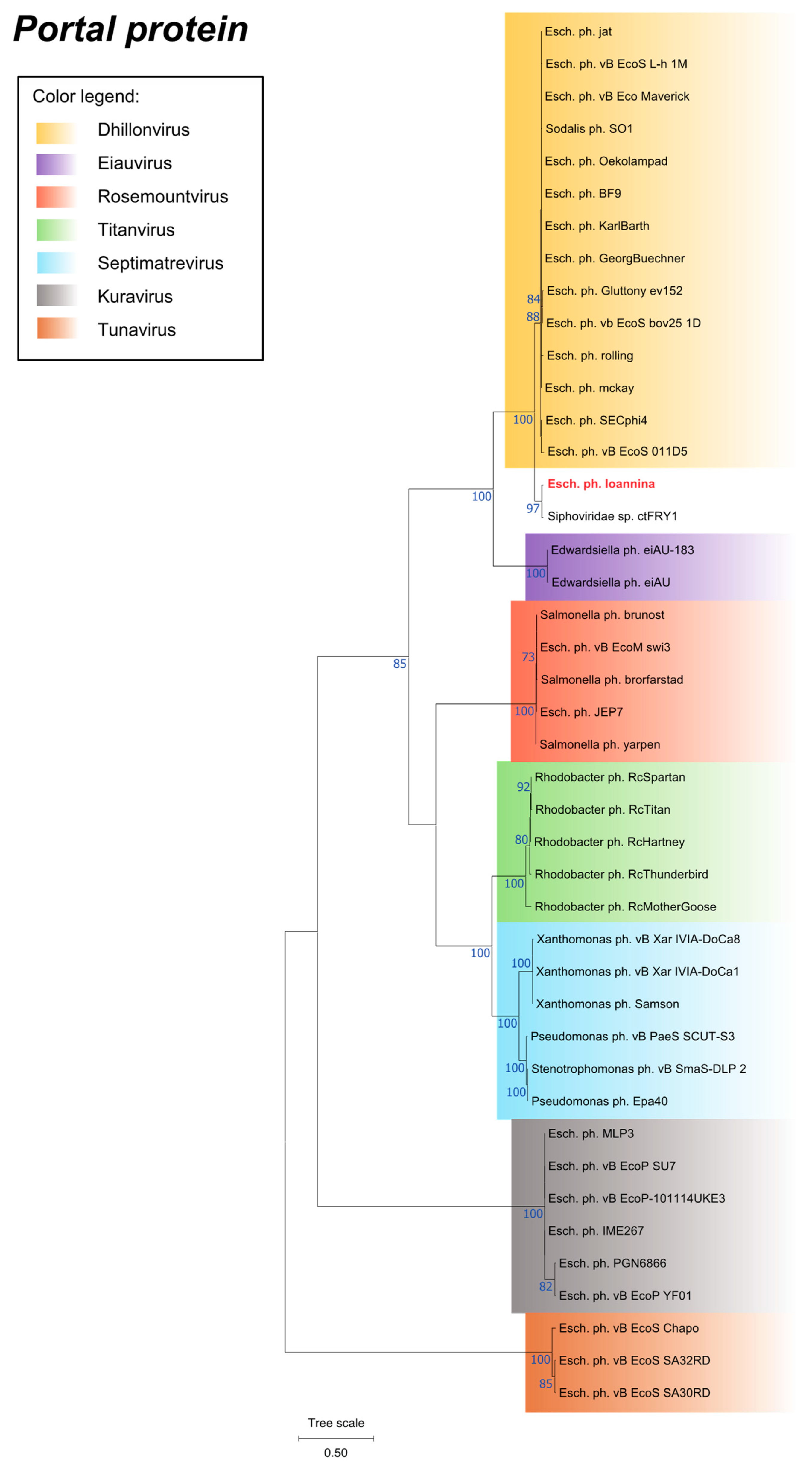
Phylogenetic analysis was performed by utilizing the MEGA11 program and was mainly based on the portal protein of Escherichia phage Ioannina and other similar phages (Figure 5). Moreover, two additional proteins, namely the large terminase subunit protein and the major capsid protein, were used to verify the phylogeny of Escherichia phage Ioannina (Supplementary Figures S1 and S2). According to the phylogenetic analysis, Escherichia phage Ioannina can be classified as a siphovirus, forming a distinct cluster within the Dhillonvirus genus, which contained only the CtFRY1 metagenome (Figure 5, Supplementary Figures S1 and S2). The classification within the Dhillonvirus genus was further supported by calculation of the intergenomic similarities and distances amongst similar phages by utilizing the VIRIDIC tool (Supplementary Figure S3).

3.8. Phylogenetic Analysis of a Unique Putative Tail Fiber Protein

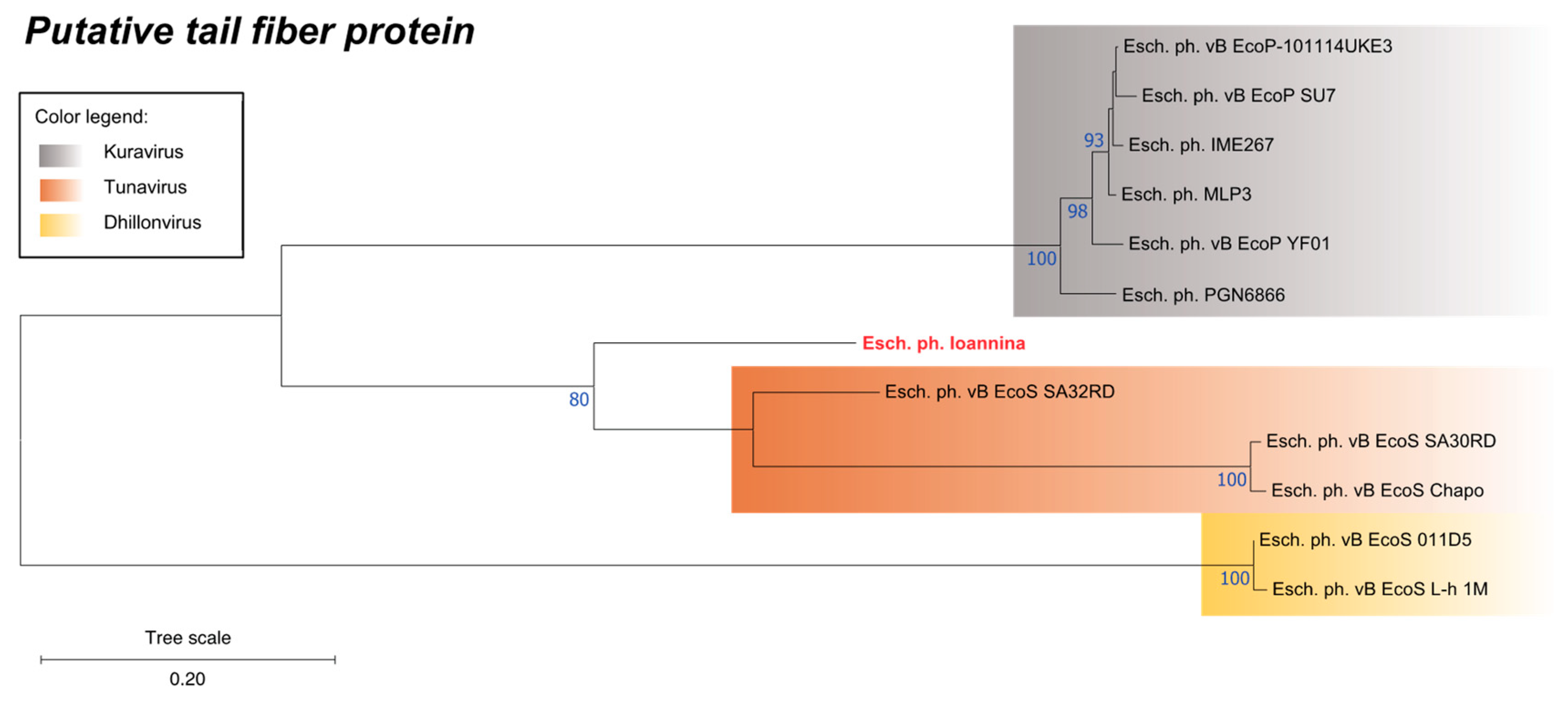
The analysis of coding sequences of the Escherichia phage Ioannina showed the presence of a CDS (CDS28) that was not present in most of the studied members of the Dhillonvirus genus (Table 2). Construction of a phylogenetic tree that encompassed homologous proteins (or predicted proteins), from related phage families, revealed a closer relationship of CDS28 with members of the Kuravirus or the Tunavirus (Drexlerviridae) genera, which was additionally visualized via MUSCLE v. 3.8 alignment (Figure 6, Supplementary file S2). It is intriguing that similar CDSs in the Dhillonvirus genus, where Escherichia phage Ioannina is predicted to belong, are more distantly related to CDS28 (Figure 6).

4. Discussion

The focus of the bacteriophage research has in the past decade been either on the isolation and characterization of bacteriophages against pathogenic and multidrug resistant bacterial strains, or on the analysis of phageome from human or environmental samples [37,38]. A definite host–virus relationship has been reported only for a minority of phage metagenomes [39]. Moreover, our knowledge on host–virus relationships involving commensal bacteria (important for mucosal homeostasis) is even more limited [40]. Importantly, phage therapy is a promising alternative for the combat against multidrug-resistant strains as our antibiotics arsenal is significantly losing its potential [41]. Phages for phage therapy have been isolated from the environment [42,43,44,45]. As the host range of phages varies, phage therapy requires the creation of phage cocktails for the prompt treatment of diseases, caused by MDR bacteria. Phage cocktails may also overcome the potential emergence of phage resistance during treatment [46]. The combination of antibiotics and phage therapy has been shown to be more effective in treating serious bacterial infections, than mono-phage therapy [47].
In this study, we isolated and characterized a lytic bacteriophage from biological wastewater treatment from the University Hospital of Ioannina, Epirus, Greece, using the strain E. coli ATCC 25922 as the host bacterium. The identified phage was lytic against E. coli ATCC 25922, as well as for a variety of E. coli isolates of clinical or environmental origin. Based on the morphological characteristics (e.g., long non-contractile tail and icosahedral head), the novel phage, namely Escherichia phage Ioannina, was classified into siphoviruses of Caudoviricetes. This classification was further refined by whole-genome sequencing of the isolated virus, and the virus was found to belong to the Dhillonvirus genus. As the similarity with the other members of Dhillonvirus genome is marginal according to ICTV demarcation criteria, there is a possibility that the novel virus belongs to a novel genus. Similar phages from hospital sewage have been isolated in the past, infecting E. coli pathogenic strains 40371 (genus Cornellvirus) and O18 (genus Dhillonvirus) [48,49]. Lytic bacteriophages infecting pathogenic E. coli O157:H7 (genus Kuttervirus) and 6 clinical E. coli isolates (genus Tequatrovirus), belonging to Ackermannviridae and myoviruses, respectively, have also been isolated from hospital wastewater [50,51].
Phages within the Dhillonvirus genus, infecting various E. coli strains, have been isolated from different sources of environmental, animal or human origin. Specifically, a previous report indicated that four different phages, which belong to the Dhillonvirus genus, were isolated from wastewater treatment plants against E. coli K-12 MG1655 [52]. Also, phages belonging to the Dhillonvirus genus have been isolated from fecal samples of healthy cattle [53], pig farm [54], slurry of birds’ feces [55] and fresh goat fecal samples [56]. Escherichia phage Gluttony_ev152 (the phylogenetically closest relative of Escherichia phage Ioannina) was isolated from feces of children (LR597646). Finally, a phage belonging to the Dhillonvirus genus has been isolated from a possibly contaminated culture of the E. coli BL21 (DE3) laboratory strain [57].
Temperature and pH are two important factors for phage viability. Escherichia phage Ioannina was tested at different thermal and pH values, in order to evaluate its lytic activity. Thermal stability tests showed that the Escherichia phage Ioannina was stable up to 40 °C with a gradual drop of activity between 40 and 55 °C and almost complete inactivation at 60 °C. In addition, it indicated a similar thermal stability between 37 and 55 °C compared to other phages of the member of siphoviruses [48,58]. Escherichia phage Ioannina viability testing, at different pH values, showed higher stability at alkaline pH (pH 10). On the other hand, the phage was sensitive to lower pH values (pH < 4). This finding was in agreement with earlier reports, identifying several members of siphoviruses resistant to alkaline pH while sensitive to acidic pH [59,60,61].
Comparative genomic analysis revealed that the Escherichia phage Ioannina indicated the highest nucleotide similarity with the “Siphoviridae sp. CtFRY1 metagenome” (BK032676.1). Phylogenetic analysis for three annotated proteins (portal protein, large terminase subunit protein and major capsid protein) showed that the CtFRY1 partial genome formed a distinct cluster within the Dhillonvirus genus. As Escherichia phage Ioannina presented similarity close to 70% (nucleotide identity of the full length genome) compared to other representatives of the Dhillonvirus genus, it is anticipated that the identification of more related phages, in the future, will lead into the emergence of a novel genus or subgenus [62].
Finally, in an attempt to identify the source of the similarity divergence between the Escherichia phage Ioannina and the other representatives from the Dhillonvirus genus, we characterized a gene (CDS28) absent in the vast majority of the species within the genus. CDS28 is predicted to encode a putative tail fiber protein. Tail fiber proteins are located at the tip of the tail and are responsible for the phage binding to a specific receptor present on the bacterial cell surface, such as lipopolysaccharides (LPS), teichoic acids and organelles [63]. Tail fiber proteins determine the host range during infection process [64]. Changes in tail fiber proteins have been shown to lead to a change in phage specificity towards the species of bacteria it infects [65].
Phylogenetic analysis of this protein, from CDS28, revealed that this gene showed a higher similarity with the Kuravirus genus of podoviruses (72.0–74.2%) and the Tunavirus genus of the family Drexlerviridae (47.0–72.5%). Interestingly, the similarity of this putative protein, with other related proteins in the Dhillonvirus genus, was only 37.8–38.7%. This observation may indicate a possible recombination event with either Kuravirus or Tunavirus (Drexlerviridae). Recombination in tail fiber protein genes have been observed much more frequently than in other regions of phage genomes, suggesting adaptive pressure towards switch of phage specificity as these proteins are involved in the interaction with the host cell [66]. In another study, representatives of Litunavirus genus (Schitoviridae) were analyzed, and the putative tail fiber region was shown to be a hotspot of recombinations with multiple phages species incorporating this region from other genera [67].
In conclusion, this study isolated and characterized a lytic bacteriophage against E. coli ATCC 25922 that is also efficiently lytic against a variety of E. coli isolates of clinical or environmental origin. Genomic analysis of the phage revealed that this novel phage, namely Escherichia phage Ioannina, belongs to a distant cluster of Dhillonviruses with a unique CDS encoding for a putative tail fiber protein. There is evidence that this CDS was incorporated through a recombination event from a different genus of phages. Escherichia phage Ioannina, due to its fast and potent lytic activity, may be used as a therapeutic tool against MDR E. coli strains, either alone or within a phage cocktail. Escherichia phage Ioannina, lacking genes encoding known virulence factors or providing antibiotic resistance, serves as a good candidate for further clinical research as use.

Supplementary Materials

The following supporting information can be downloaded at: https://www.mdpi.com/article/10.3390/cimb46090551/s1. Figure S1. Phylogenetic tree of the Escherichia phage Ioannina terminase large subunit protein constructed using the Maximum-Likelihood method of the MEGA11 software. The “Esch. ph. Ioannina” represents the Escehrichia phage Ioannina terminase large subunit protein. Bootstrap values (blue coloured text) were obtained from 100 bootstrap replicates and only those above 70 are displayed next to each node. Tree scale is displayed on the bottom left corner of the phylogenetic tree. Figure S2. Phylogenetic tree of the Escherichia phage Ioannina major capsid protein constructed using the Maximum-Likelihood method of the MEGA11 software. The “Esch. ph. Ioannina” represents the Escherichia phage Ioannina major capsid protein. Bootstrap values (blue coloured text) were obtained from 100 bootstrap replicates and only those above 70 are displayed next to each node. Tree scale is displayed on the bottom left corner of the phylogenetic tree. Figure S3. Heatmap generated by VIRIDIC tool incorporating intergenomic similarity values (right half) and alignment indicators (left half and top annotation). In the right half, the color-coding allows a rapid visualization of the clustering of the phage genomes based on intergenomic similarity: the more closely-related the genomes, the darker the color. The numbers represent the similarity values for each genome pair, rounded to the first decimal. In the left half, three indicator values are represented for each genome pair, in the order from top to bottom: aligned fraction genome 1 (for the genome found in this row), genome length ratio (for the two genomes in this pair) and aligned fraction genome 2 (for the genome found in this column). The darker colors emphasize low values, indicating genome pairs where only a small fraction of the genome was aligned (orange to white color gradient), or where there is a high difference in the length of the two genomes (black to white color gradient). The aligned genome fractions are expected to decrease with increasing the distance between the phages. Therefore, darker colors should correspond to genome pairs with low similarity values, and whiter colors to genome pairs with higher similarity values. Similarly, more closely-related phages are expected to have similar lengths. A 95% threshold was used for the species level demarcation and a 70% threshold for the genera level. According to the heatmap, Escherichia phage Ioannina belongs to the Dhillonvirus genus. The genome of the Escherichia phage Ioannina and the genome of other related dhillonviruses have 72.1 to 77.9 similarity. Table S1. List of Escherichia phage Ioannina predicted CDSs, their positions on the phage genome, respective length, BLASTp annotation, putative role in phage life cycle and predicted protein size. Table S2. BLASTn alignment statistics of phage entries closely related to the Escherichia phage Ioannina identified in this study. Nucleotide identity full-length genome value represents the value of “Query coverage” (x) “Nucleotide identity” for each phage. Supplementary File S1. Genome organization synteny plot of Escherichia phage Ioannina compared to selected highly similar phages of the genus Dhillonvirus at the nucleotide level. Supplementary File S2. MUSCLE (v3.8) amino acid sequence alignment of the putative tail fiber protein encoded by the isolated Escherichia phage Ioannina against phylogenetically closely related Escherichia phages.

Author Contributions

Conceptualization, I.K. and P.B.; Methodology, A.V., P.B. and I.K.; Software, K.K.; Validation, A.V.; Formal Analysis, A.V. and K.K.; Investigation, A.V., P.B., N.K., L.P., A.K., A.B., S.H. and V.G.G.; Resources, M.I.P., K.G., V.G.G. and I.K.; Data Curation, A.V. and K.K.; Writing—Original Draft Preparation, A.V.; Writing—Review and Editing, A.V. and I.K.; Visualization, A.V., K.K., L.P., A.K., A.B., S.H. and V.G.G.; Supervision, P.B., M.I.P., K.G. and I.K.; Project Administration, P.B. and I.K.; Funding Acquisition, M.I.P., K.G. and I.K. All authors have read and agreed to the published version of the manuscript.

Funding

This work received no specific grant from any funding agency.

Institutional Review Board Statement

Not applicable.

Informed Consent Statement

Not applicable.

Data Availability Statement

The raw sequencing datasets for the current study are available in the NCBI Sequence Read Archive repository, under the Bioproject with accession number PRJNA941078 (NCBI BioProject database, https://www.ncbi.nlm.nih.gov/bioproject/PRJNA941078, last accessed date 13 March 2023). The assembled genome of the phage identified in the study is available in the NCBI Nucleotide repository and can be accessed online via the GenBank accession number OQ589852 (NCBI Nucleotide database, https://www.ncbi.nlm.nih.gov/nucleotide, last accessed date 13 March 2023). All custom scripts developed exclusively for the purposes of this study were uploaded to Github and can be accessed online (Github, https://github.com/konskons11/MOSQ, last accessed date 13 March 2023).

Acknowledgments

The authors would like to thank Konstantinos P. Koutsoumanis from the Department of Food Science and Technology, School of Agriculture, Aristotle University of Thessaloniki, Greece, for generously supplying the clinical isolate of the E. coli O157:H7 strain. The authors extend their appreciation to Chrysoula Dioli from the Department of Biomedical Sciences, School of Health Sciences, University of West Attica, Athens, Greece, for isolating and characterizing the collection of 47 E. coli environmental and clinical isolates sourced from diverse environments (including hospital wastewater, wastewater treatment plants, and river water). These isolates encompassed clinical strains representing all four phylogenetic groups (A, B1, B2 and D), as classified by Clermont’s schema. Furthermore, the isolates exhibited a spectrum of AMR profiles, ranging from WT to MDR phenotypes.

Conflicts of Interest

The authors declare no conflicts of interest.

References

  1. Principi, N.; Silvestri, E.; Esposito, S. Advantages and Limitations of Bacteriophages for the Treatment of Bacterial Infections. Front. Pharmacol. 2019, 10, 457104. [Google Scholar] [CrossRef]
  2. Keen, E.C. A Century of Phage Research: Bacteriophages and the Shaping of Modern Biology. Bioessays 2015, 37, 6–9. [Google Scholar] [CrossRef]
  3. Naureen, Z.; Dautaj, A.; Anpilogov, K.; Camilleri, G.; Dhuli, K.; Tanzi, B.; Maltese, P.E.; Cristofoli, F.; De Antoni, L.; Beccari, T.; et al. Bacteriophages Presence in Nature and Their Role in the Natural Selection of Bacterial Populations. Acta Biomed. 2020, 91, e2020024. [Google Scholar] [CrossRef]
  4. Endersen, L.; Coffey, A. The Use of Bacteriophages for Food Safety. Curr. Opin. Food Sci. 2020, 36, 1–8. [Google Scholar] [CrossRef]
  5. Moye, Z.D.; Woolston, J.; Sulakvelidze, A. Bacteriophage Applications for Food Production and Processing. Viruses 2018, 10, 205. [Google Scholar] [CrossRef]
  6. Harada, L.K.; Silva, E.C.; Campos, W.F.; Del Fiol, F.S.; Vila, M.; Dąbrowska, K.; Krylov, V.N.; Balcão, V.M. Biotechnological Applications of Bacteriophages: State of the Art. Microbiol. Res. 2018, 212–213, 38–58. [Google Scholar] [CrossRef]
  7. Düzgüneş, N.; Sessevmez, M.; Yildirim, M. Bacteriophage Therapy of Bacterial Infections: The Rediscovered Frontier. Pharmaceuticals 2021, 14, 34. [Google Scholar] [CrossRef]
  8. Ferriol-González, C.; Domingo-Calap, P. Phages for Biofilm Removal. Antibiotics 2020, 9, 268. [Google Scholar] [CrossRef]
  9. Carascal, M.B.; dela Cruz-Papa, D.M.; Remenyi, R.; Cruz, M.C.B.; Destura, R.V. Phage Revolution against Multidrug-Resistant Clinical Pathogens in Southeast Asia. Front. Microbiol. 2022, 13, 820572. [Google Scholar] [CrossRef]
  10. Aranaga, C.; Pantoja, L.D.; Martínez, E.A.; Falco, A. Phage Therapy in the Era of Multidrug Resistance in Bacteria: A Systematic Review. Int. J. Mol. Sci. 2022, 23, 4577. [Google Scholar] [CrossRef]
  11. Manrique, P.; Bolduc, B.; Walk, S.T.; van der Oost, J.; de Vos, W.M.; Young, M.J. Healthy Human Gut Phageome. Proc. Natl. Acad. Sci. USA 2016, 113, 10400–10405. [Google Scholar] [CrossRef]
  12. Tiamani, K.; Luo, S.; Schulz, S.; Xue, J.; Costa, R.; Khan Mirzaei, M.; Deng, L. The Role of Virome in the Gastrointestinal Tract and Beyond. FEMS Microbiol. Rev. 2022, 46, fuac027. [Google Scholar] [CrossRef]
  13. Federici, S.; Nobs, S.P.; Elinav, E. Phages and Their Potential to Modulate the Microbiome and Immunity. Cell Mol. Immunol. 2021, 18, 889–904. [Google Scholar] [CrossRef]
  14. Liu, B.; Furevi, A.; Perepelov, A.V.; Guo, X.; Cao, H.; Wang, Q.; Reeves, P.R.; Knirel, Y.A.; Wang, L.; Widmalm, G. Structure and Genetics of Escherichia Coli O Antigens. FEMS Microbiol. Rev. 2020, 44, 655–683. [Google Scholar] [CrossRef]
  15. van der Hooft, J.J.J.; Goldstone, R.J.; Harris, S.; Burgess, K.E.V.; Smith, D.G.E. Substantial Extracellular Metabolic Differences Found between Phylogenetically Closely Related Probiotic and Pathogenic Strains of Escherichia Coli. Front. Microbiol. 2019, 10, 252. [Google Scholar] [CrossRef]
  16. Pang, W.; Wang, H.; Shi, L.; Sun, Y.; Wang, X.; Wang, M.; Li, J.; Wang, H.; Shi, G. Immunomodulatory Effects of Escherichia Coli ATCC 25922 on Allergic Airway Inflammation in a Mouse Model. PLoS ONE 2013, 8, e59174. [Google Scholar] [CrossRef]
  17. Minogue, T.D.; Daligault, H.A.; Davenport, K.W.; Bishop-Lilly, K.A.; Broomall, S.M.; Bruce, D.C.; Chain, P.S.; Chertkov, O.; Coyne, S.R.; Freitas, T.; et al. Complete Genome Assembly of Escherichia Coli ATCC 25922, a Serotype O6 Reference Strain. Genome Announc. 2014, 2, e00969-14. [Google Scholar] [CrossRef]
  18. Grabherr, M.G.; Haas, B.J.; Yassour, M.; Levin, J.Z.; Thompson, D.A.; Amit, I.; Adiconis, X.; Fan, L.; Raychowdhury, R.; Zeng, Q.; et al. Trinity: Reconstructing a Full-Length Transcriptome without a Genome from RNA-Seq Data. Nat. Biotechnol. 2011, 29, 644–652. [Google Scholar] [CrossRef]
  19. Altschul, S.F.; Gish, W.; Miller, W.; Myers, E.W.; Lipman, D.J. Basic Local Alignment Search Tool. J. Mol. Biol. 1990, 215, 403–410. [Google Scholar] [CrossRef]
  20. Huang, X.; Madan, A. CAP3: A DNA Sequence Assembly Program. Genome Res. 1999, 9, 868–877. [Google Scholar] [CrossRef]
  21. Chan, P.P.; Lowe, T.M. tRNAscan-SE: Searching for tRNA Genes in Genomic Sequences. Methods Mol. Biol. 2019, 1962, 1–14. [Google Scholar] [CrossRef]
  22. Sullivan, M.J.; Petty, N.K.; Beatson, S.A. Easyfig: A Genome Comparison Visualizer. Bioinformatics 2011, 27, 1009–1010. [Google Scholar] [CrossRef]
  23. Pertics, B.Z.; Kovács, T.; Schneider, G. Characterization of a Lytic Bacteriophage and Demonstration of Its Combined Lytic Effect with a K2 Depolymerase on the Hypervirulent Klebsiella Pneumoniae Strain 52145. Microorganisms 2023, 11, 669. [Google Scholar] [CrossRef]
  24. Dioli, C.; Pappa, O.; Siatravani, E.; Bratakou, S.; Tatsiopoulos, A.; Giakkoupi, P.; Miriagou, V.; Beloukas, A. Molecular Characterization and Prevalence of Antimicrobial-Resistant Escherichia Coli Isolates Derived from Clinical Specimens and Environmental Habitats. Microorganisms 2023, 11, 1399. [Google Scholar] [CrossRef]
  25. Clermont, O.; Bonacorsi, S.; Bingen, E. Rapid and Simple Determination of the Escherichia Coli Phylogenetic Group. Appl. Environ. Microbiol. 2000, 66, 4555–4558. [Google Scholar] [CrossRef] [PubMed]
  26. Kutter, E. Phage Host Range and Efficiency of Plating. In Bacteriophages: Methods and Protocols, Volume 1: Isolation, Characterization, and Interactions; Clokie, M.R.J., Kropinski, A.M., Eds.; Methods in Molecular BiologyTM; Humana Press: Totowa, NJ, USA, 2009; pp. 141–149. ISBN 978-1-60327-164-6. [Google Scholar]
  27. Yazdi, M.; Bouzari, M.; Ghaemi, E.A.; Shahin, K. Isolation, Characterization and Genomic Analysis of a Novel Bacteriophage VB_EcoS-Golestan Infecting Multidrug-Resistant Escherichia Coli Isolated from Urinary Tract Infection. Sci. Rep. 2020, 10, 7690. [Google Scholar] [CrossRef]
  28. Mardiana, M.; Teh, S.-H.; Lin, L.-C.; Lin, N.-T. Isolation and Characterization of a Novel Siphoviridae Phage, vB_AbaS_TCUP2199, Infecting Multidrug-Resistant Acinetobacter Baumannii. Viruses 2022, 14, 1240. [Google Scholar] [CrossRef] [PubMed]
  29. Tabassum, R.; Shafique, M.; Khawaja, K.A.; Alvi, I.A.; Rehman, Y.; Sheik, C.S.; Abbas, Z.; Rehman, S. ur Complete Genome Analysis of a Siphoviridae Phage TSK1 Showing Biofilm Removal Potential against Klebsiella Pneumoniae. Sci. Rep. 2018, 8, 17904. [Google Scholar] [CrossRef] [PubMed]
  30. Swanson, M.M.; Reavy, B.; Makarova, K.S.; Cock, P.J.; Hopkins, D.W.; Torrance, L.; Koonin, E.V.; Taliansky, M. Novel Bacteriophages Containing a Genome of Another Bacteriophage within Their Genomes. PLoS ONE 2012, 7, e40683. [Google Scholar] [CrossRef] [PubMed]
  31. Kans, J. Entrez Direct: E-Utilities on the Unix Command Line; National Center for Biotechnology Information (US): Bethesda, MD, USA, 2023. [Google Scholar]
  32. Tamura, K.; Stecher, G.; Kumar, S. MEGA11: Molecular Evolutionary Genetics Analysis Version 11. Mol. Biol. Evol. 2021, 38, 3022–3027. [Google Scholar] [CrossRef]
  33. Felsenstein, J. Maximum Likelihood and Minimum-Steps Methods for Estimating Evolutionary Trees from Data on Discrete Characters. Syst. Biol. 1973, 22, 240–249. [Google Scholar] [CrossRef]
  34. Felsenstein, J. Confidence limits on phylogenies: An approach using the bootstrap. Evolution 1985, 39, 783–791. [Google Scholar] [CrossRef] [PubMed]
  35. Edgar, R.C. MUSCLE: A Multiple Sequence Alignment Method with Reduced Time and Space Complexity. BMC Bioinform. 2004, 5, 113. [Google Scholar] [CrossRef]
  36. Moraru, C.; Varsani, A.; Kropinski, A.M. VIRIDIC—A Novel Tool to Calculate the Intergenomic Similarities of Prokaryote-Infecting Viruses. Viruses 2020, 12, 1268. [Google Scholar] [CrossRef]
  37. Baral, B. Phages against Killer Superbugs: An Enticing Strategy against Antibiotics-Resistant Pathogens. Front. Pharmacol. 2023, 14, 1036051. [Google Scholar] [CrossRef] [PubMed]
  38. Ezzatpour, S.; Mondragon Portocarrero, A.d.C.; Cardelle-Cobas, A.; Lamas, A.; López-Santamarina, A.; Miranda, J.M.; Aguilar, H.C. The Human Gut Virome and Its Relationship with Nontransmissible Chronic Diseases. Nutrients 2023, 15, 977. [Google Scholar] [CrossRef]
  39. Aggarwala, V.; Liang, G.; Bushman, F.D. Viral Communities of the Human Gut: Metagenomic Analysis of Composition and Dynamics. Mob. DNA 2017, 8, 12. [Google Scholar] [CrossRef]
  40. Li, N.; Ma, W.-T.; Pang, M.; Fan, Q.-L.; Hua, J.-L. The Commensal Microbiota and Viral Infection: A Comprehensive Review. Front. Immunol. 2019, 10, 1551. [Google Scholar] [CrossRef] [PubMed]
  41. Hibstu, Z.; Belew, H.; Akelew, Y.; Mengist, H.M. Phage Therapy: A Different Approach to Fight Bacterial Infections. Biologics 2022, 16, 173–186. [Google Scholar] [CrossRef]
  42. Khan Mirzaei, M.; Nilsson, A.S. Isolation of Phages for Phage Therapy: A Comparison of Spot Tests and Efficiency of Plating Analyses for Determination of Host Range and Efficacy. PLoS ONE 2015, 10, e0118557. [Google Scholar] [CrossRef]
  43. Ye, M.; Sun, M.; Huang, D.; Zhang, Z.; Zhang, H.; Zhang, S.; Hu, F.; Jiang, X.; Jiao, W. A Review of Bacteriophage Therapy for Pathogenic Bacteria Inactivation in the Soil Environment. Environ. Int. 2019, 129, 488–496. [Google Scholar] [CrossRef]
  44. Abdulhussein, A.A.; Abdulsattar, B.O. Isolation and Characterization of Two Novel Phages as a Possible Therapeutic Alternative against Multi-Drug Resistant E. Coli. Gene Rep. 2022, 28, 101644. [Google Scholar] [CrossRef]
  45. Kulshrestha, M.; Tiwari, M.; Tiwari, V. Bacteriophage Therapy against ESKAPE Bacterial Pathogens: Current Status, Strategies, Challenges, and Future Scope. Microb. Pathog. 2024, 186, 106467. [Google Scholar] [CrossRef]
  46. Mani, I. Chapter Seven—Phage and Phage Cocktails Formulations. In Progress in Molecular Biology and Translational Science; Singh, V., Ed.; Phage Therapy—Part A; Academic Press: Cambridge, MA, USA, 2023; Volume 200, pp. 159–169. [Google Scholar]
  47. Osman, A.-H.; Kotey, F.C.N.; Odoom, A.; Darkwah, S.; Yeboah, R.K.; Dayie, N.T.K.D.; Donkor, E.S. The Potential of Bacteriophage-Antibiotic Combination Therapy in Treating Infections with Multidrug-Resistant Bacteria. Antibiotics 2023, 12, 1329. [Google Scholar] [CrossRef] [PubMed]
  48. Peng, Q.; Yuan, Y. Characterization of a Newly Isolated Phage Infecting Pathogenic Escherichia Coli and Analysis of Its Mosaic Structural Genes. Sci. Rep. 2018, 8, 8086. [Google Scholar] [CrossRef]
  49. Abdelrahman, F.; Rezk, N.; Fayez, M.S.; Abdelmoteleb, M.; Atteya, R.; Elhadidy, M.; El-Shibiny, A. Isolation, Characterization, and Genomic Analysis of Three Novel E. Coli Bacteriophages That Effectively Infect E. Coli O18. Microorganisms 2022, 10, 589. [Google Scholar] [CrossRef] [PubMed]
  50. Hon, K.; Liu, S.; Camens, S.; Bouras, G.S.; Psaltis, A.J.; Wormald, P.-J.; Vreugde, S. APTC-EC-2A: A Lytic Phage Targeting Multidrug Resistant E. Coli Planktonic Cells and Biofilms. Microorganisms 2022, 10, 102. [Google Scholar] [CrossRef]
  51. Fan, C.; Tie, D.; Sun, Y.; Jiang, J.; Huang, H.; Gong, Y.; Zhao, C. Characterization and Genomic Analysis of Escherichia Coli O157:H7 Bacteriophage FEC14, a New Member of Genus Kuttervirus. Curr. Microbiol. 2021, 78, 159–166. [Google Scholar] [CrossRef] [PubMed]
  52. Olsen, N.S.; Forero-Junco, L.; Kot, W.; Hansen, L.H. Exploring the Remarkable Diversity of Culturable Escherichia Coli Phages in the Danish Wastewater Environment. Viruses 2020, 12, 986. [Google Scholar] [CrossRef]
  53. Sváb, D.; Falgenhauer, L.; Chakraborty, T.; Tóth, I. Complete Genome Sequences of Novel Bovine T4, Rv5-Like, and Dhillonviruses Effective against Escherichia Coli O157. Microbiol. Resour. Announc. 2021, 10, e01261-20. [Google Scholar] [CrossRef]
  54. Skaradzińska, A.; Śliwka, P.; Kuźmińska-Bajor, M.; Skaradziński, G.; Rząsa, A.; Friese, A.; Roschanski, N.; Murugaiyan, J.; Roesler, U.H. The Efficacy of Isolated Bacteriophages from Pig Farms against ESBL/AmpC-Producing Escherichia Coli from Pig and Turkey Farms. Front. Microbiol. 2017, 8, 530. [Google Scholar] [CrossRef] [PubMed]
  55. Sultan-Alolama, M.I.; Amin, A.; El-Tarabily, K.A.; Vijayan, R. Characterization and Genomic Analysis of Escherichia Coli O157:H7 Phage UAE_MI-01 Isolated from Birds. Int. J. Mol. Sci. 2022, 23, 14846. [Google Scholar] [CrossRef]
  56. Sellvam, D.; Lau, N.S.; Arip, Y.M. Genome Organization of Escherichia Phage YD-2008.s: A New Entry to Siphoviridae Family. Trop. Life Sci. Res. 2018, 29, 37–50. [Google Scholar] [CrossRef]
  57. Li, S.; Lu, S.; Huang, H.; Tan, L.; Ni, Q.; Shang, W.; Yang, Y.; Hu, Z.; Zhu, J.; Li, M.; et al. Comparative Analysis and Characterization of Enterobacteria Phage SSL-2009a and ‘HK578likevirus’ Bacteriophages. Virus Res. 2019, 259, 77–84. [Google Scholar] [CrossRef]
  58. Chen, L.; Liu, Q.; Fan, J.; Yan, T.; Zhang, H.; Yang, J.; Deng, D.; Liu, C.; Wei, T.; Ma, Y. Characterization and Genomic Analysis of ValSw3-3, a New Siphoviridae Bacteriophage Infecting Vibrio Alginolyticus. J. Virol. 2020, 94, e00066-20. [Google Scholar] [CrossRef]
  59. Qadri, I.; Harakeh, S.; Demeke Teklemariam, A.; Al Amri, T.; Al-Hindi, R. Isolation and Identification of a Wastewater Siphoviridae Bacteriophage Targeting Multidrug-Resistant Klebsiella Pneumoniae. Jundishapur J. Microbiol. 2021, 14, e118910. [Google Scholar] [CrossRef]
  60. Mondal, P.; Mallick, B.; Dutta, M.; Dutta, S. Isolation, Characterization, and Application of a Novel Polyvalent Lytic Phage STWB21 against Typhoidal and Nontyphoidal Salmonella Spp. Front. Microbiol. 2022, 13, 980025. [Google Scholar] [CrossRef]
  61. Chen, X.; Xi, Y.; Zhang, H.; Wang, Z.; Fan, M.; Liu, Y.; Wu, W. Characterization and Adsorption of Lactobacillus Virulent Phage P1. J. Dairy Sci. 2016, 99, 6995–7001. [Google Scholar] [CrossRef]
  62. Turner, D.; Kropinski, A.M.; Adriaenssens, E.M. A Roadmap for Genome-Based Phage Taxonomy. Viruses 2021, 13, 506. [Google Scholar] [CrossRef]
  63. Taslem Mourosi, J.; Awe, A.; Guo, W.; Batra, H.; Ganesh, H.; Wu, X.; Zhu, J. Understanding Bacteriophage Tail Fiber Interaction with Host Surface Receptor: The Key “Blueprint” for Reprogramming Phage Host Range. Int. J. Mol. Sci. 2022, 23, 12146. [Google Scholar] [CrossRef]
  64. Zhang, J.; Ning, H.; Lin, H.; She, J.; Wang, L.; Jing, Y.; Wang, J. Expansion of the Plaquing Host Range and Improvement of the Absorption Rate of a T5-like Salmonella Phage by Altering the Long Tail Fibers. Appl. Environ. Microbiol. 2022, 88, e00895-22. [Google Scholar] [CrossRef] [PubMed]
  65. Chen, M.; Zhang, L.; Abdelgader, S.A.; Yu, L.; Xu, J.; Yao, H.; Lu, C.; Zhang, W. Alterations in Gp37 Expand the Host Range of a T4-Like Phage. Appl. Environ. Microbiol. 2017, 83, e01576-17. [Google Scholar] [CrossRef]
  66. Burrowes, B.H.; Molineux, I.J.; Fralick, J.A. Directed in Vitro Evolution of Therapeutic Bacteriophages: The Appelmans Protocol. Viruses 2019, 11, 241. [Google Scholar] [CrossRef] [PubMed]
  67. Górniak, M.; Zalewska, A.; Jurczak-Kurek, A. Recombination Events in Putative Tail Fibre Gene in Litunavirus Phages Infecting Pseudomonas Aeruginosa and Their Phylogenetic Consequences. Viruses 2022, 14, 2669. [Google Scholar] [CrossRef]
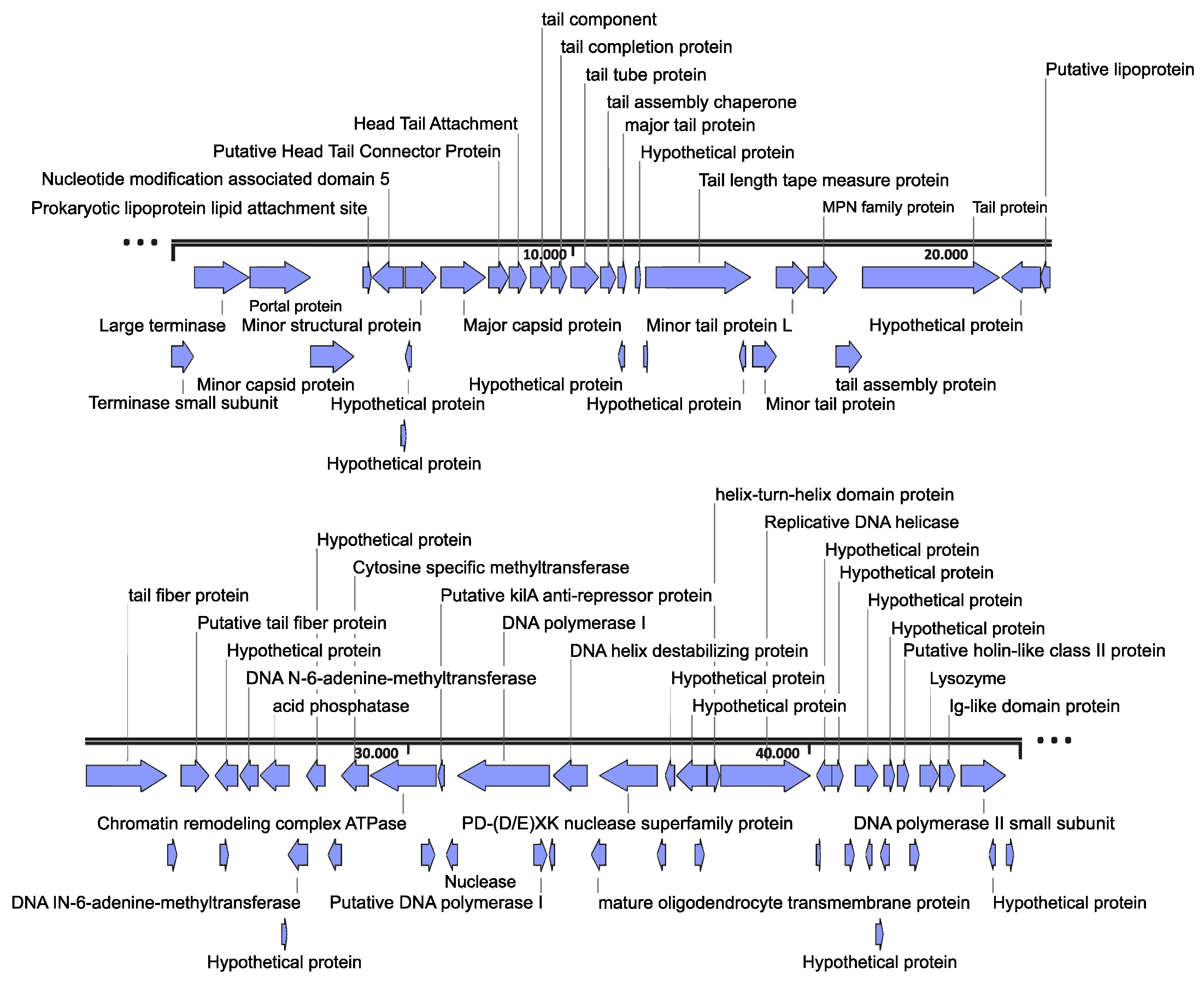
Figure 1. Map of the Escherichia phage Ioannina genome. Arrows with blue color represent predicted coding sequences (CDSs) of different phage functions. The genome map was constructed by using the SnapGene 6.0.2 tool.
Cimb 46 00551 g001
Figure 2. Escherichia phage Ioannina plaque morphology and morphology of the phage absorbed on the host. (a) Circular phage plaques with a diameter of about 5.3 ± 0.5 mm, formed on a bacterial lawn spread on MHB agar. (b) A TEM image of the phage was obtained, indicating a head and a non-contractile tail of 37 ± 3 nm and 122 ± 8 nm, respectively. UA negative staining. Scale bar represents 50 nm.
Cimb 46 00551 g002
Figure 3. One-step growth curve of Escherichia phage Ioannina infecting E. coli ATCC 25922 strain at a multiplicity of infection of 0.01. Experiments for one-step phage growth curve were carried out in triplicates, and the standard deviations of the observed data are drawn as error bars on the depicted graph.
Cimb 46 00551 g003
Figure 4. Thermal and pH stability analysis of Escherichia phage Ioannina. (a) The thermal stability of the phage after incubation at different temperatures for 1 h. (b) pH stability of the phage after incubation at different pH spectra for 1 h. Experiments for thermal and pH stability were carried out in triplicates, and the standard deviations of the observed data are drawn as error bars on the depicted graphs.
Cimb 46 00551 g004
Figure 5. Phylogenetic tree of the Escherichia phage Ioannina portal protein constructed using the Maximum Likelihood method of the MEGA11 software. The “Esch. Ph. Ioannina” represents the Escherichia phage Ioannina portal protein. Bootstrap values (blue-colored text) were obtained from 100 bootstrap replicates, and only those above 70 are displayed next to each node. The tree scale is displayed on the bottom left corner of the phylogenetic tree.
Cimb 46 00551 g005
Figure 6. Phylogenetic tree of the Escherichia phage Ioannina putative tail fiber protein constructed using the Maximum Likelihood method of the MEGA11 software. The “Esch. ph. Ioannina” represents the Escherichia phage Ioannina putative tail fiber protein. Bootstrap values (blue-colored text) were obtained from 100 bootstrap replicates, and only those above 70 are displayed next to each node. The tree scale is displayed on the bottom left corner of the phylogenetic tree.
Cimb 46 00551 g006
Table 1. Lytic activity of the Escherichia phage Ioannina on different bacterial species (environmental, clinical and reference strains). The antibiotics resistance is reported as S (susceptible to antibiotics), R (resistance to one antimicrobial agent) and MDR (resistance to at least one antimicrobial agent in more than three categories). Phylogroups of E. coli isolates are reported as per Clermont’s schema. Phage lytic activity is presented as positive (+) or negative (−).
Sample No.Bacterial Strains/
Isolates *
Samples SourceAntibiotics Resistance $Phylogroup &Phage Lytic
Activity
1Escherichia coli 823Wastewater treatment plantSA+
2Escherichia coli 668Wastewater treatment plantSB1+
3Escherichia coli 824Wastewater treatment plantSB2
4Escherichia coli 663Wastewater treatment plantSD
5Escherichia coli 792Wastewater treatment plantMDRA
6Escherichia coli 494Wastewater treatment plantMDRB1
7Escherichia coli 810Wastewater treatment plantMDRB2
8Escherichia coli 638Wastewater treatment plantMDRD
9Escherichia coli 640Wastewater treatment plantRA
10Escherichia coli 643Wastewater treatment plantRB1
11Escherichia coli 809Wastewater treatment plantRB2
12Escherichia coli 635Wastewater treatment plantRD
13Escherichia coli 865Hospital wastewaterWTA+
14Escherichia coli 866Hospital wastewaterWTB1
15Escherichia coli 843Hospital wastewaterWTB2
16Escherichia coli 580Hospital wastewaterWTD
17Escherichia coli 426Hospital wastewaterMDRD+
18Escherichia coli 858Hospital wastewaterMDRA
19Escherichia coli 546Hospital wastewaterMDRB2
20Escherichia coli 576Hospital wastewaterRB2
21Escherichia coli 545Hospital wastewaterRD+
22Escherichia coli 674Hospital wastewaterRA
23Escherichia coli 759River waterWTA
24Escherichia coli 774River waterWTB1
25Escherichia coli 624River waterWTB2
26Escherichia coli 769River waterWTD
27Escherichia coli 472River waterMDRA
28Escherichia coli 607River waterMDRB1
29Escherichia coli 737River waterMDRB2
30Escherichia coli 408River waterMDRD
31Escherichia coli 614River waterRA
32Escherichia coli 372River waterRD
33Escherichia coli 743River waterRB2
34Escherichia coli 784River waterRB1
35Escherichia coli 117ClinicalMDRA+
36Escherichia coli 60ClinicalMDRB2+
37Escherichia coli 203ClinicalMDRD+
38Escherichia coli 325ClinicalRA+
39Escherichia coli 5ClinicalRA
40Escherichia coli 264ClinicalRB1
41Escherichia coli 294ClinicalRB2
42Escherichia coli 378ClinicalRB2+
43Escherichia coli 313ClinicalRD
44Escherichia coli 324ClinicalSA+
45Escherichia coli 368ClinicalSD
46Escherichia coli 387ClinicalSB2
47Escherichia coli 301ClinicalSB1
48Escherichia coli 25922Reference strain from Becton Dickinson, France S.A.SS+
49Escherichia coli 35218Reference strain from Becton Dickinson, France S.A.SS
50Escherichia coli 13846Reference strain from Becton Dickinson, France S.A.SS
51Escherichia coli O157:H7ClinicalMDR
52Klebsiella pneumoniae 13883Reference strain from Becton Dickinson, France S.A.SS
53Klebsiella pneumoniae 700603Reference strain from Becton Dickinson, France S.A.SS
54Pseudomonas aeruginosa 27853Reference strain from Becton Dickinson, France S.A.SS
55Yersinia enterocolitica 9610Reference strain from Becton Dickinson, France S.A.SS
56Acinetobacter baumannii 17978Reference strain from Becton Dickinson, France S.A.SS
57Acinetobacter baumannii 19668Reference strain from Becton Dickinson, France S.A.SS
* The number of the isolate is the respective number as reported in Dioli et al. https://pubmed.ncbi.nlm.nih.gov/37374900/ (accessed on 23 July 2024). $ Antibiotics resistance is reported as S (susceptible to antibiotics), R (resistance to one antimicrobial agent) and MDR (resistance to at least one antimicrobial agent in more than three categories). Resistances have been previously reported, as reported in Dioli et al. https://pubmed.ncbi.nlm.nih.gov/37374900/ (accessed on 23 July 2024). & Phylogroups of E. coli isolates are reported as per Clermont’s schema, as identified in Dioli et al. https://pubmed.ncbi.nlm.nih.gov/37374900/ (accessed on 23 July 2024). Phage lytic activity is presented as positive (+) or negative (−).
Table 2. Closest putative tail fiber amino acid sequences to the Escherichia phage Ioannina putative tail fiber protein, based on the BLASTp tool.
Phage NameBLASTp Coverage (%)BLASTp Percent Identity (%)GenusFamily/
Morphotype
NCBI Accession Number
vB_EcoS_SA32RD9872.5TunavirusDrexlerviridaeUIU27553.1
PGN68669274.22KuraviruspodovirusesQKL16987.1
vB_EcoP_YF019272.89KuraviruspodovirusesWBF04932.1
IME2679272.44KuraviruspodovirusesYP_010673185.1
MLP39272.89KuraviruspodovirusesUEN68517.1
vB_EcoP-101114UKE39272.44KuraviruspodovirusesYP_010673043.1
νB_EcoP_SU79272KuraviruspodovirusesYP_010672804.1
vB_EcoS_011D59938.7DhillonvirussiphovirusesQMP82830.1
vB_EcoS_L-h 1M9937.79DhillonvirussiphovirusesUNY42316.1
vB_EcoS_SA30RD7248.02TunavirusDrexlerviridaeUIU27628.1
vB_EcoS_Chapo7247.46TunavirusDrexlerviridaeQLF82390.1
Disclaimer/Publisher’s Note: The statements, opinions and data contained in all publications are solely those of the individual author(s) and contributor(s) and not of MDPI and/or the editor(s). MDPI and/or the editor(s) disclaim responsibility for any injury to people or property resulting from any ideas, methods, instructions or products referred to in the content.