Article

Selection and Validation of Reliable Reference Genes for Liquidambar formosana Leaves with Different Leaf Colors

1
Zhejiang Key Laboratory of Forest Genetics and Breeding, Zhejiang Academy of Forestry, Hangzhou 310023, China
2
Jiangsu Academy of Forestry, Nanjing 211153, China
*
Authors to whom correspondence should be addressed.
Curr. Issues Mol. Biol. 2024, 46(9), 9449-9462; https://doi.org/10.3390/cimb46090560
Submission received: 22 July 2024 / Revised: 21 August 2024 / Accepted: 25 August 2024 / Published: 27 August 2024
(This article belongs to the Section Molecular Plant Sciences)

Abstract

:
Liquidambar formosana Hance is renowned for its rich leaf color and possesses notable advantages, such as robust adaptability, strong resistance to diseases and pests, and rapid growth, making it a preferred choice for urban greening and carbon sequestration forest initiatives. The completion of whole-genome sequencing of L. formosana has spurred an increased interest in exploring the molecular mechanisms underlying seasonal changes in leaf color, marking a significant focus in L. formosana breeding research. However, there is currently a lack of stable reference genes suitable for analyzing the expression patterns of functional genes in L. formosana exhibiting varying leaf colors. This study selected five L. formosana varieties with significant differences in leaf colors. Through the RT-qPCR analysis, and evaluation using BestKeeper, geNorm, NormFinder, Delta Ct, and RefFinder, the expression stability of 14 candidate reference genes was examined. Consequently, two reference genes (LifEF1-α and LifACT) with stable expression, suitable for RT-qPCR of L. formosana with diverse leaf colors, were identified. The stability of these selected reference genes was further validated by examining the LifbHLH137 gene, which promoted the biosynthesis of anthocyanins. This advancement facilitated molecular biology and genetic breeding investigations of L. formosana, providing essential data for the precise quantification of functional genes associated with leaf color variation.

1. Introduction

Liquidambar formosana Hance, classified under the family Hamamelidaceae and genus Liquidambar, is a tall deciduous tree esteemed as both a crucial afforestation species and an exceptional landscape ecological tree. Renowned for its rich leaf color, it predominantly thrives in southern and central China, alongside the tropical regions of Southeast Asia, with additional occurrences in the eastern United States, Mexico, and Guatemala [1]. L. formosana exhibits a tall tree shape, dense foliage, and robust adaptability, demonstrating resilience to drought and infertile soil conditions [2]. Concurrently, the escalating demand for ornamental flora and advancements in landscaping have propelled L. formosana into the spotlight because of its rich, bright color and high ornamental value, thereby establishing them as the preferred species for national greening initiatives [3].
Most autumn-colored leaf tree species are deciduous plants, mainly due to insufficient winter light and lower temperatures. The self-sufficiency cost of plants during the leaf-shedding period will exceed the capacity accumulation of photosynthesis. Therefore, plants choose to enter a brief dormancy period to reduce their own nutrient consumption [4]. Simultaneously, the accumulation of antioxidant compounds, such as anthocyanins, serves as a mechanism to cope with environmental changes. Notably, anthocyanins tend to accumulate significantly during the period when leaf color begins to change [5,6]. Chlorophyll degradation products exhibit antioxidant properties that can preserve cellular activity and mitigate aging processes [6,7]. Consequently, the degradation of chlorophyll and the synthesis of anthocyanins reflect the adaptive regulatory mechanisms of plants in response to environmental conditions. Investigating the molecular mechanisms underlying these processes holds substantial biological significance for the development of superior plant varieties through advanced scientific and technological methodologies.
Current research on L. formosana predominantly focuses on resource distribution, breeding of improved varieties, population genetics and evolution, and ecological monitoring [3,8]. However, most studies concerning the color changes of L. formosana leaves typically focus only on the phenotypic traits and lack in-depth exploration of the molecular regulatory mechanisms underlying these changes. Understanding the key regulatory factors for leaf color variation in L. formosana remains a significant research area considering the combined impact of genetic background and environmental factors on color variation. Research on the color change of L. formosana leaves primarily focuses on physiological and biochemical aspects, such as photosynthesis and pigment content [1,9], while the molecular mechanisms driving these changes still lack comprehensive genetic information, inhibiting the development and utilization of L. formosana leaf color resources. A definitive solution for generating new L. formosana germplasms with diverse leaf colors via biotechnology and gene editing remains elusive, and the molecular regulatory mechanisms governing leaf color variation remain unclear. The completion of whole-genome sequencing for L. formosana has significantly advanced molecular biology and genetic breeding research in this species [10]. The advancement of the L. formosana industry correlates with the progress of the agricultural and forestry economy and establishment of an ecological civilization. Therefore, there is an urgent need for comprehensive research into the seasonal fluctuations in L. formosana leaf color, the identification of key genes that dictate these color changes, and the examination of expression patterns and regulatory mechanisms of crucial functional genes affecting the diversity of L. formosana leaf color. This study can provide a foundation for L. formosana tree breeding.
Real-time quantitative PCR (RT-qPCR) is now widely used to study gene transcriptional expression levels due to its convenience, speed, specificity, and sensitivity. However, factors such as RNA and cDNA quality, sample dilution ratio, and experimental accuracy can affect the RT-qPCR accuracy [11]. Therefore, employing appropriate reference genes is crucial for standardized analysis of target gene expression levels [12]. The ideal reference genes are stably expressed in cells, typically encompassing housekeeping genes, such as actin, 18S Ribosomal RNA, tubulin, and ubiquitin, which maintain fundamental cellular activities [13,14]. Nevertheless, recent studies have indicated that variations in species and tissue organs can alter the transcription of housekeeping genes. The leaves from various Zantedeschia hybrida varieties were collected, and six commonly used reference genes (18S rRNA, ACT, EF1-α, GAPDH, LEU, and TUB) were assessed. The analysis that was conducted using the reference gene analysis methods and software revealed that 18S rRNA, LEU, and GAPDH exhibited greater stability across different varieties, whereas ACT, EF1-α, and 18S rRNA were the most stable across different tissues [15]. This suggested that optimal reference genes may vary across developmental stages and tissues. Moreover, the stability of reference genes can differ within the same plant under different environmental conditions. Hence, the selection of appropriate reference genes should be aligned with the specific experimental conditions to minimize experimental errors.
Currently, there is a scarcity of suitable reference genes for investigating functional genes linked to the L. formosana leaf color. Commonly used reference genes may present instability across different plants and experimental conditions, potentially affecting the accuracy of target gene identification. In this study, the leaves from five L. formosana varieties exhibiting distinct leaf color differences in September and December were used as experimental samples. Based on transcriptome data of L. formosana, 14 potential reference genes were identified. Various analyses, including fluorescence-quantitative PCR, amplification curve and melting curve analyses, LinRegPCR amplification efficiency analysis, and stability assessments using BestKeeper, geNorm, NormFinder, Delta Ct, and RefFinder, were conducted to identify two stable reference genes (LifEF1-α and LifACT) suitable for fluorescence-quantitative PCR. The stability of the selected reference genes was validated using LifbHLH137, which enhanced anthocyanin biosynthesis. This study addressed the gap in stable reference genes for studying L. formosana leaf color variation, advanced molecular biology, and genetic breeding research, and offered data support for the precise quantification of functional genes related to leaf color variation, thereby enhancing research stability and accuracy.

2. Materials and Methods

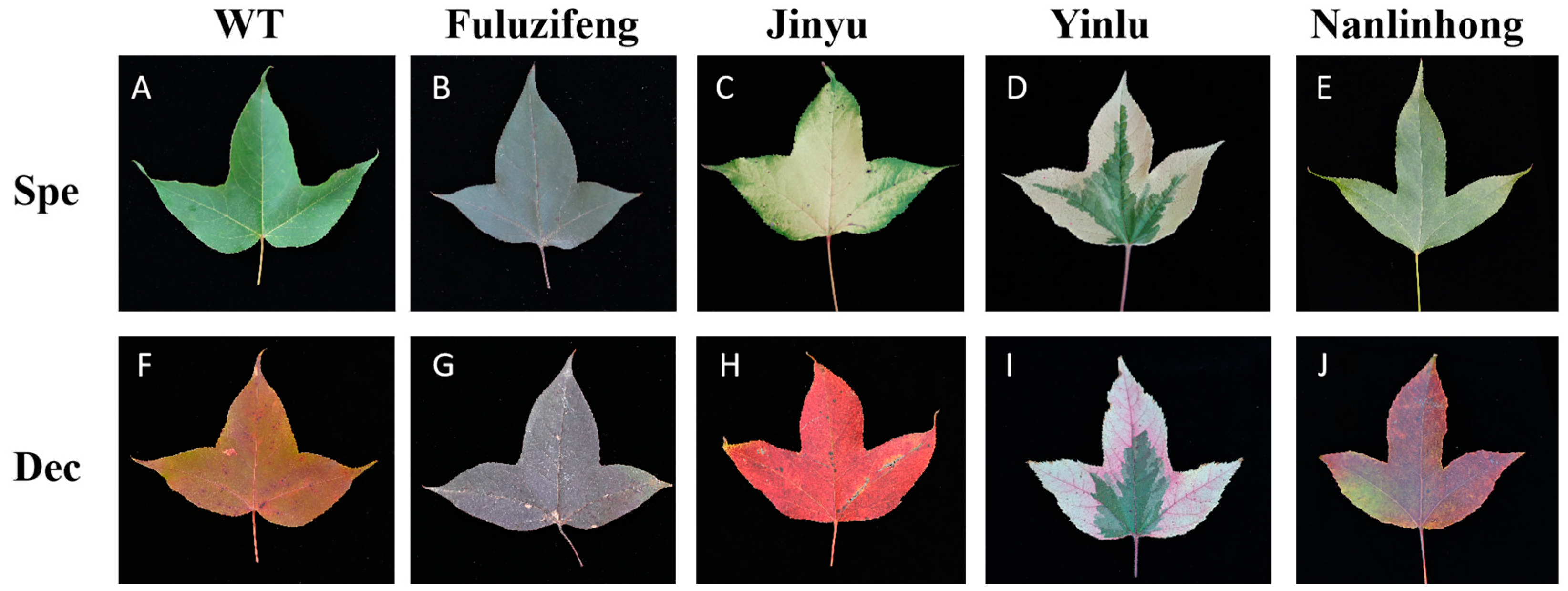
2.1. Plant Materials

For this study, experimental materials comprised the leaves from L. formosana, specifically, selected variants with distinct leaf colors: “Jinyu”, “Fuluzifeng”, “Yinlu”, and “Nanlinhong”. Three plants from each variant exhibiting consistent growth and height were randomly selected for sample collection. The complete leaves were harvested in September and December of 2023 (Figure 1). Three replicates were collected from each tree per period, which were promptly frozen in liquid nitrogen upon collection and stored at −80 °C until further applications.

2.2. Total RNA Extraction and cDNA First-Strand Synthesis

The total RNA was extracted from various leaf tissues using the RNAprep Pure Plant Plus Kit (TIANGEN, Nanjing, China). The integrity and concentration of the total RNA were assessed using 1.2% agarose gel electrophoresis and an ultra-micro spectrophotometer (Thermo Fisher, Waltham, MA, USA), respectively. After the extraction, the total RNA concentration was diluted to 100 ng/µL. The first strand of cDNA was synthesized using the Fastking gDNA Dispelling RT SuperMix (TIANGEN, Nanjing, China) and stored at −20 °C for subsequent experiments.

2.3. Primer Design and Specific Detection

From transcriptome sequencing data of L. formosan and common reference genes in plants, 14 candidate reference genes were selected: LifEF1-α, LifACT, Lifα-TUB1, Lifβ-TUB, LifARF2, LifCDC2, LifDnaJ, LifGAPDH, LifH2A, LifHIS, LifLTP, LifSAND, LifUBC, and LifUBQ. The primer design was conducted using Primer Premier 5.0, adhering to standards such as an annealing temperature of 54–62 °C, primer length of 18–25 bp, and amplification fragment length of 100–300 bp [16]. The primer specificity was confirmed using the BLAST tool, and the detailed sequences and product lengths are provided in Table 1. The primers were synthesized by Shenggong Biotechnology Co. Ltd. (Shanghai, China). The primer specificity was assessed by the PCR amplification and RT-qPCR melting curve analysis, using mixed cDNA from the leaves of five different L. formosana varieties as templates. The PCR reaction system comprised 25.0 µL, including 12.5 µL of 2× Taq Fast PCR Mix, 1.0 µL of each upstream and downstream primer at 10 µmol/L, 1.0 µL of template cDNA at 100 ng/µL, and RNase Free ddH2O supplemented to 25.0 µL.

2.4. Real-Time Fluorescence-Quantitative PCR of Candidate Reference Genes

The real-time fluorescence-quantitative PCR detection was conducted using the QIAquant 96 2plex (QIAGEN, Hilden, Germany) fluorescence-quantitative PCR system and PowerUp™ SYBR™ Green Master Mix (Applied Biosystems, Waltham, MA, USA), with cDNA from leaves of five different L. formosana varieties (“WT”, “Jinyu”, “Fuluzifeng”, “Yinlu”, and “Nanlinhong”) collected in September and December serving as templates. The reaction system comprised 10 μL of PowerUp™ SYBR™ Green Master Mix, 0.2 µL each of 10 µmol forward and reverse primers, 2.0 µL of cDNA templates at varying dilutions, and RNase-free ddH2O supplemented to a total volume of 20.0 µL. The amplification program included pre-denaturation at 95 °C for 3 min, followed by 40 cycles of 95 °C for 15 s, 60 °C for 15 s, and 72 °C for 30 s. After the amplification, the melting curve analysis was performed at 55–95 °C. All analyses were conducted in triplicates.

2.5. Establishment of Reference Gene Primer Standard Curves

Equal amounts of cDNA templates from all samples were combined and diluted with ddH2O to 5 concentrations, each of which was diluted 5 times (1, 1/5, 1/25, 1/125, and 1/625), to establish standard curves. The RT-qPCR amplification was performed using the QIAquant 96 2plex (QIAGEN, Germany) to obtain Ct values for each candidate reference gene at different template dilutions. The standard curve for each candidate reference gene was constructed by plotting the logarithmic relationship between the average Ct value and continuously diluted template cDNA. The slope (K) and correlation coefficient (R2) were derived from the standard curve, and the PCR amplification efficiency (E) was calculated using the formula: E = [5 (1/−K) − 1] × 100%, for each primer pair [17]. The comprehensive evaluation was conducted to ensure that the system (including primers and templates) and amplification program met the requirements for the RT-qPCR analysis. The criteria included a correlation coefficient R2 > 0.99 and an amplification efficiency E value between 90% and 120% [17]. All samples were subjected to three repetitions to ensure the reliability of the experimental data.

2.6. Data Analysis

(1) The geNorm version 2003 software, a fundamental programming tool, can assess reference gene stability via stability parameter M value calculation [18]. A threshold of M = 1.5 was established, where M < 1.5 indicated reference gene suitability, and lower M values indicated superior stability. Conversely, for M values exceeding 1.5, the increasing M correlated with the heightened instability of the reference gene. Consequently, this gene was unsuitable as a reference gene. In addition, when calculating multiple reference genes, the geNorm software can identify the optimal number of reference genes by assessing a new paired variation (V) value of the normalization factor for reference genes. By utilizing the ratio of Vn/n+1, the software assessed the impact of introducing new internal reference genes on the standard factor. The default threshold for the V-value was set at 0.15. If Vn/n+1 < 0.15, it indicated stability among the n internal reference genes for normalization. Conversely, if Vn/n+1 > 0.15, the introduction of the n + 1st gene became necessary. This software reliably determined a suitable number of reference genes [19].
(2) The NormFinder version 0953 software normalized candidate genes based on their expression stability within a given sample group and experimental design. Its methodology closely paralleled the geNorm, involving the calculation of ΔCt values and their subsequent transformation into 2−ΔCt values using Excel functions. These values represent the relative expression levels of candidate reference genes from which the stable expression value (S) was derived. The reference gene with the smallest stable value was deemed the most suitable. Compared to the geNorm, the NormFinder identified only one stable reference gene and did not determine the optimal number of reference genes. Nevertheless, it offers guidance for identifying the best reference gene [20].
(3) The BestKeeper version 2003 algorithm employed the repeated-pair correlation and regression analyses to conduct a correlation analysis for each reference gene. The original Ct values were directly imported into the BestKeeper Excel table using this algorithm. The coefficient of variation (CV) and standard deviation (SD) were used to comprehensively assess the stability of the reference gene. A lower CV ± SD value indicated greater stability of the reference gene. A comparison of these values determined the ranking of the final reference genes based on their stability [21].
(4) The ΔCt version 2003 algorithm discerns stable reference genes by comparing the relative expression levels of gene pairs within each sample. The consistent ΔCt values across various samples indicated stable gene expressions, whereas fluctuations indicated instability. By introducing additional genes for comparison and subsequent sorting, suitable reference genes can be selected based on the experimental criteria [22].
(5) The RefFinder version 2003 software systematically evaluated the stability of internal reference genes obtained from the geNorm version 2003, NormFinder version 0953, ΔCt version 2003, and BestKeeper version 2003 software by computing their geometric mean weights. Lower geometric means could indicate greater stability of the internal reference genes. The stability of all reference genes was comprehensively assessed through scoring, leading to the selection of the most stable internal reference genes [23].

2.7. Stability Verification of Reference Genes

LifbHLH137, known to promote anthocyanin biosynthesis, was selected to assess the stability of the selected reference genes [24]. The LifEF1-α and LifACT, identified as the most stable reference genes, were employed to analyze LifbHLH137 expression in leaves of WT, “Jinyu”, “Fuluzifeng”, “Yinlu”, and “Nanlinhong”. The unstable expressions of LifCDC2 and LifLTP genes served as the controls. The LifbHLH137 expression was evaluated using the 2−ΔΔCt method [25,26].

3. Results

3.1. RNA Quality Testing and Primer Specificity Validation

This study employed NanoDrop™ One (Thermo Fisher, Waltham, MA, USA) and 1% agarose gel to assess the total RNA mass extracted from leaves of WT, “Jinyu”, “Fuluzifeng”, “Yinlu”, and “Nanlinhong” in September and December (Figure 1). The RNA concentration ranged from 140 to 400 ng/μL, with the OD260/280 values between 2.0 and 2.3, indicating high purity without protein or phenol contamination. The agarose gel electrophoresis revealed clear main bands for each sample without dispersion or tailing. The brightness ratio of the 28S rRNA to 18S rRNA bands was approximately 2:1, indicating minimal RNA degradation and high integrity, meeting the subsequent testing requirements.
Based on the transcriptome data from L. formosan, 14 candidate reference genes with stable expression across various tissues were identified. The PCR amplification was conducted using equimolar amounts of mixed cDNA from the leaves of the five L. formosan varieties as templates. The results revealed single and intense target fragments devoid of primer dimerization or non-specific amplification. The fragment sizes ranged from 102 to 264 bp, which was consistent with expectations (Figure S1). The subsequent validation of primer specificity via the RT-qPCR revealed single dissolution peaks for each candidate reference gene free from hairpin structures or primer dimers (Figure S2). The overlap of amplification curves from repeated samples indicated appropriate annealing temperatures and excellent primer specificity for each reference gene (Figure S3). The standard curves were established using the cDNA templates with varying concentration gradients, demonstrating primer amplification efficiency (E) within an acceptable range of 90% to 120%, ranging from 99.31% to 117.49%. The linear correlation coefficients (R2) showed minimal differences, ranging from 0.9765 to 0.9961 (Table 1). These results confirmed the soundness of the primer design for the 14 candidate reference genes, ensuring favorable specificity and amplification efficiency for the RT-qPCR. They provided accurate and reliable data for reference gene stability assessments.

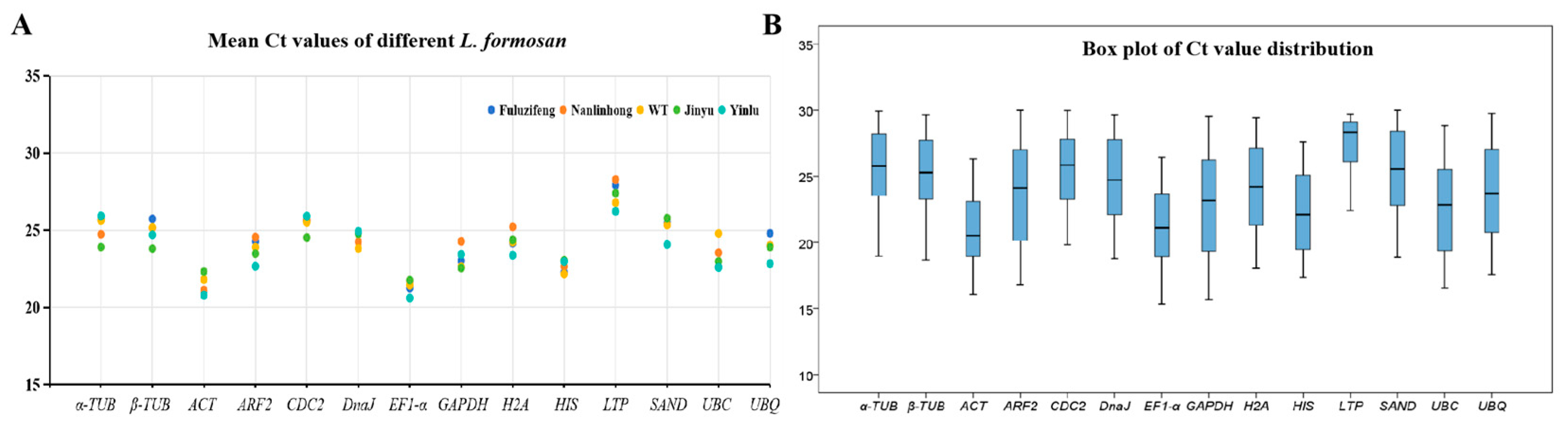
3.2. Reference Primer Amplification Ct Value Analysis

The Ct value, which is inversely related to gene expression, reflects the expression level across all experimental samples. The lower Ct values corresponded to the higher gene expression levels. The box plots of Ct values for each reference gene across different samples indicated a range of 15.69 to 29.97, indicating moderate expression abundance (Figure 2). The mean Ct values for the 14 reference genes ranged from 21.35 to 27.32. The LifEF1-α demonstrated the highest expression level across all samples (average Ct value: 21.35), whereas the LifLTP exhibited the lowest (average Ct value: 27.32). Greater gene stability correlated with a narrower range of Ct values. Preliminary observations from box plot analysis indicated notable variations in the expression levels of different reference genes in L. formosan. The Ct values of LifGAPDH presented the widest range among the 14 reference genes, spanning from 15.69 to 29.56, indicating the low expression stability. Conversely, the LifACT (16.05–26.32) and LifEF1-α (15.32–26.43) exhibited narrower Ct value ranges, suggesting a more stable gene expression (Figure 2). These findings indicated the varying expression levels of different reference genes in the leaves of different L. formosan varieties and developmental stages. Although most candidate reference genes displayed relatively high expression levels with minimal variation, ensuring their suitability as reference genes, the consistency of their expression levels across samples was less pronounced. Therefore, employing 5 statistical algorithms to evaluate the expression stability of the 14 reference genes across different colors of L. formosan leaves could be imperative for identifying the optimal reference genes for standardizing target gene expression.

3.3. Analysis of Gene Expression Stability

To assess the expression stability of the 14 candidate reference genes across various L. formosan leaf varieties, ΔCt, BestKeeper, geNorm, and NormFinder were employed for the statistical analysis of gene expression levels in diverse leaf tissues. The RefFinder was employed to rank the overall stability and identify suitable reference genes for analyzing functional gene expression patterns across different leaf colors and developmental stages of L. formosan.

3.3.1. ΔCt Analysis

The stability ranking can be determined by the dispersion of gene Ct value data, with lower ΔCt values indicating higher expression stability [20]. Table 2 presents the ranking of the 14 candidate reference genes based on their expression stability in the leaf tissues of the 5 differently colored L. formosan leaves, calculated using the ΔCt method. The analysis revealed that LifEF1-α was the most stable reference gene in the September leaves, and LifDnaJ in the December leaves. Overall, LifEF1-α was identified as the ideal reference gene, followed by LifACT, whereas LifLTP was consistently deemed the least stable. The stability ranking of the reference genes across all samples was as follows: LifEF1-α > LifACT > LifHIS > LifUBQ > LifSAND > LifH2A > LifDnaJ > Lifβ-TUB > LifGAPDH > Lifα-TUB > LifUBC > LifARF2 > LifCDC2 > LifLTP. LifEF1-α emerged as an optimal reference gene.

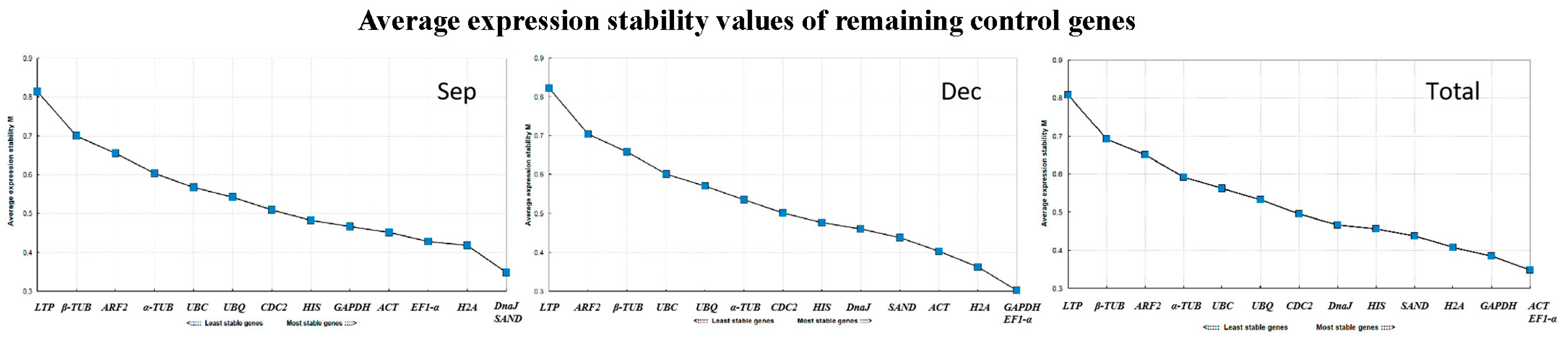
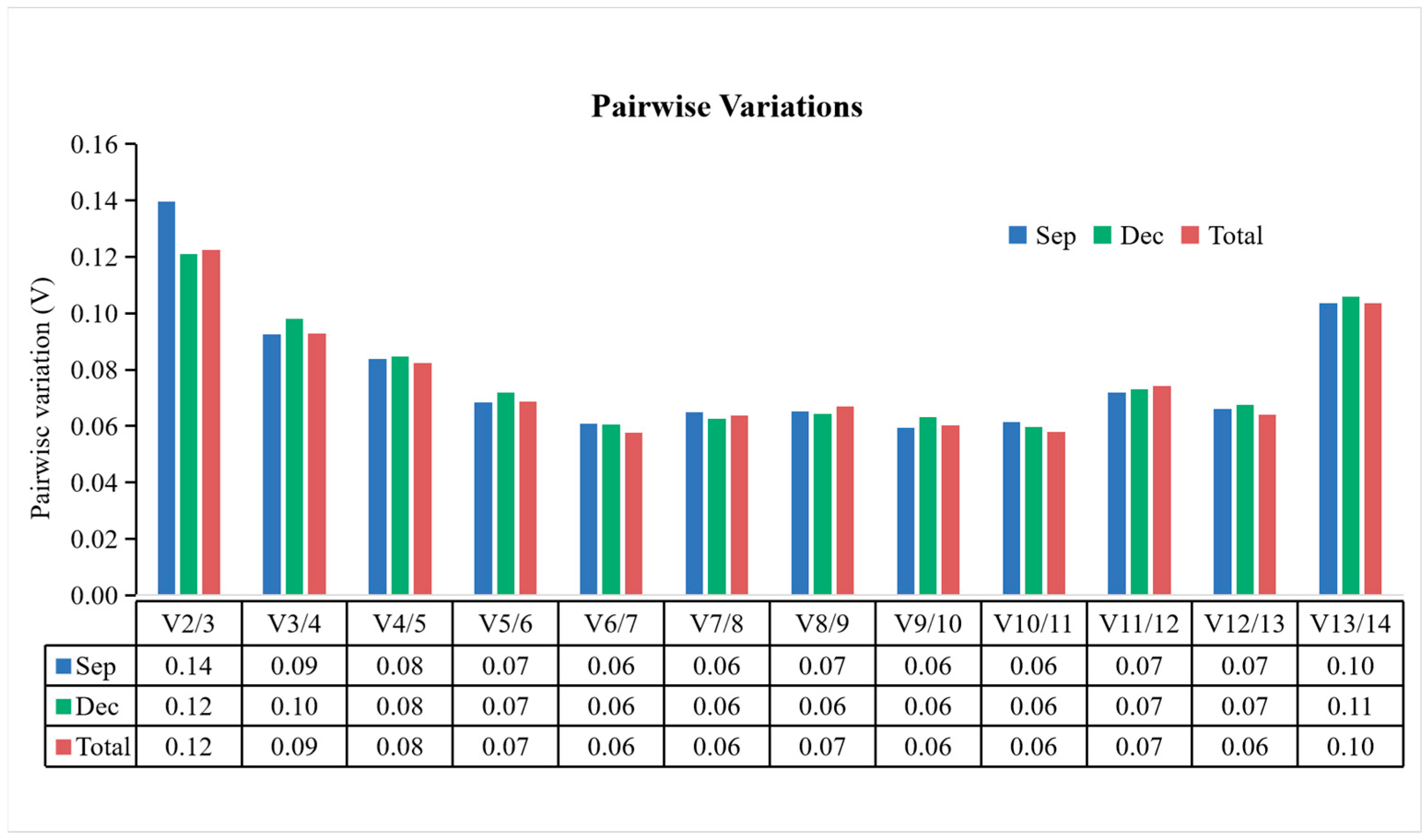
3.3.2. geNorm Analysis

Using the geNorm software, the M value was calculated, indicating the expression stability of each candidate reference gene across L. formosan leaf varieties. The software set M = 1.5 as the critical threshold, with lower M values indicating a higher stability. The analysis revealed that all 14 candidate reference genes had M values below 1.5, indicating stable and robust expression (Figure 3). Notably, LifDnaJ, LifSAND, LifH2A, LifEF1-α, LifACT, LifGAPDH, and LifHIS exhibited M values below 0.5 across all leaf samples, indicating a highly stable expression. Specifically, LifDnaJ and LifSAND demonstrated the highest expression stability in September leaves, whereas LifGAPDH and LifEF1-α demonstrated the highest stability in December leaves. Overall, LifACT and LifEF1-α were identified as the two reference genes with the highest expression stabilities across all leaf samples. These results indicated the variability in reference gene stability across the different developmental stages of the leaves.
Furthermore, the geNorm software has the capability to conduct paired difference (Vn/n+1) analysis on standardized factors of candidate reference genes to determine the optimal number of reference genes. When Vn/n+1 < 0.15, n reference genes are sufficiently stable for normalization, obviating the need to introduce the n + 1st reference gene. This study revealed that V2/3 was less than 0.15 when analyzing leaf tissues separately in September and December, as well as when analyzing all samples collectively (Figure 4), indicating that a combination of two stable genes could improve the reliability and accuracy of the quantitative results. Overall, the comprehensive analysis ranked the stability of the 14 reference genes as follows: LifACT = LifEF1-α > LifGAPDH > LifH2A > LifSAND > LifHIS > LifDnaJ > LifCDC2 > LifUBC > LifUBQ > Lifα-TUB > LifARF2 > Lifβ-TUB > LifLTP, and LifACT and LifEF1-α emerged as the most suitable reference gene combination.

3.3.3. BestKeeper Analysis

The BestKeeper primarily assesses gene stability by analyzing the standard deviation (SD) and coefficient of variation (CV) of the Ct values of candidate reference genes [21]. Smaller SD and CV values indicate greater stability, with an SD value of less than 1.0, which is considered indicative of high stability. In both the total sample and September sample, six reference genes had SD values < 1.0, whereas in the December sample, only four reference genes met this criterion (Table 3). The LifEF1-α consistently displayed SD values of <1.0 across all analyses, indicating the highest expression stability. The LifACT ranked second in expression quantity in September and December samples and third in the comprehensive analysis. Based on the SD and CV of the Ct values, reference gene stability was ranked as follows: LifEF1-α > LifSAND > LifACT > Lifβ-TUB > LifDnaJ > LifHIS > LifUBQ > LifH2A > LifLTP > Lifα-TUB > LifGAPDH > LifUBC > LifCDC2 > LifARF2 (Table 3).

3.3.4. NormFinder Analysis

The NormFinder software evaluates the gene stability based on the stable expression value (S value). Smaller S values indicate greater stability. Subsequently, the optimal reference gene is selected [22]. In this study, the stability of the 14 reference genes across 5 L. formosan leaf slices with distinct color differences was analyzed. The results revealed the variations in gene stability among different samples. The LifEF1-α was deemed the most stable gene expression in the September samples, while the LifDnaJ demonstrated the highest stability in the December samples. Overall, the LifACT demonstrated the most stable expression when all samples were analyzed comprehensively. Conversely, the LifLTP exhibited the highest S value across all the analyses, indicating that it was the most unstable reference gene. The expression stability of the reference genes in all L. formosan leaves, ranked from high to low, was as follows: LifACT > LifEF1-α > LifSAND > LifUBQ > LifHIS > LifH2A > LifDnaJ > Lifβ-TUB > LifGAPDH > Lifα-TUB > LifUBC > LifARF2 > LifCDC2 > LifLTP (Table 4).

3.3.5. RefFinder Analysis

The stability of the 14 candidate reference genes in L. formosan leaves at different developmental stages varied owing to the applications of different algorithms across software, in both within- and between-group analyses. To mitigate potential errors stemming from individual reference gene evaluation programs, we employed the RefFinder tool to comprehensively rank the results from the four aforementioned software packages. RefFinder utilized a comprehensive approach to analyze gene expression stability based on the geometric average weights of the four algorithms (GeNorm, NormFinder, ΔCt, and BestKeeper). Lower geometric average weight values indicate higher expression stability. RefFinder provided a more authoritative evaluation of the stability of the 14 candidate reference genes, yielding a more reasonable ranking. The overall stability ranking, from high to low, was as follows: LifEF1-α > LifACT > LifSAND > LifUBQ > LifHIS > LifH2A > LifDnaJ > Lifβ-TUB > LifGAPDH > Lifα-TUB > LifUBC > LifARF2 > LifCDC2 > LifLTP, suggesting that LifEF1-α and LifACT were the optimal reference gene combinations (Table 5). These two genes consistently ranked in the top five across the four algorithms, making them suitable reference genes for analyzing functional color gene expression patterns in L. formosan leaves. Additionally, the comprehensive analysis identified LifCDC2 and LifLTP as the two most unstable genes that were unsuitable for use as reference genes.

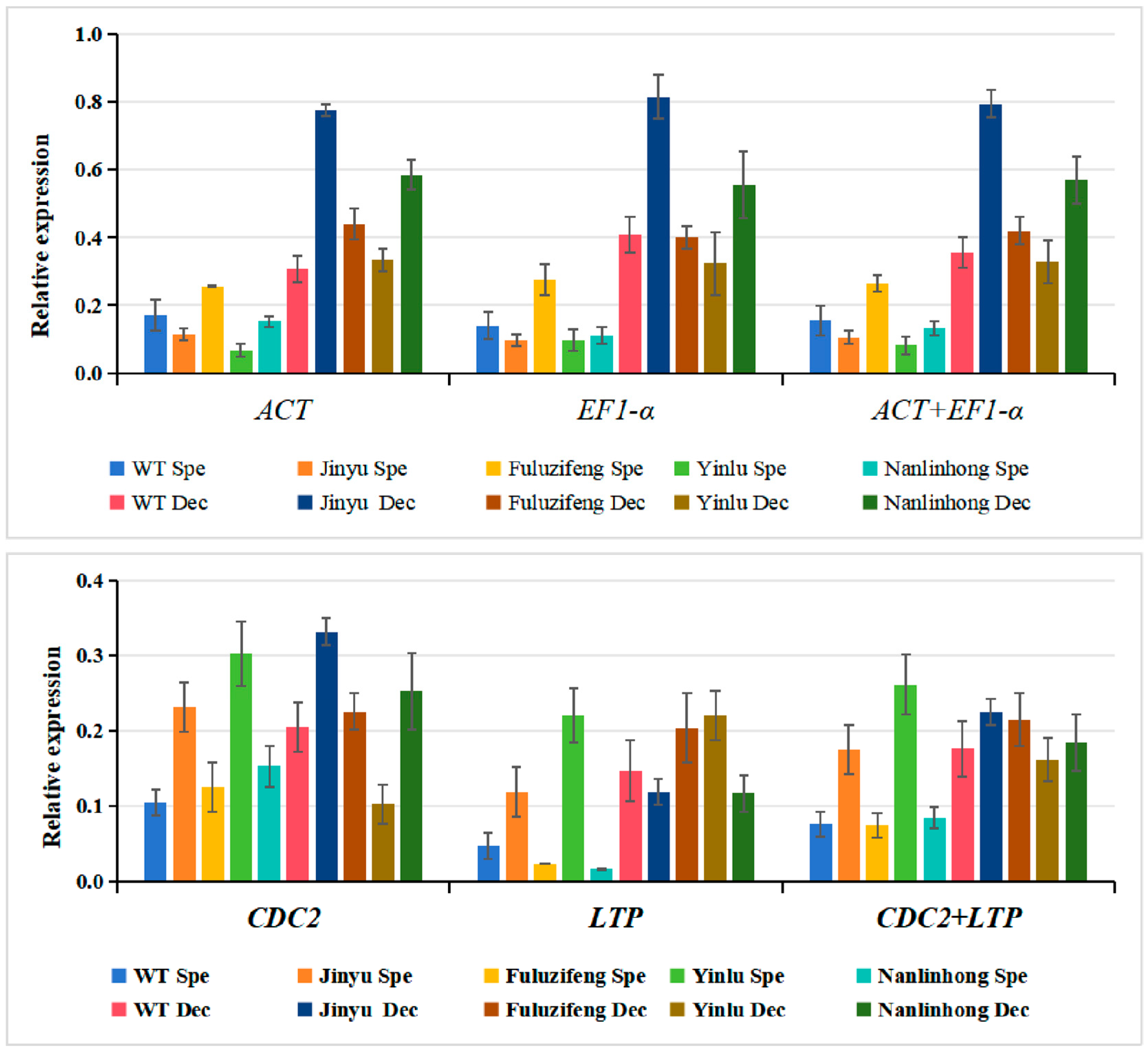
3.4. Reference Gene Stability Verification

The red and purple hues of L. formosan leaves stem from their high anthocyanin content, with cyanidin and paeoniflorin contributing to the red coloration, whereas delphinidin and geranium accumulation can enhance the purple appearance [3]. Previous investigations employed transcriptomics and metabolomics to jointly analyze the anthocyanin synthesis pathways in WT, “Jinyu”, “Fuluzifeng”, “Yinlu”, and “Nanlinhong”. The 14 candidate reference genes were stably expressed in leaves at different developmental stages (Figure S4A). The bHLH137, which promotes anthocyanin biosynthesis, was markedly upregulated during the leaf reddening (Figure S4B). The expression patterns of bHLH137 across different L. formosan varieties were examined using RT-qPCR. The LifEF1-α and LifACT, selected as optimal internal reference genes, along with their combination, LifEF1-α + LifACT, were employed for normalization. Both individual and combined corrections yielded consistent relative expression levels of the target genes. The results indicated significant seasonal variation in the bHLH137 expression across various varieties, with notable upregulation observed in the red autumn leaves. Especially in “Jinyu”, notable seasonal variation suggested that it may play a pivotal role in regulating leaf color changes. However, when utilizing the unstable expression of LifCDC2 and LifLTP genes as references, the bHLH137 expression patterns varied, lacking clear consistency (Figure 5). Inappropriate internal reference genes can lead to deviations in target gene expression levels, thereby compromising experimental accuracy. This reaffirmed the accuracy and reliability of the selected reference genes, LifEF1-α and LifACT, laying a foundation for future research on functional gene expression analysis related to seasonal shifts in L. formosan leaf color.

4. Discussion

L. formosan, a relic plant dating back to the third century, thrives in subtropical and tropical regions and displays diverse leaf colors, indicative of its adaptability to climate fluctuations [27]. Recently, there has been increased research interest in L. formosan, owing to its vibrant foliage and growing landscape value [8]. However, the lack of a comprehensive genome database has hindered reference gene screening in L. formosan, which is critical for analyzing functional gene expression patterns. Consequently, the depth of understanding of the mechanism of leaf color change in L. formosan remains limited. Although the MYB113 has been implicated in leaf color regulation, its scope is primarily limited to autumn leaves and aging factors [28]. Research on candidate transcription factors that are directly involved in seasonal leaf color regulation is scarce. To elucidate the molecular mechanisms underlying leaf color variation across seasons or varieties of L. formosan, stable reference genes are essential for data standardization. Considering the limited development of reference genes in woody plants, it is imperative to screen for stable reference gene expression in diverse L. formosan varieties to lay the groundwork for subsequent research on related functional genes.
This study employed the RT-qPCR to comprehensively assess the expression stability of 14 candidate reference genes across 5 maple trees exhibiting significant leaf color variations [29]. Currently, multiple software tools are used for the RT-qPCR reference primer screening and evaluation. However, varying the calculation principles of these software programs can lead to contradictory conclusions from the same data. For instance, in a reference gene screening study of Solanum melongena, using only two software tools (geNorm and NormFinder) resulted in inconsistent results [30]. Conversely, in a study of Chrysanthemum lavandulifolium reference genes, the utilization of at least three software tools improved reliability [31]. Therefore, in this study, four software tools, including the geNorm version 2003, NormFinder version 0953, ΔCt version 2003, and BestKeeper version 2003, were employed for reference gene screening in L. formosan. The analysis results from the ΔCt and BestKeeper indicated LifEF1-α as the most stable reference gene across all samples, whereas the NormFinder identified LifACT as the most stable reference gene. Despite variations in the results from different software analyses, the ranking of candidate reference genes based on expression stability remained consistent across all samples. The geNorm software conducted a paired difference analysis of standardized factors and determined two reference genes as optimal. To mitigate errors arising from disparate algorithms, the RefFinder, a comprehensive analysis software, was applied to calculate the geometric mean of the four software analysis results. Ultimately, LifEF1-α and LifACT were identified as optimal reference gene combinations for standardized fluorescence-quantitative analysis in L. formosan.
The reference genes identified in this study provide an urgently needed resource for analyzing the expression patterns of functional genes in different varieties of L. formosana leaves in the future. Our study did not identify an independent gene that could serve as a suitable reference gene in L. formosana leaves of varying colors. This may be attributed to variations in experimental methodologies, materials, and the type and quantity of selected reference genes, all of which can influence the stability of reference gene expression. A single reference gene may lead to inaccurate quantification of target genes, and selecting a combination of reference genes for normalization is consistent with previous studies. Through comprehensive stability ranking in Solanum lycopersicum, ACT + TUB was selected as the best reference gene combination for leaves, and TUB + UVR8 was selected as the best reference gene combination for roots [32]. Similarly, in Salix suchowensis, comprehensive analysis using five software programs (Delta Ct, BestKeeper, NormFinder, GeNorm, and RefFinder) provided stable reference gene combinations ACT and DnaJ for male and female flowers [33]. Therefore, different statistical calculations should be used to determine the most suitable reference gene for different experimental conditions. On the basis of the stability results, combinations of reference genes are recommended for use in RT-qPCR.
This study identified LifEF1-α and LifACT as reference genes for variously colored L. formosana leaves. This selection facilitates the analysis of gene expression associated with color changes in different L. formosana leaves and establishes a foundation for breeding new L. formosana varieties with diverse leaf colors through advanced scientific and technological methods. Additionally, it offers a reference framework for the selection of reference genes in related species of L. formosana, such as L. aclycina Chang, L. styraciflua L., and L. orientalis Mill. Although homologous reference genes typically exhibited conservatism among closely related species, the variations in expression stability between species were also evident. Therefore, a comprehensive analysis involving multiple reference gene types could be essential for accurately evaluating target gene expression and quantitative analysis results across different species.

5. Conclusions

In this study, we used RT-qPCR to analyze the expression of 14 candidate reference genes in L. formosana, and then analyzed their stability using 5 different statistical algorithms. We concluded that LifEF1-α and LifACT were the most suitable RT-qPCR reference genes for L. formosana leaves with different leaf colors, and LifbHLH137 was used as a target gene to validate the stability of the selected reference genes. To the best of our knowledge, this is the first study to identify the suitable reference genes for normalizing the gene expression studies using RT-qPCR in L. formosana. This lays the foundation for gene expression and other related molecular biology research on L. formosana.

Supplementary Materials

The following supporting information can be downloaded at: https://www.mdpi.com/article/10.3390/cimb46090560/s1, Figure S1: 1% agarose gel electrophoresis shows specific bands of PCR products. Figure S2: Melting curves of the 14 reference genes showing single peaks. Figure S3: Amplification plots of the 14 candidate reference genes. Figure S4: Expression profiles of the 14 candidate reference genes and the bHLH137 gene in different samples.

Author Contributions

Conceptualization, S.Y. and Y.C.; methodology, F.Z.; software, L.X.; validation, F.Z. and L.X.; formal analysis, F.Z. and L.X.; investigation, F.Z. and C.S.; resources, F.Z. and C.S.; data curation, F.Z. and L.X.; writing—original draft, F.Z.; writing—review and editing, F.Z., S.Y. and Y.C.; visualization, L.X. and C.S.; supervision, S.Y. and Y.C.; project administration, F.Z., S.Y. and Y.C.; funding acquisition, L.X., Y.C. and S.Y. All authors have read and agreed to the published version of the manuscript.

Funding

This study was financially supported by the Zhejiang Special Project for Provincial Research Institutes (grant number 2024F1065-4), the Project of Development and Research on Unordinary Ornamental Trees (grant number 2018HX014), and the 2023 Central Finance Forestry and Grassland Ecological Protection Restoration Funds (grant number Su2023).

Institutional Review Board Statement

Not applicable.

Informed Consent Statement

Not applicable.

Data Availability Statement

Data are contained within the article and Supplementary Materials.

Conflicts of Interest

The authors declare no conflicts of interest.

References

  1. Luo, Z.; Guan, H.D.; Zhang, X.P.; Liu, N. Photosynthetic capacity of senescent leaves for a subtropical broadleaf deciduous tree species Liquidambar formosana Hance. Sci. Rep. 2017, 7, 6323. [Google Scholar] [CrossRef] [PubMed]
  2. Yin, G.P.; Wang, Y.; Xiao, Y.F.; Yang, J.S.; Wang, R.J.; Jiang, Y.; Huang, R.L.; Liu, X.S.; Jiang, Y. Relationships between leaf color changes, pigment levels, enzyme activity, photosynthetic fluorescence characteristics and chloroplast ultrastructure of Liquidambar formosana Hance. J. For. Res. 2022, 33, 1559–1572. [Google Scholar] [CrossRef]
  3. Lai, J.X.; Lin, F.R.; Huang, P.; Guo, W.Y.; Han, L.B.; Zheng, Y.Q. Seasonal variation of flavonoid metabolites in leaves of Liquidambar formosana ‘Nanlinhong’ trees revealed by UPLC-ESI–MS/MS. Rev. Bras. Bot. 2021, 44, 575–586. [Google Scholar] [CrossRef]
  4. Lim, P.O.; Kim, H.J.; Gil, N.H. Leaf senescence. Annu. Rev. Plant Biol. 2007, 58, 115–136. [Google Scholar] [CrossRef]
  5. Schaberg, P.; Murakami, P.; Turner, M.; Heitz, H.; Hawley, G. Association of red coloration with senescence of sugar maple leaves in autumn. Trees–Struct. Funct. 2008, 22, 573–578. [Google Scholar] [CrossRef]
  6. Archetti, M. Phylogenetic analysis reveals a scattered distribution of autumn colours. Ann. Bot. 2009, 103, 703–713. [Google Scholar] [CrossRef]
  7. Park, S.Y.; Yu, J.W.; Park, J.S.; Li, J.; Yoo, S.C.; Lee, N.Y.; Lee, S.K.; Jeong, S.W.; Seo, H.S.; Koh, H.J.; et al. The senescence-induced staygreen protein regulates chlorophyll degradation. Plant Cell 2007, 19, 1649–1664. [Google Scholar] [CrossRef] [PubMed]
  8. Sun, R.X.; Lin, F.R.; Huang, P.; Zheng, Y.Q. Moderate genetic diversity and genetic differentiation in the relict tree Liquidambar formosana Hance revealed by genic simple sequence repeat markers. Front. Plant Sci. 2016, 7, 1411. [Google Scholar] [CrossRef] [PubMed]
  9. Li, Y.; Zhou, Y.; Chen, H.; Chen, C.; Liu, Z.; Han, C.; Wu, Q.; Yu, F. Transcriptomic analyses reveal key genes involved in pigment biosynthesis related to leaf color change of Liquidambar formosana Hance. Molecules 2022, 27, 5433. [Google Scholar] [CrossRef]
  10. Xu, W.Q.; Ren, C.Q.; Zhang, X.Y.; Comes, H.P.; Liu, X.H.; Li, Y.G.; Kettle, C.J.; Jalonen, R.; Gaisberger, H.; Ma, Y.Z.; et al. Genome sequences and population genomics reveal climatic adaptation and genomic divergence between two closely related sweetgum species. Plant J. 2024, 118, 1372–1387. [Google Scholar] [CrossRef]
  11. Gachon, C.; Mingam, A.; Charrier, B. Real-time PCR: What relevance to plant studies? J. Exp. Bot. 2004, 55, 1445–1454. [Google Scholar] [CrossRef] [PubMed]
  12. Klein, D. Quantifification using real-time PCR technology: Applications and limitations. Trends Mol. Med. 2002, 8, 257–260. [Google Scholar] [CrossRef] [PubMed]
  13. Zhang, Y.; Han, X.; Chen, S.; Zheng, L.; He, X.; Liu, M.; Qiao, G.; Wang, Y.; Zhou, R. Selection of suitable reference genes for quantitative real-time PCR gene expression analysis in Salix matsudana under different abiotic stresses. Sci. Rep. 2017, 7, 40290. [Google Scholar] [CrossRef]
  14. Ambroise, V.; Legay, S.; Guerriero, G.; Hausman, J.F.; Cuypers, A.; Sergeant, K. Selection of appropriate reference genes for gene expression analysis under abiotic stresses in Salix viminalis. Int. J. Mol. Sci. 2019, 20, 4210. [Google Scholar] [CrossRef]
  15. Zhou, L.; Zhang, Y.C.; Cai, Y.M.; Zhao, B.X.; Fu, C.Q.; Yang, L.Y. Screening of RT-qPCR reference genes in different varieties and tissues of Zantedeschia hybrida. Mol. Plant Breed 2020, 18, 3971–3979. [Google Scholar]
  16. Singh, V.K.; Mangalam, A.K.; Dwivedi, S.; Naik, S. Primer premier: Program for design of degenerate primers from a protein sequence. Biotechniques 1998, 24, 318–319. [Google Scholar] [CrossRef] [PubMed]
  17. Han, X.; Lu, M.; Chen, Y.; Zhan, Z.; Cui, Q.; Wang, Y. Selection of reliable reference genes for gene expression studies using real-time PCR in tung tree during seed development. PLoS ONE 2012, 7, e43084. [Google Scholar] [CrossRef]
  18. Andersen, C.L.; Jensen, J.L.; Ørntoft, T.F. Normalization of real-time quantitative reverse transcription-PCR data: A model-based variance estimation approach to identify genes suited for normalization, applied to bladder and colon cancer data sets. Cancer Res. 2004, 64, 5245–5250. [Google Scholar] [CrossRef]
  19. Hellemans, J.; Mortier, G.; De Paepe, A.; Speleman, F.; Vandesompele, J. qBase relative quantification framework and software for management and automated analysis of real-time quantitative PCR data. Genome Biol. 2007, 8, R19. [Google Scholar] [CrossRef]
  20. Silver, N.; Best, S.; Jiang, J.; Thein, S.L. Selection of housekeeping genes for gene expression studies in human reticulocytes using real-time PCR. BMC Mol. Biol. 2006, 7, 33. [Google Scholar] [CrossRef]
  21. Pfaffl, M.; Tichopad, A.; Prgomet, C.; Neuvians, T.P. Determination of stable housekeeping genes, differentially regulated target genes and sample integrity: BestKeeper--Excel-based tool using pair-wise correlations. Biotechnol. Lett. 2004, 26, 509–515. [Google Scholar] [CrossRef] [PubMed]
  22. Li, Z.; Li, X.; Zhang, Q.; Yuan, L.; Zhou, X. Reference gene selection for transcriptional profiling in Cryptocercus punctulatus, an evolutionary link between Isoptera and Blattodea. Sci. Rep. 2020, 10, 22169. [Google Scholar] [CrossRef]
  23. Xie, F.; Xiao, P.; Chen, D.; Xu, L.; Zhang, B. miRDeepFinder: A miRNA analysis tool for deep sequencing of plant small RNAs. Plant Mol. Biol. 2012, 80, 75–84. [Google Scholar] [CrossRef] [PubMed]
  24. Yu, D.; Wei, W.; Fan, Z.Q.; Chen, J.Y.; You, Y.L.; Huang, W.D.; Zhan, J.C. VabHLH137 promotes proanthocyanidin and anthocyanin biosynthesis and enhances resistance to Colletotrichum gloeosporioides in grapevine. Hortic. Res. 2022, 10, uhac261. [Google Scholar] [CrossRef] [PubMed]
  25. Livak, K.J.; Schmittgen, T.D. Analysis of relative gene expression data using real-time quantitative PCR and the 2−ΔΔCT method. Methods 2001, 25, 402–408. [Google Scholar] [CrossRef]
  26. Zhou, F.; Chen, Y.; Wu, H.; Yin, T. Genome-Wide comparative analysis of R2R3 MYB gene family in Populus and Salix and identification of male flower bud development-related genes. Front. Plant Sci. 2021, 12, 721558. [Google Scholar] [CrossRef]
  27. Li, J.; Bogle, A.L.; Klein, A.S. Interspecific relationships and genetic divergence of the disjunct genus Liquidambar (Hamamelidaceae) inferred from DNA sequences of plastid gene matK. Rhodora 1997, 99, 229–240. [Google Scholar]
  28. Wen, C.H.; Chu, F.H. A R2R3-MYB gene LfMYB113 is responsible for autumn leaf coloration in Formosan sweet gum (Liquidambar formosana Hance). Plant Cell Physiol. 2017, 58, 508–521. [Google Scholar]
  29. Niu, X.P.; Qi, J.M.; Zhang, G.Y.; Xu, J.T.; Tao, A.F.; Fang, P.P.; Su, J.G. Selection of reliable reference genes for quantitative real-time PCR gene expression analysis in Jute (Corchorus capsularis) under stress treatments. Front. Plant Sci. 2015, 6, 848. [Google Scholar] [CrossRef]
  30. Kanakachari, M.; Solanke, A.U.; Prabhakaran, N.; Ahmad, I.; Dhandapani, G.; Jayabalan, N.; Kumar, P.A. Evaluation of suitable reference genes for normalization of qPCR gene expression studies in Brinjal (Solanum melongena L.) during fruit developmental stages. Appl. Biochem. Biotechnol. 2016, 178, 433–450. [Google Scholar] [CrossRef]
  31. Qi, S.; Yang, L.; Wen, X.; Hong, Y.; Song, X.; Zhang, M.; Dai, S. Reference Gene selection for RT-qPCR analysis of flower development in Chrysanthemum morifolium and Chrysanthemum lavandulifolium. Front. Plant Sci. 2016, 7, 287. [Google Scholar] [CrossRef] [PubMed]
  32. Fernández, M.B.; Lukaszewicz, G.; Lamattina, L.; Cassia, R. Selection and optimization of reference genes for RT-qPCR normalization: A case study in Solanum lycopersicum exposed to UV-B. Plant Physiol. Biochem. 2021, 160, 269–280. [Google Scholar] [CrossRef] [PubMed]
  33. Zhou, F.W.; Chen, Y.N.; Wu, H.T.; Yin, T.M. A Selection of Reliable Reference Genes for Gene Expression Analysis in the Female and Male Flowers of Salix suchowensis. Plants 2022, 11, 647. [Google Scholar] [CrossRef] [PubMed]
Figure 1. The leaves of L. formosan with different leaf colors, including leaves from September (Spe) and December (Dec) of WT, “Jinyu”, “Fuluzifeng”, “Yinlu”, and “Nanlinhong”. (AE) WT, “Jinyu”, “Fuluzifeng”, “Yinlu”, and “Nanlinhong” leaves in September. (FJ) WT, “Jinyu”, “Fuluzifeng”, “Yinlu”, and “Nanlinhong” leaves in December.
Cimb 46 00560 g001
Figure 2. The distribution of Ct values of the 14 candidate reference genes in L. formosan. (A) Mean Ct values of reference genes in different L. formosan. (B) Box plot of Ct values for the 14 candidate reference genes. The median is depicted as a horizontal line, with the upper boundary of the box denoting the upper quartile, the lower boundary representing the lower quartile, and the outermost lines in each box corresponding to the maximum and minimum Ct values.
Cimb 46 00560 g002
Figure 3. GeNorm analyzed the M values of the 14 candidate reference genes. We analyzed the M values of reference genes in leaves at different developmental stages and total samples using the geNorm algorithm and provided the optimal combination of reference genes.
Cimb 46 00560 g003
Figure 4. GeNorm assessed the ideal quantity of the reference genes by conducting paired variation analysis (Vn/n+1). The threshold value for Vn/n+1 was set at 0.15, with n denoting the optimal number of reference genes.
Cimb 46 00560 g004
Figure 5. Verifying the stability of selected maple reference genes with bHLH137. Using RT-qPCR to evaluate the expression patterns of bHLH137 in different leaf colors of maple leaves, normalizing gene expression data with LifEF1-α, LifACT, or a combination of both, and using LifCDC2 and LifLTP as controls with the least stable expression. Analyzing gene expression through the 2−ΔΔCt method.
Cimb 46 00560 g005
Table 1. Primer sequences and amplification efficiency of the 14 candidate reference genes and 1 target gene in L. formosan.
GeneGene DescriptionPrimer Sequence F/R (5′-3′)Product Size (bp)Efficiency (%)R2
α-TUB1Alpha-tubulin 1AGCAACTCATCAGCGGCAAGG264101.210.9801
TGGCTCCACAACAGAGGTAGAAA
β-TUBTubulinACGAGGCTCTTTATGATATTTG264107.220.9960
GTTGGGTGAGTTCAGGGACA
ACTActinATGTTCCCTGGCATTGCAGAC181102.450.9916
ACTCATCATATTCACCCTTCG
ARF2ADP-ribosylation factor 2GTTGAATGAGGATGAGTTGAGGGAA177105.140.9813
CGTTGACGGAGCGAGTGAAGA
CDC2Cyclin-dependent kinase-putativeTGGCATTGCTTATTGTCATTC123106.400.9875
GGTGCTCTGTACCACAGGGTC
DnaJChaperone protein DnaJ 49TGGGTCAGATGAGCCCGTTTA135105.270.9923
AATTGGGTCGGTTGCATTCCT
EF1-αElongation factor 1-alphaGATTGGTGGCATTGGAACTGT124109.530.9903
GTGGTGCATCTCAACGGACTT
GAPDHGlyceraldehyde-3-phosphate dehydrogenaseGGTGGATTTGGCACATTTGGT108105.900.9944
CACTCCTCATCAGCAGGGTTT
H2AHistone H2AGCTTGAGTTGGCGGGAAATGC168103.120.9961
TTCGGAAGGAGAAGGTTGTGAATGT
HISHistone superfamily protein H3GAAGAAGCCTCACAGATACCG135105.020.9950
GTCTTGAAGTCCTGGGCAAT
LTPSeed storage/lipid transfer proteinCATCAGCAGCACAAGATTCAA102117.490.9765
AAAGCATAACAGCACAGAGGC
SANDSAND family proteinGGCTTCAGAGTTTCCATCACC157107.500.9843
GACCCAGCAGAGTAGAACATAG
UBCUbiquitin conjugating enzymeAGCCCTGCCCTTACCATTTCC141108.510.9920
GCTCCTTGCGGTTGTTTCATA
UBQUbiquitin familyAGGAGTGCCCTAATGCCGAGTG10599.310.9947
CAGCCTTCTGATAAACGTAAGTCAA
Target gene
bHLH137Basic helix-loop-helixAAGAAGCTCCAACAGGGTAC200101.770.9972
TTGTAGGGACTGGACATAGTT
Table 2. ΔCt analysis of the average SD of the 14 candidate reference genes in different samples of L. formosan.
Rank1234567891011121314
SpeGeneEF1-αDnaJSANDH2ACDC2α-TUBUBQHISUBCβ-TUBACTGAPDHARF2LTP
Stability0.630.650.670.680.690.740.750.760.780.80.921.021.11.45
DecGeneDnaJEF1-αSANDHISβ-TUBH2ACDC2UBQα-TUBUBCACTARF2GAPDHLTP
Stability0.60.640.640.650.660.660.690.750.770.790.820.961.031.6
TotalGeneEF1-αACTHISUBQSANDH2ADnaJβ-TUBGAPDHα-TUBUBCARF2CDC2LTP
Stability0.410.420.440.460.510.580.690.870.931.041.251.682.582.67
Table 3. BestKeeper software analysis of the expression stability of the candidate reference genes.
Rank1234567891011121314
SpeGeneEF1-αACTβ-TUBDnaJUBQHISLTPSANDα-TUBH2ACDC2UBCGAPDHARF2
SD0.690.760.780.860.910.921.081.191.271.341.511.571.711.93
CV2.072.362.523.213.413.473.884.874.985.375.626.027.038.41
DecGeneEF1-αACTDnaJβ-TUBα-TUBUBQSANDHISCDC2H2ALTPUBCARF2GAPDH
SD0.610.640.720.812.472.552.582.642.762.893.053.093.323.33
CV5.818.558.869.4311.4911.699.9411.6510.8811.6112.4213.2913.6114.12
TotalGeneEF1-αSANDACTβ-TUBDnaJHISUBQH2ALTPα-TUBGAPDHUBCCDC2ARF2
SD0.700.770.800.820.910.941.161.211.481.621.941.962.212.48
CV2.382.552.763.153.213.484.164.855.416.218.658.989.8510.81
Table 4. NormFinder analysis of stable values of the 14 candidate reference genes in L. formosan.
Rank1234567891011121314
SpeGeneEF1-αSANDDnaJCDC2H2Aα-TUBHISUBQUBCβ-TUBACTGAPDHARF2LTP
Stability0.1580.2490.2670.340.340.4250.4670.4960.5370.5440.740.8990.9881.381
DecGeneDnaJSANDEF1-αHISβ-TUBH2ACDC2α-TUBUBQUBCACTARF2GAPDHLTP
Stability0.1140.2420.2640.2850.2920.3290.3870.5250.5270.5830.5880.840.9411.560
TotalGeneACTEF1-αSANDUBQHISH2ADnaJβ-TUBGAPDHα-TUBUBCARF2CDC2LTP
Stability0.1760.2630.2740.3780.4110.4670.4820.4970.8961.0631.2501.4531.5331.874
Table 5. Comprehensive analysis results of RefFinder.
Rank1234567891011121314
Ranking order of leaves in September (better average)
Delta CtEF1-αDnaJSANDH2ACDC2α-TUBUBQHISUBCβ-TUBACTGAPDHARF2LTP
geNormDnaJSANDH2AEF1-αACTGAPDHHISCDC2UBQUBCα-TUBARF2β-TUBLTP
BestKeeperEF1-αACTβ-TUBDnaJUBQHISLTPSANDα-TUBH2ACDC2UBCGAPDHARF2
NormFinderEF1-αSANDDnaJCDC2H2Aα-TUBHISUBQUBCβ-TUBACTGAPDHARF2LTP
ComprehensiveEF1-αSANDDnaJH2ACDC2α-TUBHISUBQUBCβ-TUBACTGAPDHARF2LTP
Ranking order of leaves in December (better average)
Delta CtDnaJEF1-αSANDHISβ-TUBH2ACDC2UBQα-TUBUBCACTARF2GAPDHLTP
geNormGAPDHEF1-αH2AACTSANDDnaJHISCDC2α-TUBUBQUBCβ-TUBARF2LTP
BestKeeperEF1-αACTDnaJβ-TUBα-TUBUBQSANDHISCDC2H2ALTPUBCARF2GAPDH
NormFinderDnaJSANDEF1-αHISβ-TUBH2ACDC2α-TUBUBQUBCACTARF2GAPDHLTP
ComprehensiveDnaJEF1-αACTSANDβ-TUBH2ACDC2HISα-TUBUBQUBCARF2GAPDHLTP
Ranking order under total samples (better average)
Delta CtEF1-αACTHISUBQSANDH2ADnaJβ-TUBGAPDHα-TUBUBCARF2CDC2LTP
geNormACTEF1-αGAPDHH2ASANDHISDnaJCDC2UBQUBCα-TUBARF2β-TUBLTP
BestKeeperEF1-αSANDACTβ-TUBDnaJHISUBQH2ALTPα-TUBGAPDHUBCCDC2ARF2
NormFinderACTEF1-αSANDUBQHISH2ADnaJβ-TUBGAPDHα-TUBUBCARF2CDC2LTP
ComprehensiveEF1-αACTSANDUBQHISH2ADnaJβ-TUBGAPDHα-TUBUBCARF2CDC2LTP
Disclaimer/Publisher’s Note: The statements, opinions and data contained in all publications are solely those of the individual author(s) and contributor(s) and not of MDPI and/or the editor(s). MDPI and/or the editor(s) disclaim responsibility for any injury to people or property resulting from any ideas, methods, instructions or products referred to in the content.

