Review

Advances in RNA-Based Therapeutics: Challenges and Innovations in RNA Delivery Systems

School of Medicine, Shenzhen Campus of Sun Yat-sen University, Shenzhen 518107, China
*
Author to whom correspondence should be addressed.
Curr. Issues Mol. Biol. 2025, 47(1), 22; https://doi.org/10.3390/cimb47010022
Submission received: 1 December 2024 / Revised: 26 December 2024 / Accepted: 29 December 2024 / Published: 31 December 2024
(This article belongs to the Section Biochemistry, Molecular and Cellular Biology)

Abstract

:
Nucleic acids, as carriers of genetic information, have found wide applications in both medical and research fields, including gene editing, disease diagnostics, and drug development. Among various types of nucleic acids, RNA offers greater versatility compared to DNA due to its single-stranded structure, ability to directly encode proteins, and high modifiability for targeted therapeutic and regulatory applications. Despite its promising potential in biomedicine, RNA-based medicine still faces several challenges. Notably, one of the most significant technical hurdles is achieving efficient and targeted RNA delivery while minimizing immune responses. Various strategies have been developed for RNA delivery, including viral vectors, virus-like particles (VLPs), lipid nanoparticles (LNPs), and extracellular vesicles (EVs). In this review, we explore the applications of these delivery methods, highlight their advantages and limitations, and discuss recent research advancements, providing insights for the future of RNA-based therapeutics.

1. Introduction

Nucleic acid and genetic drugs are increasingly being applied in the biomedical field, covering gene therapy, vaccine development, and disease diagnostics [1,2]. RNA-based therapies include various approaches to treat different medical conditions. Messenger RNA (mRNA) therapy delivers synthetic mRNA to produce proteins and induce immune responses, with applications in vaccines, cancer immunotherapy, and genetic disorders [3]. Small interfering RNA (siRNA) silences specific genes, while antisense oligonucleotides (ASOs) and microRNAs (miRNAs) modify gene expression, offering potential treatments for diseases like viral infections, cancer, and genetic disorders [4,5,6]. Additionally, ribozyme-based therapy, which targets disease-causing RNAs, is in early development for genetic disorders [7].
RNA-based drugs have gained significant attention due to their ability to regulate gene expression post-transcriptionally, particularly through RNA interference (RNAi) mechanisms involving siRNAs and miRNAs [8]. These molecules can precisely target and degrade mRNA or inhibit its translation, enabling control over protein production without altering the genome—an advantage over DNA-based therapies [9]. RNA therapies also circumvent limitations of DNA approaches, such as the need to enter the nucleus, since RNA functions primarily in the cytoplasm. This makes RNA delivery simpler and reduces the risk of genomic integration. mRNA vaccines, for example, leverage the cell’s translational machinery to produce proteins [10,11]. Furthermore, RNA can be chemically modified to enhance stability and targeting, making it adaptable for a wide range of therapeutic applications. The success of siRNA drugs, such as Patisiran (ONPATTRO™), Bevasiranib, AGN211745 [4], and mRNA-based vaccines in combating the COVID-19 pandemic, underscores RNA’s critical role in pharmaceutical applications and heralds a new era in RNA-based medicine [12].
Despite their potential, the effective application of RNA molecules faces numerous challenges, with stable and efficient delivery into target cells being the most critical [13,14]. In recent years, significant progress has been made in RNA delivery technologies, incorporating a variety of strategies and carriers that have greatly improved delivery efficiency and safety [15]. In this review, we discuss four primary RNA delivery systems: viral vectors, virus-like particles (VLPs), lipid nanoparticles (LNPs), and extracellular vesicles (EVs) (Table 1), while also exploring other nanomaterial-based platforms. We highlight their respective advantages and limitations, summarize recent research advancements, and explore future directions and applications in RNA therapeutics (Table 2).

2. Viral Delivery

Viral vectors are among the most extensively studied RNA delivery systems, with adeno-associated virus (AAV), adenovirus (AdV), herpes simplex virus (HSV), retrovirus, and lentivirus being the most commonly used. These vectors exploit the natural infection mechanisms of viruses to efficiently introduce RNA into cells, facilitating effective gene transfer [47,48]. Viral vectors are capable of expressing RNA efficiently and persistently, thus holding significant potential in gene therapy and vaccine development. However, the immunogenicity of viral vectors and the associated risks of genomic integration limit their clinical applicability (Table 3) [49,50]. Retroviral and lentiviral vectors, for instance, have seen declining use due to the high risk of insertional mutagenesis [51]. While AdV and HSV deliver nucleic acids episomally, their use is often associated with severe inflammation, immune responses, and challenges in genetic manipulation. In contrast, AAV has emerged as a preferred vector in research and clinical settings, owing to its low immunogenicity and capacity for stable gene expression, particularly in gene therapy and RNA delivery applications [51].
AAV is a small, non-enveloped virus capable of packaging single-stranded DNA (ssDNA) [52]. It has been extensively engineered for DNA delivery by replacing all viral protein-coding sequences with a therapeutic gene expression cassette flanked by two essential packaging signals, the inverted terminal repeats (ITRs) [48]. Unlike retrovirus-derived virus-like particles (VLPs), where virus assembly and genome encapsidation occur simultaneously, the AAV genome is synthesized separately and then loaded into a pre-assembled capsid. This process occurs in a 3′ to 5′ direction, driven by the viral DNA helicase/ATPase activity of Rep proteins (Rep78, Rep68, Rep52, and Rep40) [53,54]. The non-structural proteins Rep78 and Rep68 play a dual role, not only acting as motors but also bridging the ssDNA genome to the pre-assembled capsid during packaging [52,54]. Building on these mechanisms, Bai et al. replaced the ITRs with RNA packaging signals (RPSs), allowing Rep78/68 proteins to bind RPS-harboring mRNAs and transform AAV into an RNA-packaging virus. This innovation led to the development of mRNA-carrying AAVs (RAAVs), which demonstrated high mRNA selectivity and minimal residual DNA packaging (~0.005%). In central nervous system (CNS) oncology, gene therapy has faced limited clinical efficacy due to the blood-brain barrier (BBB), a major anatomical and functional obstacle, leaving surgery as a primary treatment option [55]. Notably, RAAVs have shown the ability to mediate efficient mRNA transfer into target cells and tissues, enabling transient expression of functional proteins. The intravenous administration of RAAVs has allowed them to cross the BBB, successfully deliver mRNA into the mouse hippocampus, and achieve widespread infection of the entire brain. These properties make RAAVs a powerful tool for both basic neuroscience research and therapeutic applications [16].
In addition to mRNA delivery, AAVs have been utilized to package and deliver circular RNAs (circRNAs). Alzheimer’s disease (AD), a neurodegenerative disorder characterized by memory decline and cognitive dysfunction, is driven by amyloid-beta (Aβ) plaque deposition and neuroinflammation, with the latter playing a central etiopathogenic role [1,2,3,4,5]. Neuroinflammation exacerbates Aβ and Tau pathologies, partly through double-stranded RNA (dsRNA)-activated protein kinase R (PKR), whose overactivation contributes to synaptic and cognitive deficits [6]. PKR depletion has shown therapeutic promise in AD models [7,8,9,10]. Feng et al. developed circular RNAs with short-imperfect duplex regions (ds-cRNAs) containing ITRs for AAV packaging, designed to selectively inhibit PKR overactivation. Using AAV2/9 vectors, they achieved precise delivery of these therapeutic ds-cRNAs to neurons and microglia in AD mouse models. The localized PKR inhibition reduced neuroinflammation and Aβ plaque deposition, resulting in significant improvements in spatial learning and memory, with minimal toxicity. This study highlights the potential of AAV as a carrier for circRNA-based therapeutics in treating neurodegenerative diseases like AD [17].
A similar AAV-based approach has also been explored for amyotrophic lateral sclerosis (ALS), a condition characterized by motor neuron death caused by nuclear depletion and cytoplasmic aggregation of the splice factor TDP-43. Pathological TDP-43 is associated with stress granules (SGs), and reducing the SG-associated protein Ataxin-2 (Atxn2) using ASOs has been shown to prolong survival in TAR4/4 sporadic ALS mouse models—a strategy now under clinical investigation. Building on this, Amado et al. designed miRNAs targeting Atxn2, flanked by AAV2 145-bp ITR sequences, and packaged them into a peptide-modified AAV9 (PM-AAV9) vector engineered for enhanced central nervous system (CNS) targeting [18,56]. A single intracerebroventricular (ICV) injection of this construct early in life achieved sustained knockdown of Atxn2 throughout the mouse CNS, demonstrating its therapeutic potential in ALS models [18].

3. Virus-like Particle Delivery

Virus-like particles (VLPs) are nanoscale structures composed of viral proteins but devoid of viral genetic material, rendering them non-infectious. These VLPs can be produced in a variety of systems, including mammals, plants, insects, and bacteria, and serve as versatile carriers for delivering bio- and nanomaterials, such as drugs, vaccines, quantum dots, and imaging agents, due to the cavity within their structure [57]. Additionally, VLPs are capable of efficiently encapsulating and protecting RNA, allowing for targeted delivery with high precision and minimal risk of genome integration. Recent advancements have focused on optimizing VLP design to improve delivery efficiency and cell-specific targeting, positioning them as a safer alternative to traditional viral vectors for therapeutic applications [58,59].
Various viral structural proteins have been utilized to generate VLPs, among which those derived from the human immunodeficiency virus (HIV) are the most widely used. Zhang et al. utilized HIV-derived VLPs to develop a protective mRNA vaccine aimed at controlling the HIV/AIDS pandemic. This mRNA vaccine, which co-expressed membrane-anchored HIV-1 envelope (Env) and simian immunodeficiency virus (SIV) Gag proteins to generate VLPs, induced antibodies capable of broad neutralization and significantly reduced infection risk in rhesus macaques. Immunization with a co-formulated Env and Gag mRNA combination was more effective than immunization with Env mRNA alone in generating neutralizing antibodies. In the study, macaques were primed with a transmitted-founder clade-B Env mRNA (lacking the N276 glycan), followed by multiple booster immunizations with glycan-repaired autologous Env and bivalent heterologous Envs (clades A and C). This immunization regimen was highly immunogenic, stimulating neutralizing antibodies against prevalent HIV-1 strains (tier-2) and robust anti-Env CD4+ T-cell responses. Vaccinated animals showed a 79% reduction in per-exposure risk following repeated low-dose mucosal challenges with heterologous tier-2 simian–human immunodeficiency virus (SHIV AD8), highlighting the potential of the multiclade Env-Gag VLP mRNA platform as a promising strategy for the development of an HIV-1 vaccine [19].
Building on a similar approach, Bjorkman’s team utilized HIV structural proteins Gag and Env to develop an enhanced mRNA vaccine for SARS-CoV-2. They engineered self-assembling enveloped VLPs by incorporating the endosomal sorting complex required for transport (ESCRT) and the ALG-2-interacting protein X (ALIX) binding region into the cytoplasmic tail of the SARS-CoV-2 spike protein. This modification successfully recruited ESCRT proteins to produce spike-enriched enveloped VLPs. Preclinical studies showed that mRNA vaccines encoding this modified spike protein elicited a robust immune response, particularly against variants such as Omicron, with enhanced CD8+ T cell activity and improved antibody neutralization [20]. This hybrid approach, combining mRNA technology with VLPs, holds significant promise for the development of vaccines against COVID-19 and other pathogens.
Mammalian genomes contain domesticated genes from long terminal repeat (LTR) retroelements that have integrated throughout evolution [60,61,62,63,64]. Among these genes are homologs of the capsid protein Gag, from LTR retrotransposons and retroviruses. Zhang and his colleagues identified several mammalian Gag homologs that form VLPs, particularly PEG10, which preferentially binds to and facilitates the vesicular secretion of its own mRNA. They developed selective endogenous eNcapsidation for cellular delivery (SEND) by engineering mouse and human PEG10 to package, secrete, and deliver specific RNAs (Figure 1). In this platform, mRNA consisting of both the 5′ and 3′ untranslated regions (UTRs) of PEG10 flanking a gene of interest was efficiently packaged in recipient cells expressing endogenous fusogen, such as SYNA. The PEG10 VLPs are pseudo-typed and secreted within extracellular vesicles that mediate target RNA transfer into recipient cells. Due to its use of endogenous proteins, SEND may exhibit reduced immunogenicity compared to currently available viral vectors [21]. In addition, PEG10 is highly expressed in the thymic epithelium of the developing human thymus, playing a crucial role in T cell tolerance induction [65]. As a fully endogenous system, SEND represents a promising approach for developing minimally immunogenic delivery platforms for nucleic acid therapies [66].

4. Lipid Nanoparticle Delivery

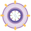
Liposomes, composed of phospholipid bilayers, are amphiphilic structures featuring a polar head group, a hydrophobic tail region, and a linker domain. Their ability to encapsulate and protect nucleic acids, combined with their biocompatibility and customizability, has made them indispensable in drug delivery systems [67,68]. Surface modifications further enable liposomes to achieve targeted delivery, enhance efficiency, and provide multifunctionality with minimal toxicity—key factors for effective RNA delivery [69].
Lipid nanoparticles (LNPs) are liposome-like structures, which are ideal for encapsulating and delivering various nucleic acids. In clinical LNPs, the surface charge is typically near-neutral under physiological conditions [70]. Interestingly, a recent study revealed that using an acidic buffer could impart positive charges to LNPs, improving their stability during nebulization and enabling efficient mRNA delivery to the lungs without significant toxicity [22]. These results indicate that the lungs may demonstrate better tolerance to positively charged nanoparticles, which frequently raise toxicity concerns following systemic injections in clinical studies. Building on these findings, Liu et al. developed a charge-assisted stabilization (CAS) strategy to enhance LNP colloidal stability via electrostatic repulsion. By incorporating peptide-lipid conjugates, the CAS-LNPs maintained structural integrity during nebulization and efficiently delivered mRNA to the lungs of mice, dogs, and pigs. These CAS-LNPs preferentially targeted lung dendritic cells, eliciting robust mucosal and systemic immune responses. Notably, they demonstrated potential as vaccines against the SARS-CoV-2 Omicron variant and as cancer vaccines for preventing lung metastases. These innovations not only address the instability and aggregation of LNPs during nebulization but also establish critical design principles for inhalable mRNA vaccines and therapeutics, opening new possibilities for treating pulmonary diseases and developing mucosal vaccines [23].
Li et al. combined machine learning with combinatorial chemistry to optimize ionizable lipids for mRNA delivery [24]. They developed a four-component reaction (4CR) platform that produced a library of 584 ionizable lipids, using machine learning to identify the most effective candidates. This approach demonstrated enhanced mRNA delivery, particularly in muscle and immune cells, while reducing off-target effects. Norimatsu et al. explored triphenylphosphonium (TPP)-modified cationic polymers to stabilize polyion complexes for mRNA delivery [71]. Their TPP-modified carriers significantly improved mRNA packaging and delivery, particularly in solid tumors, where they enhanced protein expression. Similarly, Han’s team introduced amidine-incorporated degradable lipids (AID-lipids), created through a fast one-pot multi-component reaction, to boost endosomal escape and mRNA delivery efficiency [25]. Their AID-lipid system supports applications like mRNA vaccines and gene-editing tools, offering a scalable and adaptable option for mRNA therapeutics. Additionally, Liu and colleagues developed a novel LNP system for organ-targeted mRNA therapy focused on the liver and lungs [26]. They found that cholesterol and phospholipids, though commonly used in LNP formulations, are not essential for effective delivery and instead contribute to unwanted liver accumulation. To overcome this limitation, they designed a three-component LNP system comprising nAcx-Cm lipids, permanently cationic lipids, and polyethylene glycol (PEG)-lipid. This formulation enables efficient pulmonary mRNA accumulation and translation, particularly in endothelial and epithelial cells. Compared to traditional four- or five-component LNPs containing cholesterol, the three-component system demonstrates superior efficacy, enhanced targeting specificity for lung delivery, and remarkable stability.
The further development of LNPs has led to the creation of cell-specific LNPs. Lian’s team demonstrated that incorporating stearic acid N-hydroxysuccinimide ester (SA-NHS) into LNPs enhanced bone marrow-homing (BM-homing) tropism, enabling transfection of 14 distinct cell types, including hematopoietic stem cells (HSCs) and immune cells [27]. The study highlighted the role of apolipoprotein E (ApoE) in BM-targeted delivery, potentially guiding new LNP designs for extrahepatic nucleic acid delivery in hematopoietic diseases. Li et al. explored the in situ engineering of CAR T cells using spleen-targeted LNPs, composed of DSPE-PEG2000-biotin conjugated with biotinylated anti-CD3 via streptavidin, to deliver CAR mRNA directly to T cells [28]. This approach simplified CAR T cell preparation and enhanced their anti-tumor function, showcasing the potential of organ-targeted mRNA therapies for solid tumors.
Despite their potential, LNPs face significant challenges in delivering ASOs. Byrnes et al. demonstrated that encapsulating ASOs in LNPs could enhance their activity by up to 100-fold in cultured primary brain cells compared to free ASOs. However, in vivo studies revealed a critical limitation: unlike free ASOs, LNP-delivered ASOs failed to achieve widespread mRNA downregulation across the brain following intracerebroventricular injection. This shortfall likely arises from ASO accumulation in cells lining the ventricles and blood vessels. Moreover, immune activation was observed post-dosing in a formulation-dependent manner, indicating that LNP encapsulation does not fully eliminate the cellular toxicities associated with the ASO backbone [72].

5. Extracellular Vesicles Delivery

Extracellular vesicles (EVs) have emerged as promising RNA delivery systems due to their natural ability to encapsulate and protect RNA molecules, enabling targeted delivery to specific cells [73]. Their biocompatibility and inherent targeting capabilities make them particularly attractive for therapeutic applications, especially in efficiently delivering RNA-based therapeutics [74].
Respiratory diseases, exacerbated by the COVID-19 pandemic, remain a significant global health challenge. While intramuscular mRNA-LNP vaccines have shown efficacy, their limited pulmonary bioavailability highlights the need for alternative delivery strategies. Inhaled nanoparticle therapeutics represent a promising solution, yet optimizing LNP formulations for efficient mRNA translation and pulmonary delivery remains a significant hurdle. In contrast, biological nanoparticles like EVs offer a compelling alternative, capitalizing on their natural ability to encapsulate mRNA and facilitate cellular delivery. Notably, EVs derived from pulmonary cells contain molecular components and membrane features uniquely suited to the lung microenvironment. For example, Popowski et al. demonstrated that lung-derived EVs (lung-Exo) exhibit superior distribution and retention of molecular drugs in deep lung tissues compared to synthetic nanoparticles. These lung-derived EVs thus represent a novel delivery system with enhanced pulmonary bioavailability, providing a tailored platform for delivering mRNA and protein-based therapeutics for lung diseases [29,30].
Beyond respiratory diseases, EV-based therapeutics have shown promise in other therapeutic areas. For instance, Ma et al. developed a clinically scalable method utilizing cellular nanoelectroporation with commercially available track-etched membranes (TM-nanoEP) to produce large quantities of small EVs (sEVs) loaded with therapeutic mRNAs and associated miRNAs. Unlike conventional methods that load therapeutics into EVs post-production, this approach uses parental cells as bioreactors. These cells endogenously transcribe plasmid DNAs (pDNAs), which generate therapeutic mRNAs that are subsequently packaged into multivesicular bodies (MVBs) or intraluminal vesicles (ILVs)—precursors of sEVs—and released as therapeutic sEVs (t-sEVs) via exocytosis. By optimizing electroporation parameters, TM-nanoEP achieved efficient loading of functional mRNAs (e.g., BMP-2 and VEGF-A) into abundant sEVs, significantly improving the efficacy of EV-based therapies. This method outperforms traditional approaches using naturally secreted or exogenously loaded RNAs like miRNAs and siRNAs. Moreover, the delivery of pDNAs not only enhances mRNA production but also promotes the secretion of miRNAs encoded by the pDNAs, creating a synergistic mRNA-miRNA therapeutic effect [31].
Recent advancements have further improved EVs by incorporating virus-derived structures, enhancing their potential for cancer and neurological disorder therapies. Wedge et al. explored EVs programmed by oncolytic viruses (OVs) to improve cancer treatment. These EVs, produced by virus-infected cancer cells, carry artificial miRNA (amiR-4), which targets and suppresses genes like ARID1A that contribute to cancer cell resistance against viral infections. This strategy allows EVs to spread amiR-4 to neighboring cancer cells, weakening their antiviral defenses and making them more vulnerable to viral attacks and small-molecule therapies. When combined with immunotherapies, this approach enhances cancer treatment by providing a broader, more targeted attack on cancer cells while minimizing harm to normal tissues [32]. Similarly, Gu’s team developed leukocyte-derived EVs containing retrovirus-like capsids to improve systemic mRNA delivery to neurons. This system effectively crosses the BBB, delivering mRNA in a neuroinflammation mouse model and offering an immune-inert platform for gene therapy in neurological disorders [33].
Further modifications to EV surface membranes have enhanced their utility in gene therapy for both cancer and neurological disorders. Cotto et al. developed a system for delivering mRNA to retinal photoreceptors by modifying EVs with cationic motifs. By anchoring cationic peptide carriers (CPC) and PEG groups to milk-derived EVs, they enhanced hydrophilicity and transport efficiency. This approach demonstrated excellent biocompatibility and successfully delivered mRNA to retinal cells, offering a promising method for localized gene therapy in retinal conditions [34]. In another study, Dong’s team engineered sEVs loaded with mRNA using microfluidic electroporation technology. By conjugating targeting ligands such as anti-CD71 and anti-PD-L1 antibodies to the sEVs, they achieved enhanced mRNA delivery to cancer cells, stimulating immune responses and offering a promising treatment for recurrent and drug-resistant tumors [35].
Although significant progress has been made in improving the targeting and delivery efficiency of EVs, loading large biomolecules into EVs remains a challenge. To address this, Malle’s team developed an innovative RNA-loading system based on a toehold-mediated strand displacement mechanism. The system facilitates the selective and efficient loading of mRNA into EVs by establishing a DNA-mediated fusion between mRNA-loaded liposomes and EVs. This method not only ensures efficient RNA loading but also allows for controlled RNA release and purification. The toehold structure plays a key role in precisely controlling RNA release from EVs, significantly enhancing delivery efficiency. Compared to conventional EV loading methods, this system improves RNA cargo selection and loading precision, thereby boosting the therapeutic potential of EVs for gene therapy and RNA-based treatments [75].

6. Other Nanomaterial-Based Delivery

Although RNA therapeutics have been successfully applied in the clinic using LNPs [76], challenges remain, particularly concerning toxicity. While cationic lipids in LNPs facilitate RNA encapsulation, they also carry toxicity risks [77]. For instance, cationic LNP components can interact with enzymes like protein kinase C, leading to cytotoxic effects [78,79]. High concentrations of these lipids can disrupt cellular membranes, causing cell lysis and necrosis [78,79]. To overcome these limitations, various nanoparticle carriers, including polymer-based (both natural and synthetic), inorganic, hybrid nanoparticles, and biological approaches using peptide nano-assemblies, have been actively studied [80].

6.1. Polymeric Nanoparticles

Naturally derived polymers, such as hyaluronic acid and chitosan, are widely utilized as mRNA carriers in drug delivery systems due to their biocompatibility and high modifiability. Among these, chitosan stands out for its cationic charge and ease of modification, which facilitate complex formation and surface adsorption [81]. Soliman et al. demonstrated that chitosan-based nanoparticles achieved a transfection efficiency of 60–65% in vitro by optimizing factors such as polymer length, degree of deacetylation, hyaluronic acid content, charge density, and nucleic acid composition [36]. Beyond mRNA delivery, chitosan nanoparticles have also been employed to deliver miRNA mimics, key biomarkers in cancer therapy [82]. These miRNA/chitosan complexes target macrophages to treat cardiovascular diseases, modulating gene expression both in vitro and in vivo. In mice, treatment with miRNA/chitosan nanoparticles reduced reverse cholesterol transport, highlighting their potential for addressing atherosclerotic lesions [83]. Furthermore, incorporating heparin into chitosan nanoparticles enhances transfection efficiency. Pilipenko et al. reported pH-dependent oligonucleotide release from heparin-loaded chitosan nanoparticles, driven by polyplex swelling and network collapse under mildly acidic conditions (pH 4.5). siRNA targeting vascular endothelial growth factor (VEGF) delivered via heparin-enriched chitosan nanoparticles showed 25% higher silencing efficiency in ARPE-19 cells compared to chitosan nanoparticles without heparin [37,84].
In addition to naturally derived polymers, synthetic polymers also play a critical role in RNA delivery. High-molecular-weight polyethylenimine (PEI) is considered one of the most effective non-viral cationic vectors for gene transfection. However, its high cytotoxicity limits clinical applications [85,86]. To mitigate this, fluorination was introduced to PEI, reducing cytotoxicity and enhancing its biodistribution control. Fluorinated PEI facilitated major accumulation of siRNA polyplex nanoparticles in the liver, while non-fluorinated PEI primarily delivered siRNA nanoparticles to the lungs following intravenous administration in mice [38]. Beyond cationic polymers, an anionic complex consisting of PEI, γ-polyglutamic acid, and mRNA demonstrated stable and high in vitro protein expression without cytotoxicity. Intravenous administration of this anionic complex resulted in high protein expression, predominantly in the liver and spleen [39].

6.2. Peptide-Derived Nanoparticles

Cell-penetrating peptides (CPPs) are short, cationic peptides (5–30 residues) capable of mediating intracellular delivery of diverse biological cargos, including large protein complexes and genetic materials, which typically struggle to penetrate cellular membranes. CPPs can traverse membranes via energy-dependent or independent mechanisms, making them promising candidates for peptide-based RNA delivery platforms [87,88].
While LNPs are widely used for systemic CRISPR-Cas9 delivery, their effectiveness is largely limited to the liver due to clearance mechanisms, restricting their application in extrahepatic targeting. In contrast, peptide-based nanoparticles offer advantages such as extrahepatic delivery, chemical diversity, and design flexibility [89]. Gonzalez et al. introduced a novel class of CPPs, termed “ADGN peptides”, which self-assemble into stable nanoparticles for efficient delivery of long mRNA and CRISPR components. These ADGN nanoparticles protect RNA, traverse cell membranes efficiently, and preferentially target cancer cells overexpressing the laminin receptor. In vitro, they achieved 60% CRISPR-Cas9-mediated knockout of the luciferase gene with a preference for G insertions at the target site. In vivo, systemically administered ADGN nanoparticles successfully delivered functional CRISPR-Cas9 systems to lung tumor cells, enabling effective gene knockout. Notably, their biodistribution depended on the peptide-to-RNA molar ratio, highlighting the tunability of this platform [40].
The KL4 peptide, modeled after pulmonary surfactant protein B (SP-B), has been utilized as an mRNA carrier with a linear PEG attachment. This PEG-KL4/mRNA complex exhibited superior transfection efficiency compared to naked mRNA or lipoplexes in mouse lungs after intratracheal administration [41]. Similarly, Levačić et al. developed a library of peptide-like oligoaminoamides with synthetic amino acids, histidine modifications, and bioreducible disulfide blocks, to enhance mRNA transfection efficiency [42].

6.3. Inorganic Nanoparticles

Inorganic materials possess ideal properties such as non-toxicity, hydrophilicity, biocompatibility, and high stability, making them invaluable in RNA packaging, protection, and delivery. Among these, gold nanoparticles (AuNPs) stand out due to their bioinert nature, ease of synthesis, and functionalization through thiol-gold bonds or electrostatic absorption. When functionalized with thiolated siRNAs, these nanoparticles—known as spherical nucleic acids (SNAs)—exhibit enhanced stability, high cellular uptake efficiency, and prolonged therapeutic activity. Notably, SNAs can cross the blood-brain and blood-tumor barriers to silence oncogenes and reduce tumor burden without inducing adverse side effects, thus paving the way for clinical applications [43]. Building on the SNA platform, gold cores modified with therapeutic agents or targeting ligands have been developed for gene and combination therapy. Alternatively, pristine RNAs can be noncovalently adsorbed onto polycation-capped AuNPs. For example, PEG-PEI-coated AuNPs have demonstrated potent siRNA delivery to pancreatic stellate cells, achieving effective silencing of HSP47 and reducing pancreatic cancer-associated desmoplasia [90].
Other widely applied bioinert carriers include carbon nanomaterials (e.g., carbon nanotubes, graphene, nanodiamonds) and mesoporous silica nanoparticles (MSNs). Functionalized carbon nanotubes deliver siRNAs to renal cells, halting acute kidney injury by silencing p53 and meprin-1b [44]. MSNs, with tunable pore sizes and cationic polymer coatings, enable RNA attachment and are ideal for combination therapy [45].
Dissolvable nanoparticles, such as calcium phosphate (CaP), reduce the risk of tissue accumulation and dissolve in acidic environments to release RNA cargo. Lipid-coated CaP nanoparticles significantly enhance systemic siRNA delivery to tumors [46]. Iron oxide nanoparticles, approved for imaging, serve as multifunctional platforms combining siRNA delivery, imaging, and magnetically controlled targeting for gene therapy [91]. Metal-organic frameworks (MOFs), with their high surface area, porosity, and biodegradability, offer efficient RNA delivery through coordination, encapsulation, or biomineralization, releasing RNA upon structural collapse and enhancing therapeutic efficacy [92].

7. Perspectives

Recent advancements in RNA delivery systems have shown great promise in therapeutic applications, particularly in vaccine development and disease treatment. Platforms such as viral vectors, LNPs, VLPs, and EVs each offer distinct advantages in improving the stability, specificity, and efficiency of RNA-based therapeutics (Table 1 and Table 2). Notably, LNPs have proven highly effective in the delivery of mRNA vaccines, as demonstrated by the success of COVID-19 vaccines, as well as in the development of siRNA-based drugs. However, each delivery system also presents unique challenges.
For instance, AAV have a limited DNA packaging capacity of 4.5 kb; recent studies indicate that AAV-delivered RNA molecules typically measure shorter than 2000 nt, although the maximum RNA length that AAVs can effectively accommodate is still unclear. Additionally, AAV’s transduction efficiency is dose-dependent: while higher doses can enhance efficiency, they also increase toxicity and immunogenicity [93,94,95].
In contrast, VLPs, which do not carry viral genomes, eliminate the risk of disease transmission. However, their virus-like symmetric and repetitive structures can provoke high immunogenicity. While this property makes VLPs promising vaccine candidates, it limits their use in therapeutic RNA drugs.
Furthermore, eukaryotic genomes contain domesticated genes from integrating viruses and mobile genetic elements, including homologs of the Gag protein found in LTR retrotransposons and retroviruses. Utilizing endogenous retrovirus-based delivery systems could potentially reduce immunogenicity, allowing for RNA delivery with minimal immune responses [21]. However, a major challenge remains: their transduction efficiency for targeted delivery is relatively low, highlighting the need for effective endogenous envelope glycoproteins to enhance targeting and delivery efficacy.
The toxicity of LNPs is particularly relevant in formulations containing cationic lipids, which are synthetic and whose toxicities may not be fully understood or have been observed in biological systems. In many cases, the effectiveness of a cationic lipid in LNP formulations is associated with increased toxicity. For instance, multivalent cationic lipids are more effective than monovalent ones in LNPs, but they also exhibit significantly higher toxicity to cells. Additionally, LNPs face significant challenges in delivering ASOs, as the delivered ASOs tend to accumulate in cells lining the ventricles and blood vessels.
To address the toxicity, various non-lipid nanoparticle carriers have been actively developed, including polymer-based, inorganic, hybrid nanoparticles, and biological approaches using peptide nano-assemblies. However, new challenges remain: polymeric vehicles suffer from low degradability and dose-limited toxicity with certain cationic polymers, as well as high charge density; the synthesis of inorganic nanoparticles is complex, limiting their scalability, and their low degradability may pose potential toxicity risks; peptide-derived nanoparticles often exhibit low stability and potential immunogenicity [92].
EVs have been explored as an endogenous alternative to LNPs, particularly in applications such as inhaled nanoparticle therapeutics. However, EVs present their own set of challenges, including heterogeneity, difficulties in large-scale production, and inconsistent RNA loading and targeting specificity—all of which must be addressed for successful clinical application [96].
The future of RNA delivery lies in combining the strengths of these systems to develop hybrid platforms that leverage their respective advantages. The study by Peng et al. effectively combines the advantages of LNPs and EVs by developing bio-functional lipid nanoparticles (LNP@HMVs) coated with neutrophil-bacterial hybrid cell membrane vesicles (HMVs). By integrating the targeting properties of EVs with the delivery efficiency of LNPs, this approach provides a promising strategy to combat antibiotic resistance and improve treatment outcomes in bacterial infections [97]. Zakas et al. explored a novel in vivo delivery approach based on an engineered transposase, Sleeping Beauty, delivered as an mRNA within a LNP, in combination with an rAAV-delivered transposable transgene [98]. Their findings offer the possibility of significantly reducing the dose used for clinical AAV gene editing therapies, promising to improve drug safety and reduce the cost of future gene therapies. Optimizing delivery systems through ancillary means can also be effective. Fitzgerald et al. demonstrated that using foam-based delivery carriers significantly improves transfection efficiency and homogeneity while reducing systemic exposure and targeting non-target tissues, thereby enhancing gene therapy safety [99]. Moreover, machine learning and AI are expected to play a critical role in optimizing nanoparticle design and improving targeting capabilities. Innovations in non-invasive delivery methods, such as ultrasound-based techniques, further contribute to enhancing the safety and efficiency of these therapies. A promising strategy to reduce immunogenicity involves utilizing endogenous retroviruses—rather than exogenous viruses—to construct fully endogenous delivery systems. This approach could facilitate RNA delivery with minimized immune responses. Additionally, certain endogenous retroviral envelope proteins, which are tissue-specific in their functions, can be leveraged as Env for endogenous VLPs to enhance the delivery and function of cargoed RNA. For instance, the primate-specific endogenous retroviral envelope protein ERVH48-1, which sequesters SFRP2 to regulate human cardiomyocyte development, could be used to improve RNA delivery in myocardial injury repair (Figure 1) [100].
Overall, as research progresses, RNA delivery systems will continue to evolve, making personalized medicine more achievable and providing novel treatments for previously untreatable conditions.

Funding

This work was supported by the Natural Science Foundation of Guangdong Province (2021A1515012478 to L.H.), and the Fundamental Research Funds for the Central Universities, Sun Yat-sen University (24qnpy091 to L.H.).

Conflicts of Interest

The authors declare no conflict of interest.

References

  1. Wang, F.; Li, P.; Chu, H.C.; Lo, P.K. Nucleic Acids and Their Analogues for Biomedical Applications. Biosensors 2022, 12, 93. [Google Scholar] [CrossRef] [PubMed]
  2. Bhatti, G.K.; Khullar, N.; Sidhu, I.S.; Navik, U.S.; Reddy, A.P.; Reddy, P.H.; Bhatti, J.S. Emerging role of non-coding RNA in health and disease. Metab. Brain Dis. 2021, 36, 1119–1134. [Google Scholar] [CrossRef] [PubMed]
  3. Wolff, J.A.; Malone, R.W.; Williams, P.; Chong, W.; Acsadi, G.; Jani, A.; Felgner, P.L. Direct gene transfer into mouse muscle in vivo. Science 1990, 247 Pt 1, 1465–1468. [Google Scholar] [CrossRef] [PubMed]
  4. Hu, B.; Zhong, L.; Weng, Y.; Peng, L.; Huang, Y.; Zhao, Y.; Liang, X.J. Therapeutic siRNA: State of the art. Signal Transduct. Target. Ther. 2020, 5, 101. [Google Scholar] [CrossRef] [PubMed]
  5. Muth, C.C. ASO Therapy: Hope for Genetic Neurological Diseases. JAMA 2018, 319, 644–646. [Google Scholar] [CrossRef]
  6. Vienberg, S.; Geiger, J.; Madsen, S.; Dalgaard, L.T. MicroRNAs in metabolism. Acta Physiol. 2017, 219, 346–361. [Google Scholar] [CrossRef]
  7. Lewin, A.S.; Hauswirth, W.W. Ribozyme gene therapy: Applications for molecular medicine. Trends Mol. Med. 2001, 7, 221–228. [Google Scholar] [CrossRef]
  8. Pardi, N.; Hogan, M.J.; Porter, F.W.; Weissman, D. mRNA vaccines—A new era in vaccinology. Nat. Rev. Drug Discov. 2018, 17, 261–279. [Google Scholar] [CrossRef]
  9. Malakondaiah, S.; Julius, A.; Ponnambalam, D.; Gunthoti, S.S.; Ashok, J.; Krishana, P.S.; Rebecca, J. Gene silencing by RNA interference: A review. Genome Instab. Dis. 2024, 5, 225–241. [Google Scholar] [CrossRef]
  10. Garbo, S.; Maione, R.; Tripodi, M.; Battistelli, C. Next RNA Therapeutics: The Mine of Non-Coding. Int. J. Mol. Sci. 2022, 23, 7471. [Google Scholar] [CrossRef]
  11. Saw, P.E.; Song, E. Advancements in clinical RNA therapeutics: Present developments and prospective outlooks. Cell Rep. Med. 2024, 5, 101555. [Google Scholar] [CrossRef] [PubMed]
  12. Fang, E.; Liu, X.; Li, M.; Zhang, Z.; Song, L.; Zhu, B.; Wu, X.; Liu, J.; Zhao, D.; Li, Y. Advances in COVID-19 mRNA vaccine development. Signal Transduct. Target. Ther. 2022, 7, 94. [Google Scholar] [CrossRef] [PubMed]
  13. Whitehead, K.A.; Langer, R.; Anderson, D.G. Knocking down barriers: Advances in siRNA delivery. Nat. Rev. Drug Discov. 2009, 8, 129–138. [Google Scholar] [CrossRef] [PubMed]
  14. Byun, J.; Wu, Y.; Park, J.; Kim, J.S.; Li, Q.; Choi, J.; Shin, N.; Lan, M.; Cai, Y.; Lee, J.; et al. RNA Nanomedicine: Delivery Strategies and Applications. Aaps J. 2023, 25, 95. [Google Scholar] [CrossRef]
  15. Dammes, N.; Peer, D. Paving the Road for RNA Therapeutics. Trends Pharmacol. Sci. 2020, 41, 755–775. [Google Scholar] [CrossRef]
  16. Bai, W.; Yang, D.; Zhao, Y.; Li, G.; Liu, Z.; Xiong, P.; Quan, H.; Wu, X.; Chen, P.; Kong, X.; et al. Multi-step engineered adeno-associated virus enables whole-brain mRNA delivery. bioRxiv 2024. [Google Scholar] [CrossRef]
  17. Feng, X.; Jiang, B.-W.; Zhai, S.-N.; Liu, C.-X.; Wu, H.; Zhu, B.-Q.; Wei, M.-Y.; Wei, J.; Yang, L.; Chen, L.-L. Circular RNA aptamers ameliorate AD-relevant phenotypes by targeting PKR. bioRxiv 2024. [Google Scholar] [CrossRef]
  18. Amado, D.A.; Robbins, A.B.; Smith, A.R.; Whiteman, K.R.; Chillon Bosch, G.; Chen, Y.; Fuller, J.A.; Izda, A.; Nelson, S.; Dichter, A.I.; et al. AAV-based delivery of RNAi targeting Ataxin-2 improves survival, strength, and pathology in mouse models of rapidly and slowly progressive sporadic ALS. bioRxiv 2024. [Google Scholar] [CrossRef]
  19. Zhang, P.; Narayanan, E.; Liu, Q.; Tsybovsky, Y.; Boswell, K.; Ding, S.; Hu, Z.; Follmann, D.; Lin, Y.; Miao, H.; et al. A multiclade env–gag VLP mRNA vaccine elicits tier-2 HIV-1-neutralizing antibodies and reduces the risk of heterologous SHIV infection in macaques. Nat. Med. 2021, 27, 2234–2245. [Google Scholar] [CrossRef]
  20. Hoffmann, M.A.G.; Yang, Z.; Huey-Tubman, K.E.; Cohen, A.A.; Gnanapragasam, P.N.P.; Nakatomi, L.M.; Storm, K.N.; Moon, W.J.; Lin, P.J.C.; West, A.P.; et al. ESCRT recruitment to SARS-CoV-2 spike induces virus-like particles that improve mRNA vaccines. Cell 2023, 186, 2380–2391.e9. [Google Scholar] [CrossRef]
  21. Segel, M.; Lash, B.; Song, J.; Ladha, A.; Liu, C.C.; Jin, X.; Mekhedov, S.L.; Macrae, R.K.; Koonin, E.V.; Zhang, F. Mammalian retrovirus-like protein PEG10 packages its own mRNA and can be pseudotyped for mRNA delivery. Science 2021, 373, 882–889. [Google Scholar] [CrossRef] [PubMed]
  22. Jiang, A.Y.; Witten, J.; Raji, I.O.; Eweje, F.; MacIsaac, C.; Meng, S.; Oladimeji, F.A.; Hu, Y.; Manan, R.S.; Langer, R.; et al. Combinatorial development of nebulized mRNA delivery formulations for the lungs. Nat. Nanotechnol. 2024, 19, 364–375. [Google Scholar] [CrossRef] [PubMed]
  23. Liu, S.; Wen, Y.; Shan, X.; Ma, X.; Yang, C.; Cheng, X.; Zhao, Y.; Li, J.; Mi, S.; Huo, H.; et al. Charge-assisted stabilization of lipid nanoparticles enables inhaled mRNA delivery for mucosal vaccination. Nat. Commun. 2024, 15, 9471. [Google Scholar] [CrossRef] [PubMed]
  24. Li, B.; Raji, I.O.; Gordon, A.G.R.; Sun, L.; Raimondo, T.M.; Oladimeji, F.A.; Jiang, A.Y.; Varley, A.; Langer, R.S.; Anderson, D.G. Accelerating ionizable lipid discovery for mRNA delivery using machine learning and combinatorial chemistry. Nat. Mater. 2024, 23, 1002–1008. [Google Scholar] [CrossRef]
  25. Han, X.; Alameh, M.G.; Gong, N.; Xue, L.; Ghattas, M.; Bojja, G.; Xu, J.; Zhao, G.; Warzecha, C.C.; Padilla, M.S.; et al. Fast and facile synthesis of amidine-incorporated degradable lipids for versatile mRNA delivery in vivo. Nat. Chem. 2024, 16, 1687–1697. [Google Scholar] [CrossRef]
  26. Su, K.; Shi, L.; Sheng, T.; Yan, X.; Lin, L.; Meng, C.; Wu, S.; Chen, Y.; Zhang, Y.; Wang, C.; et al. Reformulating lipid nanoparticles for organ-targeted mRNA accumulation and translation. Nat. Commun. 2024, 15, 5659. [Google Scholar] [CrossRef]
  27. Lian, X.; Chatterjee, S.; Sun, Y.; Dilliard, S.A.; Moore, S.; Xiao, Y.; Bian, X.; Yamada, K.; Sung, Y.-C.; Levine, R.M.; et al. Bone-marrow-homing lipid nanoparticles for genome editing in diseased and malignant haematopoietic stem cells. Nat. Nanotechnol. 2024, 19, 1409–1417. [Google Scholar] [CrossRef]
  28. Li, Y.; Tian, Y.; Li, C.; Fang, W.; Li, X.; Jing, Z.; Yang, Z.; Zhang, X.; Huang, Y.; Gong, J.; et al. In situ engineering of mRNA-CAR T cells using spleen-targeted ionizable lipid nanoparticles to eliminate cancer cells. Nano Today 2024, 59, 102518. [Google Scholar] [CrossRef]
  29. Popowski, K.; Lutz, H.; Hu, S.; George, A.; Dinh, P.U.; Cheng, K. Exosome therapeutics for lung regenerative medicine. J. Extracell Vesicles 2020, 9, 1785161. [Google Scholar] [CrossRef]
  30. Popowski, K.D.; López de Juan Abad, B.; George, A.; Silkstone, D.; Belcher, E.; Chung, J.; Ghodsi, A.; Lutz, H.; Davenport, J.; Flanagan, M.; et al. Inhalable exosomes outperform liposomes as mRNA and protein drug carriers to the lung. Extracell. Vesicle 2022, 1, 100002. [Google Scholar] [CrossRef]
  31. Ma, Y.; Sun, L.; Zhang, J.; Chiang, C.-L.; Pan, J.; Wang, X.; Kwak, K.J.; Li, H.; Zhao, R.; Rima, X.Y.; et al. Exosomal mRNAs for Angiogenic–Osteogenic Coupled Bone Repair. Adv. Sci. 2023, 10, 2302622. [Google Scholar] [CrossRef] [PubMed]
  32. Wedge, M.-E.; Jennings, V.A.; Crupi, M.J.F.; Poutou, J.; Jamieson, T.; Pelin, A.; Pugliese, G.; de Souza, C.T.; Petryk, J.; Laight, B.J.; et al. Virally programmed extracellular vesicles sensitize cancer cells to oncolytic virus and small molecule therapy. Nat. Commun. 2022, 13, 1898. [Google Scholar] [CrossRef] [PubMed]
  33. Gu, W.; Luozhong, S.; Cai, S.; Londhe, K.; Elkasri, N.; Hawkins, R.; Yuan, Z.; Su-Greene, K.; Yin, Y.; Cruz, M.; et al. Extracellular vesicles incorporating retrovirus-like capsids for the enhanced packaging and systemic delivery of mRNA into neurons. Nat. Biomed. Eng. 2024, 8, 415–426. [Google Scholar] [CrossRef]
  34. Millán Cotto, H.A.; Pathrikar, T.V.; Hakim, B.; Baby, H.M.; Zhang, H.; Zhao, P.; Ansaripour, R.; Amini, R.; Carrier, R.L.; Bajpayee, A.G. Cationic-motif-modified exosomes for mRNA delivery to retinal photoreceptors. J. Mater. Chem. B 2024, 12, 7384–7400. [Google Scholar] [CrossRef]
  35. Dong, S.; Liu, X.; Bi, Y.; Wang, Y.; Antony, A.; Lee, D.; Huntoon, K.; Jeong, S.; Ma, Y.; Li, X.; et al. Adaptive design of mRNA-loaded extracellular vesicles for targeted immunotherapy of cancer. Nat. Commun. 2023, 14, 6610. [Google Scholar] [CrossRef]
  36. Soliman, O.Y.; Alameh, M.G.; De Cresenzo, G.; Buschmann, M.D.; Lavertu, M. Efficiency of Chitosan/Hyaluronan-Based mRNA Delivery Systems In Vitro: Influence of Composition and Structure. J. Pharm. Sci. 2020, 109, 1581–1593. [Google Scholar] [CrossRef]
  37. Pilipenko, I.; Korzhikov-Vlakh, V.; Sharoyko, V.; Zhang, N.; Schäfer-Korting, M.; Rühl, E.; Zoschke, C.; Tennikova, T. pH-Sensitive Chitosan-Heparin Nanoparticles for Effective Delivery of Genetic Drugs into Epithelial Cells. Pharmaceutics 2019, 11, 317. [Google Scholar] [CrossRef]
  38. Xue, L.; Yan, Y.; Kos, P.; Chen, X.; Siegwart, D.J. PEI fluorination reduces toxicity and promotes liver-targeted siRNA delivery. Drug Deliv. Transl. Res. 2021, 11, 255–260. [Google Scholar] [CrossRef]
  39. Hamada, E.; Kurosaki, T.; Hashizume, J.; Harasawa, H.; Nakagawa, H.; Nakamura, T.; Kodama, Y.; Sasaki, H. Anionic Complex with Efficient Expression and Good Safety Profile for mRNA Delivery. Pharmaceutics 2021, 13, 126. [Google Scholar] [CrossRef]
  40. Guzman Gonzalez, V.; Grunenberger, A.; Nicoud, O.; Czuba, E.; Vollaire, J.; Josserand, V.; Le Guével, X.; Desai, N.; Coll, J.-L.; Divita, G.; et al. Enhanced CRISPR-Cas9 RNA system delivery using cell penetrating peptides-based nanoparticles for efficient in vitro and in vivo applications. J. Control. Release 2024, 376, 1160–1175. [Google Scholar] [CrossRef]
  41. Qiu, Y.; Man, R.C.H.; Liao, Q.; Kung, K.L.K.; Chow, M.Y.T.; Lam, J.K.W. Effective mRNA pulmonary delivery by dry powder formulation of PEGylated synthetic KL4 peptide. J. Control. Release 2019, 314, 102–115. [Google Scholar] [CrossRef] [PubMed]
  42. Krhač Levačić, A.; Berger, S.; Müller, J.; Wegner, A.; Lächelt, U.; Dohmen, C.; Rudolph, C.; Wagner, E. Dynamic mRNA polyplexes benefit from bioreducible cleavage sites for in vitro and in vivo transfer. J. Control. Release 2021, 339, 27–40. [Google Scholar] [CrossRef] [PubMed]
  43. Jensen, S.A.; Day, E.S.; Ko, C.H.; Hurley, L.A.; Luciano, J.P.; Kouri, F.M.; Merkel, T.J.; Luthi, A.J.; Patel, P.C.; Cutler, J.I.; et al. Spherical nucleic acid nanoparticle conjugates as an RNAi-based therapy for glioblastoma. Sci. Transl. Med. 2013, 5, 209ra152. [Google Scholar] [CrossRef] [PubMed]
  44. Alidori, S.; Akhavein, N.; Thorek, D.L.; Behling, K.; Romin, Y.; Queen, D.; Beattie, B.J.; Manova-Todorova, K.; Bergkvist, M.; Scheinberg, D.A.; et al. Targeted fibrillar nanocarbon RNAi treatment of acute kidney injury. Sci. Transl. Med. 2016, 8, 331ra39. [Google Scholar] [CrossRef]
  45. Meng, H.; Mai, W.X.; Zhang, H.; Xue, M.; Xia, T.; Lin, S.; Wang, X.; Zhao, Y.; Ji, Z.; Zink, J.I.; et al. Codelivery of an Optimal Drug/siRNA Combination Using Mesoporous Silica Nanoparticles To Overcome Drug Resistance in Breast Cancer in Vitro and in Vivo. ACS Nano 2013, 7, 994–1005. [Google Scholar] [CrossRef]
  46. Li, J.; Chen, Y.-C.; Tseng, Y.-C.; Mozumdar, S.; Huang, L. Biodegradable calcium phosphate nanoparticle with lipid coating for systemic siRNA delivery. J. Control. Release 2010, 142, 416–421. [Google Scholar] [CrossRef]
  47. Finer, M.; Glorioso, J. A brief account of viral vectors and their promise for gene therapy. Gene Ther. 2017, 24, 1–2. [Google Scholar] [CrossRef]
  48. Wang, D.; Tai, P.W.L.; Gao, G. Adeno-associated virus vector as a platform for gene therapy delivery. Nat. Rev. Drug Discov. 2019, 18, 358–378. [Google Scholar] [CrossRef]
  49. Thomas, C.E.; Ehrhardt, A.; Kay, M.A. Progress and problems with the use of viral vectors for gene therapy. Nat. Rev. Genet. 2003, 4, 346–358. [Google Scholar] [CrossRef]
  50. Lundstrom, K. Viral Vectors in Gene Therapy: Where Do We Stand in 2023? Viruses 2023, 15, 698. [Google Scholar] [CrossRef]
  51. Zhao, Z.; Anselmo, A.C.; Mitragotri, S. Viral vector-based gene therapies in the clinic. Bioeng. Transl. Med. 2022, 7, e10258. [Google Scholar] [CrossRef] [PubMed]
  52. Samulski, R.J.; Muzyczka, N. AAV-Mediated Gene Therapy for Research and Therapeutic Purposes. Annu. Rev. Virol. 2014, 1, 427–451. [Google Scholar] [CrossRef] [PubMed]
  53. King, J.A.; Dubielzig, R.; Grimm, D.; Kleinschmidt, J.A. DNA helicase-mediated packaging of adeno-associated virus type 2 genomes into preformed capsids. EMBO J. 2001, 20, 3282–3291. [Google Scholar] [CrossRef] [PubMed]
  54. Maurer, A.C.; Weitzman, M.D. Adeno-Associated Virus Genome Interactions Important for Vector Production and Transduction. Hum. Gene Ther. 2020, 31, 499–511. [Google Scholar] [CrossRef]
  55. Mitusova, K.; Peltek, O.O.; Karpov, T.E.; Muslimov, A.R.; Zyuzin, M.V.; Timin, A.S. Overcoming the blood-brain barrier for the therapy of malignant brain tumor: Current status and prospects of drug delivery approaches. J. Nanobiotechnology 2022, 20, 412. [Google Scholar] [CrossRef]
  56. Ranum, P.T.; Tecedor, L.; Keiser, M.S.; Chen, Y.H.; Leib, D.E.; Liu, X.; Davidson, B.L. Cochlear transduction via cerebrospinal fluid delivery of AAV in non-human primates. Mol. Ther. 2023, 31, 609–612. [Google Scholar] [CrossRef]
  57. Chung, Y.H.; Cai, H.; Steinmetz, N.F. Viral nanoparticles for drug delivery, imaging, immunotherapy, and theranostic applications. Adv. Drug Deliv. Rev. 2020, 156, 214–235. [Google Scholar] [CrossRef]
  58. Lyu, P.; Wang, L.; Lu, B. Virus-Like Particle Mediated CRISPR/Cas9 Delivery for Efficient and Safe Genome Editing. Life 2020, 10, 366. [Google Scholar] [CrossRef]
  59. Lyu, P.; Lu, B. New Advances in Using Virus-like Particles and Related Technologies for Eukaryotic Genome Editing Delivery. Int. J. Mol. Sci. 2022, 23, 8750. [Google Scholar] [CrossRef]
  60. Goodier, J.L.; Kazazian, H.H. Retrotransposons Revisited: The Restraint and Rehabilitation of Parasites. Cell 2008, 135, 23–35. [Google Scholar] [CrossRef]
  61. Geuens, T.; Bouhy, D.; Timmerman, V. The hnRNP family: Insights into their role in health and disease. Hum. Genet. 2016, 135, 851–867. [Google Scholar] [CrossRef] [PubMed]
  62. Patel, M.R.; Emerman, M.; Malik, H.S. Paleovirology—Ghosts and gifts of viruses past. Curr. Opin Virol. 2011, 1, 304–309. [Google Scholar] [CrossRef] [PubMed]
  63. Hogg, J.R.; Collins, K. RNA-based affinity purification reveals 7SK RNPs with distinct composition and regulation. RNA 2007, 13, 868–880. [Google Scholar] [CrossRef] [PubMed]
  64. Feschotte, C.; Gilbert, C. Endogenous viruses: Insights into viral evolution and impact on host biology. Nat. Rev. Genet. 2012, 13, 283–296. [Google Scholar] [CrossRef]
  65. Park, J.E.; Botting, R.A.; Domínguez Conde, C.; Popescu, D.M.; Lavaert, M.; Kunz, D.J.; Goh, I.; Stephenson, E.; Ragazzini, R.; Tuck, E.; et al. A cell atlas of human thymic development defines T cell repertoire formation. Science 2020, 367, eaay3224. [Google Scholar] [CrossRef]
  66. Pham, V.V.; Gao, M.; Meagher, J.L.; Smith, J.L.; D’Souza, V.M. A structure-based mechanism for displacement of the HEXIM adapter from 7SK small nuclear RNA. Commun. Biol. 2022, 5, 819. [Google Scholar] [CrossRef]
  67. Allen, T.M.; Cullis, P.R. Liposomal drug delivery systems: From concept to clinical applications. Adv. Drug Deliv. Rev. 2013, 65, 36–48. [Google Scholar] [CrossRef]
  68. Filipczak, N.; Pan, J.; Yalamarty, S.S.K.; Torchilin, V.P. Recent advancements in liposome technology. Adv. Drug Deliv. Rev. 2020, 156, 4–22. [Google Scholar] [CrossRef]
  69. Cullis, P.R.; Hope, M.J. Lipid Nanoparticle Systems for Enabling Gene Therapies. Mol. Ther. 2017, 25, 1467–1475. [Google Scholar] [CrossRef]
  70. Thi, T.T.H.; Suys, E.J.A.; Lee, J.S.; Nguyen, D.H.; Park, K.D.; Truong, N.P. Lipid-Based Nanoparticles in the Clinic and Clinical Trials: From Cancer Nanomedicine to COVID-19 Vaccines. Vaccines 2021, 9, 359. [Google Scholar] [CrossRef]
  71. Norimatsu, J.; Mizuno, H.L.; Watanabe, T.; Obara, T.; Nakakido, M.; Tsumoto, K.; Cabral, H.; Kuroda, D.; Anraku, Y. Triphenylphosphonium-modified catiomers enhance in vivo mRNA delivery through stabilized polyion complexation. Mater. Horiz. 2024, 11, 4711–4721. [Google Scholar] [CrossRef] [PubMed]
  72. Byrnes, A.E.; Dominguez, S.L.; Yen, C.-W.; Laufer, B.I.; Foreman, O.; Reichelt, M.; Lin, H.; Sagolla, M.; Hötzel, K.; Ngu, H.; et al. Lipid nanoparticle delivery limits antisense oligonucleotide activity and cellular distribution in the brain after intracerebroventricular injection. Mol. Ther. Nucleic Acids 2023, 32, 773–793. [Google Scholar] [CrossRef] [PubMed]
  73. Thery, C.; Witwer, K.W.; Aikawa, E.; Alcaraz, M.J.; Anderson, J.D.; Andriantsitohaina, R.; Antoniou, A.; Arab, T.; Archer, F.; Atkin-Smith, G.K.; et al. Minimal information for studies of extracellular vesicles 2018 (MISEV2018): A position statement of the International Society for Extracellular Vesicles and update of the MISEV2014 guidelines. J. Extracell. Vesicles 2018, 7, 1535750. [Google Scholar] [CrossRef] [PubMed]
  74. Barile, L.; Vassalli, G. Exosomes: Therapy delivery tools and biomarkers of diseases. Pharmacol. Ther. 2017, 174, 63–78. [Google Scholar] [CrossRef]
  75. Malle, M.G.; Song, P.; Loffler, P.M.G.; Kalisi, N.; Yan, Y.; Valero, J.; Vogel, S.; Kjems, J. Programmable RNA Loading of Extracellular Vesicles with Toehold-Release Purification. J. Am. Chem. Soc. 2024, 146, 12410–12422. [Google Scholar] [CrossRef]
  76. Hou, X.; Zaks, T.; Langer, R.; Dong, Y. Lipid nanoparticles for mRNA delivery. Nat. Rev. Mater. 2021, 6, 1078–1094. [Google Scholar] [CrossRef]
  77. Tenchov, R.; Bird, R.; Curtze, A.E.; Zhou, Q. Lipid Nanoparticles—From Liposomes to mRNA Vaccine Delivery, a Landscape of Research Diversity and Advancement. ACS Nano 2021, 15, 16982–17015. [Google Scholar] [CrossRef]
  78. Xue, H.Y.; Liu, S.; Wong, H.L. Nanotoxicity: A key obstacle to clinical translation of siRNA-based nanomedicine. Nanomedicine 2014, 9, 295–312. [Google Scholar] [CrossRef]
  79. Ho, W.; Gao, M.; Li, F.; Li, Z.; Zhang, X.Q.; Xu, X. Next-Generation Vaccines: Nanoparticle-Mediated DNA and mRNA Delivery. Adv. Healthc. Mater. 2021, 10, e2001812. [Google Scholar] [CrossRef]
  80. Smith, S.A.; Selby, L.I.; Johnston, A.P.R.; Such, G.K. The Endosomal Escape of Nanoparticles: Toward More Efficient Cellular Delivery. Bioconjug Chem. 2019, 30, 263–272. [Google Scholar] [CrossRef]
  81. Cao, Y.; Tan, Y.F.; Wong, Y.S.; Liew, M.W.J.; Venkatraman, S. Recent Advances in Chitosan-Based Carriers for Gene Delivery. Mar. Drugs 2019, 17, 381. [Google Scholar] [CrossRef] [PubMed]
  82. Lee, W.-J.; Kim, K.-J.; Hossain, M.K.; Cho, H.-Y.; Choi, J.-W. DNA–Gold Nanoparticle Conjugates for Intracellular miRNA Detection Using Surface-Enhanced Raman Spectroscopy. BioChip J. 2022, 16, 33–40. [Google Scholar] [CrossRef]
  83. Nguyen, M.A.; Wyatt, H.; Susser, L.; Geoffrion, M.; Rasheed, A.; Duchez, A.C.; Cottee, M.L.; Afolayan, E.; Farah, E.; Kahiel, Z.; et al. Delivery of MicroRNAs by Chitosan Nanoparticles to Functionally Alter Macrophage Cholesterol Efflux in Vitro and in Vivo. ACS Nano 2019, 13, 6491–6505. [Google Scholar] [CrossRef]
  84. Shtykalova, S.; Deviatkin, D.; Freund, S.; Egorova, A.; Kiselev, A. Non-Viral Carriers for Nucleic Acids Delivery: Fundamentals and Current Applications. Life 2023, 13, 903. [Google Scholar] [CrossRef]
  85. Casper, J.; Schenk, S.H.; Parhizkar, E.; Detampel, P.; Dehshahri, A.; Huwyler, J. Polyethylenimine (PEI) in gene therapy: Current status and clinical applications. J. Control. Release 2023, 362, 667–691. [Google Scholar] [CrossRef]
  86. Wang, M.; Zhao, J.; Jiang, H.; Wang, X. Tumor-targeted nano-delivery system of therapeutic RNA. Mater. Horiz. 2022, 9, 1111–1140. [Google Scholar] [CrossRef]
  87. Jones, S.W.; Christison, R.; Bundell, K.; Voyce, C.J.; Brockbank, S.M.; Newham, P.; Lindsay, M.A. Characterisation of cell-penetrating peptide-mediated peptide delivery. Br. J. Pharmacol. 2005, 145, 1093–1102. [Google Scholar] [CrossRef]
  88. Shoari, A.; Tooyserkani, R.; Tahmasebi, M.; Löwik, D. Delivery of Various Cargos into Cancer Cells and Tissues via Cell-Penetrating Peptides: A Review of the Last Decade. Pharmaceutics 2021, 13, 1391. [Google Scholar] [CrossRef]
  89. Wickline, S.A.; Hou, K.K.; Pan, H. Peptide-Based Nanoparticles for Systemic Extrahepatic Delivery of Therapeutic Nucleotides. Int. J. Mol. Sci. 2023, 24, 9455. [Google Scholar] [CrossRef]
  90. Elbakry, A.; Zaky, A.; Liebl, R.; Rachel, R.; Goepferich, A.; Breunig, M. Layer-by-Layer Assembled Gold Nanoparticles for siRNA Delivery. Nano Lett. 2009, 9, 2059–2064. [Google Scholar] [CrossRef]
  91. Li, Y.; Wang, X.; Zhang, Y.; Nie, G. Recent Advances in Nanomaterials with Inherent Optical and Magnetic Properties for Bioimaging and Imaging-Guided Nucleic Acid Therapy. Bioconjugate Chem. 2020, 31, 1234–1246. [Google Scholar] [CrossRef] [PubMed]
  92. Han, X.; Mitchell, M.J.; Nie, G. Nanomaterials for Therapeutic RNA Delivery. Matter 2020, 3, 1948–1975. [Google Scholar] [CrossRef]
  93. Lu, Q.; Wright, A.; Pan, Z.-H. AAV dose-dependent transduction efficiency in retinal ganglion cells and functional efficacy of optogenetic vision restoration. Gene Ther. 2024, 31, 572–579. [Google Scholar] [CrossRef] [PubMed]
  94. Stone, D.; Aubert, M.; Jerome, K.R. Breaching the blood-brain barrier: AAV triggers dose-dependent toxicity in the brain. Mol. Ther. Methods Clin. Dev. 2023, 31, 101105. [Google Scholar] [CrossRef] [PubMed]
  95. Costa Verdera, H.; Kuranda, K.; Mingozzi, F. AAV Vector Immunogenicity in Humans: A Long Journey to Successful Gene Transfer. Mol. Ther. 2020, 28, 723–746. [Google Scholar] [CrossRef]
  96. Elsharkasy, O.M.; Nordin, J.Z.; Hagey, D.W.; de Jong, O.G.; Schiffelers, R.M.; Andaloussi, S.E.; Vader, P. Extracellular vesicles as drug delivery systems: Why and how? Adv. Drug Deliv. Rev. 2020, 159, 332–343. [Google Scholar] [CrossRef]
  97. Peng, X.; Chen, J.; Gan, Y.; Yang, L.; Luo, Y.; Bu, C.; Huang, Y.; Chen, X.; Tan, J.; Yang, Y.Y.; et al. Biofunctional lipid nanoparticles for precision treatment and prophylaxis of bacterial infections. Sci. Adv. 2024, 10, eadk9754. [Google Scholar] [CrossRef]
  98. Zakas, P.M.; Cunningham, S.C.; Doherty, A.; van Dijk, E.B.; Ibraheim, R.; Yu, S.; Mekonnen, B.D.; Lang, B.; English, E.J.; Sun, G.; et al. Sleeping Beauty mRNA-LNP enables stable rAAV transgene expression in mouse and NHP hepatocytes and improves vector potency. Mol. Ther. 2024, 32, 3356–3371. [Google Scholar] [CrossRef]
  99. Fitzgerald, K.; Stephan, S.B.; Ma, N.; Wu, Q.V.; Stephan, M.T. Liquid foam improves potency and safety of gene therapy vectors. Nat. Commun. 2024, 15, 4523. [Google Scholar] [CrossRef]
  100. Zhang, R.; Wu, M.; Xiang, D.; Zhu, J.; Zhang, Q.; Zhong, H.; Peng, Y.; Wang, Z.; Ma, G.; Li, G.; et al. A primate-specific endogenous retroviral envelope protein sequesters SFRP2 to regulate human cardiomyocyte development. Cell Stem Cell 2024, 31, 1298–1314.e8. [Google Scholar] [CrossRef]
Figure 1. Schematic showing the components of SEND. SEND is a delivery platform that integrates an endogenous Gag homolog, a fusogen (glycoprotein), and cargo mRNA, allowing customization for specific applications.
Cimb 47 00022 g001
Table 1. Comparative features of AAVs, VLPs, lipid nanoparticles (LNPs), and EVs as RNA delivery systems.
NameSizeGenomeAdvantagesChallenges
Adeno-Associated Virus (AAV)
Cimb 47 00022 i001
20–25 nmSingle-stranded DNA, up to 4.7 kb
  • High transduction efficiency in various cell types.
  • Long-term expression in target cells.
  • Low immunogenicity and safety profile.
  • Limited packaging capacity for larger genes.
  • Pre-existing immunity in human populations can reduce effectiveness.
  • Production and purification can be complex and costly.
Virus-Like Particles (VLPs)
Cimb 47 00022 i002
20–200 nmNo viral genome; can be engineered to carry RNA
  • Mimic natural viruses, enhancing cellular uptake and immune response.
  • High stability and safety as they are non-infectious.
  • Versatile for various types of genetic material.
  • Production can be challenging and require specific conditions.
  • Limited capacity for carrying large RNA constructs.
  • Potential immunogenicity depending on the source.
Lipid Nanoparticles (LNPs)
Cimb 47 00022 i003
50–150 nmEncapsulate mRNA or other RNA molecules
  • High delivery efficiency to a wide range of cell types.
  • Protect RNA from degradation and facilitate endosomal escape.
  • Scalable and adaptable for different RNA therapeutics.
  • Formulation can impact stability and delivery efficiency.
  • Potential toxicity and immunogenicity issues.
  • Optimization is required for organ-specific delivery.
Extracellular Vesicles (EVs)
Cimb 47 00022 i004
30–1000 nmNaturally contain RNA and can load therapeutic RNA
  • Biocompatibility and low immunogenicity, as they are derived from cells.
  • Natural targeting mechanisms can enhance delivery to specific tissues.
  • Capable of delivering complex cargo, including proteins and nucleic acids.
  • Isolation and characterization can be complex and time-consuming.
  • Variability in composition and efficacy based on source.
  • Limited control over cargo loading and release mechanisms.
Table 2. RNA delivery systems and their targeting specificity for tissues and cell types.
NameModificationTargetReference
Viral deliveryReplaced AAV’s inverted terminal repeats (ITRs) with RNA packaging signals (RPSs) The blood-brain barrier (BBB)Bai et al. [16]
Developed circular RNAs with short-imperfect duplex regions (ds-cRNAs) containing ITRs for AAV packagingNeurons and MicrogliaFeng et al. [17]
Designed microRNAs (miRNAs) targeting Atxn2, flanked by AAV2 145-bp ITR sequences, and packaged them into a peptide-modified AAV9 (PM-AAV9) vector Central Nervous System (CNS)Amado et al. [18]
Virus-like particle deliveryUsed HIV-derived VLPs to develop an mRNA vaccine co-expressing HIV-1 Env and SIV Gag proteinsT cellZhang et al. [19]
Engineered self-assembling VLPs by incorporating ESCRT proteins into the SARS-CoV-2 spike protein, enhancing spike protein enrichment on VLPs. T cellHoffmann et al. [20]
Developed selective endogenous eNcapsidation for cellular delivery (SEND) by engineering mouse and human PEG10 to package, secrete, and deliver specific RNAs.Mammalian CellsSegel et al. [21]
Lipid nanoparticle deliverySynthesized a combinatorial library of ionizable, degradable lipids using reductive amination.Lung Jiang et al. [22]
Developed charge-assisted stabilized lipid nanoparticles (CAS-LNPs) by optimizing surface charges with a peptide-lipid conjugatePulmonaryLiu et al. [23]
Combined machine learning with combinatorial chemistry to identify the most effective candidates. Muscle and immune cellsLi et al. [24]
Develop a one-pot, tandem multi-component reaction based on the rationally designed amine–thiolacrylate conjugation Lung and spleenHan et al. [25]
Designed a three-component LNP system comprising nAcx-Cm lipids, permanently cationic lipids, and polyethylene glycol (PEG)-lipid.LungSu et al. [26]
Designed a three-component LNP system comprising nAcx-Cm lipids, permanently cationic lipids, and lyethylene glycol (PEG)-lipid.Liver and lungsLian et al. [27]
Explored in situ engineering of CAR T cells using spleen-targeted LNPs, composed of DSPE-PEG2000-biotin conjugated with biotinylated anti-CD3 via streptavidinT cellsLi et al. [28]
Extracellular vesicles deliveryOptimized exosomes by isolating them from lung tissues Lung Popowski et al. [29,30]
Generated exosomes enriched with angiogenesis- and osteogenesis-related mRNAs (e.g., VEGF and BMP2) by engineering bone marrow mesenchymal stem cells (BMSCs) BoneMa et al. [31]
Explored EVs programmed by oncolytic viruses (OVs)Cancer cellsWedge et al. [32]
Developed leukocyte-derived EVs containing retrovirus-like capsidsNeuronsGu et al. [33]
Anchoring cationic peptide carriers (CPC) and polyethylene glycol (PEG) groups to milk-derived EVsRetinal photoreceptorsCotto et al. [34]
Engineered sEVs loaded with mRNA using microfluidic electroporation technology and by conjugating targeting ligands such as anti-CD71 and anti-PD-L1 antibodies to the sEVsTumorsDong et al. [35]
Polymeric nanoparticlesOptimizing factors such as polymer length, degree of deacetylation, hyaluronic acid content, charge density, and nucleic acid compositionMacrophagesSoliman et al. [36]
Incorporating heparin into chitosan nanoparticlesARPE-19 cellsPilipenko et al. [37]
Fluorination was introduced to PEILiverXue et al. [38]
An anionic complex consisting of PEI, γ-polyglutamic acid, and mRNALiver and spleenHamada et al. [39]
Peptide-derived nanoparticlesIntroduced a novel class of CPPs by optimizing the sequence, which can self-assemble into stable nanoparticles and can be adjusted in concentration to determine their biodistributionLung tumor cellsGonzalez et al. [40]
The KL4 peptide, modeled after pulmonary surfactant protein B (SP-B), has been utilized as an mRNA carrier with a linear PEG attachment.LungQiu et al. [41]
Developed a library of peptide-like oligoaminoamides with synthetic amino acids, histidine modifications, and bioreducible disulfide blocksTumor CellsKrhač et al. [42]
Inorganic nanoparticlesUsed spherical nucleic acids (SNAs) with a dense shell of RNA oligonucleotides conjugated to a nanoparticle coreGlioblastoma cellsJensen et al. [43]
Functionalized nanocarbon fibers were modified with targeting ligands for specific accumulationKidney cellsAlidori et al. [44]
Mesoporous silica nanoparticles (MSNs) were designed for dual loading of chemotherapeutic drugs and siRNA, with surface functionalization to enhance endosomal escape and target delivery.Breast cancer cellsMeng et al. [45]
Lipid-coated biodegradable calcium phosphate nanoparticles (LCPs) were developed for systemic delivery, with optimized particle size and surface charge for tumor targeting.Tumor cellsLi et al. [46]
Table 3. Comparison of retrovirus, lentivirus, HSV, adenovirus, and AAV for gene delivery.
TropismCloning CapacityGenomic IntegrationDuration of Expression In VivoAdvantagesDisadvantages
RetrovirusDividing cells (hematopoietic cells).Moderate (~7–8 kb).Stable integration into the genome.Long-term (due to stable integration).Stable gene transfer in dividing cells.Insertional mutagenesis, limited tropism.
LentivirusBroad (dividing and non-dividing cells).Moderate (~7–8 kb).Stable integration into the genome (both dividing and non-dividing cells).Long-term (stable expression).Stable expression in a wide range of cells.Risk of insertional mutagenesis, immunogenicity.
Herpes Simplex Virus (HSV)Primarily neurons, can be engineered for other tissues.Large (~150 kb).Episomal (no integration).Long-term in neurons (latent potential).High cloning capacity, long-term expression in neurons.Latency and reactivation risks, immunogenicity.
AdenovirusBroad (respiratory, liver, epithelial).Moderate (~36 kb).Episomal (no integration).Short-term (due to immune response).High transduction efficiency, large cargo capacity.Short-lived expression, immune response.
Adeno-Associated Virus (AAV)Limited but can be engineered (muscle, liver, retina).Small (~4.0 kb).Low-frequency integration.Long-term (non-dividing cells).Low immunogenicity, stable expression.Small cloning capacity, pre-existing immunity.
Disclaimer/Publisher’s Note: The statements, opinions and data contained in all publications are solely those of the individual author(s) and contributor(s) and not of MDPI and/or the editor(s). MDPI and/or the editor(s) disclaim responsibility for any injury to people or property resulting from any ideas, methods, instructions or products referred to in the content.