Review

New Techniques in Hemorrhoidal Disease but the Same Old Problem: Anal Stenosis

1
Department of General Surgery, Faculty of Medicine, Gazi University, Cankaya, Ankara 06560, Turkey
2
Department of Surgery, Proctology, Ankara Memorial Hospital, Cankaya, Ankara 06520, Turkey
3
Department of General Surgery, Ankara Oncology Training and Research Hospital, Yenimahalle, Ankara 06105, Turkey
4
Faculty of Medicine, Gazi University, Cankaya, Ankara 06560, Turkey
*
Author to whom correspondence should be addressed.
Medicina 2022, 58(3), 362; https://doi.org/10.3390/medicina58030362
Submission received: 26 December 2021 / Revised: 22 February 2022 / Accepted: 23 February 2022 / Published: 1 March 2022

Abstract

:
Anal stenosis, which develops as a result of aggressive excisional hemorrhoidectomy, especially with the stoutly use of advanced technologies (LigaSure®, ultrasonic dissector, laser, etc.), has become common, causing significant deterioration in the patient’s quality of life. Although non-surgical treatment is effective for mild anal stenosis, surgical reconstruction is unavoidable for moderate to severe anal stenosis that causes distressing, severe anal pain, and inability to defecate. The problem in anal stenosis, unlike anal fissure, is that the skin does not stretch as a result of chronic fibrosis due to surgery. Therefore, the application of lateral internal sphincterotomy does not provide satisfactory results in the treatment of anal stenosis. Surgical treatment methods are based on the use of flaps of different shapes and sizes to reconstruct the anal caliber and flexibility. This article aims to summarize most-used surgical techniques for anal stenosis regarding functional and surgical outcomes.

1. Introduction

Anal stenosis (AS) is defined as anatomical or functional narrowing of the anal canal, which can result from inflammatory bowel diseases, radiation therapy, congenital malformations, or excisional hemorrhoidectomy [1,2]. The anatomical AS is related to the increased fibrous scar tissue forming, which disables stretching of the anal canal [3]. The leading cause of the anatomical AS is excisional hemorrhoidectomy that is often the chosen treatment for grade III and IV hemorrhoidal disease [4].
The incidence of AS is reported to be as high as 5%, and patients usually present with burdensome symptoms such as severe constipation, outlet obstruction, and anal pain, which cannot be alleviated with stool softeners or dietary changes [3,5,6]. The diagnosis can be made during rectal examination by visualizing the scar tissue and the extension of the anal stricture, localized or circumferential.
Milsom and Mazier described a classification system for the postsurgical AS that defines the treatment options based on the severity and the level of the stricture (Table 1) [7]. Non-operative management, including mechanical dilatation, fiber supplements, and stool softeners, may achieve good results in selected cases with mild AS [8,9]. However, operative treatment is inevitable for moderate AS refractory to non-operative management and severe AS.
This article aims to review the operative treatment methods regarding functional results, postoperative care, and complications.

2. Surgical Techniques

Several flap techniques have been described for the treatment of AS, and they can mainly be classified as advancement, island (adjacent tissue transfer), or rotational flaps [10] (Table 2). These techniques are based on delivering the more pliable anoderm into the anal canal to replace the scar tissue [1]. Depending on the extension of the stricture into the anal canal and the presence of adequate perianal skin, one of those techniques can be performed unilaterally or in several quadrants of the anal verge.
On the other hand, there is significant heterogeneity in the reported studies in terms of sample sizes (ranging from 4 to 149 patients), subjective assessment of functional outcomes (good-fair-poor) without the use of standardized scoring systems, and the evaluation of healing [11,12,13,14,15,16,17,18] (Table 3). Those terms result in choosing the surgical technique based on the surgeon’s familiarity rather than the patient’s clinical features.
a. 
Mucosal advancement flap
The mucosal advancement flap is mainly preferred for the treatment of mid-level AS. Good functional outcomes have been reported by a couple of studies that include different samples sizes [2,19]. Rakhmanine et al. reported outcomes of 95 patients in a retrospective study, and the overall complication rate was found to be 3% [20].
Technical notes of Mucosal advancement flap:
The technique starts with the excision of the scar tissue; then, a transverse incision is made proximally to the dentate line. The rectal mucosa is dissected to the level of submucosa and advanced to the anal canal to cover the excised stricture area. The exterior wound is preferred to be left open to minimize the ectropion formation [1].
b. 
House flap
The house flap is recommended if the stenosis extends from the dentate line to the perianal skin. The creation of a wide-based flap increases the anal canal diameter along its length and allows primary closure of the donor site [1]. Alver et al. reported complete healing in all 28 patients [21], whereas Sentovich et al. demonstrated healing rates of 89% with a median follow-up of 28 months [22]. A prospective randomized study revealed a clinical improvement rate of 90% with the house flap when compared to rhomboid flap (80%) and Y-V anoplasty (65%) during 1-year follow-up [23].
Technical notes of House flap:
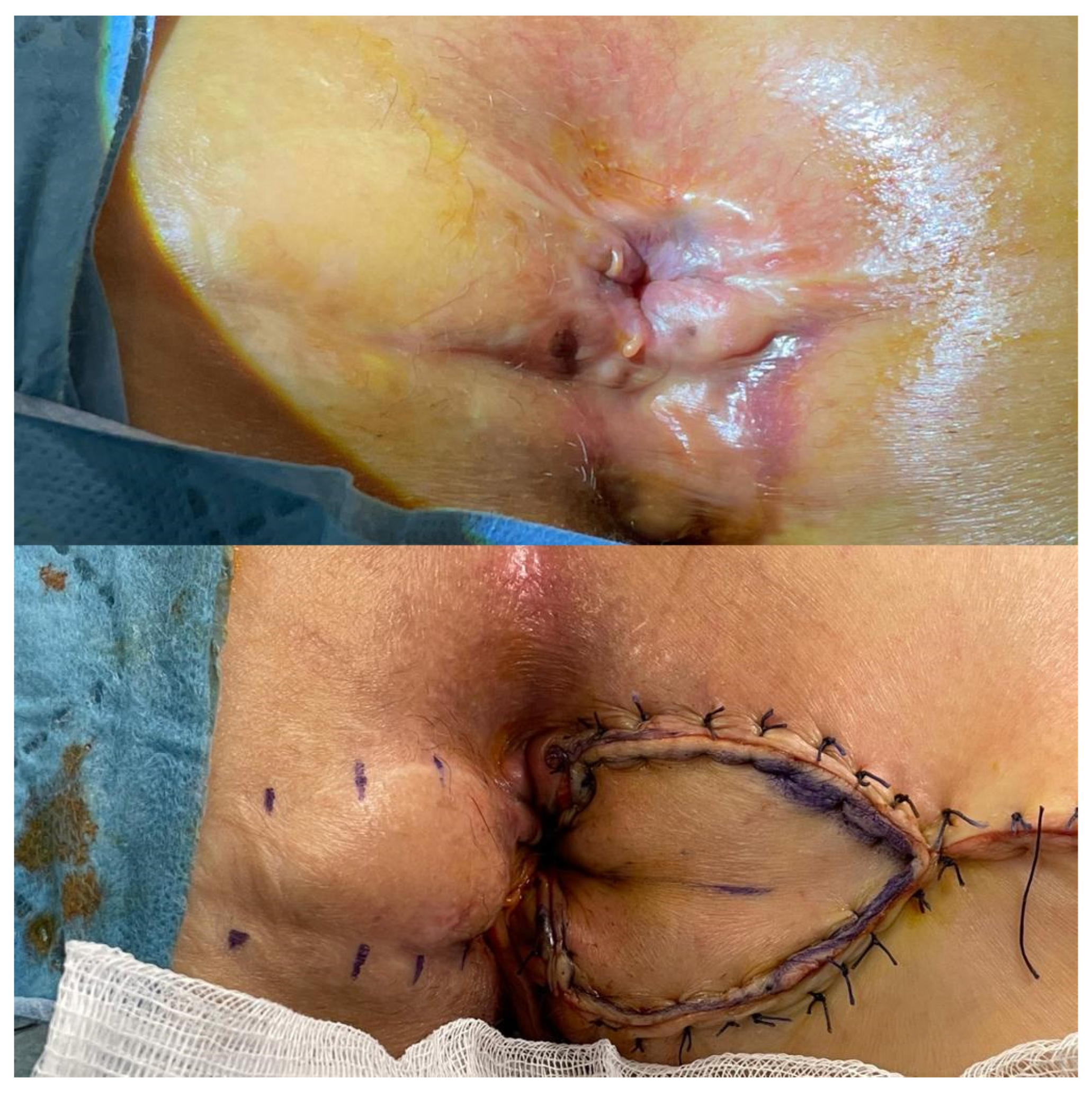
The patient is placed in the prone jack-knife position. The longitudinal incision is made from the dentate line to the end of the stenosis. Then the flap is designed as in the house shape, with the length of the “walls” of the flap corresponding to the length of the incision and the “roof” of the flap reaching the healthy perianal skin (Figure 1 and Figure 2). The flap is dissected to the depth of the ischiorectal fat to advance it into the anal canal without tension and to preserve the vascular pedicle [1,21].
c. 
Diamond flap
The diamond flap was first described by Caplin and Kodner in 1986 and has been performed for the treatment of moderate and severe AS [24]. Although the final target for anal caliber has not been standardized, Gulen et al. reported a clinical success rate of 88.9% with an eventual anal caliber of 25 to 26 mm in 18 consecutive patients. After 12-months of follow-up, the obstructed defecation syndrome scores were found to be significantly improved [25].
Technical notes of Diamond flap:
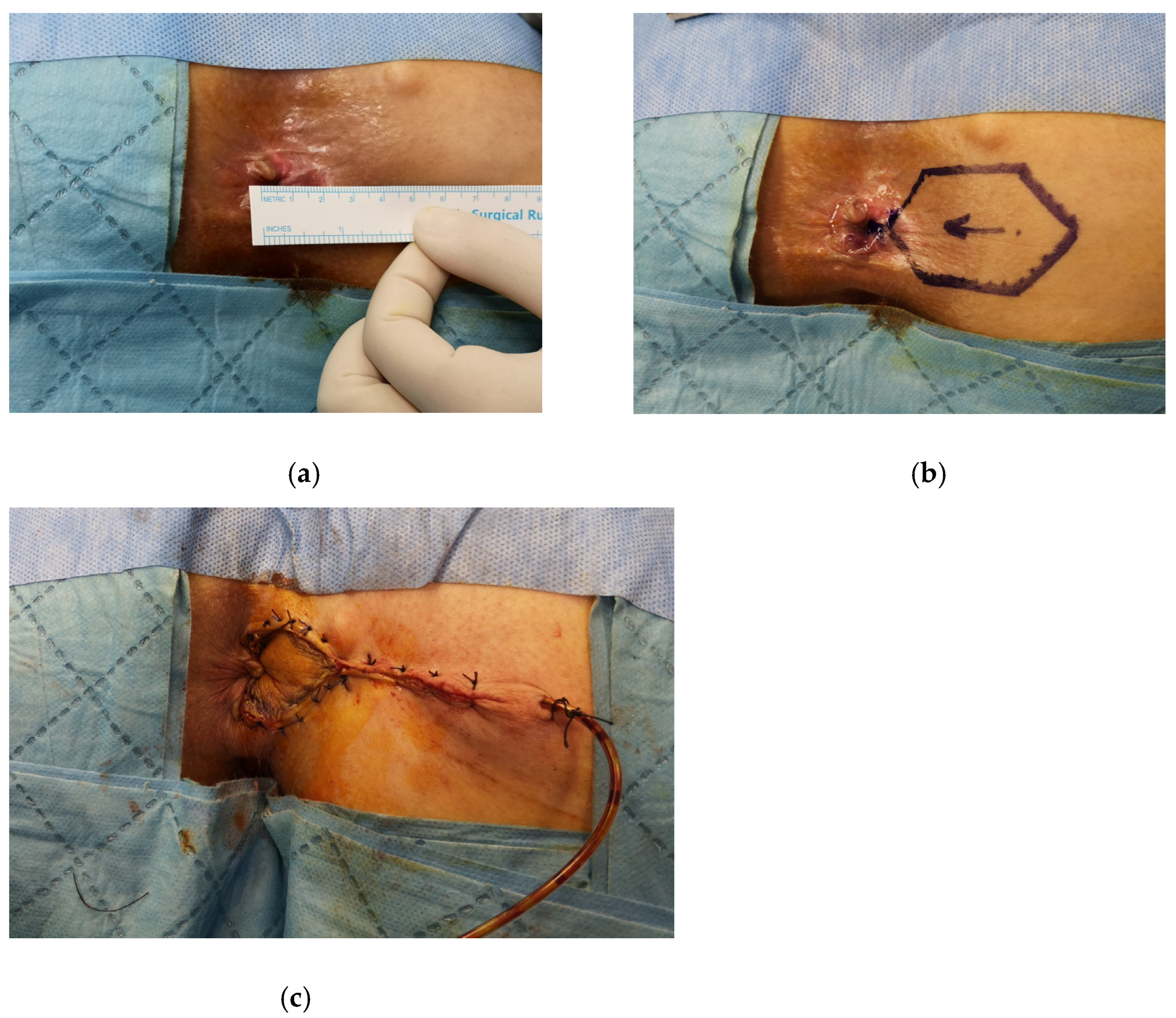
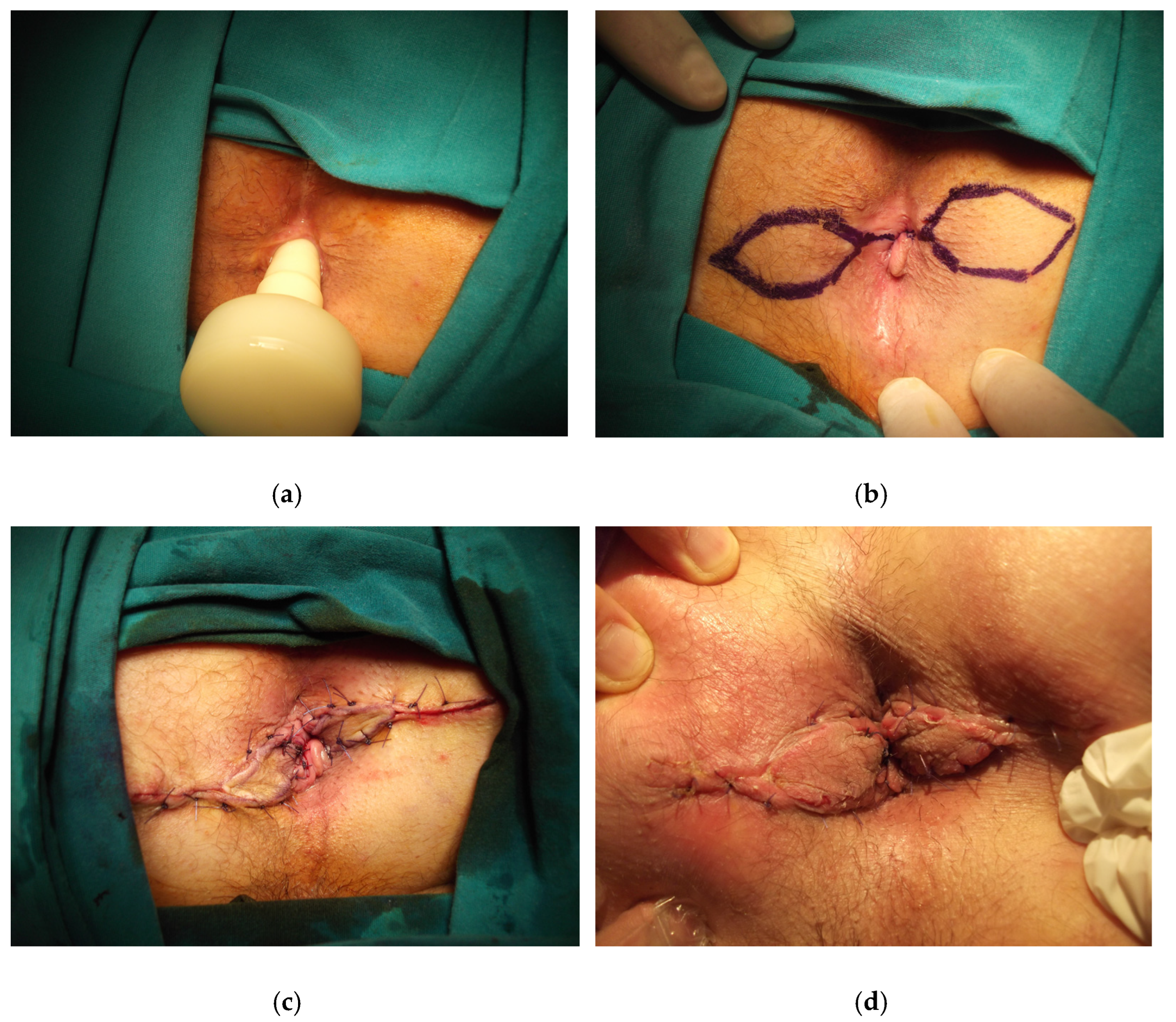
The patient is placed in the prone jack-knife position. The incision is made on the scar tissue longitudinally until it reaches the dentate line. The anal caliber is recommended to be checked during this step, and the external sphincter should be spared [26]. The diamond flap is designed as adjacent to the diseased anal canal. The flap is dissected out together with its vascular pedicle and advanced into the anal canal. The flap should be tension-free to avoid any postoperative wound complications (Figure 3, Figure 4 and Figure 5).
d. 
Y-V flap/V-Y flap
The Y-V advancement flap is performed for low and localized strictures below the dentate line, whereas the V-Y flap is used for mild to severe stricture at the dentate line [1]. Maria et al. reported 29 patients who had Y-V anoplasty in a comparative study with diamond flap, with a healing rate of 90% and an ischemic contracture of the leading edge of the flap and wound dehiscence in two patients [27]. The authors suggested that the diamond flap is a more reliable technique due to the reduced tension in the suture line and the better blood supply of the flap. Farid et al. observed less clinical improvement (65%) and higher ischemic wound complications (15%) with this technique [23].
Technical notes of Y-V flap/V-Y flap:
Initially, the vertical limb of the Y is performed to the area of stricture. The incision is extended to the perianal skin in two directions creating a V shape. Then, the tip of the V is advanced to the vertical limb of the Y incision. It can also be done in either a lateral position or just in the posterior midline. The main disadvantage regarding this technique is the tip of the V flap being prone to ischemic necrosis [10].
For the V-Y advancement flap, the first few steps are the same as the Y-V flap, but in this procedure, the wider base of the triangular Y flap is sutured to the dentate line. Then the perianal skin is approximated with sutures longitudinally behind the V shape to form the Y shape [1].
a. 
Rhomboid flap/Modified rhomboid flap
The rhomboid flap has been modified in terms of the flap size to be adjusted to each patient. Sloane et al. observed good functional results in a case series including nine patients, eight of whom underwent bilateral rhomboid flap with complete resolution of symptoms [28]. The modified rhomboid flap is demonstrated as a safe and suitable technique for the treatment of moderate and severe AS. Gallo et al. reported 0% recurrence rate and 96% success rate in a study with 50 consecutive patients, and significant improvement in the obstructed defecation syndrome scores and the quality of life were observed at 12 months [29]. The mean anal caliber was found to be 24 mm and significantly different compared to the preoperative measurement.
Technical notes of Rhomboid flap/Modified rhomboid flap:
The size of the rhomboid flap can vary between 8 to 9 cm in length and 5 cm in width at its largest point. A minimal left/right internal sphincterotomy can be performed by paying careful attention to the external sphincter. Then, the flap is relocated in the anal canal and fixed with a single-layer suture of absorbable stitches to the distal rectum. The skin sutures should be adequately spaced to avoid excessive tension with subsequent ischemia [29].
a. 
U-flap
The U-flap technique is mainly used for the treatment of AS with mucosal ectropion. The disadvantage of this technique is that the donor site is left open [1]. Pearl et al. reported a good clinical result of 92% in 25 patients during a mean follow-up of 19 months [30].
Technical notes of U-flap:
The procedure starts with the excision of the ectropion; then, a U-shaped incision is made at the adjacent perianal skin. The U flap is advanced into the anal canal to cover the wide defect resulting from the excised area of ectropion.
b. 
Rotational S-plasty
Rotational S-plasty was first described by Ferguson in 1959 for the repair of the whitehead deformity of the anus [31]. Corman et al. in 1976 adapted this technique for the treatment of AS [32]. This technique enables covering large areas of skin with an adequate blood supply. Gonzalez et al. revealed that 94% of patients had good results during a mean follow-up of 18 months [33].
Technical notes of Rotational S-plasty:
The technique starts with outlining the semicircular incision in the perianal skin, with a length of 12 to 13 cm. The base of the incision should not be shorter than its length. Then skin incision is done to the layer of subcutaneous fat lobules to preserve the blood supply of the skin flaps. The flap is then rotated and fixed to the mucosa by interrupted sutures [34].

3. Postoperative Care and Complications

Patients are usually discharged on postoperative day 1, with recommendations of daily sitz baths or showers for comfort and hygiene. Prophylactic antibiotics with metronidazole and ciprofloxacin or cephalosporins can be continued in the postoperative setting. The postoperative complications encountered commonly are urinary retention, wound dehiscence, wound infection, flap ischemia, and bleeding [13,19,20,27].

4. Conclusions

The AS due to overzealous hemorrhoidectomy is an entirely preventable disease when performed under skilled and experienced hands [35]. With better knowledge of the anorectal anatomy and delicate treatment to the anal tissue, the complications of anorectal surgery, such as anal stenosis, can be reduced. Considering the fact that with today’s technological evolution, there are many alternative techniques such as doppler-guided hemorrhoidal artery ligation and stapled hemorrhoidopexy, and excisional hemorrhoidectomy may not be the only option for many patients [36].
Nevertheless, anoplasty techniques should be in the armamentarium of colorectal surgeons. The basis of these techniques includes excision or incision of the scar tissue, preparation of the flap with careful attention to the vascular supply, advancement of the flap, and fixating the flap without tension into the anal canal. Following these steps carefully would result in a significant decrease in anoplasty complications.
As for the better technique regarding functional outcomes, we need more high-quality studies objectively evaluating patients’ quality of life and functional outcomes using standardized scoring systems and questionnaires.

Author Contributions

Conceptualization, S.L. and B.B.; H.C.K.; investigation, B.M.; resources, B.B.; writing—original draft preparation, S.L.; writing—review and editing, H.C.K.; visualization, B.M.; supervision. All authors have read and agreed to the published version of the manuscript.

Funding

This research received no external funding.

Institutional Review Board Statement

Not applicable.

Informed Consent Statement

Not applicable.

Data Availability Statement

This study did not report any new research data.

Conflicts of Interest

The authors declare no conflict of interest.

References

  1. Brisinda, G.; Vanella, S.; Cadeddu, F.; Marniga, G.; Mazzeo, P.; Brandara, F.; Maria, G. Surgical treatment of anal stenosis. World J. Gastroenterol. 2009, 15, 1921–1928. [Google Scholar] [CrossRef] [PubMed]
  2. Casadesus, D.; Villasana, L.E.; Diaz, H.; Chavez, M.; Sanchez, I.M.; Martinez, P.P.; Diaz, A. Treatment of anal stenosis: A 5-year review. ANZ J. Surg. 2007, 77, 557–559. [Google Scholar] [CrossRef] [PubMed]
  3. Eu, K.W.; Teoh, T.A.; Seow-Choen, F.; Goh, H.S. Anal stricture following haemorrhoidectomy: Early diagnosis and treat-ment. Aust. N. Z. J. Surg. 1995, 65, 101–103. [Google Scholar] [CrossRef] [PubMed]
  4. Altomare, D.F.; Picciariello, A.; Pecorella, G.; Milito, G.; Naldini, G.; Amato, A.; Ratto, C.; Perinotti, R. The Italian Haemorrhoid Survey group Surgical management of haemorrhoids: An Italian survey of over 32 000 patients over 17 years. Colorectal Dis. 2018, 20, 1117–1124. [Google Scholar] [CrossRef] [Green Version]
  5. Blumetti, J. Anal stenosis. In Complications of Anorectal Surgery: Prevention and Management, 1st ed.; Abcarian, H., Cintron, J.R., Nelson, R.L., Eds.; Springer International Publishing: Cham, Switzerland, 2017; pp. 235–246. [Google Scholar]
  6. Corman, M. Hemorrhoids. In Corman’s Colon and Rectal Surgery, 3rd ed.; Corman, M., Ed.; JB Lippincott: Philadelphia, PA, USA, 1993; pp. 54–115. [Google Scholar]
  7. Milsom, J.W.; Mazier, W.P. Classification and management of postsurgical anal stenosis. Surg. Gynecol. Obstet. 1986, 163, 60–64. [Google Scholar]
  8. Kmiot, W.A. Anal stenosis. In Surgery of the Colon, Rectum and Anus, 1st ed.; Mazier, W., Levien, D., Luchtefeld, M., Senagore, A., Eds.; WB Saunders: Philadelphia, PA, USA, 1995; pp. 340–344. [Google Scholar]
  9. Macdonald, A.; Smith, A.; McNeill, A.D.; Finlay, I.G. Manual dilatation of the anus. Br. J. Surg. 1992, 79, 1381–1382. [Google Scholar] [CrossRef]
  10. Liberman, H.; Thorson, A.G. How I do it. Anal stenosis. Am. J. Surg. 2000, 179, 325–329. [Google Scholar] [CrossRef]
  11. Nickell, W.B.; Woodward, E.R. Advancement flaps for treatment of anal stricture. Arch. Surg. 1972, 104, 223–224. [Google Scholar] [CrossRef]
  12. Oh, C.; Zinberg, J. Anoplasty for anal stricture. Dis. Colon Rectum 1982, 25, 809–810. [Google Scholar]
  13. Angelchik, P.D.; Harms, B.A.; Starling, J.R. Repair of anal stricture and mucosal ectropion with Y-V or pedicle flap anoplasty. Am. J. Surg. 1993, 166, 55–59. [Google Scholar] [CrossRef]
  14. Gingold, B.S.; Arvanitis, M. Y-V anoplasty for treatment of anal stricture. Surg. Gynecol. Obstet. 1986, 162, 241–242. [Google Scholar] [PubMed]
  15. Pidala, M.J.; A Slezak, F.; A Porter, J. Island flap anoplasty for anal canal stenosis and mucosal ectropion. Am. Surg. 1994, 60, 194–196. [Google Scholar] [PubMed]
  16. Habr-Gama, A.; Sobrado, C.W.; De Araújo, S.E.A.; Nahas, S.C.; Birbojm, I.; Nahas, C.S.R.; Kiss, D.R. Surgical treatment of anal stenosis: Assessment of 77 anoplasties. Clinics 2005, 60, 17–20. [Google Scholar] [CrossRef] [Green Version]
  17. Aitola, P.T.; Hiltunen, K.M.; Matikainen, M.J. Y-V anoplasty combined with internal sphincterotomy for stenosis of the anal canal. Eur. J. Surg. 1997, 163, 839–842. [Google Scholar] [PubMed]
  18. Filingeri, V.; Gravante, G.; Cassisa, D. Radiofrequency Y-V anoplasty in the treatment of anal stenosis. Eur. Rev. Med. Pharmacol. Sci. 2006, 10, 263–267. [Google Scholar]
  19. Carditello, A.; Milone, A.; Stilo, F.; Mollo, F.; Basile, M. Surgicaltreatment of anal stenosis following hemorrhoid sur-gery.Results of 150 combined mucosal advancement and internalsphincterotomy. Chir. Ital. 2002, 54, 841–844. [Google Scholar]
  20. Rakhmanine, M.; Rosen, L.; Khubchandani, I.; Stasik, J.; Riether, R.D. Lateral mucosal advancement anoplasty for anal stricture. Br. J. Surg. 2002, 89, 1423–1424. [Google Scholar] [CrossRef]
  21. Alver, O.; Ersoy, Y.E.; Aydemir, I.; Erguney, S.; Teksoz, S.; Apaydin, B.; Ertem, M. Use of “House” Advancement Flap in Anorectal Diseases. World J. Surg. 2008, 32, 2281–2286. [Google Scholar] [CrossRef]
  22. Sentovich, S.M.; Falk, P.M.; Christensen, M.A.; Thorson, A.G.; Blatchford, G.J.; Pitsch, R.M. Operative results of House advancement anoplasty. Br. J. Surg. 1996, 83, 1242–1244. [Google Scholar]
  23. Farid, M.; Youssef, M.; El Nakeeb, A.; Fikry, A.; El Awady, S.; Morshed, M. Comparative Study of the House Advancement Flap, Rhomboid Flap, and Y-V Anoplasty in Treatment of Anal Stenosis: A Prospective Randomized Study. Dis. Colon Rectum 2010, 53, 790–797. [Google Scholar] [CrossRef]
  24. Caplin, D.A.; Kodner, I.J. Repair of anal stricture and mucosal ectropion by simple flap procedures. Dis. Colon Rectum 1986, 29, 92–94. [Google Scholar] [CrossRef] [PubMed]
  25. Gülen, M.; Leventoğlu, S.; Ege, B.; Menteş, B.B. Surgical Treatment of Anal Stenosis with Diamond Flap Anoplasty Performed in a Calibrated Fashion. Dis. Colon Rectum 2016, 59, 230–235. [Google Scholar] [CrossRef] [PubMed]
  26. Balci, B.; Yildiz, A.; Leventoglu, S.; Mentes, B.B. Diamond-shaped flap anoplasty for severe anal stenosis—A video vignette. Colorectal Dis. 2021, 23, 1941. [Google Scholar] [CrossRef]
  27. Maria, G.; Brisinda, G.; Civello, I.M. Anoplasty for the treatment of anal stenosis. Am. J. Surg. 1998, 175, 158–160. [Google Scholar] [CrossRef]
  28. Sloane, J.A.H.; Zahid, A.; Young, C.J. Rhomboid-shaped advancement flap anoplasty to treat anal stenosis. Tech. Coloproctol. 2016, 21, 159–161. [Google Scholar] [CrossRef]
  29. Gallo, G.; Stratta, E.; Luc, A.R.; Clerico, G.; Trompetto, M. A tailored rhomboid mucocutaneous advancement flap to treat anal stenosis. Colorectal Dis. 2020, 22, 1388–1395. [Google Scholar] [CrossRef]
  30. Pearl, R.K.; Hooks, V.H., 3rd; Abcarian, H.; Orsay, C.P.; Nelson, R.L. Island flap anoplasty for the treatment of anal stricture and mucosal ectropion. Dis. Colo Rectum 1990, 33, 581–583. [Google Scholar] [CrossRef]
  31. A Ferguson, J. Repair of Whitehead deformity of the anus. Surg. Gynecol. Obstet. 1959, 108, 115–116. [Google Scholar]
  32. Corman, M.L.; Veidenheimer, M.C.; Coller, J.A. Anoplasty for Anal Stricture. Surg. Clin. N. Am. 1976, 56, 727–731. [Google Scholar] [CrossRef]
  33. González, A.R.; De Oliveira, O.; Verzaro, R.; Nogueras, J.; Wexner, S.D. Anoplasty for stenosis and other anorectal defects. Am. Surg. 1995, 61, 526–529. [Google Scholar]
  34. Oh, C.; Albanese, C. S-plasty for various anal lesions. Am. J. Surg. 1992, 163, 606–608. [Google Scholar] [CrossRef]
  35. Brisinda, G. How to treat haemorrhoids. Prevention is best; haemorrhoidectomy needs skilled operators. BMJ 2000, 321, 582–583. [Google Scholar] [CrossRef] [PubMed]
  36. Gallo, G.; Luc, A.R.; Tiesi, V.; Trompetto, M. Sign of the times: The Milligan–Morgan era. Tech. Coloproctol. 2021, 25, 1327. [Google Scholar] [CrossRef] [PubMed]
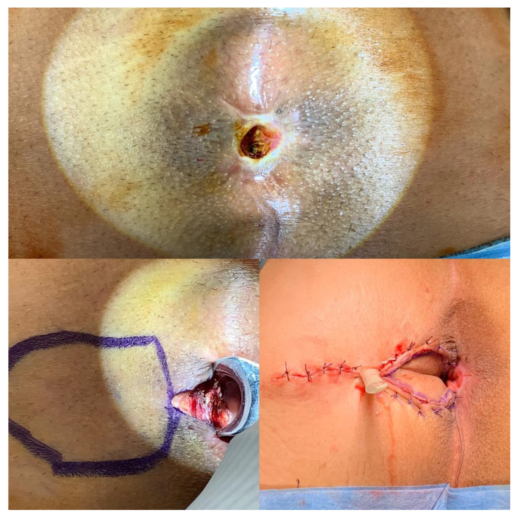
Figure 1. The house flap anoplasty is shown in a patient with severe anal stenosis.
Medicina 58 00362 g001
Figure 2. A tailored house flap anoplasty in a patient with chronic unhealing wound in the posterior anal canal.
Medicina 58 00362 g002
Figure 3. The diamond flap anoplasty is shown in a patient with severe anal stenosis, (a) the anal caliber is measured as 10 mm, (b) the right-sided anoplasty is decided according to the location of scar tissue in the anal verge, (c) the anoplasty is performed and the negative pressure drain is placed.
Medicina 58 00362 g003
Figure 4. Contralateral diamond flap anoplasty in a patient with recurrent anal stenosis.
Medicina 58 00362 g004
Figure 5. Bilateral diamond flap anoplasty in severe anal stenosis is shown, (a) the anal caliber is measured using an anal calibrator, (b) the technique is drawn, (c) the postoperative image after anoplasty, (d) the image in postoperative 1st week.
Medicina 58 00362 g005
Table 1. Classification of anal stenosis by Milsom and Mazier [7].
Classification based on the severity
Mild: Tight anal canal can be examined by a well-lubricated index finger or a medium Hill-Ferguson retractor.
Moderate: Forceful dilatation is required to insert either the index finger or a medium Hill-Ferguson retractor.
Severe: Neither the little finger nor the small Hill-Ferguson retractor can be inserted unless a forceful dilatation is employed.
Classification based on the level of stenosis
Low: Distal anal canal at least 0.5 cm below the dentate line
Middle: 0.5 cm proximal to 0.5 cm distal to the dentate line
High: Proximal to 0.5 cm above the dentate line
Table 2. Common surgical techniques used for treating anal stenosis.
Surgical TechniqueIndicationsAdvantagesDisadvantages
Mucosal advancement flapMiddle or high mild anal stenosis-The risk of ectropion unless the wound is left open
House flapModerate to severe anal stenosisProvides adequate extension in the anal canal diameter-
Diamond flapModerate to severe anal stenosisCovers the defect in the anal canal while sparing the sphincter complex -
Y-V flap/V-Y flapMild to moderate anal stenosis-Flap’s tip prone to ischemia and lacks sufficient extension of the anal canal diameter
Rhomboid flap/Modified rhomboid flapModerate to severe anal stenosisEnables a tailored-anoplasty in different sizes-
U-flapExcising the mucosal ectropion-Leaving the wound open results in delay of recovery
Rotational S-plastyModerate to severe anal stenosisProvides a large tissue rotation without compromising vascular supply-
Table 3. Several studies on functional and surgical outcomes after anoplasty.
AuthorsStudy MethodTotal N of Included PatientsIndications for Anoplasty (N of Patients)Surgical TechniquesFunctional OutcomesSurgical Outcomes (N of Patients)Mean Follow-Up (Months)
Rakhmanine et al. [20]Retrospective95Hemorrhoidectomy (35)
Chronic anal fissure (10)
Perianal fistula (4)
Anal carcinoma (1)
Various (10)
Mucosal advancement flapReported as good in 74 patients and as poor in 8 patientsAbscess (1)
Seepage of liquid stool (2)
50
Alver et al. [21]Retrospective28Chronic anal fissure (14)
Anal stenosis (8)
Rectovaginal fistula (1)
Perianal fistula (3)
Anal carcinoma (1)
Obstetric injury (1)
House flapReported as good in 8 patients with anal stenosisWound dehiscence (3)
Recurrence of rectovaginal fistula (1)
26
Sentovich et al. [22]Retrospective29Anal stenosis (21)
Ectropion (4)
Bowen’s disease (2)
Key-hole deformity (2)
Perianal fistula (1)
House flapReported as good in 26 patients and as poor in 3 patientsDonor-site separation (14)
Urinary retention (8) Sepsis (4)
28
Farid et al. [23]Prospective-randomized60Anal stenosis (60)Rhomboid flap/Y-V flap/House flapBetter anal caliber increase and improvement in GI-QLI score with house-flap 12
Gulen et al. [25]Retrospective18Anal stenosis (18)Diamond flapSignificant increase in anal caliber and improvement in ODS scoreWound dehiscence (4)35
Maria et al. [27]Comparative42Anal stenosis (42)Diamond flap/Y-V flapReported as good in 89% of patients with Y-V flap, and 100% with diamond flap Wound dehiscence (1)
Ischemia in tip of the flap (1)
24
Sloane et al. [28]Retrospective9Anal stenosis (9)Rhomboid flapReported as significant improvements in 9 patientsSingle quadrant stenosis (1)12
Gallo et al. [29]Retrospective50Anal stenosis (50)Modified rhomboid flapSignificant increase in anal caliber and improvement in ODS and CCI scoreIschemia of donor site (1)
Wound dehiscence (2)
97
Pearl et al. [30]Retrospective25Anal stenosis (20)
Ectropion (5)
Island flap
(U-shaped and Diamond-shaped)
Reported as excellent in 64% of patients and good in 25% of patients-19
Gonzalez et al. [33]Comparative17Anal stenosis (13)
Perianal fistula (2)
Key-hole deformity (1)
Chronic anal fissure (1)
Rotational S-plasty/Advancement flapReported as good in 16 patientsSepsis (1)18
Abbreviations: N, number; GI-QLI, gastrointestinal quality of life inventory; ODS, obstructed defecation syndrome; CCI, Cleveland Clinic Incontinence.
Publisher’s Note: MDPI stays neutral with regard to jurisdictional claims in published maps and institutional affiliations.