
Chronic Rupture of Achilles Tendon Caused by Haglund’s Deformity: A Case Report


Abstract
:1. Introduction
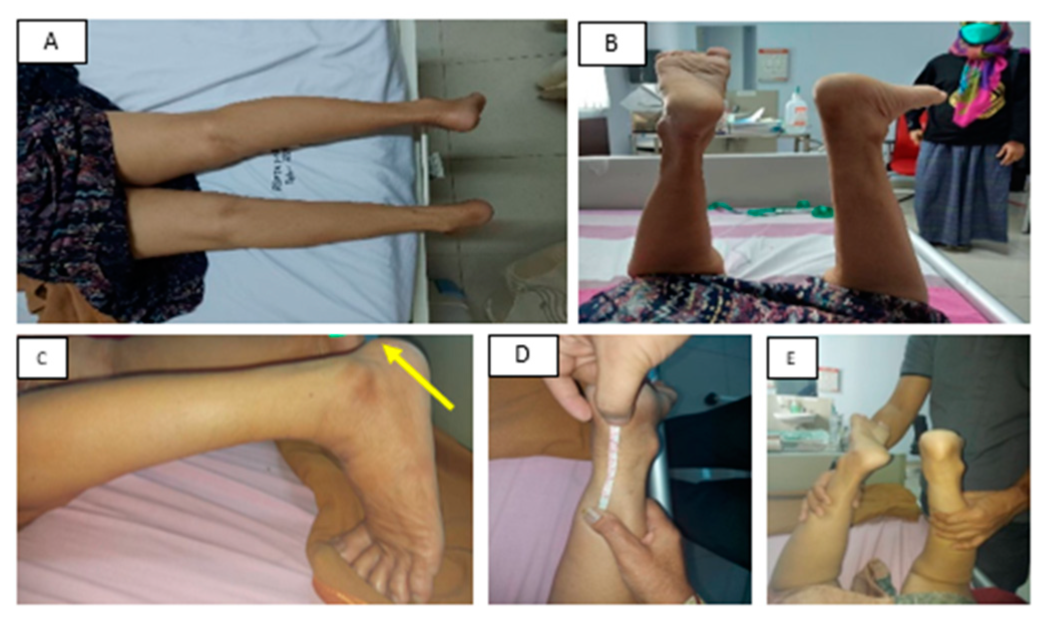
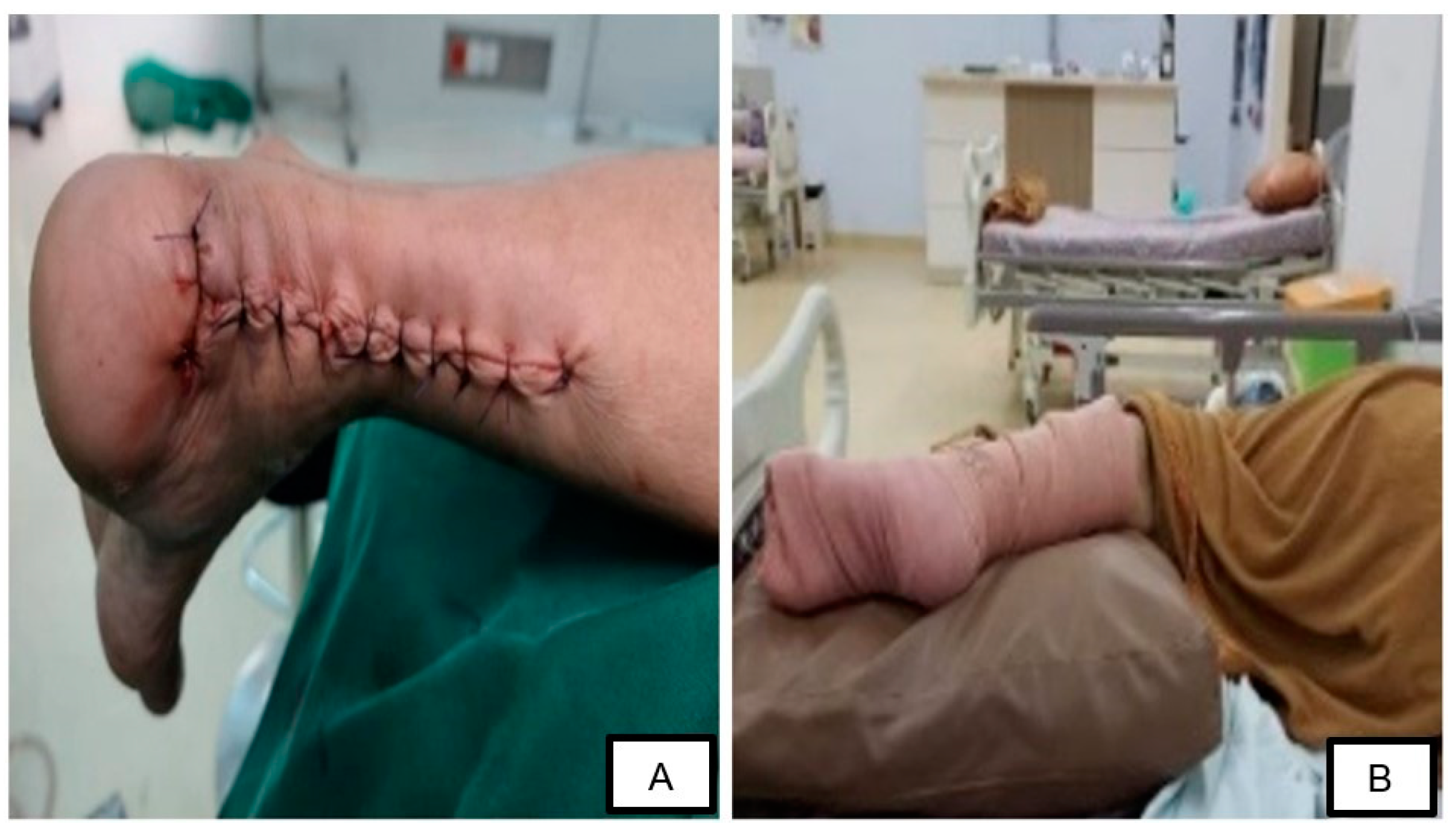
2. Clinical Case
3. Discussion
4. Conclusions
Author Contributions
Funding
Institutional Review Board Statement
Informed Consent Statement
Data Availability Statement
Acknowledgments
Conflicts of Interest
References
- Mahan, J.; Damodar, D.; Trapana, E.; Barnhill, S.; Nuno, A.U.; Smyth, N.A.; Aiyer, A.; Jose, J. Achilles tendon complex: The anatomy of its insertional footprint on the calcaneus and clinical implications. J. Orthop. 2020, 17, 221–227. [Google Scholar] [CrossRef] [PubMed]
- Thevendran, G.; Sarraf, K.M.; Patel, N.K.; Sadri, A.; Rosenfeld, P. The ruptured Achilles tendon: A current overview from biology of rupture to treatment. Musculoskelet. Surg. 2013, 97, 9–20. [Google Scholar] [CrossRef] [PubMed]
- Romero-Morales, C.; Martín-Llantino, P.J.; Calvo-Lobo, C.; Sánchez-Gómez, R.; López-López, D.; Pareja-Galeano, H.; Rodríguez-Sanz, D. Ultrasound evaluation of extrinsic foot muscles in patients with chronic non-insertional Achilles tendinopathy: A case-control study. Phys. Ther. Sport 2019, 37, 44–48. [Google Scholar] [CrossRef] [PubMed]
- Romero-Morales, C.; Martín-Llantino, P.J.; Calvo-Lobo, C.; Almazán-Polo, J.; López-López, D.; de la Cruz-Torres, B.; Palomo-López, P.; Rodríguez-Sanz, D. Intrinsic foot muscles morphological modifications in patients with Achilles tendinopathy: A novel case-control research study. Phys. Ther. Sport 2019, 40, 208–212. [Google Scholar] [CrossRef]
- Vaishya, R.; Agarwal, A.K.; Azizi, A.T.; Vijay, V. Haglund’s Syndrome: A Commonly Seen Mysterious Condition. Cureus 2016, 8, e820. [Google Scholar] [CrossRef] [PubMed]
- Madi, S.; Hillrichs, B. Haglund’s Deformity as a Cause of Acute Achilles Tendon Rupture: A Case Report. J. Foot Ankle Surg. 2022, 61, 410–413. [Google Scholar] [CrossRef] [PubMed]
- Schunck, J.; Jerosch, J. Operative treatment of Haglund’s syndrome. Basics, indications, procedures, surgical techniques, results and problems. Foot Ankle Surg. 2005, 11, 123–130. [Google Scholar] [CrossRef]
- Lemme, N.J.; Li, N.Y.; DeFroda, S.F.; Kleiner, J.; Owens, B.D. Epidemiology of Achilles Tendon Ruptures in the United States: Athletic and Nonathletic Injuries From 2012 to 2016. Orthop. J. Sport Med. 2018, 6, 2325967118808238. [Google Scholar] [CrossRef]
- Mazzone, M.F.; McCue, T. Common conditions of the Achilles tendon. Am. Fam. Physician 2002, 65, 1805–1810. [Google Scholar]
- Gravlee, J.R.; Hatch, R.L.; Galea, A.M. Achilles tendon rupture: A challenging diagnosis. J. Am. Board Fam. Pract. 2000, 13, 371–373. [Google Scholar]
- Maffulli, N.; Via, A.G.; Oliva, F. Chronic Achilles Tendon Rupture. Open Orthop. J. 2017, 11, 660–669. [Google Scholar] [CrossRef] [PubMed]
- Ochen, Y.; Beks, R.B.; van Heijl, M.; Hietbrink, F.; Leenen, L.P.H.; van der Velde, D.; Heng, M.; van der Meijden, O.; Groenwold, R.H.H.; Houwert, R.M. Operative treatment versus nonoperative treatment of Achilles tendon ruptures: Systematic review and meta-analysis. BMJ 2019, 364, k5120. [Google Scholar] [CrossRef] [PubMed] [Green Version]
- Borah, D.N.; Rai, S.; Frank, H.C.; Dutta, A. Repair of chronic Achilles tendon rupture using Bosworth’s technique. J. Orthop. Trauma Rehabil. 2020, 2210491720972713. [Google Scholar] [CrossRef]
- Apinun, J.; Jenvorapoj, S.; Arirachakaran, A.; Kongtharvonskul, J. Clinical outcomes of chronic Achilles tendon rupture treated with flexor hallucis longus grafting and flexor hallucis longus grafting plus additional augmentation: A meta-analysis. Foot Ankle Surg. 2020, 26, 717–722. [Google Scholar] [CrossRef]
- Alhaug, O.K.; Berdal, G.; Husebye, E.E.; Hvaal, K. Flexor hallucis longus tendon transfer for chronic Achilles tendon rupture. A retrospective study. Foot Ankle Surg. 2019, 25, 630–635. [Google Scholar] [CrossRef]
- Maffulli, N.; Ajis, A. Management of chronic ruptures of the Achilles tendon. J. Bone Jt. Surg. 2008, 90, 1348–1360. [Google Scholar] [CrossRef]
- Yasui, Y.; Tonogai, I.; Rosenbaum, A.J.; Shimozono, Y.; Kawano, H.; Kennedy, J.G. The Risk of Achilles Tendon Rupture in the Patients with Achilles Tendinopathy: Healthcare Database Analysis in the United States. BioMed Res. Int. 2017, 2017, 7021862. [Google Scholar] [CrossRef]
- Tarantino, D.; Palermi, S.; Sirico, F.; Corrado, B. Achilles Tendon Rupture: Mechanisms of Injury, Principles of Rehabilitation and Return to Play. J. Funct. Morphol. Kinesiol. 2020, 5, 95. [Google Scholar] [CrossRef]
- Yassin, M.; Gupta, V.; Martins, A.; Mahadevan, D.; Bhatia, M. Patient reported outcomes and satisfaction following single incision Flexor Hallucis Longus (FHL) augmentation for chronic Achilles tendon pathologies. J. Clin. Orthop. Trauma 2021, 23, 101650. [Google Scholar] [CrossRef]
- Spennacchio, P.; Vascellari, A.; Cucchi, D.; Canata, G.L.; Randelli, P. Outcome evaluation after Achilles tendon ruptures. A review of the literature. Joints 2016, 4, 52–61. [Google Scholar] [CrossRef]
- Irwin, T.A. Current concepts review: Insertional Achilles tendinopathy. Foot Ankle Int. 2010, 31, 933–939. [Google Scholar] [CrossRef] [PubMed]
- Dakin, S.G.; Newton, J.; Martinez, F.O.; Hedley, R.; Gwilym, S.; Jones, N.; Reid, H.A.B.; Wood, S.; Wells, G.; Appleton, L.; et al. Chronic inflammation is a feature of Achilles tendinopathy and rupture. Br. J. Sports Med. 2018, 52, 359–367. [Google Scholar] [CrossRef]
- Zhu, G.; Wang, Z.; Yuan, C.; Geng, X.; Zhang, C.; Huang, J.; Wang, X.; Ma, X. A radiographic study of biomechanical relationship between the Achilles tendon and plantar fascia. BioMed Res. Int. 2020, 2020, 5319640. [Google Scholar] [CrossRef] [PubMed]
- Gaston, T.E.; Daniel, J.N. Achilles Insertional Tendinopathy-Is There a Gold Standard? Arch. Bone Jt. Surg. 2021, 9, 5–8. [Google Scholar] [PubMed]
- Sofka, C.M.; Adler, R.S.; Positano, R.; Pavlov, H.; Luchs, J.S. Haglund’s syndrome: Diagnosis and treatment using sonography. HSS J. 2006, 2, 27–29. [Google Scholar] [CrossRef] [PubMed]
- Lin, Y.; Yang, L.; Yin, L.; Duan, X. Surgical strategy for the chronic Achilles tendon rupture. BioMed Res. Int. 2016, 2016, 1416971. [Google Scholar] [CrossRef] [PubMed]
- Yang, Y.; Wang, D.; Wei, L.; An, N.; Tao, L.; Jiao, C.; Guo, Q.; Hu, Y. Repair of Achilles sleeve avulsion: A new transosseous suture technique. J. Orthop. Surg. Res. 2020, 15, 224. [Google Scholar] [CrossRef]
- Elias, I.; Raikin, S.M.; Besser, M.P.; Nazarian, L.N. Outcomes of chronic insertional Achilles tendinosis using FHL autograft through single incision. Foot Ankle Int. 2009, 30, 197–204. [Google Scholar] [CrossRef]
- Schmidtberg, B.; Johnson, J.D.; Kia, C.; Baldino, J.B.; Obopilwe, E.; Cote, M.P.; Geaney, L.E. Flexor hallucis longus transfer improves Achilles tendon load to failure in surgery for non-insertional tendinopathy: A biomechanical study. J. Bone Jt. Surg. 2019, 101, 1505–1512. [Google Scholar] [CrossRef]
- Park, S.-H.; Lee, H.S.; Young, K.W.; Seo, S.G. Treatment of Acute Achilles Tendon Rupture. Clin. Orthop. Surg. 2020, 12, 1–8. [Google Scholar] [CrossRef]
- Nilsson-Helander, K.; Silbernagel, K.G.; Thomeé, R.; Faxén, E.; Olsson, N.; Eriksson, B.I.; Karlsson, J. Acute Achilles tendon rupture: A randomized, controlled study comparing surgical and nonsurgical treatments using validated outcome measures. Am. J. Sports Med. 2010, 38, 2186–2193. [Google Scholar] [CrossRef] [PubMed]
- Karlsson, J.; Nilsson-Helander, K.; Olsson, N. Achilles Tendon Rupture: An International Evidence-Based Approach to Treatment and Rehabilitation. Surgical or Non-Surgical Treatment? Aspetar Sport Med. J. 2013, 2, 350–357. [Google Scholar]


Publisher’s Note: MDPI stays neutral with regard to jurisdictional claims in published maps and institutional affiliations. |
© 2022 by the authors. Licensee MDPI, Basel, Switzerland. This article is an open access article distributed under the terms and conditions of the Creative Commons Attribution (CC BY) license (https://creativecommons.org/licenses/by/4.0/).
Share and Cite
Usman, M.A.; Murtaza, B.; Winangun, P.A.N.; Kennedy, D. Chronic Rupture of Achilles Tendon Caused by Haglund’s Deformity: A Case Report. Medicina 2022, 58, 1216. https://doi.org/10.3390/medicina58091216
Usman MA, Murtaza B, Winangun PAN, Kennedy D. Chronic Rupture of Achilles Tendon Caused by Haglund’s Deformity: A Case Report. Medicina. 2022; 58(9):1216. https://doi.org/10.3390/medicina58091216
Chicago/Turabian StyleUsman, Muhammad Andry, Benny Murtaza, Putu Acarya Nugraha Winangun, and Dave Kennedy. 2022. "Chronic Rupture of Achilles Tendon Caused by Haglund’s Deformity: A Case Report" Medicina 58, no. 9: 1216. https://doi.org/10.3390/medicina58091216
APA StyleUsman, M. A., Murtaza, B., Winangun, P. A. N., & Kennedy, D. (2022). Chronic Rupture of Achilles Tendon Caused by Haglund’s Deformity: A Case Report. Medicina, 58(9), 1216. https://doi.org/10.3390/medicina58091216