Systematic Review

Gastrectomy with or without Complete Omentectomy for Advanced Gastric Cancer: A Meta-Analysis

1
Surgical Oncology Unit, Azienda Unità Sanitaria Locale-IRCCS di Reggio Emilia, 42123 Reggio Emilia, Italy
2
Pathology Unit, Azienda Unità Sanitaria Locale-IRCCS di Reggio Emilia, 42123 Reggio Emilia, Italy
3
Pathology Unit, Azienda Ospedaliero-Universitaria, Ospedali Riuniti di Foggia, 71122 Foggia, Italy
4
Hematology Unit, CREO, Azienda Ospedaliera di Perugia, University of Perugia, 06129 Perugia, Italy
5
Pathology Unit, Azienda Ospedaliera S. Maria di Terni, University of Perugia, 05100 Terni, Italy
*
Author to whom correspondence should be addressed.
Medicina 2022, 58(9), 1241; https://doi.org/10.3390/medicina58091241
Submission received: 27 July 2022 / Revised: 1 September 2022 / Accepted: 6 September 2022 / Published: 7 September 2022
(This article belongs to the Special Issue Current and Future Directions in Gastric Surgery)

Abstract

:
Background and Objectives: Surgery remains the only possible curative treatment for advanced gastric cancer (AGC). Peritoneal metastases are estimated to occur in approximately 55–60% AGC patients. Greater omentum is the most common metastatic area in AGC. At present, omentectomy alone or bursectomy are usually carried out during gastric cancer surgery. We performed a meta-analysis in order to evaluate long-term and short-term outcomes among AGC patients, who have undergone radical gastrectomy with or without complete omentectomy (CO). Materials and Methods: We performed a systematic review following the Preferred Reporting Items for Systematic Reviews and Meta-Analyses (PRISMA) guidelines. Meta-analysis was performed by use of RevMan (Computer program) Version 5.4. Results: The eight included studies covered an approximately 20 years long study period (2000–2018). Almost all included studies were retrospective ones and originated from Asian countries. Meta-analysis indicated gastrectomy without CO as significantly associated with longer 3-year (RR: 0.94, 95% CI: 0.90–0.98, p = 0.005) and 5-year overall survivals (OS) (RR: 0.93, 95% CI: 0.88–0.98, p = 0.007). Moreover, we found longer operative time (MD: 24.00, 95% CI: −0.45–48.45, p = 0.05) and higher estimated blood loss (MD: 194.76, 95% CI: 96.40–293.13, p = 0.0001) in CO group. Conclusions: Non-complete omentectomy (NCO) group had a statistically greater rate in 3-year and 5-year OSs than the CO group, while the CO group had significantly longer operative time and higher estimated blood loss than the NCO group. Further randomized, possibly multi-center trials may turn out of paramount importance in confirming our results.

1. Introduction

According to GLOBOCAN 2020 data, gastric cancer (GC) is the fifth most frequent malignancy and the third most common cause of cancer-related death worldwide [1,2]. Gastrectomy with D2 lymphadenectomy remains the only possible curative treatment for advanced gastric cancer (AGC), in spite of recent and ongoing developments in neoadjuvant/adjuvant chemotherapy as well as molecular-targeted agents [3,4].
Although many therapeutic advancements have been achieved in recent decades, GC remains an easily progressing, recurring and metastasizing condition marked by a <35% 5-year overall survival (OS) rate [5].
Being the most common recurrence in GC, peritoneal metastases are estimated to occur in approximately 55–60% AGC patients, thus being deemed a leading factor in GC poor outcomes [5]. Peritoneal recurrence is commonly detected in patients undergoing gastrectomy for cT3 and cT4 GC [6,7]. As concerns metastatic peritoneal sites, the greater omentum is the most common metastatic area in AGC. At present, omentectomy (resection of the greater omentum) alone or bursectomy (resection of the bursa omentalis: greater omentum, anterior membrane of transverse mesocolon and pancreatic capsule) are usually carried out during GC surgery [8,9,10,11,12]. However, bursectomy did not provide a survival advantage over non-bursectomy (omentectomy alone) in patients with resectable AGC, as demonstrated by relevant results from the JCOG1001 trial [13]. In particular, the authors concluded that D2 lymphadenectomy with omentectomy alone should be performed as standard surgery for resectable cT3-T4a GC [13].
From a purely anatomical perspective, resection of the bursa omentalis or omentum alone should be an integral part of radical resection for AGC patients [14]. Both areas could represent preferential implantation sites of GC metastases, due to the presence of omental “milky spots”, i.e., clusters of immune cells in the omentum, that are supposed to play a paramount role in determining both transit and anchoring of cancer cells [8,9,10,11,12,14,15].
Several mainly retrospective observational studies analyzed the role of complete omentectomy (CO) in addition to radical gastrectomy in GC patients [16,17,18,19,20,21]. Given the presence of significant bias in the aforementioned studies, different meta-analyses have been developed in order to improve the present evidence [6,7,14,22,23,24,25,26]. Such meta-analyses demonstrated that gastrectomy without CO gives similar, even better short- and long-term results than gastrectomy with CO, although many of those studies also included populations with early gastric cancer (EGC) [6,7,14,22,23,24,25,26]. At present, outcomes of just one randomized controlled trial (RCT) are available [27], while two other RCTs are being investigated [28,29].
Following a recent non-systematic literature search, we identified another study that compared gastrectomy without CO with gastrectomy with CO for AGC [30]. Therefore, our meta-analysis aimed at providing an update on current evidence coming from a comparison between long-term and short-term outcomes among AGC patients who have undergone radical gastrectomy with or without CO.

2. Materials and Methods

The present meta-analysis was performed following the Preferred Reporting Items for Systematic Reviews and Meta-Analyses (PRISMA) statement and guidelines [31]. Since this meta-analysis was based on previously published studies and no original patient population data have been added, ethics committee approval and informed patient consent are not required.
Before the start of the search, a review protocol was entered into the PROSPERO database (CRD42022339519).

2.1. Search Strategy

PubMed/MEDLINE, Scopus, Cochrane Library (Cochrane Database of Systematic Reviews, Cochrane Central Register of Controlled Trials-CENTRAL) and Web of Science (Science and Social Science Citation Index) databases were used to identify articles of interest.
Combination of non-MeSH/MeSH terms was as follows:
-
PubMed/MEDLINE
((omentectomy[Title/Abstract]) AND (gastrectomy[Title/Abstract])) AND (gastric cancer[Title/Abstract]) Filters: English.
-
Scopus
(TITLE-ABS-KEY (omentectomy) AND TITLE-ABS-KEY (gastrectomy) AND TITLE-ABS-KEY (gastric AND cancer)) AND (LIMIT-TO (LANGUAGE, “English”)).
-
Cochrane Library
omentectomy in Title Abstract Keyword AND gastrectomy in Title Abstract Keyword AND gastric cancer in Title Abstract Keyword—(Word variations have been searched).
-
Web of Science
((Topic = (omentectomy)) AND Topic = (gastrectomy)) AND Topic = (gastric cancer).
Refined by: LANGUAGES: ENGLISH.
Final analysis was carried out in 3 June 2022.
Moreover, the reference lists of included studies and relevant reviews were manually searched.

2.2. Inclusion Criteria

Comparative population studies (case series, case-control studies, cohort studies, controlled clinical trials and randomized clinical trials) were included that involved adult patients (over 18 years of age) undergoing radical gastrectomy with or without CO for pathologically confirmed AGC. Abstracts, posters, letters to the Editor, editorials, case reports and previously published systematic reviews and/or meta-analyses were ruled out.
Furthermore, studies comparing CO with non-complete omentectomy (NCO) in patient populations having just EGC or mixed ECG + AGC data were ruled out, as well as studies involving mixed populations undergoing gastrectomy with or without CO for mixed benign and malignant gastric diseases.
Only scientific papers in the English language were included.

2.3. Outcomes

Primary outcomes included 3-year and 5-year overall survival rates, 3-year and 5-year disease-free survival (DFS) rates, overall and specific recurrence (peritoneal and other sites) of each eligible trial, while secondary outcomes included assessment of short-term ones, as operative time, estimated blood loss, number of harvested lymph nodes and postoperative data such as overall complications and major complications.

2.4. Data Extraction

Papers were selected and identified by two independent reviewers (Maurizio Zizzo and A.M.) based on title, abstracts, keywords and full texts. Following data were collected from included papers:
-
Demographic data [Author’s surname and year of publication, study type, study country, population size, gender and age, body mass index (BMI)];
-
Surgical data [surgical approach, surgical procedure];
-
Histopathological data [primary GC T stage, lymphadenectomy extension];
-
Outcomes data [3-year and 5-year OSs, 3-year and 5-year DFSs, overall and specific recurrence rates, harvested lymph nodes number, overall and major complications].
All collected results were eventually reviewed by a third independent reviewer (A.G.).

2.5. Quality Assessment

For a proper quality assessment of the different included studies, two independent reviewers analyzed the risks of biased assessments (RoB 2 and ROBINS-I) [32,33].
Version 2 Cochrane Risk-of-Bias tool for randomized trials (RoB 2) was recommended in assessing the risk of bias in randomized trials [32]. It included a fixed set of bias domains that were focused on different aspects of study design, conduct and reporting [32]. Within each domain, a series of questions (“reporting questions”) aimed at collecting data on study features that were relevant to the risk of bias [32]. A proposal for bias risk from each domain was generated by an algorithm, based on answers to reporting questions [31]. Ratings for risk of bias were “Low”, “High”, or “Some Concerns” [32].
ROBINS-I tool was developed to assess the risk of bias in non-randomized studies, that compared health outcomes of two or more interventions [33]. In order to obtain an assessment of the risk, reporting questions were used that had a substantial factual nature and aimed at easing judgment on the risk of bias [33]. Answers to the reporting questions provided a framework for domain-level judgments on the risk of bias, which then served as a basis for an overall judgment on the risk of bias in a special outcome [33]. Ratings for risk of bias judgments were “Low Risk”, “Moderate Risk”, “Severe Risk” and “Critical Risk”, keeping in mind that “Low risk” meant the risk of bias in a high-quality randomized study [33]. Only in exceptional cases will a non-randomized study be rated as low risk of bias due to confounding variables [33].

2.6. Statistical Analysis

Our meta-analysis was performed by use of “Review Manager (RevMan) [Computer program] Version 5.4. The Cochrane Collaboration, 2020” [34]. In the case of dichotomous outcomes, risk ratios (RRs) and the corresponding 95% confidence intervals (CIs) were computed according to Mantel–Haenszel (MH) method. For continuous outcomes, weighted mean differences (WMD) and corresponding 95% CI were calculated by use of the inverse variance method. In the lack of mean or standard deviation (SD) of an endpoint, it was calculated from the reported median range, or interquartile range (IQR), if provided.
I2 statistics were used to assess the statistical heterogeneity. <25, 25–50 and >50% I2 values were classified as low, moderate and high heterogeneity, respectively. Due to heterogeneity of malignant disease and patient features, in addition to discrepancies in surgical approaches and adopted methods, a random-effects model was used as the default for all statistical analyses. Statistical significance was set at p < 0.05. Moreover, subgroup analysis stratified by different study designs was performed.

3. Results

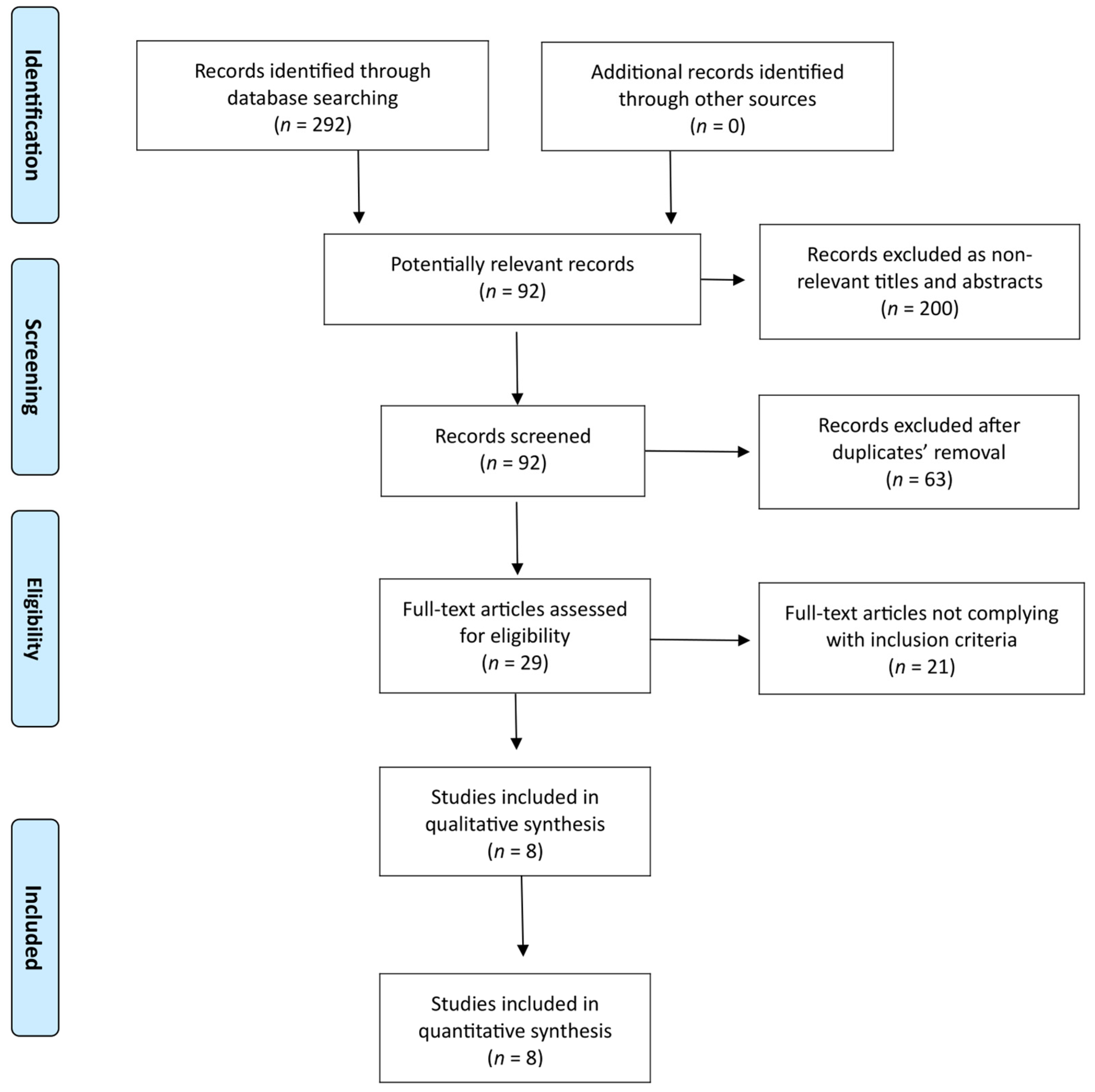
3.1. Search Results and Study Characteristics

According to the final literature search, which was performed on 3 June 2022, 292 potentially interesting studies were found (PubMed/MEDLINE: 53 records; Scopus: 148 records; Cochrane Library: 25 records; Web of Science: 66 records) (Figure 1). Although analysis covered all 292 studies, 200 ones turned out as not relevant for title and abstract, while 92 full-texts were considered eligible. Then, 63 out of 92 studies were ruled out as duplicate publications. Following the exclusion of 21 studies not complying with inclusion criteria, 8 comparative studies (seven retrospective studies and one randomized controlled study) underwent qualitative and quantitative synthesis [16,17,18,19,20,21,27,30]. No additional record was found through other sources (References list). Six out of the seven retrospective studies used propensity score matching (PSM) analysis [16,17,19,20,21,30].
According to ROBINS-I, most non-randomized studies showed moderate overall bias, except for Young et al.’s (serious) (see Supplementary Materials, Table S1). On the contrary, according to RoB 2, the only included randomized study had a low overall bias (see Supplementary Materials, Table S2).

3.2. Study and General Population Characteristics

Table 1 and Table 2 show study characteristics and demographic features of analyzed populations. The identified eight studies covered an almost 20-year period (2000–2018) [16,17,18,19,20,21,27,30]. In total, 43.2% of the general population (1122/2598) underwent gastrectomy and CO [16,17,18,19,20,21,27,30]. We recorded a male preponderance (66.4%; 1725/2598) and a 56–74-year-old population [16,17,18,19,20,21,27,30].
Moreover, 54.1% (1150/2127) and 64.5% (1372/2127) patients underwent open approach and distal gastrectomies, respectively [16,17,18,19,20,21,27,30]. Most gastrectomies were of the D2/D2+ type (75.8%; 1292/1705) [16,17,18,19,20,21,27,30]. pTNM stages III/IV were slightly predominant (51.2%; 1018/1987) [16,17,18,19,20,21,27,30].

3.3. Meta-Analyses Results

3.3.1. Overall Survival

Overall, six studies out of the eight included ones [16,17,18,19,20,21] comprising 1929 patients (CO 826, NCO 1103) who reported both 3-year and 5-year Oss (Figure 2 and Figure 3). The meta-analysis of pooled results showed that 3-year OS (RR: 0.94, 95% CI: 0.90–0.98, p = 0.005) and 5-year OS (RR: 0.93, 95% CI: 0.88–0.98, p = 0.007) in the NCO group were statistically significantly higher than in CO group. For both outcomes, the detected heterogeneity was low but statistically negligible (3-year OS–I2 = 0%, p = 0.72; 5-year OS–I2 = 5%, p = 0.38).

3.3.2. Disease-Free Survival

In total, five out of eight included studies [16,17,19,20,21] comprising 1458 patients (CO 736, NCO 722) reported both 3-year and 5-year DFSs (see Supplementary Materials, Figures S1 and S2). Meta-analysis of the pooled results showed a non-statistically significant difference between the two groups as concerned both outcomes (3-year DFS—RR: 0.97, 95% CI: 0.90–1.04, p = 0.44; 5-year DFS—RR: 0.98, 95% CI: 0.90–1.06, p = 0.60). For both outcomes, detected heterogeneity was moderate but statistically negligible (3-year DFS—I2 = 36%, p = 0.18; 5-year DFS—I2 = 40%, p = 0.16).

3.3.3. Recurrences

In total, six of the eight included studies [16,17,19,20,21,30] comprising 1880 patients (CO 910, NCO 970) reported an overall recurrence rate (see Supplementary Materials, Figure S3). Meta-analysis of pooled results showed a non-statistically significant difference between the two groups (RR: 1.13, 95% CI: 0.94–1.35, p = 0.19). Heterogeneity was low but statistically negligible (I2 = 10%, p = 0.35).
Moreover, five of the eight included studies [16,19,20,21,30] comprising 1734 patients (CO 830, NCO 904) reported recurrence rates in the peritoneum (see Supplementary Materials, Figure S4). Meta-analysis of the pooled results showed a non-statistically significant difference between the two groups (RR: 1.11, 95% CI: 0.85–1.44, p = 0.43). Heterogeneity was low but statistically non-significant (I2 = 0%, p = 0.83).
Further, five of the eight included studies [16,19,20,21,30] comprising 1734 patients (CO 830, NCO 904) reported recurrence rates in other sites (see Supplementary Materials, Figure S5). Meta-analysis of the pooled results showed a non-statistically significant difference between the two groups (RR: 1.02, 95% CI: 0.79–1.31, p = 0.88). Heterogeneity was low but statistically negligible (I2 = 0%, p = 0.88).

3.3.4. Operative Time

In total, six out of eight included studies [16,18,19,21,27,30] comprising 2312 patients (CO 972, NCO 1340) reported operative time (Figure 4). Meta-analysis of the pooled results showed a statistically significant longer operative time for the CO group compared to the NCO group (MD: 24.00, 95% CI: −0.45–48.45, p = 0.05). Heterogeneity was high and statistically significant (I2 = 94%, p < 0.00001).

3.3.5. Estimated Blood Loss

In total, five of the eight included studies [16,19,21,27,30] comprising 1841 patients (CO 882, NCO 959) reported estimated blood loss (Figure 5). Meta-analysis of the pooled results showed a statistically significant higher estimated blood loss for the CO group, when compared to the NCO group (MD: 194.76, 95% CI: 96.40–293.13, p = 0.0001). Heterogeneity was high and statistically significant (I2 = 98%, p < 0.00001).

3.3.6. Number of Lymph Nodes Harvested

In total, six of the eight included studies [17,18,19,21,27,30] comprising 2262 patients (CO 954, NCO 1308) reported harvested lymph nodes number (see Supplementary Materials, Figure S6). Meta-analysis of the pooled results showed a non-statistically significant difference between the two groups (MD: −0.52, 95% CI: −3.44–2.40, p = 0.73). Heterogeneity was high and statistically significant (I2 = 76%, p = 0.0009).

3.3.7. Overall Complications

In total, six of the eight included studies [16,18,19,20,21,30] comprising 2205 patients (CO 920, NCO 1285) reported an overall complication rate (see Supplementary Materials, Figure S7). Meta-analysis of the pooled results showed a statistically non-significant difference between the two groups (RR: 1.20, 95% CI: 0.93–1.54, p = 0.17). Heterogeneity was high and statistically relevant (I2 = 55%, p = 0.05).

3.3.8. Major Complications

In total, five of the eight included studies [19,20,21,27,30] comprising 1785 patients (CO 854, NCO 931) reported rates of major complications (see Supplementary Materials, Figure S8). Meta-analysis of the pooled results showed a statistically non-significant difference between the two groups (RR: 0.88, 95% CI: 0.51–1.52, p = 0.65). Heterogeneity was high and statistically relevant (I2 = 58%, p = 0.05).

3.3.9. Subgroup Analysis

Subgroup analysis was carried out according to discrepancies in study designs. In particular, we analyzed the different outcomes just considering PSM studies. Our subgroup analysis confirmed the outcomes of pooled analysis: higher 3-year OS and 5-year OS rates in the NCO group, longer operative time and higher estimated blood loss in the CO group (see Supplementary Materials, Figures S9–S15).

3.3.10. Publication Bias

According to the Cochrane Handbook for Systematic Reviews of Interventions (Version 5.1.0), tests for funnel plot asymmetry should only be carried out when meta-analysis includes at least 10 studies, as a smaller number of studies reduces the potential of tests to identify the case from real asymmetry [35]. As our meta-analysis included eight studies, we did not perform an analysis of publication bias.

4. Discussion

Gastrectomy with D2 lymphadenectomy represents a cornerstone in curative treatment for AGC [36]. However, despite correct radical intent gastrectomy, GC was marked by a 40–61% recurrence rate [37]. Peritoneal recurrence stood out, representing 36–45% of all kinds [38]. Omentum turned out as the most involved peritoneal site [6,7,14,22,23,24,25,26].
Omentum is a tissue stemming from the dorsal mesogastrium around the eighth week of gestation [15,39]. It consists of two mesothelial layers, mainly enclosing adipocytes embedded in a loose connective tissue and accumulations of mononuclear phagocytic cells [15,40]. Omentum has a rich vascularity with many widespread typical convolutions termed omental glomeruli, because they resemble renal glomeruli [15,40]. Omentum’s leukocytes gather in the perivascular area and form what we call “milky spots” [15,41]. Cells are arranged around omental glomeruli, which lie directly under the mesothelium [15,42,43]. They are supported by frail networks of reticular fibers that make up the organ’s structure [15,44]. In humans, milky spots include macrophages (70%), B-lymphocytes (10%), T-lymphocytes (10%), mast cells and stromal cells [15,45]. Macrophages in mature omentum are mainly scavengers [15,46]. They seem to be different from monocytic precursors in milky spots and are not dependent on bone-marrow-derived precursors [15,46]. They are dendritic in shape and have remarkable phagocytic abilities [15,46]. When activated, macrophage precursors in milky spots proliferate, then migrate to the mesothelial surface and turn into dendritic-shaped macrophages [15,46].
Omentum has turned out as a frequent site of metastatic malignancy [15,47,48]. In animals, tumor cells that are inoculated into the peritoneal cavity preferentially infiltrate omentum milky spots and grow into distinct metastases [15,47,48]. Omentum seems to be able to support not only malignant cells in milky spots, but also free intraperitoneal cells [15,47,48]. This happens thanks to omentum intrinsic angiogenic properties, as recent studies have highlighted [15,47,48,49]. In animals, omentum removal affects the survival of free intraperitoneal malignant cells and consequently reduces the local recurrence rate [15,50,51]. In this background, the omentum is often removed as part of resected malignancies in various intra-abdominal organs (ovarian cancer and GC, in particular) [15].
Despite omental tissue’s protumoral properties, authors are still debating on the need to perform CO, in addition to gastrectomy and D2 lymphadenectomy for GC [6,7,14,22,23,24,25,26]. Analyses of major guidelines in GC treatment clearly explained this topic. Japanese gastric cancer treatment guidelines 2018 (5th edition) suggested total removal of greater omentum in patients affected by T3 gastric cancer or deeper tumors, as an integral part of radical gastrectomy [52]. For patients affected by T1-T2 gastric cancer, the omentum more than 3 cm away from the gastroepiploic artery may be preserved [52]. Recommendations by the Japanese Gastric Cancer Association (JGCA) were shared by the Italian Research Group for Gastric Cancer (GIRCG) [53]. Following the aforementioned Japanese guidelines, the Chinese Society of Clinical Oncology (CSCO) stressed the important role of omentectomy by recommending total omentum removal in both D1 and D2 gastrectomy [54]. No mention of the role of omentectomy in radical gastrectomy was made by the Korean Practice Guideline for Gastric Cancer 2018 and National Comprehensive Cancer Network® (NCCN®) Guidelines for Gastric Cancer, Version 2.2022 [55].
In 2016, Jongerius et al. published a prospective multi-center cohort study (OMEGA trial), whose aim was to evaluate both incidence and risk factors for metastasis in the greater omentum, as regarded patients undergoing gastrectomy and CO for GC [56]. In a 100-patient population, the authors identified a low rate of incidence for omental metastases (5%) [56]. In addition, multivariate analysis identified that omental metastases were significantly related to microscopically non-radical resection, tumor expansion in the esophagus or duodenum, linitis plastica, tumor location in the proximal third of stomach, ≥5 cm tumor, III-IV stage of disease and category (y) pM1 [56].
Our meta-analysis aimed at updating data related to the comparison between patients who underwent gastrectomy with CO or without CO for AGC. We identified eight studies: seven retrospective ones and just one RCT [16,17,18,19,20,21,27,30]. Six out of seven retrospective studies had been developed as PSM [16,17,19,20,21,30]. Our meta-analysis indicated gastrectomy without CO as significantly associated with longer 3- and 5-year OSs, in the absence of relevant differences in terms of 3- and 5-year DFSs, overall recurrence, recurrences in peritoneum and recurrences in other site rates. Moreover, taking into account short-term outcomes, we found longer operative time and higher estimated blood loss in the CO group compared to the NCO group, with no statistically relevant differences between the two groups, as concerned the number of harvested lymph nodes, overall complications and major ones. However, it is necessary to consider that the significant differences identified between the surgical outcomes of the meta-analyzed studies (overall/major complications, operative time and estimated blood loss, in particular) could be influenced by different surgical procedures adopted. In particular, in some of the included studies it is possible to identify mixed populations of patients who underwent partial or complete omentectomy alone, as part of the bursectomy [16], or associated with splenectomy [16,17,19,20,27], transverse colectomy [17,20,27], cholecystectomy [19,27], partial pancreatectomy [20], adrenalectomy [27] and thoracotomy [27]. The above results were then confirmed by study design-based (PSM) subgroup analysis and seemed to be in line with reports in previous meta-analyses.
To date, just one RCT has been published comparing outcomes between CO and NCO gastrectomies for AGC [27]. Murakami et al. randomized a 251 overall patient population (CO: 125 patients, NCO: 126 patients) [27]. Following the exclusion of patients with peritoneal metastases or a history of laparotomy, 247 patients’ data were analyzed [27]. CO group showed a significantly longer median operative time (225 min vs. 204 min, p = 0.022) and was inclined to have higher median blood loss (260 mL vs. 210 mL, p = 0.073) [26]. Incidence of morbidity showed similar values, by recording a 10% rate in both groups (8% vs. 9%, p = 1000) [27]. No mortality was recorded in either group, although authors did not analyze survival outcomes [27].
As stated at the beginning of our discussion, surgery represents a cornerstone in the treatment of GC as well as a method that can lead to a higher chance of recovery [36,57]. Nevertheless, surgery is not always sufficient and feasible for patients affected by AGC [57]. A multimodal method that includes systemic or local therapies (neoadjuvants or adjuvants) may lead to greater disease control, ease of complete resection, and above all, improved survival outcomes [57]. Present multimodal strategies reflect significant geographical differences [58,59]. Adjuvant chemotherapy is the preferred treatment in East Asia, while neoadjuvant chemotherapy and adjuvant chemoradiation are the preferred ones in Europe and North America, respectively [58,59].
In particular, the application or non-application of neoadjuvant chemotherapy with or without radiotherapy could play a paramount role in highlighting differences between OS and DFS among groups to be compared. Different European trials that focused on the potential role of neoadjuvant chemotherapy collected interesting results [58,59]. At the end of the UK Medical Research Council Adjuvant Gastric Infusional Chemotherapy (MAGIC) trial, which included 503 resectable GC patients randomly assigned to three cycles of neoadjuvant chemotherapy (epirubicin, cisplatic and 5-fluorouracil) and surgery alone, neoadjuvant group showed significantly better 5-year OS (36% versus 23%; HR 0.75, p = 0.009) and DFS (35% versus 25%; HR 0.66, p < 0.001) [60]. The later Federation Nationale des Centres de Lutte contre le Cancer (FNCLCC)/the Federation Francophone de Cancerologie Digestive (FFCD) trial, that employed 5-fluorouracil plus cisplatin in preoperative setting and whose 224 resectable GC patients were randomly assigned to neoadjuvant chemotherapy and surgery alone, showed significantly improved R0 resection rates (87% versus 74%; p = 0.04), in addition to 5-year OS (38% versus 24%; HR 0.69, p = 0.02) and DFS (34% versus 19%; HR 0.65, p = 0.003) [61]. Similar outcomes were achieved by European Organisation for Research and Treatment of Cancer (EORTC) 40954 trial, whose 144 AGC patients were randomized to neoadjuvant 5-fluorouracil plus cisplatin or surgery alone [62]. Despite early termination, due to poor enrolment, the study highlighted a significant improvement in R0 resection rates for neoadjuvant group (81.9% versus 66.7%; p = 0.036) [62].
Two main Asian trials led to the most recent results. Korean PRODIGY trial compared neoadjuvant docetaxel, oxaliplatin and S-, followed by surgery and adjuvant S-1 with surgery and adjuvant S-1 (266 patients with resectable AGC) [63]. The study found a significant dominance of neoadjuvant chemotherapy in terms of 3-year DFS (66.3% versus 60.2%; HR 0.70, p = 0.023) [63]. Similar results were obtained from the Chinese RESOLVE trial, whose 1022 resectable AGC patients were randomized to neoadjuvant S-1 and oxaliplatin or adjuvant oxaliplatin or capecitabine/oxaliplatin, thus recording significantly improved 3-year DFS in the neoadjuvant group (62% versus 54.8%; HR 0.79, p = 0.045) [64].
With the exception of the Young et al. study [18], whose related data to neoadjuvant chemotherapy administration are unavailable, all studies included in our meta-analysis stem from Asian countries. This could be the reason why just a few patients undergoing neoadjuvant chemotherapy can be identified among the populations under analysis (Hasegawa et al. NCO 2/98 CO 6/98 [16]; Kim et al. n/a [17]; Ri et al. NCO 0/263 CO 0/263 [19]; Sakimura et al. NCO 5/70 CO 7/70 [20]; Seo et al. NCO 0/225 CO 0/225 [21]; Murakami et al. NCO 0/122 CO 0/125 [27]; Lee et al. NCO 0/174 CO 0/248 [30]). Therefore, we cannot fully rule out that the administration of neoadjuvant chemotherapy modifies long-term oncological outcomes in a more or less significant way, going so far as to nullify discrepancies between NCO and CO surgical groups.
To date, the development of further, possibly multi-center, randomized controlled trials corroborating the beneficial role of neoadjuvant chemotherapy in AGC patients is strongly needed.

Limitations

Our research bears some limitations: (i) the literature search did not include non-English written scientific articles; (ii) included studies were almost exclusively retrospective or retrospective PSM ones; (iii) data related to original baseline patient were missing, although matchings were conducted in PSM studies; (iv) most studies stemmed from Asian countries; (v) size of analyzed populations was small; (vi) significant heterogeneities existed among three short-term outcomes, which might have an adverse impact on evidence for short-term outcomes. For all these reasons, a direct comparison of results turned out as difficult.

5. Conclusions

Gastrectomy with D2 lymphadenectomy is the gold standard in AGC treatment, although peritoneal recurrence rate is both non-negligible and a major cause of poor prognosis. Omentum is one of the most common metastatic sites.
Many Authors describe CO as an integral part of AGC surgical treatment, although CO role is still highly debated. In fact, no agreement has been reached among international guidelines yet.
Our updated meta-analysis found that NCO group had a statistically greater rate in 3-year and 5-year Oss than the CO group, while the CO group had significantly longer operative time and higher estimated blood loss than the NCO group.
However, given non-negligible bias among the meta-analyzed studies, our results need an extremely careful data reading. Therefore, further randomized, possibly multi-center trials may turn out of paramount importance in confirming our results.

Supplementary Materials

The following supporting information can be downloaded at: https://www.mdpi.com/article/10.3390/medicina58091241/s1, Table S1. Retrospective studies and retrospective PSMs evaluated using ROBINS-I; Table S2. Randomized controlled trial evaluated using RoB 2.0; Figure S1. Forest plot comparing 3-year DFS between the CO and NCO groups. CI, confidence interval; M–H, Mantel–Haenszel; Figure S2. Forest plot comparing 5-year DFS between the CO and NCO groups. CI, confidence interval; M–H, Mantel–Haenszel; Figure S3. Forest plot comparing overall recurrences between the CO and NCO groups. CI, confidence interval; M–H, Mantel–Haenszel; Figure S4. Forest plot comparing recurrence in peritoneum between the CO and NCO groups. CI, confidence interval; M–H, Mantel–Haenszel; Figure S5. Forest plot comparing recurrences in other sites between the CO and NCO groups. CI, confidence interval; M–H, Mantel–Haenszel; Figure S6. Forest plot comparing number of harvested lymph nodes between the CO and NCO groups. CI, confidence interval; M–H, Mantel–Haenszel; Figure S7. Forest plot comparing overall complications between the CO and NCO groups. CI, confidence interval; M–H, Mantel–Haenszel; Figure S8. Forest plot comparing major complications between the CO and NCO groups. CI, confidence interval; M–H, Mantel–Haenszel; Figure S9. Forest plot comparing 3-year OS between the CO and NCO PSM groups. CI, confidence interval; M–H, Mantel–Haenszel; Figure S10. Forest plot comparing 5-year OS between the CO and NCO PSM groups. CI, confidence interval; M–H, Mantel–Haenszel; Figure S11. Forest plot comparing operative time between the CO and NCO PSM groups. CI, confidence interval; M–H, Mantel–Haenszel; Figure S12. Forest plot comparing estimated blood loss between the CO and NCO PSM groups. CI, confidence interval; M–H, Mantel–Haenszel; Figure S13. Forest plot comparing number of harvested lymph nodes between the CO and NCO PSM groups. CI, confidence interval; M–H, Mantel–Haenszel; Figure S14. Forest plot comparing overall complications between the CO and NCO PSM groups. CI, confidence interval; M–H, Mantel–Haenszel; Figure S15. Forest plot comparing major complications between the CO and NCO PSM groups. CI, confidence interval; M–H, Mantel–Haenszel.

Author Contributions

Conceptualization, M.Z. (Maurizio Zizzo); methodology, M.Z. (Maurizio Zizzo); validation, M.Z. (Maurizio Zizzo); formal analysis, M.Z. (Maurizio Zizzo); investigation, M.Z. (Maurizio Zizzo), M.Z. (Magda Zanelli), F.S., A.P., S.A., A.M., D.T., F.M., A.L.Z., M.N., F.G. and M.Z. (Manuel Zanni); data curation, M.Z. (Maurizio Zizzo); writing—original draft preparation, M.Z. (Maurizio Zizzo); writing—review and editing, M.Z. (Maurizio Zizzo); supervision, A.G.; project administration, M.Z. (Maurizio Zizzo). All authors have read and agreed to the published version of the manuscript.

Funding

This research received no external funding.

Institutional Review Board Statement

Local ethics committee (Comitato Etico dell’Area Vasta Emilia Nord, Italy) ruled that no formal ethics approval was required in this study.

Informed Consent Statement

Not applicable.

Data Availability Statement

The data presented in this study are available on request from the corresponding author.

Acknowledgments

We thank Daniela Masi (Azienda USL-IRCCS di Reggio Emilia) for her support in English editing. This study was partially supported by the Italian Ministry of Health–Ricerca Corrente Annual Program 2023.

Conflicts of Interest

The authors declare no conflict of interest.

References

  1. Sung, H.; Ferlay, J.; Siegel, R.L.; Laversanne, M.; Soerjomataram, I.; Jemal, A.; Bray, F. Global Cancer Statistics 2020: GLOBOCAN Estimates of Incidence and Mortality Worldwide for 36 Cancers in 185 Countries. CA Cancer J. Clin. 2021, 71, 209–249. [Google Scholar] [CrossRef] [PubMed]
  2. Smyth, E.C.; Nilsson, M.; Grabsch, H.I.; van Grieken, N.C.; Lordick, F. Gastric cancer. Lancet 2020, 396, 635–648. [Google Scholar] [CrossRef]
  3. Hu, Y.; Yoon, S.S. Extent of gastrectomy and lymphadenectomy for gastric adenocarcinoma. Surg. Oncol. 2022, 40, 101689. [Google Scholar] [CrossRef]
  4. Zizzo, M.; Zanelli, M.; Sanguedolce, F.; Torricelli, F.; Morini, A.; Tumiati, D.; Mereu, F.; Zuliani, A.L.; Palicelli, A.; Ascani, S.; et al. Robotic versus Laparoscopic Gastrectomy for Gastric Cancer: An Updated Systematic Review. Medicina 2022, 58, 834. [Google Scholar] [CrossRef] [PubMed]
  5. Ruiz Hispán, E.; Pedregal, M.; Cristobal, I.; García-Foncillas, J.; Caramés, C. Immunotherapy for Peritoneal Metastases from Gastric Cancer: Rationale, Current Practice and Ongoing Trials. J. Clin. Med. 2021, 10, 4649. [Google Scholar] [CrossRef]
  6. Zhu, A.; Yin, G.; Liu, X.; Kong, W.; Zhang, Y.; Shan, Y.; Ying, R.; Zhang, J.; Zhou, C. Efficiency of complete omentectomy in patients with resectable gastric cancer: A meta-analysis and systematic review. BMC Gastroenterol. 2021, 21, 346. [Google Scholar] [CrossRef]
  7. Lin, H.W.; Loh, E.W.; Shen, S.C.; Tam, K.W. Gastrectomy with or without omentectomy for gastric cancer: A systematic review and meta-analysis. Surgery 2022, 171, 1281–1289. [Google Scholar] [CrossRef]
  8. Marano, L.; Polom, K.; Bartoli, A.; Spaziani, A.; De Luca, R.; Lorenzon, L.; Di Martino, N.; Marrelli, D.; Roviello, F.; Castagnoli, G. Oncologic Effectiveness and Safety of Bursectomy in Patients with Advanced Gastric Cancer: A Systematic Review and Updated Meta-Analysis. J. Investig. Surg. 2018, 31, 529–538. [Google Scholar] [CrossRef]
  9. Nie, R.C.; Yuan, S.Q.; Chen, S.; Yan, S.M.; Chen, Y.M.; Chen, X.J.; Chen, G.M.; Zhou, Z.W.; Chen, Y.B.; Li, Y.F. Bursectomy for advanced gastric cancer: An update meta-analysis. World J. Surg. Oncol. 2018, 16, 66. [Google Scholar] [CrossRef]
  10. Xiong, B.; Ma, L.; Huang, W.; Cheng, Y.; Luo, H.; Wang, K. Efficiency of bursectomy in patients with resectable gastric cancer: An updated meta-analysis. Eur. J. Surg. Oncol. 2019, 45, 1483–1492. [Google Scholar] [CrossRef]
  11. Li, Y.; Li, Y.; Wang, K. Bursectomy in Gastric Cancer Surgery: A Meta-Analysis. Rev. Investig. Clin. 2019, 71, 98–105. [Google Scholar] [CrossRef] [PubMed]
  12. Perivoliotis, K.; Baloyiannis, I.; Symeonidis, D.; Tepetes, K. The role of bursectomy in the surgical management of gastric cancer: A meta-analysis and systematic review. Updates Surg. 2020, 72, 939–950. [Google Scholar] [CrossRef] [PubMed]
  13. Kurokawa, Y.; Doki, Y.; Mizusawa, J.; Terashima, M.; Katai, H.; Yoshikawa, T.; Kimura, Y.; Takiguchi, S.; Nishida, Y.; Fukushima, N.; et al. Bursectomy versus omentectomy alone for resectable gastric cancer (JCOG1001): A phase 3, open-label, randomised controlled trial. Lancet Gastroenterol. Hepatol. 2018, 3, 460–468. [Google Scholar] [CrossRef]
  14. Chai, S.W.; Wang, S.H.; Wang, C.Y.; Chen, Y.C.; Soong, R.S.; Huang, T.S. Partial Versus Total Omentectomy in Patients with Gastric Cancer: A Systemic Review and Meta-Analysis. Cancers 2021, 13, 4971. [Google Scholar] [CrossRef]
  15. Platell, C.; Cooper, D.; Papadimitriou, J.M.; Hall, J.C. The omentum. World J. Gastroenterol. 2000, 6, 169–176. [Google Scholar]
  16. Hasegawa, S.; Kunisaki, C.; Ono, H.; Oshima, T.; Fujii, S.; Taguri, M.; Morita, S.; Sato, T.; Yamada, R.; Yukawa, N.; et al. Omentum-preserving gastrectomy for advanced gastric cancer: A propensity-matched retrospective cohort study. Gastric Cancer 2013, 16, 383–388. [Google Scholar] [CrossRef]
  17. Kim, D.J.; Lee, J.H.; Kim, W. A comparison of total versus partial omentectomy for advanced gastric cancer in laparoscopic gastrectomy. World J. Surg. Oncol. 2014, 12, 64. [Google Scholar] [CrossRef]
  18. Young, S.; DiFronzo, L.A.; Ahuja, A.; Keim, L.; Papenfuss, C.; O’Connor, V.; Dehal, A. Performing Omentectomy during Gastrectomy Does Not Improve Survival: A Multi-Center Analysis of 471 Patients with Gastric Adenocarcinoma. J. Gastrointest. Surg. 2020, 24, 2856–2858. [Google Scholar] [CrossRef]
  19. Ri, M.; Nunobe, S.; Honda, M.; Akimoto, E.; Kinoshita, T.; Hori, S.; Aizawa, M.; Yabusaki, H.; Isobe, Y.; Kawakubo, H.; et al. Gastrectomy with or without omentectomy for cT3-4 gastric cancer: A multicentre cohort study. Br. J. Surg. 2020, 107, 1640–1647. [Google Scholar] [CrossRef]
  20. Sakimura, Y.; Inaki, N.; Tsuji, T.; Kadoya, S.; Bando, H. Long-term outcomes of omentum-preserving versus resecting gastrectomy for locally advanced gastric cancer with propensity score analysis. Sci. Rep. 2020, 10, 16305. [Google Scholar] [CrossRef]
  21. Seo, W.J.; Choi, S.; Roh, C.K.; Cho, M.; Kim, Y.M.; Kim, H.I.; Hyung, W.J. Omentum preservation as an oncologically comparable and surgically superior alternative to total omentectomy during radical gastrectomy for T3-T4 gastric cancer. Surgery 2021, 170, 610–616. [Google Scholar] [CrossRef] [PubMed]
  22. Chen, M.; He, F.Q.; Liao, M.S.; Yang, C.; Chen, X.-D. Gastrectomy with omentum preservation versus gastrectomy with omentectomy for locally advanced gastric cancer: A systematic review and meta-analysis. Int. J. Surg. 2021, 96, 106176. [Google Scholar] [CrossRef] [PubMed]
  23. Ishizuka, M.; Shibuya, N.; Takagi, K.; Hachiya, H.; Tago, K.; Matsumoto, T.; Shimizu, T.; Aoki, T.; Kubota, K. Omentectomy Does Not Affect the Postoperative Outcome of Patients with Locally Advanced Gastric Cancer: A Systematic Review and Meta-Analysis. J. Surg. Res. 2021, 264, 287–295. [Google Scholar] [CrossRef] [PubMed]
  24. Li, Z.; Song, M.; Zhou, Y.; Jiang, H.; Xu, L.; Hu, Z.; Liu, Y.; Jiang, Y.; Li, X. Efficacy of Omentum-Preserving Gastrectomy for Patients with Gastric Cancer: A Systematic Review and Meta-Analysis. Front. Oncol. 2021, 11, 710814. [Google Scholar] [CrossRef]
  25. Zhang, Y.X.; Liu, H.D.; Chen, Z.H.; Jin, T.; Hu, J.K.; Yang, K. Comparison of Survival and Safety Between Total Omentectomy and Partial Omentectomy for Gastric Cancer: A Meta-Analysis. Front. Surg. 2021, 8, 708545. [Google Scholar] [CrossRef]
  26. Kong, M.; Chen, H.; Zhang, R.; Sheng, H.; Li, L. Overall Survival Advantage of Omentum Preservation over Omentectomy for Advanced Gastric Cancer: A Systematic Review and Meta-Analysis. World J. Surg. 2022, 46, 1952–1961. [Google Scholar] [CrossRef]
  27. Murakami, H.; Yamada, T.; Taguri, M.; Hasegawa, S.; Yamanaka, T.; Rino, Y.; Mushiake, H.; Oshima, T.; Matsukawa, H.; Tani, K.; et al. Short-Term Outcomes from a Randomized Screening Phase II Non-inferiority Trial Comparing Omentectomy and Omentum Preservation for Locally Advanced Gastric Cancer: The TOP-G Trial. World J. Surg. 2021, 45, 1803–1811. [Google Scholar] [CrossRef]
  28. Lu, S.; Yang, Z.Y.; Yan, C.; Liu, W.T.; Ni, Z.T.; Yao, X.X.; Hua, Z.C.; Feng, R.H.; Zheng, Y.N.; Wang, Z.Q.; et al. A randomized controlled trial to evaluate omentum-preserving gastrectomy for patients with T1-T3 gastric cancer. Future Oncol. 2021, 17, 3301–3307. [Google Scholar] [CrossRef]
  29. Sato, Y.; Yamada, T.; Yoshikawa, T.; Machida, R.; Mizusawa, J.; Katayama, H.; Tokunaga, M.; Boku, N.; Terashima, M.; Stomach Cancer Study Group/Japan Clinical Oncology Group. Randomized controlled Phase III trial to evaluate omentum preserving gastrectomy for patients with advanced gastric cancer (JCOG1711, ROAD-GC). Jpn. J. Clin. Oncol. 2020, 50, 1321–1324. [Google Scholar] [CrossRef]
  30. Lee, S.; Suh, Y.S.; Kang, S.H.; Won, Y.; Park, Y.S.; Ahn, S.H.; Kim, H.H. Should total omentectomy be performed for advanced gastric cancer?: The role of omentectomy during laparoscopic gastrectomy for advanced gastric cancer. Surg. Endosc. 2022, 36, 6937–6948. [Google Scholar] [CrossRef]
  31. Moher, D.; Liberati, A.; Tetzlaff, J. Preferred reporting items for systematic reviews and meta-analyses: The PRISMA statement. Ann. Intern. Med. 2009, 151, 264–269. [Google Scholar] [CrossRef] [PubMed] [Green Version]
  32. Sterne, J.A.C.; Savović, J.; Page, M.J.; Elbers, R.G.; Blencowe, N.S.; Boutron, I.; Cates, C.J.; Cheng, H.Y.; Corbett, M.S.; Eldridge, S.M.; et al. RoB 2: A revised tool for assessing risk of bias in randomised trials. BMJ 2019, 366, l4898. [Google Scholar] [CrossRef] [PubMed]
  33. Sterne, J.A.; Hernán, M.A.; Reeves, B.C.; Savović, J.; Berkman, N.D.; Viswanathan, M.; Henry, D.; Altman, D.G.; Ansari, M.T.; Boutron, I.; et al. ROBINS-I: A tool for assessing risk of bias in non-randomised studies of interventions. BMJ 2016, 355, i4919. [Google Scholar] [CrossRef] [PubMed]
  34. Cochrane Review Manager (RevMan). Available online: https://training.cochrane.org/online-learning/core-software/revman (accessed on 3 June 2022).
  35. Cochrane Handbook for Systematic Reviews of Interventions [Version 5.1.0]. Available online: https://handbook-5-1.cochrane.org/chapter_10/10_4_3_1_recommendations_on_testing_for_funnel_plot_asymmetry.htm (accessed on 3 June 2022).
  36. Zizzo, M.; Ugoletti, L.; Manzini, L.; Castro Ruiz, C.; Nita, G.E.; Zanelli, M.; De Marco, L.; Besutti, G.; Scalzone, R.; Sassatelli, R.; et al. Management of duodenal stump fistula after gastrectomy for malignant disease: A systematic review of the literature. BMC Surg. 2019, 19, 55. [Google Scholar]
  37. Agnes, A.; Biondi, A.; Persiani, R.; Laurino, A.; Reddavid, R.; De Giuli, M.; Sicoli, F.; Cananzi, F.; De Pascale, S.; Fumagalli, U.; et al. Development of the PERI-Gastric (PEritoneal Recurrence Index) and PERI-Gram (Peritoneal Recurrence Index NomoGRAM) for predicting the risk of metachronous peritoneal carcinomatosis after gastrectomy with curative intent for gastric cancer. Gastric Cancer 2022, 25, 629–639. [Google Scholar] [CrossRef]
  38. Guner, A.; Yildirim, R. Surgical management of metastatic gastric cancer: Moving beyond the guidelines. Transl. Gastroenterol. Hepatol. 2019, 4, 58. [Google Scholar] [CrossRef]
  39. Krist, L.F.; Koenen, H.; Calame, W.; van der Harten, J.J.; van der Linder, J.C.; Eestermans, I.L.; Meyer, S.; Beelen, R.H. Ontogeny of milky spots in the human greater omentum: An immunochemical study. Anat. Rec. 1997, 249, 399–404. [Google Scholar] [CrossRef]
  40. Ackermann, P.C.; De Wet, P.D.; Loots, G.P. Microcirculation of the rat omentum studied by means of corrosion casts. Acta Anat. 1991, 140, 146–149. [Google Scholar] [CrossRef]
  41. Vanvugt, E.; Vanrijthoven, E.A.M.; Kamperdijk, E.W.A.; Beelen, R.H.J. Omental milky spots in the local immune response in the peritoneal cavity of rats. Anat. Rec. 1996, 244, 235–245. [Google Scholar] [CrossRef]
  42. Wijffels, J.F.; Hendrickx, R.J.; Steenbergen, J.J.; Eestermans, I.L.; Beelen, R.H. Milky spots in the mouse omentum may play an important role in the origin of peritoneal macrophages. Res. Immunol. 1992, 143, 401–409. [Google Scholar] [CrossRef]
  43. Shimotsuma, M.; Shields, J.W.; Simpson Morgan, M.W.; Sakuyama, A.; Shirasu, M.; Hagiwara, A.; Takahashi, T. Morpho physiological function and role of omental milky spots as omentum associated lymphoid tissue (OALT) in the peritoneal cavity. Lymphology 1993, 26, 90–101. [Google Scholar] [PubMed]
  44. Shimotsuma, M.; Kawata, M.; Hagiwara, A.; Takahashi, T. Milky spots in the human greater omentum. Macroscopic and histological identification. Acta Anat. 1989, 136, 211–216. [Google Scholar] [CrossRef] [PubMed]
  45. Zhu, H.; Naito, M.; Umezu, H.; Moriyama, H.; Takatsuka, H.; Takahashi, K.; Shultz, L.D. Macrophage differentiation and expression of macrophage colony stimulatingfactor in murine milky spots and omentum after macrophage elimination. J. Leukoc. Biol. 1997, 61, 436–444. [Google Scholar] [CrossRef] [PubMed]
  46. Dux, K. Proliferative activity of macrophages in the greater omentum of the mouse in relation to the early postnatal development of the vascular structures. J. Leukoc. Biol. 1986, 40, 445–458. [Google Scholar] [CrossRef] [PubMed]
  47. Tsujimoto, H.; Hagiwara, A.; Shimotsuma, M.; Sakakura, C.; Osaki, K.; Sasaki, S.; Ohyama, T.; Ohgaki, M.; Imanishi, T.; Yamazaki, J.; et al. Role of milky spots as selective implantation sites for malignant cells in peritoneal dissemination in mice. J. Cancer Res. Clin. Oncol. 1996, 122, 590–595. [Google Scholar] [CrossRef]
  48. Tsujimoto, H.; Takhashi, T.; Hagiwara, A.; Shimotsuma, M.; Sakakura, C.; Osaki, K.; Sasaki, S.; Shirasu, M.; Sakakibara, T.; Ohyama, T. Site-specific implantation in the milky spots of malignant cells in peritoneal dissemination: Immunohistochemical observation in mice inoculated intraperitoneally with bromodeoxyuridine labelled cells. Br. J. Cancer 1995, 71, 468–472. [Google Scholar] [CrossRef]
  49. Kersy, O.; Loewenstein, S.; Lubezky, N.; Sher, O.; Simon, N.B.; Klausner, J.M.; Lahat, G. Omental Tissue-Mediated Tumorigenesis of Gastric Cancer Peritoneal Metastases. Front. Oncol. 2019, 9, 1267. [Google Scholar] [CrossRef] [PubMed]
  50. Lawrance, R.J.; Loizidou, M.; Cooper, A.J.; Alexander, P.; Taylor, I. Importance of the omentum in the development of intra- abdominal metastases. Br. J. Surg. 1991, 78, 117–119. [Google Scholar] [CrossRef]
  51. Weese, J.L.; Ottery, F.D.; Emoto, S.E. Does omentectomy prevent malignant small bowel obstruction. Clin. Exp. Metastasis 1988, 6, 319–324. [Google Scholar] [CrossRef]
  52. Japanese Gastric Cancer Association. Japanese gastric cancer treatment guidelines 2018 (5th edition). Gastric Cancer 2021, 24, 1–21. [Google Scholar] [CrossRef]
  53. De Manzoni, G.; Marrelli, D.; Baiocchi, G.L.; Morgagni, P.; Saragoni, L.; Degiuli, M.; Donini, A.; Fumagalli, U.; Mazzei, M.A.; Pacelli, F.; et al. The Italian Research Group for Gastric Cancer (GIRCG) guidelines for gastric cancer staging and treatment: 2015. Gastric Cancer 2017, 20, 20–30. [Google Scholar] [CrossRef] [PubMed]
  54. Wang, F.H.; Zhang, X.T.; Li, Y.F.; Tang, L.; Qu, X.J.; Ying, J.E.; Zhang, J.; Sun, L.Y.; Lin, R.B.; Qiu, H.; et al. The Chinese Society of Clinical Oncology (CSCO): Clinical guidelines for the diagnosis and treatment of gastric cancer, 2021. Cancer Commun. 2021, 41, 747–795. [Google Scholar] [CrossRef]
  55. Ajani, J.A.; D’Amico, T.A.; Bentrem, D.J.; Chao, J.; Cooke, D.; Corvera, C.; Das, P.; Enzinger, P.C.; Enzler, T.; Fanta, P.; et al. Gastric Cancer, Version 2.2022, NCCN Clinical Practice Guidelines in Oncology. J. Natl. Compr. Canc. Netw. 2022, 20, 167–192. [Google Scholar] [CrossRef] [PubMed]
  56. Jongerius, E.J.; Boerma, D.; Seldenrijk, K.A.; Meijer, S.L.; Scheepers, J.J.; Smedts, F.; Lagarde, S.M.; Balague Ponz, O.; van Berge Henegouwen, M.I.; van Sandick, J.W.; et al. Role of omentectomy as part of radical surgery for gastric cancer. Br. J. Surg. 2016, 103, 1497–1503. [Google Scholar] [CrossRef] [PubMed]
  57. Yeh, J.H.; Yeh, Y.S.; Tsai, H.L.; Huang, C.W.; Chang, T.K.; Su, W.C.; Wang, J.Y. Neoadjuvant Chemoradiotherapy for Locally Advanced Gastric Cancer: Where Are We at? Cancers 2022, 14, 3026. [Google Scholar] [CrossRef] [PubMed]
  58. Fong, C.; Johnston, E.; Starling, N. Neoadjuvant and Adjuvant Therapy Approaches to Gastric Cancer. Curr. Treat. Options Oncol. 2022, 23, 1247–1268. [Google Scholar] [CrossRef]
  59. Lumish, M.A.; Ku, G.Y. Approach to Resectable Gastric Cancer: Evolving Paradigm of Neoadjuvant and Adjuvant Treatment. Curr. Treat. Options Oncol. 2022, 23, 1044–1058. [Google Scholar] [CrossRef]
  60. Cunningham, D.; Allum, W.H.; Stenning, S.P.; Thompson, J.N.; Van de Velde, C.J.; Nicolson, M.; Scarffe, J.H.; Lofts, F.J.; Falk, S.J.; Iveson, T.J.; et al. MAGIC Trial Participants. Perioperative chemotherapy versus surgery alone for resectable gastroesophageal cancer. N. Engl. J. Med. 2006, 355, 11–20. [Google Scholar] [CrossRef]
  61. Ychou, M.; Boige, V.; Pignon, J.P.; Conroy, T.; Bouché, O.; Lebreton, G.; Ducourtieux, M.; Bedenne, L.; Fabre, J.M.; Saint-Aubert, B.; et al. Perioperative chemotherapy compared with surgery alone for resectable gastroesophageal adenocarcinoma: An FNCLCC and FFCD multicenter phase III trial. J. Clin. Oncol. 2011, 29, 1715–1721. [Google Scholar] [CrossRef]
  62. Schuhmacher, C.; Gretschel, S.; Lordick, F.; Reichardt, P.; Hohenberger, W.; Eisenberger, C.F.; Haag, C.; Mauer, M.E.; Hasan, B.; Welch, J.; et al. Neoadjuvant chemotherapy compared with surgery alone for locally advanced cancer of the stomach and cardia: European Organisation for Research and Treatment of Cancer randomized trial 40954. J. Clin. Oncol. 2010, 28, 5210–5218. [Google Scholar] [CrossRef]
  63. Kang, Y.K.; Yook, J.H.; Park, Y.K.; Lee, J.S.; Kim, Y.W.; Kim, J.Y.; Ryu, M.H.; Rha, S.Y.; Chung, I.J.; Kim, I.H.; et al. PRODIGY: A Phase III Study of Neoadjuvant Docetaxel, Oxaliplatin, and S-1 Plus Surgery and Adjuvant S-1 Versus Surgery and Adjuvant S-1 for Resectable Advanced Gastric Cancer. J. Clin. Oncol. 2021, 39, 2903–2913. [Google Scholar] [CrossRef] [PubMed]
  64. Zhang, X.; Liang, H.; Li, Z.; Xue, Y.; Wang, Y.; Zhou, Z.; Yu, J.; Bu, Z.; Chen, L.; Du, Y.; et al. Perioperative or postoperative adjuvant oxaliplatin with S-1 versus adjuvant oxaliplatin with capecitabine in patients with locally advanced gastric or gastro-oesophageal junction adenocarcinoma undergoing D2 gastrectomy (RESOLVE): An open-label, superiority and non-inferiority, phase 3 randomised controlled trial. Lancet Oncol. 2021, 22, 1081–1092, Erratum in Lancet Oncol. 2021, 22, e347. [Google Scholar] [CrossRef] [PubMed]
Figure 1. PRISMA flow chart of literature search.
Medicina 58 01241 g001
Figure 2. Forest plot comparing 3-year OS between the CO and NCO groups. CI, confidence interval; M–H, Mantel–Haenszel [16,17,18,19,20,21].
Medicina 58 01241 g002
Figure 3. Forest plot comparing 5-year OS between the CO and NCO groups. CI, confidence interval; M–H, Mantel–Haenszel [16,17,18,19,20,21].
Medicina 58 01241 g003
Figure 4. Forest plot comparing operative time between the CO and NCO groups. CI, confidence interval; M–H, Mantel–Haenszel [16,18,19,21,27,30].
Medicina 58 01241 g004
Figure 5. Forest plot comparing estimated blood loss between the CO and NCO groups. CI, confidence interval; M–H, Mantel–Haenszel [16,19,21,27,30].
Medicina 58 01241 g005
Table 1. Study characteristics.
Authors/YearStudy TypeStudy
Country
Study
Period
GroupPatient
Population, n
Gender, nAge (Years), Mean or MedianBMI (kg/m2), Mean or
Median
MaleFemale
Lee et al./2022 [30]Retrospective PSMKorea2014–2018CO1741225259.9 ± 12.723.1 ± 3.7
NCO2481777161.6 ± 13.323.8 ± 3.1
Seo et al./2021 [21]Retrospective PSMKorea2003–2015CO2251319459 (49–70)23.5 (21.1–25.6)
NCO2251378856 (49–67)22.9 (21.0–24.9)
Murakami et al./2021 [27]RCTJapan2011–2018CO122893371 (30–90)22.4 (14.8–31.8)
NCO125893674 (45–89)22.2 (14.5–32.1)
Sakimura et al./2020 [20]Retrospective PSMJapan2008–2017CO70462465.0 (37–90)22.2 (15.8–30.3)
NCO70482266.5 (42–94)22.4 (16.4–32.6)
Ri et al./2020 [19]Retrospective PSMJapan2006–2012CO2631768766.7 ± 1122.4 ± 3.6
NCO2631818265.7 ± 12.922.5 ± 3.4
Young et al./2020 [18]RetrospectiveUSA2008–2016CO90622869.5 (62–77)27.4 ± 6.1
NCO38121716468 (58–76)26.2 ± 5.3
Kim et al./2014 [17]Retrospective PSMKorea2004–2011CO80562460.9 ± 11.2n/a
NCO66501662.2 ± 11n/a
Hasegawa et al./2013 [16]Retrospective PSMJapan2000–2009CO98722669.0 (40–91)n/a
NCO98722668.7 (45–91)n/a
n = number; BMI = Body Mass Index; PSM = propensity score matching; RCT = randomized controlled trial; CO = complete omentectomy; NCO = non-complete omentectomy; n/a = not available [16,17,18,19,20,21,27,30].
Table 2. General population characteristics.
Authors/YearGroupPatient
Population, n
Surgical
Approach, n
Surgical Procedure, nLymphadenectomy, npT Stage, npN Stage, npTNM Stage, n
OpenMISDGTGD1D1+D2D2+T0T1T2T3T4N0N1N2N3IIIIIIIV
Lee et al./2022 [30]CO174017410173n/an/an/an/a002278744219397410431210
NCO248024815791n/an/an/an/a0045119846535628621771500
Seo et al./2021 [21]CO22560165167580252000000100125754242660951300
NCO22569156169560222030000111114734742630991260
Murakami et al./2021 [27]CO12212207349001220020214239442925242648417
NCO12512508144001250031213142542517293840407
Sakimura et al./2020 [20]CO704129452509611512322022141420n/an/an/an/a
NCO702545442601456061627212914918n/an/an/an/a
Ri et al./2020 [19]CO26326301561071114610604721614801150281011295
NCO263263015111281461090482151450118029961317
Young et al./2020 [18]CO90n/an/an/an/an/an/an/an/an/an/an/a414753n/an/an/an/a
NCO381n/an/an/an/an/an/an/an/an/an/an/a184176205n/an/an/an/a
Kim et al./2014 [17]CO800806119027800028520401413131739240
NCO6606654120561000372903481682326170
Hasegawa et al./2013 [16]CO98980524601286000303434392518161640420
NCO988414613701385000343034412317172136410
n = number; CO = complete omentectomy; NCO = non-complete omentectomy; MIS = minimally invasive surgery; DG = Distal gastrectomy; TG = Total gastrectomy; n/a = not available [16,17,18,19,20,21,27,30].
Publisher’s Note: MDPI stays neutral with regard to jurisdictional claims in published maps and institutional affiliations.

