Article

Network Pharmacology Integrated Molecular Docking and Dynamics to Elucidate Saffron Compounds Targeting Human COX-2 Protein

1
Division of Veterinary Biochemistry, Faculty of Veterinary Sciences & Animal Husbandry, SKUAST-K, Shuhama, Alusteng, Srinagar 190006, India
2
Genome Engineering and Societal Biotechnology Lab., Division of Plant Biotechnology, SKUAST-K, Shalimar, Srinagar 190006, India
3
Department of Biochemistry, School of Biological Sciences, University of Kashmir, Hazratbal, Srinagar 190006, India
4
SDM Research Institute for Biomedical Sciences, Dharwad 580009, India
5
SALK Institute for Biological Studies, La Jolla, San Diego, CA 92037, USA
6
Department of Studies and Research in Biochemistry, Mangalore University, Mangalore 571232, India
7
Department of Clinical Pharmacy, College of Pharmacy, King Saud University, Riyadh 11451, Saudi Arabia
8
Department of Pharmacology and Toxicology, College of Pharmacy, King Saud University, Riyadh 11451, Saudi Arabia
*
Authors to whom correspondence should be addressed.
Medicina 2023, 59(12), 2058; https://doi.org/10.3390/medicina59122058
Submission received: 11 October 2023 / Revised: 5 November 2023 / Accepted: 17 November 2023 / Published: 22 November 2023
(This article belongs to the Section Pharmacology)

Abstract

:
Background and Objectives: Cyclooxygenase-2 (COX-2) is mostly linked to inflammation and has been validated as a molecular target for treating inflammatory diseases. The present study aimed to identify novel compounds that could inhibit COX-2, which is associated with various diseases including inflammation, and in such a scenario, plant-derived biomolecules have been considered as attractive candidates. Materials and Methods: In the present study, physiochemical properties and toxicity of natural compounds/drugs were determined by SWISSADME and ProTox-II. In the present study, the molecular docking binding features of saffron derivatives (crocetin, picrocrocin, quercetin, safranal, crocin, rutin, and dimethylcrocetin) against human COX-2 protein were assessed. Moreover, protein-protein interactions, topographic properties, gene enrichment analysis and molecular dynamics simulation were also determined. Results: The present study revealed that picrocrocin showed the highest binding affinity of −8.1 kcal/mol when docked against the COX-2 protein. PROCHECK analysis revealed that 90.3% of the protein residues were found in the most favored region. Compartmentalized Protein–Protein Interaction identified 90 interactions with an average interaction score of 0.62, and the highest localization score of 0.99 found in secretory pathways. The Computed Atlas of Surface Topography of Proteins was used to identify binding pockets and important residues that could serve as drug targets. Use of WEBnmα revealed protein dynamics by using normal mode analysis. Ligand and Receptor Dynamics used the Molecular Generalized Born Surface Area approach to determine the binding free energy of the protein. Gene enrichment analysis revealed that ovarian steroidogenesis, was the most significant enrichment pathway. Molecular dynamic simulations were executed for the best docked (COX-2-picrocrocin) complex, and the results displayed conformational alterations with more pronounced surface residue fluctuations in COX-2 with loss of the intra-protein hydrogen bonding network. The direct interaction of picrocrocin with various crucial amino-acid residues like GLN203, TYR385, HIS386 and 388, ASN382, and TRP387 causes modifications in these residues, which ultimately attenuates the activity of COX-2 protein. Conclusions: The present study revealed that picrocrocin was the most effective biomolecule and could be repurposed via computational approaches. However, various in vivo and in vitro observations are still needed.

1. Introduction

Inflammation is the biological response of the body to harmful stimuli and can be associated with pathogens, tissue damage, irritation, infection, and toxic compounds [1]. Inflammatory reactions are a non-specific defense against cell injury; their hallmarks are redness, swelling, and pain, and they are associated with the release of various inflammatory mediators including histamines, interleukins (ILs), bradykinin, leukotrienes, prostaglandins (PGs), and enzymes [2,3,4]. Under normal conditions, inflammation is restricted to affected tissues; however, in some cases, it may become uncontrollable, such as in cancers, asthma, diabetes mellitus, Alzheimer’s disease, and nephropathy [5,6,7,8]. Therapeutic interventions involve the use of non-steroidal anti-inflammatory drugs (NSAID) and narcotic analgesic agents. In the management and treatment of inflammation, cyclooxygenase 2 (COX-2) inhibitors are the main prominent molecules. The COX-2 enzyme is the main prostanoid (PG, prostacyclin, and thromboxane) synthesis enzyme, having two isoforms (COX-1 and COX-2) each with two activities like oxygenase and peroxidase activity, and it converts 20 carbon arachidonic acids into PGH2 through a short-lived PGG2 intermediate [2,3,4,9]. COX-1 is a constitutive protein, whereas COX-2 is an inducible protein; both the proteins are active in dimeric form, and their monomeric form consists of three domains: the epidermal growth factor (EGF) domain at N-terminal, the heme-peroxidase domain at the core, and the membrane anchor domain [10]. COX enzyme, primarily COX-2, becomes an important target for various anti-inflammatory drugs by causing amino acid substitution [11]. Several anti-inflammatory drugs like celecoxib, naproxen, celecoxib, ketorolac, acetylsalicylic acid, etoricoxib, and indomethacin are primarily used to treat inflammation [12,13,14]. All these drugs are also associated with various side effects and resistance development [15,16]. Therefore, we move towards naturally occurring herbal drugs as they act as precursors of various synthetic drugs to cure different diseases including inflammatory disease [17,18,19]. Plant secondary metabolites also regulate various biological activities like cancer, anti-bacterial, inflammation, anti-viral, migration, proliferation, and cell death [20,21,22,23]. Many natural products (culinary mushrooms, flavonoids, terpenoids, hyperforin, phenols, and alkaloids) have already been discovered that inhibit COX-2 expression/activity and exhibit fewer side effects [24,25,26].
In the traditional system of medicine, saffron (Crocus sativus) has been used as an alternative to treat various diseases in addition to its properties of flavoring, tasting, and coloring food [27]. Saffron has been widely found in Iran, Turkey, India, Italy, Greece, Egypt, Azerbaijan, Spain, and France [27,28]. Saffron is an ancient medicinal plant that possesses antioxidant, immunomodulatory, and anti-inflammatory properties [29,30,31]. Saffron has been used for the treatment of diabetes, coronary artery disorders, colorectal cancer, asthma, neurodegenerative diseases, and bronchitis [32,33,34]. The health-promoting medicinal properties of saffron are mainly due to its prominent chemical constituents such as crocins, safranal, crocetins, and carotenoids (α and β carotene, lycopene, and zeaxanthin) [35,36,37].
Recent developments in computational methods have been significant in developing the rationale for identifying natural compounds that can be used to target inflammatory proteins of interest. Computational approaches have the tremendous potential to repurpose plant compounds for drug design and development. Here, keeping in view the above-mentioned issues, computational methods were used to identify potential COX-2 inhibitors from compounds of saffron. In the present study, absorption, distribution, metabolism, excretion, and toxicity (ADMET) analysis of saffron phytocompounds and synthetic drugs was carried out. Moreover, we also aimed to determine the sequence alignment, molecular modeling, molecular docking, protein–protein interactions (PPIs), gene enrichment analysis, and topological properties of the protein. Further, in the present study, molecular dynamics simulations (MDs) of the topmost protein–ligand complex were performed.

2. Methods and Material

2.1. Ligand Selection

In the present study, crocetin, picrocrocin, quercetin, rutin, safranal, crocin, and dimethyl crocetin were the selected phytocompounds, whereas aspirin and rofecoxib were the synthetic drugs. The ligand’s three-dimensional (3D) structures were retrieved from PubChem (http://pubchem.ncbi.nlm.nih.gov/compound/ accessed on 7 June 2023).

2.2. ADMET Analysis

In the present study, the physiochemical and pharmacokinetics properties of selected natural compounds and drugs were evaluated through ADMET. SWISSADME, an online, easily available web server (http://www.swissadme.ch/ accessed on 7 June 2023), was used for determining the drug-like properties of molecules [38]. The smiles of ligands (obtained from PubChem) were then entered in input of SWISSADME to determine their ADMET characteristics. All the compounds were screened for Lipinski’s rule of five (RO5) that includes molecular weight less than 500 g/mol, LOGP < 5, topological polar surface area (TPSA) score of <140 Å2, and a number of hydrogen bond acceptors and donors less than 10 and 5, respectively [39]. The compounds with better values of leadlikeness, BBB (blood–brain barrier), bioavailability, gastrointestinal (GI) absorption, and synthetic accessibility were selected for further analysis.

2.3. ProTox-II

ProTox-II is an online web server used for determining the toxicity of selected compounds (https://ox-new.charite.de/protox_II/ last accessed on 7 June 2023). The evaluation of the toxicity of natural compounds is an essential part of drug design and development. In this study, different parameters were considered for determining toxicity, including class of toxicity, lethal dose (LD50), hepatotoxicity, immunotoxicity, mutagenicity, carcinogenicity, and cytotoxicity [40]. The predicted LD50 was obtained for crocetin (4300 mg/kg), picrocrocin (55 mg/kg), quercetin (159 mg/kg), rutin (5000 mg/kg), aspirin (250 mg/kg), and rofecoxib (2240 mg/kg). Based on the levels of toxicity, different classes were constituted, i.e., class I and II (fatal), class III (could prove toxic), class IV/V (could be harmful), and class VI (non-harmful) [40].

2.4. PROCHECK

The PROCHECK tool was used to determine the accuracy and quality of the selected protein model (http://saves.mbi.ucla.edu/ accessed on 7 June 2023) [41,42].

2.5. Selection of Protein

COX-2 with PDB ID 5f19 served as the target protein, the 3D crystal structure of which was obtained from the RCSB protein databank (http://www.rccsb.org/pdb/ accessed on 7 June 2023).

2.5.1. Protein Preparation

AutoDock Vina (UCSF-Chimera©, version 4.2.6, Torrey, Mesa, CA, USA) was used for docking analysis, which was performed as per the protocol mentioned by [43]. The target protein was prepared in AutoDock Vina by performing various steps that included the removal of water molecules from the protein first, followed by the addition of polar-charged hydrogens, and lastly addition of Gasteiger charges. In AutoDock Vina, the protein was selected as a macromolecule and the compounds as ligands. During analysis, both ligands and the target protein were saved in the pdbqt format separately.

2.5.2. Grid Dimensions

In AutoDock Vina, the center dimensions (x = 22.683, y = 40.790, and z = 39.382) and grid size of 40 × 40 × 40 with 0.375Å spacing were set. In the preparation of the configuration file during the docking process, information on both ligands and protein was used, with both candidate entities considered rigid. After completion of the docking analysis, 10 log files of the protein-ligand complex were produced with the 10 best-docked poses. For ligand–protein interaction visualization, Discovery Studio (version 2.4.1, 2016) was used.

2.6. Inhibition Constant

The inhibition constant (Ki) was calculated from the change in binding energy (ΔG in kcal/mol) formula:
Ki (µM) = exp (ΔG/RT)
where R = gas constant in kcal/mol/K−1 (1.98 × 10⁻³) and T = temperature in K (298.15).

2.7. Compartmentalized Protein–Protein Interaction Database (ComPPI)

A database that provides qualitative information on protein–protein interaction networks and their localizations was used (http://ComPPI.Linkgroup.hu/ accessed on 7 June 2023) [44]. ComPPI is a multi-species database that includes S. cerevisiae, C. elegans, D. melanogaster, and H. sapiens. ComPPI provides two novel scores (Interaction and Localization) that determine the probability of data correctness. The Localization Score is determined by the category of subcellular localization data (experimental, unknown, or predicted) as well as the number of resources. The Interaction Score outlines the likelihood of a protein–protein interaction’s subcellular localization and is dependent on the consensus of the interacting proteins’ compartment-specific Localization Scores.

2.8. Computed Atlas of Surface Topography of Proteins (CASTp)

The detailed topographic characteristics of the target protein were identified through this server (http://sts.bioe.uic.edu/ accessed on 7 June 2023). In the server input, the PDB ID or PDB format of the protein can be uploaded, and this tool is based on the alpha-shape method with a default probe radius of 1.4 Å.

2.9. WEBnm@

This tool provides simple automated computation analysis of low-frequency normal protein modes (http://apps.cbu.uib.no/WEBnm@/ accessed on 7 June 2023). This server enables us to understand the large amplitude movements that a protein undergoes. In computing the normal modes, this tool uses an elastic network model (ENM) with a C-alpha force field [45]. The diagonalization of the matrix of the second components of the energy parameter about the atom displacements in mass-weighted coordinates (Hessian matrix) constitutes a normal mode analysis (NMA). The Hessian matrix’s eigenvectors are the normal modes, and its eigenvalues are the squares of the related frequencies. In the present study, Hinsen’s approximate normal modes computation approach was used [45]. This approach accurately captures low-frequency domain motions at a low computing cost.

2.10. Ligand and Receptor Dynamics (LARMD)

Based on normal mode analysis (NMA), the LARMD tool (http://chemyang.ccnu.edu.cn/ accessed on 7 June 2023) was used to perform simulation analysis for 1 nanosecond MD time explicitly along with water. In conventional molecular dynamics, the estimation of free energy was carried out by using the LARMD server. Gibb’s free binding energy (DG bind) was calculated through conformational entropy (TDS conf) and salvation energy entropy (TDS sol) [46]. To determine entropy and enthalpy, Molecular Generalized Born Surface area (MM/GBSA) and Empirical approaches were used [47,48].

2.11. Gene Enrichment Analysis

ShinyGO is a graphical gene enrichment tool that provides valuable insights about enrichment results and gene characteristics, pathways, and protein interactions (http://bioinformatics.sdstate.edu/go/ accessed on 7 June 2023). ShinyGO operates on an amalgamation of R/Bioconductor programs and comprehensive network data along with annotation obtained from a variety of sources. This user-friendly, graphical application has made it easier to investigate gene functions and pathways in a wide range of species, including 59 plants, 256 animals, 115 archeal, and 1678 bacterial species. A Chi-squared test is used to compare the distribution of background genes on the chromosomes to the rest of the background genes in the entire genome together. The fold enrichment is calculated by dividing the proportion of genes in the list that are involved in a specific pathway by the relevant background percentage. For GO enrichment, the ShinyGO server and the Gene Ontology (GO) database were utilized at a false discovery rate (FDR) of <0.01.

2.12. Molecular Dynamics Simulation

In the present study, molecular dynamics (MD) simulation on the dock complex of COX-2 with picrocrocin (best-docked protein–ligand complex) was performed via the Desmond program (Schrodinger, LLC). For each solved protein–ligand interaction, a simple point charge (SPC) method was used in this system [49]. Na+ and Cl ions were added to each system to neutralize the charge. For the minimization and relaxation of the system, the NPT (a number of atoms (N), pressure (P), and temperature (T) ensemble from the Desmond program were used. During the retraining of the complex with picrocrocin, the system was first equilibrated by using an NVT ensemble for 200 ns. Using an NPT ensemble, a short run for equilibration and minimization was performed for 20 ns. To ensure reliable results during the study, the chain coupling method of Nose–Hoover was used to set up the NPT ensemble and run at 27 °C and a relaxation time of 5.0 ps under a pressure of 1 bar [50]. In this experiment, a 2 fs time step was used. The Martyna–Tuckermann–Klein barostat method was used to control the pressure with 2 ps relaxation time. The final run of production was performed for a 200 ns time scale. The stability of the MD simulations was calculated by using various parameters like root mean square deviation (RMSD), root mean square fluctuation (RMSF), no. of hydrogen bonds, radius of gyration (Rg), and solvent accessible surface area (SASA).

3. Results

3.1. ADMET Properties

From the ADMET analysis, it was found that all the compounds followed Lipinski’s RO5 except rutin (molecular weight 610.52 g/mol, TPSA greater than 140 Å2, number of hydrogen bond acceptors >10, and donors > 5). In the plasma membrane, a TPSA score of <140 Å2 indicates significant permeability. In the present study, all the selected compounds had a TPSA score of less than 140 Å2 except rutin, which had TPSA value of 269.63 Å2. In this study, the selected compounds all had molar refractivity (MR) scores within the acceptable range (40–130) except rutin, which had an MR score of 141.8. In this study, all the studied compounds had lipophilicity scores (XLOGP) ranging from 5.41 to −0.50, with crocetin having a score >5. Similarly, iLOGP scores of selected compounds varied from 0.46 to 3.33 respectively. All the selected compounds except rutin showed high bioavailability scores. The gastrointestinal (GI) absorption of all the studied compounds was high except rutin, which showed lower GI absorption. The blood–brain barrier (BBB) determines the capability of compounds to cross the BBB, and in this study, none of the selected compounds showed the ability to cross the BBB. In this study, all the compounds except quercetin showed no p-glycoprotein substrate (Pgp) activity. Cytochrome P450 (CYP450) inhibitors are used to determine whether a compound is going to inhibit CYP450 and its isoforms. Skin permeability (log Kp) is an important parameter in the development of transdermal drug delivery, and scores >2.5 determine lower permeability to skin. Table 1 shows the findings of the ADMET analysis.

3.2. ProTox-II Analysis

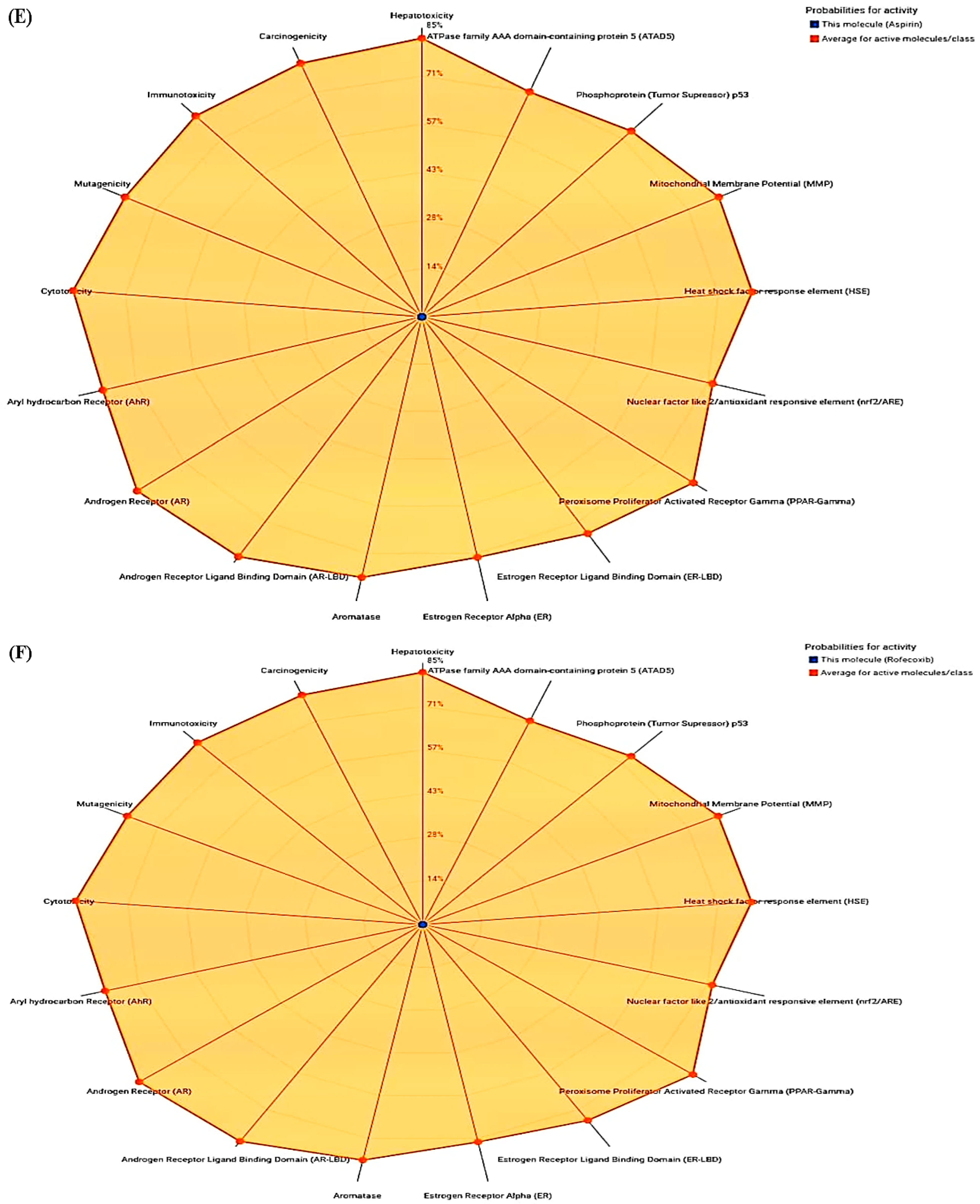
The findings of the present study revealed that crocetin, aspirin, and rofecoxib were inactive for all the toxicities. However, quercetin showed active toxicity for carcinogenicity and mutagenicity and was inactive for the rest. Moreover, rutin showed inactive activity for various toxicities except for immunotoxicity. Table 2 and Figure 1A–F show the toxicity analysis scores of selected compounds.

3.3. Molecular Docking

In the present study, docking analysis revealed that picrocrocin had the highest binding affinity of −8.1 kcal/mol. Among the drugs, rofecoxib showed the highest binding affinity of −8.3 kcal/mol, whereas aspirin reported the lowest (6.1 kcal/mol). The inhibition constant (Ki) in µM of selected saffron compounds was determined/calculated from binding affinity. Among the compounds, picrocrocin had the highest Ki value of 13.68 µM, whereas safranal reported the lowest (11.51 µM). In the present study, Table 3 shows the binding affinity and Ki of compounds and drugs.
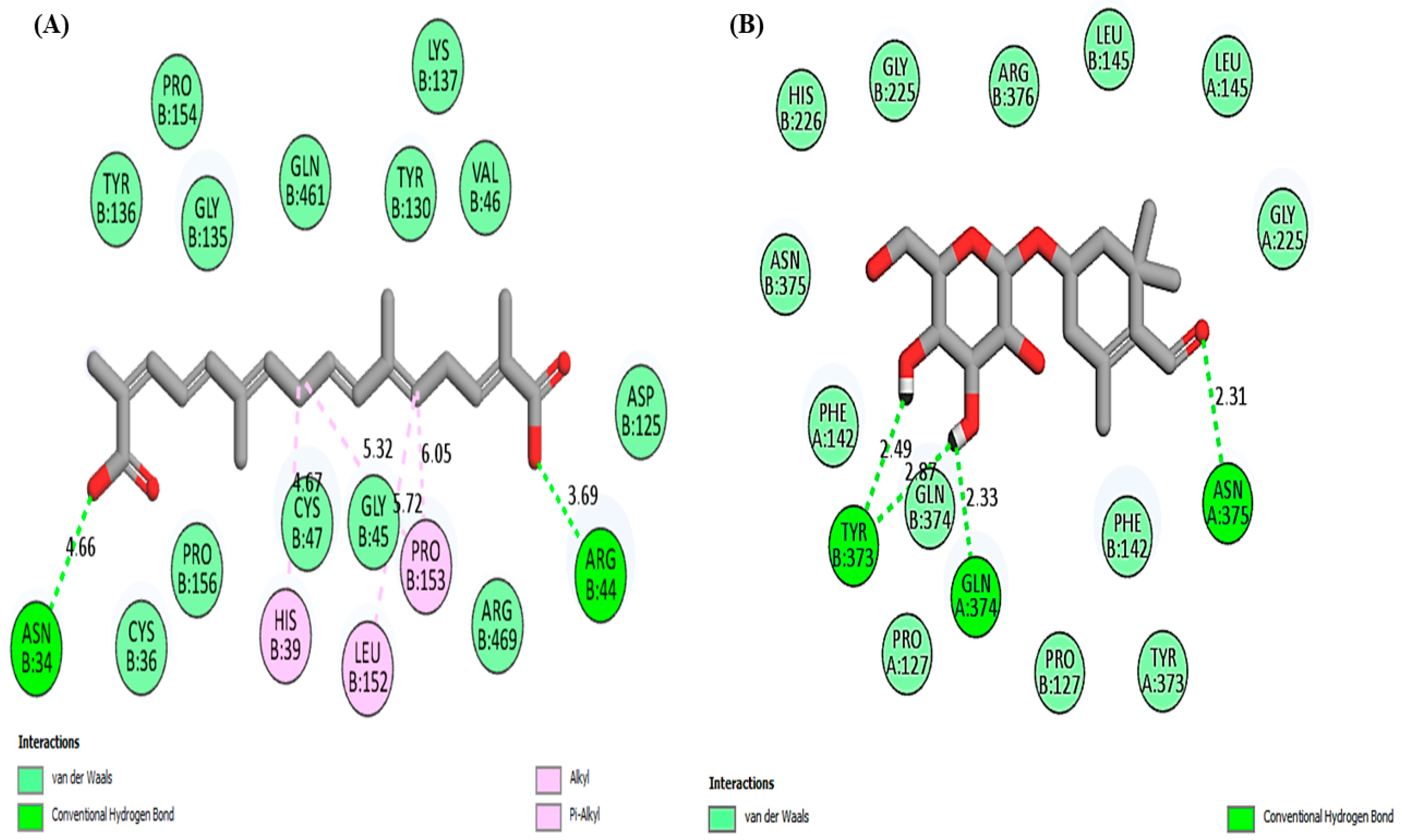
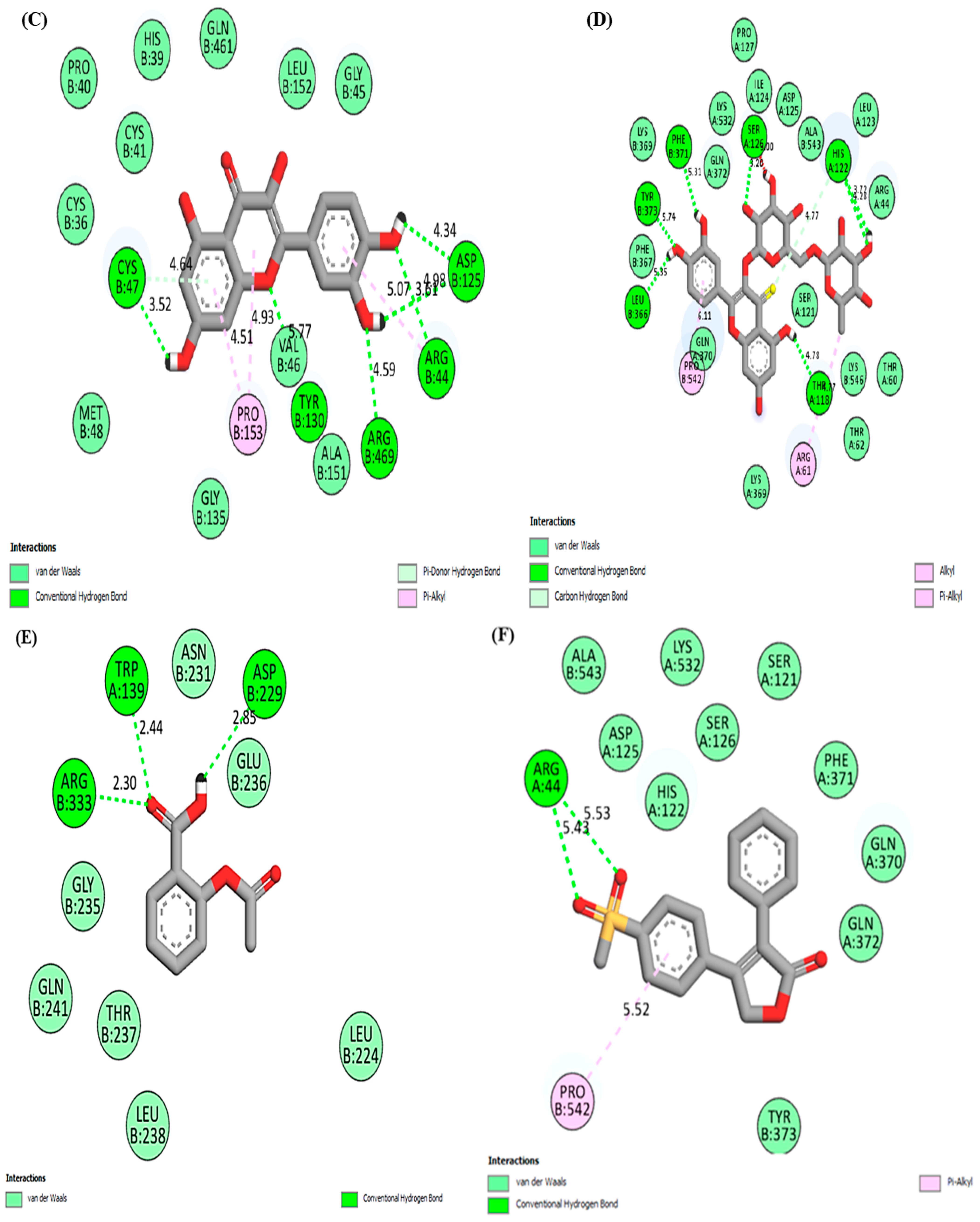
The graphical representation of 2D interactions of the COX-2 protein when docked with selected saffron compounds and known drugs are shown in Figure 2. The results showed that the compounds form different types of interactions with the amino acid residues of COX-2 protein, as shown in Table 4.

3.4. PROCHECK

The PROCHECK server helps us to evaluate the 3D geometry of protein structures, and it provides results for residues with areas represented by different colors such as red (favored), yellow (additionally allowed), pale yellow (generously allowed), and white (disallowed), as shown in Figure 3A. In human COX-2, 562 residues were found, and out of those, 428 (90.3%) residues were lying in the most favored region (A, B, L). About 45 (9.5%) residues lay in the additional allowed regions (a, b, l, p), none in generously allowed regions, and 1 (0.1%) in disallowed regions (Figure 3A). In human COX-2 protein, we found 474 (100%) non-proline and non-glycine residues, while at the end, the number of residues (except glycine and proline) were only 14. The proline and glycine residues are shown as triangles and are 38 and 36, respectively (Figure 3A). In this study, Figure 3B illustrates the hydrophobicity plot of the human COX-2 protein. The Ramachandran plots for all residue types are represented in Figure 3C. The number of amino acid residues is shown in brackets and those in unfavorable conformations (score < −3.0) are labeled. Favorable conformations are illustrated by shaded regions based on analyzing a total of 163 structures at a resolution of 2.0Å or better. Shading shows favorable conformations as obtained from an analysis of 163 structures at a resolution 2.0Å or better.

3.5. ComPPI

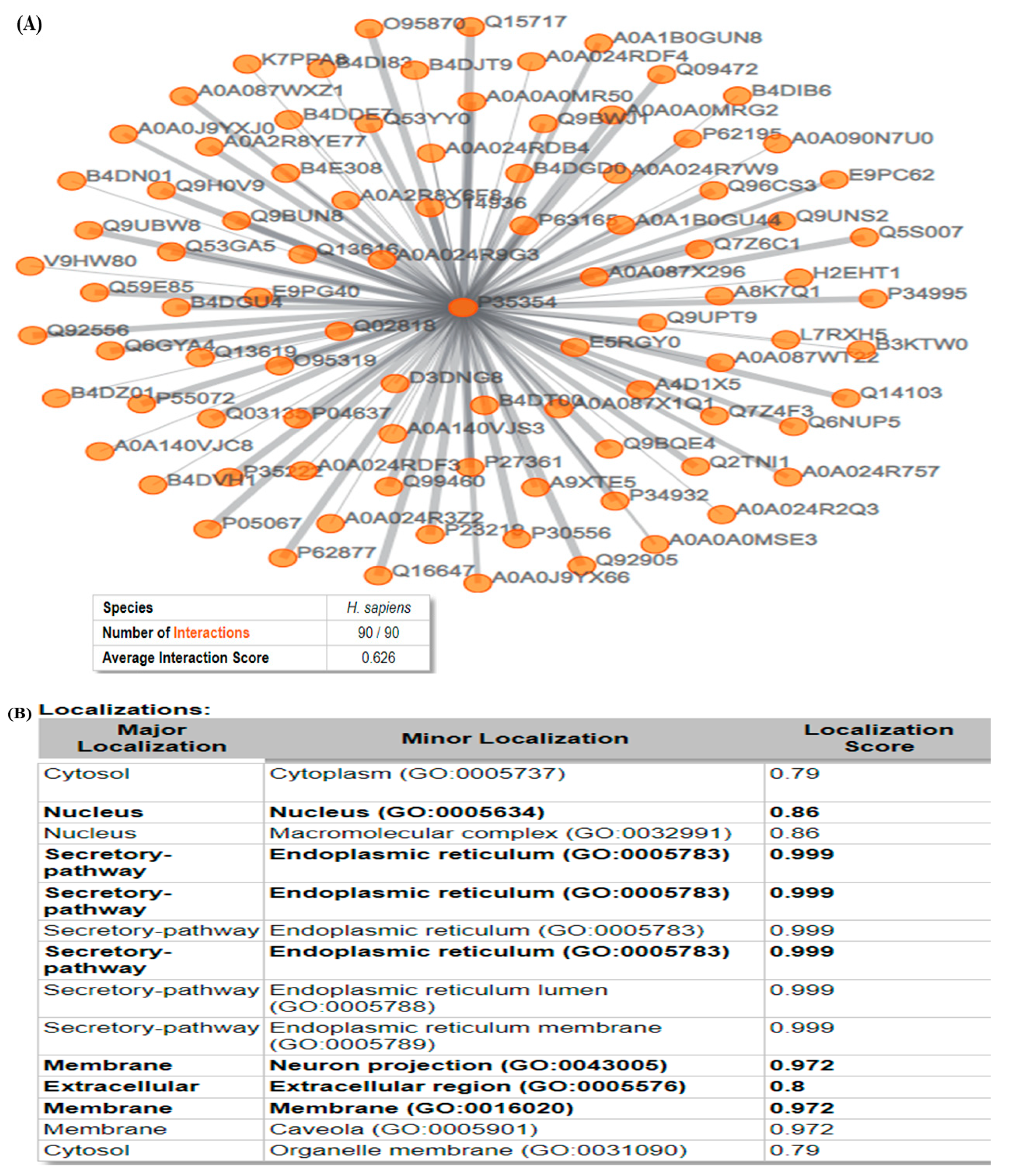
In the present study, Figure 4A shows the first-neighbor network showing the interaction map, and the width of the edges is proportional to the corresponding localization score. In the present study, the number of interactions was 90 and the average interaction score was 0.626. In this study, COX-2 showed an interaction score of 1 with Amyloid-Beta A4, Caveolin-1, Prostacyclin Synthase, Derlin-1, Vip36-Like Protein, Nucleobindin-1, Prostaglandin G/H Synthase 1, Selenoprotein S, Mitogen-activated protein kinase 3, etc. (Figure 4B). The localization of the COX-2 protein is illustrated in Figure 4B, wherein the highest score was found in secretory pathways (0.99) and the lowest in cytosol (0.79).

3.6. CASTp

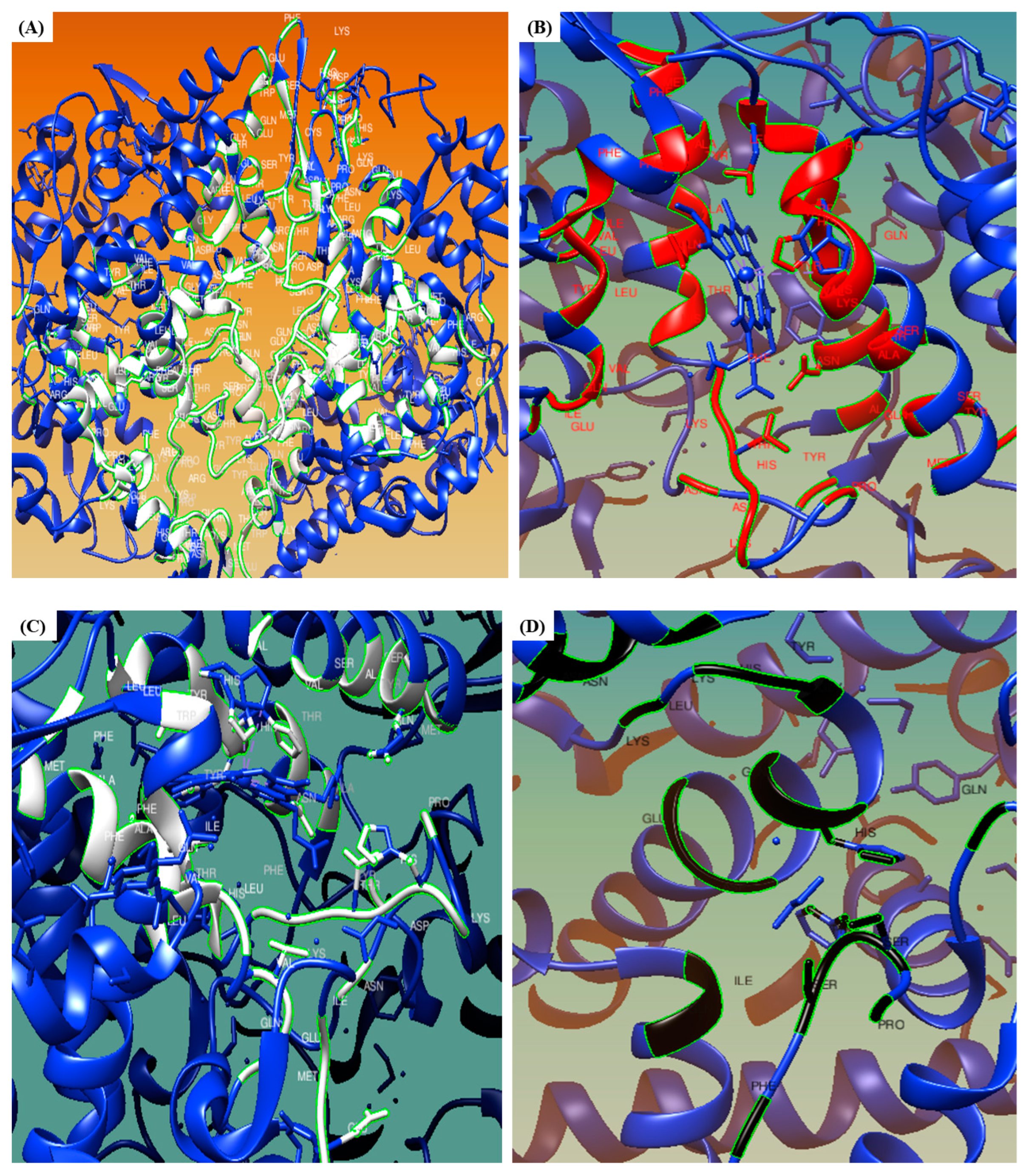
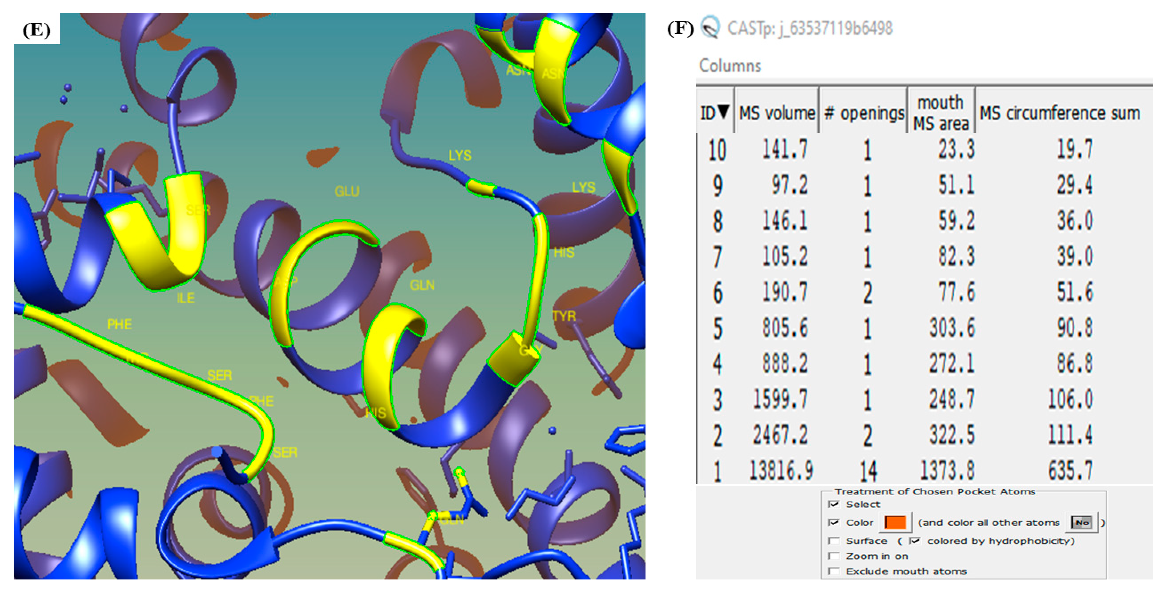
In the present study, Figure 5 illustrates the five best binding pockets on the COX-2 protein that are automatically recognized by the CASTp tool. Figure 5A represents the top-most binding pocket of COX-2 protein, whereas Figure 5B–D indicate the second, third, fourth, and fifth binding pockets, respectively. The pocket panel shows many important residues that are located in the pocket, and these pockets could serve as targets for drugs. The imprint of the pockets is illustrated in white (Figure 5A), red (Figure 5B), white (Figure 5C), black (Figure 5D), and yellow (Figure 5E), respectively. The CASTp data showing the volume, openings, and area of the binding pockets, along with their respective positions, are illustrated in Figure 5F.

3.7. WEBnmα

In the present study, the eigenvalues for the 50 lowest-frequency modes starting from mode 7 are illustrated in Figure 6A. In this study, the 14 lowest-frequency modes (7–20) had average deformation energies of 323.96, 448.04, 968.32, 1113.79, 1384.79, 1451.23, 1377.34, 1589.30, 1803.78, 1737.89, 1901.58, 2306.34, 2389.80, and 2497.08, respectively (Figure 6B).
Eigenvalues and deformation energies depict the energy related to every mode and are inversely associated with the amplitude of motion expressed by the representative modes. Along all the normal modes, the protein structure moves at once. The atomic displacement of the protein molecule is shown in Figure 6C. The square of the atomic displacement of each C-alpha atom (mode 7) is normalized, and the highest values correspond to the most displaced regions. Cluster peaks represent the significantly displaced regions on the plots, whereas isolated peaks indicate local flexibility in a low-density region (N or C-terminal).
In this study, Figure 6D represents the normalized fluctuations from mode 7 to 206, respectively. The sum of atomic displacement in each of the 200 lowest frequency modes weighted by the inverse of their corresponding eigenvalues gives the fluctuation plots; thus, in the present study, we found the highest atomic fluctuation to be 1.71 and the lowest to be 0.01. The correlation matrix describes the correlated movement of the C-alphas of protein, as illustrated in Figure 6E, wherein the covariance matrix indicates the coupling between a pair of residues, i.e., red (correlated), blue (anti-correlated), and white (uncorrelated), respectively. The coupling of two residues in the proteins ranged from −1 (anti-correlated) to 1 (correlated) and 0 (uncorrelated). The color of the sticks represents indicate correlations, i.e., red (positive) and blue (negative).

3.8. LARMD

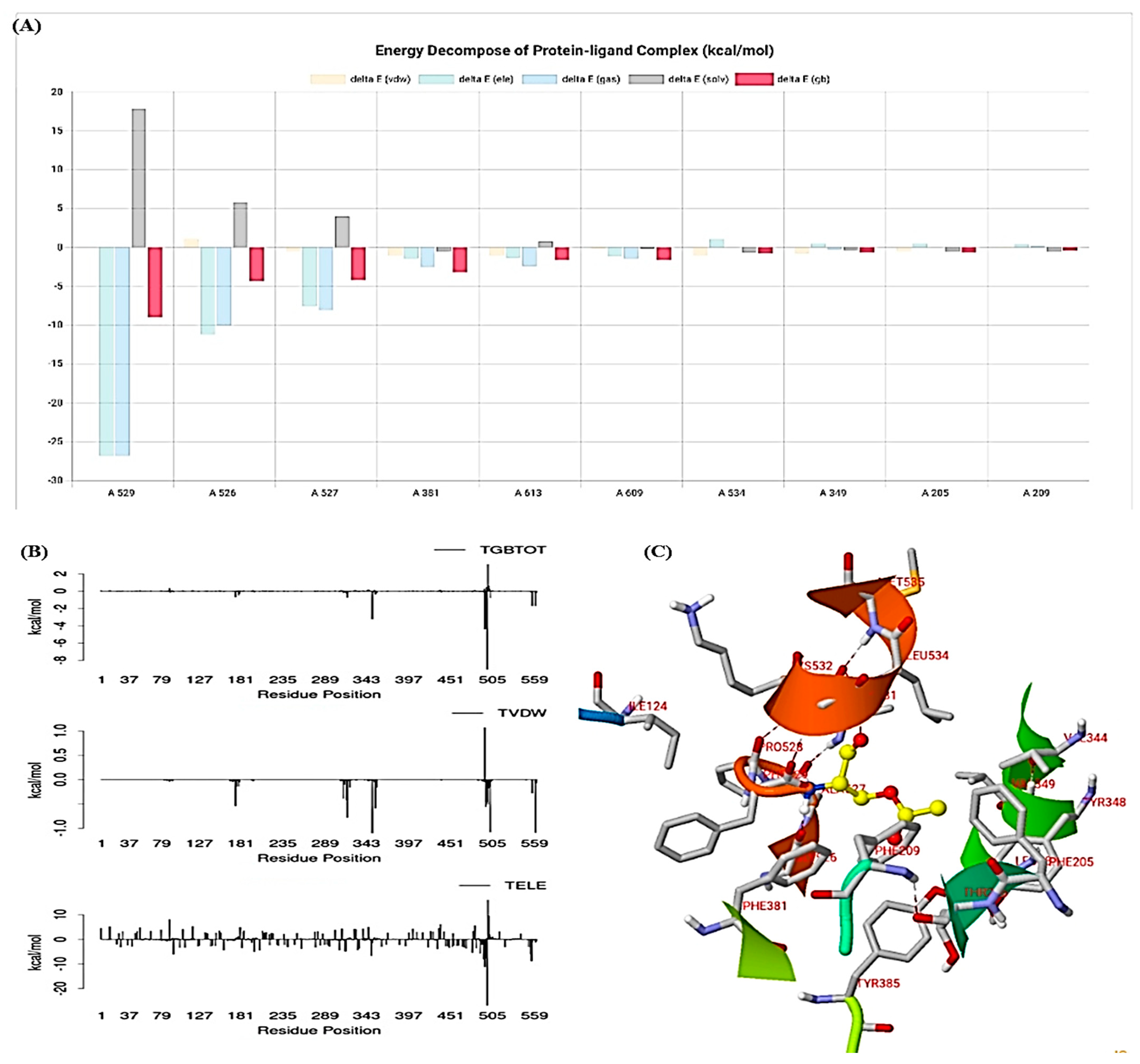
In the present study, the LARMD server provided the results of decomposition energy (kcal/mol) of the protein molecule, as illustrated in Figure 7A. The findings of MM/PB(GB)SA, consisting of van der Waals contribution (VDW), electrostatic energy (ELE), total gas phase energy (GAS), and polar and non-polar contribution to solvation (PBSL/GBSOL) of the residues, are shown in Figure 7B. Furthermore, the final binding free energy (deltaPB/deltaGB) was determined from PBTOT/GBTOT and entropy (TS). The MM/PB(GB)SA results are shown in Table 5.

3.9. ShinyGO

In the present study, Figure 8A shows the KEGG pathway enrichment with the fold enrichment scores. In the enrichment analysis, the differentially expressed genes were significantly enriched in ovarian steroidogenesis, regulation of lipolysis in adipocytes, vascular endothelial growth factors (VEGF) signaling pathway, and arachidonic acid metabolism. KEGG pathway analysis indicated that the differentially expressed genes were significantly enriched in metabolic, cancer, inflammatory, and other significant signaling pathways. Similarly, Figure 8B represents the functional enrichment analysis in which the most significantly enriched function was the positive regulation of the prostaglandin biosynthetic process and intrinsic apoptotic signaling pathway in response to osmotic stress. Table 6 and Figure 8 represent the different signaling pathways with fold enrichment scores.

3.10. Molecular Simulation Study

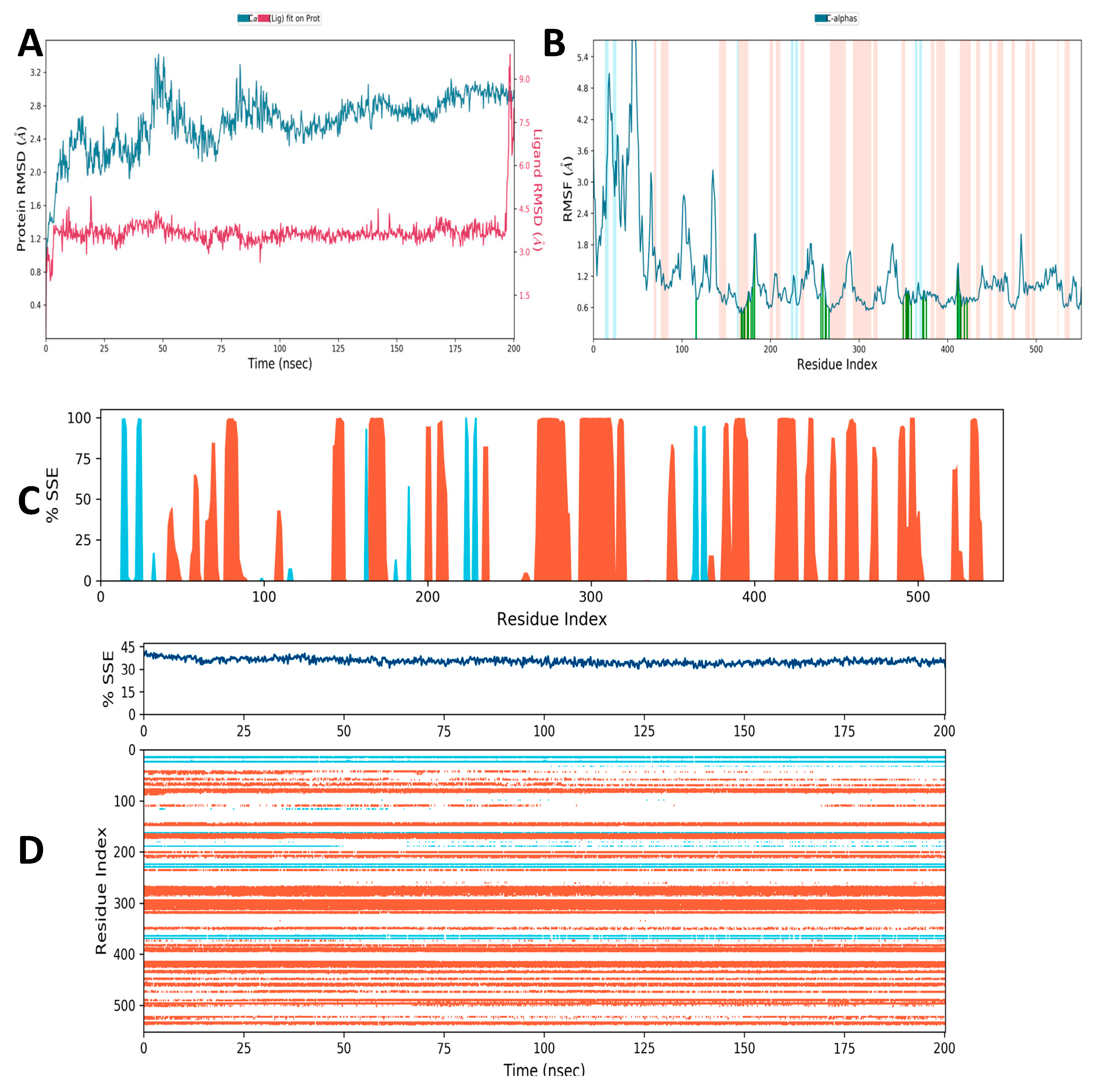
Determining the protein’s RMSD allows us to understand how the protein’s structural conformation has changed over time. This method is particularly well suited for studying minute atomic motions on femto/picosecond timescales, which are extremely challenging to measure experimentally. The RMSD was determined in a simulation of about 200 ns. The average values of RMSD of Cα-COX-2 were determined to be around 2.74 Å in the presence of picrocrocin (Figure 9A). As estimated, the RMSD values and atomic and residual fluctuations of COX-2 were found to be highest in the presence of picrocrocin. The observed structural deviations of COX-2 may be due to the greater number of hydroxyl groups and the presence of a ring structure in picrocrocin. In addition, RMSF values were also plotted against each residue of the COX-2 backbone in the presence of picrocrocin (Figure 9B). As can be seen in Figure 9B, the RMSF plot reveals higher-order residual fluctuations in the presence of picrocrocin. The peaks in the plot represent the protein areas that fluctuate most during the process of simulation. Usually, we have found that the N- and C-terminal tails fluctuate more in comparison to other regions of the protein. This analysis shows that the stability of COX-2 protein has a direct relation with the amino-acid fluctuations.

3.10.1. Secondary Structural Elements

To further investigate this, the effect of picrocrocin on the secondary structural elements (α-helical and β-strand regions) of the COX-2 protein was determined (Figure 9C,D). Helices or strands that last for more than 70% of the simulation as a whole designate these areas. The α-helical and β-strand regions are highlighted with red and blue backgrounds, respectively, and protein SSEs (secondary structure element) are visualized throughout the simulation. The plots in Figure 9C,D depict the residue index distribution throughout the protein structure. Over time, the graphs monitor each residue and SSE assignment, and at the bottom, the plot summarizes the composition of SSE for each trajectory frame during the process of simulation. The strand was 4.22%, while the helix was 31%. The COX-2 simulation times describe the changes in the residue index in the synthesis of protein SSE, such as the blue and red arrows depicting helices and strands. The changes may be due to the breakdown of the hydrogen bond network of COX-2 residues due to the conformational instability of protein in the presence of picrocrocin.
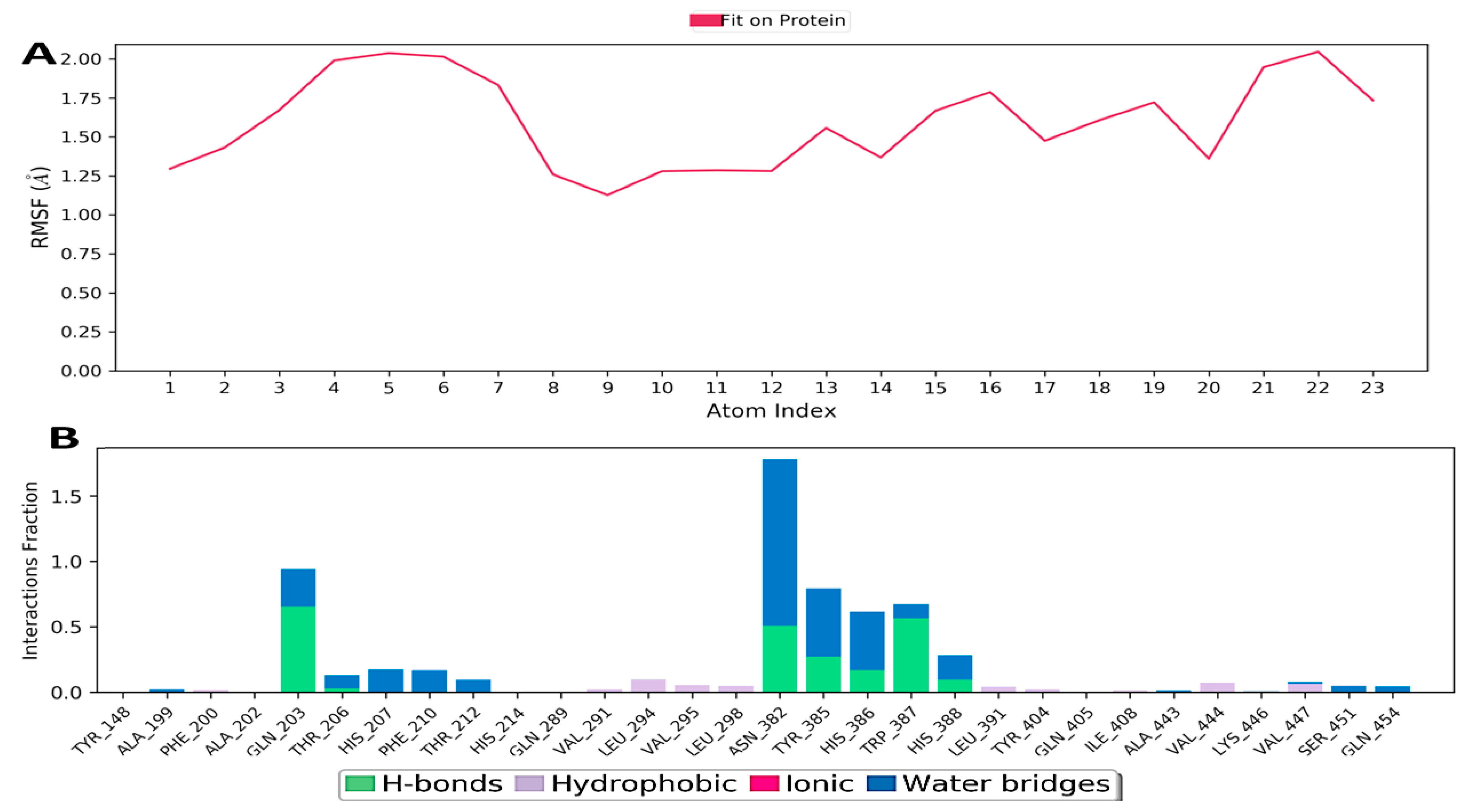
Figure 10A shows the determined Ligand Root Mean Square Fluctuation (L-RMSF). This helps in characterizing changes in the ligand atom positions. The mean of the RMSD ligand was found to be 1.5 Å. By breaking down the ligand’s fluctuations into individual atoms, the ligand RMSF displays a 2D structure. In the present study, Figure 10B summarizes the protein-ligand contacts that includes hydrogen bonds (H-bonds), hydrophobic/ionic interactions, water bridges and contacts as a timeline representation.

3.10.2. Hydrogen Bonding

Hydrogen bonding is essential for ligand–protein interaction because it has a significant effect on drug specificity, adsorption, and metabolism, i.e., drug development. The backbone acceptor and donor, side-chain acceptor, and donor are the four types of hydrogen bonding. Various geometric criteria were taken into consideration in hydrogen bonding like distance (2.5 Å) between donor and acceptor atoms, donor angle (3120°) between donor and acceptor, and acceptor angle (390°) between acceptor and donor.

3.10.3. Ionic Interaction

Ionic interaction is also known as the polar interaction between protein and ligand and is identified by the presence of metal ions within 3.4 of heavy atoms of ligand and protein (except carbon). Polar interactions are generally of two types, as mediated by the protein backbone and side chain.

3.10.4. Hydrophobic Contacts (like p-p, p-Cation, etc.)

These interactions involve the bonding between hydrophobic amino acid residues of protein with the aliphatic or aromatic carbon groups of ligands. The general criteria for the interactions are p-cation-aromatic and charged groups with 4.5 Å range, p-p—aromatic groups stacked face to face or face to edge, and for other interactions, 3.6 Å range between side chain and carbon groups.

3.10.5. Water Bridge (like Water-Protein and Water-Ligand)

This type of interaction involves a hydrogen bond and a water molecule, but it is less flexible than hydrogen bonding. The criteria involved are a distance of 2.8 Å, 3110° donor angle, and 390° acceptor angle.

3.10.6. Protein–Ligand Contacts

During this study, the total number of distinct interactions that a protein makes with the ligand is depicted in the top panel of Figure 11. In each trajectory, the lower panel of Figure 11 displays which residue of the protein interacts with the ligand. According to the scale to the right of the plot, the darker orange color indicates that some residues have many specific contacts with the ligand. It can be seen in Figure 11 that more specific interaction was shown by amino acid residues like GLN203, TYR385, HIS386 and 388, ASN382, and TRP387.

3.10.7. Torsion Profile

During the simulation trajectory (0 through 200.0 nanoseconds), the conformational evolution of rotatable bonds in the ligand is summarized in the ligand torsion figure. The 2D schematics of a ligand with colored rotatable bonds are displayed in Figure 12A’s top panel, where radial and bar plots (charts) of the same color accompany each rotatable bond torsions. How the torsion has changed is shown by radial (dial) charts, while a bar plot determines the probability density of torsion throughout the simulation. In the center of the radial map, the time evolution is represented radially outwards from the simulation’s start point.
The ligand properties like RMSD, Rg, SASA, PSA, intra-hydrogen bonding, and MoISA are represented in Figure 12B. The charts also show the potential of rotatable bonds with the help of torsional potential. The Y-axis of the plot shows the potential values in kcal/mol, and the torsion potential and histogram link might shed light on the ligand conformational strain, which is experienced by the ligand to retain protein-bound conformation.

4. Discussion

COX-2 is an important bi-functional enzyme that mediates prostaglandin synthesis during the inflammatory processes and has become a vital therapeutic target for developing anti-inflammatory medications. Synthetic COX-2 inhibitors hold great potential for treatments; however, drug toxicity creates problems, and as a result, new approaches are required. In such a scenario, natural compounds hold great potential for inhibiting inflammatory signaling pathways including COX-2, which has gained much consideration over the past few years. In this study, natural constituents of saffron (crocin, crocetin, picrocrocin, quercetin, dimethyl crocetin, and rutin) were used. The pertinent association between the identification, synthesis, and purification of compounds during drug discovery has a significant interest [51,52]. In this study, ADMET properties were used to determine the pharmacokinetics of drugs in the body. The five parameters of Lipinski’s rule were used to determine the drug-like properties of compounds, and these features are mostly important as they are related to dissolution and intestinal permeability [39,53,54,55]. In our study, all the molecules followed Lipinski’s rule except rutin, which has a molecular weight >500 g/mol, a number of hydrogen bond donors >5, and a number of acceptors >10. If a compound fails in more than two of Lipinski’s rules and violations, it cannot be consumed and hence used as a drug [39]. The compounds are classified as hydrophilic or hydrophobic when the logP scores are negative or positive. A hydrophilic compound has good water solubility, gastric tolerance, and elimination by the kidneys, whereas lipophilic compounds have lower aqueous solubility/gastric tolerance, and good plasma protein binding, elimination, and permeability across the biological membrane [56].
During clinical trials, unfavorable effects caused by drug toxicity often lead to drug breakdown [57]. In the process of drug development, it is important to predict the toxicity of compounds, and in such a situation, ProTox-II becomes a particularly significant tool [40]. The estimation of toxicities via computational analysis is much faster than determining toxic doses in animals but also decreases the amount of animal experimentation [58]. The protein targets are the toxicity sites that have been related to adverse drug reactions and effects. Generally, the toxic doses are specified as LD50 (mg/kg) of body weight.
Plant compounds with decreased toxicity and good bioavailability have been selected to determine their therapeutic potential against different diseases [59,60]. In structure-based drug designing, scoring functions are particularly important. The ranking of compounds is carried out with the help of docking algorithms as they produce scoring properties and binding poses [61,62]. Insights into protein interactions with phytocompounds are provided via computational docking methods. In the present study, our analysis indicated that the picrocrocin compound of saffron possesses the best binding affinity of −8.1 kcal/mol. Similar findings have reported that the picrocrocin molecule of saffron possesses protection against diseases [63,64]. This can be explained by the fact that the picrocrocin compound of saffron possesses significant pharmacological properties such as antioxidant, anti-inflammatory, antiproliferative, antiangiogenic, and pro-apoptotic properties [65,66,67]. Moreover, picrocrocin is effective against various diseases like cancer, Alzheimer’s, Parkinson’s, memory impairment, epilepsy, and brain ischemia [64,68,69]. The intake of saffron has been found to reduce inflammation by inhibiting the enzymatic activity of cyclooxygenase and improving the number of inflammatory markers (CRP, IL-6, and TNF-α) [66,70]. Saffron contains numerous molecules such as alkaloids, minerals, anthocyanins, carotenoids, glycosides, and flavonoids (rutin, quercetin, and kaempferol) [71].
In the present study, the PROCHECK server was used to evaluate the accuracy and stereochemical properties of the protein structure. This server analyzes the entire protein geometry with residue-by-residue geometry and provides the stereochemical quality of the predicted protein model. In this study, 90.3% of protein residues occupied the favored region, and the structure was acceptable and could be used for further in silico studies [41]. Thus, the stereochemical properties were validated using Ramachandran analysis and the PROCHECK tool, which indicated that the predicted model could be satisfactory.
Protein–protein interaction information is one of the most significant sources for proteome-wide studies [72], especially in network-associated drug design [73] and to understand human disorders [74]. In this scenario, ComPPI-built interactomes provide much wider coverage and offer the construction of highly confident data sets for analysis of protein–protein interaction at the subcellular level. High-confidence interactomes could be developed from optimized interaction and localization scores that could further help in the area of compartment-associated biological processes [75].
In the present study, the CASTp server provides a complete, thorough description of surface regions (pockets and voids) located on the surface of the protein, which are associated with binding events [76]. In a protein structure, the CASTp server, in addition to providing complete information about all atoms involved in their formation, identifies all the voids and pockets. This tool also measures the area and volume of each void and pocket. Furthermore, it measures the size of individual pocket mouth openings, which facilitates determining the availability of binding sites to different ligands and substrates. In several research studies, CASTp computation analysis has proved quite useful [77,78,79].
In this study, motion stiffness is represented by eigenvalues, and its value is proportional to the energy needed to deform the protein structure [80]. The deformation will be easier when the eigenvalues are lower and vice versa. In a protein molecule, the measure of local flexibility is provided through deformation analysis. Atomic displacement (B factors) is dependent on structural heterogeneity and provides a broad range of information about protein structure and dynamics. B-factors enable the study of protein flexibility and biological protein–protein binding locations, as well as the differentiation of crystal packing contacts [81]. The covariance matrix map depicts the related actions of several pairs of anti-correlated and correlated residues, which are represented by white, red, and blue colors that represent residue pair coupling [80].
In research studies, a good method for evaluating binding energies and revealing binding structures is provided by MM-PB(GB)SA [82]. The correctness of docking complex calculations is provided through these rescoring approaches [83,84]. Due to the high efficiency and accuracy of these computational approaches, they have been used mostly in docking calculations. However, a challenging task would be to find more advanced rescoring methods within tolerable limits.
Enrichment analysis shows the set of enriched genes associated with a particular pathway in huge-scale datasets. Functional enrichment analysis provides significant insights into identifying diseases and drug mechanisms. In functional enrichment, gene ontology is the most extensively used. Gene enrichment analysis helps to associate a disease phenotype with a group of genes/proteins, and by identifying vital characteristics, the enrichment analysis can help in the discovery of biological functions. The enrichment analysis predicts whether the fraction of pathway genes of interest is higher in comparison to outside pathway genes.
The molecular dynamic simulation shows that picrocrocin destabilizes the activity of the COX-2 protein by altering its tertiary structure, which causes more pronounced amino-acid fluctuations as determined using RMSD and RMSF values in Figure 9. It was also noted in the simulation studies of the hydrogen bond network that picrocrocin also disrupts the intra-protein hydrogen bond network, thereby causing more unfolding of the COX-2 protein. Furthermore, the direct interaction of picrocrocin with various amino acid residues like GLN203, TYR385, HIS386 and 388, ASN382, and TRP387 is very crucial for the activity of the COX-2 protein. The modification of these residues with overall structural alterations in protein presents the potential inhibitory properties of this compound; thus, picrocrocin might prove to be a potential therapeutic moiety for the treatment of various disorders like inflammation.

5. Conclusions

In silico approaches were applied to the compounds of saffron to reveal their potential against the human COX-2 protein. Our molecular docking study suggested that picrocrocin of saffron was the most potent inhibitor of the COX-2 protein. In our study, the ADMET analysis revealed the physiochemical properties and the toxicity of the compounds of saffron, while the quality and correctness of the COX-2 protein structure were predicted using molecular modeling, which predicted 90.3% of COX-2 residues as lying in the Ramachandran favored region. We know that protein–protein interactions play a significant role in the cellular functions of the living organism, and thus the protein–ligand interaction and behavior of protein motion can be determined using molecular dynamics approaches. The molecular simulation studies showed that there are overall conformational alterations with a decrease in the hydrogen bond network in the COX-2 protein in the presence of picrocrocin. Overall, compared to the currently available methods, the picrocrocin used in this study could serve as an alternative for use against inflammatory diseases. However, more research studies are required to validate the inflammatory role of picrocrocin. Thus, based on these findings, we can conclude that picrocrocin can be repurposed as a drug candidate against inflammatory diseases.

Author Contributions

Conceptualization, A.A.; Methodology, A.A.; Software, A.A.; Validation, A.A.; Formal analysis, A.A.; Investigation, A.A.; Writing—original draft preparation, A.A. and M.U.R.; Writing—review and editing., A.A., B.A.M., A.B.W., A.N., S.B.A., N.J.D., F.A. and J.P.; Visualization., A.A., Supervision, A.A., S.B.A. and M.U.R.; Funding, A.N. All authors have read and agreed to the published version of the manuscript.

Funding

The authors are thankful to the Researchers Supporting Project Number (RSP2023R124), King Saud University, Riyadh, Saudi Arabia for funding this research.

Institutional Review Board Statement

Not applicable.

Informed Consent Statement

This study did not involve any animal or human participants.

Data Availability Statement

All the data generated has been published in this manuscript.

Acknowledgments

The authors extend their appreciation to Researchers Supporting Project Number (RSP2023R124), King Saud University, Riyadh, Saudi Arabia for funding this research.

Conflicts of Interest

The authors have read the manuscript and have no conflict of interest.

References

  1. Medzhitov, R. Inflammation 2010: New Adventures of an Old Flame. Cell 2010, 140, 771–776. [Google Scholar] [CrossRef] [PubMed]
  2. Chen, L.; Deng, H.; Cui, H.; Fang, J.; Zuo, Z.; Deng, J.; Li, Y.; Wang, X.; ZhÅ, L. Inflammatory Responses and Inflammation-Associated Diseases in Organs. Oncotarget 2018, 9, 7204–7218. [Google Scholar] [CrossRef] [PubMed]
  3. Abdulkhaleq, L.A.; Assi, M.A.; Abdullah, R.; Zamri-Saad, M.; Taufiq-Yap, Y.H.; Hezmee, M.N.M. The Crucial Roles of Inflammatory Mediators in Inflammation: A Review. Vet. World 2018, 11, 627–635. [Google Scholar] [CrossRef] [PubMed]
  4. Chopade, A.R.; Sayyad, F.J.; Pore, Y.V. Molecular Docking Studies of Phytocompounds from the Phyllanthus species as Potential Chronic Pain Modulators. Sci. Pharm. 2015, 83, 243–267. [Google Scholar] [CrossRef]
  5. Leláková, V.; Šmejkal, K.; Jakubczyk, K.; Veselý, O.; Landa, P.; Václavík, J.; Bobáľ, P.; Pížová, H.; Temml, V.; Steinacher, T.; et al. Parallel In Vitro and In Silico Investigations into Anti-Inflammatory Effects of Non-Prenylated Stilbenoids. Food Chem. 2019, 285, 431–440. [Google Scholar] [CrossRef]
  6. Ahmad, S.B.; Ali, A.; Bilal, M.; Rashid, S.M.; Wani, A.B.; Bhat, R.R.; Rehman, M.U. Melatonin and Health: Insights of Melatonin Action, Biological Functions, and Associated Disorders. Cell. Mol. Neurobiol. 2023, 43, 2437–2458. [Google Scholar] [CrossRef] [PubMed]
  7. Malla, B.A.; Ali, A.; Maqbool, I.; Dar, N.A.; Ahmad, S.B.; Alsaffar, R.M.; Rehman, M.U. Insights into Molecular Docking and Dynamics to Reveal Therapeutic Potential of Natural Compounds Against P53 Protein. J. Biomol. Struct. Dyn. 2023, 41, 8762–8781. [Google Scholar] [CrossRef]
  8. Aldossari, R.M.; Ali, A.; Rehman, M.U.; Rashid, S.; Ahmad, S.B. Computational Approaches for Identification of Potential Plant Bioactives as Novel G6PD Inhibitors Using Advanced Tools and Databases. Molecules 2023, 28, 3018. [Google Scholar] [CrossRef]
  9. Ahmed, E.M.; Kassab, A.E.; El-Malah, A.A.; Hassan, M.S.A. Synthesis and Biological Evaluation of Pyridazinone Derivatives as Selective COX-2 Inhibitors and Potential Anti-Inflammatory Agents. Eur. J. Med. Chem. 2019, 171, 25–37. [Google Scholar] [CrossRef]
  10. Nandakishore, R.; Yalavarthi, P.R.; Kiran, Y.R.; Rajapranathi, M. Selective Cyclooxygenase Inhibitors: Current Status. Curr. Drug Discov. Technol. 2014, 11, 127–132. [Google Scholar] [CrossRef]
  11. Kurumbail, R.G.; Stevens, A.M.; Gierse, J.K.; McDonald, J.J.; Stegeman, R.A.; Pak, J.Y.; Gildehaus, D.; Miyashiro, J.M.; Penning, T.D.; Seibert, K.; et al. Structural Basis for Selective Inhibition of Cyclooxygenase-2 by Anti-Inflammatory Agents. Nature 1996, 384, 644–648. [Google Scholar] [CrossRef] [PubMed]
  12. Rouzer, C.A.; Marnett, L.J. Cyclooxygenases: Structural and Functional Insights. J. Lipid Res. 2009, 50, S29–S34. [Google Scholar] [CrossRef]
  13. Perrone, M.G.; Scilimati, A.; Simone, L.; Vitale, P. Selective COX-1 Inhibition: A Therapeutic Target to Be Reconsidered. Curr. Med. Chem. 2010, 17, 3769–3805. [Google Scholar] [CrossRef] [PubMed]
  14. de Souza, G.; Silva, R.J.; Milián, I.C.B.; Rosini, A.M.; de Araújo, T.E.; Teixeira, S.C.; Barbosa, B.F. Cyclooxygenase (COX)-2 Modulates Toxoplasma gondii Infection, Immune Response and Lipid Droplets Formation in Human Trophoblast Cells and Villous Explants. Sci. Rep. 2021, 11, 12709. [Google Scholar] [CrossRef]
  15. El-Sayed, N.A.; Nour, M.S.; Salem, M.A.; Arafa, R.K. New Oxadiazoles with Selective-COX-2 and EGFR Dual Inhibitory Activity: Design, Synthesis, Cytotoxicity Evaluation and In Silico Studies. Eur. J. Med. Chem. 2019, 183, 111693. [Google Scholar] [CrossRef]
  16. Kumar, R.; Saha, N.; Purohit, P.; Garg, S.K.; Seth, K.; Meena, V.S.; Dubey, S.; Dave, K.; Goyal, R.; Sharma, S.S.; et al. Cyclic Enaminone as New Chemotype for Selective Cyclooxygenase-2 Inhibitory, Anti-Inflammatory, and Analgesic Activities. Eur. J. Med. Chem. 2019, 182, 111601. [Google Scholar] [CrossRef]
  17. Andrade, P.B.; Valentão, P. Insights into Natural Products in Inflammation. Int. J. Mol. Sci. 2018, 19, 644. [Google Scholar] [CrossRef] [PubMed]
  18. Wang, Y.H.; Zeng, K.W. Natural Products as a Crucial Source of Anti-Inflammatory Drugs: Recent Trends and Advancements. Trad. Med. Res. 2019, 4, 257–268. [Google Scholar] [CrossRef]
  19. Wink, M. Modes of Action of Herbal Medicines and Plant Secondary Metabolites. Medicines 2015, 2, 251–286. [Google Scholar] [CrossRef]
  20. Schafer, H.; Wink, M. Medicinally Important Secondary Metabolites in Recombinant Microorganisms or Plants: Progress in Alkaloid Biosynthesis. Biotechnol. J. 2009, 4, 1684–1703. [Google Scholar] [CrossRef]
  21. Matsuura, H.N.; Fett-Neto, A.G. Plant Alkaloids: Main Features, Toxicity, and Mechanisms of Action. Plant Toxins 2015, 2, 1–15. [Google Scholar]
  22. Souto, A.L.; Tavares, J.F.; Da Silva, M.S.; Diniz, M.D.F.F.M.; de Athayde-Filho, P.F.; Barbosa Filho, J.M.B. Anti-Inflammatory Activity of Alkaloids: An Update from 2000 to 2010. Molecules 2011, 16, 8515–8534. [Google Scholar] [CrossRef]
  23. Alsaffar, R.M.; Ali, A.; Rashid, S.M.; Ahmad, S.B.; Alkholifi, F.K.; Kawoosa, M.S.; Ahmad, S.P.; Rehman, M.U. Zerumbone Protects Rats from Collagen-Induced Arthritis by Inhibiting Oxidative Outbursts and Inflammatory Cytokine Levels. ACS Omega 2023, 8, 2982–2991. [Google Scholar] [CrossRef]
  24. Cui, J.; Jia, J. Natural COX-2 Inhibitors as Promising Anti-Inflammatory Agents: An Update. Curr. Med. Chem. 2021, 28, 3622–3646. [Google Scholar] [CrossRef] [PubMed]
  25. Ambati, G.G.; Jachak, S.M. Natural Product Inhibitors of Cyclooxygenase (COX) Enzyme: A Review on Current Status and Future Perspectives. Curr. Med. Chem. 2021, 28, 1877–1905. [Google Scholar] [CrossRef]
  26. Chandel, P.; Rawal, R.K.; Kaur, R. Natural Products and Their Derivatives as Cyclooxygenase-2 Inhibitors. Future Med. Chem. 2018, 10, 2471–2492. [Google Scholar] [CrossRef] [PubMed]
  27. Heydari, S.; Haghayegh, G.H. Extraction and Microextraction Techniques for the Determination of Compounds from Saffron. Chem. C Trans. 2014, 2, 221–247. [Google Scholar]
  28. Patel, S.; Sarwat, M.; Khan, T.H. Mechanism Behind the Anti-tumour Potential of Saffron (Crocus sativus L.): The Molecular Perspective. Crit. Rev. Oncol. Hematol. 2017, 115, 27–35. [Google Scholar] [CrossRef]
  29. Khorasany, A.R.; Hosseinzadeh, H. Therapeutic Effects of Saffron (Crocus sativus L.) in Digestive Disorders: A Review. Iran. J. Basic Med. Sci. 2016, 19, 455–469. [Google Scholar]
  30. Christodoulou, E.; Grafakou, M.E.; Skaltsa, E.; Kadoglou, N.; Kostomitsopoulos, N.; Valsami, G. Preparation, Chemical Characterization and Determination of Crocetin’s Pharmacokinetics after Oral and Intravenous Administration of Saffron (Crocus sativus L.) Aqueous Extract to C57/BL6J Mice. J. Pharm. Pharmacol. 2019, 71, 753–764. [Google Scholar] [CrossRef]
  31. Zeinali, M.; Zirak, M.R.; Rezaee, S.A.; Karimi, G.; Hosseinzadeh, H. Immunoregulatory and Anti-Inflammatory Properties of Crocus sativus (Saffron) and Its Main Active Constituents: A Review. Iran. J. Basic Med. Sci. 2019, 22, 334–344. [Google Scholar] [CrossRef] [PubMed]
  32. Khajeh, E.; Rasmi, Y.; Kheradmand, F.; Malekinejad, H.; Aramwit, P.; Saboory, E.; Daeihassani, B.; Nasirzadeh, M. Crocetin Suppresses the Growth and Migration in HCT-116 Human Colorectal Cancer Cells by Activating the p-38 MAPK Signaling Pathway. Res. Pharm. Sci. 2020, 15, 592–601. [Google Scholar] [CrossRef] [PubMed]
  33. Zhang, J.; Wang, Y.; Dong, X.; Liu, J. Crocetin Attenuates Inflammation and Amyloid-β Accumulation in APPsw Transgenic Mice. Immun. Ageing 2018, 15, 24. [Google Scholar] [CrossRef]
  34. D’Onofrio, G.; Nabavi, S.M.; Sancarlo, D.; Greco, A.; Pieretti, S. Crocus sativus L.(Saffron) in Alzheimer’s Disease Treatment: Bioactive Effects on Cognitive Impairment. Curr. Neuropharmacol. 2021, 19, 1606–1616. [Google Scholar] [CrossRef]
  35. Dar, R.A.; Brahman, P.K.; Khurana, N.; Wagay, J.A.; Lone, Z.A.; Ganaie, M.A.; Pitre, K.S. Evaluation of Antioxidant Activity of Crocin, Podophyllotoxin and Kaempferol by Chemical, Biochemical and Electrochemical Assays. Arab. J. Chem. 2017, 10, S1119–S1128. [Google Scholar] [CrossRef]
  36. Ghorbanzadeh, V.; Mohammadi, M.; Dariushnejad, H.; Abhari, A.; Chodari, L.; Mohaddes, G. Cardioprotective Effect of Crocin Combined with Voluntary Exercise in Rat: Role of mir-126 and mir-210 in Heart Angiogenesis. Arq. Bras. Cardiol. 2017, 109, 54–62. [Google Scholar] [CrossRef]
  37. Maqbool, Z.; Arshad, M.S.; Ali, A.; Aziz, A.; Khalid, W.; Afzal, M.F.; Bangar, S.P.; Addi, M.; Hano, C.; Lorenzo, J.M. Potential Role of Phytochemical Extract from Saffron in Development of Functional Foods and Protection of Brain-Related Disorders. Oxid. Med. Cell. Longev. 2022, 2022, 6480590. [Google Scholar] [CrossRef]
  38. Daina, A.; Michielin, O.; Zoete, V. SwissADME: A Free Web Tool to Evaluate Pharmacokinetics, Drug-Likeness and Medicinal Chemistry Friendliness of Small Molecules. Sci. Rep. 2017, 7, 42717. [Google Scholar] [CrossRef]
  39. Lipinski, C.A. Lead- and Drug-like Compounds: The Rule-of-Five Revolution. Drug Discov. Today Technol. 2004, 1, 337–341. [Google Scholar] [CrossRef]
  40. Banerjee, P.; Eckert, A.O.; Schrey, A.K.; Preissner, R. ProTox-II: A Webserver for the Prediction of Toxicity of Chemicals. Nucleic Acids Res. 2018, 46, W257–W263. [Google Scholar] [CrossRef]
  41. Laskowski, R.A.; MacArthur, M.W.; Moss, D.S.; Thornton, J.M. PROCHECK: A Program to Check the Stereochemical Quality of Protein Structures. J. Appl. Crystallogr. 1993, 26, 283–291. [Google Scholar] [CrossRef]
  42. Kleywegt, G.J.; Jones, T.A. Phi/Psi-Chology: Ramachandran Revisited. Structure 1996, 4, 1395–1400. [Google Scholar] [CrossRef] [PubMed]
  43. Trott, O.; Olson, A.J. AutoDock Vina: Improving the Speed and Accuracy of Docking with a New Scoring Function, Efficient Optimization, and Multithreading. J. Comp. Chem. 2010, 31, 455–461. [Google Scholar] [CrossRef] [PubMed]
  44. Veres, D.V.; Gyurkó, D.M.; Thaler, B.; Szalay, K.Z.; Fazekas, D.; Korcsmáros, T.; Csermely, P. ComPPI: A Cellular Compartment-Specific Database for Protein–Protein Interaction Network Analysis. Nucleic Acids Res. 2014, 43, D485–D493. [Google Scholar] [CrossRef] [PubMed]
  45. Hinsen, K. The Molecular Modeling Toolkit: A New Approach to Molecular Simulations. J. Comp. Chem. 2000, 21, 79–85. [Google Scholar] [CrossRef]
  46. Raha, K.; Merz, K.M. Large-Scale Validation of a Quantum Mechanics Based Scoring Function: Predicting the Binding Affinity and the Binding Mode of a Diverse Set of Protein−Ligand Complexes. J. Med. Chem. 2005, 48, 4558–4575. [Google Scholar] [CrossRef]
  47. Hou, T.; Wang, J.; Li, Y.; Wang, W. Assessing the Performance of the MM/PBSA and MM/GBSA Methods. 1. The Accuracy of Binding Free Energy Calculations Based on Molecular Dynamics Simulations. J. Chem. Inf. Model. 2011, 51, 69–82. [Google Scholar] [CrossRef]
  48. HÅ, D.; He, X.; Ji, B.; Zhang, S.; Wang, J. How Well Does the Extended Linear Interaction Energy Method Perform in Accurate Binding Free Energy Calculations? J. Chem. Inf. Model. 2020, 60, 6624–6633. [Google Scholar] [CrossRef]
  49. Jorgensen, W.L.; Chandrasekhar, J.; Madura, J.D.; Impey, R.W.; Klein, M.L. Comparison of Simple Potential Functions for Simulating Liquid Water. J. Chem. Phys. 1983, 79, 926–935. [Google Scholar] [CrossRef]
  50. Martyna, G.J.; Tobias, D.J.; Klein, M.L. Constant Pressure Molecular Dynamics Algorithms. J. Chem. Phys. 1994, 101, 4177–4189. [Google Scholar] [CrossRef]
  51. Ali, A.; Mir, G.J.; Ayaz, A.; Maqbool, I.; Ahmad, S.B.; Mushtaq, S.; Khan, A.; Mir, T.M.; Rehman, M.U. In Silico Analysis and Molecular Docking Studies of Natural Compounds of Withania somnifera Against Bovine NLRP9. J. Mol. Model. 2023, 29, 171. [Google Scholar] [CrossRef] [PubMed]
  52. Mahmud, S.; Paul, G.K.; Afroze, M.; Islam, S.; Gupt, S.B.R.; Razu, M.H.; Biswas, S.; Zaman, S.; Uddin, M.S.; Khan, M.; et al. Efficacy of Phytochemicals Derived from Avicennia officinalis for the Management of COVID-19: A Combined In Silico and Biochemical Study. Molecules 2021, 26, 2210. [Google Scholar] [CrossRef] [PubMed]
  53. Ali, A.; Rehman, M.U.; Mushtaq, S.; Ahmad, S.B.; Khan, A.; Karan, A.; Bashir Wani, A.; Ganie, S.A.; Mir, M.U.R. Biochemical and Computational Assessment of Acute Phase Proteins in Dairy Cows Affected with Subclinical Mastitis. Curr. Issues Mol. Biol. 2023, 45, 5317–5346. [Google Scholar] [CrossRef] [PubMed]
  54. Khan, J.; Sakib, S.A.; Mahmud, S.; Khan, Z.; Islam, M.N.; Sakib, M.A.; Emran, T.B.; Simal-Gandara, J. Identification of Potential Phytochemicals from Citrus limon against Main Protease of SARS-CoV-2: Molecular Docking, Molecular Dynamic Simulations and Quantum Computations. J. Biomol. Struct. Dyn. 2022, 40, 10741–10752. [Google Scholar] [CrossRef]
  55. Rehman, M.U.; Ali, A.; Ansar, R.; Arafah, A.; Imtiyaz, Z.; Wani, T.A.; Zargar, S.; Ganie, S.A. In Silico Molecular Docking and Dynamic Analysis of Natural Compounds Against Major Nonstructural Proteins of SARS-CoV-2. J. Biomol. Struct. Dyn. 2023, 41, 9072–9088. [Google Scholar] [CrossRef]
  56. Ertl, P. Cheminformatics Analysis of Organic Substituents: Identification of the Most Common Substituents, Calculation of Substituent Properties, and Automatic Identification of Drug-Like Bioisosteric Groups. J. Chem. Inf. Comput. Sci. 2003, 43, 374–380. [Google Scholar] [CrossRef]
  57. Wang, Y.; Xing, J.; Xu, Y.; Zhou, N.; Peng, J.; Xiong, Z.; Liu, X.; Luo, X.; Luo, C.; Chen, K.; et al. In Silico ADME/T Modelling for Rational Drug Design. Q. Rev. Biophys. 2015, 48, 488–515. [Google Scholar] [CrossRef]
  58. Drwal, M.N.; Banerjee, P.; Dunkel, M.; Wettig, M.R.; Preissner, R. ProTox: A Web Server for the In Silico Prediction of Rodent Oral Toxicity. Nucleic Acids Res. 2014, 42, W53–W58. [Google Scholar] [CrossRef]
  59. Bhardwaj, V.K.; Singh, R.; Sharma, J.; Rajendran, V.; Purohit, R.; Kumar, S. Identification of Bioactive Molecules from Tea Plant as SARS-CoV-2 Main Protease Inhibitors. J. Biomol. Struct. Dyn. 2020, 39, 3449–3458. [Google Scholar] [CrossRef]
  60. Das, S.; Sarmah, S.; Lyndem, S.; Singha Roy, A. An Investigation into the Identification of Potential Inhibitors of SARS-CoV-2 Main Protease Using Molecular Docking Study. J. Biomol. Struct. Dyn. 2021, 39, 3347–3357. [Google Scholar] [CrossRef]
  61. Kairys, V.; Baranauskiene, L.; Kazlauskiene, M.; Matulis, D.; Kazlauskas, E. Binding Affinity in Drug Design: Experimental and Computational Techniques. Expert Opin. Drug Discov. 2019, 14, 755–768. [Google Scholar] [CrossRef] [PubMed]
  62. Du, X.; Li, Y.; Xia, Y.L.; Ai, S.M.; Liang, J.; Sang, P.; Ji, X.L.; Liu, S.Q. Insights into Protein–Ligand Interactions: Mechanisms, Models, and Methods. Int. J. Mol. Sci. 2016, 17, 144. [Google Scholar] [CrossRef] [PubMed]
  63. Xing, B.; Li, S.; Yang, J.; Lin, D.; Feng, Y.; Lu, J.; ShÅ, Q. Phytochemistry, Pharmacology, and Potential Clinical Applications of Saffron: A Review. J. Ethnopharmacol. 2021, 281, 114555. [Google Scholar] [CrossRef] [PubMed]
  64. Khazdair, M.R.; Boskabady, M.H.; Hosseini, M.; Rezaee, R.; Tsatsakis, A.M. The Effects of Crocus sativus (Saffron) and Its Constituents on Nervous System: A Review. Avicenna J. Phytomed. 2015, 5, 376–391. [Google Scholar] [PubMed]
  65. Boneva, B.; Marchev, A.; Amirova, K.; Ganova, P.; Georgiev, M.; Tchorbanov, A.; Mihaylova, N. Crocus sativus Extract as a Biological Agent for Disease-Modifying Therapy of Collagenase-Induced Mouse Model of Osteoarthritis. Life 2023, 13, 894. [Google Scholar] [CrossRef]
  66. Asbaghi, O.; Sadeghian, M.; Sadeghi, O.; Rigi, S.; Tan, S.C.; Shokri, A.; Mousavi, S.M. Effects of Saffron (Crocus sativus L.) Supplementation on Inflammatory Biomarkers: A Systematic Review and Meta-analysis. Phytother. Res. 2021, 35, 20–32. [Google Scholar] [CrossRef]
  67. Gezici, S. Comparative Anticancer Activity Analysis of Saffron Extracts and a Principle Component, Crocetin for Prevention and Treatment of Human Malignancies. J. Food Sci. Technol. 2019, 56, 5435–5443. [Google Scholar] [CrossRef]
  68. Colapietro, A.; Mancini, A.; D’Alessandro, A.M.; Festuccia, C. Crocetin and Crocin from Saffron in Cancer Chemotherapy and Chemoprevention. Anti Cancer Agents Med. Chem. 2019, 19, 38–47. [Google Scholar] [CrossRef]
  69. Kyriakoudi, A.; Tsimidou, M.Z.; O’Callaghan, Y.C.; Galvin, K.; O’Brien, N.M. Changes in Total and Individual Crocetin Esters upon In Vitro Gastrointestinal Digestion of Saffron Aqueous Extracts. J. Agric. Food Chem. 2013, 61, 5318–5327. [Google Scholar] [CrossRef]
  70. Husaini, A.M.; Jan, K.N.; Wani, G.A. Saffron: A Potential Drug-Supplement for Severe Acute Respiratory Syndrome Coronavirus (COVID) Management. Heliyon 2021, 7, e07068. [Google Scholar] [CrossRef]
  71. Hosseini, A.; Razavi, B.M.; Hosseinzadeh, H. Saffron (Crocus sativus) Petal as a New Pharmacological Target: A Review. Iran. J. Basic Med. Sci. 2018, 21, 1091–1099. [Google Scholar] [CrossRef] [PubMed]
  72. Koh, G.C.; Porras, P.; Aranda, B.; Hermjakob, H.; Orchard, S.E. Analyzing Protein–Protein Interaction Networks. J. Proteome Res. 2012, 11, 2014–2031. [Google Scholar] [CrossRef] [PubMed]
  73. Bulusu, K.C.; Tym, J.E.; Coker, E.A.; Schierz, A.C.; Al-Lazikani, B. canSAR: Updated Cancer Research and Drug Discovery Knowledgebase. Nucleic Acids Res. 2014, 42, D1040–D1047. [Google Scholar] [CrossRef]
  74. Vidal, M.; Cusick, M.E.; Barabási, A.L. Interactome Networks and Human Disease. Cell 2011, 144, 986–998. [Google Scholar] [CrossRef] [PubMed]
  75. Lee, K.; Sung, M.K.; Kim, J.; Kim, K.; Byun, J.; Paik, H.; Kim, B.; Huh, W.K.; Ideker, T. Proteome-Wide Remodeling of Protein Location and Function by Stress. Proc. Natl. Acad. Sci. USA 2014, 111, E3157–E3166. [Google Scholar] [CrossRef]
  76. Liang, J.; Edelsbrunner, H.; Woodward, C. Anatomy of Protein Pockets and Cavities: Measurement of Binding Site Geometry and Implications for Ligand Design. Protein Sci. 1998, 7, 1884–1897. [Google Scholar] [CrossRef]
  77. Li, H.; Raman, C.S.; Glaser, C.B.; Blasko, E.; Young, T.A.; Parkinson, J.F.; Whitlow, M.; Poulos, T.L. Crystal Structures of Zinc-Free and-Bound Heme Domain of Human Inducible Nitric-Oxide Synthase: Implications for Dimer Stability and Comparison with Endothelial Nitric-Oxide Synthase. J. Biol. Chem. 1999, 274, 21276–21284. [Google Scholar] [CrossRef]
  78. Thompson, J.; Reese-Wagoner, A.; Banaszak, L. Liver Fatty Acid Binding Protein: Species Variation and the Accommodation of Different Ligands. Biochim. Biophys. Acta 1999, 1441, 117–130. [Google Scholar] [CrossRef]
  79. Ory, J.J.; Banaszak, L.J. Studies of the Ligand Binding Reaction of Adipocyte Lipid Binding Protein Using the Fluorescent Probe 1, 8-anilinonaphthalene-8-sulfonate. Biophys. J. 1999, 77, 1107–1116. [Google Scholar] [CrossRef]
  80. Tiwari, S.P.; Fuglebakk, E.; Hollup, S.M.; Skjærven, L.; Cragnolini, T.; Grindhaug, S.H.; Reuter, N. WEBnm@v2. 0: Web Server and Services for Comparing Protein Flexibility. BMC Bioinform. 2014, 15, 427. [Google Scholar] [CrossRef]
  81. Liu, Q.; Li, Z.; Li, J. Use B-Factor Related Features for Accurate Classification Between Protein Binding Interfaces and Crystal Packing Contacts. BMC Bioinform. 2014, 15 (Suppl. S16), S3. [Google Scholar] [CrossRef] [PubMed]
  82. Chen, F.; Liu, H.; Sun, H.; Pan, P.; Li, Y.; Li, D.; Hou, T. Assessing the Performance of the MM/PBSA and MM/GBSA Methods. 6. Capability to Predict Protein–Protein Binding Free Energies and Rerank Binding Poses Generated by Protein–Protein Docking. Phys. Chem. Chem. Phys. 2016, 18, 22129–22139. [Google Scholar] [CrossRef] [PubMed]
  83. Wang, E.; Sun, H.; Wang, J.; Wang, Z.; Liu, H.; Zhang, J.Z.H.; Hou, T. End-Point Binding Free Energy Calculation with MM/PBSA and MM/GBSA: Strategies and Applications in Drug Design. Chem. Rev. 2019, 119, 9478–9508. [Google Scholar] [CrossRef] [PubMed]
  84. Weng, G.; Wang, E.; Wang, Z.; Liu, H.; Zhu, F.; Li, D.; Hou, T. HawkDock: A Web Server to Predict and Analyze the Protein–Protein Complex Based on Computational Docking and MM/GBSA. Nucleic Acids Res. 2019, 47, W322–W330. [Google Scholar] [CrossRef]
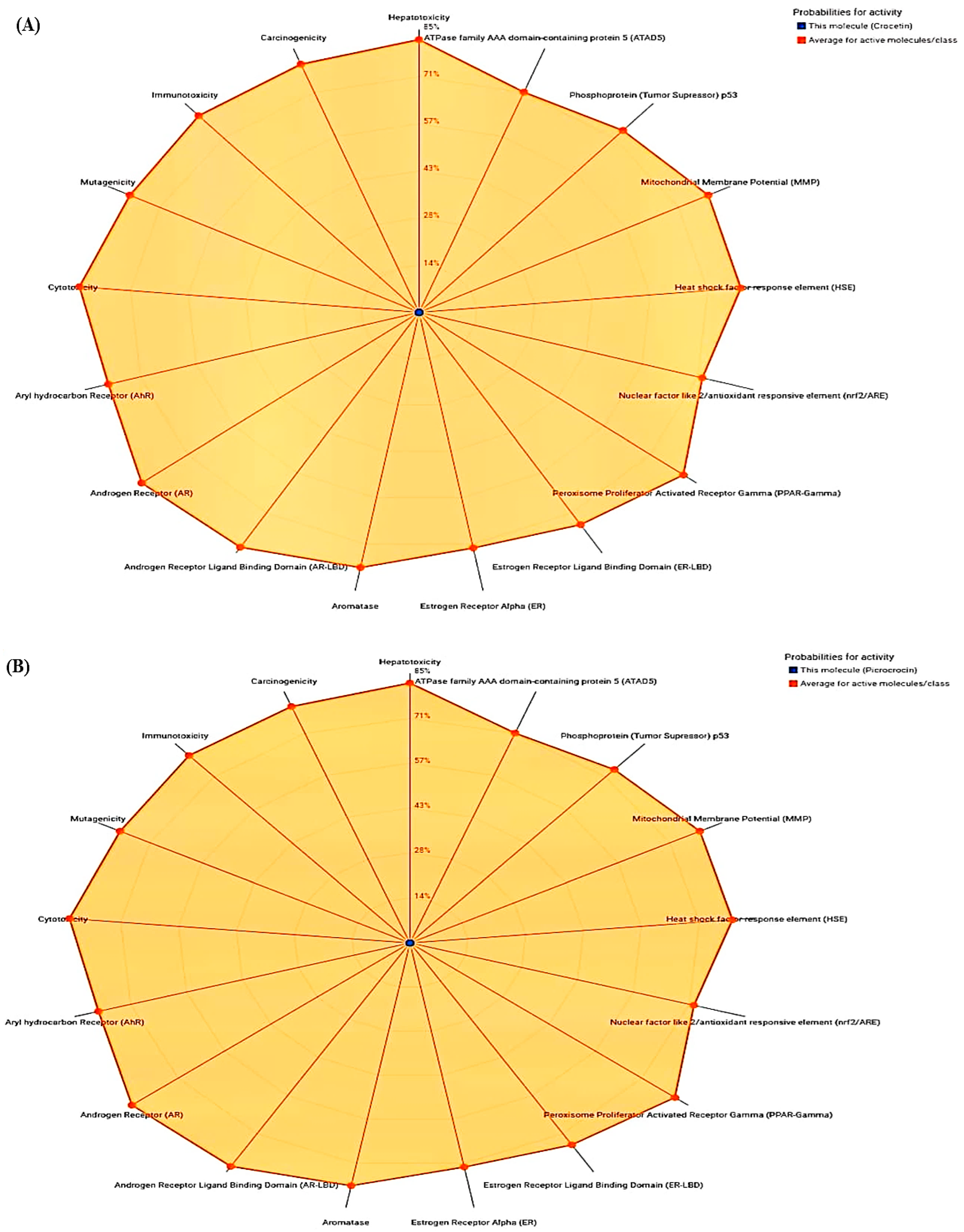
Figure 1. Radar toxicity chart analysis: (A) Crocetin, (B) Picrocrocin, (C) Quercetin, (D) Rutin, (E) Aspirin, (F) Rofecoxib.
Medicina 59 02058 g001aMedicina 59 02058 g001bMedicina 59 02058 g001c
Figure 2. Molecular docking analysis: (A) Crocetin, (B) Picrocrocin, (C) Quercetin, (D) Rutin, (E) Aspirin, (F) Rofecoxib.
Medicina 59 02058 g002aMedicina 59 02058 g002b
Figure 3. PROCHECK analysis: (A) Ramachandran plot statistics, (B) Hydrophobicity plot, (C) Ramachandran plot for all residues.
Medicina 59 02058 g003
Figure 4. ComPPI analysis: (A) First-neighbor network visualization COX-2 protein, (B) Major and Minor localizations with scores.
Medicina 59 02058 g004
Figure 5. CASTp server analysis: (AE) Different representations of pocket panel, (F) CASTp data.
Medicina 59 02058 g005aMedicina 59 02058 g005b
Figure 6. WEBnmα analysis: (A) Eigenvalues, (B) Average deformation energies, (C) Atomic displacement, (D) Fluctuation, (E) Covariance map.
Medicina 59 02058 g006
Figure 7. LARMD analysis: (A) Decomposition energy, (B) MM/PB(GB)SA analysis, (C) Binding residues.
Medicina 59 02058 g007
Figure 8. ShinyGO analysis: (A) KEGG pathway enrichment, (B) Functional enrichment.
Medicina 59 02058 g008
Figure 9. Molecular simulation depicting: (A) RMSD, (B) RMSF, (C,D) % SSE of COX-2 protein.
Medicina 59 02058 g009
Figure 10. RMSF of picrocrocin ligand: (A,B) ligand–protein interactions.
Medicina 59 02058 g010
Figure 11. Timeline representation: hydrogen-bonds, water bridges, hydrophobic and ionic interactions.
Medicina 59 02058 g011
Figure 12. Ligand torsion profile (A, Upper panel), and (B, Lower panel) ligand properties.
Medicina 59 02058 g012
Table 1. ADMET analysis of saffron constituents.
ADMET Physiochemical Properties
CompoundsDrugs
Crocetin Picrocrocin Quecertin Rutin Aspirin Rofecoxib
Molecular Weight (g/mol)328.40330.37302.24610.52180.16314.36
Topological polar surface area (TPSA) Å274.60116.45131.36269.6363.6068.82
Hydrogen bond acceptors 4771644
Hydrogen bond donors2451010
Molar refractivity 98.4881.0878.03141.844.9083.69
XLOGP5.41−0.501.54−0.331.192.27
iLOGP3.332.071.630.461.302.13
MLOGP3.52−0.88−0.56−3.891.512.62
WLOGP4.61−0.491.99−1.691.313.64
Lipinski Yes YesYes NoYesYes
GI absorption High HighHigh LowHighHigh
Bioavailability score 0.850.550.550.170.850.55
BBB permeability NoNoNo No Yes Yes
Pgp substrate NoYesNo Yes No No
CYP2D6 inhibitor NoNoYes No NoNo
PAINS (Pan assay interference compounds)001100
Leadlikeness No YesYesNoNoYes
Skin permeation (log kp) cm/s−4.46−8.67−7.05−10.26−6.55−6.61
Table 2. Predicted toxicity analysis of compounds and drugs.
Compounds Toxicity Class Hepatotoxicity Immunotoxicity CarcinogenicityMutagenicity Cytotoxicity
Crocetin 50.720.990.740.720.72
Picrocrocin30.830.830.780.730.87
Quercetin30.690.87 0.68 0.51 0.99
Rutin 50.800.98 0.910.880.64
Aspirin30.510.990.860.970.94
Rofecoxib50.550.970.710.680.76
Table 3. Selected saffron compounds with their Ki and binding affinity.
S. No.PhytocompoundsBinding Affinity (kcal/mol)Ki (µM)
1Crocetin −7.412.50
2Picrocrocin −8.113.68
3Quecertin −7.612.87
4Rutin −7.913.38
5Safranal −6.811.51
6Crocin−7.112.02
7Dimethylcrocetin −7.212.19
Drug
1Aspirin−6.110.30
2Rofecoxib −8.314.05
Table 4. Position and interactions between chosen protein and compounds.
CompoundType of InteractionPosition of Interaction
CrocetinHydrogen bondingASN34, ARG44
Van der Waals PRO154, PRO156, TYR136, TYR130, GLY135, GLY45, GLN461, VAL46, CYS36, CYS47, ARG469, ASP125
Alkyl/Pi-alkylPRO153, HIS39, LEU152
PicocroxinHydrogen bondingTYR373, GLN374, SN375
Van der WaalsGLY225, HIS226, ARG376, LEU145, ASN375, PHE142, PRO127, TYR373
QuercetinHydrogen bondingCYS47, TYR130, ARG469, ARG44, ASP125
Van der WaalsPRO40, HIS39, GLN461, LEU152, GLY45, GLY135, CYS41, CYS36, MET48
Pi-alkylPRO153
RutinHydrogen bondingSER126, HIS122, PHE371, LEU366, TYR373, THR118
Van der WaalsLYS369, THR62, THR60, LYS560, GLN370, SER121, PHE367, ARG44, GLN372, LYS369, LYS532, ILE124, ASP125, ALA543, LEU123
Alkyl/Pi-alkylARG61, PRO542
Drug
Aspirin Hydrogen bondingTRP139, ASP229, ARG333
Van der Waals LEU224, GLY235, GLU236, THR237, LEU238, GLN241
Rofecoxib Hydrogen bonding ARG44
Van der WaalsSER121, ALA543, LYS532, ASP125, SER126, PHE371, HIS122, GLN370, GLN372, TYR373
Pi-alkylPRO542
Table 5. MM/PB(GB)SA free binding energies of human COX-2 protein.
ELEVDWGASPBSOLPBTOTGBSOLGBTOTTSDelta
PB
Delta
GB
−271.82−74.73−346.55351.665.12352.92−20.6337.4542.5716.82
Table 6. Pathway analysis with fold enrichment scores.
FDR EnrichmentGenes of PathwayGeneFold EnrichmentName of Pathway
0.025356055511158.7843137Ovarian steroidogenesis
0.025356055561144.6071429Reg. of lipolysis in adipocytes
0.025356055591137.2542373VEGF signaling pathway
0.025356055611132.7540984Arachidonic acid metabolism
0.025356055681119.0882353Chemical carcinogenesis
0.025356055761106.5526316Leishmaniasis
0.02535605592188.02173913Small cell lung cancer
0.02535605593187.07526882IL-17 signaling pathway
0.025356055104177.86538462NF-kappa B signaling pathway
0.025356055104177.86538462C-type lectin receptor signaling pathway
0.025356055112172.30357143TNF signaling pathway
0.025356055112172.30357143Serotonergic synapse
0.029159463148154.71621622Retrograde endocannabinoid signaling
0.029159463154152.58441558Oxytocin signaling pathway
0.029159463161150.29813665MicroRNAs in cancer
0.032940232194141.74226804Kaposi sarcoma-associated herpesvirus infection
0.035796784224136.15178571Human cytomegalovirus infection
0.049957465331124.4652568Human papillomavirus infection
Disclaimer/Publisher’s Note: The statements, opinions and data contained in all publications are solely those of the individual author(s) and contributor(s) and not of MDPI and/or the editor(s). MDPI and/or the editor(s) disclaim responsibility for any injury to people or property resulting from any ideas, methods, instructions or products referred to in the content.