Case Report

Whole Exome Sequencing of a Patient with a Milder Phenotype of Xeroderma Pigmentosum Group C

1
Department of Dermatology, College of Medicine, Kyung Hee University, Seoul 02447, Republic of Korea
2
Division of Dermatology, Internal Related, Graduate School of Medicine, Kobe University, Kobe 653-0002, Japan
3
Department of Oral Anatomy and Developmental Biology, Graduate School, Kyung Hee University, Seoul 02447, Republic of Korea
4
Department of Medicine, Graduate School, Kyung Hee University, Seoul 02447, Republic of Korea
5
Department of Biomedical Laboratory Science, Catholic Kwandong University, Gangneung 25601, Republic of Korea
*
Authors to whom correspondence should be addressed.
These authors contributed equally to this work.
Medicina 2023, 59(4), 699; https://doi.org/10.3390/medicina59040699
Submission received: 11 February 2023 / Revised: 7 March 2023 / Accepted: 28 March 2023 / Published: 3 April 2023

Abstract

:
A 17-year-old female Korean patient (XP115KO) was previously diagnosed with Xeroderma pigmentosum group C (XPC) by Direct Sanger sequencing, which revealed a homozygous nonsense mutation in the XPC gene (rs121965088: c.1735C > T, p.Arg579Ter). While rs121965088 is associated with a poor prognosis, our patient presented with a milder phenotype. Hence, we conducted whole-exome sequencing in the patient and her family members to detect coexisting mutations that may have resulted in a milder phenotype of rs121965088 through genetic interaction. Materials and Methods: the whole-exome sequencing analysis of samples obtained from the patient and her family members (father, mother, and brother) was performed. To identify the underlying genetic cause of XPC, the extracted DNA was analyzed using Agilent’s SureSelect XT Human All Exon v5. The functional effects of the resultant variants were predicted using the SNPinfo web server, and structural changes in the XPC protein using the 3D protein modeling program SWISS-MODEL. Results: Eight biallelic variants, homozygous in the patient and heterozygous in her parents, were detected. Four were found in the XPC gene: one nonsense variant (rs121965088: c.1735C > T, p.Arg579Ter) and three silent variants (rs2227998: c.2061G > A, p. Arg687Arg; rs2279017: c.2251-6A > C, intron; rs2607775: c.-27G > C, 5′UTR). The remaining four variants were found in non-XP genes, including one frameshift variant [rs72452004 of olfactory receptor family 2 subfamily T member 35 (OR2T35)], three missense variants [rs202089462 of ALF transcription elongation factor 3 (AFF3), rs138027161 of TCR gamma alternate reading frame protein (TARP), and rs3750575 of annexin A7 (ANXA7)]. Conclusions: potential candidates for genetic interactions with rs121965088 were found. The rs2279017 and rs2607775 of XPC involved mutations in the intron region, which affected RNA splicing and protein translation. The genetic variants of AFF3, TARP, and ANXA7 are all frameshift or missense mutations, inevitably disturbing the translation and function of the resultant proteins. Further research on their functions in DNA repair pathways may reveal undiscovered cellular relationships within xeroderma pigmentosum.

1. Introduction

Xeroderma pigmentosum (XP) is an autosomal recessive genodermatosis that is associated with DNA repair defects. The disease comprises eight complementation groups (XP-A to XP g and XP-V), which represent functional deficiencies in different genes [damage specific DNA binding protein 2 (DDB2), ERCC excision repair, TFIIH core complex helicase subunit (ERCC1 to 5), DNA polymerase eta (POLH), XPA, DNA damage recognition and repair factor (XPA), and XPC complex subunit, DNA damage recognition and repair factor (XPC)]. Proteins encoded by DDB2, ERCC1 to 5, XPA, and XPC participate in the nucleotide excisional repair (NER) system, whereas those by POLH are in translation with DNA synthesis. Depending on the complementation groups, XP patients demonstrate a great variety of clinical features and prognoses [1,2,3,4]. From mild manifestations such as lentiginous pigmentation, photophobia, and conjunctivitis, the disease may progress into lethal conditions such as cutaneous neoplasm, ocular surface cancer, internal organ malignancy, and epileptic seizure. Because such diversified manifestations progressively appear with aging, frequent and meticulous follow-ups are required throughout the patient’s lifetime. Consequently, diagnostic confirmation and the precise identification of complementation groups are crucial in predicting clinical symptoms and providing early medical intervention [5,6,7,8].
Throughout time, various diagnostic tools have been developed for XP. Originally, specialized laboratory tests examined the cellular and chromosomal instabilities of XP cells after their exposure to DNA-damaging agents such as ultraviolet (UV) irradiation, chemical carcinogens, or cytotoxic drugs [9,10,11]. In the cellular hypersensitivity test, the colony-forming ability of XP patients’ fibroblasts was measured after UV radiation [10,11]. Similarly, the extent of chromosomal breakage and chromatid aberration was observed in XP cells in response to chemical mutagens. More advanced methods have directly evaluated the repair ability of XP cells [11]. The unscheduled DNA synthesis (UDS) test quantifies newly incorporated nucleotides in XP fibroblasts after UV-induced DNA damage [9]. XP patients only achieve 10–20% of UDS compared to normal subjects. The fusion of identical XP subtype cells leads to UDS recovery, while different ones show no improvement. Other complementation analysis laboratory tests include Western blot and Northern blot, which measure the decrease or absence of specific XP proteins or their mRNA levels [11]. Nevertheless, advancements in molecular science now enable the direct identification of pathogenic mutations in XP genes.
DNA sequencing tools such as single gene testing, targeted gene sequencing (TGS), whole exome sequencing (WES), and whole genome sequencing (WGS) enable rapid and precise XP diagnosis and complementation group analysis [10]. Currently, each method has been recommended for different conditions. When clinical characteristics and regional XP prevalence strongly indicate a certain complementation group, single-gene testing is performed on the associated pathogenic XP gene. Alternatively, patients not biased toward a subtype most often perform TGS, which simultaneously sequences multiple pathogenic XP genes. Lastly, when TGS fails to detect mutations in XP-suspected patients, large-scale genome sequencing (WGS or WES) is recommended to discover the genotypes resembling XP clinical features and exclude XP diagnosis.
A 17-year-old female Korean patient (XP115KO) was previously diagnosed with XPC [12]. This patient received regular follow-ups at our hospital for 15 years, from the age of 2 to 17 years old. Unscheduled DNA synthesis and Direct Sanger sequencing for 16 exons of XPC were performed at Kobe University in Japan when she was 6 years old. Direct Sanger sequencing revealed a homozygous nonsense mutation in the XPC gene (rs121965088: c.1735C > T, p.Arg579Ter). According to ClinVar [13], a public database of human genome mutations and their relationship to clinical phenotypes, including 10 genetic variants of XPC, are currently acknowledged to have pathogenic significance. Among these, rs121965088 is associated with poor prognosis, including multiple skin cancers, internal organ malignancy, and early death [14]. Interestingly, our patient presented with a milder phenotype. Her height and weight were within the normal range, and there were no neurological complications. On metabolic analysis, baseline plasma cortisol and adrenocorticotropic hormone (ACTH) were normal, and no abnormalities were found in the serum glucose, α2-globulins, aspartate transaminase (AST), and alanine transaminase (ALT). First, skin cancer was found at the age of 11 years; however, there have been no additional skin cancers to date (current age, 17 years) and no internal organ malignancies.
The prognosis of XP depends on early diagnosis and strict sun avoidance [15]. Our patient was diagnosed at 6 years of age and practiced diligent sun protection; therefore, her mild phenotype was expected. Additionally, undiscovered genetic variants may have alleviated this phenotype. Essential genes, such as DNA repair genes, are five times more likely to be involved in genetic interactions—with the synergy between two or more genetic mutations creating an unexpected phenotype—than are non-essential genes [16,17,18,19]. Moreover, a “positive” genetic interaction, which reduces the disadvantageous phenotype of a certain mutation, generally involves proteostasis [16], which refers to the intracellular system that maintains protein homeostasis. Since patients with XPC possess mutated endonuclease proteins that fail to recognize and incise damaged DNA, positive genetic interactions may partially restore the DNA repair pathway.
We hypothesized that the genetic interaction—the synergy between two or more mutations creating an unexpected phenotype—may have resulted in a milder phenotype of rs121965088. Hence, we conducted whole-exome sequencing in the patient and her family members to (1) identify pathogenic XP mutations affecting the protein-coding regions, (2) detect coexisting mutations in non-pathogenic XP genes associated with DNA repair or other genetic disorders, and (3) examine the Mendelian inheritance pattern in the identified biallelic mutations.

2. Methods

2.1. Subjects and DNA Extraction

We performed the whole-exome sequencing analysis of the samples obtained from the patient and her family members (father, mother, and brother). This family was not related by consanguinity. The study was approved by the institutional review board on 7 July 2017 (No. 2017-05-042-003).
For whole exome sequencing, genomic DNA from the blood was extracted using a Roche DNA Extraction kit (Roche, Indianapolis, IN, USA). Absorbance in 260, 280, and 230 nm for 2 μL of each DNA sample was measured in duplicate using Nanodrop for DNA quality check. The 260/280 ratio was used as an indicator of purity for the DNA samples. For whole exome sequencing, the optimal value of the 260/280 ratio for pure DNA was set to 1.8 or higher, and the experiment was conducted.

2.2. Exome Sequencing and Bioinformatics Pipeline

To identify the underlying genetic cause of XPC, the extracted DNA was analyzed using Agilent’s SureSelect XT Human All Exon v5 (Agilent Technologies, Santa Clara, CA, USA).
After sequencing, reads were aligned to an indexed human reference genome (GRCh37/hg19) with the Burrows–Wheeler Aligner (BWA). After alignment, further analysis steps, including (1) data preprocessing and (2) somatic short variant discovery, were used by the Genome Analysis Toolkit (GATK). Additionally, the preparation of the variant calling part was performed on the recalibrated files with detailed steps following GATK best practices. Firstly, Haplotype Caller was used with individual samples in the GVCF mode. Then, all samples were combined in a datastore using the Genomics DB Import function. Next, a joint genotyping of all samples with Genotype GVCFs was performed. To filter out possible false positives, a Variant Quality Score Recalibration was applied to the data. Finally, identified variants were saved in a variant call format. Single nucleotide polymorphisms (SNPs) and indels were annotated using snpEff, which classified the variants.
The functional effects of the resultant variants were predicted using the SNPinfo web server (https://snpinfo.niehs.nih.gov, accessed on 1 January 2022). Structural changes in the XPC protein were visualized using the 3D protein modeling program SWISS-MODEL.

3. Results

The nucleotide sequences of XPC patients, parents, and siblings obtained through whole exome sequencing were compared and analyzed. Since XPC disease is an autosomal recessive disease, we focused on finding a homozygous mutation that were not seen in the parents and siblings. As a result of the analysis, the parts showing homozygous mutation in the XPC patients, unlike their families, were found in the olfactory receptor family 2 subfamily T member 35 (OR2T35), ALF transcription elongation factor 3 (AFF3), TCR gamma alternate reading frame protein (TARP), and annexin A7 (ANXA7) genes.
Eight biallelic variants, including homozygous in the patient and heterozygous in her parents, were detected (Table 1). Among these, four were found in the XPC gene: one nonsense variant (rs121965088: c.1735C > T, p.Arg579Ter) and three silent variants [rs2227998: c.2061G > A, p. Arg687Arg; rs2279017: c.2251-6A > C, intron; rs2607775: c.-27G > C, 5′ untranslated region (UTR)]. When simulated by SWISS-MODEL, rs121965088 caused premature termination during Rad4 protein unit translation, creating a defective and misfolded XPC protein (Figure 1). The remaining four variants were found in non-XP genes: one frameshift variant (rs72452004 of OR2T35) and three missense variants (rs202089462 of AFF3, rs138027161 of TARP, and rs3750575 of ANXA7).

4. Discussion

In this report, we describe the 15-year follow-up of a Korean girl presenting with a good prognosis despite having the homozygous nonsense mutation of rs12195088 in the XPC gene. The whole-exome sequencing analysis of our patient with XP and her family members revealed four biallelic variants in XPC (rs121965088, rs2227998, rs2279017, and rs2607775) and four in non-XP genes (rs72452004 of OR2T35, rs202089462 of AFF3, rs138027161 of TARP, and rs3750575 of ANXA7).
No novel ‘pathogenic’ biallelic variants were detected in pathogenic XP genes. The additionally identified three silent mutations of XPC (rs2227998, rs2279017, and rs2607775) have already been classified as ‘benign’ variants by the American College of Medical Genetics and Genomics (ACMG) guideline. Nevertheless, since rs2279017 and rs2607775 are located in the intron and 5′UTR region of XPC, they may affect RNA splicing and alter the messenger RNA sequence. For instance, the rs2607775 is located in the transcription factor-binding site of the 5′UTR, and previous studies have reported its association with increased cancer risks, including hepatocellular cancer risk, gastric cancer, and colorectal cancer [36,37,38]. The XPC gene repairs the DNA damage caused by internal and external factors, and it is known that many cancers occur due to the abnormality of XPC [39]. Therefore, their influence on XP phenotypes, when combined with underlying pathogenic XP variants, needs further study.
Similarly, four biallelic variants located on non-pathogenic XP genes (rs72452004 of OR2T35, rs202089462 of AFF3, rs138027161 of TARP, and rs3750575 of ANXA7) had no acknowledged pathogenicity relating to XP or other genetic disorders; they have no reports in ClinVar or meaningful reports in the literature. While rs3750575 was once published to have a higher frequency in Japanese patients with dissecting aortic aneurysms in an exome-wide association study [35], no additional significance has been reported since. Nonetheless, since all four mutations are either frameshift or missense mutations, protein translation—and potentially protein function—are inevitably disturbed. Additionally, proteins encoded from OR2T35, AFF3, TARP, and ANXA7, respectively, involve the G-protein-coupled receptor, nuclear transcriptional activator, T cell receptor gamma protein, and calcium-dependent phospholipid binding proteins according to the National Center for Biotechnology Information. Since such proteins are components of major cellular pathways, their concurrence with pathogenic XP variants may influence DNA repair pathways.
Along with pathogenic genotype identification, DNA sequence analysis provides insights into the “genotype-to-phenotype relationship” in XP [40,41,42,43,44]. DNA repair disorders do not possess a one-to-one correlation between molecular genotypes and clinical phenotypes [2,40,45,46]. Mutations in different genes may present with identical clinical manifestations, while genetic variants within a gene may exhibit contrasting phenotypes. Accordingly, XP has shown great diversity in clinical phenotypes within the same complementation groups yet relative uniformity within identical genetic variants in the ClinVar database. In addition, genetic interactions may once again diverge phenotypes within a genetic variant [1,47]. Thus, large-scale genome studies such as WGS and WES enable a comprehensive understanding of genotype–phenotype relations and underlying etiologies of XP.

5. Conclusions

In conclusion, we performed a long-term follow-up of an XPC patient with a milder phenotype despite having a mutation (rs12195088) with a poor prognosis. WES was performed on her and her entire family in order to identify possible genetic features. We identified the eight variants related to XPC. Among the eight variants, four variants were located on the XPC gene, and three variants (rs2279017, rs2227998, and rs2607775) might be indirectly related to the protein function of XPC. One variant, rs121965088, had been reported to affect the protein of XPC in a previous paper [11]. The remaining four variants, rs72452004, rs202089462, rs138027161, and rs3750575 were located in OR2T35, AFF3, TARP, and ANXA7 genes, respectively. As the variant of each gene is located in the axon region, it might affect the protein by substituting the amino acid constituting the protein.
The limitation of this study is that biological experiments were not performed. Additional biological studies were needed for each variant, for example, direct changes in the RNA expression or protein structure.

Author Contributions

Conceptualization: K.-H.J. Data curation: S.K.K. Data analysis: S.K.K., J.J.A., J.Y.R. and J.L. Investigation: J.J.A., J.Y.R. and J.L. Methodology: J.J.A., J.Y.R. and J.L. Project administration: M.-H.L. Resources: C.N. and M.-H.L. Software: J.-I.S. Supervision: K.-H.J. Validation: C.N. and M.-H.L. Visualization: S.K.K. Writing—original draft: J.-I.S., S.K.K. and K.-H.J. Writing—review and editing: J.-I.S., S.K.K. and K.-H.J. All authors have read and agreed to the published version of the manuscript.

Funding

This research was supported by “Regional Innovation Strategy (RIS)” through the National Research Foundation of Korea (NRF) and funded by the Ministry of Education (MOE) (2022RIS-005).

Institutional Review Board Statement

The study was approved by the institutional review board on 7 July 2017 (No. 2017-05-042-003).

Informed Consent Statement

Written informed consent has been obtained from the patient(s) to publish this paper.

Data Availability Statement

Not applicable.

Conflicts of Interest

The authors declare no conflict of interest.

References

  1. Leung, A.K.; Barankin, B.; Lam, J.M.; Leong, K.F.; Hon, K.L. Xeroderma pigmentosum: An updated review. Drugs Context 2022, 11, 2022-2-5. [Google Scholar] [CrossRef]
  2. Fassihi, H. Spotlight on ‘xeroderma pigmentosum’. Photochem. Photobiol. Sci. 2013, 12, 78–84. [Google Scholar] [CrossRef] [PubMed]
  3. Abeti, R.; Zeitlberger, A.; Peelo, C.; Fassihi, H.; Sarkany, R.P.E.; Lehmann, A.R.; Giunti, P. Xeroderma pigmentosum: Overview of pharmacology and novel therapeutic strategies for neurological symptoms. Br. J. Pharmacol. 2019, 176, 4293–4301. [Google Scholar] [CrossRef] [PubMed] [Green Version]
  4. Halkud, R.; Shenoy, A.M.; Naik, S.M.; Chavan, P.; Sidappa, K.T.; Biswas, S. Xeroderma pigmentosum: Clinicopathological review of the multiple oculocutaneous malignancies and complications. Indian J. Surg. Oncol. 2014, 5, 120–124. [Google Scholar] [CrossRef] [Green Version]
  5. Black, J.O. Xeroderma Pigmentosum. Head Neck Pathol. 2016, 10, 139–144. [Google Scholar] [CrossRef] [Green Version]
  6. Piccione, M.; Fortina, A.B.; Ferri, G.; Andolina, G.; Beretta, L.; Cividini, A.; De Marni, E.; Caroppo, F.; Citernesi, U.; Di Liddo, R. Xeroderma Pigmentosum: General Aspects and Management. J. Pers. Med. 2021, 11, 1146. [Google Scholar] [CrossRef] [PubMed]
  7. Brambullo, T.; Colonna, M.R.; Vindigni, V.; Piaserico, S.; Masciopinto, G.; Galeano, M.; Costa, A.L.; Bassetto, F. Xeroderma Pigmentosum: A Genetic Condition Skin Cancer Correlated—A Systematic Review. BioMed Res. Int. 2022, 2022, 8549532. [Google Scholar] [CrossRef]
  8. Hossain, M.; Hasan, A.; Shawan, M.M.A.K.; Banik, S.; Jahan, I. Current Therapeutic Strategies of Xeroderma Pigmentosum. Indian J. Dermatol. 2021, 66, 660–667. [Google Scholar] [CrossRef]
  9. Lehmann, A.R.; McGibbon, D.; Stefanini, M. Xeroderma pigmentosum. Orphanet. J. Rare Dis. 2011, 6, 70. [Google Scholar] [CrossRef] [Green Version]
  10. DiGiovanna, J.J.; Kraemer, K.H. Shining a light on xeroderma pigmentosum. J. Investig. Dermatol. 2012, 132 Pt 2, 785–796. [Google Scholar] [CrossRef] [Green Version]
  11. DiGiovanna, J.J.; Rünger, T.M.; Kraemer, K.H. Hereditary Disorders of Genome Instability and DNA Repair. In Fitzpatrick’s Dermatology, 9th ed.; Kang, S., Amagai, M., Bruckner, A.L., Enk, A.H., Margolis, D.J., McMichael, A.J., Orringer, J.S., Eds.; McGraw Hill: New York, NY, USA, 2019. [Google Scholar]
  12. Masaki, T.; Nakano, E.; Okamura, K.; Ono, R.; Sugasawa, K.; Lee, M.-H.; Suzuki, T.; Nishigori, C. A case of xeroderma pigmentosum complementation group C with diverse clinical features. Br. J. Dermatol. 2018, 178, 1451–1452. [Google Scholar] [CrossRef] [PubMed]
  13. Landrum, M.J.; Lee, J.M.; Benson, M.; Brown, G.R.; Chao, C.; Chitipiralla, S.; Gu, B.; Hart, J.; Hoffman, D.; Jang, W.; et al. ClinVar: Improving access to variant interpretations and supporting evidence. Nucleic Acids Res. 2018, 46, D1062–D1067. [Google Scholar] [CrossRef] [PubMed] [Green Version]
  14. Gozukara, E.M.; Khan, S.; Metin, A.; Emmert, S.; Busch, D.B.; Shahlavi, T.; Coleman, D.M.; Miller, M.; Chinsomboon, N.; Stefanini, M.; et al. A Stop Codon in Xeroderma Pigmentosum Group C Families in Turkey and Italy: Molecular Genetic Evidence for a Common Ancestor. J. Investig. Dermatol. 2001, 117, 197–204. [Google Scholar] [CrossRef] [PubMed] [Green Version]
  15. Lucero, R.; Horowitz, D. Xeroderma Pigmentosum; StatPearls Publishing: Treasure Island, FL, USA, 2022. [Google Scholar]
  16. Costanzo, M.; VanderSluis, B.; Koch, E.N.; Baryshnikova, A.; Pons, C.; Tan, G.; Wang, W.; Usaj, M.; Hanchard, J.; Lee, S.D.; et al. A global genetic interaction network maps a wiring diagram of cellular function. Science 2016, 353, aaf1420. [Google Scholar] [CrossRef] [Green Version]
  17. Boucher, B.; Jenna, S. Genetic interaction networks: Better understand to better predict. Front. Genet. 2013, 4, 290. [Google Scholar] [CrossRef] [Green Version]
  18. Shell, S.M.; Zou, Y. Other proteins interacting with XP proteins. Adv. Exp. Med. Biol. 2008, 637, 103–112. [Google Scholar]
  19. Hipp, M.S.; Kasturi, P.; Hartl, F.U. The proteostasis network and its decline in ageing. Nat. Rev. Mol. Cell Biol. 2019, 20, 421–435. [Google Scholar] [CrossRef]
  20. Xue, P.; Gao, L.; Xiao, S.; Zhang, G.; Xiao, M.; Zhang, Q.; Zheng, X.; Cai, Y.; Jin, C.; Yang, J.; et al. Genetic polymorphisms in xrcc1, cd3eap, ppp1r13l, xpb, xpc, and xpf and the risk of chronic benzene poisoning in a chinese occupational population. PLoS ONE 2015, 10, e0144458. [Google Scholar] [CrossRef]
  21. Richards, S.; Aziz, N.; Bale, S.; Bick, D.; Das, S.; Gastier-Foster, J.; Grody, W.W.; Hegde, M.; Lyon, E.; Spector, E.; et al. Standards and guidelines for the interpretation of sequence variants: A joint consensus recommendation of the american college of medical genetics and genomics and the association for molecular pathology. Genet. Med. 2015, 17, 405–424. [Google Scholar] [CrossRef] [Green Version]
  22. Gil, J.; Gaj, P.; Misiak, B.; Ostrowski, J.; Karpinski, P.; Jarczynska, A.; Kielan, W.; Sasiadek, M.M. Cyp1a1 ile462val polymorphism and colorectal cancer risk in polish patients. Med. Oncol. 2014, 31, 72. [Google Scholar] [CrossRef] [Green Version]
  23. Rondelli, C.M.; El-Zein, R.A.; Wickliffe, J.K.; Etzel, C.J.; Abdel-Rahman, S.Z. A comprehensive haplotype analysis of the xpc genomic sequence reveals a cluster of genetic variants associated with sensitivity to tobacco-smoke mutagens. Toxicol. Sci. 2010, 115, 41–50. [Google Scholar] [CrossRef] [Green Version]
  24. Joshi, A.D.; Corral, R.; Siegmund, K.D.; Haile, R.W.; Le Marchand, L.; Martinez, M.E.; Ahnen, D.J.; Sandler, R.S.; Lance, P.; Stern, M.C. Red meat and poultry intake, polymorphisms in the nucleotide excision repair and mismatch repair pathways and colorectal cancer risk. Carcinogenesis 2009, 30, 472–479. [Google Scholar] [CrossRef]
  25. Richards, C.S.; Bale, S.; Bellissimo, D.B.; Das, S.; Grody, W.W.; Hegde, M.R.; Lyon, E.; Ward, B.E.; Molecular Subcommittee of the ACMG Laboratory Quality Assurance Committee. ACMG recommendations for standards for interpretation and reporting of sequence variations. Genet. Med. 2008, 10, 294–300. [Google Scholar] [CrossRef] [Green Version]
  26. Bodian, D.L.; McCutcheon, J.N.; Kothiyal, P.; Huddleston, K.C.; Iyer, R.K.; Vockley, J.G.; Niederhuber, J.E. Germline variation in cancer-susceptibility genes in a healthy, ancestrally diverse cohort: Implications for individual genome sequencing. PLoS ONE 2014, 9, e94554. [Google Scholar] [CrossRef] [PubMed]
  27. Doherty, J.A.; Weiss, N.S.; Fish, S.; Fan, W.; Loomis, M.M.; Sakoda, L.C.; Rossing, M.A.; Zhao, L.P.; Chen, C. Polymorphisms in nucleotide excision repair genes and endometrial cancer risk. Cancer Epidemiol. Biomark. Prev. 2011, 20, 1873–1882. [Google Scholar] [CrossRef] [PubMed] [Green Version]
  28. Roberts, M.R.; Shields, P.G.; Ambrosone, C.B.; Nie, J.; Marian, C.; Krishnan, S.S.; Goerlitz, D.S.; Modali, R.; Seddon, M.; Lehman, T.; et al. Single-nucleotide polymorphisms in DNA repair genes and association with breast cancer risk in the web study. Carcinogenesis 2011, 32, 1223–1230. [Google Scholar] [CrossRef] [Green Version]
  29. Chavanne, F.; Broughton, B.C.; Pietra, D.; Nardo, T.; Browitt, A.; Lehmann, A.R.; Stefanini, M. Mutations in the xpc gene in families with xeroderma pigmentosum and consequences at the cell, protein, and transcript levels. Cancer Res. 2000, 60, 1974–1982. [Google Scholar] [PubMed]
  30. Li, Y.K.; Xu, Q.; Sun, L.P.; Gong, Y.H.; Jing, J.J.; Xing, C.Z.; Yuan, Y. Nucleotide excision repair pathway gene polymorphisms are associated with risk and prognosis of colorectal cancer. World J. Gastroenterol. 2020, 26, 307–323. [Google Scholar] [CrossRef]
  31. Liang, X.H.; Yan, D.; Zhao, J.X.; Ding, W.; Xu, X.J.; Wang, X.Y. Interaction of polymorphisms in xeroderma pigmentosum group c with cigarette smoking and pancreatic cancer risk. Oncol. Lett. 2018, 16, 5631–5638. [Google Scholar] [CrossRef] [Green Version]
  32. Hua, R.X.; Zhuo, Z.J.; Shen, G.P.; Zhu, J.; Zhang, S.D.; Xue, W.Q.; Li, X.Z.; Zhang, P.F.; He, J.; Jia, W.H. Polymorphisms in the xpc gene and gastric cancer susceptibility in a southern chinese population. Onco. Targets Ther. 2016, 9, 5513–5519. [Google Scholar] [PubMed] [Green Version]
  33. Perez-Mayoral, J.; Pacheco-Torres, A.L.; Morales, L.; Acosta-Rodriguez, H.; Matta, J.L.; Dutil, J. Genetic polymorphisms in rad23b and xpc modulate DNA repair capacity and breast cancer risk in puerto rican women. Mol. Carcinog. 2013, 52 (Suppl. 1), E127–E138. [Google Scholar] [CrossRef] [PubMed] [Green Version]
  34. Bai, Y.; Xu, L.; Yang, X.; Hu, Z.; Yuan, J.; Wang, F.; Shao, M.; Yuan, W.; Qian, J.; Ma, H.; et al. Sequence variations in DNA repair gene xpc is associated with lung cancer risk in a chinese population: A case-control study. BMC Cancer 2007, 7, 81. [Google Scholar] [CrossRef] [PubMed] [Green Version]
  35. Yamada, Y.; Sakuma, J.; Takeuchi, I.; Yasukochi, Y.; Kato, K.; Oguri, M.; Fujimaki, T.; Horibe, H.; Muramatsu, M.; Sawabe, M.; et al. Identification of egflam, spatc1l and rnase13 as novel susceptibility loci for aortic aneurysm in japanese individuals by exome-wide association studies. Int. J. Mol. Med. 2017, 39, 1091–1100. [Google Scholar] [CrossRef] [PubMed] [Green Version]
  36. Wang, B.; Xu, Q.; Yang, H.-W.; Sun, L.-P.; Yuan, Y. The association of six polymorphisms of five genes involved in three steps of nucleotide excision repair pathways with hepatocellular cancer risk. Oncotarget 2016, 7, 20357–20367. [Google Scholar] [CrossRef] [Green Version]
  37. Liu, J.; Sun, L.; Xu, Q.; Tu, H.; He, C.; Xing, C.; Yuan, Y. Association of nucleotide excision repair pathway gene polymorphisms with gastric cancer and atrophic gastritis risks. Oncotarget 2016, 7, 6972–6983. [Google Scholar] [CrossRef] [Green Version]
  38. Hu, X.; Qin, W.; Li, S.; He, M.; Wang, Y.; Guan, S.; Zhao, H.; Yao, W.; Wei, M.; Liu, M.; et al. Polymorphisms in DNA repair pathway genes and ABCG2 gene in advanced colorectal cancer: Correlation with tumor characteristics and clinical outcome in oxaliplatin-based chemotherapy. Cancer Manag. Res. 2018, 11, 285–297. [Google Scholar] [CrossRef] [Green Version]
  39. Vineis, P.; Manuguerra, M.; Kavvoura, F.K.; Guarrera, S.; Allione, A.; Rosa, F.; Di Gregorio, A.; Polidoro, S.; Saletta, F.; Ioannidis, J.P.A.; et al. A Field Synopsis on Low-Penetrance Variants in DNA Repair Genes and Cancer Susceptibility. Gynecol. Oncol. 2009, 101, 24–36. [Google Scholar] [CrossRef]
  40. Costanzo, M.; Kuzmin, E.; van Leeuwen, J.; Mair, B.; Moffat, J.; Boone, C.; Andrews, B. Global Genetic Networks and the Genotype-to-Phenotype Relationship. Cell 2019, 177, 85–100. [Google Scholar] [CrossRef] [Green Version]
  41. Lehmann, J.; Seebode, C.; Martens, M.C.; Emmert, S. Xeroderma Pigmentosum—Facts and Perspectives. Anticancer Res. 2018, 38, 1159–1164. [Google Scholar]
  42. Nussinov, R.; Tsai, C.-J.; Jang, H. Protein ensembles link genotype to phenotype. PLoS Comput. Biol. 2019, 15, e1006648. [Google Scholar] [CrossRef] [Green Version]
  43. Sun, Z.; Zhang, J.; Guo, Y.; Ni, C.; Liang, J.; Cheng, R.; Li, M.; Yao, Z. Genotype-phenotype correlation of xeroderma pigmentosum in a Chinese Han population. Br. J. Dermatol. 2015, 172, 1096–1102. [Google Scholar] [CrossRef] [PubMed]
  44. Tong, A.H.Y.; Lesage, G.; Bader, G.D.; Ding, H.; Xu, H.; Xin, X.; Young, J.; Berriz, G.F.; Brost, R.L.; Chang, M.; et al. Global Mapping of the Yeast Genetic Interaction Network. Science 2004, 303, 808–813. [Google Scholar] [CrossRef] [PubMed] [Green Version]
  45. Kraemer, K.H.; Patronas, N.J.; Schiffmann, R.; Brooks, B.P.; Tamura, D.; DiGiovanna, J.J. Xeroderma pigmentosum, trichothiodystrophy and Cockayne syndrome: A complex genotype-phenotype relationship. Neuroscience 2007, 145, 1388–1396. [Google Scholar] [CrossRef] [PubMed] [Green Version]
  46. Pigliucci, M. Genotype-phenotype mapping and the end of the ‘genes as blueprint’ metaphor. Philos. Trans. R. Soc. Lond. B Biol. Sci. 2010, 365, 557–566. [Google Scholar] [CrossRef] [PubMed] [Green Version]
  47. Baryshnikova, A.; Costanzo, M.; Myers, C.L.; Andrews, B.; Boone, C. Genetic Interaction Networks: Toward an Understanding of Heritability. Annu. Rev. Genom. Hum. Genet. 2013, 14, 111–133. [Google Scholar] [CrossRef]
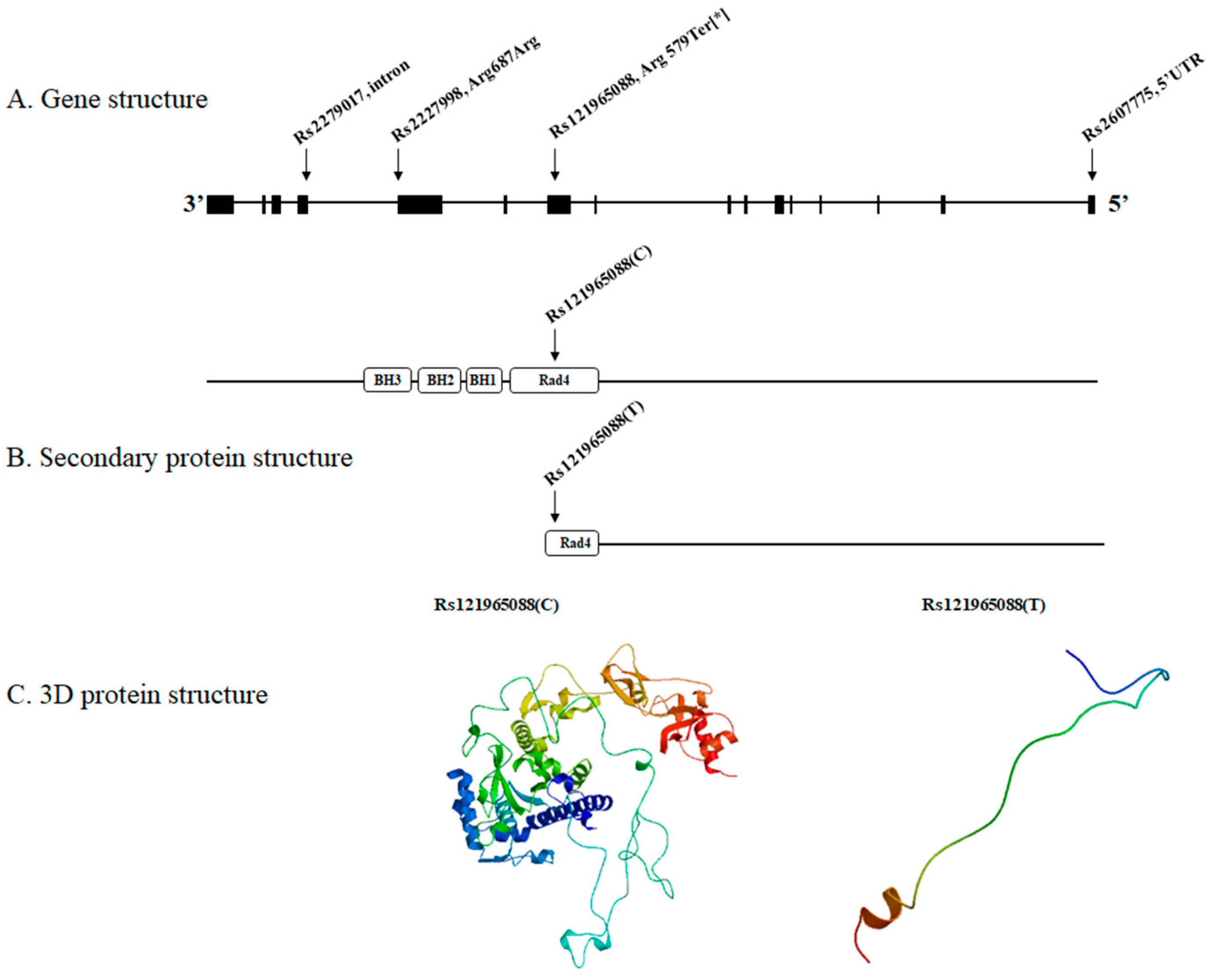
Figure 1. (A) The four XPC complex subunit, DNA damage recognition and repair factor (XPC) variants (rs121965088, rs2227998, rs2279017 and rs2607775) of our patient located on the XPC gene. (B) The normal XPC protein is constructed by the consecutive translation of Rad4, BH1, BH2, and BH3 protein units. However, rs121965088 (*) generates a stop codon and results in premature termination during the Rad4 translation. (C) Consequently, a defective and misfolded XPC protein is produced. The resultant protein architecture is displayed by the protein 3D modeling program SWISS-MODEL.
Medicina 59 00699 g001
Table 1. Eight biallelic variants identified in the patient and her family members.
Geners IDChromosome
Number
PatientBrotherFatherMotherProtein ResidueEffect on Amino AcidPrevious Study
XPCrs2279017Chr 31/10/10/10/1IntronNo effect[20,21,22,23,24,25]
XPCrs2227998Chr 31/10/10/10/1Arg687ArgSynonymous[21,26,27,28]
XPCrs121965088Chr 31/10/10/10/1Arg579TerNon-synonymous[14,29]
XPCrs2607775Chr 31/10/10/10/15′UTRNo effect[21,30,31,32,33,34]
OR2T35rs72452004Chr 11/10/10/10/1Cys203TerNon-synonymous
AFF3rs202089462Chr 21/10/10/10/1Thr619SerNon-synonymous
TARPrs138027161Chr 71/10/10/10/1Arg100GlyNon-synonymous
ANXA7rs3750575Chr 101/10/10/10/1Arg419GlnNon-synonymous[35]
XPC complex subunit, DNA damage recognition and repair factor (XPC); olfactory receptor family 2 subfamily T member 35 (OR2T35); ALF transcription elongation factor 3 (AFF3); TCR gamma alternate reading frame protein (TARP); annexin A7 (ANXA7); untranslated region (UTR); 1/1, homozygous; 0/1, heterozygous.
Disclaimer/Publisher’s Note: The statements, opinions and data contained in all publications are solely those of the individual author(s) and contributor(s) and not of MDPI and/or the editor(s). MDPI and/or the editor(s) disclaim responsibility for any injury to people or property resulting from any ideas, methods, instructions or products referred to in the content.