Article

BRAF V600E Mutation of Non-Small Cell Lung Cancer in Korean Patients

1
School of Medicine, Pusan National University, Beomeori, Mulgeum-eop, Yangsan 50612, Republic of Korea
2
Department of Thoracic and Cardiovascular Surgery, Biomedical Research Institute, Pusan National University Hospital, Busan 49241, Republic of Korea
3
Department of Pathology, Biomedical Research Institute, Pusan National University Hospital, Busan 49241, Republic of Korea
4
Department of Internal Medicine, Biomedical Research Institute, Pusan National University Hospital, Busan 49241, Republic of Korea
5
Department of Radiology, Biomedical Research Institute, Yangsan Pusan National University Hospital, Busan 50612, Republic of Korea
6
Department of Pathology, Seegene Medical Center, Busan 48792, Republic of Korea
7
Department of Pathology, Biomedical Research Institute, Yangsan Pusan National University Hospital, Busan 50612, Republic of Korea
*
Author to whom correspondence should be addressed.
Medicina 2023, 59(6), 1085; https://doi.org/10.3390/medicina59061085
Submission received: 4 May 2023 / Revised: 28 May 2023 / Accepted: 30 May 2023 / Published: 4 June 2023

Abstract

:
Background and Objectives: BRAF mutational status in resected non-small cell lung cancer (NSCLC) in the Korean population is poorly understood. We explored BRAF (particularly BRAF V600E) mutational status among Korean patients with NSCLC. Materials and Methods: This study included 378 patients with resected primary NSCLC who were enrolled from January 2015 to December 2017. The authors obtained formalin-fixed paraffin-embedded (FFPE) tissue blocks and performed peptide nucleic acid (PNA)-clamping polymerase chain reaction (PCR) for detecting BRAF V600, real-time PCR for detecting BRAF V600E, and immunohistochemical analyses using the mutation-specific Ventana VE1 monoclonal antibody. For positive cases in any methods mentioned above, direct Sanger sequencing was additionally performed. Results: The PNA-clamping method revealed the BRAF V600 mutation in 5 (1.3%) of the 378 patients. Among these five patients, real-time PCR, direct Sanger sequencing detected BRAF V600E mutations in three (0.8%) patients. Thus, two cases showed differences in their PNA-clamping and the others. Direct Sanger sequencing of PNA-clamping PCR product was performed for two cases showing negative results on direct Sanger sequencing; both contained BRAF mutations other than V600E. All patients harboring BRAF mutations had adenocarcinomas, and all patients with V600E mutation exhibited minor micropapillary components. Conclusions: Despite the low incidence of the BRAF mutation among Korean patients with NSCLC, lung adenocarcinoma patients with micropapillary components should be prioritized in terms of BRAF mutation testing. Immunohistochemical staining using Ventana VE1 antibody may serve as a screening examination for BRAF V600E.

1. Introduction

The BRAF gene is responsible for encoding the V-Raf murine sarcoma viral homolog B (BRAF) kinase, which plays a crucial role in cellular signaling, survival and proliferation [1]. BRAF gene is located on chromosome arm 7q34 and is composed of 18 exons [2]. BRAF is associated with mitogen-activated protein kinase (MAPK) pathways including the rat sarcoma (RAS), rapidly accelerated fibrosarcoma (RAF), mitogen-activated protein/extracellular signal regulated kinase (MEK), extracellular signal-regulated kinase (ERK), and mitogen-activated protein kinase. Mutations in the BRAF gene lead to sustained activation of the MAPK pathway, causing it to become a potential oncogenic driver [1]. Almost 300 different BRAF mutations were discovered in melanoma, colorectal cancer, papillary thyroid carcinoma and non-small cell lung cancers (NSCLCs) [3,4]. In addition, BRAF mutations have been classified into three classes. Class I BRAF mutation is RAS-independent and has higher kinase activity even in a monomer state. Class I mutation occurs in the valine residue at amino acid position 600 of exon 15; thus, it includes V600 mutations. Class II BRAF mutation has an intermediate kinase activity but should form homodimers to be fully activated. Finally, Class III BRAF mutation has an impaired kinase activity that requires RAS activation. Class II and III mutations occur either in the glycine of the G loop in exon 11 or in the activation part in exon 15 [5,6]. According to Owsley et al., Class I BRAF mutations represented the majority (62.1%) of all BRAF -mutant cases (2.4% of all cancers) in 114,662 different tumor sequencing analyses [7].
Now, dabrafenib (BRAF inhibitor) and trametinib (MEK inhibitor) combination therapy is the preferred first-line therapy for the BRAF V600E-mutation-positive lung cancer according to the NCCN (National Comprehensive Cancer Network) guidelines. In the French AcSe program, four patients with V600 non-E mutated lung cancer treated with vemurafenib monotherapy had outcomes comparable to the activity of vemurafenib in the BRAF V600E mutation [8]. Consequently, a clinical trial targeting V600 non-E mutation in lung cancer, corresponding to Class I BRAF mutation, is ongoing to evaluate the activity of dabrafenib and trametinib (NCT04775095). However, Class II and III BRAF mutations are not considered to respond to approved BRAF inhibitors [6,8]. Therefore, the evaluation of BRAF V600 of exon 15 mutational status, beyond V600E, could become more important.
According to the NCCN guidelines, real-time polymerase chain reaction (PCR), Sanger sequencing, and next-generation sequencing (NGS) are the most commonly recommended methods for BRAF mutation examination and immunohistochemistry, with an anti-BRAF p. V600E-specific monoclonal antibody recommended only after extensive validation.
In this study, BRAF V600 mutation, particularly the BRAF V600E mutational status, was explored with real-time PCR, peptide nucleic acid (PNA)-mediated clamping PCR, direct Sanger sequencing, and immunohistochemistry, which are relatively more feasible to use than NGS. The clinical and pathologic characteristics of the BRAF V600E mutation in non-small cell lung cancers were also investigated.

2. Materials and Methods

2.1. Patients, Tissue Specimens, and DNA

This study was performed retrospectively. Three hundred and sixty-eight patients who underwent surgical resection for primary non-small cell lung cancer between 2015 and 2017 at Pusan National University Hospital were included. Among them, five patients had synchronous primary lung cancer. The final cohort was 378 cases of primary non-small cell lung cancers. Formalin-fixed paraffin-embedded (FFPE) tissue blocks, which were made at the time of diagnosis, were used. Clinicopathological data were retrieved from the electric medical records and pathologic reports. Genomic DNA was extracted from FFPE blocks using Maxwell 16 FFPE LEV DNA Purification (Promega corp).

2.2. PNA-Mediated Clamping PCR (PNA Clamping PCR)

PNA Clamp BRAF mutation detection kit (Seegene, Seoul, Korea) was used. Extracted DNA was mixed with a PNA probe, primers (5′-AAACTCTTCATAATGCTTGCTCTG (forward) and 5′-GGCCAAAAATTTAATCAGTGGA (reverse)). SYBR green PCR master mix and all reactions totaled 20 μL. Real-time PCR reaction was performed according to the manufacturer’s instructions using a CFX96 real-time PCR system (BioRad, Pleasanton, CA, USA). The PNA probe sequences were complementary to wild-type (V600). The PNA probe hybridizes to the wild-type BRAF sequence, inhibiting the amplification of the wild-type allele and enhancing preferential amplification of mutant sequences. The positive signal was detected by the intercalation of SYBR green fluorescent dye. The cycle threshold (CT) value was automatically calculated. The delta (ΔCT) value was calculated by subtracting the CT value of a test sample from the standard CT value of a control sample (ΔCT = Standard CT − Sample CT). The cutoff for the presence of mutant was ΔCT of 2. BRAF V600 PNA clamping PCR was performed in all 378 cases of non-small cell carcinoma.

2.3. Real-Time PCR

The real-time PCR used the Real-Q BRAF V600E detection kit (Real-Q; Biosewoom, Seoul, Republic of Korea). Real-time PCR was performed with CFX96 real-time PCR Detection system (Bio-Rad) according to the manufacture’s instruction. The master mixture contained 12.5 μL of the 2X PCR reaction mixture and 2.5 μL of the BRAF probe and primer mixture. A total of 15 μL of the master mixture was dispensed into PCR tubes. Then, the extracted DNA of 10 μL (containing 50 ng of DNA) was added to each PCR tube. The sample was considered positive for V600E mutation when both the sample and the internal control were amplified and both CT value of the sample and the internal control were less than 40. If a sample showing the difference between CT value of the sample and the internal control was more than 13, the test was repeated. BRAF V600E real-time PCR was performed in all 378 cases of non-small cell carcinoma.

2.4. Immunohistochemistry

Immunohistochemistry was performed on the same FFPE block used for molecular testing. An automatic staining device (BenchMark XT, Ventana Medical Systems, Tucson, AZ, USA) was used for staining. All samples were cut into 3 μm thick sections and the sections were deparaffinized in an EZ prep. The slides were pretreated with CC1 (cell conditioner 1, pH8.4 buffer) for 64 min antigen retrieval and followed by pre-primary antibody peroxidase inhibition. Then, the slides were incubated with the Ventana BRAF V600E (VE1) mouse monoclonal primary antibody, and Hematoxylin II ® and Bluing Reagent was used for counterstaining. A sample known to have V600E mutation was used as a positive control. A case was considered to be positive when a signal was present in the cytoplasm [9]. Any nuclear staining was ignored.

2.5. Direct Sanger Sequencing

BRAF exon 15, which potentially contains the c.1799 T > A transversion mutation, was amplified from genomic DNA by PCR using primers 5′-AAACTCTTCATAATGCTTGCTCTG (forward) and 5′-GGCCAAAAATTTAATCAGTGGA (reverse). Amplification was performed under the following conditions: 1 cycle at 94 °C for 5 min, 40 cycles of denaturation at 94 °C for 30 s, annealing at 63 °C for 30 s, and extension at 72 °C for 30 s; then a final extension at 72°C for 5 min using BioRad C1000 (Pleasanton, CA). After purification of the PCR products, direct bidirectional sequencing was performed using the ABI 3730XL DNA Analyzer. Additionally, direct bidirectional sequencing was repeated using the BRAF PNA clamping PCR product, which is rich in mutant alleles, to detect the variants of low level. Direct Sanger sequencing using extracted DNA from FFPE blocks was performed in 5 cases of any positive results for BRAF V600 PNA clamping, BRAF V600E real-time PCR and immunohistochemistry for VE1. Particularly, direct Sanger sequencing using PNA clamping PCR product was conducted in cases of discordance in other methods.

3. Results

3.1. Clinicopathologic Characteristics of Resected Non-Small Cell Lung Cancers

A total cohort of 378 patients with resected non-small cell carcinoma was included in this study. All included patients were Korean. Basic data for included patients are shown in Table 1. Patient age ranged from 36 to 86 years (mean: 66.84 ± 8.76 years). The size of the cancer ranged from 0.9 cm to 10.0 cm (mean: 3.37 ± 1.55 cm). There were 238 males (63.0%) and 140 females (37.0%). The study cohort included 255 cases of adenocarcinoma (67.5%), 91 cases of squamous cell carcinoma (24.1%), 5 cases of adenosquamous cell carcinoma (1.3%), 9 cases of large cell neuroendocrine carcinoma (2.4%), 15 cases of sarcomatoid carcinoma (4%) and others (3 cases, 0.8%). Three hundred and five patients (80.7%) had early-stage disease (stage I and II) and the remaining 73 patients (19.3%) had advanced disease (stage III and IV). Among the patients, 168 (44.4%) patients never smoked, 112 patients (29.6%) were ex-smokers and the other 98 patients (25.9%) were current smokers. Information about an EGFR-activating mutation and ALK fluorescence in situ hybridization (FISH)/ALK D5F3 CDx Ventana immunohistochemistry was retrieved from the prior pathologic reports in electronic medical records. One hundred and twenty patients (31.7%) had EGFR-activating mutations and 11 patients (2.9%) had ALK translocation.

3.2. BRAF V600 PNA Clamping and BRAF V600E Real-Time PCR

BRAF V600 mutation was detected in five cases (1.3%) using a PNA clamping method among 378 non-small cell carcinoma cases (Table 2 and Table 3). By using BRAF real-time PCR, a BRAF V600E mutation was detected in 3 patients (0.8%) among the total 378 cohort (Table 2 and Table 4), and all these positive cases for real-time PCR had positive results in PNA clamping PCR. There were two discordant cases between PNA clamping and real-time PCR.

3.3. Immunohistochemistry for VE1

Immunohistochemistry for VE1 was performed in all included patients with full-face sections of FFPE blocks. Regarding the results of immunohistochemistry, three patients (0.8%) showed positive staining for tumor cytoplasm (Figure 1, Table 2 and Table 5). All three cases with positive staining showed diffuse positivity for tumor cells. However, two cases had heterogenous staining intensity, though all tumor cells were positive. The other case had diffuse positivity with homogeneous intensity for tumor cytoplasm. The detailed information of staining is shown in Table 5. However, two patients, with positive results for PNA clamping, had negative immunostaining. Regarding the results of immunohistochemistry for VE1, there were two cases showing discordance with the PNA clamping method, and there was no discordant case with real-time PCR.

3.4. Direct Sequencing

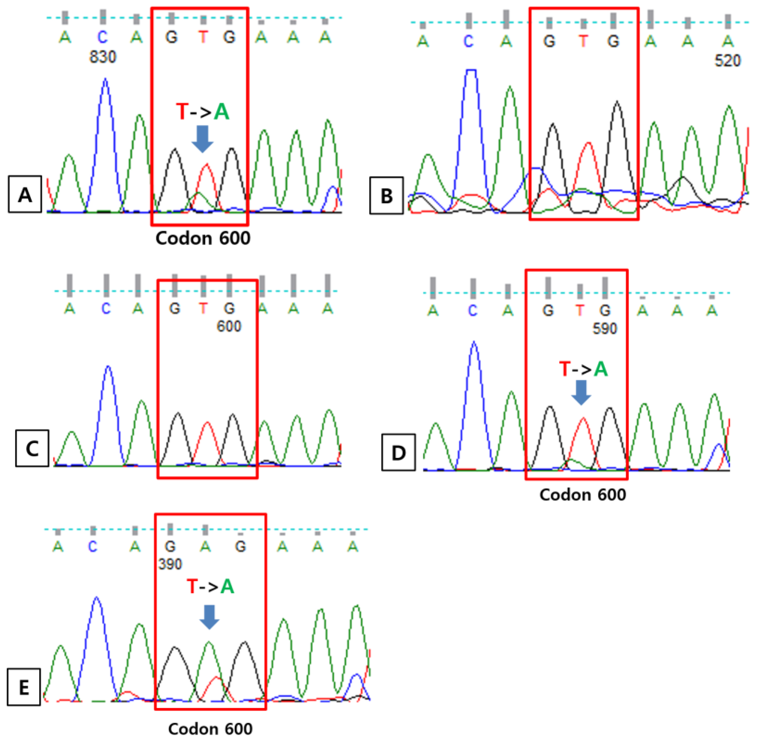
There were five patients (1.3%) who had positive results for BRAF PNA clamping, real-time PCR and immunohistochemistry. For these five patients, direct Sanger sequencing was performed. The results of Sanger sequencing were the same with those of real-time PCR and immunohistochemistry (Figure 2). Considering the PNA clamping method is a very sensitive method to detect a low allele level of mutation, direct sequencing using a clamping PCR product was performed [10]. Regarding the results of sequencing using a clamping PCR product, all cases showed mutation for the BRAF gene other than the V600E genotype (Figure 3). Finally, there were five mutated cases (1.3%) for BRAF in the total cohort. Among them, three cases (0.8%) had a V600E mutation and the other two had V600K and V600V/V601E mutations, respectively. Among the total number of BRAF mutations, V600E genotype was present in three cases, comprising 60% of the BRAF mutant.

3.5. Clinicopathologic Aspects of BRAF Mutation in Lung Cancers

There were three cases (0.8%) of the BRAF V600E mutation among 378 non-small cell carcinomas. It was 1.2% among 255 adenocarcinoma and 246 non-small cell carcinomas without EGFR/ALK aberrations. In addition, it was 2.3% among 129 adenocarcinomas without EGFR/ALK aberrations. There were two cases of BRAF mutation other than V600E, comprising 0.5% of all non-small cell carcinoma, 0.8% of adenocarcinoma and non-small cell carcinoma without EGFR/ALK aberrations and 1.5% of adenocarcinoma without EGFR/ALK aberrations (Table 6).
Among the V600E mutated patients, one patient was a never-smoker and the other two were ever-smokers. There was a micropapillary component in all the V600E-mutated cases (Table 7). All patients harboring a BRAF mutation had no concomitant EGFR or ALK aberrations. Detailed clinicopathologic characteristics of individual patients with a BRAF mutation are listed in Table 7.

4. Discussion

In this study, the BRAF V600 mutation incidence was found in five patients (1.3%) among all cases of non-small cell carcinoma, and the BRAF V600E mutation was present in three patients (0.8%) with adenocarcinoma. It is relatively low when compared to most reports from the Western population [11,12,13,14]. On the other hand, the incidence is similar to that of Japanese patients [15]. In this study, there were 2.3% and 1.5% of the BRAF V600E mutation and BRAF V600 non-E mutation, respectively, among adenocarcinoma without EGFR/ALK alterations. According to one Korean dataset, there were four patients (1.8%) with a BRAF mutation among 222 Stage III/IV lung adenocarcinoma patients without EGFR/ALK aberrations [16]. The difference from these data probably resulted from the difference in stage distribution of the study cohort. This study included 305 cases (80.7%) of early-stage (Stage I and II) disease, contrary to their advanced stage cohort. This study included 123 cases of non-adenocarcinoma patients, and none of these patients harbored a BRAF mutation. However, other data reported the detection of a BRAF mutation in non-adenocarcinoma patients, though the incidence was very low [11,13,14,15,17]. Among five BRAF V600-mutated lung cancer patients, two patients (40%) were never-smokers and three (60%) were ever-smokers. This is in accordance with the molecular testing guideline for the selection of patients with lung cancer for treatment, suggesting that BRAF mutational testing should be performed on all advanced adenocarcinoma patients, irrespective of clinical characteristics [18].
BRAF V600 non-E mutation was present in two cases. However, the result of direct sequencing showed the wild type of BRAF using amplified DNA extracted from FFPE blocks following sequencing using PNA clamping product detected mutation. Through PNA clamping PCR, the wild-type alleles are inhibited in the amplification process by hybridization with PNA, resulting in mutant enrichment. Though detected mutation was not the V600E genotype in this study, this result suggests that sequencing using the PNA clamping PCR product can help the detection of a mutant of low level in suspected or equivocal cases. In addition, Zengarini et al. presented some treatment effects on BRAF V600K mutated melanoma patients [19], and there is also an ongoing phase 2 clinical trial on the application of dabrafenib and trametinib in tumors with the BRAF V600E or V600K mutation including non-small cell lung cancer (ClinicalTrials.gov Identifier: NCT04439292). Additionally, BRAF V600E real-time PCR showed both 100% of sensitivity and specificity. All these results are in accordance with the principles of molecular and biomarker analysis for BRAF by NCCN guideline: “Real-time PCR, Sanger sequencing (ideally paired with tumor enrichment), and NGS are the most commonly deployed methodologies for examining BRAF mutation status” (NCCN Guidelines Version 3.2023).
All patients harboring the BRAF V600 mutation had adenocarcinoma and all patients with the V600E genotype had a micropapillary component. The result is similar to those of prior reports [9,20,21]. Theis may suggest that lung adenocarcinoma with micropapillary should be first considered to conduct BRAF mutation testing.
The VE1 mouse monoclonal antibody was utilized in this study. VE1 antibody is a mutation-specific antibody able to differentiate a V600E-mutated protein from wild-type and other BRAF-mutated proteins [22]. In this study, immunohistochemistry for VE1 showed both 100% of sensitivity and specificity. Gow et al. validated the usefulness of the Ventana VE1 antibody in lung cancer [20]. They reported that immunohistochemistry for VE1 antibody showed a 96.6% sensitivity to detect the BRAF V600E mutation and a 98.6% specificity to discriminate tumors without the BRAF V600E mutation. However, one positive case affecting the specificity value had weak positive cytoplasmic staining in 5% of tumor cells and the case had ROS1 gene fusion. According to their criteria, the case was considered to be negative. Ilie et al. reported that VE1 immunohistochemistry is specific and sensitive to detect the BRAF V600E mutation [9]. Similar results were shown by Hofman et al., suggesting that VE1 staining is a rapid, specific and very sensitive method [23]. In addition, Chang et al. reported that VE1 immunohistochemistry showed almost perfect interobserver agreement, suggesting that this could be a screening test for BRAF mutation [24]. In present study, BRAF V600E mutated cases showed diffuse (100% of proportion) positivity, though the intensity was heterogeneous in two cases. Overall, these results suggest that immunohistochemistry for the Ventana VE1 antibody can be a useful screening tool in lung cancers, especially for small biopsy specimens, which must be handled with care to obtain the maximum information for treatment choice. Moreover, immunohistochemistry has many advantages over molecular diagnostics, namely because it needs much less tissue and the turn-around time is far shorter.
There are limitations in this study. The detection rate of BRAF mutation was only 1.3% of the study cohort, so statistical analyses could not be performed. In addition, these data are from one single institution, which makes it difficult to generalize these findings. However, this study cohort was composed of only the Korean population, and for all experiments, we only used consecutive resected samples of primary lung cancer.

5. Conclusions

BRAF V600 mutation status in resected primary non-small cell carcinoma was tested. There were five cases (1.3%) of a BRAF V600 mutation among 378 non-small cell carcinomas, comprising three cases of a BRAF V600E mutation and two cases of a BRAF V600 non-E mutation. All cases harboring a BRAF V600 mutation were adenocarcinoma without EGFR mutation and ALK translocation. All three cases of a BRAF V600E mutation had micropapillary component. Immunohistochemistry for Ventana VE1 antibody can be a useful screening method to detect a BRAF V600E mutation.
This study preliminarily suggests that the incidence of a BRAF V600E mutation might be low in Korean population. In addition, adenocarcinoma showing micropapillary component, especially without EGFR/ALK aberration, should be first considered for BRAF testing, including immunohistochemistry.

Author Contributions

Data curation, J.S.C., J.L., S.J.L. and D.H.S.; Formal analysis, Y.J.J. and Y.D.K.; Methodology, C.H.L., M.K.L. and J.S.E.; Writing—original draft, H.Y.A. and A.K.; Writing—review and editing, A.K. All authors have read and agreed to the published version of the manuscript.

Funding

This work was supported by a 2-Year Research Grant of Pusan National University.

Institutional Review Board Statement

The study was conducted in accordance with the Declaration of Helsinki, and approved by the Institutional Review Board of Pusan National Universitiy Hospital (IRB No 2107-011-105), approval date 28 July 2017.

Informed Consent Statement

Patient consent was waived because the research involves no more than minimal risk and waiver of informed consent will not adversely affect the rights and welfare of the subjects.

Data Availability Statement

Not applicable.

Acknowledgments

We send special thanks to Myoungsuk Jo and Jungeun Ko who provided technical support. This research was partly presented in AACR 2020 (abstract 2343).

Conflicts of Interest

The authors declare no conflict of interest.

References

  1. Yan, N.; Guo, S.; Zhang, H.; Zhang, Z.; Shen, S.; Li, X. BRAF-Mutated Non-Small Cell Lung Cancer: Current Treatment Status and Future Perspective. Front. Oncol. 2022, 12, 863043. [Google Scholar] [CrossRef] [PubMed]
  2. Raman, M.; Chen, W.; Cobb, M.H. Differential regulation and properties of MAPKs. Oncogene 2007, 26, 3100–3112. [Google Scholar] [CrossRef] [PubMed] [Green Version]
  3. Forbes, S.A.; Bindal, N.; Bamford, S.; Cole, C.; Kok, C.Y.; Beare, D.; Jia, M.; Shepherd, R.; Leung, K.; Menzies, A.; et al. COSMIC: Mining complete cancer genomes in the Catalogue of Somatic Mutations in Cancer. Nucleic Acids Res. 2011, 39, D945–D950. [Google Scholar] [CrossRef] [Green Version]
  4. Tabbò, F.; Pisano, C.; Mazieres, J.; Mezquita, L.; Nadal, E.; Planchard, D.; Pradines, A.; Santamaria, D.; Swalduz, A.; Ambrogio, C.; et al. How far we have come targeting BRAF-mutant non-small cell lung cancer (NSCLC). Cancer Treat. Rev. 2022, 103, 102335. [Google Scholar] [CrossRef] [PubMed]
  5. Leonetti, A.; Facchinetti, F.; Rossi, G.; Minari, R.; Conti, A.; Friboulet, L.; Tiseo, M.; Planchard, D. BRAF in non-small cell lung cancer (NSCLC): Pickaxing another brick in the wall. Cancer Treat. Rev. 2018, 66, 82–94. [Google Scholar] [CrossRef]
  6. Riudavets, M.; Cascetta, P.; Planchard, D. Targeting BRAF-mutant non-small cell lung cancer: Current status and future directions. Lung Cancer 2022, 169, 102–114. [Google Scholar] [CrossRef] [PubMed]
  7. Owsley, J.; Stein, M.K.; Porter, J.; In, G.K.; Salem, M.; O’Day, S.; Elliott, A.; Poorman, K.; Gibney, G.; VanderWalde, A. Prevalence of class I-III BRAF mutations among 114,662 cancer patients in a large genomic database. Exp. Biol. Med. (Maywood) 2021, 246, 31–39. [Google Scholar] [CrossRef]
  8. Mazieres, J.; Cropet, C.; Montané, L.; Barlesi, F.; Souquet, P.J.; Quantin, X.; Dubos-Arvis, C.; Otto, J.; Favier, L.; Avrillon, V.; et al. Vemurafenib in non-small-cell lung cancer patients with BRAF. Ann. Oncol. 2020, 31, 289–294. [Google Scholar] [CrossRef] [Green Version]
  9. Ilie, M.; Long, E.; Hofman, V.; Dadone, B.; Marquette, C.H.; Mouroux, J.; Vignaud, J.M.; Begueret, H.; Merlio, J.P.; Capper, D.; et al. Diagnostic value of immunohistochemistry for the detection of the BRAFV600E mutation in primary lung adenocarcinoma Caucasian patients. Ann. Oncol. 2013, 24, 742–748. [Google Scholar] [CrossRef]
  10. Kang, S.H.; Pyo, J.Y.; Yang, S.W.; Hong, S.W. Detection of BRAF V600E mutation with thyroid tissue using pyrosequencing: Comparison with PNA-clamping and real-time PCR. Am. J. Clin. Pathol. 2013, 139, 759–764. [Google Scholar] [CrossRef] [Green Version]
  11. Luk, P.P.; Yu, B.; Ng, C.C.; Mercorella, B.; Selinger, C.; Lum, T.; Kao, S.; O’Toole, S.A.; Cooper, W.A. BRAF mutations in non-small cell lung cancer. Transl. Lung Cancer Res. 2015, 4, 142–148. [Google Scholar] [CrossRef] [PubMed]
  12. Paik, P.K.; Arcila, M.E.; Fara, M.; Sima, C.S.; Miller, V.A.; Kris, M.G.; Ladanyi, M.; Riely, G.J. Clinical characteristics of patients with lung adenocarcinomas harboring BRAF mutations. J. Clin. Oncol. 2011, 29, 2046–2051. [Google Scholar] [CrossRef] [PubMed] [Green Version]
  13. Marchetti, A.; Felicioni, L.; Malatesta, S.; Grazia Sciarrotta, M.; Guetti, L.; Chella, A.; Viola, P.; Pullara, C.; Mucilli, F.; Buttitta, F. Clinical features and outcome of patients with non-small-cell lung cancer harboring BRAF mutations. J. Clin. Oncol. 2011, 29, 3574–3579. [Google Scholar] [CrossRef] [PubMed]
  14. Tissot, C.; Couraud, S.; Tanguy, R.; Bringuier, P.P.; Girard, N.; Souquet, P.J. Clinical characteristics and outcome of patients with lung cancer harboring BRAF mutations. Lung Cancer 2016, 91, 23–28. [Google Scholar] [CrossRef] [PubMed]
  15. Kinno, T.; Tsuta, K.; Shiraishi, K.; Mizukami, T.; Suzuki, M.; Yoshida, A.; Suzuki, K.; Asamura, H.; Furuta, K.; Kohno, T.; et al. Clinicopathological features of nonsmall cell lung carcinomas with BRAF mutations. Ann. Oncol. 2014, 25, 138–142. [Google Scholar] [CrossRef]
  16. Kim, H.C.; Kang, Y.R.; Ji, W.; Kim, Y.J.; Yoon, S.; Lee, J.C.; Choi, C.M. Frequency and clinical features of BRAF mutations among patients with stage III/IV lung adenocarcinoma without EGFR/ALK aberrations. Onco Targets Ther. 2019, 12, 6045–6052. [Google Scholar] [CrossRef] [Green Version]
  17. Brustugun, O.T.; Khattak, A.M.; Trømborg, A.K.; Beigi, M.; Beiske, K.; Lund-Iversen, M.; Helland, Å. BRAF-mutations in non-small cell lung cancer. Lung Cancer 2014, 84, 36–38. [Google Scholar] [CrossRef]
  18. Kalemkerian, G.P.; Narula, N.; Kennedy, E.B.; Biermann, W.A.; Donington, J.; Leighl, N.B.; Lew, M.; Pantelas, J.; Ramalingam, S.S.; Reck, M.; et al. Molecular Testing Guideline for the Selection of Patients With Lung Cancer for Treatment With Targeted Tyrosine Kinase Inhibitors: American Society of Clinical Oncology Endorsement of the College of American Pathologists/International Association for the Study of Lung Cancer/Association for Molecular Pathology Clinical Practice Guideline Update. J. Clin. Oncol. 2018, 36, 911–919. [Google Scholar] [CrossRef]
  19. Zengarini, C.; Mussi, M.; Veronesi, G.; Alessandrini, A.; Lambertini, M.; Dika, E. BRAF V600K vs. BRAF V600E: A comparison of clinical and dermoscopic characteristics and response to immunotherapies and targeted therapies. Clin. Exp. Dermatol. 2022, 47, 1131–1136. [Google Scholar] [CrossRef]
  20. Gow, C.H.; Hsieh, M.S.; Lin, Y.T.; Liu, Y.N.; Shih, J.Y. Validation of Immunohistochemistry for the Detection of. Cancers 2019, 11, 866. [Google Scholar] [CrossRef] [Green Version]
  21. Hwang, I.; Choi, Y.L.; Lee, H.; Hwang, S.; Lee, B.; Yang, H.; Chelakkot, C.; Han, J. Selection Strategies and Practical Application of BRAF V600E-Mutated Non-Small Cell Lung Carcinoma. Cancer Res. Treat. 2022, 54, 782–792. [Google Scholar] [CrossRef] [PubMed]
  22. Capper, D.; Preusser, M.; Habel, A.; Sahm, F.; Ackermann, U.; Schindler, G.; Pusch, S.; Mechtersheimer, G.; Zentgraf, H.; von Deimling, A. Assessment of BRAF V600E mutation status by immunohistochemistry with a mutation-specific monoclonal antibody. Acta Neuropathol. 2011, 122, 11–19. [Google Scholar] [CrossRef] [PubMed]
  23. Hofman, V.; Benzaquen, J.; Heeke, S.; Lassalle, S.; Poudenx, M.; Long, E.; Lantéri, E.; Bordone, O.; Lespinet, V.; Tanga, V.; et al. Real-world assessment of the BRAF status in non-squamous cell lung carcinoma using VE1 immunohistochemistry: A single laboratory experience (LPCE, Nice, France). Lung Cancer 2020, 145, 58–62. [Google Scholar] [CrossRef] [PubMed]
  24. Chang, S.; Choi, Y.L.; Shim, H.S.; Lee, G.K.; Ha, S.Y.; Group, K.C.P.S. Usefulness of BRAF VE1 immunohistochemistry in non-small cell lung cancers: A multi-institutional study by 15 pathologists in Korea. J. Pathol. Transl. Med. 2022, 56, 334–341. [Google Scholar] [CrossRef] [PubMed]
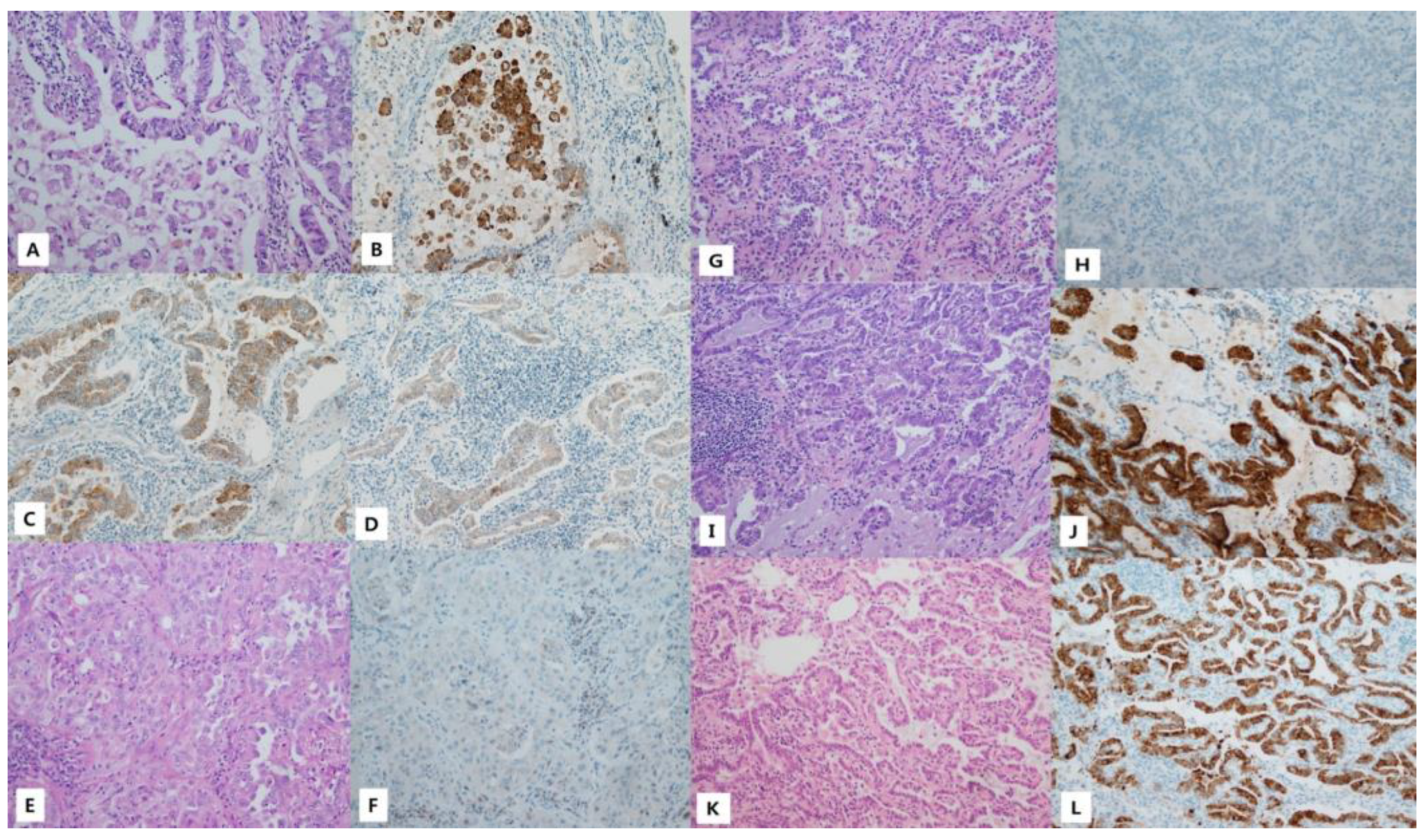
Figure 1. Histologic feature and immunohistochemistry. (AD). Patient 142; all ×200. (A) A Hematoxylin and eosin (H&E)-stained section showing acinar and micropapillary structures. (B) Immunohistochemistry for VE1 (staining intensity 3). (C) Immunohistochemistry for VE1 (staining intensity 2). (D) Immunohistochemistry for VE1 (staining intensity 1). (E,F) Patient number 270; all ×200. (E) An H&E-stained section showing solid and acinar growth patterns. (F) Negative immunostaining for VE1. (G,H) Patient number 324; all ×200. (G) An H&E-stained section showing an acinar growth pattern. (H) Negative immunostaining for VE1. (I,J) Patient number 348; all ×200. (I) An H&E-stained section showing an acinar pattern and a few micropapillary structures. (J) Immunohistochemistry for VE1 (staining intensity 3). (K,L) Patient number 358; all ×200. (K) An H&E-stained section showing an acinar pattern and a few micropapillary structures. (L) Immunohistochemistry for VE1 (staining intensity 2).
Medicina 59 01085 g001
Figure 2. Results of direct Sanger sequencing. (A) Case number 142 carries the c.1799 T > A (p. V600E) mutation. (B) Case number 270 is wild-type for B-type Raf kinase (BRAF). (C) Case number 324 is wild-type for BRAF. (D) Case number 348 carries the c.1799 T > A (p. V600E) mutation. (E) Case number 358 carries the c.1799 T > A (p. V600E) mutation.
Medicina 59 01085 g002
Figure 3. Direct sequencing of peptide nucleic acid (PNA) polymerase chain reaction (PCR) products reveals two p. V600E-negative cases. (A) Case number 270 carries the c.1798_1799 CA > TT (p. V600K) for B-type Raf kinase (BRAF) mutation. (B) Case number 324 carries the c.1800 C > T (p. V600V) and c.1801 T > C (p. K601E) BRAF mutations.
Medicina 59 01085 g003
Table 1. Clinical characteristics of study population.
CharacteristicsNumber (%)
Age (years)66.84 ± 8.76
Sex
Male238 (63.0)
Female140 (37.0)
Smoking status
Never-smoker168 (44.4)
Current smoker98 (25.9)
Ex-smoker112 (29.6)
Pack-years among ever-smokers (years) *34.68 ± 19.81
Tumor size (cm)3.37 ± 1.55
Histologic type
ADC255 (67.5)
SqCC91 (24.1)
SC15 (4.0)
LCNEC9 (2.4)
ADSqCC5 (1.3)
Other3 (0.8)
Differentiation
WD19 (5.0)
MD269 (71.2)
PD90 (23.8)
Stage
Early (I–II)305 (80.7)
Advanced (III–IV)73 (19.3)
EGFR mutation
Absent258 (68.3)
Present120 (31.7)
ALK translocation
Absent367 (97.1)
Present11 (2.9)
Ethnicity Korean378 (100.0)
* Among the ever-smokers, pack-year data were unavailable in six cases. ADC, Adenocarcinoma; SqCC, Squamous cell carcinoma; SC, sarcomatoid carcinoma; LCNEC, Large-cell neuroendocrine carcinoma; ADSqCC, Adenocasquamous cell carcinoma WD, Well-differentiated; MD, Moderately differentiated; PD, Poorly differentiated; EGFR, epidermal growth factor receptor; ALK, anaplastic lymphoma kinase.
Table 2. The results of BRAF mutation according to BRAF mutation assay.
Case NumberBRAF V600 PNA ClampingBRAF V600E Real-Time PCRIHC for VE1Direct Sanger SequencingDirect Sanger Sequencing
of the Clamping PCR Product
142+++V600ENot done
270+WTV600K
324+WTV600V, K601E
348+++V600ENot done
358+++V600ENot done
BRAF, B-type Raf kinase; IHC, immunohistochemistry; PNA, peptide nucleic acid; PCR, polymerase chain reaction; WT, Wild-type; +, positive; −, negative.
Table 3. The results of BRAF V600 on PNA-clamping.
Case
Number
DNA Loading (ng)Cycle Threshold (CT)ΔCT-2ΔCT-1
Non-PNAV600V600V600
Clamping Control24.3836.3311.95−1.33
Positive Control24.03305.975
1421028.5931.73.113.3
2701026.9132.445.532.56
3241025.9831.825.843.18
3481026.0230.34.284.7
3581026.2131.465.263.54
1422527.1531.133.973.87
2702526.3132.426.12.58
3242525.1531.896.743.11
3482525.2229.64.385.4
3582525.2631.025.763.98
BRAF, B-type Raf kinase; PNA, peptide nucleic acid, PC, positive control, ΔCT = standard CT—sample CT.
Table 4. The results of BRAF V600E on real-time PCR.
Patient NumberInternal Control CTSample CTResult
14225.931.9+
27024.6NA
32426.6NA
34824.729.6+
35824.430.6+
BRAF, B-type Raf kinase; CT, cycle threshold; NA, not applicable; +, positive; −, negative.
Table 5. Immunohistochemistry results of patients who were V600E-positive on either or both BRAF PNA-clamping and BRAF real-time PCR.
Case NumberProportion
of Cytoplasm
Positive Rate
Intensity PatternIntensity Scores
of Tumor Cells
Result
142100Heterogeneous3+: 40%
2+: 50%
1+: 10%
0: 0%
+
2700Homogeneous 3+: 0%
2+: 0%
1+: 0%
0: 100%
3240Homogeneous 3+: 0%
2+: 0%
1+: 0%
0: 100%
348100Homogeneous3+: 100%
2+: 0%
1+: 0%
0: 0%
+
358100Heterogeneous3+:70%
2+:30%
1+: 0%
0: 0%
+
BRAF, B-type Raf kinase; +, positive; −, negative.
Table 6. Incidence of BRAF mutations.
BRAF
Mutation
BRAF
V600E Mutation
BRAF
Non-V600E Mutation
N (%)N (%)N (%)
NSCLC (n = 378)5 (1.3)3 (0.8)2 (0.5)
Adenocarcinomas (n = 255)5 (2.0)3 (1.2)2 (0.8)
NSCLC lacking EGFR mutation and ALK translocation (n = 246)5 (2.0)3 (1.2)2 (0.8)
Adenocarcinomas lacking EGFR mutation and ALK translocation (n = 129)5 (3.8)3 (2.3)2 (1.5)
Table 7. Clinicopathological characteristics of individual patients with BRAF mutations.
Case No.BRAF MutationAge (Years)SexSmoking
Status
Pack YearsPredominant Histological SubtypePresent of Micropapillary ComponentpT
Size (cm)
pN StagepM StageStage
142V600E72MaleCurrent50Solid+6.520IIIA
270V600K83MaleCurrent15Acinar-3.600IB
324V600V, K601E67FemaleNever0Acinar-1.600IA
348V600E51FemaleNever0Acinar+2.200IA
358V600E52MaleEx-smoker7.5Acinar+1.800IA
Disclaimer/Publisher’s Note: The statements, opinions and data contained in all publications are solely those of the individual author(s) and contributor(s) and not of MDPI and/or the editor(s). MDPI and/or the editor(s) disclaim responsibility for any injury to people or property resulting from any ideas, methods, instructions or products referred to in the content.