Systematic Review

An Effectiveness Evaluation of Nucleo-Annuloplasty for Lumbar Discogenic Lesions Using Disc-FX: A Scoping Review

1
Department of Orthopedics, The First Affiliated Hospital of Xiamen University, School of Medicine, Xiamen University, Xiamen 361000, China
2
The School of Clinical Medicine, Fujian Medical University, Fuzhou 350122, China
3
Division of Neurosurgery, Department of Surgery, Changhua Christian Hospital, Changhua 500209, Taiwan
4
Department of Leisure Industry Management, National Chin-Yi University of Technology, Taichung 41170, Taiwan
*
Author to whom correspondence should be addressed.
Medicina 2023, 59(7), 1291; https://doi.org/10.3390/medicina59071291
Submission received: 9 May 2023 / Revised: 20 May 2023 / Accepted: 30 June 2023 / Published: 13 July 2023
(This article belongs to the Special Issue Recent Advances in Endoscopic Spine Surgery)

Abstract

:
Background and Objectives: Degenerative disk disease is a widespread chronic condition that causes diskogenic pain. Diskogenic pain can be treated with various therapy methods. Disc-FX is a revolutionary, minimally invasive, percutaneous nucleo-annuloplasty method that combines manual diskectomy with nuclear and annular remodeling using radiofrequency ablation to relieve diskogenic pain. In this study, the technical features, clinical outcomes, and complications of Disc-FX are summarized. Materials and Methods: A comprehensive literature review was performed. By exploring several databases, we collected studies on Disc-FX for treating diskogenic pain. The outcomes included perioperative data, clinical results, and complications. Results: In the 15 studies included, data from 570 patients were collected. L4–L5 was the most frequently operated level, and most cases underwent single-level procedures. The follow-up period for these patients ranged from 2 months to 24 months. One study reported a procedure time between 35 and 60 min, whereas the remaining studies reported a procedure time of less than 30 min. The mean visual analog scale score decreased from 7.22 preoperatively to 1.81 at the final follow-up. The mean numerical rating scale score decreased from 6.98 preoperatively to 3.9 at the final follow-up. The mean Japanese Orthopaedic Association score improved from 16.26 preoperatively to 25.88 in the final follow-up. The mean Oswestry Disability Index score decreased from 35.37 preoperatively to 14.66 at the final follow-up. The mean satisfaction rate (based on the Macnab criteria) was 87.6% (range, 78.4–95.2%). The total incidence of postoperative transient pain was 8.77% (50/570) after nucleo-annuloplasty using Disc-FX, and recurrence was 1.58% (9/570). Conclusions: According to our comprehensive evaluation, using percutaneous nucleo-annuloplasty for treating lumbar diskogenic diseases provided considerable pain alleviation and improved functional outcomes with fewer complications. Disc-FX is a safe and effective procedure that is a good treatment option for patients with diskogenic pain.

1. Introduction

In the past, individuals with degenerative disk disease or lumbar disk herniation resulting in diskogenic pain were treated conservatively with rest, medications, injections, and/or physical therapy [1,2,3]. Unfortunately, these treatments have not always been effective. Those for whom previous conservative treatments have not been effective are forced to live with their symptoms or consider spine surgery [4]. If these patients undergo traditional spinal decompression surgery, they may require weeks or months of recuperation, which will have a greater impact on their daily lives and work.
Minimally invasive approaches for treating degenerative disk diseases have several advantages, including greater preservation of the spine architecture, less tissue loss, and decreased risk [5,6]. In recent years, it has been demonstrated that spinal degeneration is not an isolated process, but is closely related to disc degeneration, endplate degeneration, facet joint degeneration, and fatty infiltration/atrophy of the paraspinal muscles. Therefore, the use of a minimally invasive approach is not just for the protection of one structure, but for the long-term protection of all structures [7,8]. However, controversy exists regarding the surgical indications for diskogenic pain. There may be no need for traditional microsurgical diskectomy [9,10], and the use of full endoscopic lumbar diskectomy may also be problematic [11,12]. With the progressive development of minimally invasive surgical techniques and instrumentation, the Disc-FX (Elliquence, LLC, Baldwin, NY, USA) system of fluoroscopically guided nucleus pulposus removal, radiofrequency ablation, and annuloplasty is gradually being introduced in clinical practice as a new promising approach to treating diskogenic pain and contained-type herniated nucleus pulposus [13]. Disc-FX is the option of choice for those who have exhausted conservative treatment options but are not yet ready for major spine surgery [14]. It is an outpatient procedure where the patient can go home the same day.
The results of several studies suggest that nucleo-annuloplasty using the Disc-FX system may be a reasonable treatment option for those with degenerative disk diseases [13,15]. However, no robust study has demonstrated the efficacy and outcomes of Disc-FX, particularly long-term results. Therefore, this study was designed to systematically review the literature to present a summary of the clinical outcomes and complications related to this technique in degenerative disk diseases resulting in diskogenic pain.

2. Materials and Methods

2.1. Literature Search

This systematic review followed the Preferred Reporting Items for Systematic Reviews and Meta-Analyses (PRISMA) criteria. From database inception to 15 May 2022, papers published in English or Chinese were searched for from the Cochrane Library, PubMed, Web of Science, China National Knowledge Internet, and Wanfang Data. The following keyword combinations were used to obtain the highest search sensitivity: “Disc-FX”, “nucleo-annuloplasty”, or “intradiscal radiofrequency”. Furthermore, we discovered pertinent papers from references to help with our search. Two researchers separately evaluated the titles and abstracts of all search results. The relevance of those whose material looked to be relevant was then evaluated further. Discussions with a third party were used to settle any disagreements. To prevent any possible data duplication, when analyzing studies from the same institution or authors, we only evaluated the most recent study or the study with a longer follow-up.

2.2. Data Extraction

All relevant clinical research and original papers were searched for, including prospective, retrospective, and randomized controlled trials. The collected data included the authors, year of publication, type of study, number of patients, level of surgery, follow-up duration, perioperative data, clinical outcomes, and complications. The primary outcomes were visual analog scale (VAS) scores for back and/or leg pain, numerical rating scale (NRS) scores, Japanese Orthopaedic Association (JOA) scores, and Oswestry Disability Index (ODI) scores, which were measured preoperatively and in the final follow-up. Additionally, Macnab’s outcome evaluation of patient satisfaction was performed. All data were summaries of qualitative research. No statistical analysis or meta-analysis was performed.

2.3. Quality Assessment

The Newcastle–Ottawa Scale (NOS) was utilized to evaluate the quality of non-randomized trials. Each study was assessed on the basis of selection, comparability, and exposure/outcome. Employing these criteria, we regarded papers that received five or more stars in our review.

2.4. Surgical Technique

All procedures were performed under local anesthesia in the operating room. On the Jackson table, each patient was placed in the prone position. Magnetic resonance imaging (MRI) was used to plan skin entrance locations before surgery. A standard posterolateral approach was used. Under fluoroscopic assistance, the skin entrance location was indicated. The pathological side was the chosen approach side. In the lateral view, the puncture needle was inserted through the Kambin’s triangle at an angle of 30–45° to the dorsal plane, with the needle tip positioned in the posterior third of the intervertebral disk. The needle core was withdrawn, and diskography was performed by injecting a mixture of iontophoresis and melanin in a 9:1 ratio. A guidewire was placed through the puncture needle, and a 0.7 cm incision was made in the skin. Under fluoroscopic guidance, a cannula was introduced through the guidewire over the annular (in anteroposterior and lateral views). A pair of gripping forceps was used for manual diskectomy, and diseased disks were removed separately. The Surgi-Max system (Elliquence, LLC, Baldwin, NY, USA) can control the radiofrequency-generating device using a unique design of bipolar radiofrequency electrodes that can be flexed (Trigger-Flex), extended, and steered to enter the nucleus pulposus through the working channel. First, a radiofrequency of 1.7 MHz was used to ablate the nucleus pulposus with the Bipolar Turbo mode, once for 3–4 s, repeated 4–6 times; then, the Trigger-Flex tip was bent to the dorsal side; we fluoroscopically determined that the Trigger-Flex tip is located in the contralateral fibrous ring, and we slowly pulled out the fibrous ring to the ipsilateral side with 1.7 MHz (Bipolar Hemo mode), once for 4–6 s, which was repeated 4–6 times. The intervertebral space was repeatedly irrigated with saline through the working channel until the irrigation solution was clear. The working channel was removed, without the need for suturing the incision; then, the band-aid patch was applied. The patients were usually dismissed the same or the next day.

3. Results

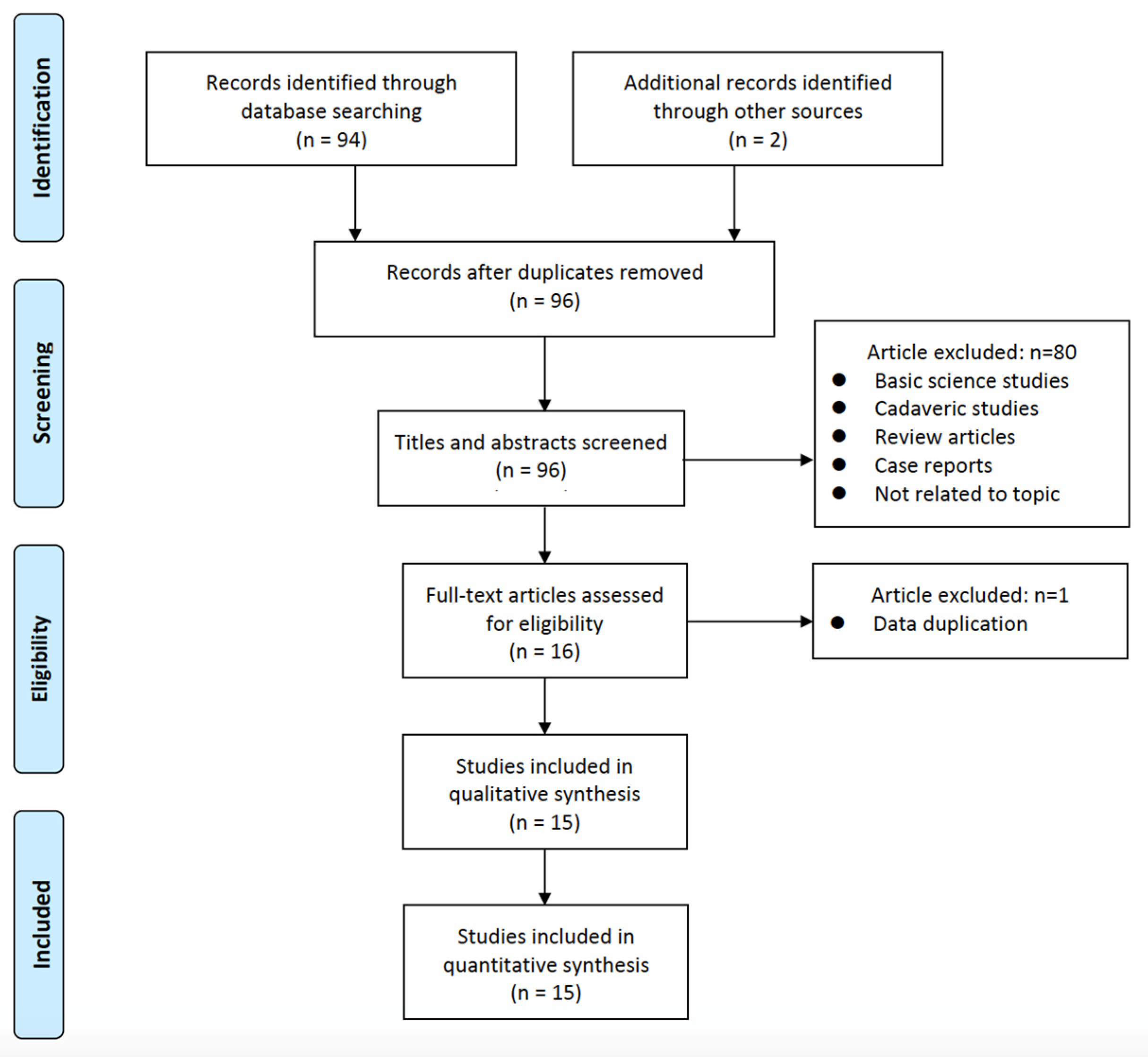
3.1. Selection of Studies and Quality Evaluation

Through the database search, 96 studies were discovered. Following a title and abstract screening, which eliminated 5 irrelevant articles, the remaining 17 possibly relevant articles were obtained. Following a thorough examination of the complete text, 15 articles were finally included in this review [13,16,17,18,19,20,21,22,23,24,25,26,27,28,29]. Figure 1 shows a flowchart for a specific study. Table S1 shows the PRISMA checklist.
All 15 studies used a retrospective comparative cohort design with moderate to high quality as assessed by the NOS (Table 1).

3.2. Study Characteristics and Outcomes

Table 2 presents the selected study characteristics. Of these, 1 was a prospective study and the remaining 14 were retrospective studies. The Disc-FX procedure was performed in 570 patients. The L4–L5 was the most commonly operated level, and most surgeries were single-level surgeries. The follow-up period for these patients ranged from 2 months to 24 months.
Eight papers (Table 3) provided operative times, and all but one paper reported operative times ranging from 35 to 60 min, whereas the remaining paper reported an operative time of less than 30 min. Only five studies reported estimated blood loss, none of which exceeded 10 mL.
Ten studies (Table 3) provided VAS scores. The mean VAS scores decreased from 7.22 preoperatively to 1.81 at the final follow-up. Two studies provided VAS scores for back pain. The mean VAS scores for back pain decreased from 4.84 preoperatively to 2.02 at the final follow-up. Two studies provided VAS scores for leg pain. The mean VAS scores for leg pain decreased from 3.84 preoperatively to 0.96 at the final follow-up. Three studies provided NRS scores. The mean NRS scores decreased from 6.98 preoperatively to 3.9 at the final follow-up (Table 3).
Five studies provided JOA scores. The mean JOA scores improved from 16.26 preoperatively to 25.88 at the final follow-up (Table 3). Seven studies provided ODI scores. The mean ODI scores decreased from 35.37 preoperatively to 14.66 at the final follow-up (Table 3). The mean satisfaction rate (based on the Macnab criteria) was 87.6% (range, 78.4–95.2%) from eight studies.

3.3. Complications

Regarding postoperative complications, three papers reported postoperative transient pain. The total incidence of postoperative transient pain was 8.77% (50/570) after the Disc-FX procedure. The total recurrence rate was 1.58% (9/570) after the Disc-FX procedure reported in four articles. Furthermore, some niche complications have been reported, such as nerve injury (2), hematoma (1), infection (1), lumbar venous plexus injury (1), and reoperation (1).

4. Discussion

In this systematic review, we found that postoperative VAS and NRS scores for diskogenic pain using the Disc-FX system were significantly lower than preoperative scores, postoperative ODI scores were lower than preoperative ones, postoperative JOA scores were significantly higher than preoperative scores, and fewer surgical adverse events occurred; additionally, the patient satisfaction rate was 87.6% (range, 78.4–95.2%).

4.1. Mechanism and Diagnosis of Diskogenic Pain

Diskogenic pain is discomfort caused by damaged intervertebral disks, and its primary cause is the degeneration and deterioration of the intervertebral disks and the formation of fissures or ruptures in their fibrous rings that separate from each other, stimulating pain receptors distributed on the surface of the fibrous rings of the disks and causing pain [30,31]. Additionally, inflammatory mediators acting on injury receptors at the terminal nerve fibers in the intervertebral disk can cause direct electrophysiological changes or make them extremely sensitive, thus causing pain [32].
Diskogenic pain is difficult to diagnose because it often has only subjective symptoms and lacks objective auxiliary examination indicators. Since patients with diskogenic pain lack obvious manifestations of lumbar disk herniation, locating the painful intervertebral space is often impossible based on symptoms and physical examination, and locating the source of pain using MRI is difficult, whereas diskography can screen normal physiologically degenerated disks from pathologically degenerated disks, thus effectively clarifying the responsible segment and avoiding overtreatment and mistreatment [33,34]. However, the patient’s fear of pain, the intensity of pain during imaging, sensory sensitivities associated with chronic pain disorders, and social factors can contribute to false positives. It can be seen that there is no objective test that can be used independently as a basis for the diagnosis of diskogenic pain; however, it needs to be analyzed and judged along with symptoms and signs. To accurately assess the effectiveness of the Disc-FX system in treating diskogenic pain and contained lumbar disk herniation, a clear diagnosis is necessary.

4.2. Features and Advantages of the Disc-FX System

Several conservative treatments have been used for diskogenic pain, including opioids, non-steroidal anti-inflammatory drugs, and physical therapies, such as acupuncture, massage, and physical therapy [35]. Although the use of medication can achieve short-term therapeutic effects, the potential risks of long-term medication are also greatly increased [35]. The failure of physical therapy to achieve good results in treating diskogenic pain in some patients may be related to the complex pathogenesis of the disease. Intradiscal electrotherapy, with its high and difficult-to-regulate temperature, probably causes damage to the surrounding tissues and secondary inflammatory reactions, and its long-term efficacy is uncertain [36]. A sinuvertebral nerve block is a rapid and precise intervention performed under local anesthesia for the treatment of diskogenic pain. A recent study [37] reported on 32 patients with diskogenic pain who underwent a sinuvertebral nerve block. The patients’ VSA and ODI scores decreased significantly immediately after surgery, from 5.75 and 32.59 preoperatively to 2.5 and 17.28 on postoperative day 3, respectively. However, there was a general rebound in the mid- to long-term postoperative period, 3.53 and 19.63 at 1 month, as well as 3.78 and 21.44 at 3 months postoperatively. Eighteen of these patients (56.25%) were observed to experience varying degrees of pain recurrence at 3 months. According to our review, 10 studies provided VAS scores after Disc-FX treatment for diskogenic pain. The mean VAS score decreased from 7.22 preoperatively to 1.81 at the final follow-up. Two studies provided VAS scores for back pain. The mean VAS score for back pain decreased from 4.84 preoperatively to 2.02 at the final follow-up. Two studies provided VAS scores for leg pain. The mean VAS score for leg pain decreased from 3.84 preoperatively to 0.96 at the final follow-up. Seven studies provided ODI scores. The mean ODI score decreased from 35.37 preoperatively to 14.66 at the final follow-up. It is evident that Disc-FX greatly outperforms sinuvertebral nerve block in terms of long-term outcomes for treating individuals with diskogenic pain. In addition, nucleus pulposus removal alone cannot completely remove inflammatory factors and inflammatory tissue from the disc, so a combination of different minimally invasive approaches may have a greater therapeutic effect.
The Disc-FX system is an innovative, minimally invasive technique based on the Yeung endoscopy spine system technology developed by Yeung, which is versatile and fills the gap between interventional and open surgery with multiple functions, such as tissue extraction, nucleus pulposus removal, radiofrequency ablation, annuloplasty, irrigation/aspiration, and observation. The Disc-FX system for treating diskogenic pain is closely related to its possible mechanism of occurrence, which can achieve intra-disk decompression, the inactivation of sinus nerve terminals growing into the ruptured annulus fibrosus fissure, and the destruction of inflammatory factors [14]. Intraoperative methylene blue can eliminate the nociceptive allergic reaction caused by local tissue inflammation and can make normal tissue (nerve root) often stain light blue-green, the degenerated nucleus pulposus and inflammatory granulation tissue stain dark blue, the normal nucleus pulposus tissue stain light blue, and the endplate and fiber ring to not stain, which can help the subsequent clamping and distinguish different tissue types. By changing the position and direction of the working channel, the miniature nucleus pulposus clamp can fully eat away the diseased nucleus pulposus and granulation tissue, reduce the pressure in the intervertebral disk, and reduce the irritation to the nerve terminals.
The Disc-FX system employs a radiofrequency of 1.7 MHz from the Elliquence Surgi-Max generator to achieve nucleus ablation (Bipolar Turbo mode) and annular remodeling (Bipolar Hemo mode), respectively [13]. While conventional radiofrequency mainly destroys cells through heat, the Surgi-Max bipolar radiofrequency system uses high frequencies to resonate with cellular fluid, causing cells to rupture from within; the low temperature output makes the bipolar head resistant to adhesion, minimizing tissue trauma and allowing for the maximum preservation of healthy tissue. Finally, repeated irrigation with saline through the working channel can effectively remove chemical pain mediators, toxic metabolites, any byproducts of radiofrequency treatment and nucleus pulposus, and inflammatory tissue debris from the disk; improve the disk microenvironment; and reduce or eliminate pressure and chemically mediated diskogenic pain.

4.3. Indications, Contraindications, and Limitations of the Disc-FX System

The Disc-FX system’s 3 mm outer working channel clearly offers less trauma than conventional endoscopic spine surgery and does not require the steep learning curve and lengthy training required for microscopic surgery. However, the careful monitoring of surgical indications remains necessary to achieve optimal results. The indications for surgery using the Disc-FX system should be strictly limited to diskogenic low back pain because of the mild inclusion of disk herniation and fibular ring tears, particularly in young and middle-aged patients with normal or mildly reduced disk height, while severe disk herniation, free nucleus pulposus, and significant nerve root symptoms should be avoided.
Nevertheless, the Disc-FX system also has its limitations. (1) Because of the restriction of the high iliac crest, the working channel cannot be placed in the optimal position at the L5–S1 level in some patients, and for the L2–L3 level and above, the puncture path is closer to the midline to avoid the kidney. (2) Because all surgeries are performed under X-ray guidance, some are blindfolded, and controlling the quantity of nucleus pulposus tissue excised is difficult, particularly for patients of varying ages and degrees of degeneration, and the amount of nucleus pulposus tissue excised frequently varies substantially. (3) The Disc-FX system is indicated in cases with an intact fibrous ring and posterior longitudinal ligament, although it has also applied in cases with fibrous ring tears. (4) Unlike full endoscopic spine surgery, which has multiple surgical approaches, Disc-FX has only one posterolateral approach, limiting its extensive use. (5) Finally, it is not a fully visualized surgical approach.

4.4. Management and Prevention of Complications

The following are possible complications associated with the Disc-FX system: (1) Postoperative transient pain is caused by recurrent episodes of transient pain caused by the space formed after the disk is removed and then filled with blood or other tissues causing compression or intraoperative manipulation that irritates the nerve roots and causes edema [38]; this complication is usually relieved by symptomatic treatment and physical therapy. (2) Recurrence may be attributed to the patient having more deteriorated disk tissue that was not removed intraoperatively and the patient having previously engaged in hard physical labor, which impaired the repair of the annulus fibrosus, resulting in recurrence. To avoid recurrence or induced disk herniation in patients with severe intraoperative diskography tears, the degenerated nucleus pulposus should be removed during surgery, and the amount of postoperative activity should be gradual, avoiding strenuous exercise for 3 weeks, and heavy physical exertion should be gradually resumed after surgery. (3) Infections may be related to the patient being elderly, being immunocompromised, or having diabetes, which may improve with intravenous drips of sensitive antibiotics and effective glycemic control. (4) Hematomas, which present as a persistent postoperative exacerbation of symptoms, are relieved via a secondary laminectomy to remove the hematoma. In addition to careful and gentle intraoperative handling, for middle-aged and elderly patients who have been taking aspirin for a long time, the procedure should be performed only after 2 weeks of drug withdrawal. (5) Finally, other rare complications include nerve root injuries and vessel ruptures. These can be prevented by focusing on the patient’s preoperative examination and perioperative management and by strict asepsis and careful manipulation during the procedure.

4.5. Strengths and Limitations

This is the first systematic review of the efficacy of nucleo-annuloplasty using Disc-FX for treating lumbar diskogenic lesions. However, this review also has some limitations. This review is of a low evidence-based grade because it consists mainly of retrospective studies; there are no randomized controlled trials or prospective comparative studies to refer to. Additionally, there was variation in the duration of follow-up in this study, which may have provided the incomplete reporting of complication rates. Finally, some studies failed to disclose accurate data on issues such as age, gender, and follow-up duration.

5. Conclusions

According to our comprehensive evaluation, percutaneous nucleo-annuloplasty for lumbar diskogenic diseases provided considerable pain alleviation and improved functional outcomes with fewer complications. Disc-FX is a safe and effective procedure that is a good treatment option for patients with diskogenic pain. Existing research, including several that are still underway, are primarily confined to small cohorts and short-term follow-ups. Further prospective studies and randomized trials with large sample sizes and long-term follow-ups should be conducted.

Supplementary Materials

The following supporting information can be downloaded at: https://www.mdpi.com/article/10.3390/medicina59071291/s1, Table S1: PRISMA checklist. Reference [39] is cited in Supplementary Materials.

Author Contributions

Conceptualization, C.-M.C.; software, G.-X.L.; formal analysis, S.-W.J.; writing—original draft preparation, G.-X.L.; writing—review and editing, C.-M.C. All authors have read and agreed to the published version of the manuscript.

Funding

This research was funded by the Natural Science Foundation of Fujian Province, grant number 2021J05282; the Xiamen Municipal Bureau of Science and Technology (3502Z20224033); and the “Xiamen Health System Discipline Leaders and their Backup Candidates, Senior Management Talent Training Candidates Training Program (2021)”. The funder had no role in the design of the study; the collection, analysis, or interpretation of the data; or in the writing of the manuscript.

Institutional Review Board Statement

This kind of study does not require ethical approval.

Informed Consent Statement

Informed consent was waived due to the retrospective and non-interventional nature of this analysis.

Data Availability Statement

The datasets are presented within the manuscript.

Acknowledgments

The author (G.X.L.) wishes to acknowledge the financial support of the “Xiamen Health High-Level Talent Training Program”.

Conflicts of Interest

The authors declare no conflict of interest.

References

  1. Lin, G.X.; Xu, W.B.; Kotheeranurak, V.; Chen, C.M.; Deng, Z.H.; Zhu, M.T. Comparison of oblique and transforaminal approaches to lumbar interbody fusion for lumbar degenerative disease: An updated meta-analysis. Front. Surg. 2022, 9, 1004870. [Google Scholar] [CrossRef] [PubMed]
  2. Berthelot, J.M.; Delecrin, J.; Maugars, Y.; Passuti, N. Contribution of centralization phenomenon to the diagnosis, prognosis, and treatment of diskogenic low back pain. Jt. Bone Spine 2007, 74, 319–323. [Google Scholar] [CrossRef] [PubMed]
  3. Maus, T.P.; Aprill, C.N. Lumbar diskogenic pain, provocation diskography, and imaging correlates. Radiol. Clin. N. Am. 2012, 50, 681–704. [Google Scholar] [CrossRef] [PubMed]
  4. Ravikanth, R. A review of discogenic pain management by interventional techniques. J. Craniovertebr. Junction Spine 2020, 11, 4–8. [Google Scholar] [CrossRef]
  5. Gushcha, A.O.; Yusupova, A.R. Minimally invasive surgery and modern view on the treatment of degenerative spine diseases. Zh Vopr. Neirokhir Im. N. N. Burdenko 2021, 85, 5–9. [Google Scholar] [CrossRef] [PubMed]
  6. Nguyen, K.M.L.; Nguyen, D.T.D. Minimally Invasive Treatment for Degenerative Lumbar Spine. Tech. Vasc. Interv. Radiol. 2020, 23, 100700. [Google Scholar] [CrossRef] [PubMed]
  7. Eksi, M.S.; Ozcan-Eksi, E.E.; Orhun, O.; Turgut, V.U.; Pamir, M.N. Proposal for a new scoring system for spinal degeneration: Mo-Fi-Disc. Clin. Neurol. Neurosurg. 2020, 198, 106120. [Google Scholar] [CrossRef]
  8. Teichtahl, A.J.; Urquhart, D.M.; Wang, Y.; Wluka, A.E.; O’Sullivan, R.; Jones, G.; Cicuttini, F.M. Lumbar disc degeneration is associated with modic change and high paraspinal fat content—A 3.0T magnetic resonance imaging study. BMC Musculoskelet. Disord. 2016, 17, 439. [Google Scholar] [CrossRef] [Green Version]
  9. Clark, A.J.; Safaee, M.M.; Khan, N.R.; Brown, M.T.; Foley, K.T. Tubular microdiscectomy: Techniques, complication avoidance, and review of the literature. Neurosurg. Focus. 2017, 43, E7. [Google Scholar] [CrossRef] [Green Version]
  10. Hoff-Olsen, P.; Wiberg, J. Small bowel perforation as a complication of microsurgical lumbar diskectomy. A case report and brief review of the literature. Am. J. Forensic Med. Pathol. 2001, 22, 319–321. [Google Scholar] [CrossRef]
  11. Aiyer, R.; Noori, S.; Schirripa, F.; Schirripa, M.; Jain, S.; Aboud, T.; Mehta, N.; Elowitz, E.; Pahuta, M.; Datta, S. A systematic review of full endoscopic versus micro-endoscopic or open discectomy for lumbar disc herniation. Pain Manag. 2022, 12, 87–104. [Google Scholar] [CrossRef] [PubMed]
  12. Li, X.C.; Zhong, C.F.; Deng, G.B.; Liang, R.W.; Huang, C.M. Full-Endoscopic Procedures Versus Traditional Discectomy Surgery for Discectomy: A Systematic Review and Meta-analysis of Current Global Clinical Trials. Pain Physician 2016, 19, 103–118. [Google Scholar] [PubMed]
  13. Kumar, N.; Zaw, A.S.; Kumar, N.; Sonawane, D.; Hey, H.W.D.; Kumar, A. Annulo-Nucleoplasty Using Disc-Fx in the Management of Degenerative Lumbar Disc Pathology: How Long Can the Effect Last? Glob. Spine J. 2018, 8, 365–373. [Google Scholar] [CrossRef] [PubMed] [Green Version]
  14. Kumar, N.; Kumar, A.; Siddharth, M.S.; Sambhav, P.S.; Tan, J. Annulo-nucleoplasty using Disc-FX in the management of lumbar disc pathology: Early results. Int. J. Spine Surg. 2014, 8, 18. [Google Scholar] [CrossRef] [Green Version]
  15. Beyaz, S.G.; Inanmaz, M.E.; Zengin, E.S.; Ulgen, A.M. Combined Use of High Radiofrequency Disk Ablation, Annulus Modulation, and Manual Nucleotomy in a Patient with Extruded Disk Herniation. Pain Pract. 2016, 16, E74–E80. [Google Scholar] [CrossRef]
  16. Bai, Y.; Xi, C.; Zhao, W.; Mu, X.; Tan, F.; Wang, L.; Ma, Y. Analysis of clinical efficacy on percutaneous discectomy combined with radiofrequency ablation by Disc-FX in contained lumbar disc herniation. Chin. J. Pain Med. 2011, 17, 280–283. [Google Scholar]
  17. Chen, S.Y.Y.; Mei, W. Disc-FX system for minimally invasive treatment of accommodative lumbar disc herniation. Chin. Orthop. 2014, 26, 46–47. [Google Scholar]
  18. Hirano, Y.P.S.; Bansil, R.; Watnabe, K. Advantages of Endoscope-Assisted Disc-FX in Surgical Management of Lumbar Disc Herniation: A Report of 10 Cases. J. Spine 2018, S7, 1–4. [Google Scholar]
  19. Lu, Z.Y.Y.; Fan, F.; Chen, S.; Zheng, S.; Sun, Y.; Bao, C.; Wang, Y. Clinical analysis of the 3-in-1 technique of Disc-FX system for discogenic low back pain. Chin. J. Parct. Med. 2013, 40, 65–67. [Google Scholar]
  20. Liao, X.J.J.; Xiong, D.; Yi, W.; Zhang, Q.; Zheng, H.; Luo, Y.; Linag, H.; Zhang, D. The comparison of clinical outcomes of percutaneous lumbar discectomy with Disc-FX system with fenestration and decompression for contained lumbar disc herniation with radicular pain. Chin. J. Pain Med. 2011, 17, 25–29. [Google Scholar]
  21. Ma, Y.W.P. Analysis of changes in serum substance P and inflammatory factors before and after minimally invasive Disc-FX surgery for discogenic low back pain and evaluation of early efficacy. J. Cervicodynia Lumbodynia 2021, 42, 386–388. [Google Scholar]
  22. Ou, J. Clinical efficacy of the treatment of discectomy, radiofrequency ablation and annulus fibrosus angioplasty under the guidance of Disc-FX system for contained lumbar disc herniation. Chin. J. Mod. Med. 2013, 23, 91–93. [Google Scholar]
  23. Park, C.H.; Lee, S.H. Efficacy of Nucleo-Annuloplasty Using Disc-Fx in Lumbar Disc Herniation. J. Spine 2015, 4, 1–4. [Google Scholar]
  24. Park, C.H.; Lee, K.K.; Lee, S.H. Efficacy of transforaminal laser annuloplasty versus intradiscal radiofrequency annuloplasty for discogenic low back pain. Korean J. Pain 2019, 32, 113–119. [Google Scholar] [CrossRef] [Green Version]
  25. Wang, J.Z.; Xia, Q.; Zheng, B.; Shi, K.; Ma, W.; Liu, J. Efficacy of lumbar discectomy radiofrequency ablation and annuloplasty with Disc-FX system. Chin. J. Anesthesiol. 2013, 33, 430–432. [Google Scholar]
  26. Xi, J.M.; Hu, M.; Chui, X.; Zhao, W.; Mu, X. Early clinical efficacy analysis of Disc-FX system percutaneous disc removal and radiofrequency ablation for inclusive lumbar disc herniation. Chin. J. Bone Jt. Surg. 2012, 5, 252–254. [Google Scholar]
  27. Yam, J.O.; Cheung, P.; Chiang, L.F.; Wan, S.Y.S.; Mok, W.Y. Efficacy and safety of nucleo-annuloplasty using radiofrequency ablation for discogenic back pain in a local Hong Kong population. J. Orthop. Trauma Rehabil. 2021, 1–8. [Google Scholar] [CrossRef]
  28. Zhang, W.W.; Jiao, J.; Wang, Y.; Yang, D.; Ding, W.; Shen, Y. Primary results of the 3-in-1 technique of Disc-FX system for the discogenic low back pain. Chin. J. Orthop. 2011, 31, 1049–1055. [Google Scholar]
  29. Zhang, J.S.; Su, S.; Lin, D. Disc-FX system-assisted percutaneous nucleus pulposus clamping with radiofrequency ablation for discogenic low back pain. J. Spinal Surg. 2015, 13, 7–10. [Google Scholar]
  30. Kim, H.S.; Wu, P.H.; Jang, I.T. Lumbar Degenerative Disease Part 1: Anatomy and Pathophysiology of Intervertebral Discogenic Pain and Radiofrequency Ablation of Basivertebral and Sinuvertebral Nerve Treatment for Chronic Discogenic Back Pain: A Prospective Case Series and Review of Literature. Int. J. Mol. Sci. 2020, 21, 1483. [Google Scholar]
  31. Simon, J.; McAuliffe, M.; Shamim, F.; Vuong, N.; Tahaei, A. Discogenic low back pain. Phys. Med. Rehabil. Clin. N. Am. 2014, 25, 305–317. [Google Scholar] [CrossRef] [PubMed]
  32. Urits, I.; Burshtein, A.; Sharma, M.; Testa, L.; Gold, P.A.; Orhurhu, V.; Viswanath, O.; Jones, M.R.; Sidransky, M.A.; Spektor, B.; et al. Low Back Pain, a Comprehensive Review: Pathophysiology, Diagnosis, and Treatment. Curr. Pain Headache Rep. 2019, 23, 23. [Google Scholar] [CrossRef] [PubMed]
  33. Bogduk, N.; Aprill, C.; Derby, R. Lumbar discogenic pain: State-of-the-art review. Pain Med. 2013, 14, 813–836. [Google Scholar] [CrossRef] [PubMed] [Green Version]
  34. Yang, S.; Boudier-Reveret, M.; Chang, M.C. Use of Pulsed Radiofrequency for the Treatment of Discogenic Back Pain: A Narrative Review. Pain Pract. 2021, 21, 594–601. [Google Scholar] [CrossRef]
  35. Zhao, L.; Manchikanti, L.; Kaye, A.D.; Abd-Elsayed, A. Treatment of Discogenic Low Back Pain: Current Treatment Strategies and Future Options-a Literature Review. Curr. Pain Headache Rep. 2019, 23, 86. [Google Scholar] [CrossRef]
  36. Gelalis, I.; Gkiatas, I.; Spiliotis, A.; Papadopoulos, D.; Pakos, E.; Vekris, M.; Korompilias, A. Current Concepts in Intradiscal Percutaneous Minimally Invasive Procedures for Chronic Low Back Pain. Asian J. Neurosurg. 2019, 14, 657–669. [Google Scholar] [CrossRef]
  37. Liu, Z.; Ma, R.; Fan, C.; Chen, J.; Zhang, R.; Zheng, Z.; Xu, Y.; Liu, Z.; Zhao, Q.; Li, Q. Sinuvertebral nerve block treats discogenic low back pain: A retrospective cohort study. Ann. Transl. Med. 2022, 10, 1219. [Google Scholar] [CrossRef]
  38. Zhang, C.; Li, Z.; Yu, K.; Wang, Y. A Postoperative Phenomenon of Percutaneous Endoscopic Lumbar Discectomy: Rebound Pain. Orthop. Surg. 2021, 13, 2196–2205. [Google Scholar] [CrossRef]
  39. Page, M.J.; McKenzie, J.E.; Bossuyt, P.M.; Boutron, I.; Hoffmann, T.C.; Mulrow, C.D.; Shamseer, L.; Tetzlaff, J.M.; Akl, E.A.; Brennan, S.E.; et al. The PRISMA 2020 statement: An updated guideline for reporting systematic reviews. BMJ 2021, 372, n71. [Google Scholar] [CrossRef]
Figure 1. Flowchart of specific studies.
Medicina 59 01291 g001
Table 1. Quality assessment of the included studies.
StudiesSelectionComparabilityExposureTotal Scores (of 9)
Is the Case Definition Adequate?Representativeness of the CasesSelection of ControlsDefinition of ControlsComparability of Cases and Controls on the Basis of the Design or AnalysisAscertainment of ExposureSame Method of Ascertainment for Cases and ControlsNon-Response Rate
Bai et al. [16], 2011☆☆ 8
Chen et al. [17], 2014 7
Hirano et al. [18], 2018 7
Kumar et al. [13], 2018☆☆9
Lu et al. [19], 2013 7
Liao et al. [20], 2011☆☆ 8
Ma et al. [21], 2021☆☆ 8
Ou et al. [22], 2013☆☆9
Park et al. [23], 2015☆☆ 8
Park et al. [24], 2019☆☆ 8
Wang et al. [25], 2013☆☆ 8
Xi et al. [26], 2012☆☆9
Yam et al. [27], 2021☆☆9
Zhang et al [28].,2011☆☆ 8
Zhang et al. [29], 2015☆☆9
The NOS score evaluates the quality of the literature using the semi-quantitative principle of the star system, except for comparability which can be rated up to 2 stars, the rest of the entries can be rated up to 1 star out of 9 stars, the higher the score indicates the higher quality of the study.
Table 2. Characteristics from reviewed studies.
Authors and YearStudy TypeStudy PeriodNo. of Patients (Levels)Operated LevelsAge (Years), Mean; RangeSex (M:F)Follow-Up Period (mo)Complications (n)
Bai et al. [16], 2011Retrospective2010.07–2010.1036 (NR)NR47.5; 18–7716:206None
Chen et al. [17], 2014Retrospective2011.10–2013.0236 (36)L3–4 (5); L4–5 (21); L5–S1 (10)43.5; 26–6521:156–12None
Hirano et al. [18], 2018RetrospectiveNR10 (10)L1–2 (1); L3–4 (2); L4–5 (6); L5–S1 (2)47.2; 30–728:26None
Kumar et al. [13], 2018Prospective2010.09–2014.1251 (66)L2–3 (1); L3–4 (8); L4–5 (27); L5–S1 (30)41: 20–6338:1324Infection (1), postoperative transient pain (16)
Lu et al. [19], 2013Retrospective2011.02–2012.0535 (35)NR36.5; 27–5623:1214.8Recurrent (1)
Liao et al. [20], 2011Retrospective2008.05–2010.0525 (NR)NRNRNR12None
Ma et al. [21], 2021Retrospective2013.01–2015.1256 (NR)NR51.2 ± 12.4; 43–6532:243Recurrent (5), nerve root injury (2); postoperative transient pain (8)
Ou et al. [22], 2013Retrospective2010.09–2011.1062 (70)L3–4 (5); L4–5 (36); L5–S1 (29)45.3 ± 16.9: 21–7525:376None
Park et al. [23], 2015RetrospectiveNR43 (NR)NR44.9; 22–7730:136None
Park et al. [24], 2019RetrospectiveNR43 (NR)NR56.7 ± 14.1; NR20:236None
Wang et al. [25], 2013RetrospectiveNR28 (NR)NRNR; 27–73NR2Recurrent (2), lumbar venous plexus injury (1)
Xi et al. [26], 2012Retrospective2010.07–2011.0636 (73)L2–3 (4); L3–4 (13); L4–5 (32); L5–S1 (24)56; 18–7716:2012Infection (1), postoperative hematoma (1), postoperative transient pain (26)
Yam et al. [27], 2021Retrospective2017–201916 (24)L2–3 (1); L3–4 (3); L4–5 (12); L5–S1 (8)NR; 23–6913:3>6Re-operation (1)
Zhang et al [28], 2011Retrospective2010.02–2011.0240 (47)L3–4 (3); L4–5 (23); L5–S1 (21)38.7; 32–5822:1813.8 (6–18)Recurrent (1)
Zhang et al. [29], 2015Retrospective2010.06–2011.0553 (NR)NRNR; 29–5624:2924None
NR: not reported.
Table 3. Outcomes from reviewed studies.
Authors and YearOperative Time (mins)Blood Loss (mL)PreoperativeFinal Follow-UpMacnab
Pain Rating ScaleFunctional Rating ScalePain Rating ScaleFunctional Rating Scale
Bai et al. [16], 201115–200–5VAS: 6.5 ± 1.8JOA: 19.3 ± 3.6; ODI: 18.7 ± 11.8VAS: 2.5 ± 2.2JOA: 25.5 ± 3.2; ODI: 9.1 ± 8.581.3%
Chen et al. [17], 201415–305VAS: 8.5 ± 1.4JOA: 12.3 ± 1.2VAS: 2.1 ± 0.8JOA: 25.5 ± 2.1NR
Hirano et al. [18], 2018NRNRVAS: 8.0JOA: 13.0VAS: 1.2JOA: 25.9NR
Kumar et al. [13], 2018NRNRVAS: 6.69 ± 0.93ODI: 47.80 ± 17.92VAS: 2.85 ± 1.76ODI: 19.63 ± 14.1478.4%
Lu et al. [19], 201329NRVAS: 6.70 ± 1.26ODI: 19.9 ± 6.8VAS: 1.05 ± 0.66ODI: 8.6 ± 4.5NR
Liao et al. [20], 201130 ± 52 ± 1VAS: 7.8 ± 0.4ODI: 39.1 ± 3.9VAS: 2.0 ± 0.1ODI: 11.4 ± 1.6NR
Ma et al. [21], 2021NRNRVAS: 6.2 ± 1.5JOA: 17.4 ± 3.8VAS: 2.2 ± 1.3JOA: 25.9 ± 1.389.3%
Ou et al. [22], 201325.16 ± 3.210–5VASB: 3.07 ± 1.15; VASL: 6.72 ± 1.26NRVASB: 0.98 ± 0.54; VASL: 0.97 ± 0.58NR95.2%
Park et al. [23], 2015NRNRNRS: 7.4 ± 0.8NRNRS: 3.7 ± 1.9NRNR
Park et al. [24], 2019NRNRNRS: 7.3 ± 0.8ODI: 57.2 ±10.0NRS: 3.6 ± 1.8ODI: 22.1 ± 8.4NR
Wang et al. [25], 2013NRNRVAS: 8.0 ± 1.2NRVAS: 1.2 ± 0.8NR93%
Xi et al. [26], 2012180–5VAS: 6.5 ± 1.8JOA: 19.3 ± 3.6; ODI: 18.7 ± 11.8VAS: 2.1 ± 1.7JOA: 26.6 ± 2.4; ODI: 7.7 ± 6.578.9%
Yam et al. [27], 2021NRNRNRS: 6.25ODI: 46.25NRS: 4.4ODI: 24.12NR
Zhang et al [28], 201126<0VASB: 6.60 ± 1.47; VASL: 0.95 ± 0.63NRVASB: 3.05 ± 1.23; VASL: 0.95 ± 0.54NR92.5%
Zhang et al. [29], 201535–60NRVAS: 7.3 ± 1.1NRVAS: 0.9 ± 0.2NR92.4%
NR: not reported; VAS: visual analog scale; VASB: visual analog scale scores for back pain; VASL: visual analog scale for leg pain; NRS: numerical rating scale scores; JOA: Japanese Orthopaedic Association scores; ODI: Oswestry Disability Index.
Disclaimer/Publisher’s Note: The statements, opinions and data contained in all publications are solely those of the individual author(s) and contributor(s) and not of MDPI and/or the editor(s). MDPI and/or the editor(s) disclaim responsibility for any injury to people or property resulting from any ideas, methods, instructions or products referred to in the content.

