
Reflections on Postpartum Hysterectomy as a Possible Complication of Cesarean Myomectomy: A Long Debate


Abstract
1. Introduction
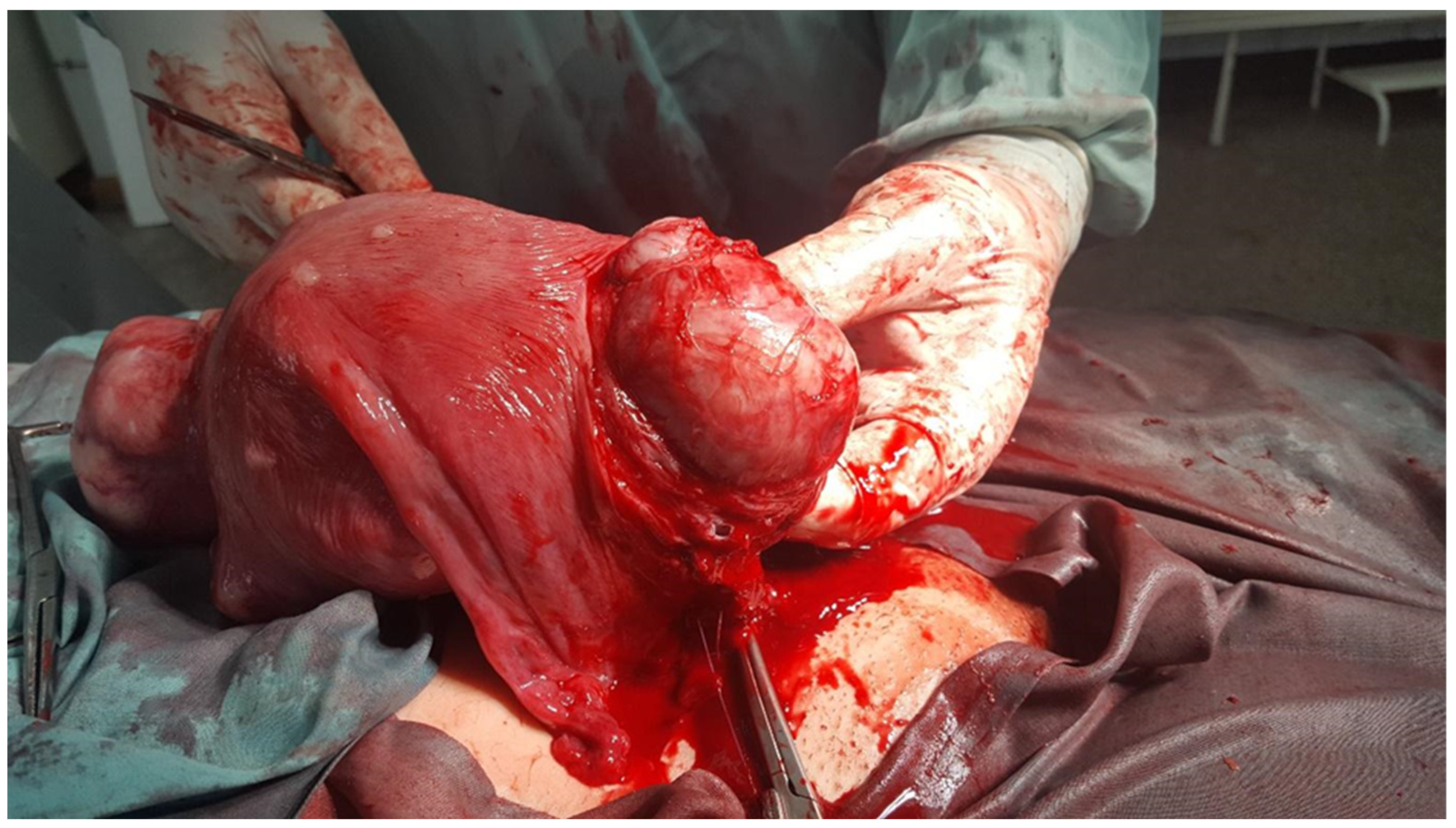
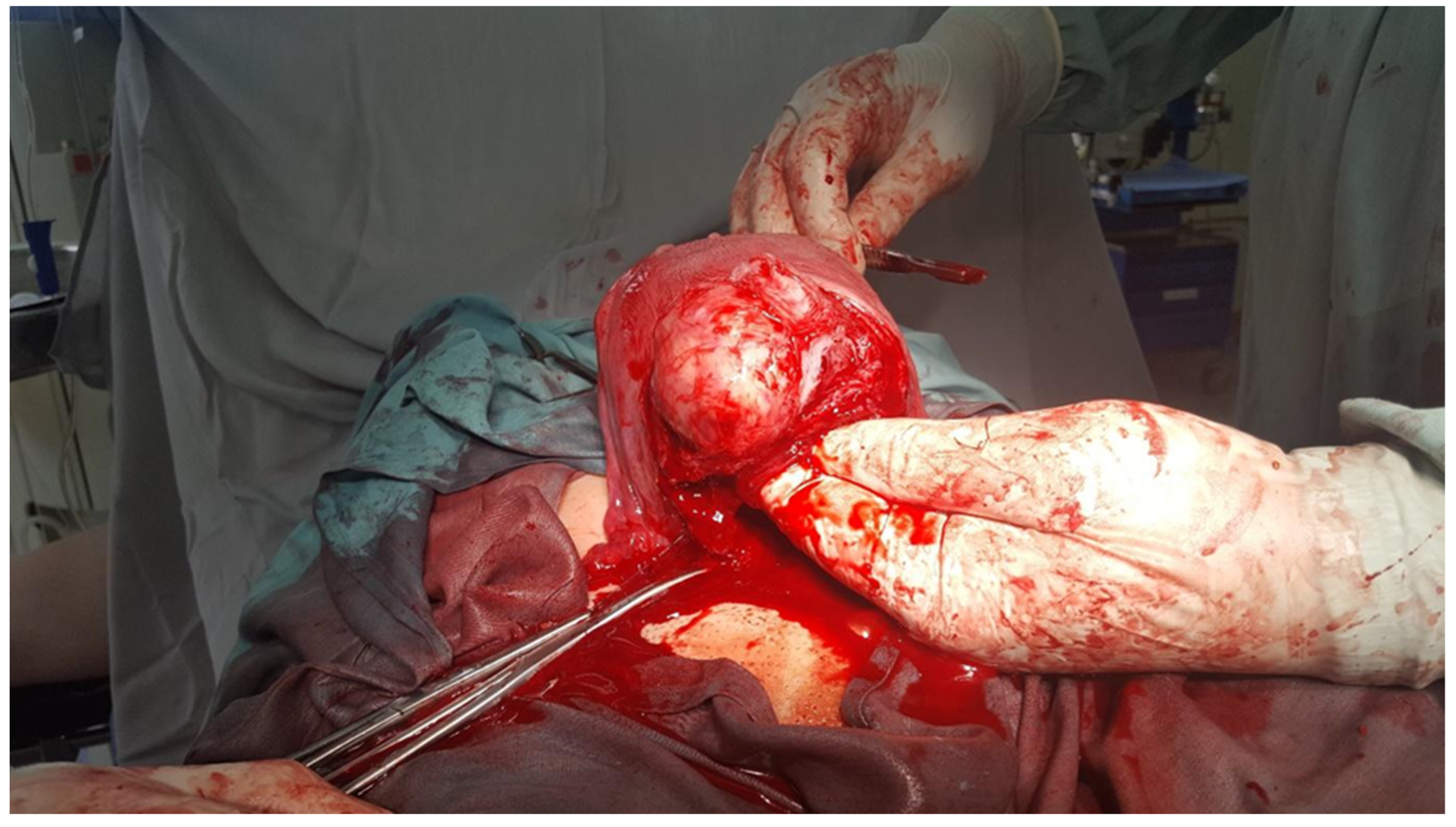
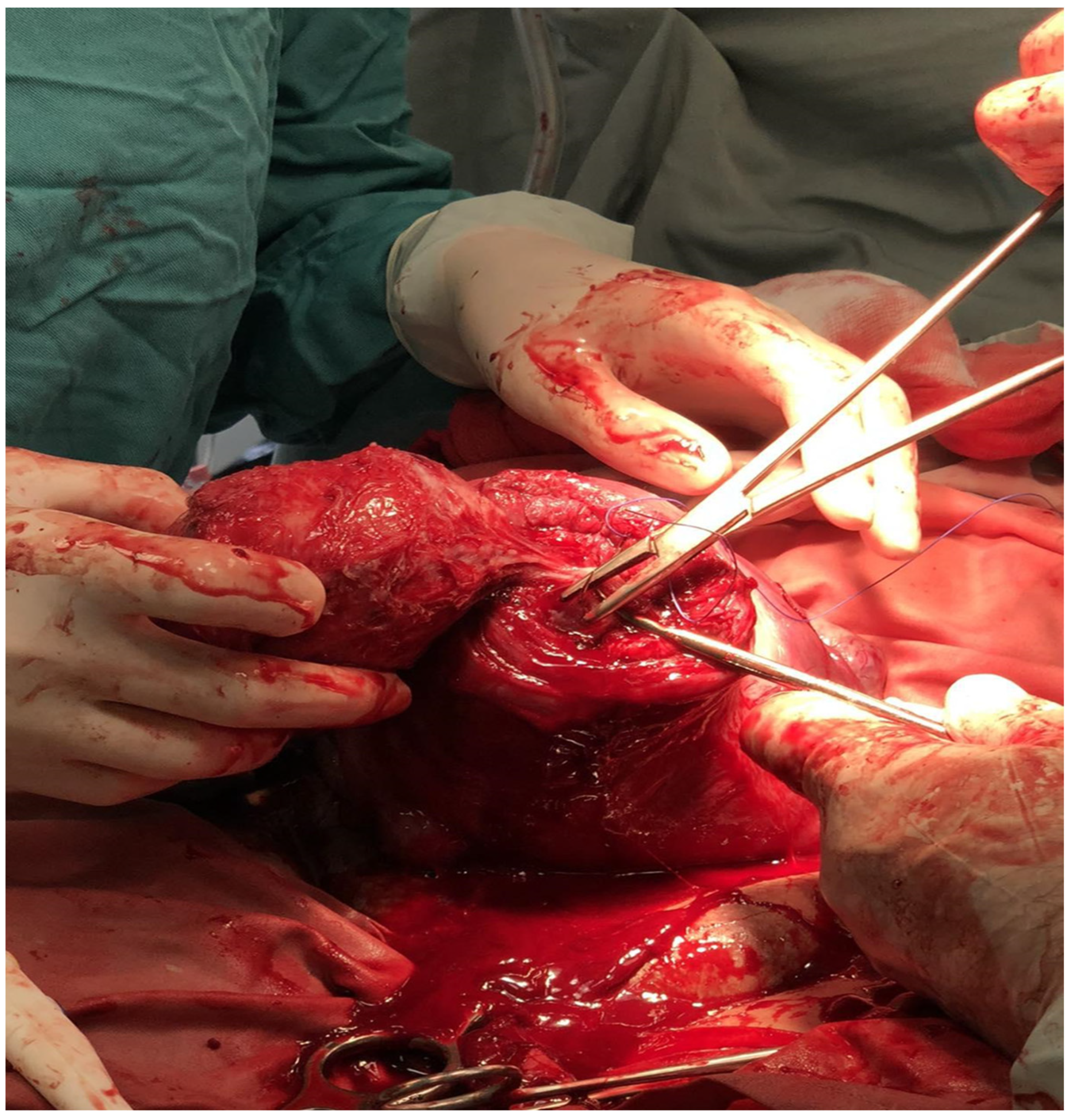
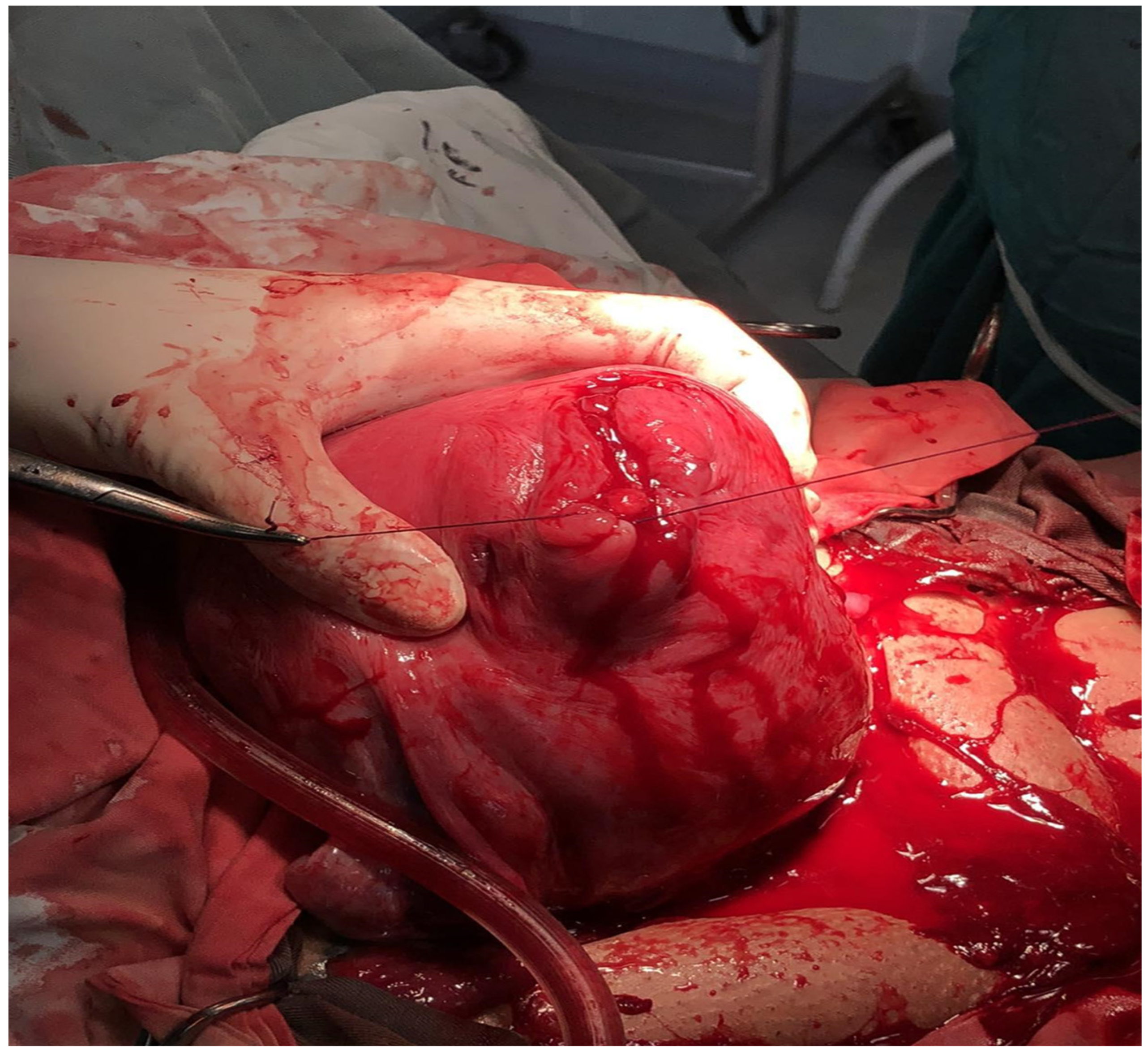
1.1. Case 1
1.2. Case 2
2. Discussion
2.1. The Most Common Complications of a Cesarean Myomectomy
2.2. Treatment Options for Bleeding Following CM That Preserve the Uterus
3. Conclusions
Author Contributions
Funding
Institutional Review Board Statement
Informed Consent Statement
Data Availability Statement
Conflicts of Interest
List of Abbreviations
| Uterine fibroids | UFs |
| Cesarean myomectomy | CM |
| Cesarean section | CS |
| Lower uterine segment | LUS |
| Red blood cells | RBCs |
| Intensive care unit | ICU |
| Plasma expander solution | HES |
| Disseminated intravascular coagulation | DIC |
| Postpartum hemorrhage | PPH |
References
- Sparic, R.; Mirkovic, L.; Malvasi, A.; Tinelli, A. Epidemiology of Uterine Myomas: A Review. Int. J. Fertil. Steril. 2016, 9, 424–435. [Google Scholar] [CrossRef] [PubMed]
- Tinelli, A.; Sparic, R.; Kadija, S.; Babovic, I.; Tinelli, R.; Mynbaev, O.A.; Malvasi, A. Myomas: Anatomy and related issues. Minerva Ginecol. 2016, 68, 261–273. [Google Scholar] [PubMed]
- Sparić, R.; Malvasi, A.; Tinelli, A. Analysis of clinical, biological and obstetric factors influencing the decision to perform cesarean myomectomy. Ginekol. Pol. 2015, 86, 40–45. [Google Scholar] [CrossRef]
- Sparić, R. Uterine myomas in pregnancy, childbirth and puerperium. Srp. Arh. Celok. Lek. 2014, 142, 118–124. [Google Scholar] [CrossRef] [PubMed]
- Klatsky, P.C.; Tran, N.D.; Caughey, A.B.; Fujimoto, V.Y. Fibroids and reproductive outcomes: A systematic literature review from conception to delivery. Am. J. Obstet. Gynecol. 2008, 198, 357–366. [Google Scholar] [CrossRef] [PubMed]
- Chuang, J.; Tsai, H.W.; Hwang, J.L. Fetal compression syndrome caused by myoma in pregnancy, a case report. Acta Obstet. Gynecol. Scan. 2001, 80, 472–473. [Google Scholar] [CrossRef]
- Aydeniz, B.; Wallwiener, D.; Kocer, C.; Grischke, E.M.; Diel, I.J.; Sohn, C.; Bastert, G. Der Stellenwert myombedingt Komplikationen in der Schwangerschaft. Z. Geburtshilfe Neonatol. 1998, 202, 154–158. [Google Scholar]
- Hasan, F.; Arumugam, K.; Sivanesaratnam, V. Uterine leiomyomata in pregnancy. Int. J. Gynecol. Obstet. 1991, 34, 45–48. [Google Scholar] [CrossRef] [PubMed]
- Winer-Muram, H.T.; Muram, D.; Gillieson, M.S. Uterine myomas in pregnancy. J. Can. Assoc. Radiol. 1984, 35, 168–170. [Google Scholar] [CrossRef]
- Vergani, P.; Ghidini, A.; Strobelt, N.; Roncaglia, N.; Locatelli, A.; Lapinski, R.H.; Mangioni, C. Do uterine leiomyomas influence pregnancy outcome? Am. J. Perinatol. 1994, 11, 356–358. [Google Scholar] [CrossRef]
- Girault, A.; Le Ray, C.; Chapron, C.; Goffinet, F.; Marcellin, L. Leiomyomatous uterus and preterm birth: An exposed/unexposed monocentric cohort study. Am. J. Obstet. Gynecol. 2018, 219, 410.e1–410.e7. [Google Scholar] [CrossRef]
- Sparić, R.; Malvasi, A.; Kadija, S.; Babović, I.; Nejković, L.; Tinelli, A. Cesarean myomectomy trends and controversies: An appraisal. J. Matern.-Fetal Neonatal Med. 2017, 30, 1114–1123. [Google Scholar] [CrossRef] [PubMed]
- Burton, C.A.; Grimes, D.A.; March, C.M. Surgical management of leiomyomata during pregnancy. Obstet. Gynecol. 1989, 74, 707–709. [Google Scholar] [PubMed]
- Hsieh, T.T.; Cheng, B.J.; Liou, J.D.; Chiu, T.H. Incidental myomectomy in cesarean section. Changgeng Yi Xue Za Zhi 1989, 12, 13–20. [Google Scholar] [PubMed]
- Michalas, S.P.; Oreopoulou, F.V.; Papageorgiou, J.S. Myomectomy during pregnancy and caesarean section. Hum. Reprod. 1995, 10, 1869–1870. [Google Scholar] [CrossRef] [PubMed]
- Sparić, R.; Radunović, N.; Tinelli, A.; Radević, O.; Kadija, S. Large myomas as a complicating factor necessitating cesarean myomectomy followed by cesarean hysterectomy. Srp. Arh. Celok. Lek. 2018, 146, 452–455. [Google Scholar] [CrossRef]
- Li, H.; Du, J.; Jin, L.; Shi, Z.; Liu, M. Myomectomy during cesarean section. Acta Obstet. Gynecol. Scand. 2009, 88, 183–186. [Google Scholar] [CrossRef] [PubMed]
- Exacoustòs, C.; Rosati, P. Ultrasound diagnosis of uterine myomas and complications in pregnancy. Obstet. Gynecol. 1993, 82, 97–101. [Google Scholar] [CrossRef]
- Pattanaik, T.; Kumar Pati, B.; Samal, S. Caesarean myomectomy: A descriptive study of clinical outcome. Int. J. Reprod. Contracept. Obstet. Gynecol. 2014, 3, 172+. Available online: https://link.gale.com/apps/doc/A377577865/HRCA?u=anon~ba57e4b9&sid=googleScholar&xid=818dc5e8 (accessed on 9 March 2024). [CrossRef]
- Seffah, J.D. Re-laparotomy after Cesarean section. Int. J. Gynaecol. Obstet. 2005, 88, 253–257. [Google Scholar] [CrossRef]
- Tian, J.; Hu, W. Cervical leiomyomas in pregnancy: Report of 17 cases. Aust. N. Z. J. Obstet. Gynaecol. 2012, 52, 258–261. [Google Scholar] [CrossRef]
- Dedes, I.; Schaffer, L.; Zimmermann, R.; Burkhardt, T.; Haslinger, C. Outcome and risk factors of cesarean delivery with and without cesarean myomectomy in women with uterine myomatas. Arch. Gynecol. Obstet. 2017, 295, 27–32. [Google Scholar] [CrossRef] [PubMed]
- Koike, T.; Minakami, H.; Kosuge, S.; Usui, R.; Matsubara, S.; Izumi, A.; Sato, I. Uterine leiomyomas in pregnancy: Its influence on obstetric performance. J. Obstet. Gynecol. 1999, 25, 309–313. [Google Scholar] [CrossRef] [PubMed]
- Tinelli, A.; Nezhat, C.; Likić-Ladjević, I.; Andjić, M.; Tomašević, D.; Papoutsis, D.; Stefanović, R.; Sparić, R. Myomectomy during cesarean section or non-caesarean myomectomy in reproductive surgery: This is the dilemma. Clin. Exp. Obstet. Gynecol. 2021, 48, 1250–1258. [Google Scholar] [CrossRef]
- Holmgren, C.M. Uterine rupture associated with VBAC. Clin. Obstet. Gynecol. 2012, 55, 978–987. [Google Scholar] [CrossRef] [PubMed]
- Dai, Q.H.; Zhang, L.; Chen, A.E. Prognostic and reproductive outcomes in women who had uterine myomas removed during cesarean section and sutured using different techniques. BMC Women’s Health 2024, 24, 7. [Google Scholar] [CrossRef] [PubMed]
- Akbas, M.; Mihmanli, V.; Bulut, B.; Temel Yuksel, I.; Karahisar, G.; Demirayak, G. Myomectomy for intramural fibroids during caesarean section: A therapeutic dilemma. J. Obstet. Gynaecol. 2017, 37, 141–145. [Google Scholar] [CrossRef] [PubMed]
- Ortac, F.; Güngőr, M.; Sőnmezer, M. Myomectomy during cesarean section. Int. J. Gynecol. Obstet. 1999, 67, 189–190. [Google Scholar] [CrossRef] [PubMed]
- Okogbo, F.O.; Ezechi, O.C.; Loto, O.M.; Ezeobi, P.M. Uterine leiomyomata in South Western Nigeria: A clinical study of presentations and management outcome. Afr. Health Sci. 2011, 11, 271–278. [Google Scholar]
- Velasco, V.R. Update of the conservative management of the uterine leiomyomata. An. Real Acad. Nac. Med. 2006, 123, 841–862. [Google Scholar]
- Doğan, S.; Özyüncü, Ö.; Atak, Z. Fibroids during pregnancy: Effects on pregnancy and neonatal outcomes. J. Reprod. Med. 2016, 61, 52–57. [Google Scholar] [PubMed]
- Claeys, J.; Hellendoorn, I.; Hamerlynck, T.; Bosteels, J.; Weyers, S. The risk of uterine rupture after myomectomy: A systemic review of the literature and meta-analysis. Gynecol. Surg. 2014, 11, 197–206. [Google Scholar] [CrossRef]
- Kim, H.S.; Oh, S.Y.; Choi, S.J.; Park, H.S.; Cho, G.J.; Chung, J.H. Uterine rupture in pregnancies following myomectomy: A multicenter case series. Obstet. Gynecol. Sci. 2016, 59, 454–462. [Google Scholar] [CrossRef] [PubMed]
- Amatya Vaidya, S.; Jha, R. Caesarean Myomectomy among Patients Undergoing Lower Segment Caesarean Section in a Tertiary Care Center. J. Nepal Med. Assoc. 2021, 59, 220–224. [Google Scholar] [CrossRef] [PubMed]
- Spariç, R. Intraoperative hemorrhage as a complication of cesarean myomectomy: Analysis of risk factors. Vojnosanit. Pregl. 2016, 73, 415–421. [Google Scholar] [CrossRef] [PubMed]
- Zhao, R.; Wang, X.; Zou, L.; Zhang, W. Outcomes of Myomectomy at the Time of Cesarean Section among Pregnant Women with Uterine Fibroids: A Retrospective Cohort Study. BioMed Res. Int. 2019, 2019, 7576934. [Google Scholar] [CrossRef] [PubMed]
- Guler, A.E.; Guler, Z.Ç.D.; Kinci, M.F.; Mungan, M.T. Myomectomy During Cesarean Section: Why Do We Abstain from? J. Obstet. Gynecol. India 2020, 70, 133–137. [Google Scholar] [CrossRef] [PubMed]
- Tabakova, N. Myomectomy during Abdominal Delivery. Available online: https://repository.mu-varna.bg/handle/nls/618 (accessed on 9 March 2024).
- Ramesh Kumar, R.; Patıl, M.; Sa, S. The utility of cesarean myomectomy as a safe procedure. J. Clin. Diagn. Res. 2014, 8, 0005–0008. [Google Scholar]
- Pergialiotis, V.; Sinanidis, I.; Louloudis, I.E.; Vichos, T.; Perrea, D.N.; Doumouchtsis, S.K. Perioperative Complications of Cesarean Delivery Myomectomy: A Meta-analysis. Obstet. Gynecol. 2017, 130, 1295–1303. [Google Scholar] [CrossRef]
- Goyal, M.; Dawood, A.S.; Elbohoty, S.B.; Abbas, A.M.; Singh, P.; Melana, N.; Singh, S. Cesarean myomectomy in the last ten years; A true shift from contraindication to indication: A systematic review and meta-analysis. Eur. J. Obstet. Gynecol. Reprod. Biol. 2021, 256, 145–157. [Google Scholar] [CrossRef]
- Youshanloie, M.; Vaezi, M.; Pashazadeh, F. Consequences of Concurrent Myomectomy and Caesarean Section versus Caesarean Section Alone in the Last Two Decades: Systematic Review and Meta-Analysis. Curr. Women’s Health Rev. 2023, 19, e151122210902. [Google Scholar] [CrossRef]
- Huang, Y.; Ming, X.; Li, Z. Feasibility and safety of performing cesarean myomectomy: A systematic review and meta-analysis. J. Matern.-Fetal Neonatal Med. 2020, 35, 2619–2627. [Google Scholar] [CrossRef] [PubMed]
- Lee, Y.E.; Park, S.; Lee, K.Y.; Song, J.E. Risk factors based on myoma characteristics for predicting postoperative complications following cesarean myomectomy. PLoS ONE 2023, 18, e0280953. [Google Scholar] [CrossRef] [PubMed]
- Sapmaz, E.; Celik, H.; Altungül, A. Bilateral ascending uterine artery ligation vs. Tourniquet use for hemostasis in cesarean myomectomy. A comparison. J. Reprod. Med. 2003, 48, 950–954. [Google Scholar]
- Tinelli, A. Bleeding during laparoscopic myomectomy? It depends on the biology-based technique. Fertil. Steril. 2022, 118, 995–996. [Google Scholar] [CrossRef]


Disclaimer/Publisher’s Note: The statements, opinions and data contained in all publications are solely those of the individual author(s) and contributor(s) and not of MDPI and/or the editor(s). MDPI and/or the editor(s) disclaim responsibility for any injury to people or property resulting from any ideas, methods, instructions or products referred to in the content. |
© 2024 by the authors. Licensee MDPI, Basel, Switzerland. This article is an open access article distributed under the terms and conditions of the Creative Commons Attribution (CC BY) license (https://creativecommons.org/licenses/by/4.0/).
Share and Cite
Tabakova, N.; Sparić, R.; Tinelli, A. Reflections on Postpartum Hysterectomy as a Possible Complication of Cesarean Myomectomy: A Long Debate. Medicina 2024, 60, 594. https://doi.org/10.3390/medicina60040594
Tabakova N, Sparić R, Tinelli A. Reflections on Postpartum Hysterectomy as a Possible Complication of Cesarean Myomectomy: A Long Debate. Medicina. 2024; 60(4):594. https://doi.org/10.3390/medicina60040594
Chicago/Turabian StyleTabakova, Nikoleta, Radmila Sparić, and Andrea Tinelli. 2024. "Reflections on Postpartum Hysterectomy as a Possible Complication of Cesarean Myomectomy: A Long Debate" Medicina 60, no. 4: 594. https://doi.org/10.3390/medicina60040594
APA StyleTabakova, N., Sparić, R., & Tinelli, A. (2024). Reflections on Postpartum Hysterectomy as a Possible Complication of Cesarean Myomectomy: A Long Debate. Medicina, 60(4), 594. https://doi.org/10.3390/medicina60040594