
Beyond Tumors: Gastric Syphilis Emulating a Gastric Neoplasia
Abstract
1. Introduction
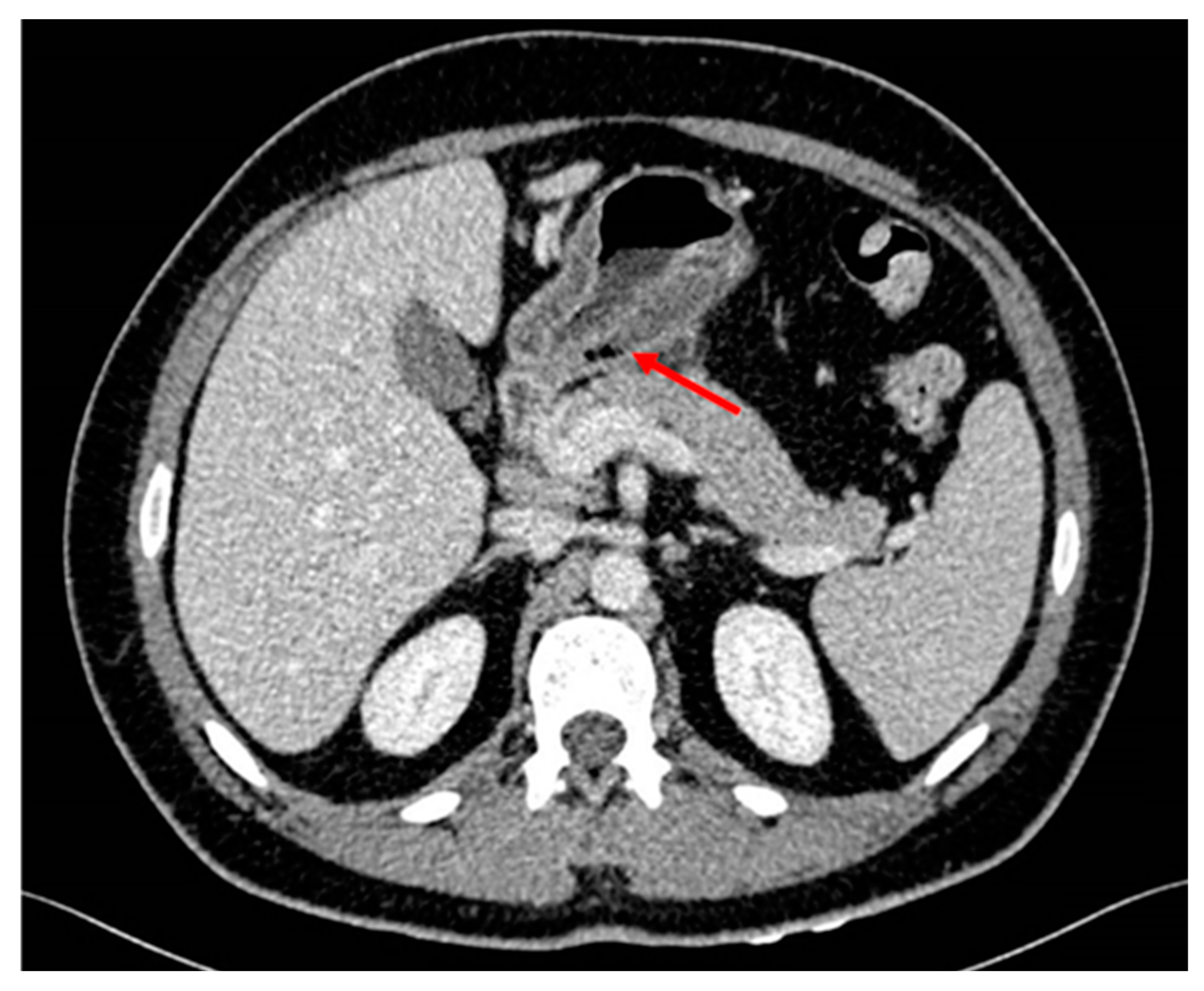
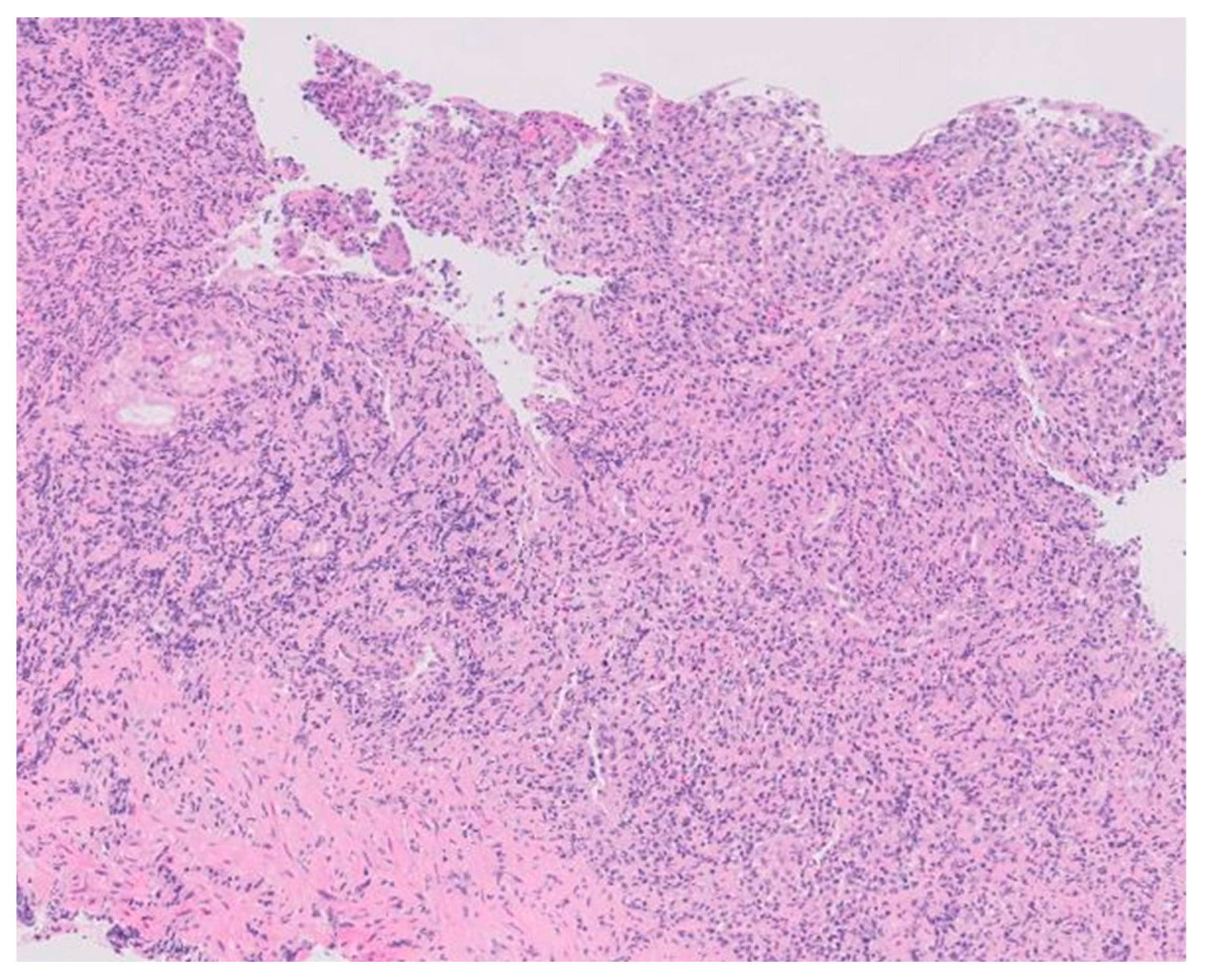
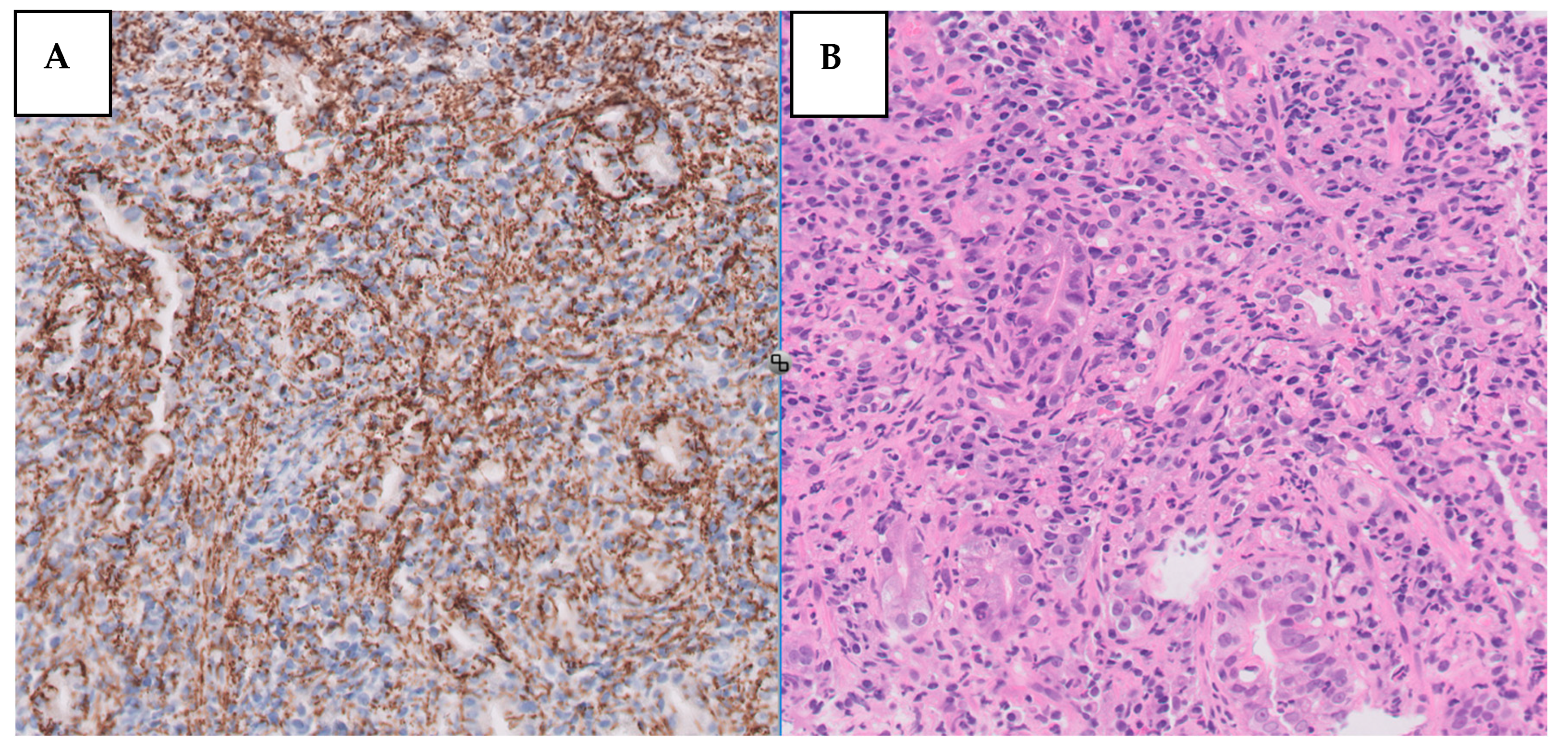
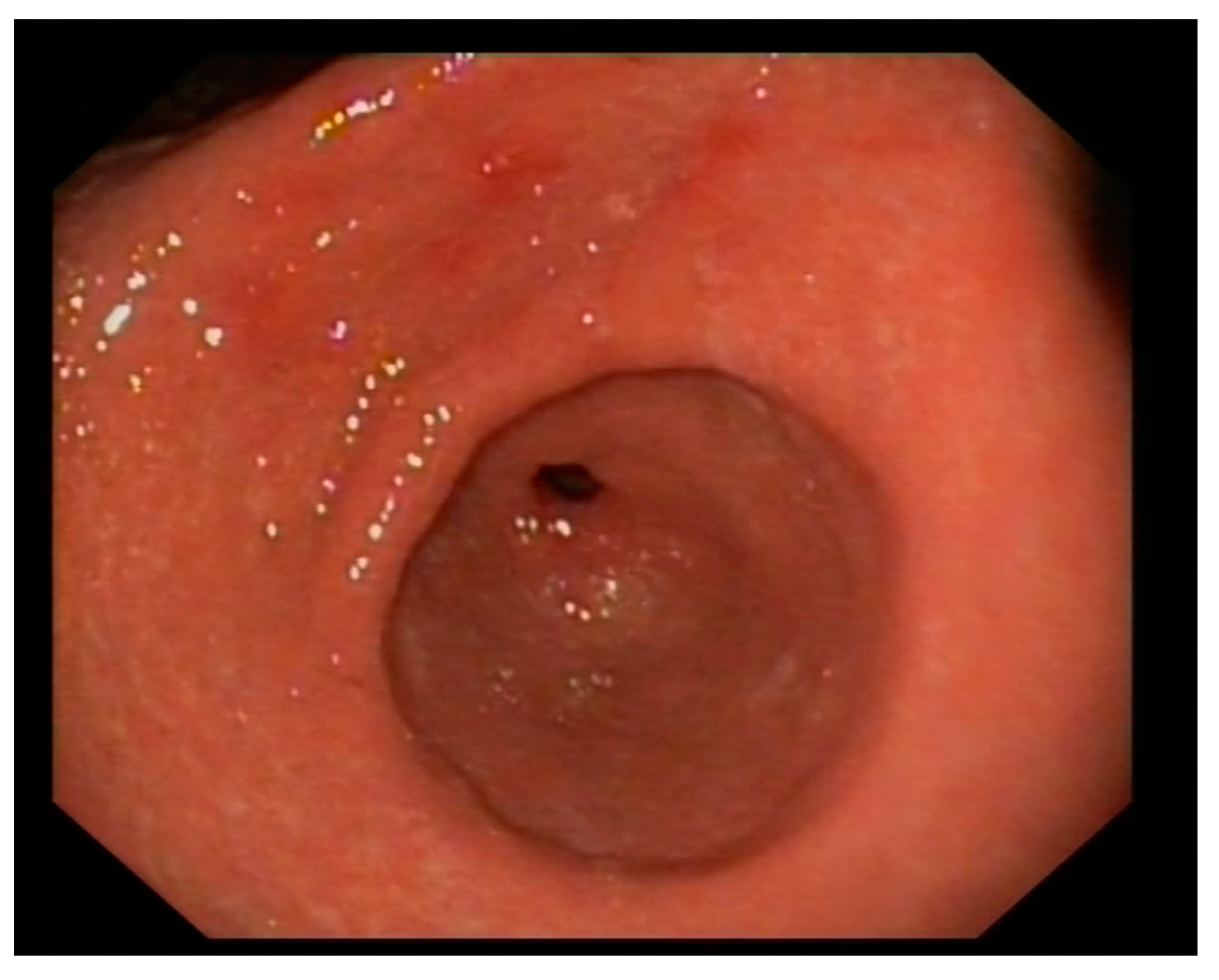
2. Case Description
3. Discussion
4. Conclusions
Author Contributions
Funding
Data Availability Statement
Conflicts of Interest
References
- Chaudhry, S.; Akinlusi, I.; Shi, T.; Cervantes, J. Secondary Syphilis: Pathophysiology, Clinical Manifestations, and Diagnostic Testing. Venereology 2023, 2, 65–75. [Google Scholar] [CrossRef]
- Sinagra, E.; Macaione, I.; Stella, M.; Shahini, E.; Maida, M.; Pompei, G.; Rossi, F.; Conoscenti, G.; Alloro, R.; Di Ganci, S.; et al. Gastric Syphilis Presenting as a Nodal Inflammatory Pseudotumor Mimicking a Neoplasm: Don’t Forget the Treponema! Case Report and Scoping Review of the Literature of the Last 65 Years. Gastroenterol. Insights 2023, 14, 178–190. [Google Scholar] [CrossRef]
- Okamoto, K.; Hatakeyama, S.; Umezawa, M.; Hayashi, S. Gastric syphilis: The great imitator in the stomach. IDCases 2018, 12, 97–98. [Google Scholar] [CrossRef] [PubMed]
- Morton, C.B. Syphilis of the stomach. Arch. Surg. 1932, 25, 880–889. [Google Scholar] [CrossRef]
- Graham, E.A. Surgical treatment of syphilis of the stomach. Ann. Surg. 1922, 76, 449–456. [Google Scholar] [CrossRef] [PubMed]
- Cai, J.; Ji, D.; Guan, J.L. Gastric syphilis. IDCases 2017, 8, 87–88. [Google Scholar] [CrossRef] [PubMed]
- Souza Varella Frazão, M.; Guimarães Vilaça, T.; Olavo Aragão Andrade Carneiro, F.; Toma, K.; Eliane Reina-Forster, C.; Ryoka Baba, E.; Cheng, S.; Ferreira de Souza, T.; Guimarães Hourneaux de Moura, E.; Sakai, P. Endoscopic aspects of gastric syphilis. Case Rep. Med. 2012, 2012, 646525. [Google Scholar] [CrossRef] [PubMed]
- Cao, J.; Zhu, J.; Xiang, Y.; Peng, P.; Liu, Q.; Fu, H.; Huang, Y. Gastric Syphilis Mimicking Lymphoma: A Case Report. Infect. Drug Resist. 2023, 16, 4539–4544. [Google Scholar] [CrossRef] [PubMed]
- Mylona, E.E.; Baraboutis, I.G.; Papastamopoulos, V.; Tsagalou, E.P.; Vryonis, E.; Samarkos, M.; Fanourgiakis, P.; Skoutelis, A. Gastric syphilis: A systematic review of published cases of the last 50 years. Sex. Transm. Dis. 2010, 37, 177–183. [Google Scholar] [CrossRef] [PubMed]
- Verma, R.; Al Elshafey, M.; Oza, T.; Azouz, A.; White, C. A Giant Syphilitic Gastric Ulcer. ACG Case Rep. J. 2022, 9, e00819. [Google Scholar] [CrossRef] [PubMed]
- Lan, Y.M.; Yang, S.W.; Dai, M.G.; Ye, B.; He, F.Y. Gastric syphilis mimicking gastric cancer: A case report. World J. Clin. Cases 2021, 9, 7798–7804. [Google Scholar] [CrossRef] [PubMed]
- Long, B.W.; Johnston, J.H.; Wetzel, W.; Flowers, R.H., 3rd; Haick, A. Gastric syphilis: Endoscopic and histological features mimicking lymphoma. Am. J. Gastroenterol. 1995, 90, 1504–1507. [Google Scholar] [PubMed]
- Machlowska, J.; Baj, J.; Sitarz, M.; Maciejewski, R.; Sitarz, R. Gastric Cancer: Epidemiology, Risk Factors, Classification, Genomic Characteristics and Treatment Strategies. Int. J. Mol. Sci. 2020, 21, 4012. [Google Scholar] [CrossRef] [PubMed]


| Comparison Chart between Case Reports | ||||||
|---|---|---|---|---|---|---|
| Case Reports | Time of Infection | Digestive History | Manifestations | Conventional Symptoms | Site of Infection/Suspicion of Gastric Cancer | Final Diagnosis |
| Guerrero C, Castañeda A et al. | Unknown | Gastric cancer diagnosed in his father at the age of 45 | Weight loss, hematemesis, epigastric pain, early satiety | No | Antrum/Yes | Pathological anatomy |
| Okamoto et al. [5] | Not described | Absent | Epigastric pain, nausea, emesis, weight loss | Yes, Skin Rash | Greater curvature/No | Pathological anatomy |
| Lan YM et al. [2] | Same sexual partner for 1 year but his partner was promiscuous | Absent | Epigastric and mesogastric pain, nausea, weight loss | No | Antrum-pylorus/Yes | Pathological anatomy |
| Sinagra E et al. [3] | Not described | Absent | Epigastric pain, nausea, anorexia, vomiting, weight loss | Yes, skin rash and lymphadenophaty | Antrum/Yes | Pathological anatomy |
| Verma R et al. [7] | Not described | Absent | Hematemesis, epigastric pain | No | Antrum/Yes | Pathological anatomy |
Disclaimer/Publisher’s Note: The statements, opinions and data contained in all publications are solely those of the individual author(s) and contributor(s) and not of MDPI and/or the editor(s). MDPI and/or the editor(s) disclaim responsibility for any injury to people or property resulting from any ideas, methods, instructions or products referred to in the content. |
© 2024 by the authors. Licensee MDPI, Basel, Switzerland. This article is an open access article distributed under the terms and conditions of the Creative Commons Attribution (CC BY) license (https://creativecommons.org/licenses/by/4.0/).
Share and Cite
Guerrero Muñoz, C.; Castañeda Agredo, A.; Romero Valle, M.J.; Silva Silva, A.M.; León del Campo, M.; Azpirtarte Sánchez, C.; Domínguez Fraga, M.; Milán Pilo, M.V.; Polo Lorduy, B.A.; González Guirado, A.; et al. Beyond Tumors: Gastric Syphilis Emulating a Gastric Neoplasia. Medicina 2024, 60, 629. https://doi.org/10.3390/medicina60040629
Guerrero Muñoz C, Castañeda Agredo A, Romero Valle MJ, Silva Silva AM, León del Campo M, Azpirtarte Sánchez C, Domínguez Fraga M, Milán Pilo MV, Polo Lorduy BA, González Guirado A, et al. Beyond Tumors: Gastric Syphilis Emulating a Gastric Neoplasia. Medicina. 2024; 60(4):629. https://doi.org/10.3390/medicina60040629
Chicago/Turabian StyleGuerrero Muñoz, Claudia, Andres Castañeda Agredo, Maria Jose Romero Valle, Andrés Mauricio Silva Silva, Marta León del Campo, Claudia Azpirtarte Sánchez, Marta Domínguez Fraga, Manuel Vicente Milán Pilo, Benjamin Arturo Polo Lorduy, Agustina González Guirado, and et al. 2024. "Beyond Tumors: Gastric Syphilis Emulating a Gastric Neoplasia" Medicina 60, no. 4: 629. https://doi.org/10.3390/medicina60040629
APA StyleGuerrero Muñoz, C., Castañeda Agredo, A., Romero Valle, M. J., Silva Silva, A. M., León del Campo, M., Azpirtarte Sánchez, C., Domínguez Fraga, M., Milán Pilo, M. V., Polo Lorduy, B. A., González Guirado, A., Martín Relloso, M. J., Sánchez-Fayos Calabuig, P., & Bosch Esteva, O. (2024). Beyond Tumors: Gastric Syphilis Emulating a Gastric Neoplasia. Medicina, 60(4), 629. https://doi.org/10.3390/medicina60040629