
Effects of Upper Limb Control on the Less-Affected Side on Upper Limb Function, Respiration, Balance, and Activities of Daily Living in Stroke


Abstract
:1. Instruction
2. Materials and Methods
2.1. Participants
2.2. Experimental Procedure
Real-Time Feedback
2.3. Training Program
2.4. Outcome Measurements
2.4.1. Upper Limb Function Assessment
2.4.2. Respiratory Assessment
2.4.3. Balance Assessment
2.4.4. Activities of Daily Living
2.5. Data Analysis
3. Results
3.1. General Characteristics of the Participants
3.2. Changes in Upper Limb Function of Affected Side
3.3. Changes in Respiration Function
3.4. Changes in Balance
3.5. Changes in Activities of Daily Living
4. Discussion
5. Conclusions
Author Contributions
Funding
Institutional Review Board Statement
Informed Consent Statement
Data Availability Statement
Conflicts of Interest
References
- Lee, L.J.; Coppieters, M.W.; Hodges, P.W. Anticipatory, postural adjustments to arm movement reveal complex control of paraspinal muscles in the thorax. J. Electromyogr. Kinesiol. 2009, 19, 46–54. [Google Scholar] [CrossRef] [PubMed]
- Ruhland, J.L.; van Kan, P.L. Medial, pontine hemorrhagic stroke. Phys. Ther. 2003, 83, 552–566. [Google Scholar] [CrossRef]
- Suvada, K.C.; Deol, J.; Dewald, J.P.A.; Acosta, A.M. A method for quantifying trunk motor control during reaching in individuals post hemiparetic stroke. In Proceedings of the 42nd Annual International Conference of the IEEE Engineering in Medicine and Biology Society, Montreal, QC, Canada, 20–24 July 2020; pp. 3743–3746. [Google Scholar]
- McPherson, J.G.; Chen, A.; Ellis, M.D.; Yao, J.; Heckman, C.J.; Dewald, J.P.A. Progressive, recruitment of contralesional cortico-reticulospinal pathways drives motor impairment post stroke. J. Physiol. 2018, 596, 1211–1225. [Google Scholar] [CrossRef] [PubMed]
- Chen, L.; Lo, W.L.; Mao, Y.R.; Ding, M.H.; Lin, Q.; Li, H.; Huang, D.F. Effect of virtual reality on postural and balance control in patients with stroke: A systematic literature review. Biomed Res. Int. 2016, 7309272. [Google Scholar] [CrossRef]
- Pandian, S.; Arya, K.N.; Kumar, D. Effect, of motor training involving the less-affected side (MTLA) in post-stroke subjects: A pilot randomized controlled trial. Top. Stroke Rehabil. 2015, 22, 357–367. [Google Scholar] [CrossRef] [PubMed]
- Van Criekinge, T.; Saeys, W.; Hallemans, A.; Velghe, S.; Viskens, P.J.; Vereeck, L.; Truijen, S. Trunk, biomechanics during hemiplegic gait after stroke: A systematic review. Gait Posture 2017, 54, 133–143. [Google Scholar] [CrossRef]
- Pandian, S.; Arya, K.N.; Kumar, D. Does, motor training of the nonparetic side influences balance and function in chronic stroke? A pilot RCT. Sci. World J. 2014, 17, 769726. [Google Scholar] [CrossRef]
- Verheyden, G.; Nieuwboer, A.; Feys, H.; Thijs, V.; Vaes, K.; De Weerdt, W. Discriminant, ability of the Trunk Impairment Scale: A comparison between stroke patients and healthy individuals. Disabil. Rehabil. 2005, 27, 1023–1028. [Google Scholar] [CrossRef]
- Ryerson, S.; Byl, N.N.; Brown, D.A.; Wong, R.A.; Hidler, J.M. Altered, trunk position sense and its relation to balance functions in people post-stroke. J. Neurol. Phys. Ther. 2008, 32, 14–20. [Google Scholar] [CrossRef]
- Titus, A.W.; Hillier, S.; Louw, Q.A.; Inglis-Jassiem, G. An, analysis of trunk kinematics and gait parameters in people with stroke. Afr. J. Disabil. 2018, 7, 310. [Google Scholar] [CrossRef]
- Valdes, B.A.; Glegg SM, N.; Van der Loos, H.F.M. Trunk, compensation during bimanual reaching at different heights by healthy and hemiparetic adults. J. Mot. Behav. 2017, 49, 580–592. [Google Scholar] [CrossRef]
- Curuk, E.; Lee, Y.; Aruin, A.S. Individuals, with stroke use asymmetrical anticipatory postural adjustments when counteracting external perturbations. Mot. Control. 2019, 23, 461–471. [Google Scholar] [CrossRef]
- Schepens, B.; Drew, T. Independent, and convergent signals from the pontomedullary reticular formation contribute to the control of posture and movement during reaching in the cat. J. Neurophysiol. 2004, 92, 2217–2238. [Google Scholar] [CrossRef]
- Alhwoaimel, N.; Turk, R.; Warner, M.; Verheyden, G.; Thijs, L.; Wee, S.K.; Hughes, A.M. Do trunk exercises improve trunk and upper limb performance, post stroke? A systematic review and meta-analysis. NeuroRehabilitation 2018, 43, 395–412. [Google Scholar] [CrossRef]
- Jo, M.R.; Kim, N.S.; Jung, J.H. The effects of respiratory muscle training on respiratory function, respiratory muscle strength, and cough capacity in stroke patients. J. Korean Soc. Phys. Med. 2014, 9, 399–405. [Google Scholar] [CrossRef]
- Kim, J.O.; Lee, B.H. Effect of upper limb coordination exercise during standing on the paretic side on balance, gait ability and activities of daily living in persons with stroke. Phys. Ther. Rehabil. Sci. 2017, 6, 53–58. [Google Scholar] [CrossRef]
- Chan, I.H.; Fong, K.N.; Chan, D.Y.; Wang, A.Q.; Cheng, E.K.; Chau, P.H.; Cheung, H.K. Effects, of arm weight support training to promote recovery of upper limb function for subacute patients after stroke with different levels of arm impairments. BioMed Res. Int. 2016, 2016, 9346374. [Google Scholar] [CrossRef] [PubMed]
- De Baets, L.; Van Deun, S.; Monari, D.; Jaspers, E. Three-dimensional kinematics of the scapula and trunk, and associated scapular muscle timing in individuals with stroke. Hum. Mov. Sci. 2016, 48, 82–90. [Google Scholar] [CrossRef] [PubMed]
- Michimata, A.; Kondo, T.; Suzukamo, Y.; Chiba, M.; Izumi, S.I. The manual function test: Norms for 20-to 90-year-olds and effects of age, gender, and hand dominance on dexterity. Tohoku J. Exp. Med. 2008, 214, 257–267. [Google Scholar] [CrossRef]
- Krukowska, J.; Bugajski, M.; Sienkiewicz, M.; Czernicki, J. The influence of NDT-Bobath and PNF methods on the field support and total path length measure foot pressure (COP) in patients after stroke. Neurol. Neurochirugia Pol. 2016, 50, 449–454. [Google Scholar] [CrossRef]
- Shah, S.; Vanclay, F.; Cooper, B. Improving, the sensitivity of the Barthel Index for stroke rehabilitation. J. Clin. Epidemiol. 1989, 42, 703–709. [Google Scholar] [CrossRef] [PubMed]
- Choi, Y.I.; Kim, W.H.; Park, E.Y.; Kim, E.J. The validity, reliability and discriminative index of the korean version of modified barthel index (K-MBI) in stroke patients. J. Korea Assoc. Inf. Sci. 2012, 13, 4119–4125. [Google Scholar]
- Arya, K.N.; Pandian, S.; Kumar, V. Effect, of activity-based mirror therapy on lower limb motor-recovery and gait in stroke: A randomised controlled trial. Neuropsychol. Rehabil. 2019, 29, 1193–1210. [Google Scholar] [CrossRef] [PubMed]
- Silva, C.C.; Silva, A.; Sousa, A.; Pinheiro, A.R.; Bourlinova, C.; Silva, A.; Salazar, A.; Borges, C.; Crasto, C.; Correia, M.V.; et al. Co-activation, of upper limb muscles during reaching in post-stroke subjects: An analysis of the contralesional and ipsilesional limbs. J. Electromyogr. Kinesiol. 2014, 24, 731–738. [Google Scholar] [CrossRef] [PubMed]
- Baker, R.; Pain, L.M.; Richardson, D.; Agur, A.M. Effect of trunk-restraint training on function and compensatory trunk, shoulder and elbow patterns during post-stroke reach: A systematic review. Disabil. Rehabil. 2015, 37, 553–562. [Google Scholar]
- Schneemilch, C. General anaesthesia for neurological diseases. Anasthesiol. Intensivmed. Notfallmedizin Schmerzther. AINS 2010, 45, 336–344. [Google Scholar] [CrossRef] [PubMed]
- Jandt, S.R.; da Sil Caballero, R.M.; Junior LA, F.; Dias, A.S. Correlation between trunk control, respiratory muscle strength and spirometry in patients with stroke: An observational study. Physiother. Res. Int. 2011, 16, 218–224. [Google Scholar] [CrossRef] [PubMed]
- Lipska, L.; Visokai, V.; Levy, M.; Koznar, B.; Zaruba, P. Celiac, axis stenosis and lethal liver ischemia after pancreaticoduodenectomy. Hepatogastroenterology 2009, 56, 1203–1206. [Google Scholar]
- Themudo, R.E.; Lindahl, B.; Johansson, L.; Venge, P.; Ahlström, H.; Barbier, C.E.; Bjerner, T. Unrecognized, myocardial scars detected by delayed–enhanced MRI are associated with increased levels of NT-proBNP. Coron. Artery Dis. 2011, 22, 158–164. [Google Scholar] [CrossRef]
- Park, C.H.; You, Y.Y.; Choi, Y.E. Change of balance and gait parameters while sit to stand with different foot position in chronic stroke patient. Korean Soc. Med. Ther. Sci. 2017, 9, 51–59. [Google Scholar]
- Kanekar, N.; Aruin, A.S. Improvement, of anticipatory postural adjustments for balance control: Effect of a single training session. J. Electromyogr. Kinesiol. 2015, 25, 400–405. [Google Scholar] [CrossRef] [PubMed]
- Kang, Y.J.; Ku, J.; Han, K.; Kim, S.I.; Yu, T.W.; Lee, J.H.; Park, C.I. Development, and clinical trial of virtual reality-based cognitive assessment in people with stroke: Preliminary study. CyberPsychol. Behav. 2008, 11, 329–339. [Google Scholar] [CrossRef] [PubMed]

| Sequence | Intermediate and Finishing Postures |
|---|---|
| 1 | Seat the patient on the mat and stabilize their feet and ankles near the midline. |
| 2 | Hold the pelvis and grasp the proximal femurs to stabilize them near the midline. Perform steps 1 and 2 bilaterally. |
| 3 | Place both hands on the table and align the trunk with the midline. |
| 4 | The therapist stabilizes the pelvis by holding it with both hands. |
| 5 | The therapist holds the patient’s lower trunk and performs trunk extension. |
| 6 | The therapist holds the patient’s lower trunk and performs trunk flexion. |
| 7 | The therapist holds the patient’s trunk and moves toward the affected side. |
| 8 | Therapist holds the patient’s trunk and moves toward the non-affected side before stabilizing in the center. |
| 1 | The patient wears the HMD and prepares on the mat. |
| 2 | Prepare for trunk control exercise program (10 min). |
| 3 | The therapist holds the patient’s trunk and upper limb on the affected side and waits. |
| 4 | Play the intervention video with real-time feedback from the HMD connected to the laptop, along with the verbal command from the assistant to “start”. |
| 5 | The patient follows the movements while watching the video on the HMD, and the therapist conducts treatment while adjusting the posture on the affected side (20 min). |
| 6 | Treatment focuses on enhancing posture control on the affected side and promoting mobility of the upper limb. This is achieved by adjusting the posture of the trunk and upper limb on the affected side, leading to improved stability on the non-affected side and increased mobility of the upper limb. |
| Weeks | Intermediate and Finishing Postures | Picture |
|---|---|---|
| 1st week | A. Stabilize the trunk and secure the paralyzed arm on the assistive table. B. Flex and extend the fingers of the non-paralyzed side to regulate tension. C. Repeat the movement of flexing and then extending the wrist of the non-paralyzed side. D. Repeat the movement of pronating and then supinating the wrist of the non-paralyzed side. E. All movements are performed in collaboration with the therapist, who assists in controlling the patient’s upper limb strength while actively participating. |  |
| 2nd week | A. Stabilize the trunk and secure the paralyzed arm on the assistive table. B. Flex the elbow of the non-paralyzed side and then extend it again. C. Bend the elbow of the non-paralyzed side and then flex the shoulder joint. D. Bend the elbow of the non-paralyzed side and then bring the shoulder joint forward. E. All movements are conducted with a therapist, actively assisting while regulating the patient’s upper limb strength. | 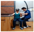 |
| 3rd week | A. Stabilize the trunk and secure the paralyzed arm on the assistive table. B. In a flexed position of the non-paralyzed elbow, move the shoulder joint backward. C. In a bent position of the paralyzed elbow, move the shoulder joint upward. D. In a flexed position of the non-paralyzed elbow, move the shoulder joint downward. E. All movements are conducted with a therapist, actively assisting while regulating the patient’s upper limb strength. |  |
| 4th week | A. Stabilize the trunk and secure the paralyzed arm on the assistive table. B. In the bent position of the non-paralyzed elbow, flex the shoulder joint upward. C. In the bent position of the non-paralyzed elbow, flex and extend the upper limb. D. In the bent position of the non-paralyzed elbow, flex and extend the lower limb. E. All movements are conducted with a therapist, actively assisting while regulating the patient’s upper limb strength. |  |
| Characteristics | ULD-LA Group (n = 14) | ULD-A Group (n = 14) | t (p) |
|---|---|---|---|
| Sex (M/F) | 9/5 | 6/8 | −1.520 (0.617) |
| Age (years) | 61.15 (1.61) | 63.32 (4.21) | 1.886 (0.637) |
| Height (cm) | 167.64 (7.92) | 161.42 (5.15) | 2.213 (0.334) |
| Weight (kg) | 67.86 (12.60) | 66.54 (2.93) | 1.746 (0.945) |
| Lesion sites (Rt/Lt) | 7/7 | 7/7 | 0.366 (1.000) |
| Onset period (month) | 10.71 (4.23) | 12.57 (5.15) | 1.325 (0.617) |
| ULD-LA (n = 14) | ULD-A (n = 14) | Time F (p) | Group F (p) | Time × Group F (p) | ||
|---|---|---|---|---|---|---|
| A | 0 w | 70.12 (18.15) a | 60.61 (11.25) | 3.072 (0.091) | 11.426 (0.002) * | 0.296 (0.591) |
| 4 w | 74.62 (20.14) | 61.62 (13.67) | ||||
| 6 w | 78.93 (21.66) | 62.63 (10.85) | ||||
| B | 0 w | 61.92 (17.18) | 53.48 (20.13) | 10.234 (0.004) * | 9.954 (0.004) * | 0.113 (0.039) * |
| 4 w | 62.56 (18.53) | 55.04 (18.44) | ||||
| 6 w | 69.28 (17.94) | 54.47 (16.59) | ||||
| C | 0 w | 112.32 (43.58) | 87.43 (51.53) | 2.782 (0.107) | 5.397 (0.028) * | 4.635 (0.041) * |
| 4 w | 127.42 (45.40) | 88.13 (52.01) | ||||
| 6 w | 130.32 (41.94) | 89.13 (48.55) | ||||
| D | 0 w | 36.14 (17.33) | 32.91 (15.68) | 5.455 (0.027) * | 3.388 (0.077) | 2.343 (0.138) |
| 4 w | 41.64 (15.22) | 31.21 (31.22)) | ||||
| 6 w | 50.54 (34.81) | 38.01 (19.83) | ||||
| E | 0 w | 17.62 (9.63) | 17.91 (10.22) | 1.064 (0.312) | 0.894 (0.033) * | 1.064 (0.312) |
| 4 w | 21.21 (8.68) | 18.41 (9.77) | ||||
| 6 w | 22.42 (8.88) | 17.94 (9.15) | ||||
| ULD-LA (n = 14) | ULD-A (n = 14) | Time F (p) | Group F (p) | Time × Group F (p) | ||
|---|---|---|---|---|---|---|
| A | 0 w | 3.14 (0.43) a | 2.68 (1.78) | 1.180 (0.287) | 0820 (0.374) | 6.007 (0.021) * |
| 4 w | 3.34 (0.36) | 2.07 (1.65) | ||||
| 6 w | 3.94 (0.49) | 2.56 (0.24) | ||||
| B | 0 w | 1.83 (0.19) | 1.28 (0.24) | 1.465 (0.237) | 3.296 (0.081) | 8.696 (0.007) * |
| 4 w | 1.84 (0.16) | 1.27 (0.13) | ||||
| 6 w | 2.13 (0.21) | 1.36 (0.44) | ||||
| C | 0 w | 2.61 (0.39) | 2.18 (0.40) | 2.225 (0.148) | 6.813 (0.015) | 2.245 (0.146) |
| 4 w | 3.11 (0.45) | 2.17 (0.16) | ||||
| 6 w | 3.41 (0.45) | 2.46 (0.27) | ||||
| ULD-LA (n = 14) | ULD-A (n = 14) | Time F (p) | Group F (p) | Time × Group F (p) | ||
|---|---|---|---|---|---|---|
| A | 0 w | 1057.86 (149.11) a | 1096.11 (171.52) | 12.776 (0.001) * | 5.275 (0.030) * | 12.741 (0.035) * |
| 4 w | 1010.54 (186.41) | 1183.28 (210.54) | ||||
| 6 w | 919.22 (155.63) | 1066.21 (191.23) | ||||
| B | 0 w | 916.12 (157.71) | 1271.22 (101.64) | 30.504 (0.000) * | 7.753 (0.010) * | 0.273 (0.042) * |
| 4 w | 793.01 (161.41) | 1216.23 (119.14) | ||||
| 6 w | 734.09 (161.63) | 1211 (121.16) | ||||
| C | 0 w | 19.21 (3.56) | 27.94 (2.05) | 0.767 (0.000) * | 1.168 (0.290) | 1.787 (0.193) |
| 4 w | 17.99 (27.48) | 64.67 (19.52) | ||||
| 6 w | 16.64 (23.04) | 58.21 (16.43) | ||||
| ULD-LA (n = 14) | ULD-A (n = 14) | Time F (p) | Group F (p) | Time × Group F (p) | ||
|---|---|---|---|---|---|---|
| KMBI | 0 w | 74.31 (3.58) a | 65.06 (2.64) | 2.747 (0.044) * | 3.003 (0.035) * | 0.277 (0.03) * |
| 4 w | 77.41 (4.51) | 70.66 (2.27) | ||||
| 6 w | 79.19 (3.05) | 70.97 (3.19) | ||||
Disclaimer/Publisher’s Note: The statements, opinions and data contained in all publications are solely those of the individual author(s) and contributor(s) and not of MDPI and/or the editor(s). MDPI and/or the editor(s) disclaim responsibility for any injury to people or property resulting from any ideas, methods, instructions or products referred to in the content. |
© 2024 by the authors. Licensee MDPI, Basel, Switzerland. This article is an open access article distributed under the terms and conditions of the Creative Commons Attribution (CC BY) license (https://creativecommons.org/licenses/by/4.0/).
Share and Cite
Kim, J.-O.; Lee, M.-Y.; Lee, B.-H. Effects of Upper Limb Control on the Less-Affected Side on Upper Limb Function, Respiration, Balance, and Activities of Daily Living in Stroke. Medicina 2024, 60, 937. https://doi.org/10.3390/medicina60060937
Kim J-O, Lee M-Y, Lee B-H. Effects of Upper Limb Control on the Less-Affected Side on Upper Limb Function, Respiration, Balance, and Activities of Daily Living in Stroke. Medicina. 2024; 60(6):937. https://doi.org/10.3390/medicina60060937
Chicago/Turabian StyleKim, Ju-O, Mi-Young Lee, and Byoung-Hee Lee. 2024. "Effects of Upper Limb Control on the Less-Affected Side on Upper Limb Function, Respiration, Balance, and Activities of Daily Living in Stroke" Medicina 60, no. 6: 937. https://doi.org/10.3390/medicina60060937