
Predictive Utility of Changes in Optic Nerve Sheath Diameter after Cardiac Arrest for Neurologic Outcomes

 , ,
, ,
Abstract
:1. Introduction
2. Materials and Methods
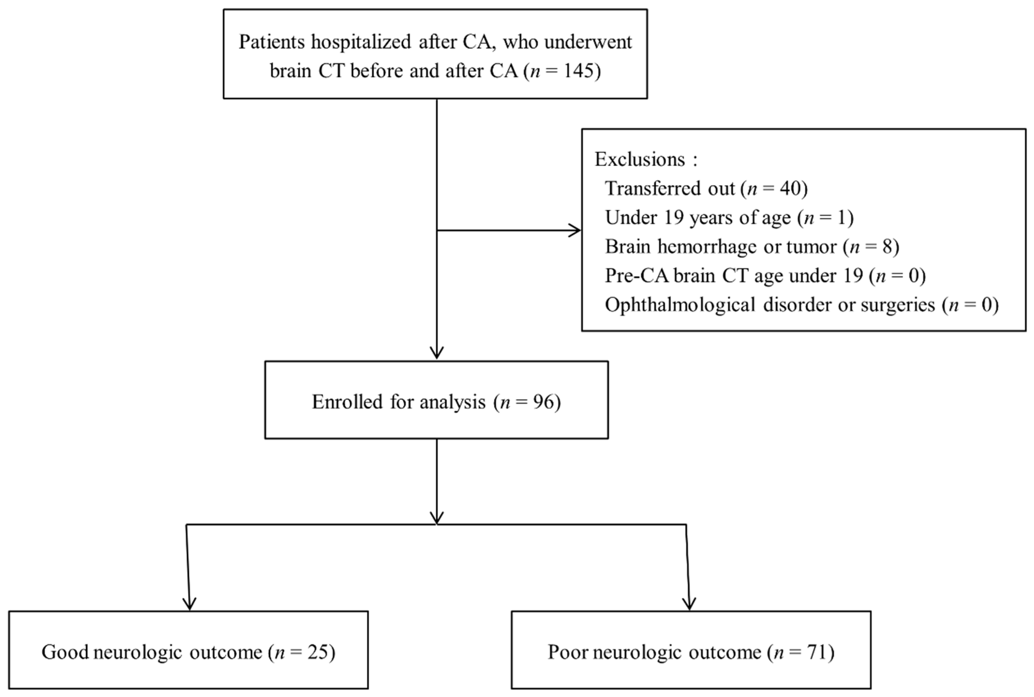
2.1. Study Design and Population
2.2. Data Collection
2.3. ONSD Measurements Using Brain CT
2.4. Sample Size
2.5. Statistical Analysis
3. Results
3.1. Baseline Characteristics
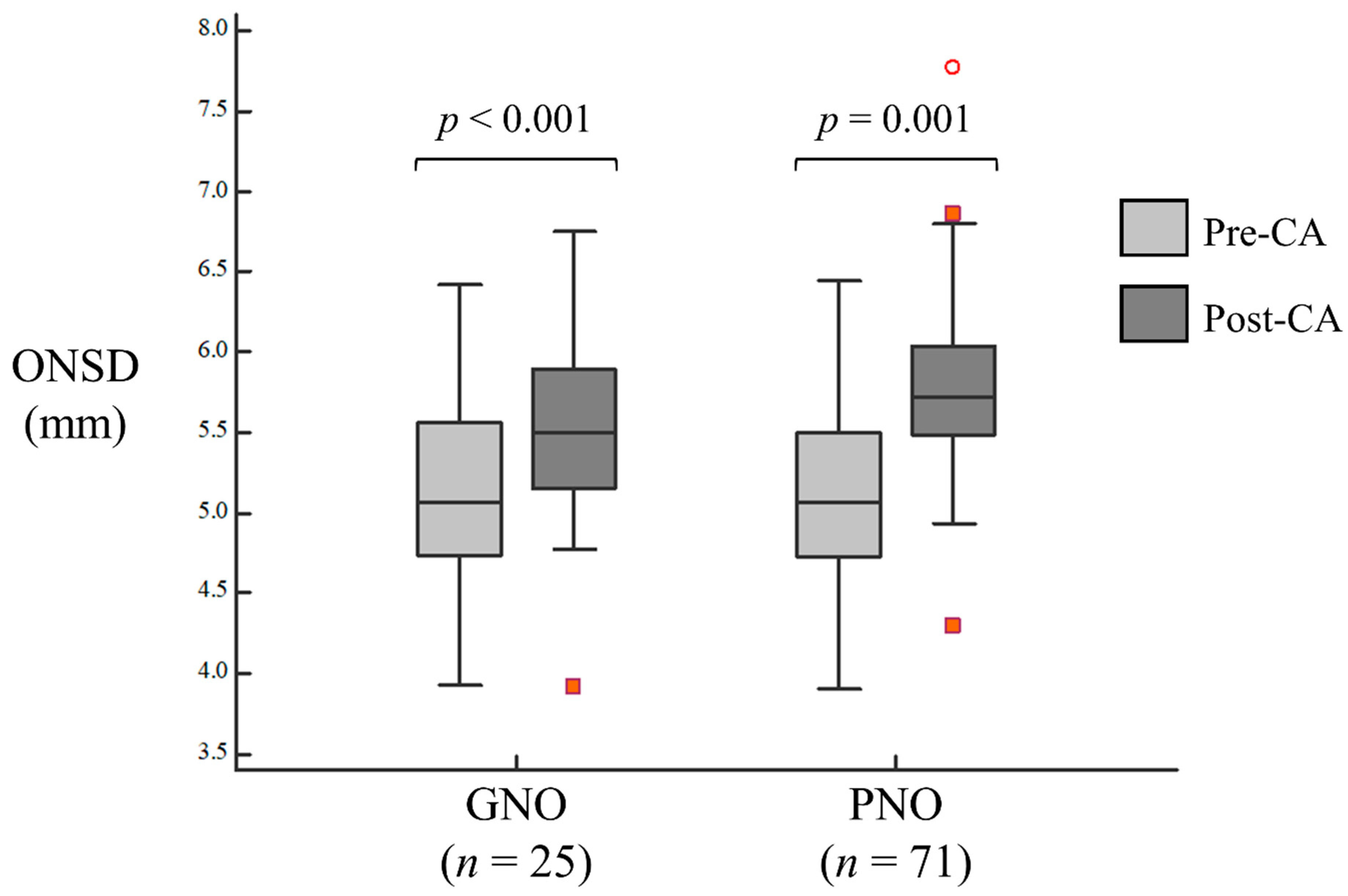
3.2. Comparison of Pre-CA and Post-CA ONSDs
3.3. The Association between ONSD Changes and Neurologic Outcomes
3.4. Diagnostic Value of ONSD Changes for Predicting the Neurologic Outcome
4. Discussion
5. Conclusions
Supplementary Materials
Author Contributions
Funding
Institutional Review Board Statement
Informed Consent Statement
Data Availability Statement
Acknowledgments
Conflicts of Interest
Abbreviations
| ONSD | optic nerve sheath diameter |
| CA | cardiac arrest |
| CT | computed tomography |
| GNO | good neurologic outcome |
| PNO | poor neurologic outcome |
| ICP | intracranial pressure |
| ROSC | return of spontaneous circulation |
| GWR | gray-to-white matter ratio |
| CPR | cardiopulmonary resuscitation |
| TTM | targeted temperature management |
| CPCs | Cerebral Performance Categories |
| PACS | picture archiving and communication system |
| IQR | interquartile range |
| AUC | area under curve |
| ROC | receiver operating characteristic |
| CI | confidence interval |
| PPV | positive predictive value |
| NPV | negative predictive value |
| CSF | cerebrospinal fluid |
| BMI | body mass index |
| ETD | eyeball transverse diameter |
References
- Gueugniaud, P.Y.; Garcia-Darennes, F.; Gaussorgues, P.; Bancalari, G.; Petit, P.; Robert, D. Prognostic significance of early intracranial and cerebral perfusion pressures in post-cardiac arrest anoxic coma. Intensive Care Med. 1991, 17, 392–398. [Google Scholar] [CrossRef] [PubMed]
- Sundgreen, C.; Larsen, F.S.; Herzog, T.M.; Knudsen, G.M.; Boesgaard, S.; Aldershvile, J. Autoregulation of cerebral blood flow in patients resuscitated from cardiac arrest. Stroke 2001, 32, 128–132. [Google Scholar] [CrossRef] [PubMed] [Green Version]
- Metter, R.B.; Rittenberger, J.C.; Guyette, F.X.; Callaway, C.W. Association between a quantitative CT scan measure of brain edema and outcome after cardiac arrest. Resuscitation 2011, 82, 1180–1185. [Google Scholar] [CrossRef] [PubMed] [Green Version]
- Iida, K.; Satoh, H.; Arita, K.; Nakahara, T.; Kurisu, K.; Ohtani, M. Delayed hyperemia causing intracranial hypertension after cardiopulmonary resuscitation. Crit. Care Med. 1997, 25, 971–976. [Google Scholar] [CrossRef] [PubMed]
- Sandroni, C.; Cavallaro, F.; Callaway, C.W.; Sanna, T.; D’Arrigo, S.; Kuiper, M.; Della Marca, G.; Nolan, J.P. Predictors of poor neurological outcome in adult comatose survivors of cardiac arrest: A systematic review and meta-analysis. Part 1: Patients not treated with therapeutic hypothermia. Resuscitation 2013, 84, 1310–1323. [Google Scholar] [CrossRef]
- Young, G.B. Clinical practice. Neurologic prognosis after cardiac arrest. N. Engl. J. Med. 2009, 361, 605–611. [Google Scholar] [CrossRef]
- Cloostermans, M.C.; van Meulen, F.B.; Eertman, C.J.; Hom, H.W.; van Putten, M.J. Continuous electroencephalography monitoring for early prediction of neurological outcome in postanoxic patients after cardiac arrest: A prospective cohort study. Crit. Care Med. 2012, 40, 2867–2875. [Google Scholar] [CrossRef]
- Sandroni, C.; Cariou, A.; Cavallaro, F.; Cronberg, T.; Friberg, H.; Hoedemaekers, C.; Horn, J.; Nolan, J.P.; Rossetti, A.O.; Soar, J. Prognostication in comatose survivors of cardiac arrest: An advisory statement from the European Resuscitation Council and the European Society of Intensive Care Medicine. Resuscitation 2014, 85, 1779–1789. [Google Scholar] [CrossRef]
- Kamps, M.J.A.; Horn, J.; Oddo, M.; Fugate, J.E.; Storm, C.; Cronberg, T.; Wijman, C.A.; Wu, O.; Binnekade, J.M.; Hoedemaekers, C.W.E. Prognostication of neurologic outcome in cardiac arrest patients after mild therapeutic hypothermia: A meta-analysis of the current literature. Intensive Care Med. 2013, 39, 1671–1682. [Google Scholar] [CrossRef]
- Geocadin, R.G.; Callaway, C.W.; Fink, E.L.; Golan, E.; Greer, D.M.; Ko, N.U.; Lang, E.; Licht, D.J.; Marino, B.S.; McNair, N.D.; et al. Standards for Studies of Neurological Prognostication in Comatose Survivors of Cardiac Arrest: A Scientific Statement from the American Heart Association. Circulation 2019, 140, e517–e542. [Google Scholar] [CrossRef] [PubMed]
- Lee, Y.H.; Oh, Y.T.; Ahn, H.C.; Kim, H.S.; Han, S.J.; Lee, J.J.; Lee, T.H.; Seo, J.Y.; Shin, D.H.; Ha, S.O.; et al. The prognostic value of the grey-to-white matter ratio in cardiac arrest patients treated with extracorporeal membrane oxygenation. Resuscitation 2016, 99, 50–55. [Google Scholar] [CrossRef]
- Lee, B.K.; Kim, W.Y.; Shin, J.; Oh, J.S.; Wee, J.H.; Cha, K.C.; Park, Y.; Choi, J.H.; Jeung, K.W.; Korean Hypothermia Network Investigators. Prognostic value of gray matter to white matter ratio in hypoxic and non-hypoxic cardiac arrest with non-cardiac etiology. Am. J. Emerg. Med. 2016, 34, 1583–1588. [Google Scholar] [CrossRef] [PubMed]
- Chae, M.K.; Ko, E.; Lee, J.H.; Lee, T.R.; Yoon, H.; Hwang, S.Y.; Cha, W.C.; Shin, T.G.; Sim, M.S.; Jo, I.J.; et al. Better prognostic value with combined optic nerve sheath diameter and grey-to-white matter ratio on initial brain computed tomography in post-cardiac arrest patients. Resuscitation 2016, 104, 40–45. [Google Scholar] [CrossRef] [PubMed]
- Kim, Y.H.; Lee, J.H.; Hong, C.K.; Cho, K.W.; Yeo, J.H.; Kang, M.J.; Kim, Y.W.; Lee, K.Y.; Kim, J.J.; Hwang, S.Y. Feasibility of optic nerve sheath diameter measured on initial brain computed tomography as an early neurologic outcome predictor after cardiac arrest. Acad. Emerg. Med. 2014, 21, 1121–1128. [Google Scholar]
- Ryu, J.-A.; Chung, C.R.; Cho, Y.H.; Sung, K.; Suh, G.Y.; Park, T.K.; Bin Song, Y.; Hahn, J.-Y.; Choi, J.-H.; Gwon, H.-C.; et al. The association of findings on brain computed tomography with neurologic outcomes following extracorporeal cardiopulmonary resuscitation. Crit Care 2017, 21, 15. [Google Scholar] [CrossRef] [Green Version]
- Sekhon, M.S.; Griesdale, D.E.; Robba, C.; McGlashan, N.; Needham, E.; Walland, K.; Shook, A.C.; Smielewski, P.; Czosnyka, M.; Gupta, A.K.; et al. Optic nerve sheath diameter on computed tomography is correlated with simultaneously measured intracranial pressure in patients with severe traumatic brain injury. Intensive Care Med. 2014, 40, 1267–1274. [Google Scholar] [CrossRef]
- Rajajee, V.; Fletcher, J.J.; Rochlen, L.R.; Jacobs, T.L. Comparison of accuracy of optic nerve ultrasound for the detection of intracranial hypertension in the setting of acutely fluctuating vs stable intracranial pressure: Post-hoc analysis of data from a prospective, blinded single center study. Crit Care 2012, 16, R79. [Google Scholar] [CrossRef] [PubMed] [Green Version]
- Chelly, J.; Deye, N.; Guichard, J.-P.; Vodovar, D.; Vong, L.; Jochmans, S.; Thieulot-Rolin, N.; Sy, O.; Serbource-Goguel, J.; Vinsonneau, C.; et al. The optic nerve sheath diameter as a useful tool for early prediction of outcome after cardiac arrest: A prospective pilot study. Resuscitation 2016, 103, 7–13. [Google Scholar] [CrossRef] [PubMed]
- Lee, S.H.; Jong Yun, S. Diagnostic performance of optic nerve sheath diameter for predicting neurologic outcome in post-cardiac arrest patients: A systematic review and meta-analysis. Resuscitation 2019, 138, 59–67. [Google Scholar] [CrossRef] [PubMed]
- Zhang, Y.W.; Zhang, S.; Gao, H.; Li, C.; Zhang, M.X. Prognostic Role of Optic Nerve Sheath Diameter for Neurological Outcomes in Post-Cardiac Arrest Patients: A Systematic Review and Meta-Analysis. BioMed Res. Int. 2020, 5219367. [Google Scholar]
- You, Y.; Park, J.; Min, J.; Yoo, I.; Jeong, W.; Cho, Y.; Ryu, S.; Lee, J.; Kim, S.; Cho, S.; et al. Relationship between time related serum albumin concentration, optic nerve sheath diameter, cerebrospinal fluid pressure, and neurological prognosis in cardiac arrest survivors. Resuscitation 2018, 131, 42–47. [Google Scholar] [CrossRef] [PubMed]
- Park, J.S.; You, Y.; Min, J.H.; Yoo, I.; Jeong, W.; Cho, Y.; Ryu, S.; Lee, J.; Kim, S.W.; Cho, S.U.; et al. Study on the timing of severe blood-brain barrier disruption using cerebrospinal fluid-serum albumin quotient in post cardiac arrest patients treated with targeted temperature management. Resuscitation 2018, 135, 118–123. [Google Scholar] [CrossRef] [PubMed]
- Selhorst, J.B.; Chen, Y. The optic nerve. Semin. Neurol. 2009, 29, 29–35. [Google Scholar] [CrossRef] [PubMed]
- Robba, C.; Santori, G.; Czosnyka, M.; Corradi, F.; Bragazzi, N.; Padayachy, L.; Taccone, F.S.; Citerio, G. Optic nerve sheath diameter measured sonographically as non-invasive estimator of intracranial pressure: A systematic review and meta-analysis. Intensive Care Med. 2018, 44, 1284–1294. [Google Scholar] [CrossRef] [PubMed]
- Lee, D.H.; Lee, S.H.; Oh, J.H.; Cho, I.S.; Lee, Y.H.; Han, C.; Choi, W.J.; Sohn, Y.D.; KORHN Investigators. Optic nerve sheath diameter measured using early unenhanced brain computed tomography shows no correlation with neurological outcomes in patients undergoing targeted temperature management after cardiac arrest. Resuscitation 2018, 128, 144–150. [Google Scholar] [CrossRef]
- Rush, B.; Wormsbecker, A.; Berger, L.; Wiskar, K.; Sekhon, M.S.; Griesdale, D.E. Optic nerve sheath diameter on computed tomography not predictive of neurological status post-cardiac arrest. CJEM 2017, 19, 181–185. [Google Scholar] [CrossRef] [PubMed] [Green Version]
- Kim, D.H.; Jun, J.S.; Kim, R. Ultrasonographic measurement of the optic nerve sheath diameter and its association with eyeball transverse diameter in 585 healthy volunteers. Sci. Rep. 2017, 7, 15906. [Google Scholar] [CrossRef] [PubMed]
- Wang, L.; Feng, L.; Yao, Y.; Deng, F.; Wang, Y.; Feng, J.; Xing, Y. Ultrasonographic Evaluation of Optic Nerve Sheath Diameter among Healthy Chinese Adults. Ultrasound Med. Biol. 2016, 42, 683–688. [Google Scholar] [CrossRef] [PubMed]
- Maude, R.R.; Hossain, M.A.; Hassan, M.U.; Osbourne, S.; Sayeed, K.L.A.; Karim, M.R.; Samad, R.; Borooah, S.; Dhillon, B.; Day, N.P.J.; et al. Transorbital sonographic evaluation of normal optic nerve sheath diameter in healthy volunteers in Bangladesh. PLoS ONE 2013, 8, e81013. [Google Scholar] [CrossRef] [PubMed]
- Romagnuolo, L.; Tayal, V.; Tomaszewski, C.; Saunders, T.; Norton, H.J. Optic nerve sheath diameter does not change with patient position. Am. J. Emerg. Med. 2005, 23, 686–688. [Google Scholar] [CrossRef] [PubMed]
- Bhandari, D.; Udupi Bidkar, P.; Adinarayanan, S.; Narmadhalakshmi, K.; Srinivasan, S. Measurement of changes in optic nerve sheath diameter using ultrasound and computed tomography scan before and after the ventriculoperitoneal shunt surgery in patients with hydrocephalus—A prospective observational trial. Br. J. Neurosurg. 2019, 33, 125–130. [Google Scholar] [CrossRef]
- Kang, C.; Min, J.H.; Park, J.S.; Lee, B.; Lee, D.; Chae, M.K. Relationship between optic nerve sheath diameter measured by magnetic resonance imaging, intracranial pressure, and neurological outcome in cardiac arrest survivors who underwent targeted temperature management. Resuscitation 2019, 145, 43–49. [Google Scholar] [CrossRef] [PubMed]
- Geeraerts, T.; Newcombe, V.F.; Coles, J.P.; Abate, M.G.; Perkes, I.E.; Hutchinson, P.J.A.; Outtrim, J.G.; Chatfield, D.A.; Menon, D.K. Use of T2-weighted magnetic resonance imaging of the optic nerve sheath to detect raised intracranial pressure. Crit. Care 2008, 12, R114. [Google Scholar] [CrossRef] [PubMed] [Green Version]
- Kim, D.H.; Jun, J.S.; Kim, R. Measurement of the optic nerve sheath diameter with magnetic resonance imaging and its association with eyeball diameter in healthy adults. J. Clin. Neurol. 2018, 14, 345–350. [Google Scholar] [CrossRef] [PubMed]
- Perkins, G.D.; Jacobs, I.G.; Nadkarni, V.M.; Berg, R.A.; Bhanji, F.; Biarent, D.; Bossaert, L.L.; Brett, S.J.; Chamberlain, D.; de Caen, A.R.; et al. Cardiac arrest and cardiopulmonary resuscitation outcome reports: Update of the Utstein resuscitation registry templates for out-of-hospital cardiac arrest: A statement for healthcare professionals from a task force of the international liaison committee. Circulation 2015, 132, 1286–1300. [Google Scholar] [CrossRef] [PubMed]


| Total (n = 96) | GNO (n = 25) | PNO (n = 71) | p-Value | |
|---|---|---|---|---|
| Demographics | ||||
| Age, year | 70 (58–79) | 60 (52–67) | 75 (61–80) | <0.001 |
| Sex, male | 54 (56.3) | 15 (60.0) | 39 (54.9) | 0.660 |
| Comorbidities | ||||
| HTN | 52 (54.2) | 14 (56.0) | 38 (53.5) | 0.831 |
| DM | 37 (38.5) | 6 (24.0) | 31 (43.7) | 0.082 |
| MI | 16 (16.7) | 4 (16.0) | 12 (16.9) | 1.000 |
| Etiology | ||||
| Cardiac | 23 (24.0) | 14 (56.0) | 9 (12.7) | <0.001 |
| Respiratory | 40 (41.7) | 8 (32.0) | 32 (45.1) | 0.254 |
| Others | 33 (34.4) | 3 (12.0) | 30 (42.3) | 0.006 |
| Resuscitation | ||||
| Location of arrest, OHCA | 76 (79.2) | 16 (64.0) | 60 (84.5) | 0.030 |
| Witnessed | 72 (75.0) | 19 (76.0) | 53 (74.6) | 0.893 |
| Bystander CPR | 61 (63.5) | 18 (72.0) | 43 (60.6) | 0.307 |
| Shockable rhythm | 11 (11.5) | 8 (32.0) | 3 (4.2) | 0.001 |
| No-flow time, min | 10 (0–21) | 4 (0–9) | 11 (2–25) | 0.003 |
| Low-flow time, min | 10 (6–16) | 6 (3–10) | 11 (8–18) | 0.004 |
| TTM | 6 (6.3) | 3 (12.0) | 3 (4.2) | 0.180 |
| CT to ROSC interval *, month | 27 (6–55) | 40 (6–55) | 23 (6–53) | 0.780 |
| ROSC to CT interval †, min | 104 (51–171) | 60 (33–118) | 113 (60–200) | 0.017 |
| Total (n = 96) | GNO (n = 25) | PNO (n = 71) | p-Value | |
|---|---|---|---|---|
| Optic nerve sheath diameter | ||||
| Pre-CA, mm | 5.07 (4.73–5.52) | 5.06 (4.76–5.53) | 5.07 (4.73–5.52) | 0.967 |
| Post-CA, mm | 5.66 (5.41–6.01) | 5.50 (5.16–5.88) | 5.72 (5.49–6.04) | 0.075 |
| Optic nerve sheath diameter changes between pre-CA and post-CA | ||||
| Amount of change, mm | 0.57 (0.25–0.84) | 0.30 (0.18–0.65) | 0.63 (0.32–0.87) | 0.030 |
| Rate of change, % | 11.10 (4.70–17.21) | 5.26 (3.85–14.15) | 12.29 (5.83–18.74) | 0.041 |
| Variables | Adjusted OR (95% CI) | p-Value |
|---|---|---|
| Age, year | 1.115 (1.031–1.206) | 0.006 |
| DM | 3.358 (0.636–17.733) | 0.154 |
| Shockable rhythm | 0.084 (0.008–0.911) | 0.042 |
| No-flow time, min | 1.113 (1.003–1.235) | 0.043 |
| Low-flow time, min | 1.123 (1.024–1.231) | 0.013 |
| TTM | 0.119 (0.008–1.794) | 0.124 |
| Location of arrest, OHCA | 0.833 (0.115–6.014) | 0.856 |
| ROSC to CT interval *, min | 0.999 (0.999–1.000) | 0.086 |
| Etiology, cardiac | 0.080 (0.012–0.558) | 0.011 |
| Rate of change, % | 1.075 (0.990–1.167) | 0.084 |
| Cut-Off, % | Sensitivity | Specificity | PPV | NPV | |
|---|---|---|---|---|---|
| Rate of change for predicting PNO | >27.2 | 0.085 | 1.000 | 1.000 | 0.278 |
| Rate of change for predicting GNO | ≤5.83 | 0.600 | 0.761 | 0.469 | 0.844 |
Publisher’s Note: MDPI stays neutral with regard to jurisdictional claims in published maps and institutional affiliations. |
© 2021 by the authors. Licensee MDPI, Basel, Switzerland. This article is an open access article distributed under the terms and conditions of the Creative Commons Attribution (CC BY) license (https://creativecommons.org/licenses/by/4.0/).
Share and Cite
Lee, H.; Lee, J.; Shin, H.; Kim, C.; Choi, H.-J.; Kang, B.-S. Predictive Utility of Changes in Optic Nerve Sheath Diameter after Cardiac Arrest for Neurologic Outcomes. Int. J. Environ. Res. Public Health 2021, 18, 6567. https://doi.org/10.3390/ijerph18126567
Lee H, Lee J, Shin H, Kim C, Choi H-J, Kang B-S. Predictive Utility of Changes in Optic Nerve Sheath Diameter after Cardiac Arrest for Neurologic Outcomes. International Journal of Environmental Research and Public Health. 2021; 18(12):6567. https://doi.org/10.3390/ijerph18126567
Chicago/Turabian StyleLee, Heekyung, Joonkee Lee, Hyungoo Shin, Changsun Kim, Hyuk-Joong Choi, and Bo-Seung Kang. 2021. "Predictive Utility of Changes in Optic Nerve Sheath Diameter after Cardiac Arrest for Neurologic Outcomes" International Journal of Environmental Research and Public Health 18, no. 12: 6567. https://doi.org/10.3390/ijerph18126567