Review

Effects of Ursolic Acid on Colorectal Cancer: A Review of Recent Evidence

1
Department of Health Sciences, Faculty of Applied Health Sciences, Brock University, St. Catharines, ON L2S 3A1, Canada
2
Centre for Bone and Muscle Health, Applied Health Sciences, Brock University, St. Catharines, ON L2S 3A1, Canada
*
Author to whom correspondence should be addressed.
Nutraceuticals 2024, 4(3), 373-394; https://doi.org/10.3390/nutraceuticals4030022
Submission received: 23 May 2024 / Revised: 28 June 2024 / Accepted: 4 July 2024 / Published: 8 July 2024
(This article belongs to the Special Issue Functional Foods as a New Therapeutic Strategy 2.0)

Abstract

:
Colorectal cancer is the third leading cause of cancer-related deaths, and the five-year survival rate of the metastatic disease is less than 15%. Treatment approaches include surgery, systemic chemotherapy and radiotherapy. The aggressive nature and low five-year survival rate of metastatic colorectal cancer indicate a need for new treatment options to help combat this disease. Ursolic acid is a pentacyclic triterpenoid naturally occurring in many plants, with high concentrations found in cranberries. This review summarizes evidence from the last ten years of the effects of ursolic acid on colorectal cancer. Overall, the available studies indicate that the treatment of colon cancer cells with ursolic acid results in a significant inhibition of proliferation and induction of apoptosis. In addition, the limited in vivo studies indicate a significant reduction in tumor volume and tumor angiogenesis in animal models of colorectal cancer administered ursolic acid. More in vivo animal studies are required to better understand the potential anticancer properties of ursolic acid and to form the basis for human clinical trials.

1. Introduction

Colorectal cancer (CRC) accounts for approximately 10% of yearly diagnosed cancer cases, causing nearly 900,000 deaths worldwide annually [1], and it is the fourth most common cancer globally [2]. In the United States, it is the third most common cause of cancer-related deaths, causing approximately 53,200 deaths every year [2,3]. The 5-year survival for localized CRC is approximately 91% but only 14% for metastatic CRC [2,4]. Many of the cases and deaths from CRC are due to modifiable risk factors including smoking, high alcohol consumption, excess body weight, unhealthy diet and low physical activity [1,2,3]. Age and gender are factors in developing CRC. Although more than 50% of diagnoses occur in people over the age of 65, incidence rates in people under 50 years of age are on the rise and men are 33% more likely to develop this disease compared with women [2]. Heritable factors account for only 12–35% of CRC cases, therefore the majority of cases are sporadic [5].
Colorectal cancer arises from the glandular epithelial cells of the large intestine. The sporadic growth of CRC is typically due to the accumulation of genetic mutations and/or epigenetic modifications such as methylation in key cellular signaling pathways (resulting in hyper-proliferative cells [1,3,6,7]) and the transformation of normal glandular epithelial cells to adenocarcinomas [8]. CRC pathogenesis includes the following main steps: from a normal colon epithelium to aberrant crypt focus formation to polyp/adenoma formation to adenocarcinoma [8]. The crypts and polyps are benign but are considered to be pre-cancerous.
CRC is divided into four unique molecular subtypes known as the consensus molecular subtypes or CMSs. CMS1 is associated with immune evasion mechanisms, CMS2 is the largest subtype and is known as the canonical subtype, CMS3 is a metabolic subtype and CMS4 is known as the mesenchymal subtype [9].
A person’s diet and lifestyle are key risk factors in the development of colorectal cancer as they influence gut microbiota, which has been established to play a pivotal role in colorectal carcinogenesis [10,11,12]. Bacteria in the gut play an important role in regulating digestion, absorbing nutrients and providing critical immune functions [12]. The wide diversity in microorganisms that make up an individual’s gut microbiome contributes to making colorectal cancer a heterogenous cancer type [4,13,14].
The heterogeneity seen in CRC [14] makes its treatment challenging. Another cause of difficulty in treating colorectal cancer is that CRC cells are prone to developing resistance to chemotherapy drugs [14,15].
The main function of the colon in normal physiology is to reabsorb water, minerals and nutrients for use throughout the body, and malfunctions in these processes, which happen in CRC, lead to dehydration and malnourishment [12,16]. Symptoms of CRC can vary depending on the region of the colon that is effected but typically include dehydration, fatigue, anemia, abdominal pain, altered bowel movement (diarrhea or constipation), weight loss and blood in the stool [17,18].
Surgery is often the first treatment option when CRC is diagnosed early as it is relatively easy to remove the primary tumor. However, once the tumor develops metastatic properties, surgery is no longer the best/most effective method of treatment. Systemic chemotherapy is used most often in patients with CRC. There are many (approximately 35) Federal Drug Administration (FDA)-approved drugs currently in use to treat CRC. The five most commonly used FDA-approved drugs to treat CRC are 5-fluorouracil, capecitabine, irinotecan, oxaliplatin and trifluridine/tipiracil [3]. Many of the approved chemotherapy drugs work by interrupting DNA synthesis or causing DNA breaks [3]. Chemotherapy drugs that are currently available and in use are associated with severe side effects including anemia, diarrhea, gastrointestinal perforations and cardiac ischemia among others. Novel compounds with less side effects and high efficacy are needed to treat metastatic and drug-resistant CRC.
Traditionally, plant-derived chemicals have been developed into agents used to treat cancer. Some examples of plant-derived chemotherapy agents include paclitaxel, derived from the bark of the Pacific yew tree, Taxus brevifolia, and the semi-synthetic docetaxel, a taxoid derived from a precursor extracted from the leaves of the European yew tree, Taxus baccata [19,20]. These chemotherapy medications are being used in treatments against prostate, breast and lung cancers. The search for novel plant-derived chemicals with strong anticancer potential is on-going and is an important field of research. Ursolic acid (UA), a pentacyclic triterpenoid (Figure 1) (chemical formula C30H48O3), is found in the leaves and fruits of more than 120 plant species. Originally, it was isolated and identified in the 1920s in the epicuticular waxes of apples [21].
Fruits such as cranberries, black elderberries, apples and pears contain substantial levels of UA [22,23], as does olive oil [24]. Many flowering plants also contain UA, with high levels found in lavender, white deadnettle, marigold, rosinweed, basil, rosemary, daylily and olive tree leaves [24,25,26,27] (Table 1).
There is substantial evidence showing that UA exhibits a wide range of biological activities [35,36], including anti-inflammatory [37], neuroprotective [38,39], antidiabetic [40] and anticancer properties [41,42,43,44].
This review article provides a summary of research data published in the last 30 years that examine the effects of UA against CRC in vitro and in vivo. Although there are a number of published reviews examining UA and its effects against cancer (PubMed search listed 125), only one focused specifically on colorectal cancer [41]. A PubMed search was performed using the keywords ursolic acid and colorectal cancer (63 results) and ursolic acid and colon cancer (66 results). Articles that were specific to colorectal cancer or colon cancer and ursolic acid were included in this review. Articles were excluded if they were not specific to these cancer types, did not use ursolic acid in their treatments, were review papers or were not available in English.

2. Effects of Ursolic Acid against Colorectal/Colon Cancer

2.1. Effects of Ursolic Acid against Colorectal/Colon Cancer: In Vitro Evidence

A significant number of studies (Table 2) have examined the effects of ursolic acid in colorectal cells in vitro.
Human colorectal cancer cells (HCT15) treated with UA (24 to 72 h) had increased cell fragmentation and death, with an IC50 value of 30 µM UA. There was an accumulation of cells in the G0/G1 phase of the cell cycle. These data showed significant anticancer activity of UA through the induction of colorectal cancer cell cycle arrest [45].
UA inhibited the growth of HT-29 colon cancer cells and increased apoptosis, as indicated by the decreased levels of the antiapoptotic proteins Bcl-2 and Bcl-xL and the increased activation of the apoptotic markers caspases −3 and −9 [46]. UA treatment decreased the phosphorylation of EGFR, ERK1/2, p38 MAPK and JNK signaling molecules. The use of the EGFR inhibitor AG1478 or the MEK inhibitor U0126 in combination with UA enhanced the inhibitory cell proliferation response. Together, these data indicate the inhibition of colorectal cancer cell and the suppression of the EGFR/MAPK signaling pathway [46].
Treatment of HCT15 and CO115 human colon cancer cells with UA decreased proliferation and increased apoptosis [47]. UA decreased phosphorylation of Akt in CO115 cells compared with the controls. In HCT15 cells, UA significantly reduced the protein level of KRAS. Based on these results, the authors concluded UA may act on the PI3K and Ras pathways.
UA induced apoptosis in HT-29 colorectal cancer cells, an effect that was associated with increased p38 phosphorylation and COX-2 levels [48]. The elevation of COX-2 was suggested to play a role in UA-induced apoptosis resistance, as the use of a p38-specific inhibitor or COX-2 siRNA resulted in an increase in UA-mediated apoptosis [48]. In subsequent studies, the same team of researchers examined signaling upstream of p38 and found that treatment of UA HT-29 colorectal cancer cells with UA increased cytosolic ATP and expression of the ATP receptor P2Y2 [49]. The activation of P2Y2 by UA treatment resulted in the activation of Src, downstream phosphorylation/activation of p38, induction of COX-2 and apoptosis resistance [49].
Human HTC-116 colon cancer cells treated with UA had decreased cell viability and increased apoptosis, as indicated by the increased nuclear fragmentation and reduced cell migration [50]. UA treatment increased the levels of reactive oxygen species (ROS) and the increase was attenuated with the addition of NAC, a ROS inhibitor. BCL-2, survivin, NFkB and SP1 mRNA levels were decreased, while BAX, P21 and P53 mRNA levels were increased with UA treatment. In addition, mRNA levels of FN1, CDH2, CTNNB1 and TWIST (cell migration-associated genes) were decreased following UA treatment. HT-29 colorectal cancer cells treated with UA (20, 40 and 80 µM) had a significant decrease in phosphorylation of STAT3, Akt and p70S6K [51] (Table 2). Treated cells had a decrease in mRNA and protein levels of sonic hedgehog (SHH) and Gli-1, both proteins associated with angiogenesis. This treatment also inhibited protein and mRNA levels of vascular endothelial growth factor (VEGF-1) and basic fibroblast growth factor (bFGF), two more angiogenesis markers. Human umbilical vein endothelial cells (HUVECs) treated with the same concentrations of UA had significantly reduced viability and migration [51]. Although these data suggest a role for UA to act as an anti-angiogenesis agent in CRC cells, the researchers did not examine the functional effects of UA.
The same HT-29 colorectal cancer cells were found in another study to have had reduced viability and survival with UA treatment [52]. Fluorescence-activated cell sorting (FACS) analysis following UA treatment revealed less cells in the S phase of the cell cycle and an increased number of apoptotic cells. UA-induced apoptosis was also seen by an increase in DNA fragmentation. Treatment with UA (20, 40 and 80 µM) reduced protein and mRNA levels of cyclin D1 and CDK4 and increased p21. The protein and mRNA levels of anti-apoptotic Bcl-2 was decreased with UA while expression of pro-apoptotic Bax was increased. UA reduced phosphorylation of mitogen-activated protein (MAP) kinases Erk1/2, JNK and p38 [52].
Ursolic acid (25 µM) inhibited phosphorylation of signal transducer and activator of transcription-3 (STAT3) and increased cleavage of caspase 3 in human HT29, HCT116 and SW480 colorectal cancer cells [53] (Table 2). Inhibition of p-STAT3 was associated with a significantly reduced viability in these cells. The IC50 for UA-induced inhibition of viability in these cells was lower than the IC50 of resveratrol or capsaicin, two other common dietary compounds reported to have antiproliferative properties, suggesting that UA is a more potent inhibitor. In these three CRC cell lines, UA completely inhibited the formation of tumor spheres when grown in anchorage-independent conditions [53].
Colorectal cancer cells SW480 and LoVo had reduced viability and survival when exposed to UA [54], while the same concentration of UA did not significantly affect CCD841 normal epithelial-like cells, indicating a sparing of normal/healthy cells. In addition, treatment with UA inhibited cell migration, an effect that was associated with a decreased mRNA level of MMP-9 and an increased mRNA level of CDH1 (key molecules in invasion and migration). Treatment of SW480 cells with UA reduced phosphorylation of Akt, mTOR and ERK yet increased phosphorylation of PTEN. Pre-treating cells with Akt inhibitor (LY294002, 5 µM) or ERK inhibitor (U0126, 20 µM) prior to UA treatment abrogated the UA-induced reduction in viability. UA (20 and 40 µM) inhibited protein and mRNA levels of COX-2 and decreased prostaglandin E2 (PGE2) production. UA induced translocation of NF-kB and p300 from the nuclei to the cytoplasm and attenuated p300-mediated NF-kB and CREB2 acetylation. Finally, UA induced apoptosis and caused an increase in cleavage of apoptosis signaling molecules PARP (caspases -3, -8 and -9) [54].
Human HCT116 and HT29 CRC cells treated with UA (20, 40, 60 or 80 µM) had increased apoptosis, as seen by TUNEL staining (Table 2) [55]. UA treatment increased the cleavage of apoptotic proteins PARP and caspase-3 and reduced phosphorylation levels of signaling molecules Janus kinase 2 (JAK2) and signal transducer and activator of transcription 3 (STAT3). Nuclear translocation of STAT3 was also blocked with UA treatment. qRT-PCR analysis showed that UA increased the mRNA level of miR-4500 in the HCT116 cells. Application of an miR-4500 inhibitor reversed the UA-induced cytotoxicity and apoptosis. These findings provide evidence that, in CRC cells, UA induces apoptosis by upregulating miR-4500 and inhibiting the JAK2/STAT3 signaling pathway [55].
Ursolic acid treatment of HCT116 and HCT-8 colon cancer cell lines resulted in significantly decreased cell viability, altered cell morphology and decreased cell migration and invasion [56]. These effects were associated with downregulation of proteins in the TGF-β1 signaling pathway (total protein levels of TGF-β1, p-Smad/2/3, p-FAK, ZEB1 and N-cadherin). qt-PCR analysis showed that UA increased mRNA levels of miR-200a and miR-200c in both cell lines, and that HCT-8 cells also had an increase in miR-200b. Together, these results suggest that UA acts to inhibit CRC cell viability, migration and invasion through modulating the TGF-β1/ZEB1/miR200 signaling pathway [56].
Treatment of human colon cancer cells SW620 and HCT116 with UA (10–60 µM) resulted in decreased cell viability, clone formation and inducement of caspase-3-mediated apoptosis [57]. UA induced a decrease in cell migration and epithelial–mesenchymal transition (EMT), which was associated with an increased protein level of E-cadherin, while protein levels of vimentin, integrin, twist and Zeb1 (metastasis biomarkers) were decreased. These findings suggest that UA has antiproliferative and antimetastatic properties via the inhibition of EMT in colorectal cancer [57].
SW-480 and HCT-116 human colorectal cancer cells had a decreased viability when treated with UA (5–100 µM) [58]. Flow cytometry analysis of the cell cycle showed that UA treatment increased the number of cells in the S phase in both CRC cell lines. UA significantly decreased mRNA and protein levels of genes associated with cell cycle progression (CCNB1, CDK1, CCND1, CCNA2 and CDC20). mRNA levels of CDK2, CKS2 and CCNB2 were also decreased with UA. Fluorescence staining with Ethd-1 showed an increased number of injured cells following treatment with 30 µM UA and a significant increase in damaged cells when treated with the CCNB1 inhibitor (R0-3306; 10 µM), suggesting that CCNB1 and its associated targets are involved in the anticancer effects of UA. These results suggest that UA inhibits the proliferation of colon adenocarcinoma cells by downregulating CCNB1, thus blocking the division of cells [58].
Human CRC cells (HT-29) had reduced viability and colony formation and increased apoptosis following treatment with UA (2.5–40 µM) [59]. Nuclear fragile X mental retardation interacting protein 1 (NUFIP1) mRNA and protein levels were reduced following UA treatment and this effect was increased in sh-NUFIP1 knockdown models. When NUFIP1 was knocked down, HCT116 cells had increased protein levels of tumor suppressors p53 and p21. These findings suggest that NUFIP1 is an oncogene that drives CRC development and can be counteracted with UA treatment. Further studies are needed to explore the interaction between UA and NUFIP1 in more detail [59].
Human colorectal cancer RKO cells treated with UA had reduced cell viability [60]. Flow cytometry revealed a G0/G1 cell cycle phase arrest. Further analysis showed increased activity of caspases -3, -8 and -9. UA reduced the protein level of Bcl-2 yet increased the Bax protein level. UA-induced apoptosis was associated with an increase in levels of ROS. When RKO cells were grown in suspension (a detached condition), there was again a UA-induced decrease in cell growth. In detached conditions, UA induced anoikis (another form of programmed cell death that occurs in anchorage-dependent cells when they are detached from their surrounding extracellular matrix). Increased anoikis was associated with a decrease in phosphorylation of anoikis-related proteins FAK, PI3K and Akt. UA treatment of RKO cells also inhibited epithelial–mesenchymal transition. UA downregulated N-cadherin expression and upregulated E-cadherin. These findings together show that, in colorectal cancer cells, UA is able to induce caspase-dependent apoptosis and FAK/PI3K/Akt signaling-related anoikis, suggesting it has potential as an effective treatment against anchorage-dependent and anchorage-independent cancers [60]. The authors suggest that they saw a UA-induced time dependent effect; however, the data presented here do not appear to support that, and more detail is required to show that there was a true time dependent effect. While this paper begins to examine anoikis, it does not provide strong enough evidence that this event is what is occurring or being induced with their UA treatment protocol over other forms of cell death; more evidence for the initiation of anoikis would make the claim stronger.
Ursolic acid inhibited the proliferation and migration of CRC cells HCT-116hSMO−(smoothened (SMO) gene knockdown cell line) [61]. Smoothened is a g-protein coupled receptor important in the canonical hedgehog signaling pathway; however, the non-canonical hedgehog pathway can be activated without the involvement of SMO. In this study, UA (10, 20, 30 and 40 µM) reduced cell proliferation in SMO knockdown cells and HCT-116 cells at the same concentrations. Normal human colon mucosa epithelial cells (NCM460) did not show any significant changes in proliferation with UA (15–300 µM), suggesting cancer cell specific effects. Wound healing assays showed significantly decreased cell migration with UA treatment in both the normal and knockdown cells. Apoptosis was induced in the HCT-116hSMO− cells, as examined by flow cytometry. Furthermore, the apoptosis-related protein BCL-2 level was reduced, while the BAX level was increased. qRT-PCR analysis showed a decreased mRNA level of BCL-2 and increased mRNA levels for BAX, caspase-9 and caspase-3. Signaling molecules important in the hedgehog pathway were examined with UA treatment and showed decreased protein and mRNA levels of MYC (c-Myc), glioma-associated oncogene (GLI1) and sonic hedgehog (SHH), while protein and mRNA levels of suppressor of fused (SUFU) were increased. Finally, this research group found that UA decreased the phosphorylation of Akt and suggested that the suppression of Akt signaling inhibited the hedgehog signaling cascade [61]. Together, these results show that UA exerted its anticancer effects against CRC cells through the suppression of Akt signaling-dependent activation of SMO-independent hedgehog signaling. Importantly, this paper provides evidence that UA did not exert cytotoxic effects against normal colon tissue, suggesting that it may exert its anticancer effects with lower overall side effects.
Treatment of SW620 human colorectal cancer cells with UA reduced proliferation and colony formation and increased apoptosis [62]. UA inhibited the rate of cell migration in a wound healing assay. UA at the same concentrations showed no effect on NCM460 (normal human colonic cells), indicating a cancer cell specific effect. UA caused cell cycle arrest in the G0/G1 phase, which was associated with a decrease in mRNA levels of c-Myc and cyclin D1 (common cell cycle markers). These effects were associated with a decrease in Wnt/β-catenin signaling. UA increased mRNA and protein levels of GSK-3β and decreased mRNA levels of β-catenin, Wnt4 and TCF4. Phosphorylation of GSK3β and Wnt4 was decreased, while the phosphorylation level of β-catenin increased [62].
Zhang et al. [63] found that treatment of HCT-116 and SW480 cells with UA resulted in significant inhibition of migration and invasion that was associated with reduced ADP-ribosylation factor like GTPase 4C (ARL4C) and MMP2 levels and reduced phosphorylation of Akt and mTOR. The inhibitory effect of UA on cell migration and invasion and on MMP2 were reversed by ARL4C overexpression. In addition, UA, the Akt inhibitor LY294002 and the mTOR inhibitor rapamycin increased ARL4C ubiquitination, an effect reversed by the proteasome inhibitor MG-132 [63]. Altogether, these data indicate that UA inhibits CRC cell migration and invasion by inhibiting Akt-mTOR signaling and increasing ubiquitination of ARL4C, resulting in its degradation.
Using a series of database and computational methods, a protein interaction (PPI) network was created. Using this, PPI Zhao et al. (2021) concluded that UA can target multiple signaling pathways [64]. Analysis revealed 113 potential targets of UA in colon cancer cells; the core targets were interleukin-6 (IL-6), mitogen-activated protein kinase 3 (MAPK3), vascular endothelial growth factor receptor (VEGFA), caspase-3, mitogen-activated protein kinase 8, tumor necrosis factor, cyclin D1 and STAT3 [64]. The computational analysis performed in this paper provided strong evidence for UA to act against colon cancer cells through a wide variety of signaling pathways; however, experiments based off these data need to be performed using in vitro and in vivo models to confirm that UA does have these effects in a physiological model.
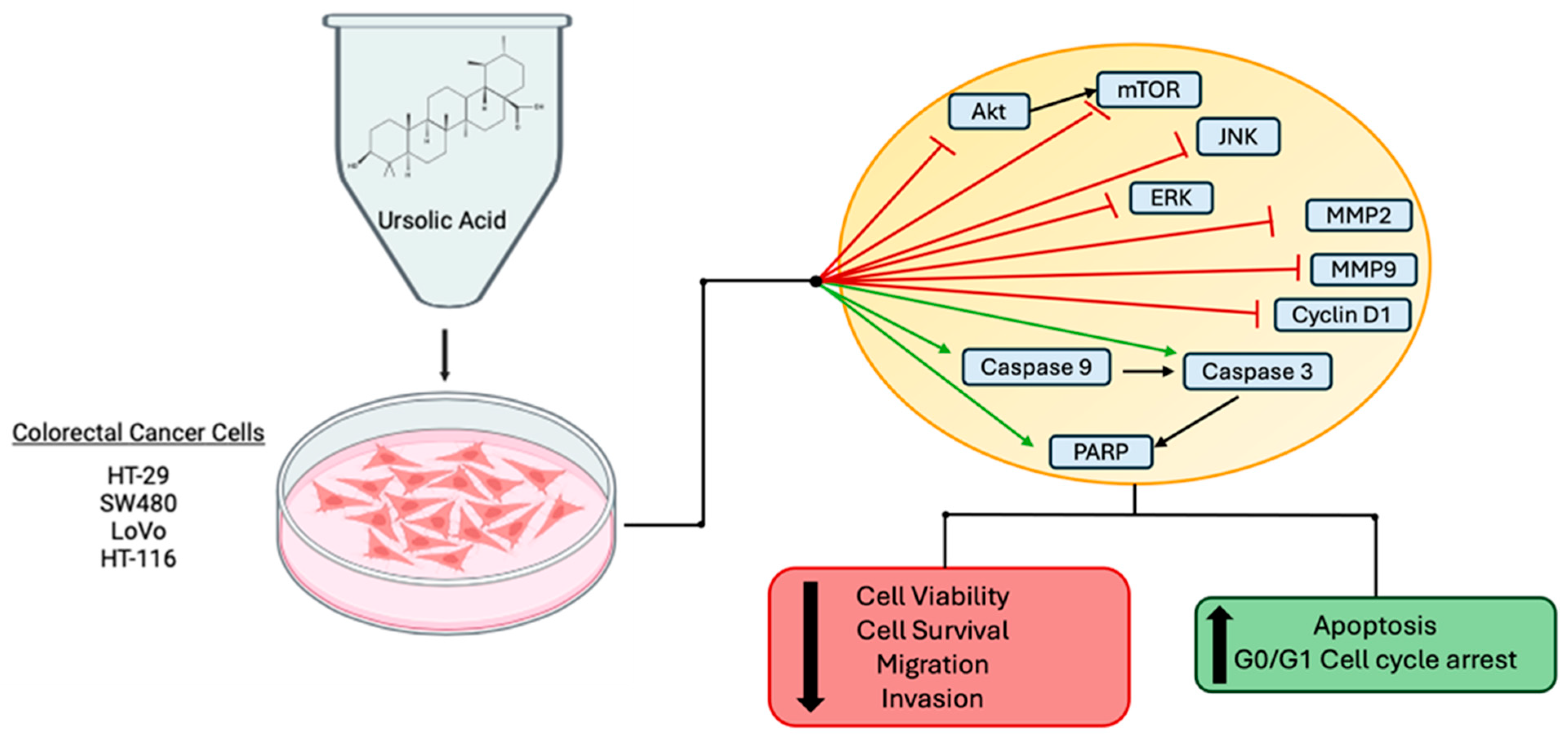
Overall, these studies indicate that the treatment of colorectal cancer cells with ursolic acid leads to decreased cell viability, survival, migration and invasion and increased apoptosis and cell cycle arrest. These effects are associated with decreased phosphorylation of signaling proteins Akt, mTOR, JNK and ERK and increased levels of cleaved PARP, casp-3 and casp-9 (Figure 2).

2.2. Effects of UA in Combination with Chemotherapy Agents and Radiation

Colorectal cancer can be treated systemically with chemotherapy agents, and there are several drugs with FDA approval for use against CRC; however, their efficacy is often reduced due to patients developing drug resistance. To help overcome drug resistance and harmful side effects experienced with many chemotherapy agents, researchers are examining other compounds that can be given in combination to enhance the efficacy of the chemo drugs, reducing the required dosage and lowering the severity of the side effects. A number of studies have examined the effects of UA treatment in combination with other agents.
Colon cancer cells SW480 and LoVo had reduced cell viability and migration when treated with UA in combination with 1 mM melatonin (MT) [65] (Table 3). This treatment induced apoptosis, caused changes in cell morphology and modulated cytochrome c/caspase, MMP9/COX-2 and p300/NF-kB signaling. UA combined with MT triggered the release of cytochrome c from the mitochondria, inducing cleavage of caspase (-3 and -9) and PARP proteins. UA plus MT treatment inhibited MMP-9 and COX-2 protein levels and promoted p300 and NF-kB translocation from the nucleus to the cytoplasm. The effects of the combined treatment were greater than UA or MT treatment alone [65].
HCT15 cells treated with a combination of UA and 5-fluorouracil (5-FU, 100 µM) (a chemotherapy agent used in CRC) showed a significant increase in TUNEL positive cells compared with either compound alone [66]. The combined treatment increased the phosphorylation level of JNK while decreasing the phosphorylation level of mTOR (p46), and increased protein levels of autophagy markers LC3-I, LC3-II and p62. The combined treatment also increased p53 protein levels. Based on these results, the authors concluded that UA is able to enhance the apoptotic effects of 5-FU by activating JNK signaling [66]. This study provides evidence that UA has the potential to act synergistically to enhance the effects of 5-FU (a chemotherapy drug already in use).
Ursolic acid’s ability to enhance the efficacy of oxaliplatin (OXL) treatment was examined in human CRC cells (SW480, SW620, LoVo and RKO) [67]. The combination treatment of SW620 cells reduced viability, induced apoptosis and reduced mitochondrial membrane potential. Cleaved caspase -3, -8 and -9 were increased, while levels of anti-apoptotic markers Bcl-xL, Bcl-2 and survivin were decreased. A drug combination index analysis revealed a synergistic effect between UA and oxaliplatin for the inhibition of proliferation in all cell lines tested; RKO cells had the greater interaction, with 0.68 synergy. Multiple signaling pathways appeared to be involved in the anticancer effects, seen as the treatment causing changes in levels of proteins associated with MAPK, PI3K/Akt and NF-kB signaling [67].
Treatment with UA caused colon cancer cells (RKO, LoVo and SW480) to become more sensitive to treatment with chemotherapy agents 5-FU or oxaliplatin [68] (Table 3). This occurred by the inhibition of MDR1 via HIF-1α under hypoxic conditions; UA alone and the combinations also inhibited HIF-1α in normoxic conditions. Additionally, UA downregulated VEGF protein levels and inhibited angiogenesis. These findings indicate that UA could act as a chemosensitizer in colon cancer by inhibiting HIF-1α, MDR1 and VEGF [68].
Human CRC cells HCT8 and SW480 treated with UA (20 µmol/L) and oxaliplatin (Oxa) (0.4 µmol/L) had reduced proliferation, increased apoptosis and ROS production [69]. The combination treatment significantly increased these effects compared with either parent compound alone. The protein level of apoptotic marker cleaved caspase-3 was increased. ROS levels, detected by DCFH-DA assay, were significantly increased with the combination treatment compared with either drug alone. Similarly, the combination treatment increased the protein level of NADPH (a primary resource for ROS production). The combination treatment significantly reduced mRNA and protein levels of permeability glycoprotein (p-gp), MRP and BCRP (genes associated with drug resistance) [69]. Based on these results, the authors conclude that UA enhances the anticancer effects of oxaliplatin in CRC cells via the ROS-mediated inhibition of drug resistance.
Another research group, Zheng et al. 2020, also examined the synergistic effect of ursolic acid and oxaliplatin (Oxa), using RKO cells [70]. The combination treatment (UA and Oxa) enhanced apoptosis and reduced survival when compared with treatment with either compound alone. Increased activity of caspase -3, -8 and -9 and a significant increase in cleaved PARP protein levels was seen. In RKO cells, the combination treatment decreased the protein levels of the X-linked inhibitor of apoptosis (XIAP) and survivin. These results provide evidence that UA and Oxa have a synergistic effect and may be used together in the treatment of CRC [70].
Human HT-29 colon cancer cells treated with UA (5 µg/mL) and oleanolic acid (OA, 100 µg/mL) had reduced viability [71]. UA combined with camptohecin-11 (CPT-11, 0.075 µg/mL) had no cytotoxic effect. Normal healthy cells (CCD 841) did not show cytotoxicity with either treatment protocol. UA combined with OA or CPT-11 inhibited the migration of HT-29 and SW620 CRC cells. HT-29 cells had reduced protein levels of MMP2 with both treatment protocols, while SW620 cells had reduced protein levels of MMP9 with both treatments. Both HT-29 and SW620 cells treated with UA combined with CPT-11 had reduced levels of urokinase-type plasminogen activator receptor (uPAR) immunofluorescence [71].
Ursolic acid in combination with chemotherapy agent doxorubicin (DOX) was examined in human colon cancer cell line HCT116 and human colorectal cancer cell line HT-29; the results showed a synergistic effect [72]. UA combined with DOX significantly decreased cell proliferation and colony formation, a response that was greater than with either compound alone. The combination treatment significantly increased the number of apoptotic cells and levels of cleaved caspase 9 and cleaved PARP. A wound healing assay revealed the significant inhibition of migration associated with an increase in protein levels of E-cadherin, MMP-9 and uPA (metastasis markers with the combined treatment). Cell cycle arrest in the G1 phase was seen and protein levels of cell cycle markers CDK4, CDK6 and cyclin D1 were all decreased. The combined treatment decreased phosphorylation levels of Akt and GSK3β and reduced c-Myc levels. UA combined with DOX increased the protein levels of Hippo signaling pathway proteins Rassf1A, Mst1, Mst2, Sav1 and CTGF and increased phosphorylation levels of Mob1 and Yap. The inhibition of Akt signaling with PI3K inhibitor (LY2940002) further increased the protein levels of Hippo pathway proteins, and the synergistic anticancer effects seen with the combined treatment occurred through the Akt/Hippo signaling pathway [72].
The combination treatment of ursolic acid and sorafenib (an FDA-approved tyrosine kinase inhibitor drug) showed synergistic anticancer effects in HCT-116 and LoVo colon cancer cells. The combined treatment significantly decreased cell viability and survival and increased apoptosis compared with either compound alone [73]. The protein levels of cleaved PARP, cleaved caspases −9 and −8 and LC3 I and II increased with the combined treatment.
Mouse colon cancer cells (CT26) exposed to 15 Gy of gamma irradiation and then treated with UA had significantly decreased survival and increased apoptosis compared with each treatment alone [74]. Irradiation and UA combined decreased caspase3 and Bcl2 protein levels and increased the level of cleaved PARP. The combination treatment significantly increased the level of cellular ROS (DCF assay) and increased mitochondrial ROS (DHR 123 assay), while levels of GSH were decreased [74].
The above studies provide evidence that the combination of ursolic acid with the chemotherapy drugs 5-fluorouracil, oxaliplatin and doxorubicin and the tyrosine kinase inhibitor sorafenib or in combination with irradiation result in significant enhanced responses. The combination treatment increased the level of apoptosis in CRC cells above the level of either treatment alone (Figure 3).

2.3. Effects of Ursolic Acid on Animal Models of Colorectal Cancer

This section summarizes articles that examined the effects of UA utilizing animal models of colorectal cancer.
The daily intraperitoneal administration of UA inhibited the growth of tumors in mice xenografted with human CRC cells (HT-29), evidenced by reduced tumor volume and weight [52] (Table 4). No change in the body weight of the animals was seen. Tumor samples from UA-treated mice had reduced PCNA cell staining. Consistent with the in vitro results reported in Section 2.1, tumor tissue samples from mice treated with UA had reduced mRNA and protein levels of cyclin D1 and CDK4, while p21 mRNA and protein levels were increased. Tumor samples had an increase in the percent of TUNEL positive cells. UA treatment decreased the protein level of Bcl-2 in tumor samples yet increased the protein and mRNA levels of Bax. Reduced phosphorylation of the MAP kinases, Erk1/2, JNK and p38 and reduced phosphorylation of STAT3 were seen in the tumor samples, suggesting that UA acts to reduce tumor growth in CRC xenografts via the inhibition of these signaling pathways [52].
Ursolic acid (12.5 mg/kg, daily) inhibited CRC tumor volume yet did not affect the body weight of mice xenografted with human HT-29 colorectal cancer cells, suggesting that UA acts as an anticancer agent in vivo, with minimal toxicity to the animal [51]. UA treatment in these mice caused reduced intratumoral microvessel density and decreased the protein level of CD31 (a vascular differential marker). This research group used a chick embryo chorioallantoic membrane model treated with UA (0.25 mg for 72 h), which resulted in a reduced total number of blood vessels. A bio-plex phosphoprotein assay showed that UA decreased phosphorylation of STAT3, Akt and p70S6K. The immunohistochemistry of tumor samples showed decreased protein levels of sonic hedgehog (SHH) and Gli-1 with the UA treatment, suggesting that UA suppresses the sonic hedgehog signaling pathway. Immunohistochemistry analysis further showed a decrease in levels of VEGF-A and bFGF (important markers of angiogenesis). Taken together, the findings from this study support a role for UA as an anticancer treatment through its ability to inhibit angiogenesis in tumors through multiple signaling pathways [51].
Ursolic acid (10 mg/kg) treatment (by daily intraperitoneal injection for 13 days) suppressed the growth of tumors in mice xenografted with human colon cancer cells (HCT116) [53] (Table 4). The tumor volume was calculated by caliper measurements and found to be reduced in the UA-treated mice compared with the controls [53]. Unfortunately, no tumor tissue examination or other measurements were performed in this study.
Female nude mice xenografted with human CRC (HCT15) cells had reduced tumor size when treated with UA (75 mg/kg orally daily for 14 days) [66]. Immunohistochemical analysis of excised tumor tissues showed a slight increase in the protein level of p62 and the phosphorylation level of JNK following UA treatment, while levels of LC3 and Ki67 were not changed. These results suggest that UA reduces tumor growth in vivo, possibly through JNK signaling and induced autophagy [66].
Nude mice xenografted with smoothed knockdown HCT-116hSMO− cells and treated with UA (10, 20 or 40 mg/kg) for 12 consecutive days had decreased tumor volume and weight compared with no-treatment mice [61]. Analysis of the extracted tumors showed that UA induced apoptosis in the tumor cells through the inhibition of Bcl-2 and increased BAX (protein and mRNA). Tumor tissues had increased mRNA levels of caspases -9 and -3, increased mRNA and protein levels of SUFU and reduced mRNA and protein levels of c-Myc, GLO1 and SHH. These results were consistent with the cell culture data from the same research group (reported above, Section 2.1) [61]. The combined results from this study provide evidence that UA acts through a complex signaling mechanism involving non-canonical hedgehog signaling and inhibited Akt signaling.
Human SW620 CRC cell-xenografted mice treated with UA (15, 30 and 60 mg/kg) by intragastric administration had a significantly higher body weight after 16 days compared with the controls, and a significantly decreased tumor weight and volume [62]. Analysis of the tumors showed that UA treatment induced apoptosis; mRNA and protein levels of Bcl-2 were decreased, while mRNA and protein levels of Bax were increased. UA treatment increased mRNA levels of caspases -3 and -9. UA-treated mice had increased mRNA and protein levels of GSK3β and decreased mRNA and protein levels of β-catenin, WNT4, TCF4 and LEF1. The phosphorylation level of β-catenin increased, while the level of GSK3β decreased. There was a decrease in the level of nuclear β-catenin protein [62].
Zhang et al. [63] injected HCT-116 cells into the tail vein of male BALB/c-nude mice, establishing a lung metastatic model of colon cancer and found that daily administration of UA (20 mg/kg) for 42 days resulted in a significant decrease in lung metastases (accompanied by prevention of weight loss). Lung tissue immunohistochemical analysis revealed that the expression of ARL4C was significantly decreased in animals administered UA compared with the control untreated animals. These data indicate that UA could inhibit the metastasis of colon cancer to the lungs.
Mice xenografted with human CRC SW620 cells were treated with UA (20 mg), OXL (10 mg) or a combination for five consecutive days and sacrificed on day 50 for tumor and tissue examination [67]. The combination of UA and OXL caused a decrease in tumor volume and tumor weight above that seen with either compound alone. Immunohistochemistry showed that the combination treatment led to a decrease in Ki-67 protein levels and an increase in TUNEL positive cells. Serum concentrations of ALT and AST showed that OXA caused impaired liver function, which was attenuated with the addition of UA to the treatment. Tumor tissues examined with immunohistochemistry and Western blot showed that the antitumor effects were associated with the inhibition of Akt and ERK1/2 signaling. Phosphorylation levels of ERK1/2, Akt and IKKα were reduced following the combined treatment [67]. These results suggest that UA enhances the effect of the chemotherapy agent OXL and contributes to reducing the OXL-associated side effects.
The combination of ursolic acid and oxaliplatin increased the survival time and reduced the tumor volume of mice xenografted with human CRC (HCT8 or SW480) cells [69]. The anticancer effects with the combined treatment were enhanced compared with either compound alone.
Mice xenografted with HCT116 human colon cancer cells were treated with UA (10 mg/kg/day), DOX (2 mg/kg/twice weekly) or combined UA and DOX by intraperitoneal injection [72] (Table 4). Following treatment, the mice were sacrificed and tumor and serum samples were collected. Serum samples showed no significant change in biochemical markers ALT, AST, BUN and creatinine, suggesting that the treatments did not impair liver or kidney function of the animals. The body weight of the animals did not differ between the treatment groups. Tumor volume was significantly decreased with UA or DOX treatment, and the decrease was enhanced with the combined UA and DOX treatment. Cell proliferation marker Ki67 was reduced with the combination treatment when examined with immunohistochemistry. Tumors from UA- and DOX-treated animals had reduced protein levels of CTGF, reduced phosphorylation levels of Akt, increased protein levels of Rassf1A, Mst1, Mst2 and Sav1 and increased phosphorylation levels of Mob1 and Yap [72]. These results are consistent with in vitro findings previously reported by this group (Section 2.2 above). The results from this study, both in vitro and in vivo, show that the combination of UA and DOX have strong anticancer effects over either treatment alone.
Nude mice were orthotopically implanted with luciferase-transfected HCT116 human colorectal cancer cells and treated via oral gavage with UA (250 mg/kg daily), capecitabine (CAP) (60 mg/kg twice/week) or both in combination. Tumor growth was measured on days 7, 14, 21 and 28 by bioluminescence imaging and showed reduced growth with UA treatment. Importantly, greater growth inhibition of the tumor was seen with the combination treatment over 28 days [75]. The excised tumors had significantly decreased tumor volume and weight only with the combination treatment. The sacrificed animals were examined for metastatic growths; UA, CAP and combination treatment mice all had reduced metastases in the liver, intestine, lung and spleen. Significant levels of UA (measured by HPLC) were detected in serum (480 ng/mL) and tissue (356 mg/mL) samples that were collected four hours after UA oral administration. These data suggest that UA reached the animals’ circulation and infiltrated the tumor. Extracted tumor tissues were further analyzed by immunohistochemistry and Western blot and it was found that UA enhanced the effects of capecitabine. UA inhibited levels of nuclear p65 and β-catenin, reduced protein levels of cyclin D1, cMyc, Bcl2, Bcl-xl and survivin and reduced phosphorylation levels of STAT3 and EGFR. The metastasis markers ICAM-1, VEGF and MMP-9 were reduced in tumor tissues with the UA treatment of animals, and expression of tumor suppressor genes p53 and p21 were increased with either UA or the combined treatment. Immunohistochemical analysis also showed a reduced expression of proliferation markers Ki67 and CD31 [75]. This paper provides strong evidence that UA acts as an anticancer drug against colorectal cancer and also acts as a capecitabine chemosensitizing agent. Importantly, this paper shows that the oral administration of UA results in significant UA serum levels and that UA is able to enter the tumor tissue, achieving detectable levels in the tumor.
Although limited in vivo animal studies exist, the evidence indicates that oral or intraperitoneal administration of UA in mice xenografted with human CRC cells causes reduced tumor volume and tumor weight. Oral administration also reduces cancer metastasis (Figure 4).
A search of ClinicalTrials.gov (accessed on 22 June 2024) for the disease “colorectal cancer” or “colon cancer” and the term “ursolic acid” found no results. A search for “cancer” and “ursolic acid” also had no results at this time. Overall, there are currently no clinical trials examining UA against CRC or any other cancer.

2.4. Bioavailability and Potential Toxicity of UA

Similar to many other polyphenols, UA has limited bioavailability due to its low water solubility and high molecular weight (456.7 g/mol) [76,77]. Studies examining the bioavailability of UA are limited. Yin et al. administered UA in mice (0.05% UA added to the diet) for 8 weeks, then measured UA plasma levels and examined UA tissue distribution. The plasma level of UA was 580 ng/mL or 1.26 µM. Tissues that showed UA accumulation included the liver (9.7 µg/g), colon (6.4 µg/g), kidney (5.9 µg/g), heart (3.9 µg/g), bladder (2.9 µg/g) and brain (1.6 µg/g) [77]. In another study, oral administration of UA (100 mg/kg body weight) in rats resulted in a plasma concentration of 300 ng/mL (656.89 nM) within one hour. The concentration decreased after 4 h, but the animals maintained a plasma concentration of about 100 ng/mL (218.96 nM) for 12 h [76]. Although there are currently no studies examining UA plasma levels in humans after oral administration of the parent UA compound, a study by Xia et al. [78] administered in humans UA nanoliposomes via intravenous infusion at a dose of 98 mg/m2. These subjects had peak plasma concentrations of 3404.6 ng/mL (7454.78 nM) four hours after infusion. This peak was followed by a rapid decline, about 10-fold, and the concentration 6 h post-infusion was approximately 300 mg/mL (656.89 nM). There was another 10-fold decline and, at 16 h post-infusion, the subjects had approximately 30 ng/mL (65.69 nM) UA plasma concentration [78]. The above studies suggest that UA administration results in plasma UA levels in the nano- to micromolar range. The concentrations found in the plasma were comparable to concentrations of UA used in many of the in vitro studies showing potent anticancer effects.
A 2013 study by Wang et al. administered ursolic acid liposomes via a 4 h intravenous infusion in healthy adult volunteers and patients with advanced solid tumors. The maximum tolerated dose was found to be 98 mg/m2 and the dose-limiting toxicity presented as diarrhea and hepatotoxicity. UA liposomes showed a linear pharmacokinetic profile [79].
Mice that were orally given one dose of 10 mg/kg of body weight UA had an elevated neutrophil count, serum glutamic oxaloacetic transaminase (SGOT), alkaline phosphatase (ALP) and urea levels and a decrease in white blood cells, lymphocytes and platelets. Some alterations were also seen in the architecture of the liver, kidney and spleen tissue that were recoverable, indicating that the toxic effects caused with oral administration are reversable [80]. An acute oral toxicity study in which mice were given a single dose of UA and then observed for 2 weeks for signs of toxicity or mortality showed the oral LD50 of UA was >2000 mg/kg in mice [80].

3. Discussion/Conclusions

Colorectal cancer is the fourth most diagnosed cancer worldwide [4]. It is the second leading cause of cancer-related deaths among men and third leading cause of cancer-related deaths among women. The incidence rate is continuing to rise, and new treatment options are needed to overcome treatment resistance commonly seen in CRCs.
Many medications, including current chemotherapy drugs such as paclitaxel and docetaxel [19,20], were originally derived from plants. Other plant-derived compounds may have strong anticancer effects and they need to be studied. In this paper, studies examining the pentacyclic triterpenoid ursolic acid and its effects against colorectal cancer were reviewed.
The in vitro studies all showed reduced proliferation and survival and increased levels of apoptosis of colorectal cancer cells treated with UA (Table 2 and Figure 2). Most of the research groups used concentrations of UA ranging from 20 to 80 µM. Only one study with low (2.5 µM) UA concentration found significant inhibitory effects [59].
Some studies examined the effects of ursolic acid when it was given in combination with compounds that are already known to inhibit CRC. Oxaliplatin, a platinum compound that inhibits DNA synthesis and is therefore cytotoxic, is currently used in the treatment of CRC but is associated with severe side effects. 5-fluorouracil is another currently used chemotherapy agent given for CRC; this drug works systemically by inhibiting thymidylate synthase thus blocking DNA replication. Doxorubicin is an antibiotic, originally derived from Streptomyces peucetius bacterium, that interacts with DNA by intercalation and inhibits macromolecule biosynthesis. All these currently accepted treatments act on DNA on a systemic level that causes many severe systemic side effects. Results of the articles summarized in this review (Table 3) suggest that the doses of some of these compounds may be reduced when combined with UA, thereby reducing the occurrence and severity of side effects.
The administration of UA in mice xenografted with human colorectal cancer cells showed decreased tumor growth and reduced tumor volumes, weights and size. Importantly, none of the animal studies reported reduced weight or premature death of the treated animals, suggesting that UA is safe and has limited side effects.
The evidence from the studies reviewed here indicates that UA is active against colorectal cancer cells. UA inhibits human colorectal cancer cell proliferation, survival and migration and induces apoptosis. In addition, the evidence indicates that UA may act as a chemosensitizer to enhance the effectiveness of currently approved drugs in the treatment of CRC. The limited evidence from in vivo animal studies indicates that UA administration reduces CRC tumor growth without causing side effects.
More studies should be performed to examine the anticancer potential of UA in animals before considering human clinical trials.

Author Contributions

A.K. formulated the review topic, performed searches and created figures. A.K. and E.T. composed the entirety of the manuscript. All authors have read and agreed to the published version of the manuscript.

Funding

This review received no external funding.

Institutional Review Board Statement

Not applicable.

Informed Consent Statement

Not applicable.

Data Availability Statement

Not applicable.

Conflicts of Interest

The authors declare no conflicts of interest.

References

  1. Dekker, E.; Tanis, P.J.; Vleugels, J.L.A.; Kasi, P.M.; Wallace, M.B. Colorectal Cancer. Lancet 2019, 394, 1467–1480. [Google Scholar] [CrossRef] [PubMed]
  2. Siegel, R.L.; Wagle, N.S.; Cercek, A.; Smith, R.A.; Jemal, A. Colorectal Cancer Statistics, 2023. CA A Cancer J. Clin. 2023, 73, 233–254. [Google Scholar] [CrossRef] [PubMed]
  3. Biller, L.H.; Schrag, D. Diagnosis and Treatment of Metastatic Colorectal Cancer: A Review. JAMA 2021, 325, 669–685. [Google Scholar] [CrossRef] [PubMed]
  4. Bray, F.; Ferlay, J.; Soerjomataram, I.; Siegel, R.L.; Torre, L.A.; Jemal, A. Global Cancer Statistics 2018: GLOBOCAN Estimates of Incidence and Mortality Worldwide for 36 Cancers in 185 Countries. CA A Cancer J. Clin. 2018, 68, 394–424. [Google Scholar] [CrossRef] [PubMed]
  5. Schubert, S.A.; Morreau, H.; de Miranda, N.F.C.C.; van Wezel, T. The Missing Heritability of Familial Colorectal Cancer. Mutagenesis 2020, 35, 221–231. [Google Scholar] [CrossRef]
  6. Li, J.; Ma, X.; Chakravarti, D.; Shalapour, S.; DePinho, R.A. Genetic and Biological Hallmarks of Colorectal Cancer. Genes Dev. 2021, 35, 787–820. [Google Scholar] [CrossRef] [PubMed]
  7. Dariya, B.; Aliya, S.; Merchant, N.; Alam, A.; Nagaraju, G.P. Colorectal Cancer Biology, Diagnosis, and Therapeutic Approaches. Crit. Rev. Oncog. 2020, 25, 71–94. [Google Scholar] [CrossRef] [PubMed]
  8. Grady, W.M.; Markowitz, S.D. The Molecular Pathogenesis of Colorectal Cancer and Its Potential Application to Colorectal Cancer Screening. Dig. Dis. Sci. 2015, 60, 762–772. [Google Scholar] [CrossRef]
  9. Kasi, A.; Handa, S.; Bhatti, S.; Umar, S.; Bansal, A.; Sun, W. Molecular Pathogenesis and Classification of Colorectal Carcinoma. Curr. Color. Cancer Rep. 2020, 16, 97–106. [Google Scholar] [CrossRef]
  10. Pandey, H.; Tang, D.W.T.; Wong, S.H.; Lal, D. Gut Microbiota in Colorectal Cancer: Biological Role and Therapeutic Opportunities. Cancers 2023, 15, 866. [Google Scholar] [CrossRef]
  11. Liu, Y.; Lau, H.C.-H.; Cheng, W.Y.; Yu, J. Gut Microbiome in Colorectal Cancer: Clinical Diagnosis and Treatment. Genom. Proteom. Bioinform. 2023, 21, 84–96. [Google Scholar] [CrossRef] [PubMed]
  12. Rinninella, E.; Raoul, P.; Cintoni, M.; Franceschi, F.; Miggiano, G.A.D.; Gasbarrini, A.; Mele, M.C. What Is the Healthy Gut Microbiota Composition? A Changing Ecosystem across Age, Environment, Diet, and Diseases. Microorganisms 2019, 7, 14. [Google Scholar] [CrossRef] [PubMed]
  13. Rothschild, D.; Weissbrod, O.; Barkan, E.; Kurilshikov, A.; Korem, T.; Zeevi, D.; Costea, P.I.; Godneva, A.; Kalka, I.N.; Bar, N.; et al. Environment Dominates over Host Genetics in Shaping Human Gut Microbiota. Nature 2018, 555, 210–215. [Google Scholar] [CrossRef] [PubMed]
  14. Saoudi González, N.; Salvà, F.; Ros, J.; Baraibar, I.; Rodríguez-Castells, M.; García, A.; Alcaráz, A.; Vega, S.; Bueno, S.; Tabernero, J.; et al. Unravelling the Complexity of Colorectal Cancer: Heterogeneity, Clonal Evolution, and Clinical Implications. Cancers 2023, 15, 4020. [Google Scholar] [CrossRef]
  15. Ashique, S.; Bhowmick, M.; Pal, R.; Khatoon, H.; Kumar, P.; Sharma, H.; Garg, A.; Kumar, S.; Das, U. Multi Drug Resistance in Colorectal Cancer- Approaches to Overcome, Advancements and Future Success. Adv. Cancer Biol.-Metastasis 2024, 10, 100114. [Google Scholar] [CrossRef]
  16. Sellers, R.S.; Morton, D. The Colon: From Banal to Brilliant. Toxicol. Pathol. 2014, 42, 67–81. [Google Scholar] [CrossRef] [PubMed]
  17. Fritz, C.D.L.; Otegbeye, E.E.; Zong, X.; Demb, J.; Nickel, K.B.; Olsen, M.A.; Mutch, M.; Davidson, N.O.; Gupta, S.; Cao, Y. Red-Flag Signs and Symptoms for Earlier Diagnosis of Early-Onset Colorectal Cancer. JNCI J. Natl. Cancer Inst. 2023, 115, 909–916. [Google Scholar] [CrossRef] [PubMed]
  18. Briggs, N.L.; Ton, M.; Malen, R.C.; Reedy, A.M.; Cohen, S.A.; Phipps, A.I.; Burnett-Hartman, A.N.; Newcomb, P.A. Colorectal Cancer Pre-Diagnostic Symptoms Are Associated with Anatomic Cancer Site. BMC Gastroenterol. 2024, 24, 65. [Google Scholar] [CrossRef]
  19. Bissery, M.C.; Nohynek, G.; Sanderink, G.J.; Lavelle, F. Docetaxel (Taxotere): A Review of Preclinical and Clinical Experience. Part I: Preclinical Experience. Anticancer Drugs 1995, 6, 339–355. [Google Scholar] [CrossRef]
  20. Weaver, B.A. How Taxol/Paclitaxel Kills Cancer Cells. Mol. Biol. Cell 2014, 25, 2677–2681. [Google Scholar] [CrossRef]
  21. Panda, S.S.; Thangaraju, M.; Lokeshwar, B.L. Ursolic Acid Analogs as Potential Therapeutics for Cancer. Molecules 2022, 27, 8981. [Google Scholar] [CrossRef] [PubMed]
  22. Butkeviciute, A.; Viskelis, J.; Liaudanskas, M.; Viskelis, P.; Bobinas, C.; Janulis, V. Variation of Triterpenes in Apples Stored in a Controlled Atmosphere. Molecules 2021, 26, 3639. [Google Scholar] [CrossRef] [PubMed]
  23. Kondo, M.; MacKinnon, S.L.; Craft, C.C.; Matchett, M.D.; Hurta, R.A.R.; Neto, C.C. Ursolic Acid and Its Esters: Occurrence in Cranberries and Other Vaccinium Fruit and Effects on Matrix Metalloproteinase Activity in DU145 Prostate Tumor Cells: Anti-Tumor Activity and Content of Ursolic Acid from Vaccinium Fruit. J. Sci. Food Agric. 2011, 91, 789–796. [Google Scholar] [CrossRef] [PubMed]
  24. Allouche, Y.; Jiménez, A.; Uceda, M.; Aguilera, M.P.; Gaforio, J.J.; Beltrán, G. Triterpenic Content and Chemometric Analysis of Virgin Olive Oils from Forty Olive Cultivars. J. Agric. Food Chem. 2009, 57, 3604–3610. [Google Scholar] [CrossRef]
  25. Guinda, Á.; Rada, M.; Delgado, T.; Gutiérrez-Adánez, P.; Castellano, J.M. Pentacyclic Triterpenoids from Olive Fruit and Leaf. J. Agric. Food Chem. 2010, 58, 9685–9691. [Google Scholar] [CrossRef] [PubMed]
  26. Ivanov, I.; Petkova, N.; Tumbarski, Y.; Vrancheva, R.; Stoyanova, M. Lavender Waste-Promising Source of Triterpenoids and Polyphenols with Antioxidant and Antimicrobial Activity. Ind. Technol. 2018, 5, 26–32. [Google Scholar]
  27. Silva, M.G.V.; Vieira, I.G.P.; Mendes, F.N.P.; Albuquerque, I.L.; dos Santos, R.N.; Silva, F.O.; Morais, S.M. Variation of Ursolic Acid Content in Eight Ocimum Species from Northeastern Brazil. Molecules 2008, 13, 2482–2487. [Google Scholar] [CrossRef] [PubMed]
  28. He, X.; Liu, R.H. Triterpenoids Isolated from Apple Peels Have Potent Antiproliferative Activity and May Be Partially Responsible for Apple’s Anticancer Activity. J. Agric. Food Chem. 2007, 55, 4366–4370. [Google Scholar] [CrossRef] [PubMed]
  29. Li, X.; Wang, T.; Zhou, B.; Gao, W.; Cao, J.; Huang, L. Chemical Composition and Antioxidant and Anti-Inflammatory Potential of Peels and Flesh from 10 Different Pear Varieties (Pyrus Spp.). Food Chem. 2014, 152, 531–538. [Google Scholar] [CrossRef]
  30. Sun, L.; Tao, S.; Zhang, S. Characterization and Quantification of Polyphenols and Triterpenoids in Thinned Young Fruits of Ten Pear Varieties by UPLC-Q TRAP-MS/MS. Molecules 2019, 24, 159. [Google Scholar] [CrossRef]
  31. Jäger, S.; Trojan, H.; Kopp, T.; Laszczyk, M.; Scheffler, A. Pentacyclic Triterpene Distribution in Various Plants–Rich Sources for a New Group of Multi-Potent Plant Extracts. Molecules 2009, 14, 2016–2031. [Google Scholar] [CrossRef] [PubMed]
  32. Zannou, O.; İPekci, B.; Koca, İ.; Odabaş, H.İ. Assessing Ursolic Acid Contents of Some Commonly Consumed Herbs Grown in Turkey. Gümüşhane Üniversitesi Fen Bilim. Enstitüsü Derg. 2022, 12, 301–308. [Google Scholar] [CrossRef]
  33. Wójciak-Kosior, M.; Sowa, I.; Kocjan, R.; Nowak, R. Effect of Different Extraction Techniques on Quantification of Oleanolic and Ursolic Acid in Lamii Albi Flos. Ind. Crops Prod. 2013, 44, 373–377. [Google Scholar] [CrossRef]
  34. Kowalski, R. Studies of Selected Plant Raw Materials as Alternative Sources of Triterpenes of Oleanolic and Ursolic Acid Types. J. Agric. Food Chem. 2007, 55, 656–662. [Google Scholar] [CrossRef] [PubMed]
  35. Mlala, S.; Oyedeji, A.O.; Gondwe, M.; Oyedeji, O.O. Ursolic Acid and Its Derivatives as Bioactive Agents. Molecules 2019, 24, 2751. [Google Scholar] [CrossRef] [PubMed]
  36. Woźniak, Ł.; Skąpska, S.; Marszałek, K. Ursolic Acid—A Pentacyclic Triterpenoid with a Wide Spectrum of Pharmacological Activities. Molecules 2015, 20, 20614–20641. [Google Scholar] [CrossRef] [PubMed]
  37. Zhao, J.; Zheng, H.; Sui, Z.; Jing, F.; Quan, X.; Zhao, W.; Liu, G. Ursolic Acid Exhibits Anti-Inflammatory Effects through Blocking TLR4-MyD88 Pathway Mediated by Autophagy. Cytokine 2019, 123, 154726. [Google Scholar] [CrossRef] [PubMed]
  38. Gudoityte, E.; Arandarcikaite, O.; Mazeikiene, I.; Bendokas, V.; Liobikas, J. Ursolic and Oleanolic Acids: Plant Metabolites with Neuroprotective Potential. Int. J. Mol. Sci. 2021, 22, 4599. [Google Scholar] [CrossRef]
  39. Alam, M.; Ali, S.; Ahmed, S.; Elasbali, A.M.; Adnan, M.; Islam, A.; Hassan, M.I.; Yadav, D.K. Therapeutic Potential of Ursolic Acid in Cancer and Diabetic Neuropathy Diseases. Int. J. Mol. Sci. 2021, 22, 12162. [Google Scholar] [CrossRef]
  40. Dai, Y.; Sun, L.; Tan, Y.; Xu, W.; Liu, S.; Zhou, J.; Hu, Y.; Lin, J.; Yao, X.; Mi, P.; et al. Recent Progress in the Development of Ursolic Acid Derivatives as Anti-Diabetes and Anti-Cardiovascular Agents. Chem. Biol. Drug Des. 2023, 102, 1643–1657. [Google Scholar] [CrossRef]
  41. Chan, E.W.C.; Soon, C.Y.; Tan, J.B.L.; Wong, S.K.; Hui, Y.W. Ursolic Acid: An Overview on Its Cytotoxic Activities against Breast and Colorectal Cancer Cells. J. Integr. Med. 2019, 17, 155–160. [Google Scholar] [CrossRef]
  42. Kornel, A.; Nadile, M.; Tsiani, E. Evidence of the Beneficial Effects of Ursolic Acid against Lung Cancer. Molecules 2022, 27, 7466. [Google Scholar] [CrossRef] [PubMed]
  43. Kornel, A.; Nadile, M.; Retsidou, M.I.; Sakellakis, M.; Gioti, K.; Beloukas, A.; Sze, N.S.K.; Klentrou, P.; Tsiani, E. Ursolic Acid against Prostate and Urogenital Cancers: A Review of In Vitro and In Vivo Studies. Int. J. Mol. Sci. 2023, 24, 7414. [Google Scholar] [CrossRef] [PubMed]
  44. Yin, R.; Li, T.; Tian, J.X.; Xi, P.; Liu, R.H. Ursolic Acid, a Potential Anticancer Compound for Breast Cancer Therapy. Crit. Rev. Food Sci. Nutr. 2018, 58, 568–574. [Google Scholar] [CrossRef] [PubMed]
  45. Li, J.; Guo, W.-J.; Yang, Q.-Y. Effects of Ursolic Acid and Oleanolic Acid on Human Colon Carcinoma Cell Line HCT15. World J. Gastroenterol. 2002, 8, 493–495. [Google Scholar] [CrossRef] [PubMed]
  46. Shan, J.; Xuan, Y.; Zheng, S.; Dong, Q.; Zhang, S. Ursolic Acid Inhibits Proliferation and Induces Apoptosis of HT-29 Colon Cancer Cells by Inhibiting the EGFR/MAPK Pathway. J. Zhejiang Univ. Sci. B 2009, 10, 668–674. [Google Scholar] [CrossRef]
  47. Xavier, C.P.R.; Lima, C.F.; Preto, A.; Seruca, R.; Fernandes-Ferreira, M.; Pereira-Wilson, C. Luteolin, Quercetin and Ursolic Acid Are Potent Inhibitors of Proliferation and Inducers of Apoptosis in Both KRAS and BRAF Mutated Human Colorectal Cancer Cells. Cancer Lett. 2009, 281, 162–170. [Google Scholar] [CrossRef] [PubMed]
  48. Limami, Y.; Pinon, A.; Leger, D.Y.; Mousseau, Y.; Cook-Moreau, J.; Beneytout, J.-L.; Delage, C.; Liagre, B.; Simon, A. HT-29 Colorectal Cancer Cells Undergoing Apoptosis Overexpress COX-2 to Delay Ursolic Acid-Induced Cell Death. Biochimie 2011, 93, 749–757. [Google Scholar] [CrossRef]
  49. Limami, Y.; Pinon, A.; Leger, D.Y.; Pinault, E.; Delage, C.; Beneytout, J.-L.; Simon, A.; Liagre, B. The P2Y2/Src/P38/COX-2 Pathway Is Involved in the Resistance to Ursolic Acid-Induced Apoptosis in Colorectal and Prostate Cancer Cells. Biochimie 2012, 94, 1754–1763. [Google Scholar] [CrossRef]
  50. Rawat, L.; Nayak, V. Ursolic Acid Disturbs ROS Homeostasis and Regulates Survival-Associated Gene Expression to Induce Apoptosis in Intestinal Cancer Cells. Toxicol. Res. 2021, 10, 369–375. [Google Scholar] [CrossRef]
  51. Lin, J.; Chen, Y.; Wei, L.; Hong, Z.; Sferra, T.J.; Peng, J. Ursolic Acid Inhibits Colorectal Cancer Angiogenesis through Suppression of Multiple Signaling Pathways. Int. J. Oncol. 2013, 43, 1666–1674. [Google Scholar] [CrossRef] [PubMed]
  52. Lin, J.; Chen, Y.; Wei, L.; Shen, A.; Sferra, T.J.; Hong, Z.; Peng, J. Ursolic Acid Promotes Colorectal Cancer Cell Apoptosis and Inhibits Cell Proliferation via Modulation of Multiple Signaling Pathways. Int. J. Oncol. 2013, 43, 1235–1243. [Google Scholar] [CrossRef] [PubMed]
  53. Wang, W.; Zhao, C.; Jou, D.; Lü, J.; Zhang, C.; Lin, L.; Lin, J. Ursolic Acid Inhibits the Growth of Colon Cancer-Initiating Cells by Targeting STAT3. Anticancer Res. 2013, 33, 4279–4284. [Google Scholar]
  54. Wang, J.; Liu, L.; Qiu, H.; Zhang, X.; Guo, W.; Chen, W.; Tian, Y.; Fu, L.; Shi, D.; Cheng, J.; et al. Ursolic Acid Simultaneously Targets Multiple Signaling Pathways to Suppress Proliferation and Induce Apoptosis in Colon Cancer Cells. PLoS ONE 2013, 8, e63872. [Google Scholar] [CrossRef] [PubMed]
  55. Kim, K.; Shin, E.A.; Jung, J.H.; Park, J.E.; Kim, D.S.; Shim, B.S.; Kim, S.-H. Ursolic Acid Induces Apoptosis in Colorectal Cancer Cells Partially via Upregulation of MicroRNA-4500 and Inhibition of JAK2/STAT3 Phosphorylation. Int. J. Mol. Sci. 2018, 20, 114. [Google Scholar] [CrossRef] [PubMed]
  56. Zhang, L.; Cai, Q.-Y.; Liu, J.; Peng, J.; Chen, Y.-Q.; Sferra, T.J.; Lin, J.-M. Ursolic Acid Suppresses the Invasive Potential of Colorectal Cancer Cells by Regulating the TGF-Β1/ZEB1/miR-200c Signaling Pathway. Oncol. Lett. 2019, 18, 3274–3282. [Google Scholar] [CrossRef] [PubMed]
  57. Wang, X.; Wang, T.; Yi, F.; Duan, C.; Wang, Q.; He, N.; Zhu, L.; Li, Q.; Deng, W. Ursolic Acid Inhibits Tumor Growth via Epithelial-to-Mesenchymal Transition in Colorectal Cancer Cells. Biol. Pharm. Bull. 2019, 42, 685–691. [Google Scholar] [CrossRef]
  58. Yang, M.; Hu, C.; Cao, Y.; Liang, W.; Yang, X.; Xiao, T. Ursolic Acid Regulates Cell Cycle and Proliferation in Colon Adenocarcinoma by Suppressing Cyclin B1. Front. Pharmacol. 2020, 11, 622212. [Google Scholar] [CrossRef] [PubMed]
  59. Shen, A.; Wu, M.; Liu, L.; Chen, Y.; Chen, X.; Zhuang, M.; Xie, Q.; Cheng, Y.; Li, J.; Shen, Z.; et al. Targeting NUFIP1 Suppresses Growth and Induces Senescence of Colorectal Cancer Cells. Front. Oncol. 2021, 11, 681425. [Google Scholar] [CrossRef]
  60. Zheng, J.-L.; Wang, S.-S.; Shen, K.-P.; Chen, L.; Peng, X.; Chen, J.-F.; An, H.-M.; Hu, B. Ursolic Acid Induces Apoptosis and Anoikis in Colorectal Carcinoma RKO Cells. BMC Complement. Med. Ther. 2021, 21, 52. [Google Scholar] [CrossRef]
  61. Chen, L.; Liu, M.; Yang, H.; Ren, S.; Sun, Q.; Zhao, H.; Ming, T.; Tang, S.; Tao, Q.; Zeng, S.; et al. Ursolic Acid Inhibits the Activation of Smoothened-Independent Non-Canonical Hedgehog Pathway in Colorectal Cancer by Suppressing AKT Signaling Cascade. Phytother. Res. 2022, 36, 3555–3570. [Google Scholar] [CrossRef]
  62. Zhao, H.; Tang, S.; Tao, Q.; Ming, T.; Lei, J.; Liang, Y.; Peng, Y.; Wang, M.; Liu, M.; Yang, H.; et al. Ursolic Acid Suppresses Colorectal Cancer by Down-Regulation of Wnt/β-Catenin Signaling Pathway Activity. J. Agric. Food Chem. 2023, 71, 3981–3993. [Google Scholar] [CrossRef]
  63. Zhang, M.; Xiang, F.; Sun, Y.; Liu, R.; Li, Q.; Gu, Q.; Kang, X.; Wu, R. Ursolic Acid Inhibits the Metastasis of Colon Cancer by Downregulating ARL4C Expression. Oncol. Rep. 2024, 51, 27. [Google Scholar] [CrossRef] [PubMed]
  64. Zhao, J.; Leng, P.; Xu, W.; Sun, J.-L.; Ni, B.-B.; Liu, G.-W. Investigating the Multitarget Pharmacological Mechanism of Ursolic Acid Acting on Colon Cancer: A Network Pharmacology Approach. Evid. Based Complement. Altern. Med. 2021, 2021, 9980949. [Google Scholar] [CrossRef]
  65. Wang, J.; Guo, W.; Chen, W.; Yu, W.; Tian, Y.; Fu, L.; Shi, D.; Tong, B.; Xiao, X.; Huang, W.; et al. Melatonin Potentiates the Antiproliferative and Pro-Apoptotic Effects of Ursolic Acid in Colon Cancer Cells by Modulating Multiple Signaling Pathways. J. Pineal. Res. 2013, 54, 406–416. [Google Scholar] [CrossRef] [PubMed]
  66. Xavier, C.P.R.; Lima, C.F.; Pedro, D.F.N.; Wilson, J.M.; Kristiansen, K.; Pereira-Wilson, C. Ursolic Acid Induces Cell Death and Modulates Autophagy through JNK Pathway in Apoptosis-Resistant Colorectal Cancer Cells. J. Nutr. Biochem. 2013, 24, 706–712. [Google Scholar] [CrossRef]
  67. Shan, J.; Xuan, Y.; Zhang, Q.; Zhu, C.; Liu, Z.; Zhang, S. Ursolic Acid Synergistically Enhances the Therapeutic Effects of Oxaliplatin in Colorectal Cancer. Protein. Cell 2016, 7, 571–585. [Google Scholar] [CrossRef] [PubMed]
  68. Shan, J.-Z.; Xuan, Y.-Y.; Zhang, Q.; Huang, J.-J. Ursolic Acid Sensitized Colon Cancer Cells to Chemotherapy under Hypoxia by Inhibiting MDR1 through HIF-1α. J. Zhejiang Univ. Sci. B 2016, 17, 672–682. [Google Scholar] [CrossRef]
  69. Zhang, Y.; Huang, L.; Shi, H.; Chen, H.; Tao, J.; Shen, R.; Wang, T. Ursolic Acid Enhances the Therapeutic Effects of Oxaliplatin in Colorectal Cancer by Inhibition of Drug Resistance. Cancer Sci. 2018, 109, 94–102. [Google Scholar] [CrossRef]
  70. Zheng, J.-L.; Wang, S.-S.; Shen, K.-P.; Huang, X.-W.; Li, M.; Chen, L.; Peng, X.; An, H.-M.; Hu, B. Ursolic Acid Potentiated Oxaliplatin to Induce Apoptosis in Colorectal Cancer RKO Cells. Pharmazie 2020, 75, 246–249. [Google Scholar]
  71. Piet, M.; Paduch, R. Ursolic and Oleanolic Acids in Combination Therapy Inhibit Migration of Colon Cancer Cells through Down-Regulation of the uPA/uPAR-Dependent MMPs Pathway. Chem. Biol. Interact. 2022, 368, 110202. [Google Scholar] [CrossRef]
  72. Hu, D.; Meng, R.Y.; Nguyen, T.V.; Chai, O.H.; Park, B.H.; Lee, J.-S.; Kim, S.M. Inhibition of Colorectal Cancer Tumorigenesis by Ursolic Acid and Doxorubicin Is Mediated by Targeting the Akt Signaling Pathway and Activating the Hippo Signaling Pathway. Mol. Med. Rep. 2023, 27, 11. [Google Scholar] [CrossRef] [PubMed]
  73. Li, H.; Yu, Y.; Liu, Y.; Luo, Z.; Law, B.Y.K.; Zheng, Y.; Huang, X.; Li, W. Ursolic Acid Enhances the Antitumor Effects of Sorafenib Associated with Mcl-1-Related Apoptosis and SLC7A11-Dependent Ferroptosis in Human Cancer. Pharmacol. Res. 2022, 182, 106306. [Google Scholar] [CrossRef] [PubMed]
  74. Koh, S.J.; Tak, J.K.; Kim, S.T.; Nam, W.S.; Kim, S.Y.; Park, K.M.; Park, J.-W. Sensitization of Ionizing Radiation-Induced Apoptosis by Ursolic Acid. Free Radic. Res. 2012, 46, 339–345. [Google Scholar] [CrossRef]
  75. Prasad, S.; Yadav, V.R.; Sung, B.; Reuter, S.; Kannappan, R.; Deorukhkar, A.; Diagaradjane, P.; Wei, C.; Baladandayuthapani, V.; Krishnan, S.; et al. Ursolic Acid Inhibits Growth and Metastasis of Human Colorectal Cancer in an Orthotopic Nude Mouse Model by Targeting Multiple Cell Signaling Pathways: Chemosensitization with Capecitabine. Clin. Cancer Res. 2012, 18, 4942–4953. [Google Scholar] [CrossRef]
  76. Yang, L.; Sun, Z.; Zu, Y.; Zhao, C.; Sun, X.; Zhang, Z.; Zhang, L. Physicochemical Properties and Oral Bioavailability of Ursolic Acid Nanoparticles Using Supercritical Anti-Solvent (SAS) Process. Food Chem. 2012, 132, 319–325. [Google Scholar] [CrossRef]
  77. Yin, M.-C.; Lin, M.-C.; Mong, M.-C.; Lin, C.-Y. Bioavailability, Distribution, and Antioxidative Effects of Selected Triterpenes in Mice. J. Agric. Food Chem. 2012, 60, 7697–7701. [Google Scholar] [CrossRef] [PubMed]
  78. Xia, Y.; Wei, G.; Si, D.; Liu, C. Quantitation of Ursolic Acid in Human Plasma by Ultra Performance Liquid Chromatography Tandem Mass Spectrometry and Its Pharmacokinetic Study. J. Chromatogr. B 2011, 879, 219–224. [Google Scholar] [CrossRef]
  79. Wang, X.-H.; Zhou, S.-Y.; Qian, Z.-Z.; Zhang, H.-L.; Qiu, L.-H.; Song, Z.; Zhao, J.; Wang, P.; Hao, X.-S.; Wang, H.-Q. Evaluation of Toxicity and Single-Dose Pharmacokinetics of Intravenous Ursolic Acid Liposomes in Healthy Adult Volunteers and Patients with Advanced Solid Tumors. Expert Opin. Drug Metab. Toxicol. 2013, 9, 117–125. [Google Scholar] [CrossRef]
  80. Mishra, V.; Soren, A.D.; Yadav, A.K. Toxicological Evaluations of Betulinic Acid and Ursolic Acid; Common Constituents of Houttuynia Cordata Used as an Anthelmintic by the Naga Tribes in North-East India. Future J. Pharm. Sci. 2021, 7, 39. [Google Scholar] [CrossRef]
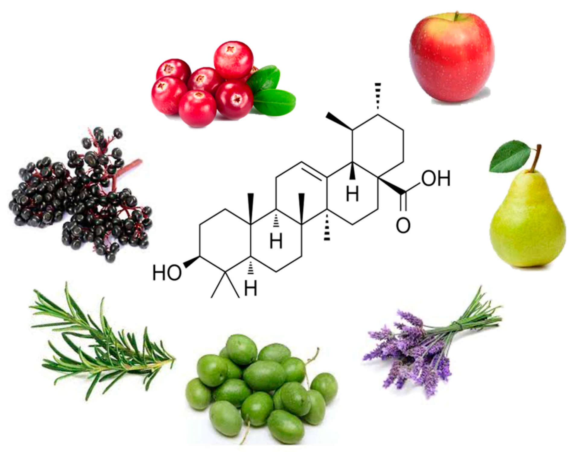
Figure 1. Chemical structure of ursolic acid and some of the plants (apples, pears, cranberries, elderberries, lavender, olives and rosemary) that contain high concentrations of UA. This chemical structure was created in BioRender.com (https://www.biorender.com; accessed 23 May 2024) and the image was created using Microsoft PowerPoint 2024.
Nutraceuticals 04 00022 g001
Figure 2. Summary of the effects of ursolic acid in human colorectal cancer cells. This figure was created using BioRender.com and Microsoft PowerPoint based on studies presented in Table 2 above. Green arrows show stimulation; red arrows show inhibition; black arrows show established pathways.
Nutraceuticals 04 00022 g002
Figure 3. Ursolic acid has synergistic effects with 5-fluorouracil (5-FU), oxaliplatin (OXA), doxorubicin (DOX), sorafenib and gamma irradiation. This figure was created with Microsoft PowerPoint 2024 based on the studies presented in Table 3. (↑: increased, ↓: reduced).
Nutraceuticals 04 00022 g003
Figure 4. Ursolic acid reduced tumor growth in animals xenografted with human CRC cells. This figure was created with BioRender.com. ↓: reduced.
Nutraceuticals 04 00022 g004
Table 1. Ursolic acid (UA) concentrations in different plants and fruits.
Source
Common and Botanical Name
Concentration of UA
(FW = Fresh Weight)
(DW = Dry Weight)
Reference
FruitsApple (peel)
Malus
1.52 mg/g DW[28]
Apple (whole fruit)
Malus
0.77 ± 0.1 mg/g to
1.85 ± 0.17 mg/g
[22]
Cranberry
Vaccinium macrocarpon
0.46–1.09 mg/g FW[23]
Pear
pyrus
0.3481 mg/g (mature fruit)
0.1293 mg/g FW (young fruit)
[29,30]
HerbsBasil
Ocimum tenuiflorum
20.2 mg/g DW[27]
Rosemary
Rosmarinus officinalis
15.8–29.5 mg/g[31]
Thyme
Thymus vulgaris
9.4 mg/g DW[31,32]
Oregano
Origanum vulgare
2.8 mg/g DW[31]
Sage
Salvia officinalus
18 mg/g DW[31]
Flowering plantsLavender
Lavandula
106.7–153.1 mg/g F.W.
3.463–6.484 mg/g D.W.
10.5 mg/g (flowers)
[26,31]
White deadnettle
Lamii albi flos
39.1–110.4 mg/g D.W.[33]
Oleander leaves
Nerium oleander
12.7 mg/g DW[31]
Rosinweed
Silphium sp. Flowers
17.95–22.05 mg/g D.W.[34]
Olive leaves
Olea europeae
1.8 mg/g DW[31]
OtherArabica coffee leaves
Coffea arabic
18 mg/g DW[31]
Table 2. Effects of ursolic acid against colorectal cancer: in vitro studies.
Cell TypeDose/DurationEffectsMechanismReference
HCT15UA
30 µM
↓ Cell viability
Cell cycle arrest (G0/G1 phase)
Not examined[45]
HT-29UA
10, 20 and 40 µM
↓ Cell proliferation
↑ Apoptosis
↓ p-EGFR
↓ p-ERK ½
↓ p-p38
↓ p-JNK
↓ Bcl-2
↓ Bcl-xL
↑ Cleaved caspase 3
↑ Cleaved caspase 9
[46]
HCT15
CO115
UA 2.5 and 4 µM (HCT15)
and
UA 10 and 15 µM (CO115)
↓ Cell proliferation
↑ Apoptosis
↓ p-AKT
↓ KRAS
[47]
HT-29UA
20 and 30 µM
↓ Cell proliferation
↑ Apoptosis
↑ Caspase 3 activity
↑ DNA fragmentation
↑ Cleaved Parp
↑ PGE2 concentration
↓ p-ERK
↑ p-p38
↑COX-2
[48]
HT-29UA
25 µM
↑ ATP in cytosol
↑ P2Y2 mRNA
↑ COX-2 protein
↑ DNA fragmentation
↑p-p38↑ p-Src protein
[49]
HCT-116UA
5, 34.7 and 50 µM
↓ Cell viability
↑ Apoptosis
↑ ROS
↓ Cell migration
↓ BCL-2 protein
↓ Survivin protein
↓ NFkB
↓ SP1 protein
↑ BAX mRNA
↑ P21 mRNA
↑ P53 mRNA
↓ FN1 mRNA
↓CDH2
↓↓CTNNB1
↓ Twist
[50]
HT-29UA
20, 40 and 80 µM
↓ SHH protein and mRNA
↓ Gli-1 protein and mRNA
↓ VEGF-A protein and mRNA
↓ bFGF protein and mRNA
[51]
HUVECUA
20, 40 and 80 µM
↓ Cell viability
↓ Cell migration
Not examined
HT-29UA
20, 40 and 80 µM
↓ Cell viability
↓ Cell survival
↓ Cell cycle progression
↑ Apoptosis
↓ Cells in s-phase
↓ Cyclin D1 protein and mRNA
↓ CDK4 protein and mRNA
↑ p21 protein and mRNA
↑ DNA fragmentation
↓ Bcl-2 protein and mRNA
↑ Bax protein and mRNA
↓ p-Erk1/2 protein
↓ p-JNK protein
↓ p-p38 protein
[52]
HCT116
HT29
SW480
UA
25 µM
↓ Cell viability
↓ Tumor sphere formation
↓ p-STAT3 protein
↑ Cleaved caspase 3
[53]
SW480
LoVo
UA
20, 40 and 60 µM
↓ Cell viability
↓ Colony formation
↓ Cell migration
↑ Apoptosis
↓ MMP9 mRNA
↑ CDH1 mRNA
↓ p-Akt
↓ p-mTOR
↑ p-PTEN
↓ p-JNK
↓ p-ERK
↓ COX-2 protein and mRNA
↓ PGE2
↑ NF-kB translocation
↑ p300 translocation
↑ Cleaved PARP
↑ Cleaved caspase -3, -8 and -9
[54]
HCT116
HT29
UA
20, 40, 60 and 80 µM
↑ Apoptosis
↓ Cell viability
↑ TUNEL positive cells
↑ Cleaved PARP
↑ Cleaved caspase-3
↓ p-JAK2
↓ p-STAT3
↓ STAT3 nuclear translocation
↓ miR-4500 mRNA expression
[55]
HCT116
HCT-8
UA
40 µM
↓ Cell viability
↓ Cell migration
↓ Cell invasion
↓ TGF-β1 protein
↓ p-Smad2/3
↓ p-FAK
↓ ZEB1
↓ N-cadherin
↑ miR-200a mRNA
↑ miR-200c mRNA
[56]
SW620
HCT116
UA
10, 30 and 60 µM
↓ Cell viability
↓ Clone formation
↓ Cell migration
↓ EMT
↑ Caspase 3 activity
↓ Mesenchymal phenotype
↑ E-cad protein
↓ Integrin protein
↓ Vimentin protein
↓ Twist protein
↓ Zeb1 protein
[57]
SW-480
HCT116
UA
10 µM
↓ Cell viability
↑ Cell injury
Cell cycle arrest (S phase)
↓ CCNB1 mRNA and protein
↓ CDK1 mRNA and protein
↓ CDK2 mRNA
↓ CCND1 mRNA and protein
↓ CCNA2 mRNA and protein
↓ CDC20 mRNA and protein
↓ CKS2 mRNA
↓ CCNB2 mRNA
[58]
HT-29
HCT116
UA
2.5–40 µM
↓ Cell viability
↓ Cell number
↓ Colony formation
↑ Apoptosis
↓ NUFIP1 mRNA[59]
RKOUA
14, 17 and 20 µM
Conventional conditions
↓ Cell viability
↑ Apoptosis
↑ ROS
Cell cycle arrest (G0/G1 phase)
↑ Casp-3, -8 and -9 activity
↑ Bax protein
↓ Bcl-2 protein
[60]
UA
25, 28, 31 µM
24 h
Poly-HEMA coated plates
↓ Cell viability
↑ Apoptosis
↑ Anoikis
↓ p-FAK
↓ p-PI3K
↓ p-Akt
↓ N-cadherin
↑ E-cadherin
HCT-116hSMO−UA
20 µM
↓ Cell proliferation
↓ Migration
↑ Apoptosis
↓ Bcl-2 protein and mRNA
↑ Bax protein and mRNA
↑ Caspase -3 and -9 mRNA
↓ c-Myc protein and mRNA
↓ GLI1 protein and mRNA
↓ SHH protein and mRNA
↓ SUFU protein and mRNA
↓ p-Akt protein
[61]
SW620UA
7.5, 15 and 30 µM
↓ Cell proliferation
↓ Migration
↑ Apoptosis
Cell cycle arrest–G0/G1 phase
↓ c-Myc protein
↓ Cyclin D1 protein
↓ Wnt4 mRNA and protein
↓ TCF4 mRNA and protein
↓ LEF1 mRNA and protein
↑ GSK3β mRNA and protein
↓ p-GSK3-β protein
↓ β-catenin mRNA and protein
↑ p-β-catenin
[62]
HCT-116 and SW480UA
15 µM
↓ Migration
↓ Invasion
↓ p-Akt
↓ p-mTOR
↓ ARL4C
↓ MMP2
[63]
N/AUA–computational model↓ Cell proliferation
↑ Apoptosis
↓ Angiogenesis
[64]
Table legend: ↑ increased, ↓ reduced, p—phosphorylated.
Table 3. Effects of UA in combination with chemotherapy agents.
Cell TypeDose/DurationEffectsMechanismReference
SW480
LoVo
UA 20, 40 and 60 µM
Melatonin 1 mM
↓ Cell viability
↓ Cell migration
↑ Apoptosis
↓ MMP9 mRNA expression
↑ Cleaved PARP
↑ Cleaved caspase -3 and -9
↓ COX-2 protein and mRNA
↑ p300 cytoplasmic translocation
↑ NF-kB cytoplasmic translocation
[65]
HCT15UA 4 µM
5-FU 100 µM
↑ Apoptosis↑ p-JNK p46 protein
↓ p-mTOR protein
↑ LC3-I protein
↑ LC3-II protein
↑ p62 protein
↑ p53 levels
[66]
SW480
SW620
LoVo
RKO
UA 10 µmol/L
Oxaliplatin 0.4 µmol/L
↓ Cell viability
↑ Apoptosis
↓ Mitochondrial membrane potential
↑ Cleaved caspase -3, -8 and -9
↓ p-B-Raf
↓ p-MEK1/2
↓ p-ERK1/2
↓ p-Akt
↓ p-p38
↓ p-JNK
↓ p-IKKα
↓ p-IkBα
↓ p-p65
↓ p-NF-kB (plasma and nucleus)
[67]
RKO
LoVo
SW480
UA 20 and 40 µM/L
5-FU 4 and 8 µM/L
Oxaliplatin 0.5, 1 and 1.5 µM/L
↓ Cell viability
↑ Chemosensitivity (hypoxia)
↑ Apoptosis
↓ MDR1 protein and mRNA expression
↓ HIF-1α protein and mRNA
↓ VEGF
[68]
HCT8
SW480
UA 20 µmo/L
Oxa 0.4 µmol/L
↓ Cell viability
↑ Apoptosis
↑ Cleaved caspase -3
↑ ROS
↑ NAPDH protein
↓ P-gp mRNA and protein
↓ MRP mRNA and protein
↓ BCRP mRNA and protein
[69]
RKOUA 15 µM
Oxa 2.5 µM
48 h
↓ Cell survival
↑ Apoptosis
↑ Caspase -3, -8 and -9 activity
↑ Cleaved PARP
↓ Survivin protein
↓ XIAP protein
[70]
HT-29
SW 620
UA 5 µg/mL
OA 100 µg/mL
CPT-11 0.075 µg/ml
↓ Cell viability
↓ Migration
N/A[71]
HCT116
HT-29
UA 15 µM
DOX 1.5 µM
↓ Cell proliferation
↑ Apoptosis
↓ Colony formation
↓ Cell migration
G1 cell cycle arrest
↑ Cleaved caspase -9
↑ Cleaved PARP
↑ E-cadherin
↓ MMP-9
↓ uPA
↓ CDK4 and CDK6
↓ cyclin D1
↓ p-Akt
↓ p-GSK-3β
↓ c-Myc
↑ Rassf1A
↑ Mst1 and Mst2
↑ Sav1
↑ p-Mob1
↑ p-Yap
↓ CTGF
[72]
LoVo
HCT116
UA 10 mM
Sorafenib 10 mM
↓ Cell viability
↓ Colony formation
↑ Apoptosis
↑ ROS
↑ Cleaved PARP
↑ Cleaved caspases 9 and 8
↑ LC3 I and II
↓ Mcl-1
↑ Bim
↑ MDA
↓ GSH
[73]
CT26
Mouse colon cancer cells
UA
15 µM
15 Gy radiation
↓ Cell survival
↑ Apoptosis
↑ Ros
↓ GSH
↓ Casp 3
↓ Bcl2
↑ Cleaved PARP
[74]
Table legend: ↑ increased, ↓ reduced, p—phosphorylated.
Table 4. Effects of ursolic acid on animal models of colorectal cancer.
ModelDose/DurationEffectsMechanismReference
Male BALB/c athymic mice xenografted with HT-29 cells (1.5 × 10⁶)UA
12.5 mg/kg
Daily intraperitoneal injection
↓ Tumor volume
↓ Tumor weight
↓ PCNA
↓ cyclin D1 protein and mRNA
↓ CDK4 protein and mRNA
↑ p21 protein and mRNA
↑ TUNEL
↓ Bcl-2 protein and mRNA
↑ Bax protein and mRNA
↓ p-STAT3
↓ p-Erk1/2
↓ p-JNK
↓ p-p38
[52]
Male BALB/c athymic mice xenografted with HT-29 cells (1.5 × 10⁶)UA
12.5 mg/kg
Daily intraperitoneal injection
↓ Tumor volume↓ CD31 positive cells
↓ p-STAT3
↓ p-Akt
↓ p-p70S6K
↓ SHH positive cells and mRNA
↓ Gli-1 positive cells and mRNA
↓ VEGF-A positive and mRNA
↓ bFGF positive cells and mRNA
[51]
Chick chorioallantoic membraneUA
0.25 mg
72 h
↓ Number of blood vessels
Female athymic nude mice xenografted with HCT116 cells (1 × 10⁷)UA
10 mg/mg
Daily
intraperitoneal injection
↓ Tumor volumeN/A[53]
Female nude mice
xenografted with HCT15 cells (106 cells)
UA
75 mg/kg
daily
Orally in Nutella
↓ Tumor size↑ p62 (ns)
↑ p-JNK (ns)
[66]
Male BALB/c nude mice
HCT-116hSMO− cells (1 × 10⁷)
UA
10, 20 or 40 mg/kg
Intraperitoneal injection
12 consecutive days
↓ Tumor weight
↓ Tumor volume
↓ BCL-2 protein and mRNA
↑ BAX protein and mRNA
↑ Caspase -3 and -9 mRNA
↓ c-Myc protein and mRNA
↓ GLI1
↓ SHH
↓ SUFU
↓ p-Akt
[61]
Nude mice
xenografted with SW620 cells (1 × 10⁷)
UA
15, 30 or 60 mg/kg
Intragastrical
↑ Body weight
↓ Tumor weight
↓ Tumor volume
↑ Apoptosis
↑ GSK3β mRNA and protein
↓ β-catenin mRNA and protein
↓ WNT4 mRNA and protein
↓ TCF4 mRNA and protein
↓ LEF1 mRNA and protein
↑ p-β-catenin
↓ p-GSK3β
↓ Nuclear β-catenin
[62]
Male BALB/c-nude mice
HCT-116 cells (5 × 10⁶) injected in tail vein
UA
20 mg/kg
Intraperitoneal
Daily/42 days
↓ Lung metastasis↓ ARL4C[63]
Female nude mice
xenografted with SW620 cells
UA
20 mg/kg
Oxaliplatin 10 mg/kg
↓ Tumor weight
↓ Tumor volume
↓ p-ERK1/2
↓ p-Akt
↓ p-IKKα
↓ Ki-67 pos cells
↑ TUNEL pos cells
[67]
Female nude mice
xenografted with HCT8 or SW480 cells (1 × 10⁵)
UA
10 mg/kg
Oxaliplatin
10 mg/kg
↑ Animal survival time
↓ Tumor volume
N/A[69]
Athymic nude mice xenografted with HCT116 cellsUA
10 mg/kg/day
DOX
2 mg/kg/twice weekly
↓ Tumor weight
↓ Tumor volume
↓ Ki67
↓ p-Akt
↑ Rassf1A
↑ Mst1
↑ Mst2
↑ Sav1
↑ p-Mob1
↑ p-Yap
↓ CTGF
[72]
Male athymic nu/nu mice
Luciferase-transfected HCT116 cells
UA
250 mg/kg
Orally, daily
Capecitabine
60 mg/kg
Orally, 2/week
combination
↓ Tumor growth
↓ Tumor volume
↓ Tumor weight
↓ Metastasis
↓ Ki67
↓ CD31
↓ Nuclear p65
↓ β-catenin
↓ p-STAT3
↓ Cyclin D1 protein
↓ cMyc
↓ p-EGFR protein
↓ Bcl-2 protein
↓ Bcl-xl protein
↓ Survivin protein
↓ ICAM-1
↓ VEGF
↓ MMP9
↓ p53
↓ p21
[75]
Table legend: ↑ increased, ↓ reduced, p—phosphorylated.
Disclaimer/Publisher’s Note: The statements, opinions and data contained in all publications are solely those of the individual author(s) and contributor(s) and not of MDPI and/or the editor(s). MDPI and/or the editor(s) disclaim responsibility for any injury to people or property resulting from any ideas, methods, instructions or products referred to in the content.