Article

Diagnostic Limitations and Aspects of the Lumbosacral Transitional Vertebrae (LSTV)

1
Department of Orthopaedic and Trauma Surgery, Paracelsus Medical University Salzburg, 5020 Salzburg, Austria
2
Division for Orthopaedics and Traumatology, Center for Clinical Medicine, Department of Medicine, Faculty of Medicine and Dentistry, Danube Private University, 3500 Krems, Austria
*
Author to whom correspondence should be addressed.
Appl. Sci. 2022, 12(21), 10830; https://doi.org/10.3390/app122110830
Submission received: 2 October 2022 / Revised: 20 October 2022 / Accepted: 24 October 2022 / Published: 25 October 2022
(This article belongs to the Special Issue Intervertebral Disc Regeneration II)

Abstract

:
The regeneration of an intervertebral disc can only be successful if the cause of the degeneration is known and eliminated. The lumbosacral transitional vertebrae (LSTV) offer itself as a model for IVD (intervertebral disc) regeneration. The aim of this work is to support this statement. In our scoliosis outpatient clinic, 1482 patients were radiologically examined, and ambiguous lumbosacral junction underwent MRI examination. Patients with Castellvi classification type II–IV were included and the results are compared with the current literature in PubMed (12 October 2022). The LSTV are discussed as a possible IVD model. A total of 115 patients were diagnosed with LSTV Castellvi type II–IV. A Castellvi distribution type IIA (n-55), IIB (n-24), IIIA (n-20), IIIB (n-10) and IV (n-6) can be found. In all, 64 patients (55.7%) reported recurrent low-back pain (LBP). Scoliosis (Cobb angle >10°) was also confirmed in 72 patients (58 female and 14 male) and 56 (75.7%) had unilateral pathology. The wide variation in the literature regarding the prevalence of the LSTV (4.6–35.6%) is reasoned by the doubtful diagnosis of Castellvi type I. The LSTV present segments with reduced to absent mobility and at the same time leads to overload of the adjacent segments. This possibility of differentiation is seen as the potential for a spinal model.

1. Introduction

In the introduction, the state of knowledge of the lumbosacral transitional vertebrae (LSTV) is shown on the basis of the current literature. The degeneration of the intervertebral discs is the focus of the work. The literature is reviewed for links between degeneration and regeneration of the intervertebral discs. Attention is paid to the clinical symptoms of lumbar complaints and radiological criteria.
The lumbosacral transitional vertebrae (LSTV) are a congenital malformation of the spine. It is characterized by enlargement of the L5 transverse process(es), with potential pseudoarticulation or fusion with the sacrum. It leads to altered rotational movement of the lower spine, which gives rise to low-back pain (LBP). A diagnosis of Bertolotti syndrome is given to patients experiencing pain caused by the presence of the LSTV. Estimates of the prevalence of the LSTV in the general population vary widely in the literature, ranging from 4.0% to 35.9%. Apazidis et al. have found that type IA is the most common, with a prevalence of 14.7%. However, type I is generally considered to be clinically insignificant [1].
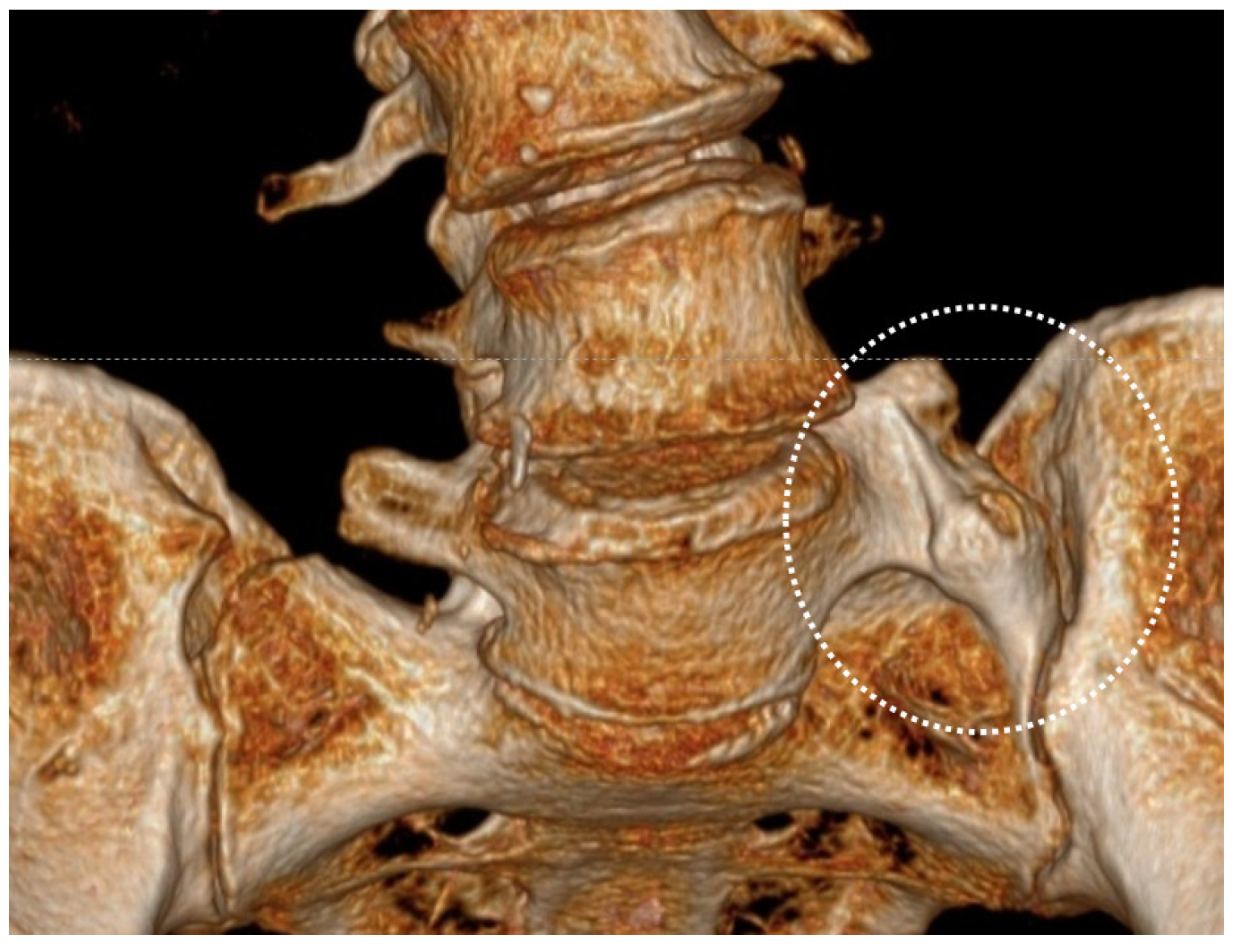
In the Castellvi classification, only type I uses a dimension of >19 mm. Castellvi types II–IV, on the other hand, are defined by a structural change as pseudarthrosis or ossification. This is important because a differentiation between normal structure without altered biomechanics and pathological structure with expected symptoms of discomfort cannot be justified by a measurement of 19 mm. The scattering of the prevalence, which is repeatedly found in the literature, is only one indication of this (Figure 1 and Figure 2).
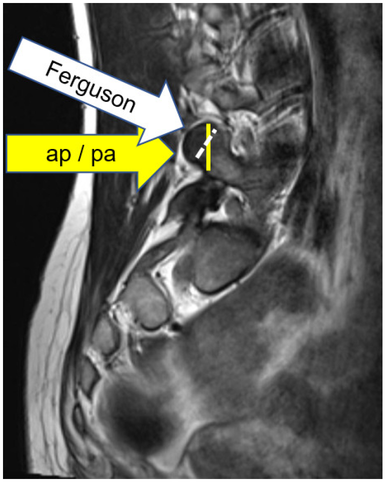
The topic of the inaccuracy of the representation of the L5 transverse process will therefore be discussed in detail at this point. In Figure 1, Castellvi type I is represented as a sketch in such a way that a slight to more severe restriction of the mobility of the affected segment can be assumed (Figure 1). As reflected in the review of the following international literature, this problem has not yet been conclusively addressed. The problem of the imaging quality of the LSTV is presented in a value-neutral manner, even though this topic is repeatedly addressed in the further course. Depending on the pelvic shape and pelvic tilt, the radiological ap image of the lumbosacral junction leads to superimpositions in the case of an enlarged L5 transverse process, which do not always permit measurement and assignment with certainty. The measurement of the L5 transverse process (>19 mm) is thus not guaranteed. As outlined in Figure 3, the imaging result is significantly influenced by the radiological beam path. The imaging technique according to Fergusion with a tube tilt of 30°–40° cranially and caudally suggests improved imaging (Figure 3). However, the dimension of 19 mm remains a variable dependent on many factors (patient body size, tube–object distance, object–film distance, etc.). The pelvic setting in relation to the spine is not only crucial for visualizing pathology, but also an important criterion for possible causes of complaints. Especially the expected disc load in the context of the LSTV is crucial. If now again the question of the cause of complaints in Castellvi type I arises, it becomes clear that a cause differentiation between the LSTV and a pathological pelvic setting is currently hardly possible. No conclusive answer can be found in the literature either. An interaction between the LSTV and a hip complaint can be assumed in individual cases. We therefore always recommend a simultaneous hip examination (Figure 3 and Figure 4).
The availability of an MRI examination significantly improves the diagnosis. However, even this examination technique must be specific to the question of the LSTV. Initial referrals of our own to various institutes yielded astonishing results that rarely allowed a diagnosis to be made. This ranged from imaging of only the sagittal and axial slices, which is often sufficient for disc assessment, to imaging that did not portray the L5 transverse process. The combination with a 1.5 Tesla device and a delivered slice thickness of 3 mm also does not allow a more accurate Castellvi type I assessment. These experiences led to arranging for standardized images, as described in the method below, after consultation with one MRI institute. These findings of our own and the still sparse literature at the beginning of the evaluation period led to the decision not to pursue Castellvi type I further for the time being (Figure 5).
In a special issue on the subject of “Intervertebral Disc Regeneration”, the question of the ligamentous situation at the lumbosacral junction must also be considered. The findings available in the literature to date are presented, and the question of the quality of the imaging is part of the problems described above. In particular, the question of mobility restriction of the affected segment and the associated intervertebral disc stress becomes the focus of consideration. This makes it obvious that the Castellvi classification type I is not suitable for differentiating between a normal segment L5/S1 with a pronounced L5 transverse process and a pathological change. This illustration should clarify why the assessment of Castellvi type I was excluded in this study, although these changes are crucial not only for disc degeneration but even more so for the outcome of disc regenerative treatment.
In the work of Min Tang, a prevalence of 15.8% for the LSTV is diagnosed in 5860 radiographs. In all, 44.8% of them are classified as type I [2]. Regarding the question of diagnosis of the LSTV comparing X-ray and MRI, a good agreement was established very early [3].
In the work of the last 10 years, the problem of classification of the LSTV is increasingly worked on. Thus, the imaging quality and reliable assignment of the LSTV, especially of type I, is becoming the focus of discussion.
Farshad et al. advocate measuring segmental differences. Measurement methods in the sagittal slices of MRI images are described for identification of LSTV. However, the variability of the vertebral bodies makes it difficult to accurately define L5 and S1 [4].
Of importance is the total number of vertebral bodies diagnosed with 23 (sacralization) or 25 (lumbarization) with a normal value of 24 vertebral bodies. Thus, a total imaging of the spine is necessary [5].
Quantitative measurements are used to try to find new parameters for the diagnosis and differentiation of the LSTV. Especially LSTV type I with a height of the transverse process of >19 mm forms an uncertainty in the presentation and therefore in the diagnosis. The previously mentioned large differences in the prevalence of the LSTV are partly due to this [6].
In the article by Konin et al., attention to the identification and correct numbering of the LSTV and recognition of imaging findings related to the development of LBP are comprehensively addressed [7].
In previous work, the iliolumbar ligament is recommended for identification of L5/S1 [8]. However, attempts to identify L5/S1 using the iliolumbar ligament have not been successful to date [9].
The iliolumbar ligament in its anatomical structure and as a possible cause of complaints is currently coming into focus. In an MRI study by McGrath et al., unilateral pathology (types IIA and IIIA) shows a thinner ligament than on the unaffected side. Bilateral pathology (types IIB, IIIB, and IV) shows no side difference and no difference from the healthy control group. However, the differentiation of LSTV type I is not answered [10].
For a special issue, “Intervertebral Disc Regeneration II”, the cause of LBP and disc degeneration is the focus of the LSTV.
The relationships between IVD degeneration (intervertebral disc degeneration) and the LSTV are differentiated in the literature. In LSTV type I, the highest numbers are reported in some papers; however, no pathological effect on the disc segment is described. In contrast, in LSTV type II–IV, the adjacent cranial disc segment is reported as the segment most exposed to degeneration. The distinction between unilateral and bilateral pathology forms another focus, which is also associated with the question of scoliotic change. A clear prediction cannot be derived so far.
Pathologies of adjacent structures as a cause of complaints are also increasingly subject to clarification. The segmental nervous assignment was examined thereby, too.
The disc degeneration is concentrated on the cranially adjacent segment L4/5. In contrast, the disc of the affected segment of the LSTV type II–IV is mechanically protected by the restriction of movement. However, this general statement still lacks a differentiated assessment dependent on the type of the LSTV. The biomechanical workup of motion restriction and the associated disc loading or unloading are at the beginning of scientific interest [11].
A paper by Ashour et al. states that the incidence of disc herniation is decreased in the segment of the LSTV. In contrast, the incidence of disc herniation or spondylolisthesis in the cranial segment L4-5 is increased in the presence of the LSTV. Therefore, the LSTV are considered a risk factor for degenerative disc changes in the level above the transitional vertebra [12]. In the LSTV type II group, compression of the L5 nerve due to a bony or synovial cause of nearthrosis has also been reported [13]. The question of nerve innervation in the LSTV has also been raised by intraoperative EMG studies, and no pathological distribution was found compared with the normal spine [14]. A significant association between lumbar stenosis and the LSTV is described in a retrospective cross-sectional study [15]. The bony structures show altered trabecular distribution in the LSTV compared to healthy spine in a cadaveric study [16]. In a prospective cadaver study by Golubovsky et al. the segmental mobility of the affected segment and adjacent segments was measured and described as follows:
The results of this study suggest that patients with Bertolotti syndrome have significantly altered spinal biomechanics and may develop pain due to increased loading forces at the LSTV joint during ipsilateral lateral flexion and axial rotation. In addition, increased motion in the upper levels in the presence of the LSTV can lead to degeneration over time [17].
After outlining the pathological changes and movement limitations, the focus turns to the possible causes of LBP:
There are different answers to the question of whether LSTV type I causes symptoms, although diagnostic uncertainty remains as described above. There is also no clear answer to the distinction between unilateral and bilateral changes as the cause of symptoms. However, the more stable the constructs of the LSTV are (type II–IV), the more likely degeneration of the cranially adjacent segment is responsible for the complaints.
Lumbar complaints (LBP) in the presence of the LSTV were originally noted by Mario Bertolotti in 1917 and are therefore referred to as “Bertolotti syndrome” [18].
Quinlan et al. found Bertolotti syndrome in 769 MRI scans of LBP patients in 4.6% of the patients studied. In patients with LBP younger than 30 years, 11.4% showed LSTV [19].
A high frequency of the LSTV was found in young male patients with LBP, and it was mostly subtype I. LSTV type I and associated disc and facet degeneration were notable in this group [20].
A review of the literature on the problem of discomfort in the LSTV (Bertolotti syndrome) shows that comparable changes are found with and without discomfort.
LSTVs pose a diagnostic dilemma for the treating physician, as they may remain undetected on plain radiographs and even on advanced imaging; moreover, even if the malformation is recognized, patients with the LSTV may be asymptomatic or have nonspecific symptoms, such as low-back pain with or without radicular symptoms.
Special attention should be given to younger patients under 30 years of age with chronic lumbar symptoms. Here, diagnostic exclusion or confirmation of the LSTV is required [21].
In lumbalgia, differences in muscle mass distribution are also associated with lordosis of the lumbar spine [22]. The LSTV are associated with a reduction in muscle volume and an increase in muscle degeneration of both the lumbar and trunk muscles compared to healthy individuals [23]. A differential relationship between lumbalgia and the type of the LSTV can be established. However, the segment affected by the LSTV must be distinguished from the adjacent segment as the cause of the symptoms [24]. Unilateral LSTV result in asymmetric biomechanical stresses. The side with the extra L5/S1 joint bears a greater proportion of the load, resulting in lateral tilt with scoliotic curvature. This local loading leads to greater wear and unilateral muscle activity. In addition, asymmetric motion may also affect disc degeneration [25]. The LSTV may also be associated with spina bifida occulta as another pathological change [26,27].
The topic of scoliosis is addressed in 16 papers. In a study in the Turkish Army, the LSTV were diagnosed in 12.9% of 3132 men [28]. Another regional assignment of prevalence to the southern European population is reported by Vinha A. et al. with 24.9% for the group LSTV Castellvi type II, III and IV [29]. In the work of Garg B. et al., a prevalence of LSTV in 198 patients with scoliosis is found in 18.2% [30].
The change in segmental mobility is measured in a paper by Becker L. et al. and summarized as follows: Patients with the LSTV show reduced range of motion in the transitional segment and a significantly increased distribution of motion to the cranially adjacent segment on flexion–extension radiographs. The increased proportion of mobility in the cranial adjacent segment may explain the higher rates of degeneration within the segment [31]. The influence of the LSTV on pelvic alignment with acetabular orientation is also described by the same group [32].
The impact of the LSTV on surgical interventions is also scientifically discussed but is not the focus of this article. An improvement of the patients’ quality of life is described by surgical interventions. This concerns both the lysis of LSTV and the fusion of the segment depending on the degenerative changes [33]. However, surgical problems due to the altered anatomy, especially of the ventral vascular plant, must also be considered in surgical planning [34].
The LSTV have a wide range in diagnosis, especially in type I, which is reflected in the widely scattered prevalence data. The biomechanical limitations and the possible causes of the symptoms are not yet sufficiently differentiated. In particular, there is little information about the intervertebral disc in the LSTV. The aim of this paper is to investigate the diagnosis of the LSTV in relation to LBP. The prevalence of the LSTV in the literature and in our own work should be documented with respect to the problem of diagnosis of LSTV, especially of Castellvi type I.

2. Materials and Methods

The present article follows the current PRISMA criteria from 2020. The structure of the contribution is shown in a flow diagram [35] (Table 1).
Diagnosis of LSTV: In our scoliosis outpatient clinic 1482 patients were examined by one consultant orthopaedic surgeon (2014–2021) with more than 25 years of experience in that field.
Each radiological, structurally ambiguous assignment of the lumbosacral junction was submitted to further MRI examination with a 3.5 Tesla device (sagittal, coronal, axial and adjustment to the segment L5/S1 with a slice thickness of 3 mm, Siemens Medical Group).
Patients with Castellvi I classification (height of L5 transverse process >19 mm) are not included in the study. This decision was made in the initial phase of the study, because in our own unpublished studies the imaging inaccuracy allows an exact measurement of the L5 transverse process only in the Ferguson image. However, this additional X-ray is not permitted for reasons of radiation hygiene. On the other hand, 19 mm should be evaluated differently for a very tall person than for a short person.
Only patients with Castellvi classification type II–IV are included in the study.

3. Results

In 1482 spine patients examined, 115 patients were diagnosed with LSTV Castellvi II–IV (male n-32, female n-83).
In the Castellvi IIA (n-55) and IIB (n-24) groups, spondylolysis of the adjacent segment was seen in two patients and lumbarization in one patient, respectively. In patients with Castellvi IIIA (n-20), spondylolysis of the adjacent segment was found in three patients and there was lumbarization in one patient. In classification IIIB (n-10), spondylolisthesis (Meyerding 2) was seen. Six patients showed Castellvi type IV.
A total of 64 (55.7%) of all patients reported recurrent LBP. Of these, 23 patients showed orthograde lumbosacral transition and 41 patients showed scoliosis.
In unilateral pathology (IIA, IIIA, n-75), 39 complained of symptoms, and in bilateral pathology (IIB, IIIB, IV, n-40), 26 patients did. Only five patients belong to group IIIA and thus have neither unilateral nor pseudarthrosis as cause of pain.
In lumbalgia, a clear age-dependent differentiation is evident. In children and adolescents <16 years, 26.7% complained of chronic complaints. In the age group 16–30 years, it was 35.5%, and in >30 years it was 88.6% who reported chronic complaints.
Five patients were therefore already in psychological treatment. In the anamnesis, up to 30 visits to the doctor for lumbalgia can be recorded up to the time of the diagnosis of LSTV. Three patients with recurrent lumbalgia have already undergone spinal surgery without the LSTV being known to the patients.
In 72 patients, LSTV Castellvi II–IV and scoliosis was confirmed (58 female and 14 male). The curvature extent of scolioses showed a mean Cobb angel of 24.3° with a range from 11°–55° (n-42) in children and in adults a mean of 32.4° with a range from 12°–66° (n-30) in the adults.
Of the 72 patients with scoliosis, 56 showed unilateral pathology (IIA and IIIA) and 16 bilateral pathologies (IIB, IIIB, IV, Table 2).
A positive family history existed in 20 patients from the scoliosis group (n = 72). In one family, the mother and daughter were affected type IIA, and in another case it was twins with IIB with scoliotic component. Other malformations were raised: longitudinal malformation of the right upper extremity, spina bifida, other vertebral body malformations (n-3), a sacral cyst, syringomyelia (n-2) and a fork rib.
When comparing our own data with the results in the literature, it should be noted that in our own data only LSTV Castellvi type II, III and IV are collected. The data from the literature refer to the total sum of LSTV Castellvi type I–IV. If Castellvi type I is shown in the data, the value is given in parentheses (Table 3).

4. Discussion

  • Castellvi type I:
In the initial phase of the examination, the Castellvi type I problem with its enlargement of the L5 transverse process of >19 mm becomes obvious. The standard radiograph of the lumbar spine, as well as the spinal X-ray for scoliosis clarification, does not result in a reliable orthograde representation of the L5 transverse process. Lordosis and pelvic tilt, as well as scoliosis, prevent standardized imaging. The magnification factor that inevitably occurs in a radiograph as a function of object–film distance and tube distance make the height indication of the L5 transverse process an unsuitable measurement for Castellvi I classification.
Only the Ferguson image (30° angle AP radiograph) serves as a standard method for detecting LSTV. Additional imaging is prohibited for radiation hygiene reasons. However, the problem of the magnification factor remains.
The alternative is an MRI examination, which provides additional information in all three planes in space. The standard images for disc assessment (coronal and sagittal) are not sufficient for the assessment of LSTV. The frequently used slice thickness of 3 mm and the quality of the device (<3.5 Tesla) form another uncertainty factor for diagnostics. If in the literature there are reported (in many cases not specified) investigations with a CT and MRI slice thickness of 0.6 mm–3 mm, this should not be considered an insignificant representation and therefore also not an inaccurate measurement [3,4,6,10].
However, the height of the L5 transverse process must also depend on the overall size of the patient, so that a measurement >19 mm is only of limited use for classification.
In conclusion, the shape of the L5 transverse process could be more indicative of pathology than the measurement alone.
In our own work, because of the intractability of the exact classification of Castellvi type I, we did not work on this type further. This does not mean that Castellvi type I cannot be responsible for complaints [8,9,10]. It should be emphasized that the scoliosis outpatient clinic, with its patients and the frequent oblique position of L5 compared to S1, presents an additional problem of presentation [1,2].
This raises the following questions:
Is pathology of the ligamentous apparatus (iliolumbar ligament) responsible for the shape of the L5 transverse process? If so, the shape of the L5 transvers process would be the expected identifying feature.
A standardized clarification of the ligamentous apparatus and appropriate classification must be required.
If it is a pathology with an effect on the intervertebral disc, then any interventional procedure on the intervertebral disc must also take into account a possible pathology due to LSTV. This assessment, at first sight very critical, becomes crucial when a new treatment modality is aimed at repairing the disc [37,38,39].
If the incidence of the LSTV indeed reaches values as reported in some literature between 4% and 35.9% and the LSTV leads to treatment failure, this would be crucial for success and failure for this new treatment modality [21].
  • LSTV type II:
In unilateral or bilateral pseudarthrosis, limitation of motion of the L5/S1 segment is assured. Therefore, the pseudarthrosis and the intervertebral disc must be differentiated as the cause of the complaint. It is to be expected that the pseudarthrosis cannot follow the age-related process of height reduction in the intervertebral disc without complaints. Corresponding complaints are thus to be expected. For this differentiation, further MRI criteria have to be defined.
We would like to point out another aspect to this.
As patients age, degenerative de novo curvatures or increases in curvature occur. This can lead to a contact zone of the L5 transverse process with the os sacrum (os ileum). If there is no previous X-ray from youth, the assignment as Castellvi II is also fraught with uncertainty. For the question of the IVD, which is the focus of this article, this may have no clinical significance. The literature provides the following evidence in this regard:
Type II LSTV had significant effects in promoting transitional and adjacent disc degeneration.
Thus, Castellvi type III/IV LSTV predisposed the adjacent spinal components to degeneration and protected the transitional discs [31,40].
  • LSTV type III:
In its unilateral form, stress loading is inevitable. In its bilateral form, stability of the L5/S1 segment is assumed. In both cases, L4/L5 overload is expected due to the lack of L5/S1 mobility [17,31].
  • LSTV type IV:
This is a bilateral form with a bony bridging and pseudarthrosis. In this case, an increased load on the adjacent segment L4/L5 may be assumed. Moreover, the pseudarthrosis may be responsible for discomfort since there is no symmetrical situation.
  • Disc degeneration:
In a paper by Ahmed Ashour et al., the incidence for disc prolapse is found to be decreased in the segment of LSTV. In contrast, the incidence for disc herniation or spondylolisthesis in the cranial segment L4-5 is increased in the presence of LSTV [12]. Hence, the LSTV are considered a risk factor for disc degenerative changes at the level above the transitional vertebra level. The IVD only makes sense (can be successful) if it does not affect the adjacent segment of the LSTV. Thus, a differential diagnosis of the LSTV is important before any IVD intervention.
Low-Back Pain (Bertolotti syndrome):
Explanation of own data:
A total of 64 patients of the total group (55.7%) reported recurrent LBP (orthograde lumbar spine n = 23, scoliosis n = 41).
The low number of LBP can be explained by the fact that the age limit of the patients is very low (scoliosis outpatient clinic). Additionally, the differentiation according to age shows a rapid increase in complaints in the patient distribution, and the dominance of scoliosis with 64.1% is striking.
Basis for an intervertebral disc model with different calculable limitation of motion:
A prospective cadaveric study measured the segmental range of motion of the affected segment and adjacent segments.
A biomechanical analysis of the tissue, joints and associated motion restriction (range of motion, resulting torques and the LSTV joint forces) and histological assessment of the segments was performed. The aim of this work is surgical therapy.
This study’s results indicate that patients with Bertolotti syndrome have significantly altered spinal biomechanics and may develop pain due to increased loading forces at the LSTV joint with ipsilateral lateral bending and axial rotation. In addition, increased motion at superior levels when the LSTV are present may lead to degeneration over time.
However, the model can be adapted to address disc treatment issues.
The LSTV are a model given in nature for a differential study of motion restriction on the affected and adjacent disc segments [17,31].
The preceding illustration suggests the LSTV as a potential model for IVD research. The LSTV provide segments with reduced to absent mobility and at the same time an overloaded adjacent segment.
MRI imaging capabilities should differentiate between complaint origin in the LSTV and disc degeneration.
The model representation should also be suitable for further FEM calculation [36,37,38,39,40].
  • Disc model:
The work shown here offers the LSTV as a disc model for further investigation. As a model for the degeneration of the lumbosacral junction and thus clarification of the cause of the complaint, treatment options become delineable. It can thus lead to a further gain in knowledge for regenerative disc treatment. We are well aware of the limitations of the model, especially in the area of Castellvi type I. Knowing the limitations of imaging will be an advantage for every practitioner and will lead to the avoidance of frustrated therapy attempts in cases of mechanically persisting pathology.
  • Limitation of own data:
The data were collected in the spine outpatient clinic with a focus on scoliosis. This explains the high number of scolioses. This is clearly shown in Table 3 in comparison with the literature.
The lack of inclusion of LSTV Castellvi I cases prevents direct comparison with the prevalence data in the literature. A wide dispersion is shown, which cannot be narrowed down by our own work. This will be discussed in detail in the discussion.
Through cooperation with an MRI institute, an attempt was made to provide standardized imaging as compensation and thus to ensure a reliable assignment of the patients LSTV Castellvi II, III and IV.

5. Conclusions

This paper clarifies the diagnosis of the LSTV as a possible cause of LBP. From the prevalence of LSTV, in the literature and in our own work, it becomes clear that this is a clinically non-negligible number of patients. In the wide dispersion of prevalence, the problem of diagnosis of LSTV Castellvi type I becomes apparent.
Despite these aforementioned diagnostic problems, the LSTV offer the full spectrum of possible segmental movement limitations. This ranges from a slight limitation of the range of motion, without clinical relevance, to complete bony fixation. Thus, the cranially adjacent disc segment is also included in a possible functional model of the IVD.
This in vivo IVD model is thus suitable to provide further insights into the degeneration mechanism. Knowing the cause of degeneration is the basic prerequisite for successful regeneration treatment using the IVD. For the therapy decision of an IVD regeneration or a necessary alternative treatment procedure, the presented model can provide information in the future.

Author Contributions

Data curation, F.L.; Investigation, F.L.; Methodology, K.T.; Writing—original draft, F.L.; Writing—review & editing, K.T. All authors have read and agreed to the published version of the manuscript.

Funding

This research received no external funding.

Institutional Review Board Statement

Ethical review and approval were waived for this study because only radiographs were analyzed.

Informed Consent Statement

Not applicable.

Data Availability Statement

Not applicable.

Conflicts of Interest

The authors declare no conflict of interest.

Abbreviations

LSTVLumbo Sacral Transitional Vertebra
IVDInterVertebral Disc
MRIMagnetic resonance imaging
FEMFinite Element Model
IDRIntervertebral Disc Regeneration
LBPLow-Back Pain

References

  1. Apazidis, A.; Ricart, P.A.; Diefenbach, C.M.; Spivak, J.M. The prevalence of transitional vertebrae in the lumbar spine. Spine J. 2011, 11, 858–862. [Google Scholar] [CrossRef] [PubMed]
  2. Tang, M.; Yang, X.F.; Yang, S.W.; Han, P.; Ma, Y.M.; Yu, H.; Zhu, B. Lumbosacral transitional vertebra in a population-based study of 5860 individuals: Prevalence and relationship to low back pain. Eur. J. Radiol. 2014, 83, 1679–1682. [Google Scholar] [CrossRef] [PubMed]
  3. O’Driscoll, C.M.; Irwin, A.; Saifuddin, A. Variations in morphology of the lumbosacral junction on sagittal MRI: Correlation with plain radiography. Skelet. Radiol. 1996, 25, 225–230. [Google Scholar] [CrossRef]
  4. Farshad, M.; Aichmair, A.; Hughes, A.P.; Herzog, R.J.; Farshad-Amacker, N.A. A reliable measurement for identifying a lumbosacral transitional vertebra with a solid bony bridge on a single-slice midsagittal MRI or plain lateral radiograph. Bone Jt. J. 2013, 95, 1533–1537. [Google Scholar] [CrossRef]
  5. Okamoto, M.; Hasegawa, K.; Hatsushikano, S.; Kobayashi, K.; Sakamoto, M.; Ohashi, M.; Watanabe, K. Influence of lumbosacral transitional vertebrae on spinopelvic parameters using biplanar slot scanning full body stereoradiography-analysis of 291 healthy volunteers. J. Orthop. Sci. 2022, 27, 751–759. [Google Scholar] [CrossRef] [PubMed]
  6. Zhou, S.; Du, L.; Liu, X.; Wang, Q.; Zhao, J.; Lv, Y.; Yang, H. Quantitative measurements at the lumbosacral junction are more reliable parameters for identifying and numbering lumbosacral transitional vertebrae. Eur. Radiol. 2022, 32, 5650–5658. [Google Scholar] [CrossRef]
  7. Konin, G.P.; Walz, D.M. Lumbosacral transitional vertebrae: Classification, imaging findings, and clinical relevance. Am. J. Neuroradiol. 2010, 31, 1778–1786. [Google Scholar] [CrossRef] [Green Version]
  8. Hughes, R.J.; Saifuddin, A. Numbering of lumbosacral transitional vertebrae on MRI: Role of the iliolumbar ligaments. Am. J. Roentgenol. 2006, 187, W59–W65. [Google Scholar] [CrossRef]
  9. Farshad-Amacker, N.A.; Lurie, B.; Herzog, R.J.; Farshad, M. Is the iliolumbar ligament a reliable identifier of the L5 vertebra in lumbosacral transitional anomalies? Eur. Radiol. 2014, 24, 2623–2630. [Google Scholar] [CrossRef]
  10. McGrath, K.A.; Lee, J.; Thompson, N.R.; Kanasz, J.; Steinmetz, M.P. Identifying the relationship between lumbar sacralization and adjacent ligamentous anatomy in patients with Bertolotti syndrome and healthy controls. J. Neurosurg. Spine 2022, 37, 200–207. [Google Scholar] [CrossRef]
  11. Zhang, B.; Wang, L.; Wang, H.; Guo, Q.; Lu, X.; Chen, D. Lumbosacral Transitional Vertebra: Possible Role in the Pathogenesis of Adolescent Lumbar Disc Herniation. World Neurosurg. 2017, 107, 983–989. [Google Scholar] [CrossRef] [PubMed]
  12. Ashour, A.; Hassan, A.; Aly, M.; Nafady, M.A. Prevalence of Bertolotti’s Syndrome in Lumbosacral Surgery Procedures. Cureus 2022, 14, e26341. [Google Scholar] [CrossRef] [PubMed]
  13. Kanematsu, R.; Hanakita, J.; Takahashi, T.; Minami, M.; Tomita, Y.; Honda, F. Extraforaminal entrapment of the fifth lumbar spinal nerve by nearthrosis in patients with lumbosacral transitional vertebrae. Eur. Spine J. 2020, 29, 2215–2221. [Google Scholar] [CrossRef] [PubMed]
  14. Hinterdorfer, P.; Parsaei, B.; Stieglbauer, K.; Sonnberger, M.; Fischer, J.; Wurm, G. Segmental innervation in lumbosacral transitional vertebrae (LSTV): A comparative clinical and intraoperative EMG study. J. Neurol. Neurosurg. Psychiatry 2010, 81, 734–741. [Google Scholar] [CrossRef]
  15. Abbas, J.; Peled, N.; Hershkovitz, I.; Hamoud, K. Is Lumbosacral Transitional Vertebra Associated with Degenerative Lumbar Spinal Stenosis? Biomed. Res. Int. 2019, 10, 2019. [Google Scholar] [CrossRef] [Green Version]
  16. Mahato, N.K. Trabecular bone structure in lumbosacral transitional vertebrae: Distribution and densities across sagittal vertebral body segments. Spine J. 2013, 13, 932–937. [Google Scholar] [CrossRef]
  17. Golubovsky, J.L.; Colbrunn, R.W.; Klatte, R.S.; Nagle, T.F.; Briskin, I.N.; Chakravarthy, V.B.; Gillespie, C.M.; Reith, J.D.; Jasty, N.; Benzel, E.C.; et al. Development of a novel in vitro cadaveric model for analysis of biomechanics and surgical treatment of Bertolotti syndrome. Spine J. 2020, 20, 638–656. [Google Scholar] [CrossRef]
  18. Bertolotti, M. Contributo alla conoscenza dei vizi di differenziazione regionale del rachide con speciale riguardo all assimilazione sacrale della v. lombare. Radiol. Med. 1917, 4, 113–144. [Google Scholar]
  19. Quinlan, J.F.; Duke, D.; Eustace, S. Bertolotti’s syndrome: A cause of back pain in young people. J. Bone Jt. Surg. Br. 2006, 88, 1183–1186. [Google Scholar] [CrossRef]
  20. Apaydin, M.; Uluc, M.E.; Sezgin, G. Lumbosacral transitional vertebra in the young men population with low back pain: Anatomical considerations and degenerations (transitional vertebra types in the young men population with low back pain). Radiol. Med. 2019, 124, 375–381. [Google Scholar] [CrossRef]
  21. McGrath, K.; Schmidt, E.; Rabah, N.; Abubakr, M.; Steinmetz, M. Clinical assessment and management of Bertolotti Syndrome: A review of the literature. Spine J. 2021, 21, 1286–1296. [Google Scholar] [CrossRef] [PubMed]
  22. Ulger, F.E.B.; Illeez, O.G. The Effect of Lumbosacral Transitional Vertebrae (LSTV) on Paraspinal Muscle Volume in Patients with Low Back Pain. Acad. Radiol. 2020, 27, 944–950. [Google Scholar] [CrossRef] [PubMed]
  23. Becker, L.; Ziegeler, K.; Diekhoff, T.; Palmowski, Y.; Pumberger, M.; Schömig, F. Musculature adaption in patients with lumbosacral transitional vertebrae: A matched-pair analysis of 46 patients. Skelet. Radiol. 2021, 50, 1697–1704. [Google Scholar] [CrossRef] [PubMed]
  24. Hanhivaara, J.; Määttä, J.H.; Karppinen, J.; Niinimäki, J.; Nevalainen, M.T. The Association of Lumbosacral Transitional Vertebrae with Low Back Pain and Lumbar Degenerative Findings in MRI: A Large Cohort Study. Spine 2022, 47, 153–162. [Google Scholar] [CrossRef] [PubMed]
  25. Mahato, N.K. Lumbosacral transitional vertebrae: Variations in low back structure, biomechanics, and stress patterns. J. Chiropr. Med. 2012, 11, 134–135. [Google Scholar] [CrossRef] [Green Version]
  26. Morimoto, M.; Sugiura, K.; Higashino, K.; Manabe, H.; Tezuka, F.; Wada, K.; Yamashita, K.; Takao, S.; Sairyo, K. Association of spinal anomalies with spondylolysis and spina bifida occulta. Eur. Spine J. 2022, 31, 858–864. [Google Scholar] [CrossRef]
  27. Sharma, A.; Kumar, A.; Kapila, A. Co-existence of spina bifida occulta and lumbosacral transitional vertebra in patients presenting with lower back pain. Reumatologia 2022, 60, 70–75. [Google Scholar] [CrossRef]
  28. Fidan, F.; Çay, N.; Asiltürk, M.; Veizi, E. The incidence of congenital lumbosacral malformations in young male Turkish military school candidates population. J. Orthop. Sci. 2021, 11, S0949–S2658. [Google Scholar] [CrossRef]
  29. Vinha, A.; Bártolo, J.; Lemos, C.; Cordeiro, F.; Rodrigues-Pinto, R. Lumbosacral transitional vertebrae: Prevalence in a southern European population and its association with low back pain. Eur. Spine J. 2022, 1–7. [Google Scholar] [CrossRef]
  30. Garg, B.; Mehta, N.; Goyal, A.; Rangaswamy, N.; Upadhayay, A. Variations in the Number of Thoracic and Lumbar Vertebrae in Patients With Adolescent Idiopathic Scoliosis: A Retrospective, Observational Study. Int. J. Spine Surg. 2021, 15, 359–367. [Google Scholar] [CrossRef]
  31. Becker, L.; Schönnagel, L.; Mihalache, T.V.; Haffer, H.; Schömig, F.; Schmidt, H.; Pumberger, M. Lumbosacral transitional vertebrae alter the distribution of lumbar mobility-Preliminary results of a radiographic evaluation. PLoS ONE 2022, 17, e0274581. [Google Scholar] [CrossRef] [PubMed]
  32. Becker, L.; Taheri, N.; Haffer, H.; Muellner, M.; Hipfl, C.; Ziegeler, K.; Diekhoff, T.; Pumberger, M. Lumbosacral Transitional Vertebrae Influence on Acetabular Orientation and Pelvic Tilt. J. Clin. Med. 2022, 11, 5153. [Google Scholar] [CrossRef]
  33. McGrath, K.A.; Thompson, N.R.; Fisher, E.; Kanasz, J.; Golubovsky, J.L.; Steinmetz, M.P. Quality-of-life and postoperative satisfaction following pseudoarthrectomy in patients with Bertolotti syndrome. Spine J. 2022, 22, 1292–1300. [Google Scholar] [CrossRef] [PubMed]
  34. Becker, L.; Amini, D.A.; Ziegeler, K.; Muellner, M.; Diekhoff, T.; Hughes, A.P.; Pumberger, M. Approach-related anatomical differences in patients with lumbo-sacral transitional vertebrae undergoing lumbar fusion surgery at level L4/5. Arch. Orthop. Trauma Surg. 2022, 1–7. [Google Scholar] [CrossRef] [PubMed]
  35. Page, M.J.; McKenzie, J.E.; Bossuyt, P.M.; Boutron, I.; Hoffmann, T.C.; Mulrow, C.M.; Shamseer, L.; Tetzlaff, J.M.; Akl, E.A.; Brennan, S.E.; et al. The PRISMA 2020 statement: An updated guideline for reporting systematic reviews. BMJ 2021, 372, n71. [Google Scholar] [CrossRef]
  36. Hanhivaara, J.; Määttä, J.H.; Niinimäki, J.; Nevalainen, M.T. Lumbosacral transitional vertebrae are associated with lumbar degeneration: Retrospective evaluation of 3855 consecutive abdominal CT scans. Eur. Radiol. 2020, 30, 3409–3416. [Google Scholar] [CrossRef] [Green Version]
  37. Griffith, J.F.; Xiao, F.; Hilkens, A.; Han, I.; Griffith, Y.; Chi, J.; Leung, S. Increased vertebral body area, disc and facet joint degeneration throughout the lumbar spine in patients with lumbosacral transitional vertebrae. Eur. Radiol. 2022, 32, 6238–6246. [Google Scholar] [CrossRef]
  38. Guerrero, J.; Häckel, S.; Croft, A.S.; Hoppe, S.; Albers, C.E.; Gantenbein, B. The nucleus pulposus microenvironment in the intervertebral disc: The fountain of youth? Eur. Cell. Mater. 2021, 41, 707–738. [Google Scholar] [CrossRef]
  39. Bermudez-Lekerika, P.; Crump, K.B.; Tseranidou, S.; Nüesch, A.; Kanelis, E.; Alminnawi, A.; Baumgartner, L.; Muñoz-Moya, E.; Compte, R.; Gualdi, F.; et al. Immuno-Modulatory Effects of Intervertebral Disc Cells. Front. Cell Dev. Biol. 2022, 10, 924692. [Google Scholar] [CrossRef]
  40. Cheng, L.; Jiang, C.; Huang, J.; Jin, J.; Guan, M.; Wang, Y. Lumbosacral Transitional Vertebra Contributed to Lumbar Spine Degeneration: An MR Study of Clinical Patients. J. Clin. Med. 2022, 11, 2339. [Google Scholar] [CrossRef]
Figure 1. Castellvi classification.
Applsci 12 10830 g001
Figure 2. Castellvi IIIA (dotted area shows pathology).
Applsci 12 10830 g002
Figure 3. Course of the X-ray beam.
Applsci 12 10830 g003
Figure 4. Pelvic setting.
Applsci 12 10830 g004
Figure 5. Representation of the L5 transverse process(es) in MRI or CT.
Applsci 12 10830 g005
Table 1. Structure of the contribution.
(1) Introduction:

Presentation of current literature of LSTV:
Prevalence, diagnosis, Castellvi classification, LBP, Bertolotti syndrome, scoliosis, etc.

Flow diagram to Disc: PubMed (12 October 2022):
LSTV n = 150
Prevalence n = 50
Castellvi classification n = 51
LBP n = 59
Bertolotti syndrome n = 19
Disc n = 45
Scoliosis n = 16
Surgery n = 80
(2) Materials and Methods:
-
Current literature focusing on LSTV
-
The own LSTV-data (2014–2021)
-
Gender, age, Castellvi classification, total number of vertebral bodies, low-back pain, scoliosis, concomitant pathologies, family history.
-
Comparison of international literature with own data
-
Discussion of segmental limitation according to Castellvi classification.
-
LSTV degeneration mechanism as a model for the mechanical limits of IVD regeneration.
(3) Results:
Results of own LSTV-data (2014–2021) (gender, age, Castellvi classification, total number of vertebral bodies, low-back pain, scoliosis, concomitant pathologies, family history).
Castellvi classification
total number of vertebral bodies
low-back pain (Bertolotti syndrome)
scoliosis
gender
age
concomitant pathologies
family history
(4) Discussion of the literature and own data of LSTV:
-
Diagnostic limits of LSTV Castellvi type I
-
Functional segmental limitation according to Castellvi classification
-
LSTV degeneration mechanism as a model for the mechanical limits of IVD regeneration
(5) Summary:
LSTV as an experimental model for intervertebral disc degeneration/regeneration
Table 2. Data of the own investigation.
Spine patients (all)n-1482
LSTV (all)    ♀ n-83   ♂ n-32n-115
CastellviIIAn-55IIBn-24
IIIAn-20IIIBn-10
IVn-6
Lumbalgia (all)    ♀ n-46   ♂ n-18n-64
CastellviIIAn-30IIBn-13
IIIAn-11IIIBn-5
IVn-5
Scoliosis (all)    ♀ n-58   ♂ n-14n-72
CastellviIIAn-42IIBn-11
IIIAn-14IIIBn-3
IVn-2
Table 3. Own data in comparison to the literature.
Own Data
Type II–IV
Literature
Type I–IV
LSTV total7.8%20.9% (+14.7% Type I)[1]
15.8%[2]
10.7% (+21.3% Type I)[20]
21.1%[24]
12.9%[28]
9.3% (+19.7% Type I)[36]
31%[37]
LBP total4.3%4.6%[19]
Bertolotti syndrome55.7%40.0%[13]
30%[11]
24.9%[29]
Scoliosis and LSTV62.6%18.2%[30]
Publisher’s Note: MDPI stays neutral with regard to jurisdictional claims in published maps and institutional affiliations.