Article

Differences in Macromolecular Structure Evolution during the Pyrolysis of Vitrinite and Inertinite Based on In Situ FTIR and XRD Measurements

College of Geoscience and Surveying Engineering, China University of Mining and Technology, Beijing 100083, China
*
Author to whom correspondence should be addressed.
Energies 2022, 15(15), 5334; https://doi.org/10.3390/en15155334
Submission received: 15 June 2022 / Revised: 20 July 2022 / Accepted: 21 July 2022 / Published: 22 July 2022

Abstract

:
An accurate understanding of molecular structure evolution during pyrolysis is essential for the clean utilization of coal. In this study, middle-rank coal was taken as the research subject, and vitrinite and inertinite samples were stripped from coal using a hand picking and sink–float separation process. In situ Fourier transform infrared (FTIR) spectroscopy and in situ X-ray diffraction (XRD) were performed to compare the macromolecular structure changes between vitrinite and inertinite during pyrolysis. The results show that the aromaticity (I), the polycondensation degree of aromatic rings (DOC), the average lateral sizes (La) of basic structure unit (BSU), and the stacking heights (Lc) of BSU in both vitrinite and inertinite during pyrolysis increase continuously with increasing temperature. The values of these parameters for inertinite are higher than those of vitrinite, suggesting that the aromatization degree of inertinite has always been higher than that of vitrinite. In situ FTIR spectroscopy shows that the macromolecular structure evolution of vitrinite and inertinite could be divided into three stages based on temperature: 30–200 °C, 200–300 °C, and 300–500 °C. The content of C–O–C, aromatic C=O, O–H groups, and aromatic ring C=C gradually decreases, while that of the CH2 in aliphatic hydrocarbons increases between 30 °C and 200 °C. The 200–300 °C stage is mainly filled by the synergistic effects of aliphatic and aromatic groups. The content of aliphatic groups, C–O–C groups, aromatic C=O, and aromatic ring C=C of both vitrinite and inertinite decreases greatly. The 300–500 °C stage is dominated by the aromatization and condensation of macromolecules. The substituents of the aromatic system gradually detach, leading to an increase in I and DOC. From 30 °C to 1000 °C, in situ XRD results reveal a difference in macromolecular structural evolution between vitrinite and inertinite. The arrangement of aromatic layers in vitrinite tends to be ordered during pyrolysis, whereas there are no significant changes in the inertinite. However, the aromatic layers of inertinite are always more compact than that of vitrinite. In addition, the aliphatic side chains of inertinite are more stable than that of vitrinite during the pyrolysis process.

1. Introduction

The macromolecular structure of coal fundamentally affects its physical and chemical properties, which determines the gasification and liquefaction of coal [1]. The aromatic structure, aliphatic groups, and oxygen content of vitrinite are quite different from that of inertinite, which explains the difference in the macromolecular structure of those two macerals in different types of coal [2]. Therefore, examining the differences in the molecular structure evolution between the vitrinite and inertinite during pyrolysis can provide a theoretical basis for the clean and efficient utilization of coal.
Currently, research on the macromolecular structure of coal mainly involves taking a whole coal sample or the vitrinite as the research subject. Coal pyrolysis is divided into three stages: the activation stage, the fast pyrolysis stage, and the recombination/condensation stage [3,4]. Vitrinite with different degrees of thermal evolution is characterized by thermal stimulation of the molecular structure under laboratory conditions. The aliphatic carbon decreases gradually during the pyrolysis process, and the relative content of aromatic carbon continues to increase during pyrolysis [5]. Inertinite has less aliphatic carbon than vitrinite, but more aromatic carbon. Moreover, the alkane side chain length on the aromatic ring of inertinite is also shorter than that of vitrinite, and the aromatic rings of inertinite are more condensed. Coal rich in inertinite has a more ordered molecular structure and a higher content of crystalline accumulation [6,7,8,9]. The extent of the change in aromatization of inertinite during pyrolysis is less than that of vitrinite with the increase in the degree of coalification [9,10,11,12]. Therefore, the macromolecular structures between the vitrinite and inertinite are significantly different. However, most of the current studies related to conventional X-ray diffraction (XRD) and Fourier transform infrared (FTIR) spectroscopy explored the evolution characteristics of the macromolecular structure based on whole coal without the separation of macerals [13,14,15]. The research on a single coal maceral mostly focused on analysis of hydrocracking and depolymerization products, etc. [16]. There is a lack of research on the evolution of single-maceral macromolecular structures. A few studies have focused on the macromolecular structures of vitrinite, but less on the inertinite. This paper focuses on the difference of macromolecular structure evolution between vitrinite and inertinite in the process of pyrolysis, which can be expected to promote the understanding of evolution of macromolecular structures in coal.
For the normal pyrolysis experiment, coal samples are usually pyrolyzed under different temperatures, and then are examined by other tests [16,17], however, samples will continue to be pyrolyzed under the residual heat before samples are taken out from the experimental facility, so the experimental results may be not accurate. The experiments of in situ FTIR and in situ XRD have the advantages of in situ monitoring the variation on macromolecular structures, reducing the impact of the external environment.
A middle-rank coal sample was taken as the research subject in this study. Vitrinite and inertinite samples were separated using the methods of hand picking and the sink–float separation process. The aromatic structures, oxygen-containing functional groups, and aliphatic structures were examined by in situ FTIR and in situ XRD experiments. Then, the differences in the evolution of the macromolecular structure of the vitrinite and inertinite during pyrolysis were revealed.

2. Samples Preparation and Methods

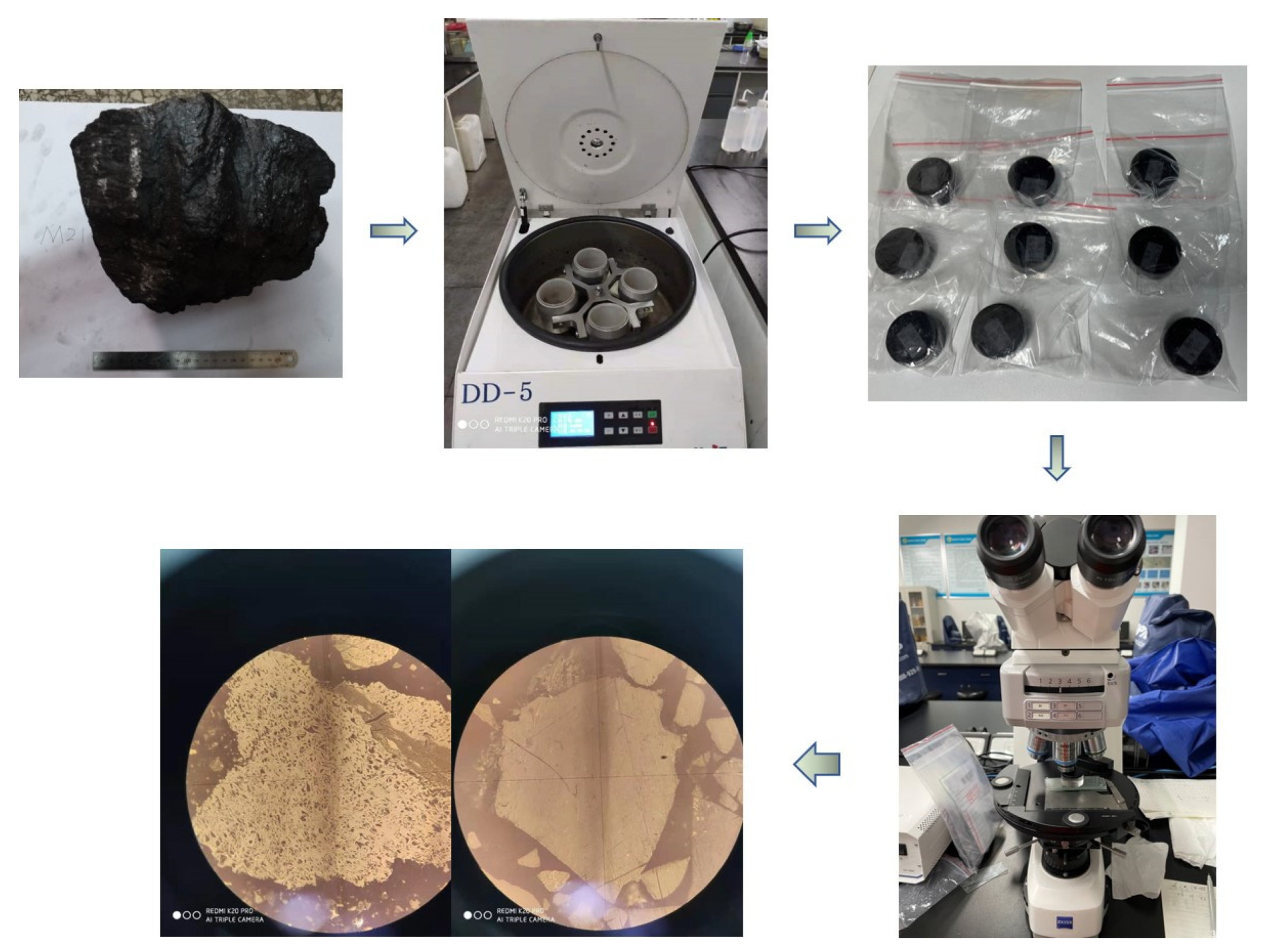
2.1. Samples Preparation

The reflectance (Ro) of the middle-rank coal collected in the Muli area of Qinghai Province (NW China) is 0.73%, and proximate analysis data of moisture, ash, volatile matter, and fixed carbon contents is 1.31%, 1.33%, 27.76%, and 69.6% in wt.%, respectively. The macerals were separated by hand selection and float–sink centrifugation, resulting in vitrinite and inertinite samples (Figure 1). First, the vitrinite has a brighter luster when observed with the naked eye, while the inertinite has a strong silky luster and a more obvious fibrous structure; therefore, the sample was separated into vitrinite-rich and inertinite-rich groups by the hand picking. Then, due to the density difference of the vitrinite and inertinite (the vitrinite density was 1.2–1.25 g/cm3, and the inertinite density was 1.3–1.35 g/cm3), the float–sink centrifugation was performed. Samples were fully mixed with ZnCl2 heavy fluids with different density values of 1.2 g/cm3, 1.25 g/cm3, 1.3 g/cm3 and 1.35 g/cm3 in turn, and the vitrinite was floated, while the inertinite was sunk in the ZnCl2 solution after centrifugation. As such, the vitrinite and inertinite samples were totally separated. After extraction from the ZnCl2 heavy fluids, the separated vitrinite and inertinite samples were washed with deionized water to eliminate ZnCl2. Then, the samples were placed in a drying oven at 60 °C and dried for 24 h for the subsequent experiments.
According to GB/T 15588–2013 and GB/T 8899–2013, the quantitative statistics of separated samples were carried out, and revealed that vitrinite and inertinite content was 90% and 95%, respectively (Table 1). The samples were crushed to 200 mesh (75 μm) and acid washed. Then, 15 g samples were placed in a plastic beaker and mixed with 80 mL of HCl (mass fraction 36%) and stirred in a water bath for 4 h at a constant temperature of 60 °C. The HCl solution was filtered off with suction, and 80 mL of HF solution (mass fraction 40%) was then added. The same water bath and acid washing operations were repeated. Finally, the residual samples were washed and filtered with ultrapure water until the filtrate showed no precipitation when AgNO3 solution was added. The acid-washed samples were filtered through filter paper, placed in an oven at 60 °C for vacuum drying for 24 h, and deashed samples were obtained.

2.2. Experimental Process

In situ FTIR spectroscopy and in situ XRD allow in situ measurements and reduce the influence of the external environment [18,19]. Physicochemical information can be obtained while the samples are being monitored. Compared to conventional FTIR spectroscopy and XRD, in situ FTIR spectroscopy and in situ XRD allow continuous heating, in situ measurements, fast speeds, and high sensitivity [20,21,22].

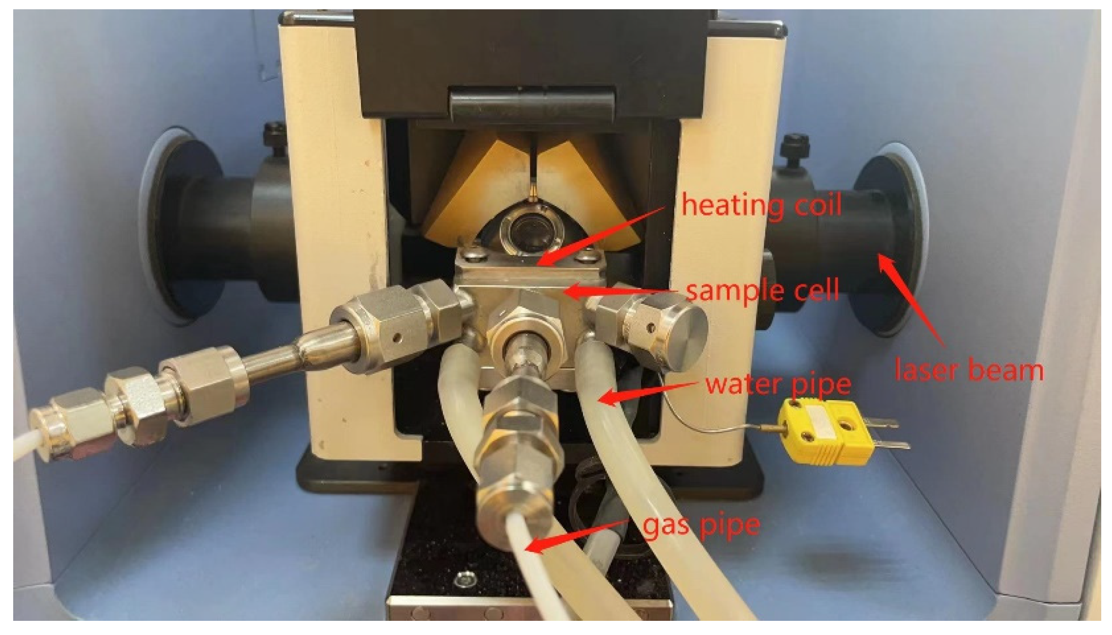
2.2.1. In Situ FTIR Spectroscopy
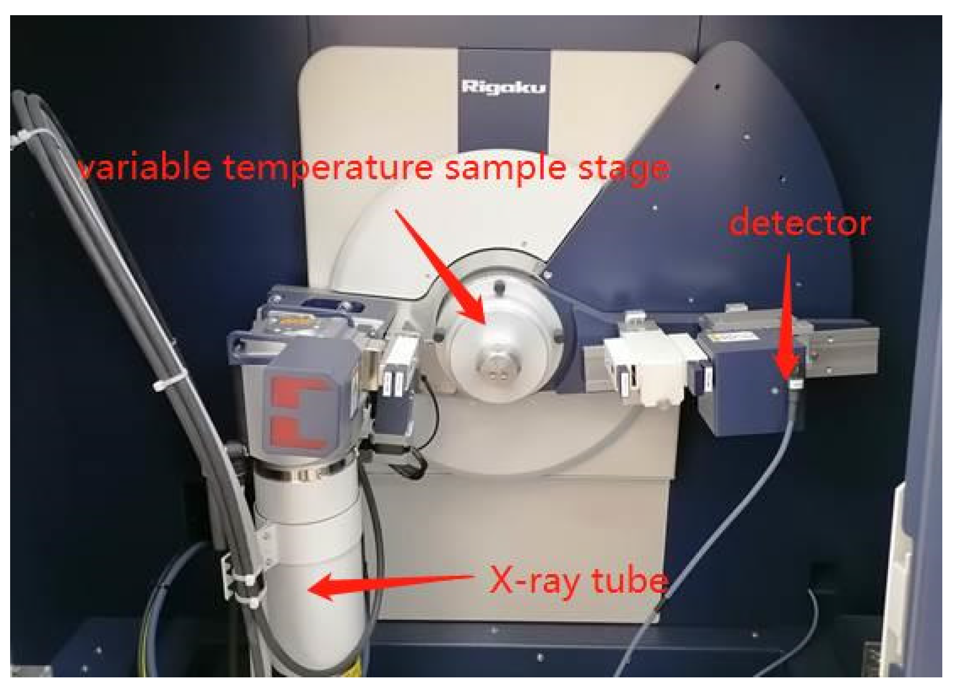
Experiments were performed using Bruker TENSOR II equipment (Figure 2). The prepared dry samples were mixed with KBr at a ratio of 1:100 and pressed into flakes in the sample cell. The heating coil in the bottom of the cover provided different temperatures for samples with electricity. Atmosphere gas was transported by gas pipes. Water was conveyed through water pipes to cool the experimental device. The acquisition wavenumber range was 4000–800 cm−1. The number of scans, temperature range, and heating rate were 64 times, 0–500 °C, and 4 °C/min, respectively. The specific test temperature points were room temperature (30 °C), 100 °C, 200 °C, 300 °C, and 500 °C. After 2 h of heat preservation at each temperature point, the samples were purged with nitrogen, and the FTIR spectrum was obtained.
A series of parameters related to the macromolecular structure in coal can be obtained from the in situ FTIR data by peak fitting. The absorption bands of the peaks in FTIR spectroscopy can be generally divided into four sections [23]: hydroxyl groups (3700–3200 cm−1), aliphatic hydrocarbons (3000–2800 cm−1), oxygenated groups (1800–1000 cm−1), and aromatic hydrocarbons (900–700 cm−1). Peak fitting analysis was performed in these four sections using a software named Peakfit, and the functional groups were assigned based on references (Table 2).
The differences in the molecular structure of the vitrinite and inertinite during the pyrolysis process were examined. According to previous research, the following three parameters are selected to characterize the macromolecular structure [25,26,27,28]:
The aromaticity of coal (I) is expressed as a ratio of the spectrum area in 900–800 cm−1 (A900–800 cm−1) to that in 3000–2800 cm−1 (A3000–2800 cm−1) because the wavenumbers of this experiment start from 800 cm−1. The 900–800 cm−1 segment represents the absorption bands of aromatic structures, and the 3000–2800 cm−1 segment represents the C–H stretching vibrations of aliphatic structures. A higher value of I indicates a higher aromatization level.
In the degree of polycondensation of aromatic rings (DOC) (A900–800 cm−1/A1600 cm−1), the 1600 cm−1 segment represents the aromatic C=C vibration of the aromatic nucleus. A higher DOC value of coal indicates a higher condensation degree of the aromatic rings.
The length of the aliphatic side chains can be represented by CH2/CH3 (A2900–2940 cm−1/A2940–3000 cm−1). The 2900–2940 cm−1 segment represents the asymmetric stretching vibration of alkane CH2, while 2940–3000 cm−1 represents the asymmetric stretching vibration of alkane CH3. A larger ratio indicates longer the aliphatic chains on the aromatic rings and worse compactness of the aromatic structure.

2.2.2. In Situ XRD

The experiments were carried out using a Rigaku SmartLab X-ray diffractometer (Figure 3). The samples were placed on a temperature-changing stage, and the temperature control device controlled the temperature-changing stage to heat up the samples. The X-ray tube provided X-rays which were reflected on the upper surface of samples, and received by the detector. The 200-mesh powder samples were placed in an argon test atmosphere. The test angle range was 5–80°. The scanning speed was 3°/min, and the test temperature ranged from room temperature (30 °C) to 1100 °C. The specific test temperature points were 200 °C, 400 °C, 600 °C, 800 °C, and 1000 °C. Each point was held for three minutes and tested. The heating rate was 15 °C/min, and the in situ XRD pattern was obtained.
Coal has a complex macromolecular structure, and the XRD experimental data can characterize the aggregation state of the coal macromolecular structure. According to the Bragg equation and the Scherrer equation, the crystallite structure parameters, d002, Lc, and La, of the coal samples can be calculated [29,30,31,32]:
d 002 = λ 2 sin θ 002
L c = 0.9 λ β 002 cos θ 002
L a = 1.84 λ β 100 cos θ 100
λ is the X-ray wavelength (λ = 0.154056 nm);
θ is the characteristic peak position;
d002 is the interlayer spacing (nm);
Lc is the average height of coal crystallites (nm);
β is the full-width half-maximum (°);
La is the average diameter of coal crystallites (nm).

3. Results and Analysis

3.1. Analysis of the In Situ FTIR Results

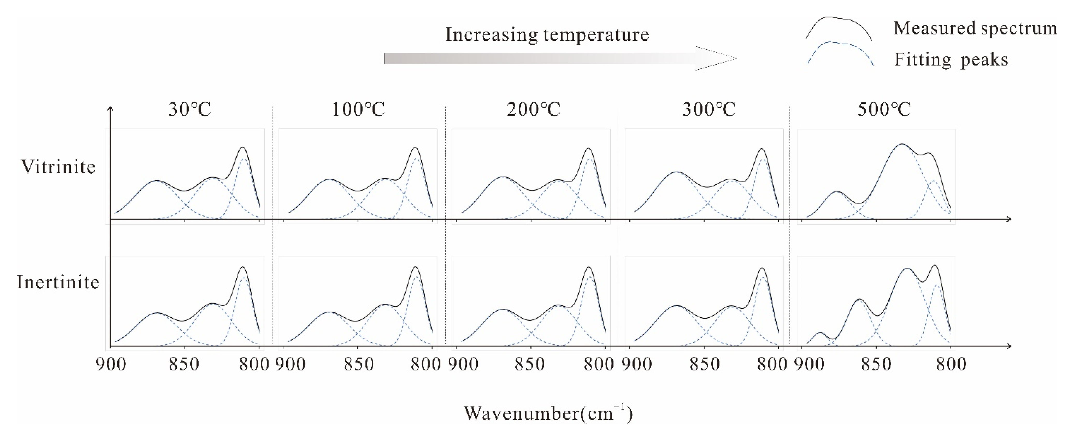
3.1.1. In Situ FTIR Spectrum Characteristics

Figure 4 presents the results of the in situ FTIR spectrum of the samples. The peak shape of vitrinite at 3200–3700 and 3000–2800 cm−1 gradually becomes flat with increasing temperature, indicating that the content of hydroxyl and aliphatic structures gradually decreased. The peak shape of the 1800–1000 cm−1 and 900–800 cm1 segments becomes gradually sharp, indicating that the contents of oxygen-containing functional groups and aromatic structures increased gradually. The peak shape of the 3200–3700 cm−1 segment of inertinite has no significant change, suggesting that the hydroxyl content of the inertinite does not change significantly. The peak shape of the 3000–2800 cm−1 segment gradually becomes flat, indicating that the content of aliphatic structures gradually decreases. The peak shape of the 1800–1000 cm−1 segment does not change significantly at 30–300 °C, but becomes sharp at 300–500 °C, indicating that the content of oxygen-containing functional groups in inertinite increases significantly. The peak shape of the 900–800 cm−1 segment has a small sharp change at 30–300 °C and becomes sharper at 300–500 °C, suggesting that the aromatic structures increase gradually during the pyrolysis process. Therefore, in the pyrolysis process, the changes in the macromolecular structure of vitrinite and inertinite with temperature show differences.

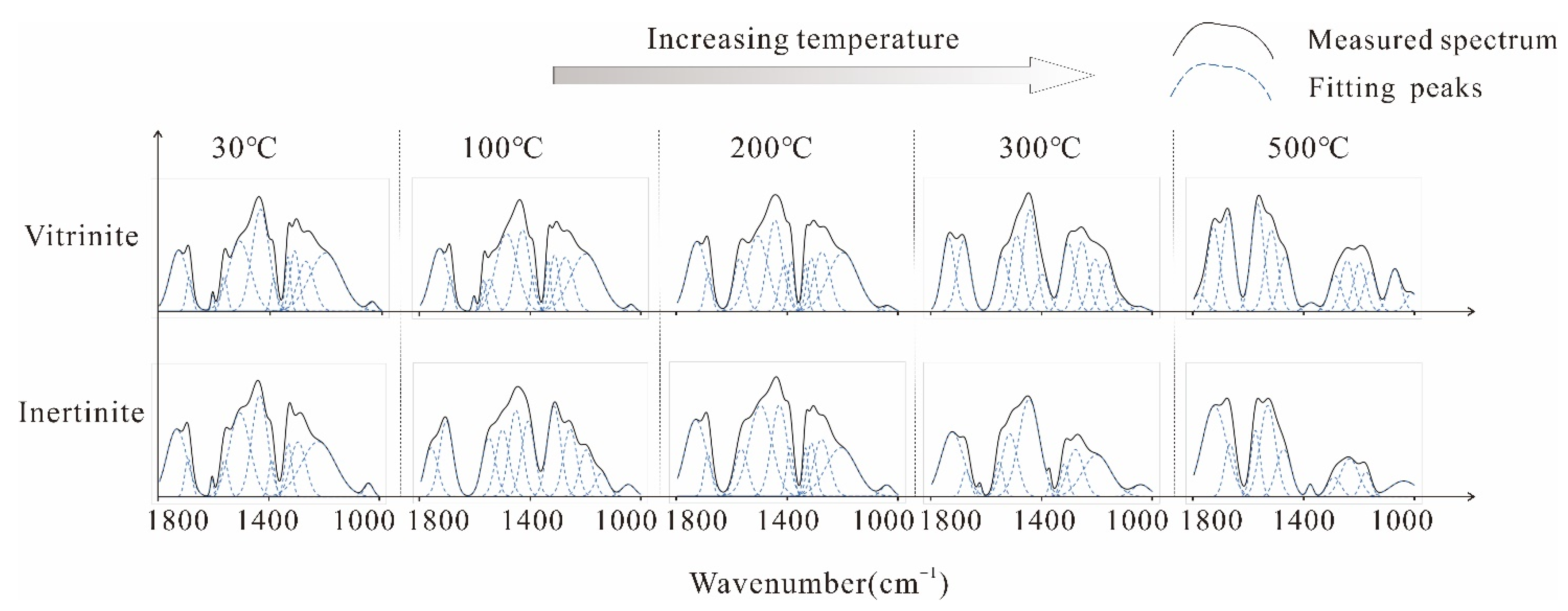
Aromatic Structure Absorption Bands

The wavenumber range of the aromatic structure is 800–900 cm−1, which is assigned to the out-of-plane deformation vibration region of the H atom adjacent to the aromatic ring [33]. The peak at 810 cm−1 represents the aromatic nucleus (C–H), and three adjacent H deformations, of which the hydrogen atom is called a type III hydrogen atom on the aromatic nucleus. The type III hydrogen atom on the aromatic nucleus represents the direction of the polycondensation process of middle-high rank bituminous coal. Hence, it can indicate the degree of aromatic ring polycondensation [34]. The peak of 868 cm−1 is assigned to the aromatic nucleus (C–H), and one adjacent H deformation. This hydrogen atom is called a type I hydrogen atom on the aromatic nucleus, with two adjacent and more than two adjacent hydrogen atomic states [34]. The peak at 3000–3200 cm−1 represents the C–H stretching vibration of the aromatic nucleus [35].
At 30 °C, the content of each group in vitrinite is smaller than that in inertinite. When heated to 200°C, the aromatic nucleus (C–H) with three adjacent H deformations, the aromatic nucleus (C–H) with five adjacent H deformations, and the aromatic nucleus C–H do not change significantly in vitrinite and inertinite. At 200–300 °C, both vitrinite and inertinite show an increase in the content of aromatic nuclei (C–H), one adjacent H deformation, and aromatic nucleus C–H. At the 300–500 °C stage, many aromatic nuclei (C–H) and three adjacent H deformations come into being in vitrinite and inertinite. The number of aromatic nuclei (C–H), one adjacent H deformation, and the aromatic nucleus C–H is reduced (Figure 5 and Figure 6). Hence, the aromatic structure content of vitrinite is less than that in inertinite. With increasing temperature during pyrolysis, coalification continues, and the degree of aromatization of both vitrinite and inertinite increases gradually. However, the degree of aromatization in inertinite is still much higher than that in vitrinite.

Oxygen-Containing Functional Groups Absorption Band

The wavenumber range, 1000–1800 cm−1, characterizes the absorption band of oxygen-containing functional groups, and the assignment of this segment is complicated. Ether oxygen C–O–C includes alkyl and aryl ethers. The peak near 1039 cm−1 is assigned to the stretching vibration of alkyl ether [36]. The peak near 1100 cm−1 represents the stretching vibration of aryl ether [37]. The peak at 1200–1340 cm−1 is assigned to the Ar–OH stretching vibration [38]. The characteristic peaks around 1360–1440 cm−1 represent the aliphatic CH2 and CH3 vibrations, respectively [13,24]. The peak at 1600 cm−1 represents the stretching vibration of the aromatic ring C=C [24]. The peak near 1700 cm−1 characterizes the stretching vibration of aromatic C=O [39].
At 30 °C, the total contents of oxygen-containing functional groups in both vitrinite and inertinite are similar; however, there are slightly more aliphatic CH2 and CH3 and fewer Ar–OH groups, C–O–C groups, aromatic ring C=C, and aromatic C=O in vitrinite. During pyrolysis up to 200 °C, both vitrinite and inertinite show a slight decrease in the content of C–O–C groups, aromatic C=O, and aromatic ring C=C. The content of aliphatic CH2 and CH3 decrease significantly, the content of Ar–OH groups increases significantly, and the total amount of oxygen-containing functional groups shows a decreasing trend. At 200–300 °C, the content of C–O–C groups, aromatic C=O, and aromatic ring C=C of vitrinite and inertinite are reduced greatly. However, the content of aliphatic CH2 and CH3 and Ar–OH groups increases considerably, and the total amount of oxygen-containing functional groups greatly increases. At 300–500 °C, the content of C–O–C groups and aromatic ring C=C of vitrinite and inertinite increase significantly; however, the content of aliphatic CH2 and CH3 and Ar–OH groups decreases significantly. The content of aromatic C=O of vitrinite does not change significantly, but that of inertinite decreases. The total amount of oxygen-containing functional groups decreases significantly (Figure 7 and Figure 8). Coalification is enhanced continuously with increasing temperature, and the oxygen-containing functional groups of vitrinite and inertinite detach.

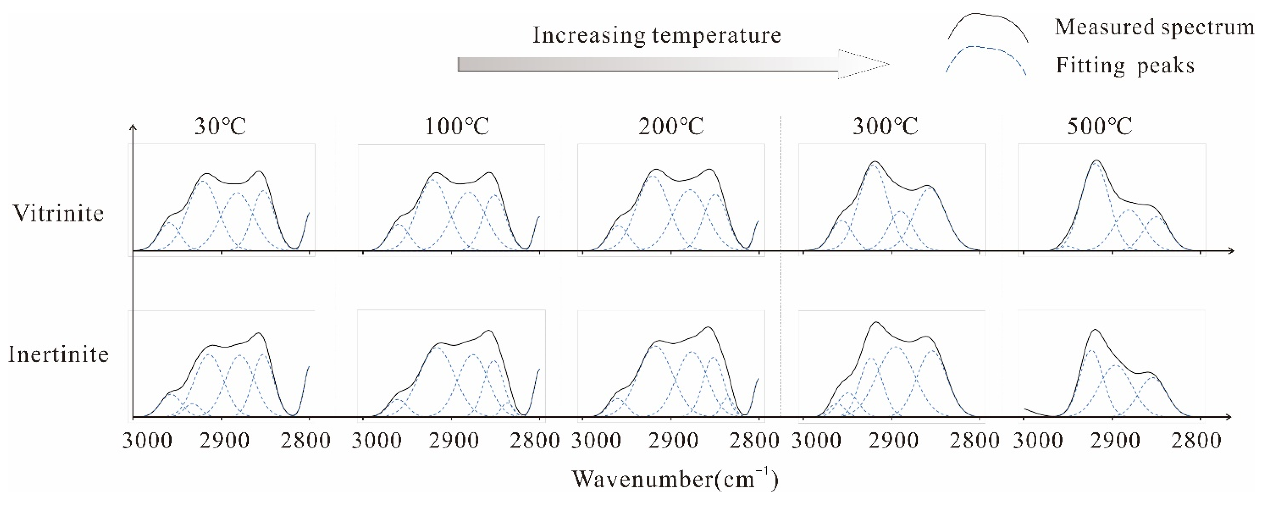
Aliphatic Structure Absorption Bands

The peak at 3000–2800 cm−1 represents the aliphatic structure, which belongs to the C–H stretching vibration region of aliphatic hydrocarbons [24]. There are obvious peaks near 2850 cm−1 and 2920 cm−1, which are respectively assigned to the stretching vibrations of symmetrical CH2 and asymmetric CH2 [40]. The characteristic peaks near 2870 cm−1 and 2950 cm−1 are assigned to the stretching vibrations of symmetrical CH3 and asymmetric CH3, respectively [40].
At 30 °C, the CH2 content in both vitrinite and inertinite is higher than the CH3 content; vitrinite has more CH2 and CH3 than inertinite. When heating to 200 °C, the CH2 content of vitrinite and inertinite and the CH3 content of vitrinite increases slightly, while the CH3 content of inertinite shows no significant change. The content of each group in vitrinite is always higher than that of inertinite. At 200–300°C, the CH2 and CH3 contents of vitrinite and inertinite decrease. In vitrinite, the change rate of the CH2 content is lower than that of the CH3 content; however, inertinite shows an opposite trend. At 300–500 °C, the CH2 and CH3 content of vitrinite and inertinite are reduced greatly. The reduction rate of the vitrinite is higher than that of the inertinite, but the content of each group in vitrinite is still higher than that of inertinite (Figure 9 and Figure 10). Hence, during the pyrolysis process, the content of aliphatic side chains in vitrinite and inertinite decreases gradually with the continuous increase in temperature. However, the aliphatic hydrocarbon content of vitrinite is always higher than that of inertinite. In both vitrinite and inertinite, the CH2 content is always higher than CH3 content. Aliphatic hydrocarbons in coal exist mostly in the form of long chains with few branches. At this stage, the coal structure is loose, which is more conducive to coal pyrolysis to form colloids [13,40].

Hydroxyl Absorption Band

The peak at 3200–3700 cm−1 characterizes the hydroxyl absorption band, and there are strong hydroxyl absorption peaks in this segment, which mainly includes three types: cyclic OH groups, O–H–π-H bonds, and free OH groups. The peak at 3180–3240 cm−1 characterizes the cyclic OH groups [41]. The peak around 3300 cm−1 represents the OH-ether O hydrogen bonds [35,41], and the peaks around 3350–3400 cm−1 represent self-associated OH bonds [40]. The peak at 3500 cm−1 characterizes the O–H–π–H bonds formed with π electrons on the aromatic rings [42], and the peak at 3600–3700 cm−1 is assigned to free OH groups [33].
At 30 °C, vitrinite has a higher content of O–H–π–H bonds than inertinite, and the content of free O–H groups of vitrinite is much smaller than that of inertinite. When heating to 300 °C, in both vitrinite and inertinite, the content of O–H–π–H bonds gradually increases, and the content of free O–H groups decreases gradually. The change rate of each group content in vitrinite is higher than that in inertinite. The O–H–π-H bond content of vitrinite is much more than that of inertinite, and the content of free O–H groups is much less than that of inertinite. At the stage of 300–500°C, the O–H–π-H bond content of the vitrinite decreases to a certain extent, but that of the inertinite increases; additionally, the content of free O–H groups in both vitrinite and inertinite increases gradually, and the content of self-associated OH bonds arises gradually (Figure 11 and Figure 12).

3.1.2. In Situ FTIR Structural Parameter Characteristics

Aromatic Structure

As shown in Figure 13a, the I of both vitrinite and inertinite shows an exponential growth trend during the pyrolysis process, in which I is relatively stable from 30 °C to 200 °C, and increases gradually from 200 °C to 500 °C, suggesting that during pyrolysis, the proportion of aromatic structures of coal increases gradually, and the degree of aromatization of coal increases continuously. At 30–300 °C, there is no significant difference in I between vitrinite and inertinite. At 300–500 °C, the I of inertinite is higher than that of vitrinite.
As shown in Figure 13b, the DOC of both vitrinite and inertinite has a linear positive correlation with temperature. The DOC of inertinite is slightly higher than that of vitrinite. In the pyrolysis process, the DOC of vitrinite and inertinite increases gradually. Combining Figure 4 and Figure 13a, at the stage of 30–300 °C, the I shows a slow growth trend, and the DOC increases gradually. At the stage of 30–300 °C, the aromatic substitution reaction and condensation reaction of aromatic rings are not obvious, but at 300–500 °C, with increasing temperature, the condensation reaction of aromatic rings gradually intensifies, and the aromatic substitution reaction occurs violently, leading to an increase in the degree of condensed cyclization.

Aliphatic Structure

As shown in Figure 14, there are some differences in the changes of CH2/CH3 in the vitrinite and inertinite during the pyrolysis process. With increasing temperature, the CH2/CH3 value of vitrinite shows a parabolic trend and reaches the lowest value at 200 °C. This suggests that the CH2 content of vitrinite decreases continuously during the pyrolysis process and reaches the minimum at 200 °C. After 200 °C, the space between the aromatic rings decreases gradually, and the structure of coal becomes more compact. The CH2/CH3 value of inertinite does not show a clear correlation, indicating that temperatures below 500 °C have no apparent catalytic effect on the detachment of aliphatic side chains in the inertinite.

3.2. Analysis of In Situ XRD Results

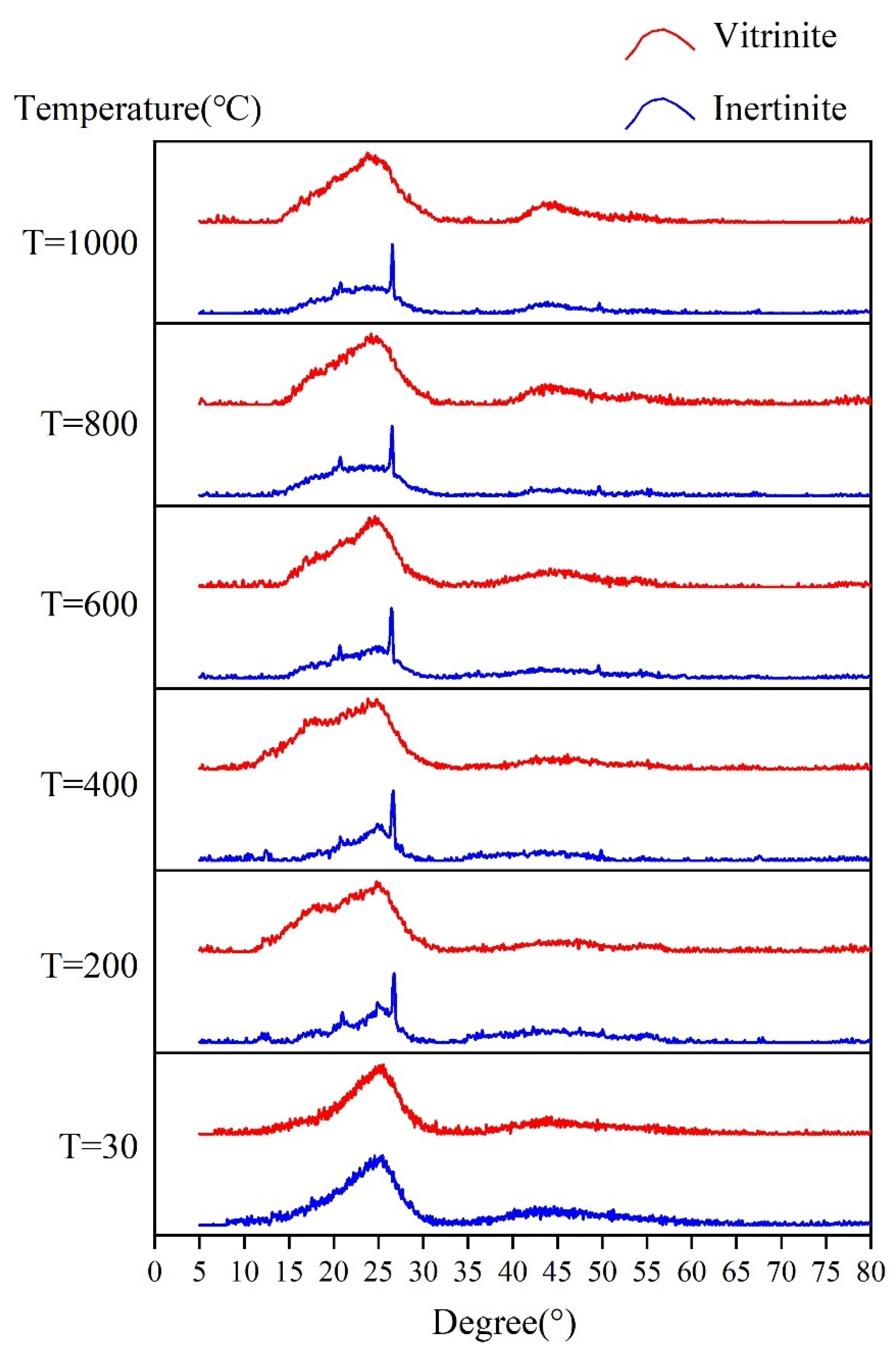
3.2.1. In Situ XRD Pattern Characteristics

Figure 15 shows the spectrum obtained by in situ XRD. Two XRD peaks are observed near 26° and 43°, which correspond to the 002 and 100 peaks, respectively, representing the structural characteristics of aromatic rings [43]. The 002 diffraction band is generated by the local parallel stacking of curved layers [44]. With the continuous increase in temperature, the two XRD peaks are gradually sharpened to a certain extent. Compared to that of the vitrinite, the peak shape of the inertinite is sharper, and the peak value is higher. Table 3 lists the calculated results of the spectra in the two intervals of 10–35°and 35–50°, respectively.

3.2.2. In Situ XRD Structural Parameters

As shown in Figure 16a, the d002 value of the vitrinite is much higher than that of the inertinite. With increasing temperature, the d002 of the vitrinite shows a downward trend. According to this experiment, the d002 of vitrinite decreases by approximately 0.013 nm, while that of inertinite shows no obvious change. The arrangement of aromatic layers in vitrinite gradually becomes regular during the pyrolysis process, while there is no significant change in inertinite. However, the aromatic interlayer spacing of inertinite is much smaller than that of vitrinite, suggesting that the aromatic layers of the inertinite are more regular and compact. The Lc of vitrinite and inertinite shows an obvious upward trend with increasing temperature, and the Lc of the inertinite is larger than that of the vitrinite (Figure 16b). The average stacking thickness of the microcrystalline layers of the inertinite increases gradually, but the layer-stacking thickness of vitrinite is still smaller than that of inertinite. The La values of both vitrinite and inertinite increase with increasing temperature, and the increase of the La of vitrinite is slightly larger than that of inertinite. In this experiment, the La values of inertinite and vitrinite increase by approximately 1.6 and 1.7 nm, respectively. However, the La value of inertinite is still higher than that of vitrinite (Figure 16c). Hence, the plane extensibility of the basic structural unit (BSU) of both vitrinite and inertinite increases significantly during the pyrolysis process. At the 600–1000 °C stage, the increase rate of the plane extensibility of BSU of vitrinite is higher than that of inertinite. However, the plane extensibility of vitrinite is still smaller than that of inertinite.

4. Discussion

The macromolecular structure evolution characteristics of vitrinite and inertinite during pyrolysis include the following.
(1)
During pyrolysis, the macromolecular structures of vitrinite and inertinite change with increasing temperature. The DOC of vitrinite and inertinite increases, and the plane extensibility and the Lc increase continuously, indicating that both the vitrinite and inertinite of low–middle rank coals have an evolution trend of increasing degree of aromatization in the early stages of pyrolysis. The increase in aromatization is reflected mainly in the continuous expansion of the aromatic structural units. The differences in the structural evolution of vitrinite and inertinite are reflected mainly by the continuous decrease in the d002 peak of vitrinite with increasing temperature, in which the reduced value is 0.013 nm, but there is no obvious change in inertinite. Second, the I, DOC, La, and Lc of inertinite are always higher than those of vitrinite, indicating that the degree of inertinite aromatization is always higher than that of vitrinite.
(2)
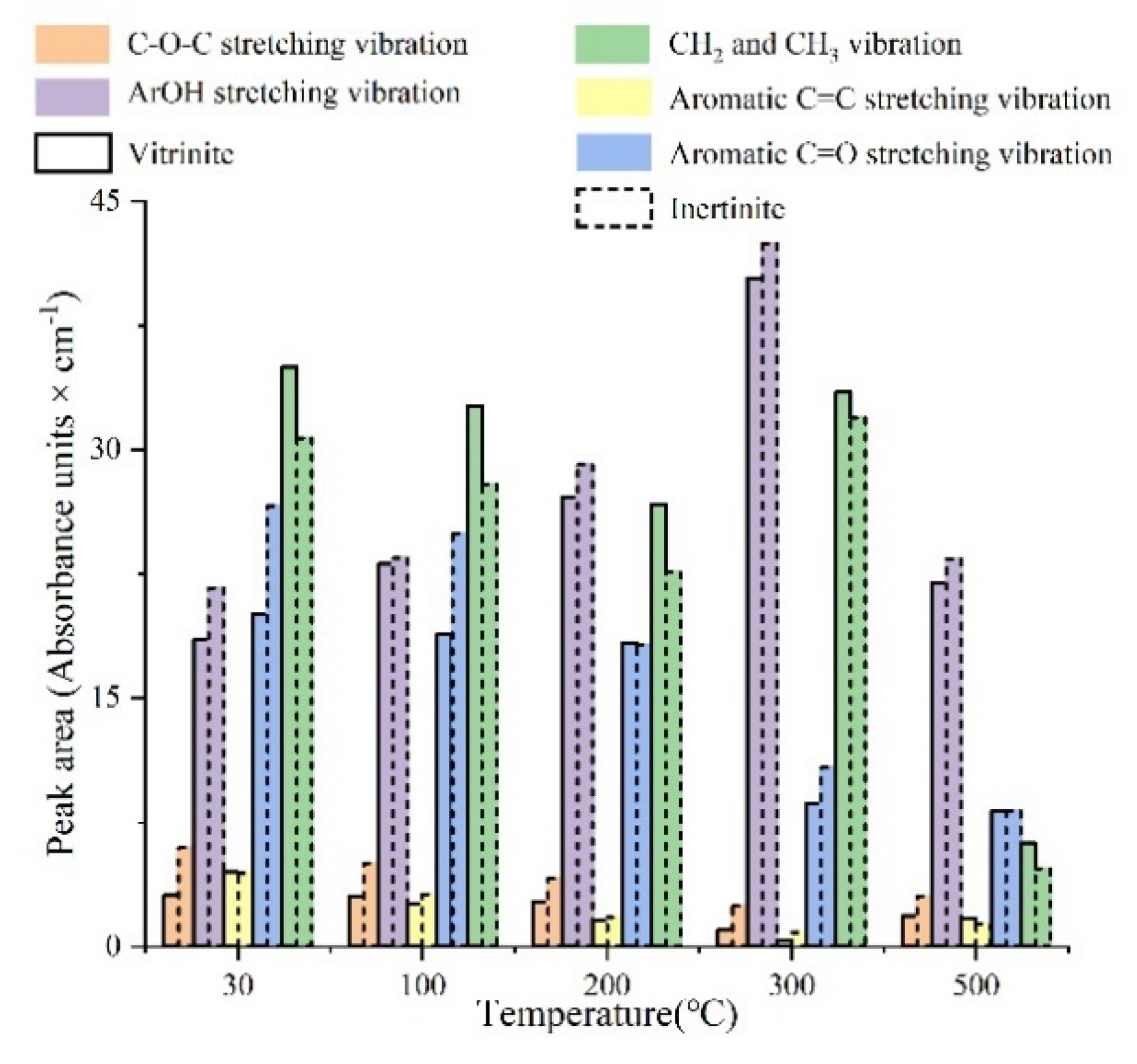
The pyrolysis process of vitrinite and inertinite can be divided into three stages. Between 30 °C and 200 °C, the content of aromatic groups in vitrinite and inertinite shows no obvious change, while that of aliphatic groups increases slightly. The total amount of oxygen-containing functional groups decreases gradually. The content of C–O–C groups, aromatic C=O, and aromatic ring C=C has a small decrease in a relatively stable situation. The content of aliphatic CH2 and CH3 decreases significantly, and that of Ar–OH groups increases significantly (Figure 17). The content of O–H–π-H bonds increases greatly, and that of free O–H groups decreases greatly. There is no significant change in the I and DOC (Figure 13), which suggests that aliphatic and aromatic structures are enriched slightly at this stage. The enlargement of the aromatic structural system is not obvious in this stage, and the detachment of oxygen-containing functional groups and the enrichment of aliphatic hydrocarbons are the dominating chemical reactions [45].
The differences in the structural evolution of vitrinite and inertinite at 30–200 °C are mainly described in three aspects (Figure 14): oxygen-containing functional groups, aliphatic structures, and hydroxyl structures. Compared with inertinite, the aromatic C=O content of vitrinite does not change significantly. With the increasing temperature, the aliphatic structure content of vitrinite and inertinite increases, and the CH2/CH3 value gradually decreases in vitrinite; however, there is no obvious change in inertinite, suggesting that the degree of branching of vitrinite increases or the alicyclic hydrocarbon is observed at this stage [46].
At 200–300 °C, both vitrinite and inertinite have a slight increase in the aromatic nucleus (CH), one adjacent H deformation, and an increase in the content of CH in the aromatic nucleus. The CH2 and CH3 content in the aliphatic structure of the vitrinite and inertinite decreases. On the other hand, the total amount of oxygen-containing functional groups increases. There is a decrease in C–O–C groups, aromatic C=O, and aromatic ring C=C in both vitrinite and inertinite, while the contents of aliphatic CH2 and CH3 and Ar–O–H groups increases greatly (Figure 17), and the I and DOC also increase (Figure 13). This stage is filled mainly by the synergistic effects of aliphatic and aromatic groups. The continuous detachment of aliphatic substances leads to a continuous increase in the aromatic structure system. The ether bonds in the oxygen-containing functional groups are broken and detached, and the conversion to phenolic hydroxyl groups and aliphatic CH2 and CH3 causes their increases.
The differences in structure evolution between vitrinite and inertinite at 200–300 °C are shown mainly in the aliphatic structure. The reduction of CH2 content in the aliphatic structure of vitrinite is much smaller than that of inertinite, and the decrease in CH3 content is much larger than that of inertinite. The change rate of the CH2 content in vitrinite is lower than that of the CH3 content, but the opposite is true for inertinite. The CH2/CH3 value of vitrinite increases gradually, and that of inertinite remains basically unchanged (Figure 14). This suggests that the length of aliphatic hydrocarbons and their branched chains of the vitrinite continue to increase at this stage, and the alicyclic hydrocarbons in the aromatic structure system continue to detach.
At 300–500 °C, many aromatic nuclei (C–H) and three adjacent H deformations in vitrinite and inertinite come into being. The content of aromatic nuclei (C–H) and one adjacent H deformation reduces with the increasing temperature. The total amount of oxygen-containing functional groups is reduced significantly, and the content of ether bonds and aromatic ring C=C increase significantly, while aliphatic CH2 and CH3 and Ar–O–H groups decrease significantly. The CH2 and CH3 content in the aliphatic structure decreases significantly (Figure 17), and the I and DOC increase significantly (Figure 13). Hence, this stage is dominated by the aromatization and condensation of macromolecules, and the substituents of the aromatic system gradually detach.
The structural evolution differences between vitrinite and inertinite at 300–500 °C are shown mainly in the fact that the aromatic C=O content of vitrinite has no obvious change. With increasing temperature, the content of CH2 and CH3 decreases, suggesting that the CH2/CH3 value of vitrinite increases gradually (Figure 14), caused by the detachment of alicyclic hydrocarbons in the aromatic system [47]. At this stage, many aliphatic hydrocarbons are detached, resulting in an increase in the content of active sites and the expansion of the aromatic structure system. The content of aromatic C=O of the inertinite decreases, but that of C–O–C groups increases, and the CH2/CH3 value remains basically unchanged (Figure 14), suggesting that the aromatic structure system of the inertinite continues to expand at this stage, which is influenced by the detachment of aliphatic hydrocarbons and the aromatic C=O breaking and splicing.

5. Conclusions

(1)
During the pyrolysis process, both the inertinite and vitrinite show an increasing degree of aromatization. With increasing pyrolysis temperature, aromaticity (I), polycondensation degree of aromatic rings (DOC), average lateral sizes (La) of BSU, and stacking heights (Lc) of BSU in vitrinite and inertinite increase, but the aromatization level of inertinite has always been higher than that of vitrinite.
(2)
In the temperature range of 30–500 °C for the in situ FTIR spectroscopy, the macromolecular structure evolution of vitrinite and inertinite is divided into three stages: 30–200 °C, 200–300 °C, and 300–500 °C. At 30–200 °C, the detachment of oxygen-containing functional groups and the enrichment of aliphatic hydrocarbons occurs. The 200–300 °C stage is filled mainly by the synergistic effects of aliphatic and aromatic groups. The stage of 300–500 °C is dominated by the aromatization and condensation of macromolecules. The substituents of the aromatic system gradually detach, leading to an increase in I and DOC.
(3)
In the temperature range of 30–1000 °C by in situ XRD, the difference in macromolecular structure evolution between vitrinite and inertinite is manifested mainly by the arrangement of aromatic layers, which tends to be more and more ordered in vitrinite regular during the pyrolysis process, while there is no significant change in inertinite. However, the aromatic layers of inertinite are always more compact than that of vitrinite. In addition, the aliphatic side chains of inertinite are more stable than that of vitrinite during pyrolysis.

Author Contributions

Data curation, M.Z. and L.D.; formal analysis, M.Z.; methodology, A.W.; resources, A.W. and Y.W.; supervision, D.C.; writing—original draft, M.Z. All authors have read and agreed to the published version of the manuscript.

Funding

This study is financially supported by the National Key Research and Development Plan of China (2021YFC2902004), and the National Natural Science Foundation of China (grant No. 42072197, 41902170, 41972174).

Institutional Review Board Statement

Not applicable.

Informed Consent Statement

Not applicable.

Data Availability Statement

Data available on request due to restrictions.

Conflicts of Interest

The authors declare no conflict of interest.

References

  1. Cao, D.; Wei, Y.; Wang, A.; Wang, L.; Liu, Z.; Qin, R.; Shu, Z.; Chen, G. The evolution difference of macromolecular structures and its dynamic mechanism of coal macerals: Research status and prospect. Coal Geol. Explor. 2021, 49, 12–20, (In Chinese with English Abstract). [Google Scholar]
  2. Louw, E.B.; Mitchell, G.D.; Wang, J.; Winans, R.; Mathews, J.P. Constitution of drop-tube-generated coal chars from vitrinite-and inertinite-rich South African coals. Energy Fuels 2016, 30, 112–120. [Google Scholar] [CrossRef]
  3. Gao, M.; Li, X.; Ren, C.; Wang, Z.; Pan, Y.; Guo, L. Construction of a multicomponent molecular model of Fugu coal for ReaxFF-MD pyrolysis simulation. Energy Fuels 2019, 33, 2848–2858. [Google Scholar] [CrossRef]
  4. Gao, M.; Li, X.; Guo, L. Pyrolysis simulations of Fugu coal by large-scale ReaxFF molecular dynamics. Fuel Process. Technol. 2018, 178, 197–205. [Google Scholar] [CrossRef]
  5. Wang, A.; Wei, Y.; Cao, D.; Ding, L.; Zhao, M. Effect of different functional groups on CH4 adsorption heat and surface free energy of vitrinite during coalification. Appl. Surf. Sci. 2022, 597, 153748. [Google Scholar] [CrossRef]
  6. Zhou, H.; Wu, C.; Pan, J.; Wang, Z.; Niu, Q.; Du, M. Research on molecular structure characteristics of vitrinite and inertinite from bituminous coal with FTIR, micro-Raman, and XRD spectroscopy. Energy Fuels 2021, 35, 1322–1335. [Google Scholar] [CrossRef]
  7. Kidena, K.; Katsuyama, M.; Murata, S.; Nomura, M.; Chikada, T. Study on plasticity of maceral concentrates in terms of their structural features. Energy Fuels 2002, 16, 1231–1238. [Google Scholar] [CrossRef]
  8. Van Niekerk, D.; Mathews, J. Molecular representations of Permian-aged vitrinite-rich and inertinite-rich South African coals. Fuel 2010, 89, 73–82. [Google Scholar] [CrossRef]
  9. Maroto-Valer, M.M.; Taulbee, D.N.; Andrésen, J.M.; Hower, J.C.; Snape, C.E. Quantitative 13C NMR study of structural variations within the vitrinite and inertinite maceral groups for a semifusinite-rich bituminous coal. Fuel 1998, 77, 805–813. [Google Scholar] [CrossRef]
  10. Van Niekerk, D.; Pugmire, R.; Solum, M.S.; Painter, P.C.; Mathews, J.P. Structural characterization of vitrinite-rich and inertinite-rich Permian-aged South African bituminous coals. Int. J. Coal Geol. 2008, 76, 290–300. [Google Scholar] [CrossRef]
  11. Wang, A.; Li, J.; Cao, D.; Wei, Y.; Ding, L.; Zhao, M. Comparison of nanopore structure evolution in vitrinite and inertinite of tectonically deformed coals: A case study in the Wutongzhuang coal mine of Hebei province, North China. Front. Earth Sci. 2022, 10, 93. [Google Scholar] [CrossRef]
  12. Wang, A.; Cao, D.; Wei, Y.; Liu, Z. Macromolecular structure controlling micro mechanical properties of vitrinite and inertinite in tectonically deformed coals—A case study in Fengfeng coal mine of Taihangshan fault zone (North China). Energies 2020, 13, 6618. [Google Scholar] [CrossRef]
  13. Jiang, J.; Zhang, S.; Longhurst, P.; Yang, W.; Zheng, S. Molecular structure characterization of bituminous coal in Northern China via XRD, Raman and FTIR spectroscopy. Spectrochim. Acta Part A Mol. Biomol. Spectrosc. 2021, 255, 119724. [Google Scholar] [CrossRef] [PubMed]
  14. Yan, J.; Lei, Z.; Li, Z.; Wang, Z.; Ren, S.; Kang, S.; Wang, X.; Shui, H. Molecular structure characterization of low-medium rank coals via XRD, solid state 13C NMR and FTIR spectroscopy. Fuel 2020, 268, 117038. [Google Scholar] [CrossRef]
  15. Jiang, J.; Yang, W.; Cheng, Y.; Liu, Z.; Zhang, Q.; Zhao, K. Molecular structure characterization of middle-high rank coal via XRD, Raman and FTIR spectroscopy: Implications for coalification. Fuel 2019, 239, 559–572. [Google Scholar] [CrossRef]
  16. Zhao, X.; Zong, Z.; Cao, J.; Ma, Y.; Han, L.; Liu, G.; Zhao, W.; Wei, X. Difference in chemical composition of carbon disulfide-extractable fraction between vitrinite and inertinite from Shenfu-Dongsheng and Pingshuo coals. Fuel 2008, 87, 565–575. [Google Scholar] [CrossRef]
  17. Wang, H.; Shi, X.; Tian, J.; Pan, J.; Yao, L.; Feng, X. Macromolecular Structure Changes of Tectonically Deformed Coal: Evidence from Coal Pyrolysis, 13C NMR, and XRD Experiments. Energy Fuels 2021, 35, 8711–8722. [Google Scholar] [CrossRef]
  18. Yu, X.; Yu, C.; Jiang, Q.; Ran, Q.; Liu, J.; Yang, B. In-situ X-ray Diffraction Analysis on the Role of Hardening Accelerator in Early Hydration of Cement. Mater. Rep. 2020, 34, 2058–2062, (In Chinese with English Abstract). [Google Scholar]
  19. Tay, F.H.; Kazarian, S. Study of petroleum heat-exchanger deposits with ATR-FTIR spectroscopic imaging. Energy Fuels 2009, 23, 4059–4067. [Google Scholar] [CrossRef]
  20. Wang, A.; Wei, Y.; Yuan, Y.; Li, C.; Li, Y.; Cao, D. Coalbed methane reservoirs’ pore-structure characterization of different macrolithotypes in the southern Junggar Basin of Northwest China. Mar. Pet. Geol. 2017, 86, 675–688. [Google Scholar] [CrossRef]
  21. De Souza, A.V.A.; da Silva, J.F.C. Biodiesel synthesis evaluated by using real-time ATR-FTIR. Org. Process. Res. Dev. 2013, 17, 127–132. [Google Scholar] [CrossRef]
  22. Gabrienko, A.A.; Morozov, E.V.; Subramani, V.; Martyanov, O.N.; Kazarian, S.G. Chemical visualization of asphaltenes aggregation processes studied in situ with ATR-FTIR spectroscopic imaging and NMR imaging. J. Phys. Chem. C 2015, 119, 2646–2660. [Google Scholar] [CrossRef]
  23. Wang, A.; Li, J.; Wei, Y.; Yang, C.; Nie, J.; Cao, D. Gas migration for terrestrial gas hydrates in the Juhugeng mining area of Muli basin, Qilian Mountains, Northwest China. Energy Explor. Exploit. 2020, 38, 989–1013. [Google Scholar] [CrossRef]
  24. Wang, A.; Cao, D.; Wei, Y.; Nie, J.; Qin, R. Comparison of nanopore evolution in vitrinite and inertinite in coalbed methane reservoirs during coalification. J. Nat. Gas Sci. Eng. 2020, 78, 103289. [Google Scholar] [CrossRef]
  25. Wu, D.; Liu, G.; Sun, R.; Fan, X. Investigation of structural characteristics of thermally metamorphosed coal by FTIR spectroscopy and X-ray diffraction. Energy Fuels 2013, 27, 5823–5830. [Google Scholar]
  26. Guo, Y.; Bustin, R. Micro-FTIR spectroscopy of liptinite macerals in coal. Int. J. Coal Geol. 1998, 36, 259–275. [Google Scholar] [CrossRef]
  27. Mastalerz, M.; Bustin, R. Application of reflectance micro-Fourier transform infrared spectrometry in studying coal macerals: Comparison with other Fourier transform infrared techniques. Fuel 1995, 74, 536–542. [Google Scholar] [CrossRef]
  28. Cao, D.; Zhang, H.; Dong, Y.; Yang, C. Nanoscale Microscopic Features and Evolution Sequence of Coal-Based Graphite. J. Nanosci. Nanotechnol. 2017, 17, 6276–6283. [Google Scholar] [CrossRef]
  29. Bragg, W.H.; Bragg, W.L. X-rays and Crystal Structure; George Bell and Sons, Limited: London, UK, 1915. [Google Scholar]
  30. Bragg, W.H. IX Bakerian Lecture—X-rays and crystal structure. In Philosophical Transactions of the Royal Society of London. Series A, Containing Papers of a Mathematical or Physical Character; Royal Society: London, UK, 1915; Volume 215, pp. 253–274. [Google Scholar]
  31. Debye, P.; Scherrer, P. Interferenzen an regellos orientierten Teilchen im Röntgenlicht. I. Nachr. Ges. Wiss. Göttingen Math. Phys. Kl. 1916, 1916, 1–15. [Google Scholar]
  32. Lu, L.; Sahajwalla, V.; Kong, C.; Harris, D. Quantitative X-ray diffraction analysis and its application to various coals. Carbon 2001, 39, 1821–1833. [Google Scholar] [CrossRef]
  33. Zhu, H.; Zhao, H.; Wei, H.; Wang, W.; Wang, H.; Li, K.; Lu, X.; Tan, B. Investigation into the thermal behavior and FTIR micro-characteristics of re-oxidation coal. Combust Flame 2020, 216, 354–368. [Google Scholar] [CrossRef]
  34. Liu, Z.; Wei, Y.; Ning, S.; Jia, X.; Qin, R.; Cao, D. The differences of element geochemical characteristics of the main coal seams in the Ningdong coalfield, Ordos Basin. J. Geochem. Explor. 2019, 202, 77–91. [Google Scholar]
  35. Chen, Y.; Mastalerz, M.; Schimmelmann, A. Characterization of chemical functional groups in macerals across different coal ranks via micro-FTIR spectroscopy. Int. J. Coal Geol. 2012, 104, 22–33. [Google Scholar] [CrossRef]
  36. Qin, R.; Wang, A.; Cao, D.; Wei, Y.; Ding, L.; Li, J. Effect of peat mire evolution on pore structure characteristics in thick coal seam: Examples from Xishanyao Formation (Middle Jurassic), Yili Basin, China. Energy Explor. Exploit. 2020, 38, 1484–1514. [Google Scholar] [CrossRef]
  37. Ibarra, J.; Munoz, E.; Moliner, R. FTIR study of the evolution of coal structure during the coalification process. Org. Geochem. 1996, 24, 725–735. [Google Scholar] [CrossRef]
  38. Manoj, B.; Kunjomana, A.; Chandrasekharan, K. Chemical leaching of low rank coal and its characterization using SEM/EDAX and FTIR. J. Miner. Mater. Charact. Eng. 2009, 8, 821–832. [Google Scholar] [CrossRef]
  39. Zhao, J.; Wang, T.; Deng, J.; Shu, C.-M.; Zeng, Q.; Guo, T.; Zhang, Y. Microcharacteristic analysis of CH4 emissions under different conditions during coal spontaneous combustion with high-temperature oxidation and in situ FTIR. Energy 2020, 209, 118494. [Google Scholar] [CrossRef]
  40. Saikia, B.K.; Boruah, R.K.; Gogoi, P. FT-IR and XRD analysis of coal from Makum coalfield of Assam. J. Earth Syst. Sci. 2007, 116, 575–579. [Google Scholar] [CrossRef]
  41. Chen, C.; Gao, J.; Yan, Y. Observation of the type of hydrogen bonds in coal by FTIR. Energy Fuels 1998, 12, 446–449. [Google Scholar] [CrossRef]
  42. Wu, D.; Zhang, H.; Hu, G.; Zhang, W. Fine characterization of the macromolecular structure of huainan coal using XRD, FTIR, 13C-CP/MAS NMR, SEM, and AFM techniques. Molecules 2020, 25, 2661. [Google Scholar] [CrossRef]
  43. Qu, X.; Wang, J. X-ray analysis of coal. Coal Geol. Explor. 1980, 2, 33–40, (In Chinese with English Abstract). [Google Scholar]
  44. Zhang, D.; Xian, X. X-ray analysis of coal structure. J. Xi’an Inst. Min. Technol. 1990, 3, 42–49, (In Chinese with English Abstract). [Google Scholar]
  45. Tissot, B.P.; du Petrole, E.N.S.; Welte, D.H. Petroleum Formation and Occurrence. A New Approach to Oil and Gas Exploration. [Book in German]; Springer: Berlin/Heidelberg, Germany, 1978. [Google Scholar]
  46. Wang, S.-H.; Griffiths, P.R. Resolution enhancement of diffuse reflectance ir spectra of coals by Fourier self-deconvolution: 1. CH stretching and bending modes. Fuel 1985, 64, 229–236. [Google Scholar] [CrossRef]
  47. Morga, R. Chemical structure of semifusinite and fusinite of steam and coking coal from the Upper Silesian Coal Basin (Poland) and its changes during heating as inferred from micro-FTIR analysis. Int. J. Coal Geol. 2010, 84, 1–15. [Google Scholar] [CrossRef]
Figure 1. Step diagram of separation and quantification of macerals.
Energies 15 05334 g001
Figure 2. In situ Fourier transform infrared (FTIR) device.
Energies 15 05334 g002
Figure 3. In situ X-ray diffraction (XRD) device.
Energies 15 05334 g003
Figure 4. In situ FTIR spectra of vitrinite and inertinite.
Energies 15 05334 g004
Figure 5. In situ FTIR peak fitting diagram of the aromatic structure of samples.
Energies 15 05334 g005
Figure 6. Fitting results of the aromatic structure of samples.
Energies 15 05334 g006
Figure 7. Fitting diagram of in situ FTIR spectra of oxygen-containing functional groups in samples.
Energies 15 05334 g007
Figure 8. Peak fitting results of oxygen-containing functional groups in samples.
Energies 15 05334 g008
Figure 9. In situ FTIR peak fitting diagram of the aliphatic structure of samples.
Energies 15 05334 g009
Figure 10. Peak fitting results of the aliphatic structures of samples.
Energies 15 05334 g010
Figure 11. Fitting diagram of in situ FTIR spectrum of the hydroxyl absorption band of samples.
Energies 15 05334 g011
Figure 12. Peak fitting results of the hydroxyl absorption band of samples.
Energies 15 05334 g012
Figure 13. Aromatic structure parameters of samples.
Energies 15 05334 g013
Figure 14. Aliphatic structure parameters of samples.
Energies 15 05334 g014
Figure 15. In situ XRD pattern.
Energies 15 05334 g015
Figure 16. In situ XRD microcrystalline structure parameters.
Energies 15 05334 g016
Figure 17. Changes and differences in the functional groups of vitrinite and inertinite.
Energies 15 05334 g017
Table 1. Quantitative results of macerals.
No.Density Interval (g/cm3)Ro,max (%)Macerals (%)Notes
VitriniteInertiniteLiptinite
11.3–1.350.6989082Vitrinite
21.35–1.41.5265950Inertinite
Table 2. Assignment of the Fourier transform infrared (FTIR) spectral range [24].
Wavenumber RangeAbsorption Peak Assignment
3600–3700free OH bonds
3500OH–π–H bonds
3350–3400self-associated OH bonds
3300OH–ether O hydrogen bonds
3180–3240cyclic OH groups
3200–3000the C–H stretching vibration of the aromatic nucleus
2940–3000aliphatic CH3 asymmetric stretching vibration
2940–2900aliphatic CH2 asymmetric stretching vibration
2863aliphatic CH3 symmetric stretching vibration
2848aliphatic CH2 symmetric stretching vibration
1700the stretching vibration of aromatic C=O
1650−1520the stretching vibration of the aromatic ring C=C
1440−1360aliphatic CH3, and CH2 deformation vibration
1340–1200Ar–OH
1100aryl ether
1039alkyl ether
868aromatic nucleus (CH), one adjacent H deformation
810aromatic nucleus (CH), three adjacent H deformation
Table 3. In situ X-ray diffraction (XRD) structural parameters.
Maceral CategoryTemperature (°C)002 (°)d002 (nm)FWHMLc (nm)100 (°)La (nm)
Inertinite3025.5790.347966.8411.19143.1041.093
20026.0520.341756.5961.23642.9981.074
40025.5940.347765.8041.40443.4011.595
60025.3540.350995.3421.52444.2991.738
80025.5730.348044.7171.72742.8481.912
100025.4910.349153.5522.29342.9552.720
Vitrinite3024.3200.365678.0141.01442.9591.094
20024.8050.358646.2041.31144.3881.301
40024.9190.357035.2331.55544.2511.350
60024.9830.356135.6191.44843.5111.503
80025.1040.354445.1381.58442.7641.742
100025.2290.352714.3381.87744.9682.757
Publisher’s Note: MDPI stays neutral with regard to jurisdictional claims in published maps and institutional affiliations.

