Article

One of Nature’s Puzzles Is Assembled: Analog of the Earth’s Most Complex Mineral, Ewingite, Synthesized in a Laboratory

by
Olga S. Tyumentseva
1,
Ilya V. Kornyakov
1,2,
Anatoly V. Kasatkin
3,
Jakub Plášil
4,
Maria G. Krzhizhanovskaya
1,
Sergey V. Krivovichev
1,5,
Peter C. Burns
6,7 and
Vladislav V. Gurzhiy
1,*
1
Department of Crystallography, St. Petersburg State University, University Emb. 7/9, 199034 St. Petersburg, Russia
2
Laboratory of Nature-Inspired Technologies and Environmental Safety of the Arctic, Kola Science Centre, Russian Academy of Sciences, Fersmana 14, 184209 Apatity, Russia
3
Fersman Mineralogical Museum of the Russian Academy of Sciences, Leninskiy Pr. 18, 2, 119071 Apatity, Russia
4
Institute of Physics ASCR, v.v.i., Na Slovance 2, 818221 Prague, Czech Republic
5
Nanomaterials Research Centre, Kola Science Centre, Russian Academy of Sciences, Fersmana 14, 184209 Apatity, Russia
6
Department of Civil and Environmental Engineering and Earth Sciences, University of Notre Dame, Notre Dame, IN 46556, USA
7
Department of Chemistry and Biochemistry, University of Notre Dame, Notre Dame, IN 46556, USA
*
Author to whom correspondence should be addressed.
Materials 2022, 15(19), 6643; https://doi.org/10.3390/ma15196643
Submission received: 24 August 2022 / Revised: 19 September 2022 / Accepted: 23 September 2022 / Published: 25 September 2022

Abstract

:
Through the combination of low-temperature hydrothermal synthesis and room-temperature evaporation, a synthetic phase similar in composition and crystal structure to the Earth’s most complex mineral, ewingite, was obtained. The crystal structures of both natural and synthetic compounds are based on supertetrahedral uranyl-carbonate nanoclusters that are arranged according to the cubic body-centered lattice principle. The structure and composition of the uranyl carbonate nanocluster were refined using the data on synthetic material. Although the stability of natural ewingite is higher (according to visual observation and experimental studies), the synthetic phase can be regarded as a primary and/or metastable reaction product which further re-crystallizes into a more stable form under environmental conditions.

1. Introduction

It is well known that modern preparative chemistry emerged as a new field of science from mineralogical studies about 500 years ago. One of the founding fathers of this discipline was Georgius Agricola, who worked in the city of St. Joachimsthal (modern Jáchymov, Czech Republic) in the middle of the 16th century. This region of Ore Mountains, including Joachimsthal, has long been known for its unique geological and mineralogical treasures [1,2].
Georgius Agricola brought silver mining to a novel metallurgical level [3], whereas at the end of the 19th century, Pierre and Marie Curie discovered polonium and radium from the Joachimsthal uranium ores [4], and 50 years later the same uranium ores served as the basis for the creation of the USSR’s nuclear industry. Although uranium mining ceased in 1964, its remains in the Jáchymov ore district continue to provide scientists with exceptional and diverse mineralogical samples. Many new U-bearing minerals have been described in Jáchymov recently (e.g., see [5,6,7,8,9,10,11,12]), including one of the most amazing and structurally the most complex known mineral, ewingite, Mg8Ca8[(UO2)24O4(OH)12(CO3)30](H2O)138 [13]. Ewingite was recently found in samples from the abandoned Plavno mine in the Jáchymov ore district and has an unprecedented structure based on uranyl carbonate nanoclusters. Ewingite is formed during the alteration processes of uranium ores (mainly uraninite, UO2+x) upon contact with carbonate-enriched groundwater, although the precise conditions and mechanisms of its formation are still unknown.
Herein, we report the synthesis and characterization of the novel synthetic compound Ca21(H3O)6[(UO2)24O4(OH)12(CO3)36](HCO3)4(H2O)88, which contains ewingite-like uranyl carbonate nanoclusters, thus linking preparative chemistry with descriptive mineralogy.

2. Materials and Methods
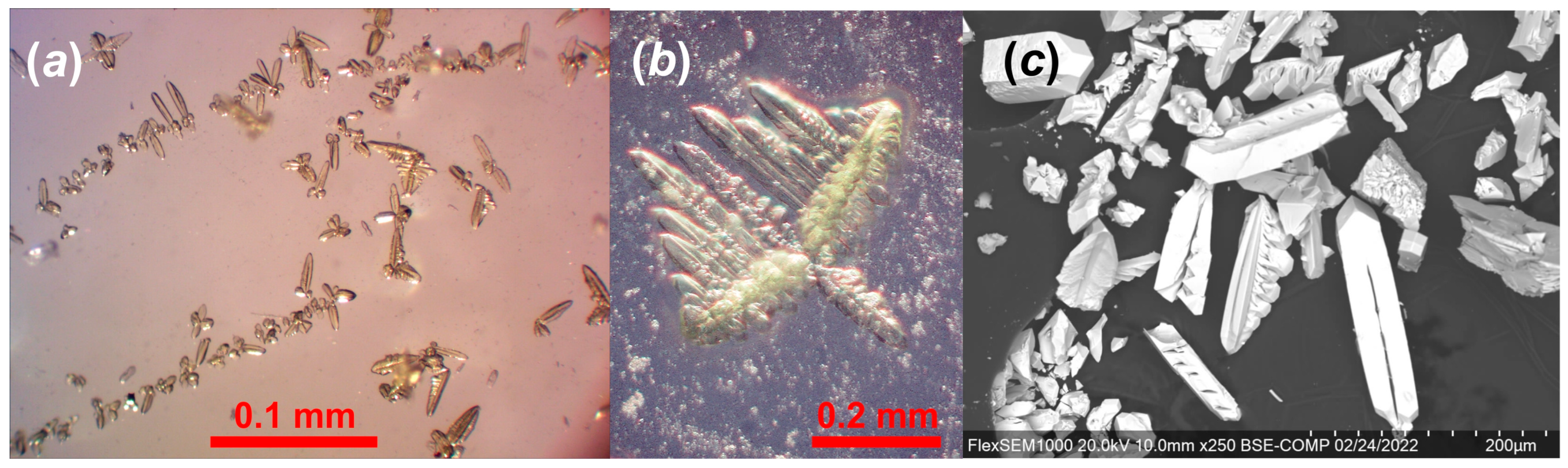
2.1. Synthesis

Caution: While isotopically depleted U was used in these experiments, precautions for handling radioactive materials should be followed.
An aqueous solution of 0.0725 g (0.14 mmol) of uranyl nitrate dissolved in 2 mL of deionized distilled water was heated on a hot plate at 70 °C for 4–5 min. Then, 0.074 g (0.7 mmol) of CaCl2 and 0.095 g (1.0 mmol) of (NH4)2CO3 were added to the hot solution, which was stirred until all solid dissolved. The resulting yellowish transparent solution, which yielded a pH of 7, was left to evaporate in a watch glass at room temperature. Crystals of the synthetic ewingite-like compound (SE) begin to form after 12–18 h as the first precipitate, mostly in the form of fine- or cryptocrystalline powders with rare individual dendritic aggregates (Figure 1a,b). Shortly thereafter, massive crystallization of calcite started, making it almost impossible to select pure SE. This process was slowed by reducing the temperature to 4–5 °C, which also yielded slightly larger dendritic aggregates with single crystalline dendrite tips separated by up to 10–20 μm (single findings up to 50 μm). The primary and relatively pure crystallization of SE was observed in a laboratory without intensive ventilation without intensive ventilation, without air conditioning system and without heaters, respectively.

2.2. Chemical Composition

The chemical analysis of SE was carried out with a Hitachi FlexSEM 1000 (Tokyo, Japan) scanning electron microscope (Figure 1c) equipped with an EDS Xplore Contact 30 detector and Oxford AZtecLive STD (Oxford, UK) system of analysis. The accelerating voltage was 20 kV and beam current was 5 nA. Only Ca, U, C, and O were detected, with elements with atomic numbers higher than that of beryllium below detection limits. The following standards and X-ray lines were used: Ca—CaF2, Kα; U—UO2, Mβ.
The chemical composition of SE is (wt.%, CO2 and H2O calculated based on structure): CaO 9.98, UO3 58.90, CO2 15.06, H2O 16.19, total 100.13. The empirical formula based on 278 O apfu is Ca20.79U24.07C40H210O278, or, taking into consideration the structural data, Ca20.79(H3O)6U24.07O52(CO3)36(HCO3)4(OH)12(H2O)88 (Figure 1c). N, Na, and K were not detected, resulting in the assignment of hydronium cations H3O+ as counter-ions in the structure of SE.

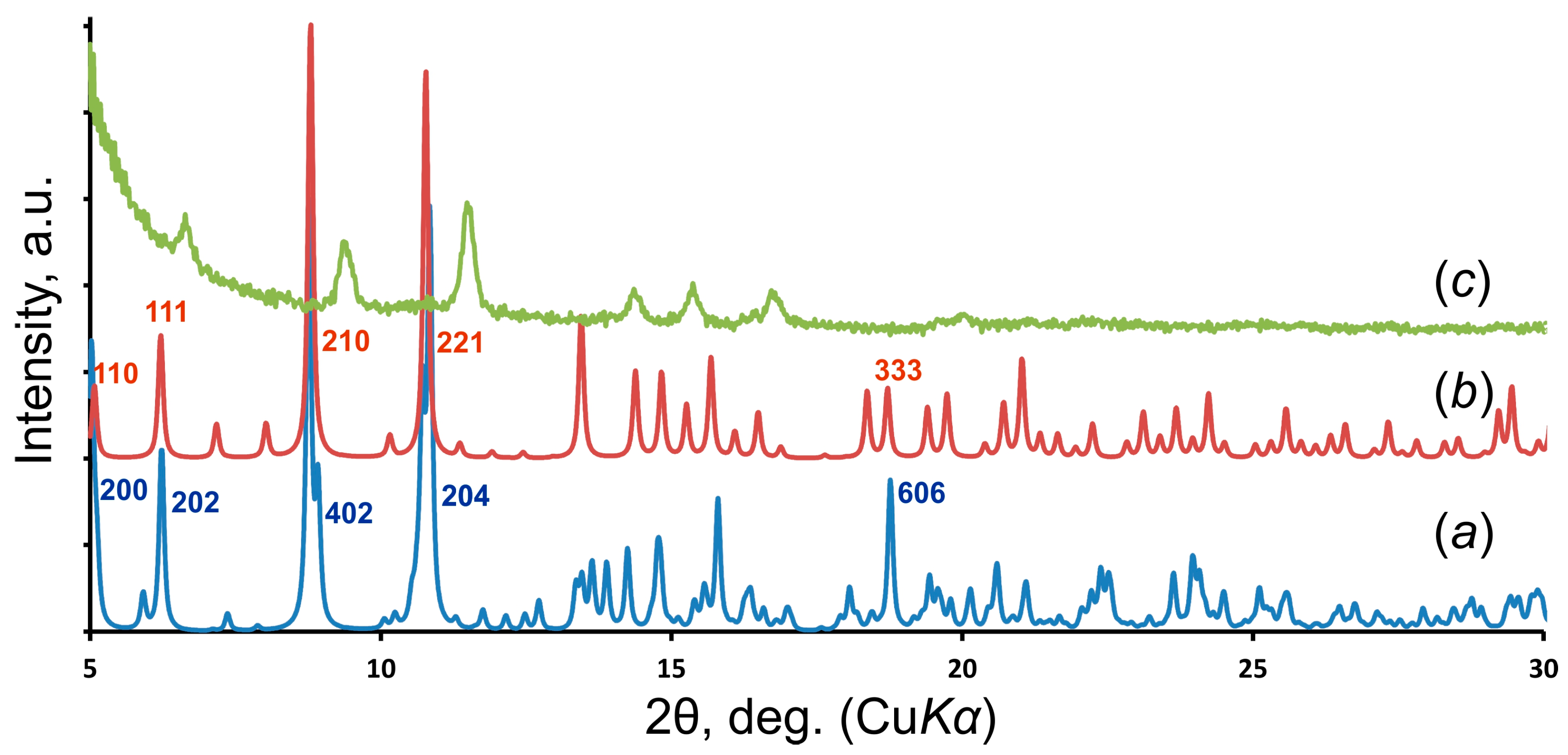
2.3. X-ray Diffraction Studies

The crystal structure of SE was determined from a greenish-yellow translucent dendrite tip with the dimensions 52 × 23 × 21 μm3 using single-crystal X-ray diffraction data collected by a Bruker Kappa Apex II Duo diffractometer at room temperature (microfocused MoKα radiation; 50 kV/0.6 mA; frame width 0.5°; exposure time 360 s/frame; Madison, WI, USA). Diffraction data were processed in the CrysAlisPro [14] program. The crystal structure of SE was solved by the dual-space algorithm and refined using the SHELX [15,16] programs incorporated in the OLEX2 [17] program package. The crystal structure of SE is tetragonal (P-4n2; a = 24.6098(2), c = 24.6246(4) Å; V = 14,913.7(3) Å3; Z = 2; R1 = 0.060; CSD 2166564 [for cif see Supplementary Materials]). The final model included coordinates and anisotropic displacement parameters for all non-H atoms. H atoms were not localized. The crystal structure of SE was refined as a 2-component twin using [010/-100/001] matrix with a statistically equal contribution of components (0.514(14)/0.486(14)).
Fragments of SE manually selected from synthesis products were used for the collection of powder diffraction data with a Rigaku Ultima IV diffractometer (CoKα radiation; 40 kV/30 mA; θ–θ Bragg–Brentano geometry; PSD D-Tex Ultra detector; Tokyo, Japan). A Rigaku SHT-1500 chamber was employed for experiments with SE in air over the temperature range of 25–800 °C; a Pt strip (20 × 12 × 2 mm3) was used as the heating element and sample holder. The temperature steps varied from 5 to 20 °C depending on the temperature range. The heating rate was 2 °C/min. The collection time at each temperature step was about 30 min. The irreversibility of the observed phase transformations was verified by collecting PXRD data following cooling.
Several fragments of SE were ground in an agate mortar for PXRD data collection using a Rigaku Ultima IV powder X-ray diffractometer (CuKα radiation; 40 kV/30 mA; θ–θ Bragg–Brentano geometry; PSD D-Tex Ultra detector; Tokyo, Japan) equipped with a background-free Si-single crystal sample holder at room temperature.

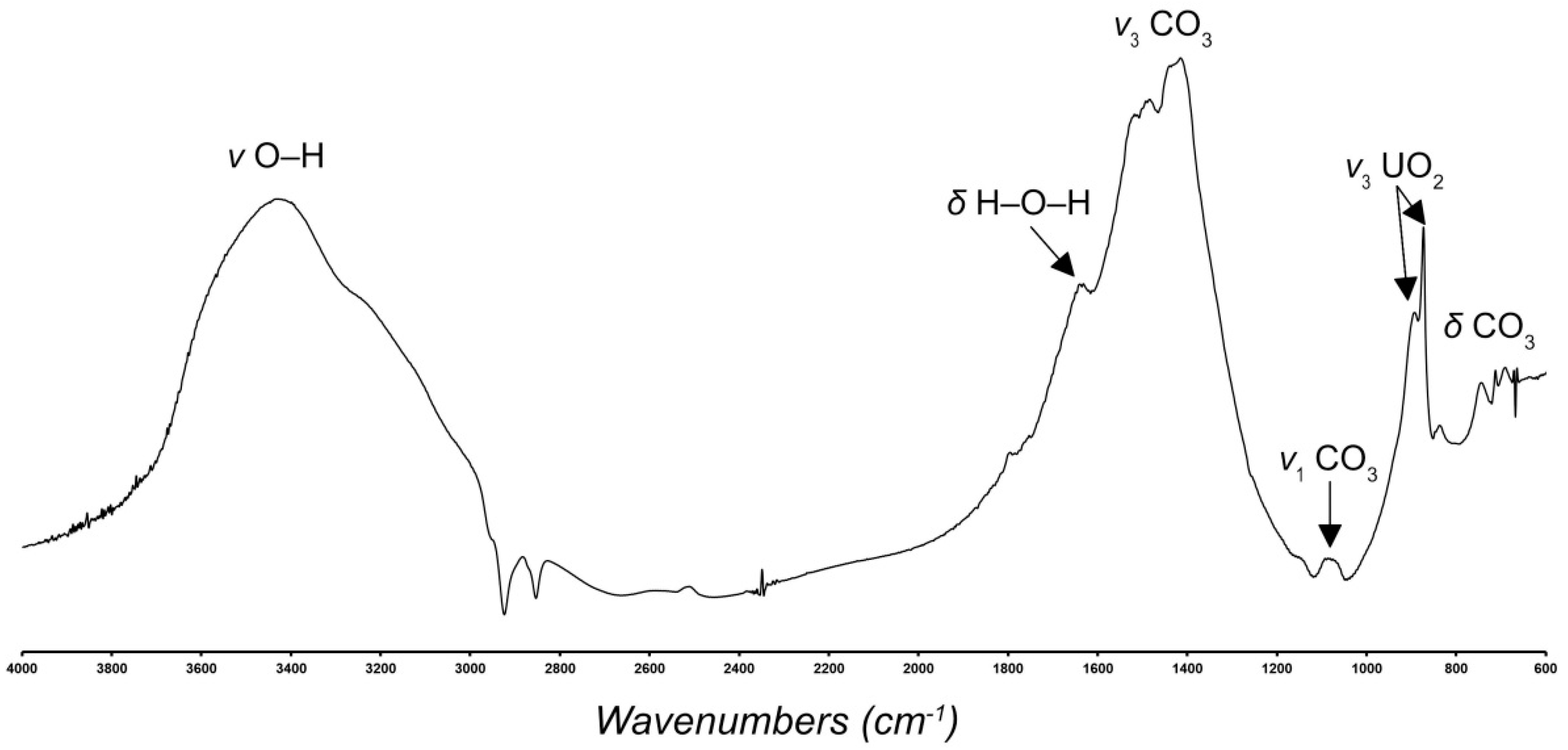
2.4. Infrared Spectroscopy

The IR spectrum of SE was recorded on a Bruker Vertex 70 (Bremen, Germany) spectrometer via the Attenuated Total Reflection Method in the region 4000–500 cm−1 (Figure 2). A MIRacle ATR accessory (Pike Technology; Fitchburg, WI, USA) with a Ge ATR crystal was used. IR spectroscopy is consistent with the structural data.
Infrared bands in the region above 2500 cm−1 (at 3590, 3420, 3230, 3130, 3000, 2950, 2900, 2890, 2830, 2730, and 2580 cm−1) are attributed to the ν O–H stretching frequencies of structurally nonequivalent H2O molecules forming the H-bonding network in the structure of SE. According to Libowitzky [18], the set of bands in this region corresponds to the approximate O–H∙∙∙O hydrogen bond lengths ranging between 2.6 and 3.2 Å. A band observed at 1645 cm−1 is attributed to the ν2 (δ) bending vibrations of symmetrically distinct H2O molecules. Bands in the region from 1570 to 1200 cm−1 (1570, 1525, 1487, 1445 and 1402 cm−1) are assigned to ν3 antisymmetric stretching vibrations of structurally nonequivalent CO3 planar groups. The splitting of the ν3 bands indicates the bidentate bonding of carbonate groups to uranyl ions in the crystal structure. The weak band at 1065 cm−1 is assigned as ν1 symmetric stretching vibrations of CO3 groups. The band caused by ν2 (δ) out-of-plane bending vibrations of CO3 groups is at 833 cm−1. Note that the ν2 (δ) CO3 bending vibrations may overlap with the ν1 UO22+ symmetric stretching vibrations. Bands at 739, 711, 687, 671, and 665 are assigned to the ν4 (δ) in-plane bending CO3 vibrations.
Two strong bands at 891 and 873 cm−1 are attributed to the ν3 antisymmetric stretching modes of UO22+.

3. Results and Discussion

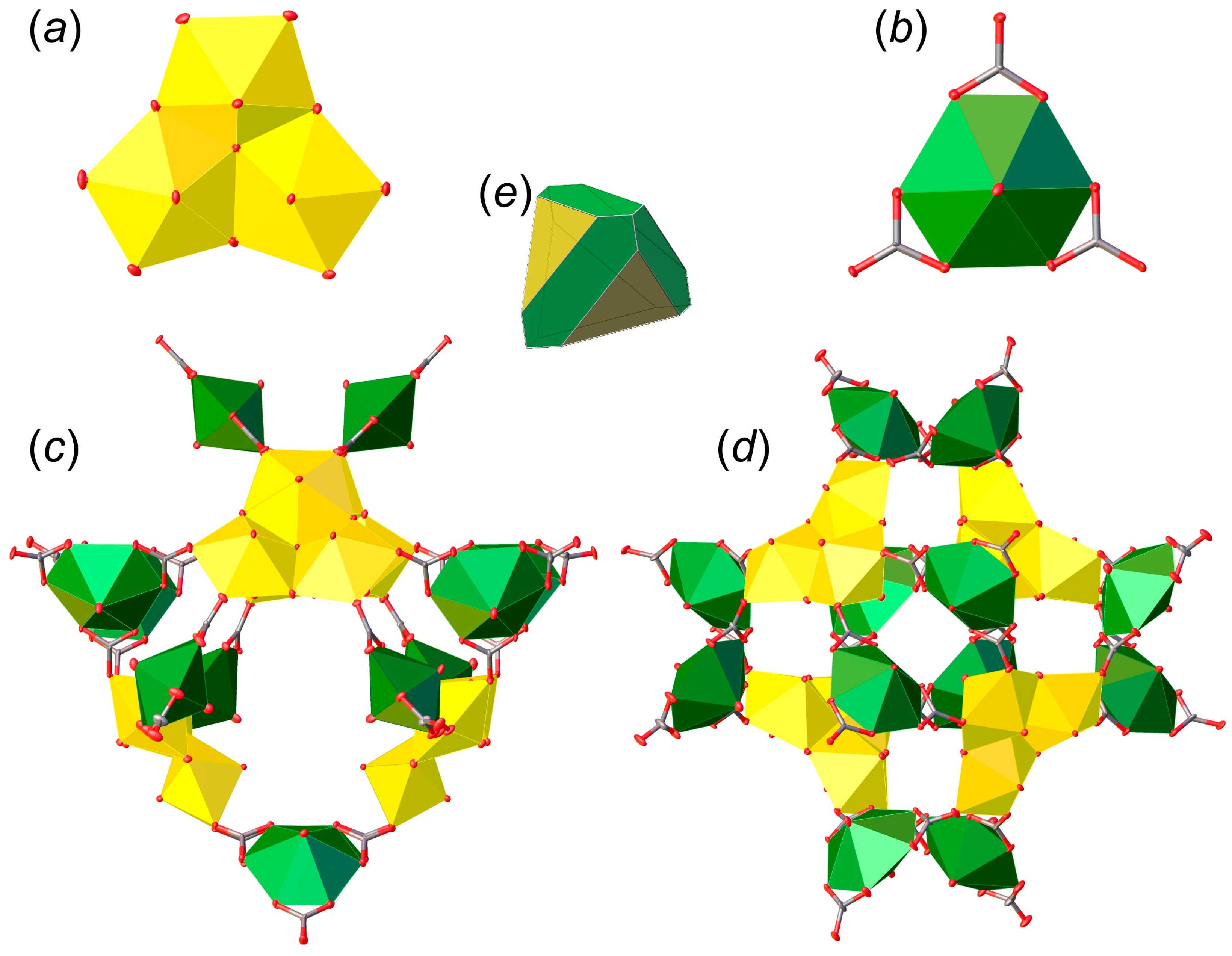
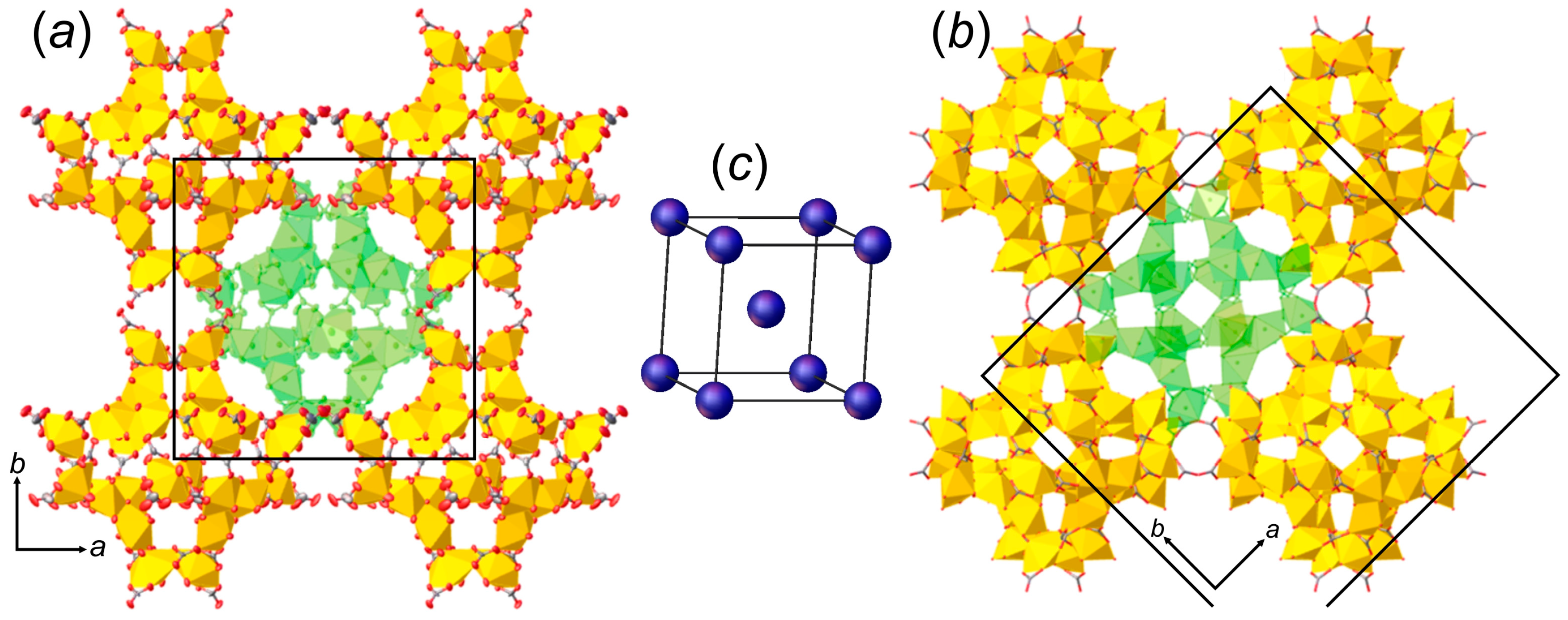
3.1. Structure Descriptions

There are six crystallographically independent U6+ cations in the structure of SE, each of which is strongly bonded to two O2− atoms, forming approximately linear O2−≡U6+≡O2− uranyl cations (Ur) with U6+≡O2− bond lengths ranging from 1.69(2) to 1.801(16) Å. The Ur1, Ur2, and Ur3 ions are equatorially coordinated by five oxygen atoms each, resulting in the formation of pentagonal bipyramids (U1–3–Oeq = 2.232(16)–2.443(17) Å), whereas the Ur4, Ur5, and Ur6 ions are coordinated by six oxygen atoms belonging to three carbonate groups, forming hexagonal bipyramids (U4–6–Oeq = 2.40(2)–2.458(18) Å). There are ten unique carbonate groups in the structure of SE, nine of which link to the equatorial edges of U-centered hexagonal bipyramids (C–O = 1.19(3)–1.35(4) Å), while the tenth group is not linked to U atoms, and due to the presence of one elongated and two short C–O bonds (1.47(4), 1.27(4) and 1.29(4) Å) is defined as an (HCO3) group.
There are two types of fundamental building units (FBU) in the structure of SE. FBU-1 consists of three Ur1–3 pentagonal bipyramids sharing common equatorial edges, with one μ3-O atom common to all three bipyramids, forming a trimeric unit (Figure 3a). The O atoms shared between two adjacent uranyl ions are hydroxyl groups, while the O atoms of the equatorial planes that do not bridge uranyl ions are from monodentate carbonate groups that link the FBU-1 and FBU-2 units. The FBU-2 unit is formed by the Ur4, Ur5, and Ur6 hexagonal bipyramids, in which each uranyl ion shares edges with three (CO3)2− triangular groups (Figure 3b) to form uranyl tricarbonate clusters (UTC) that are common U-bearing structural units in uranyl carbonates [19]. Four FBU-1 and twelve FBU-2 are arranged to form a complex uranyl carbonate [(UO2)24O4(OH)12(CO3)36]44– supertetrahedral nanocluster (Figure 3c,d). The cluster can be described as a combination of a tetrahedron and a cube, where the FBU-1 trimers correspond to faces of the tetrahedron, whereas two FBU-2 units delineate elongated faces of the cube (Figure 3e).
Isolated uranyl carbonate supertetrahedral nanoclusters in SE are separated by a network of Ca2+ cations and H2O molecules, both of which also occur inside the cluster. Five of the eight Ca sites are fully occupied. The Ca–O bond lengths range from 2.09(3) to 2.717(17) Å. Disorder (site splitting) and partial occupancies occur for the H2O sites. Calcium cations do not directly bridge between clusters. A complex H-bonding network involving interstitial H2O molecules, H3O+ ions, and (HCO3) groups link supertetrahedral nanoclusters into the crystal structure of SE.
There are several notable differences between the crystal structures of ewingite and SE. Ewingite contains both Ca and Mg, the latter of which does not occur in SE. Although tetragonal, the unit cell of ewingite is larger than SE (I41/acd; a = 35.142(2), c = 47.974(3) Å; V = 59,245(8) Å3) [13]. However, the arrangement of supertetrahedral nanoclusters in both structures is nearly identical and can be described as a body-centered cubic (α-Fe type) packing with the distances between the centers of clusters at ~21.3 Å (Figure 4). The structure symmetries are related through the following sub-/supergroup sequence: P-4n2 → I-42d (a = 2a, b = 2b, c = 2c) ← I41/acd—which explains the eightfold difference in the unit-cell volume of ewingite compared to SE. Symmetry breaking in ewingite may be attributed to the distribution of Ca and Mg ions, as well as to the higher H2O content.
The most significant difference between the two structures is that supertetrahedral nanoclusters in the structure of ewingite are formed from three FBUs: four trimers of pentagonal bipyramids, six uranyl tricarbonate, and six uranyl bicarbonate moieties [13]. The structure of SE has 12 uranyl tricarbonate units. It cannot be excluded that, due to the low quality of the natural ewingite crystals studied previously, some atoms of carbonate groups were not located in the difference-Fourier maps.
The similarity of the structural architectures of both compounds is clearly seen from the comparison of their powder XRD patterns (Figure 5a,b), where the overlapping basal peaks correspond to the supertetrahedral nanocluster packing. It should be also noted that slight grinding in an agate mortar results in the reduction in the unit cell observed in the XRD pattern from the shift of the basal reflections, which are responsible for the general cluster arrangement, to the far angular region, while reflections responsible for more subtle structural interactions are extinct (Figure 5c).

3.2. Structural Complexity Analysis

Ewingite is the mineral with the most complex crystal structure known to date and possesses the highest amount of structural information observed in minerals. The theory of structural complexity was developed by S.V. Krivovichev [20,21,22,23] and successfully implemented in recent works [24,25,26,27]. Olds et al. proposed that the information content of ewingite is c.a. 23,000 bits/cell [13]. Our recent consideration of the disorder of sites and the H atom assignment indicate that the structural complexity of ewingite is likely in the range of 19,500–21,500 bits/cell, depending on the hydration state [19]. The information-based complexity parameters for ewingite and SE are given in Table 1. Although the hydration state of SE probably may also vary, the structural complexity of the synthetic compound is half that of ewingite at 9515.770 bits/cell. The contribution of factors such as the H-bonding system, interstitial substructure, and cluster stacking to the overall structural complexity [28] in both compounds is comparable, which points to the similarity of substructural units’ arrangement and their role in ewingite and SE. The most significant difference is the topological complexity of the cluster, which is attributed to the aforementioned absence of specific carbonate groups in the structural model of ewingite. The tendency for the complexity parameters of synthetic compounds to usually be lower than those of minerals with identical or genetically similar topologies of U-bearing complexes was recently described for uranyl sulfates [29,30] and uranyl selenites [31] and seems to be quite common in general. On the other hand, the complexity of a crystal structure contributes negatively to the configurational entropy of the crystalline phase [32], although metastable polymorphs are usually structurally simpler than their more thermodynamically stable counterparts [33,34].
According to visual observation and experimental studies, in terms of their preservation, ewingite is more stable in air than SE, in which polycrystalline aggregates undergo degradation even with insignificant mechanical stress, which can be seen from the experimental PXRD pattern (Figure 5c). The indexation of the SE PXRD pattern by the Pawley method using the TOPAS software [35] resulted in the following unit cell: tetragonal; a = 23.12(1), c = 22.92(3) Å (Figure 6a). The high-temperature PXRD experiment demonstrates that SE readily loses its crystallinity, and traces from the basal reflections, which correspond to the uranyl carbonate nanocluster packing, disappear entirely after 80 °C (Figure 6b). Later, starting at c.a. 420 °C under continuous heating, the CaUO4 (PDF 01-085-0577; ICDD PDF-2 Database, release 2019 [36]) phase crystallizes.
The recent findings relating to the unnamed phase, equivalent to a now-described synthetic compound, at several localities in nature (Jáchymov, Czech Republic; Annaberg, Germany; Red Canyon district, Colorado, USA) have documented that the SE occurred there without the presence of ewingite (unpublished observations of J.P.). Furthermore, the first chemical analyses of the natural material also suggested a large chemical variability (namely in the Ca:Mg ratio), while the single-crystal X-ray diffraction patterns are very similar and also similar to those of SE. Nevertheless, these findings require deeper study, which is beyond the scope of this paper.
Keeping in mind the similar architecture of ewingite and SE as well as findings relating to the novel natural phase, it can be assumed that the formation of a particular crystalline form is highly dependent on the chemical composition of ground waters. On the other hand, since ewingite is apparently more stable, it is plausible that the SE phase can be regarded as a primary and/or metastable reaction product which further re-crystallizes into a more stable form under environmental conditions.

Supplementary Materials

The following supporting information can be downloaded at https://www.mdpi.com/article/10.3390/ma15196643/s1: CSD 2166564: contains the supplementary crystallographic data for this paper.

Author Contributions

Conceptualization, O.S.T. and V.V.G.; Methodology, O.S.T., A.V.K., M.G.K. and V.V.G.; Investigation, O.S.T., I.V.K., A.V.K., M.G.K. and V.V.G.; Writing—Original Draft Preparation, I.V.K., J.P., S.V.K., P.C.B. and V.V.G.; Writing—Review and Editing, J.P., S.V.K., P.C.B. and V.V.G.; Visualization, I.V.K., A.V.K. and V.V.G. All authors have read and agreed to the published version of the manuscript.

Funding

This work was supported by the Russian Science Foundation (no. 19-17-00038), and by the Czech Science Foundation (no. 20-11949S to J.P.).

Acknowledgments

The XRD and spectroscopy studies were performed at the X-ray Diffraction Centre of St. Petersburg State University.

Conflicts of Interest

The authors declare no conflict of interest.

References

  1. Pauliš, P.; Babka, K.; Sejkora, J.; Škácha, P. Uranové Minerály České Republiky a Jejich Nejvýznamnější Naleziště; Kuttna: Kutná Hora, Czech Republic, 2016; 570p. (In Czech) [Google Scholar]
  2. Škácha, P.; Plášil, J.; Horák, V. Jáchymov Mineralogická Perla Krušnohoří; Academia: Prague, Czech Republic, 2019; 682p. (In Czech) [Google Scholar]
  3. Agricola, G. De Re Metallica, Translation from the 1st Latin edition of, 1556; Hoover, H.C., Hoover, L.H., Eds.; The Mining Magazine, Salisbury House: London, UK, 1912; 640p. [Google Scholar]
  4. Lide, D.R. (Ed.) CRC Handbook of Chemistry and Physics, 86th ed.; CRC Press: Boca Raton, FL, USA, 2005. [Google Scholar]
  5. Plášil, J.; Mills, S.J.; Fejfarová, K.; Dušek, M.; Novák, M.; Škoda, R.; Čejka, J.; Sejkora, J. The crystal structure of natural zippeite, K1.85H+0.15[(UO2)4O2(SO4)2(OH)2](H2O)4, from Jáchymov, Czech Republic. Can. Mineral. 2011, 49, 1089–1103. [Google Scholar] [CrossRef]
  6. Plášil, J.; Hloušek, J.; Veselovský, F.; Fejfarová, K.; Dušek, M.; Škoda, R.; Novák, M.; Čejka, J.; Sejkora, J.; Ondruš, P. Adolfpateraite, K(UO2)(SO4)(OH)(H2O), a new uranyl sulphate mineral from Jáchymov, Czech Republic. Am. Mineral. 2012, 97, 447–454. [Google Scholar] [CrossRef]
  7. Plášil, J.; Veselovský, F.; Hloušek, J.; Šák, M.; Sejkora, J.; Cejka, J.; Škácha, P.; Kasatkin, A.V. Mathesiusite, K5(UO2)4(SO4)4(VO5)(H2O)4, a new uranyl vanadate-sulfate from Jáchymov, Czech Republic. Am. Mineral. 2014, 99, 625–632. [Google Scholar] [CrossRef]
  8. Plášil, J.; Hloušek, J.; Kasatkin, A.V.; Škoda, R.; Novák, M.; Čejka, J. Geschieberite, K2(UO2)(SO4)2(H2O)2, a new uranyl sulfate mineral from Jáchymov. Mineral. Mag. 2015, 79, 205–216. [Google Scholar] [CrossRef]
  9. Plášil, J.; Hloušek, J.; Kasatkin, A.V.; Belakovskiy, D.I.; Čejka, J.; Chernyshov, D. Ježekite, Na8[(UO2)(CO3)3](SO4)2·3H2O, a new uranyl mineral from Jáchymov, Czech Republic. J. Geosci. 2015, 60, 259–267. [Google Scholar] [CrossRef]
  10. Plášil, J.; Hlousek, J.; Kasatkin, A.V.; Novak, M.; Cejka, J.; Lapcak, L. Svornostite, K2Mg[(UO2)(SO4)2]2∙8H2O, a new uranyl sulfate mineral from Jáchymov, Czech Republic. J. Geosci. 2015, 60, 113–121. [Google Scholar] [CrossRef]
  11. Plášil, J.; Škácha, P.; Sejkora, J.; Kampf, A.R.; Škoda, R.; Čejka, J.; Hloušek, J.; Kasatkin, A.V.; Pavlíček, R.; Babka, K. Plavnoite, a new K-Mn member of the zippeite group from Jáchymov, Czech Republic. Eur. J. Mineral. 2017, 29, 117–128. [Google Scholar] [CrossRef]
  12. Olds, T.; Plášil, J.; Kampf, A.; Dal Bo, F.; Burns, P. Paddlewheelite, a New Uranyl Carbonate from the Jáchymov District, Bohemia, Czech Republic. Minerals 2018, 8, 511. [Google Scholar] [CrossRef]
  13. Olds, T.; Plášil, J.; Kampf, A.; Simonetti, A.; Sadergaski, L.; Chen, Y.-S.; Burns, P. Ewingite: Earth’s most complex mineral. Geology 2017, 45, 1007–1010. [Google Scholar] [CrossRef]
  14. CrysAlisPro Software System, Version 1.171.41.103a; Rigaku Oxford Diffraction: Oxford, UK, 2021.
  15. Sheldrick, G.M. SHELXT—Integrated space-group and crystal structure determination. Acta Crystallogr. 2015, A71, 3–8. [Google Scholar] [CrossRef] [Green Version]
  16. Sheldrick, G.M. Crystal structure refinement with SHELXL. Acta Crystallogr. 2015, C71, 3–8. [Google Scholar]
  17. Dolomanov, O.V.; Bourhis, L.J.; Gildea, R.J.; Howard, J.A.K.; Puschmann, H. OLEX2: A complete structure solution, refinement and analysis program. J. Appl. Cryst. 2009, 42, 339–341. [Google Scholar] [CrossRef]
  18. Libowitzky, E. Correlation of O-H stretching frequencies and O-H…O hydrogen bond lengths in minerals. Monatsh. Chem. 1999, 130, 1047–1059. [Google Scholar] [CrossRef]
  19. Gurzhiy, V.V.; Kalashnikova, S.A.; Kuporev, I.V.; Plášil, J. Crystal chemistry and structural complexity of the uranyl carbonate minerals and synthetic compounds. Crystals 2021, 11, 704. [Google Scholar] [CrossRef]
  20. Krivovichev, S.V. Topological complexity of crystal structures: Quantitative approach. Acta Crystallogr. 2012, A68, 393–398. [Google Scholar] [CrossRef]
  21. Krivovichev, S.V. Structural complexity of minerals: Information storage and processing in the mineral world. Mineral. Mag. 2013, 77, 275–326. [Google Scholar] [CrossRef]
  22. Krivovichev, S.V. Which inorganic structures are the most complex? Angew. Chem. Int. Ed. 2014, 53, 654–661. [Google Scholar] [CrossRef]
  23. Krivovichev, S.V. Structural complexity of minerals and mineral parageneses: Information and its evolution in the mineral world. In Highlights in Mineralogical Crystallography; Danisi, R., Armbruster, T., Eds.; Walter de Gruyter GmbH: Berlin, Germany; Boston, MA, USA, 2015; pp. 31–73. [Google Scholar]
  24. Krivovichev, S.V.; Hawthorne, F.C.; Williams, P.A. Structural complexity and crystallization: The Ostwald sequence of phases in the Cu2(OH)3Cl system (botallackite–atacamite–clinoatacamite). Struct. Chem. 2017, 28, 153–159. [Google Scholar] [CrossRef]
  25. Izatulina, A.R.; Gurzhiy, V.V.; Krzhizhanovskaya, M.G.; Kuz’mina, M.A.; Leoni, M.; Frank-Kamenetskaya, O.V. Hydrated Calcium Oxalates: Crystal Structures, Thermal Stability and Phase Evolution. Cryst. Growth Des. 2018, 18, 5465–5478. [Google Scholar] [CrossRef]
  26. Plášil, J. Structural complexity of uranophane and uranophane-β: Implications for their formation and occurrence. Eur. J. Mineral. 2018, 30, 253–257. [Google Scholar] [CrossRef]
  27. Gurzhiy, V.V.; Tyumentseva, O.S.; Izatulina, A.R.; Krivovichev, S.V.; Tananaev, I.G. Chemically Induced Polytypic Phase Transitions in the Mg[(UO2)(TO4)2(H2O)](H2O)4 (T = S, Se) System. Inorg. Chem. 2019, 58, 14760−14768. [Google Scholar] [CrossRef]
  28. Krivovichev, S.V. Ladders of information: What contributes to the structural complexity in inorganic crystals. Z. Kristallogr. 2018, 233, 155–161. [Google Scholar] [CrossRef]
  29. Gurzhiy, V.V.; Plášil, J. Structural complexity of natural uranyl sulfates. Acta Crystallogr. 2019, B75, 39–48. [Google Scholar] [CrossRef] [PubMed]
  30. Tyumentseva, O.S.; Kornyakov, I.V.; Britvin, S.N.; Zolotarev, A.A.; Gurzhiy, V.V. Crystallographic Insights into Uranyl Sulfate Minerals Formation: Synthesis and Crystal Structures of Three Novel Cesium Uranyl Sulfates. Crystals 2019, 9, 660. [Google Scholar] [CrossRef]
  31. Gurzhiy, V.V.; Kuporev, I.V.; Kovrugin, V.M.; Murashko, M.N.; Kasatkin, A.V.; Plášil, J. Crystal chemistry and structural complexity of natural and synthetic uranyl selenites. Crystals 2019, 9, 639. [Google Scholar] [CrossRef]
  32. Krivovichev, S.V. Structural complexity and configurational entropy of crystalline solids. Acta Crystallogr. 2016, B72, 274–276. [Google Scholar]
  33. Gurzhiy, V.V.; Tyumentseva, O.S.; Krivovichev, S.V.; Tananaev, I.G. Selective Se-for-S substitution in Cs-bearing uranyl compounds. J. Solid State Chem. 2017, 248, 126–133. [Google Scholar] [CrossRef]
  34. Kornyakov, I.V.; Tyumentseva, O.S.; Krivovichev, S.V.; Gurzhiy, V.V. Dimensional evolution in hydrated K+-bearing uranyl sulfates: From 2D-sheets to 3D frameworks. CrystEngComm. 2020, 22, 4621–4629. [Google Scholar] [CrossRef]
  35. Bruker AXS. Topas, V5.0: General Profile and Structure Analysis Software for Powder Diffraction Data; Bruker: Karlsruhe, Germany, 2014. [Google Scholar]
  36. Loopstra, B.O.; Rietveld, H.M. The structure of some alkaline-earth metal uranates. Acta Crystallogr. 1969, B25, 787–791. [Google Scholar] [CrossRef]
Figure 1. The onset of crystallization of the synthetic ewingite-like compound (SE) (a), a dendritic aggregate of SE surrounded by fine crystalline calcite powder (b), and an SEM image of SE crystals (c).
Materials 15 06643 g001
Figure 2. Powder infrared absorption spectrum of the SE.
Materials 15 06643 g002
Figure 3. Fundamental building units (FBU) in the structure of SE (a,b), and their arrangement into a supertetrahedral nanocluster (c,d) that is similar to the combination of a tetrahedron and a cube (e). Legend: uranyl pentagonal bipyramids = yellow; uranyl hexagonal bipyramids = green; different coloring of Ur polyhedra is used for clarity; red = O atoms; grey = C atoms.
Materials 15 06643 g003
Figure 4. Arrangement of supertetrahedral nanoclusters in the structures of SE (a) and ewingite (b), which is similar to the body-centered cubic (α-Fe type) packing (c). Interstitial ions and molecules are omitted for clarity; central nanocluster in the body-centered cubic packing is green-colored. Legend: U-centered polyhedra = yellow; red = O atoms; grey = C atoms.
Materials 15 06643 g004
Figure 5. PXRD patterns calculated from the structural data of the ewingite (a), SE (b), and pattern collected during the PXRD measurement of ground SE sample (c).
Materials 15 06643 g005
Figure 6. PXRD patterns of SE as a function of temperature (25–800 °C) under heating in air (a) and indexation of the SE PXRD pattern (b).
Materials 15 06643 g006
Table 1. Structural complexity parameters for ewingite and SE.
Complexes That Contribute to Structural Complexity [27]Ewingite [18]SE
υ, AtomsIG,total, Bits/Cell (IG, Bits/Atom)Contribution, %υIG,total, Bits/Cell (IG, Bits/Atom)Contribution, %
Topological complexity of the cluster2201271.820 (5.781)6.92441447.100 (5.931)15.2
Structural complexity of the cluster2201271.82002441447.1000
Stacking of clusters8803813.88620.84881506.18015.8
Interstitial structure5884531.05624.73182499.19026.3
H-bonding10408717.65047.64844063.30042.7
Structural complexity of the entire structure250818,335.988 (7.311)10012909515.77 (7.377)100
Publisher’s Note: MDPI stays neutral with regard to jurisdictional claims in published maps and institutional affiliations.

