Article

Failure Analysis of Ultra-High Molecular Weight Polyethylene Tibial Insert in Total Knee Arthroplasty

1
Faculty of Material Science and Engineering, University Politehnica of Bucharest, 313 Splaiul Independentei, District 6, 060042 Bucharest, Romania
2
Faculty of Electrical Engineering, University Politehnica of Bucharest, 313 Splaiul Independentei, District 6, 060042 Bucharest, Romania
3
Academy of Romanian Scientists, 54 Splaiul Independentei, 050094 Bucharest, Romania
4
Faculty of Medicine, Titu Maiorescu University, 67A Gheorghe Petrascu Street, 031593 Bucharest, Romania
5
Department of Orthopedics and Trauma I, Colentina Clinical Hospital, 19-21 Soseaua Stefan cel Mare, 020125 Bucharest, Romania
6
Medical Ortovit Ltd., 8 Miron Costin Street, 011098 Bucharest, Romania
7
National Institute for Research and Development in Microtechnologies IMT-Bucharest, 126A Erou Iancu Nicolae Street, 077190 Bucharest, Romania
8
Department of Morphological Sciences, Carol Davila University of Medicine and Pharmacy, 37 Dionisie Lupu Street, 020021 Bucharest, Romania
*
Authors to whom correspondence should be addressed.
Materials 2022, 15(20), 7102; https://doi.org/10.3390/ma15207102
Submission received: 7 September 2022 / Revised: 9 October 2022 / Accepted: 10 October 2022 / Published: 13 October 2022
(This article belongs to the Section Biomaterials)

Abstract

:
Knee osteoarthritis is treated based on total knee arthroplasty (TKA) interventions. The most frequent failure cause identified in surgical practice is due to wear and oxidation processes of the prothesis’ tibial insert. This component is usually manufactured from ultra-high molecular weight polyethylene (UHMWPE). To estimate the clinical complications related to a specific prosthesis design, we investigated four UHMWPE tibial inserts retrieved from patients from Clinical Hospital Colentina, Bucharest, Romania. For the initial analysis of the polyethylene degradation modes, macrophotography was chosen. A light stereomicroscope was used to estimate the structural performance and the implant surface degradation. Scanning electron microscopy confirmed the optical results and fulfilled the computation of the Hood index. The oxidation process in UHMWPE was analyzed based on Fourier-transform infrared spectroscopy (FTIR). The crystallinity degree and the oxidation index were computed in good agreement with the existing standards. Mechanical characterization was conducted based on the small punch test. The elastic modulus, initial peak load, ultimate load, and ultimate displacement were estimated. Based on the aforementioned experimental tests, a variation between 9 and 32 was found in the case of the Hood score. The oxidation index has a value of 1.33 for the reference sample and a maximum of 9.78 for a retrieved sample.

1. Introduction

Knee arthroplasty as a surgical intervention is considered an efficient treatment for osteoarthritis. It is a standard procedure in almost all hospitals in Europe and the United States of America (USA). The statistical data collected in the USA report about 1 million surgical interventions performed yearly [1]. In 2010 there were reported 719,000 total knee arthroplasties, of which 10–15% were secondary revision operations. These types of interventions are performed after implant failure. It is expected that in the year 2030, the number of primary TKA will increase, with a percentage of 673%, equal to about 3.48 million procedures.
Regarding the secondary surgical interventions, it is estimated that an increase of 401% between 2022 and 2030 will occur [2]. The main causes of the TKA procedures are the average life expectancy of the population and the high level of physical activity. Unfortunately, these surgical interventions are performed at a younger age than in the past. About 85% of patients reported an excellent post-operator course after a primary TKA [3,4]. Approximately 20% of patients suffered from long-time chronic pain, and supplementary measures and painkillers had to be taken into account [5,6].
The typical knee prosthesis failures are abnormal joint movements, aseptic loosening, infection, periprosthetic fracture, ligament/flexion instability, arthrofibrosis, and material particles’ side effects. In practice, it was concluded that a knee implant works improperly due to multiple causes mentioned above. The revision surgery is a complex procedure, and it is associated with a high risk of infection, complications, prolonged hospitalization time, and a short life expectancy for the secondary prosthesis [7,8].
The main functions of orthopedic knee implants include articular function restauration and pain relief [9,10]. The implant life expectancy depends on the device design and the used biomaterials [11,12]. All the materials must be Food and Drug Administration (FDA) approved or carry the Conformitè Europëenne certificate [13,14].
Usually, the knee implant femoral component, placed at the distal part of the femoral bone, is made from a binary Co-Cr or a ternary Co-Cr-Mo alloy [15]. Oxidized zirconium (OXINIUM) was introduced by Smith + Nephew as an alternative to the Co-based alloys to reduce the polyethylene particle quantity and limit the number of failed prostheses. This material offers a superior advantage because it combines metallic alloy durability with ceramic material biocompatibility, exhibiting a high corrosion resistance [16,17]. The implants made from OXINIUM are characterized by a 15 years life expectancy and a very low revision intervention rate [18,19]. In the case of patients that exhibit nickel allergies, the femoral component can be manufactured from ceramic materials such as alumina (Al2O3) [20,21], yttria-stabilized zirconia (Y-TZP) [22,23], and zirconia toughened alumina (ZTA) [24]. The ceramic components’ main disadvantage is implant squeezing and reduced mechanical characteristics [25]. Recently, femoral components made from polyetheretherketone (PEEK) received FDA approval due to the good mechanical properties of the material. PEEK has a value of Young’s modulus close to the one of the human bone, so the stress-shielding effect is prohibited [26,27]. By using polymeric materials, the complications of the metallic particles’ emission are avoided. The principal drawback of PEEK is that an important bioinertia characterizes this material. In order to increase its biocompatibility, different solutions, such as surface treatments, are searched [28,29].
The tibial component positioned at the tibial bone proximal part is manufactured from a titanium alloy (Ti6Al4V) [30,31]. This ternary alloy exhibits adequate mechanical properties and can sustain the mechanical stress that appears during the patient gait cycle. Unfortunately, metallic ion emission can be observed in some cases, and allergic, teratogenic, toxic, and carcinogenic effects are evidenced during secondary surgical interventions. Zimmer—Biomet has developed tibial components made from porous tantalum (Ta), denoted as Trabecular MetalTM, with excellent mechanical properties, no stress shielding effects, and high biocompatibility [32,33]. Recent studies present alternative materials such as reinforced PEEK with carbon fiber, the so-called PEEK-OPTIMA, and Zeniva ZA-600 CF [34] and concluded that polymeric-based materials could be considered an alternative for titanium alloy in some cases. In the case of old age or overweight patients, UHMWPE tibial components are proposed, and good outcomes are reported because these two categories of people have a limited range of motion and reduced physical outdoor activities [35]. Finally, the tibial component of the BPK-S Integration prosthesis developed by Peter Brehm GmbH, Weisendorf, Germany, was made from BIOLOX® ceramic material [36].
The tibial insert represents the vulnerable part of a knee implant because this component is usually the first that fails since it is made from polymeric materials [37]. The most used material is ultra-high molecular weight polyethylene (UHMWPE), whose properties were improved over time to decrease wear and oxidation [38,39]. Different variants such as highly cross-linked polyethylene (HXLPE) or HXLPE combined with a synthetic analog of vitamin E (alpha-tocopherol) were proposed to prevent the oxidative degradation effect and to reduce the number of failed knees prosthesis due to osteolysis and aseptic loosening [37,40]. UHMWPE is a particular type of polyethylene, being a linear homopolymer, half crystalline with high molecular mass. The material that is used in the orthopedic field has a molecular weight between (3.5–6) × 106 g/mol and a crystallinity grade of about 50–55% [37]. According to the American standard ASTM, UHMWPE must have a molecular weight higher than 3.1 × 10⁶ a.m.u. (atomic mass units), since the standard ISO 11542 (ISO, 2001) recommends a molecular weight of at least 1 × 10⁶ a.m.u [38]. The ultra-high molecular weight polyethylene exhibits three phases. A crystalline lamellar phase, in which the polymeric chains are organized in an orthorhombic crystalline matrix, and an amorphous phase, in which the macromolecules form random wires or act as molecule ties that go out from a crystalline lamella and enter another one. A third phase partial order, which intercalates macromolecules with tight or loose chains, is present in the material. It can be noticed that the crystalline phase has an important contribution to increased mechanical rigidity, while the amorphous phase offers ductility and resilience [39]. When the polyethylene quality is improper, mechanical stresses can result in polymeric particles that induce osteolysis, an acute inflammatory process. It was observed that this phenomenon occurs due to an inadequate sterilization process with gamma radiations. The standards recommend that the sterilization process of the material be carried out in an inert ethylene oxide atmosphere or gas plasma. The cross-linked effect is induced through irradiation, increasing the material wear resistance by a high amount [37]. The most used types of UHMWPE are manufactured by Ticona (Summit, NJ) and are denominated GUR 1020 and GUR 1050 [41]. The GUR 1020 material has an average molecular weight of 3.5 × 10⁶ g/mol, a density of 0.93 g/mL, yield stress higher than 17 MPa, impact strength higher than 210 kJ/m2, and an average particle size of 140 μm. Regarding the GUR 1030 type, the material molecular weight is comprised of between (5.5–6) × 106 g/mol. It has a density of 0.93 g/mL, a tensile modulus of 680 MPa, and an impact strength higher than 130 kJ/m2 [41]. The tibial insert can also be manufactured from PEEK, showing increased resistance to degradative processes reported in previous cases and good mechanical properties [35].
In some clinical cases, replacing the native human patella with a dedicated part is necessary. There were proposed different solutions, such as a combination between a metallic base and a thin polyethylene surface, but major failure reports were found in the literature. Today, the worldwide accepted solution uses a UHMWPE component [42].
Osteolysis due to metallic, ceramic, or polymeric particles represents one of the leading causes of aseptic loosening. After the knee prosthesis is introduced into the human body, it becomes a particle source due to wear and corrosion. The resulting particles infiltrate the patient’s circulatory system, or they can agglomerate in neighboring tissues such as bone or bone marrow. They have a dangerous potential to interact with osteoblasts, preosteoblasts, mesenchymal stem cells (MSCs), macrophages, osteoclasts, and fibroblasts [43]. Figure 1 presents the adverse effect of the particles against the functions of the cells placed in the implant vicinity. The pro-inflammatory effect of the macrophages and osteoblasts has an important contribution to a chronic inflammatory environment near the implant and osteoclastogenesis [44,45]. In addition, a negative influence of the osteogenic differentiation and osteoblasts or MSCs mineralization was put in evidence in the literature [46].
Retrieval analysis of well-functioning and failed implants is of great importance in the orthopedic biomaterials field because surgical technique, implant design, and material quality can be improved in order to repair joints affected by diseases or trauma [14,47]. The standard ASTM F561-97 recognizes the importance of this analysis and concludes that two factors are met to obtain a successful implant integration [48]. The first one is related to the material, and the second one is biologically dependent [49]. Some authors identified the existence of a dynamic interface placed at the implant-tissue boundary [50]. Osseointegration was defined in [51] as a direct structural and functional link between the load stress implant and vital bone. The main types of cells that act in bone healing and formation are the osteoblasts, that form bone at a rate of 0.17 mm3/day; osteocytes, which are considered an important stress detector for bone; and osteoclasts, that resorb bone at a rate of 100 μm/day [50]. Three different modalities of bone response were identified. The first one is called fibrointegration, which appears in a case of an extent trauma and consists of fibrous tissue apparition around the prosthesis. Then if new blood vessels are not formed, dead bone can be seen in the implant vicinity, and if all the conditions are met, normal bone can be observed in the region of the prosthesis. This last response is called osseointegration. Three types of osseointegration are present: biointegration, mechanical integration, and chemical integration. Biointegration is possible when active biomaterials that link collagen fibers to the bone, such as calcium phosphate coatings, are used. Mechanical integration is considered when the bone is connected to the implant with the help of screws, vents, or cement. Chemical integration is obtained when chemical or physical bonds such as covalent bonds, hydrogen bonds, or van der Waals forces appear. The main factors identified in the literature [52] that influence the osteointegration process are surgical technique, the biocompatibility of the materials, implant design and surface, quality of the patient bone, and loading conditions of the prosthesis. The materials can be classified according to their biocompatibility as biotolerant, bioinert, and bioactive. Regarding the stress conditions, it was noticed that implant design and geometry have a strong influence on the loads applied and can be a critical factor in the retrieval analysis. Additionally, aspects linked to the surgical technique, such as implant alignment and surgical insert point of the prosthesis, are important in stress management. The surface state influences the bonds between the bone and the implant, and critical factors such as coatings or roughness must be considered for good osseointegration.
There are many studies regarding the retrieval analysis of UHMWPE tibial inserts, and the main failure reasons are due to oxidation and wear phenomena. Liza et al. [53] investigated the failure analysis of a UHMWPE tibial insert from Apollo® Total Knee System that was removed after 10 years of service. Using different testing methods such as scanning electron microscopy (SEM), infinite focus microscope (IFM) coupled with energy disperse spectroscopy (EDS), Fourier-transform spectroscopy (FTIR), and gel permeation chromatography (GPC), they concluded that the UHMWPE material exhibited high oxidation and wear degradation. Moreover, delamination, folding, pitting, and scratching were predominant. The retrieved sample had a reduced molecular weight contributing to the implant failure. Kurz et al. [54,55] and Edidin et al. [56,57] presented a mechanical test strategy adapted to UHMWPE tibial inserts called the small punch test. They have prepared small, thin disks indented by a hemispherical punch, determining a biaxial tension state. Mechanical quantities such as Young’s modulus, ultimate load, ultimate displacement, and work to failure were measured and correlated with the in vivo service. Cho et al. [58] showed that microscopic surface asperities on the knee prosthesis’s metallic femoral components have an important role in increasing the wear process of UHMWPE tibial inserts. Medel et al. [59] collected 119 tibial inserts, from which 29 were gamma sterilized in normal atmospheric conditions and 90 were conventional gamma sterilized using nitrogen atmosphere. They have found a direct link between in vivo oxidation and fatigue deterioration. It was concluded that oxidation strongly influences delamination since pitting damage is not increased by this phenomenon. Berry et al. [60] made a similar study on 132 gamma-air and 174 gamma-inert-sterilized UHMWPE tibial inserts. They noticed that gamma-inert-sterilized tibial inserts reached the critical oxidation index value at an average of 13 years after the primary surgery. Cerquiglini et al. [61] studied the new antioxidant polyethylene retrieval analysis. They used 24 Press Fit Condylar (PFC) Sigma (DePuy) and 17 Attune Knee System (DePuy) with fixed bearing and rotating platform design. A higher value of the Hood score was found in the case of Attune tibial inserts on the backside surface. Material properties’ modifications were reported due to implant design and potentially reduced implant performance was foreseen. Raman spectroscopy was used by Tone et al. [62] to analyze the wear rate and creep of e-beam-sterilized conventional UHMWPE tibial insert. The main conclusion is that the body mass index (BMI) greatly influences implant failure, and a separation of the creep and wear components of thickness reduction was made. There are few recent papers regarding failure analysis of primary cemented Zimmer Biomet knee implants. Our study fulfills this research and sustains the conclusions of Granquist et al. [63], that found a minimum 5-year survival time for total hip or knee arthroplasty. Additionally, the paper brings new information about the potential of microscopy techniques in failure explant analysis.
In the paper, we investigated four UHMWPE tibial inserts retrieved from patients from Clinical Hospital Colentina, Bucharest, Romania. We performed macrophotography, stereomicroscopy, and scanning electron microscopy investigations to establish the samples’ wear grade and to correlate it with the Hood index computation. We checked the chemical composition, crystallinity grade, and oxidation index through Fourier-transform infrared spectroscopy (FTIR). We analyzed the material’s mechanical behavior based on the small punch test.

2. Materials and Methods

2.1. Experimental Samples: Patients Data and Associated Pathologies

The investigated UHMWPE tibial inserts were retrieved from four patients from Clinical Hospital Colentina, Bucharest, Romania. Similarly, failed implants were selected for comparative analysis and reference after different implant life spans. Happily, there are just a few revision surgeries per year in the Orthopedy Section of the hospital mentioned above. The surgical technique was improved, and usually, the failure causes are due to UHMWPE tibial insert deterioration or improper cementation of the prostheses. In Colentina Hospital, there are implanted only cemented prostheses. A standard protocol is followed for the retrieved implants. After the implant is extracted, mandatory antibacterial and antimicrobial tests are performed. If their results are positive future material investigations are stopped and the prosthesis are destroyed. In the case of negative results, the implants undergo a gamma sterilization procedure, and other material tests are recommended. Our retrieved UHMWPE tibial inserts received from Clinical Hospital Colentina laboratories negative results for all the tested pathogens. All cases had the primary surgical intervention performed in the Orthopedy Section, with a time-to-failure period comprised between 5 and 15 years. The age span of the patients ranged between 54 and 69 years (lower than 70 years). Other criteria for patient selection were Caucasian race, non-smoking, and BMI lower than 30 (non-obese). Table 1 presents the experimental samples and patient data with knee implant failure and associated pathologies.

2.2. Characterization Methods

To analyze the samples’ surface morphology, in order to accurately identify their degradation modes and to compute the Hood index according to [64] macrophotography, optical and scanning electron microscopy methods were used. The macroscopic analysis was performed with a Canon EOS camera. The optical images were obtained with a Nikon SMZ1270i stereomicroscope, using NIS—Elements D software in Koehler illumination mode. The images were acquired with a Pixelink camera, which permits the sample surface visualization in real-time and the acquisition of high-quality images. The scanning electron microscopy analysis was performed based on a Philips XL 30 ESEM TMP microscope (FEI/Philips, Eindhoven, Netherlands) that has a low vacuum secondary electron detector (SE) and a backscattered electrons detector (BSE). The images were processed via ImageJ 1.50 software (Bethesda, MD, USA).
The oxidation index and the crystallinity percent of the retrieved tibial inserts were computed based on experimental measurements made with a JASCO 6200 FT-IR Spectrometer with a Golden Gate-type attenuated total reflection device (ATR) (Tokyo, Japan). This device has a measurement wavenumber range between 7800 cm−1 and 350 cm−1 and executes accurate measurements based on a 28° Michelson interferometer Corner cube mirror with DSP control, auto-alignment mechanism, and sealed structure. The measurements were obtained and processed via Spectra Measurements and Spectra Analysis software.
Mechanical characterization was carried out using TTS 190 equipment from Unconventional Testing Machines based on the small punch test method following SIST EN 10371:2021 [65]. The cylindrical samples with a diameter of 8 mm and a thickness of 0.5 mm were cut with the GE76 tool for specimen manufacturing. For each retrieved UHMWPE tibial insert, two samples were perpendicularly collected from the deteriorated articulating surface of each condyle. The mechanical tests were made in equibiaxial tension at a rate of 0.5 mm/min. Initial stiffness, initial peak load, ultimate load, ultimate displacement, and work to failure were determined from the load-displacement dependencies described in [54,55].
Analyzing the wear mechanisms that lead to knee implant failure and investigating the clinical conditions and techniques that influence this phenomenon represents an important step in designing and manufacturing new knee implants. These new models must exhibit a low wear rate and high life expectancy.
The most common failure cause is due to polyethylene particles that result from the tibial insert wear process [66,67]. Generally, the wear phenomenon of the articular components is very complex, including different mechanisms [68,69]. Mechanical, physical, and chemical interactions can be simultaneously observed. Delamination and adhesive wear are the most pronounced wear effects [70,71].
Hood et al. [64] identified, based on personal experience and literature review [72,73], seven deterioration modes of surface degradation in UHMWPE. The first mode consists of burnishing (M1), which is characterized by the apparition of highly polished zones that can be visualized through optical microscopy [74]. Scratching (M2) is described through the presence of aligned lines placed in an anteroposterior direction and included in the deteriorated area. Another surface deterioration mode is described as punctual defects (M3), denoted as pitting or crater formation. It is characterized by the apparition of irregularly shaped depressions with 2–3 mm across and 1–2 mm deep. Surface deformation (M4) is a term used to describe a permanent deformation present on or around the articular surface due to UHMWPE’s flow and/or creep. Delamination (M5) consists of wide zones on the tibial component surface, in which a large sheet of material was removed [75]. These zones are usually placed parallel to the articulating surface. Debris (M6) is a general term that includes pressed particles entirely or partially included into the surface, impacting the polishing effect. This deterioration mode has as main cause the polymethylmethacrylate (PMMA) cement or even human bone damage [76]. Abrasion or adhesive wear affected zone (M7) is usually described in the literature as having a tufted or shredded appearance. It is due to prolonged contact and mechanical stresses between the implant and PMMA cement or knee bones [77].
In order to establish the grade of the deterioration process and its precise localization on the tibial component insert, we have adopted the ten-section model proposed by Hood et al. [64] (Figure 2). The severity of each deterioration mode was subjectively graded, starting with 0 and ending with 3. Grade 0 means that the defect is absent; grade 1 is correlated with the defect apparition on less than 10% of the surface since grades 2 and 3 are attributed when the deterioration mode widening is present in 10–50% and over 50% of the section surface [64].

3. Results and Discussion

3.1. Surface Deterioration Mechanisms

Following macrophotography analysis, a first assessment of the UHMWPE degradation modes was performed. The computation of the Hood index for each sample was finalized after a comparison between macrophotography, stereomicroscopy, and scanning electron microscopy results was performed.
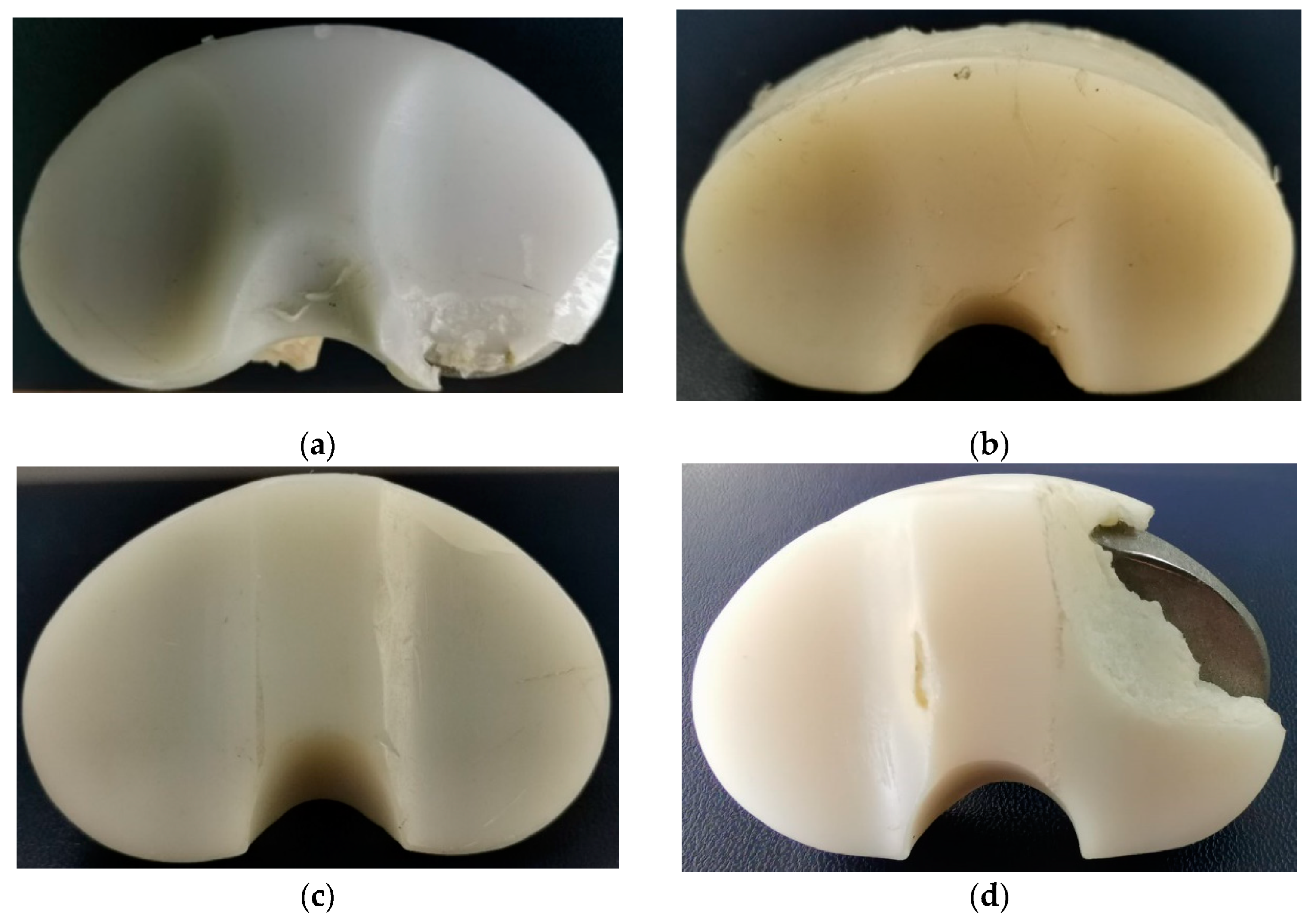
In Figure 3, the four investigated samples are presented, denoted as IT1, IT2, IT3, and IT4.
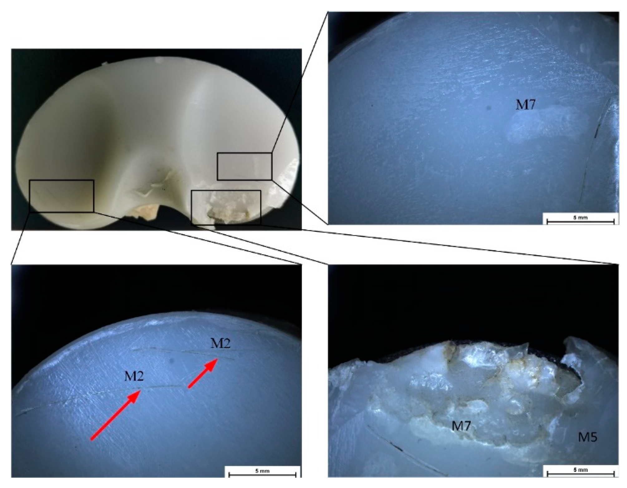
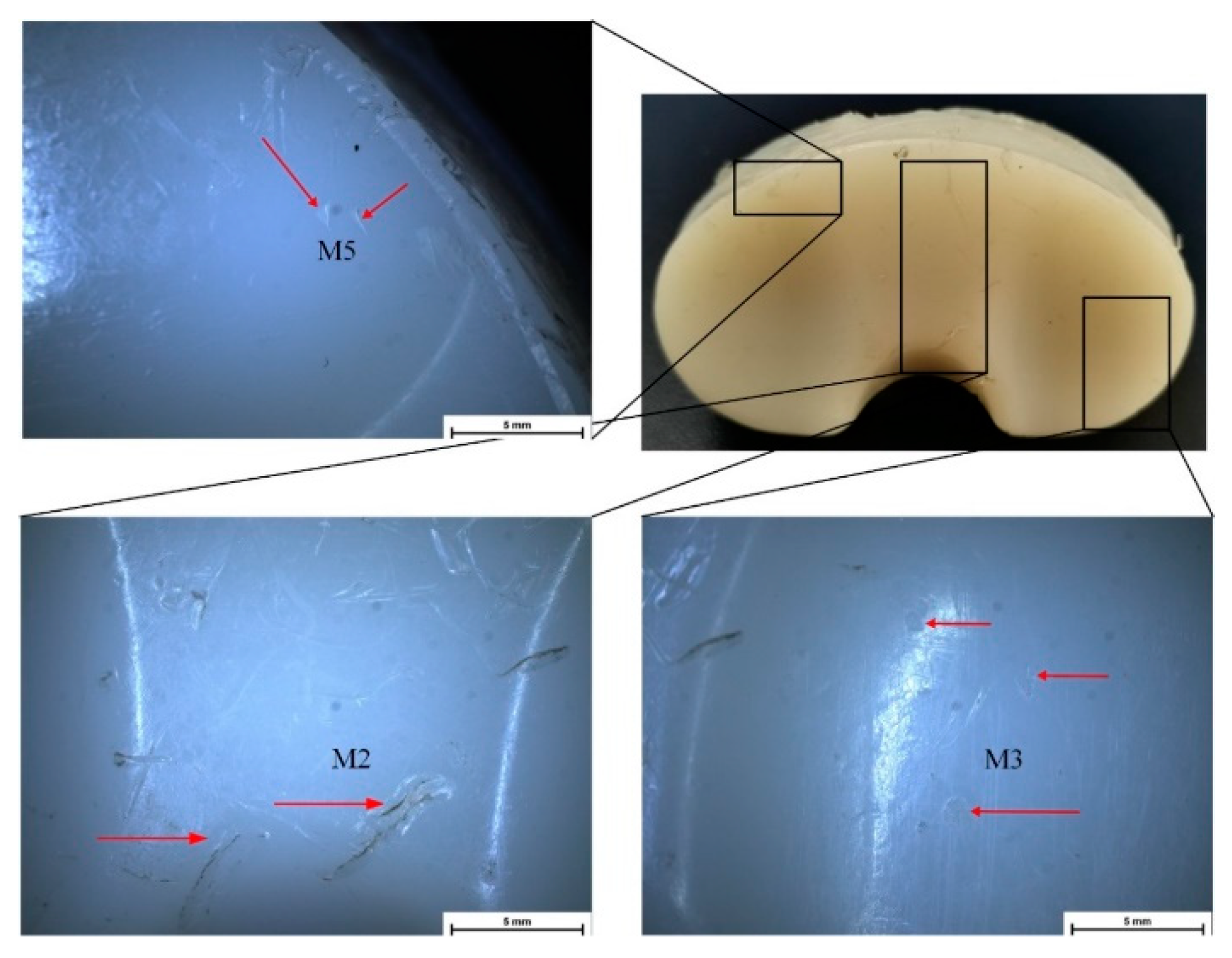
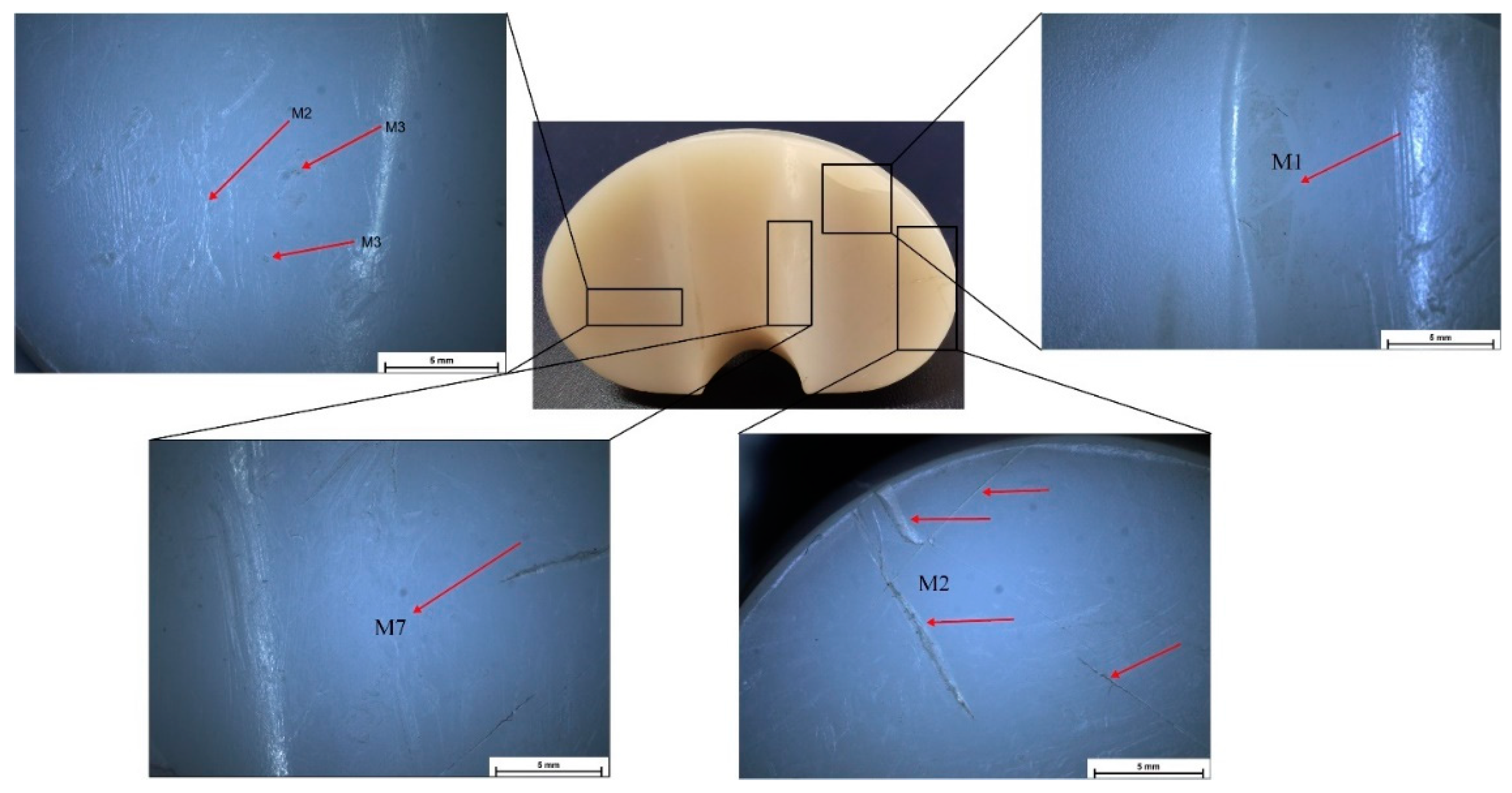
Stereomicroscopy is a much more accurate method for deterioration modes’ identification because some defects are impossible to identify in macrophotographs or with the naked eye. In Figure 4, Figure 5, Figure 6 and Figure 7, some defect images are presented, obtained with a Nikon SMZ1270i stereomicroscope. In the case of the IT1 sample, the M2, M7, and M5 deterioration modes were put in evidence. For IT2, the M2, M3, and M5 existence confirmed the macrophotography conclusion. The tibial insert IT3 exhibited M1, M2, M3, and M7 defects, and the IT4 sample, which is the most damaged, presented M1, M2, M3, M4, M5, and M7 surface degradation mechanisms.
A Philips XL 30 ESEM TMP microscope was used to characterize the surface morphology of the UHMWPE tibial inserts. The results obtained through macrophotography and stereomicroscopy were reinforced and confirmed by the scanning electron microscope images. In the case of IT1, the presence of delamination (M5), scratching (M2), and abrasive wear (M7) was clearly put in evidence (Figure 8). IT2 tibial insert exhibited scratching (M2), punctiform defects (M3), delamination (M5), and abrasive wear (M7) (Figure 9). For the IT3 sample, SEM images (Figure 10) showed punctiform defects existence (M3) and abrasive wear presence (M7), although following the results obtained through previous methods, we have to take into consideration when we compute the Hood index to also account for the scratching (M2) and burnishing (M1) modes. It can be noticed that the M1 deterioration mode cannot be easily observed through SEM investigations. Scanning electron microscopy confirmed that the IT4 component is the most damaged one, its surface exhibiting punctiform defects (M3), abrasive wear (M7), scratching (M2), and delamination (M5) (Figure 11). Through stereomicroscopy and macrophotography, we put additional evidence of the existence of burnishing (M1) and surface deformation (M4) modes.
Through SEM microscopy, the retrieved tibial component surface can be investigated at a micrometric level. This type of analysis is very accurate. However, some deformation modes, such as burnishing and surface deformation, cannot be observed. Consequently, this method must always be combined with macrophotography and/or stereomicroscopy to obtain a precise result and an exact computation of the Hood index. From the SEM images, it can be concluded that debris of PMMA cement or bone are absent. This deterioration mode can be put in evidence only through scanning electron microscopy because, in the case of the macroscopic observations, it is impossible to be observed.
In Table 2 are presented the Hood index values computed based on experimental observations for the four retrieved tibial inserts.
From Table 2, it can be noticed that the tibial insert surface deterioration varies from patient to patient, being influenced by different factors such as insert material, the shape of the investigated component, patient height and weight, the articular range of motions during daily activities, and surgical technique regarding the implant alignment and viable soft tissue quantity. Many of the variables mentioned above cannot be controlled, so developing new high-quality biomaterials with an increased wear resistance is seen as a worldwide necessity.
Usually, the polyethylene insert deteriorates due to lower interactions between polymer chains by comparing it with those determined in the case of atoms’ interactions in metallic or ceramic materials [61,74]. We have found for our retrieved UHMWPE inserts that delamination (M5) is the most important deterioration mode that is in good agreement with the literature [78,79]. Delamination is found for three samples, being much more pronounced in the case of the IT4 sample. We can observe that M5 is present in four sections and in three zones, to which we have attributed the maximum number of points. Burnishing (M1) is a surface deterioration mode that can be seen only in the case of IT3 and IT4 samples. Many scratching (M2) defects can be observed for all the samples, and their position varies from sample to sample. The most pronounced scratching is noticed for the IT3 sample in sections six and seven. Punctiform defects (M3) are present for IT2, IT3, and IT4 samples, and surface deformation (M4) is visible for IT1 and IT4 samples, being more accentuated in the case of the last sample. Polyethylene abrasive wear (M7) is a deterioration mode that was observed for all the samples with maximum allocated points for the IT4 sample in three sections. Cement or bone debris (M6) are absent because SEM micrographs did not show their existence.
By analyzing the Hood index values presented in Table 2, we can conclude that the IT4 sample is the most damaged one. In this case, we detected five deterioration modes with a Hood score of 32. Samples IT1 and IT2 have a Hood index of 9 and IT3 of 10.
Nowadays, it is estimated that all total knee implants have a survival rate of about 95% after 10 years post-surgical intervention, 88.7% after 15 years, and 82% after 25 years [80]. The life span of polyethylene components is limited to a maximum of 20 years [81,82]. Thus, the main failure reason in TKA is exclusively due to UHMWPE particles that appear due to wear and oxidation mechanisms [83,84]. This phenomenon is seen in the young and active population, and many cases of premature implant failure due to polymer particle-induced osteolysis are reported in the literature [85,86].

3.2. Oxidative Degradation Analysis

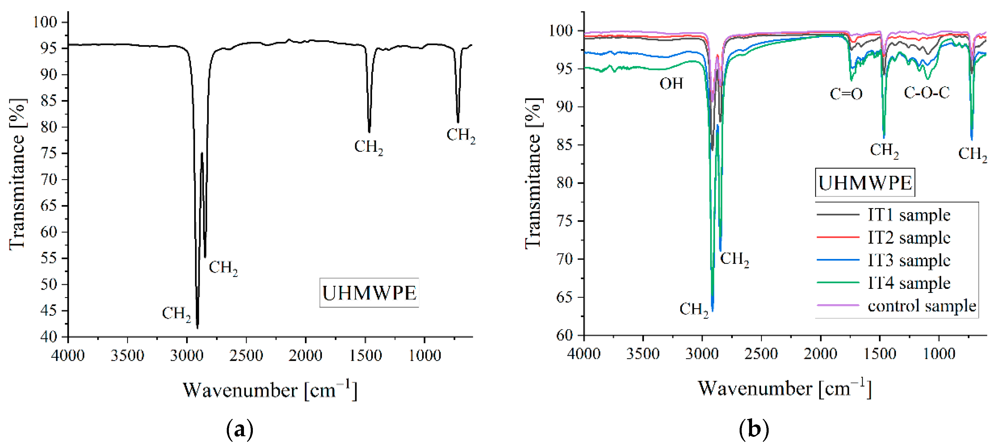
Oxidative degradation of the UHMWPE represents one of the main failure reasons for total knee implants [62,87]. This process can estimate the material’s life expectancy in accordance with international standards [88,89]. The average life span of UHMWPE tibial inserts is estimated at 15–20 years, being strongly influenced by its deterioration grade [67]. Fourier-transform infrared spectroscopy (FTIR) represents the experimental method of choice for the oxidation process identification in polymeric materials.
The oxidation mechanism of short-chain hydrocarbons such as UHMWPE is made according to the Bolland cycle [88]. FTIR analysis can identify all the functional groups that correspond to the Bolland cycle and offers information regarding the chemical bond vibrations. In order to accurately estimate the oxidation grade of the samples, we have introduced a control specimen that was never used in the patient body.
The oxidation process of UHMWPE can be quantified based on FTIR experiments through a standard protocol [89,90]. The dimensionless oxidation index (OI) is computed based on a normalization procedure that uses the height or the area of the peak associated with the carbonyl vibration (C=O) placed at a wavenumber of about 1720 cm−1. There are two standard protocols described, which use the normalization of the quantities mentioned above with the peak height or area placed at 1370 cm−1 or 2022 cm−1 [90,91]. These two reference peaks are associated with UHMWPE material’s crystalline or amorphous phase. As the polyethylene oxidates after the irradiation manufacturing step, the broken molecular chains recrystallize, and as a direct consequence, the mass density and crystallinity grade increase. It can be concluded that the material’s crystalline and amorphous phases evolve since the polymer degrades and the absorption peaks associated with these states become modified.
The spectra were determined in the range of 4000 cm−1 to 600 cm−1 at a resolution of 4 cm−1 after 64 scans. Firstly, a background measurement was performed for each sample to eliminate environmental influences such as carbon dioxide content or air humidity. We cut small parts from the control and retrieved tibial inserts with a plane surface because in our measurements it was involved an attenuated total reflectance accessory (ATR) that permits spectra measurements without additional surface treatments.
In Figure 12, the standard ideal spectrum for UHMWPE [92] and the spectra obtained for control and retrieved samples are presented.
In Table 3, the wavenumbers that correspond to the peak identified in the FTIR spectra and the reference wavenumber used for the oxidation index computation are presented.
The oxidation index was calculated as the ratio between the area of the carbonyl peak (AC=O), determined for each sample, and the area that corresponds to the reference peak placed at about 1370 cm−1 (Aref) (Equation (1)).
OI = AC=O/Aref,
The areas mentioned above were computed with the Spectra Analysis software. The obtained results are shown in Table 4.
It can be concluded that the control sample has the lowest oxidation index since the IT4 sample has the highest value. This fact is in good accordance with the Hood index computation because this specimen had the most pronounced surface deterioration. Although the IT2 sample exhibited a reduced Hood index value, its oxidation index is high. This fact shows the importance of FTIR analysis because, in some cases, the overall material deterioration cannot be identified only with visual investigation methods. The samples IT1 and IT3 have similar oxidation and Hood index values.
The FTIR analysis permits the crystallinity index computation based on an empirical formula developed by Zerbi et al. [93] that provides the amorphous phase content according to Equation (2).
X = (|1 − Ia/Ib|/1.233)/(1 + Ia/Ib) × 100,
where Ia and Ib correspond to the absorption intensities of the peaks localized at 1474 cm−1 and 1464 cm−1. The constant value of 1.233 is given by a relationship between peaks that characterize the crystalline phase of the material. Based on the Spectra Measurements software and deconvolution function, which gives us an adequate tool for separating of the overlap peaks from the FTIR spectra, we have identified the height of the peaks mentioned above and computed the amorphous substance content for each sample (Table 5).
Low content of amorphous substance is equivalent to a high crystallinity grade. It is well known that the crystallinity index increases while the UHMWPE degrades due to the oxidation process. It can be noticed that the IT4 sample has the lowest amorphous substance percent and the highest value of the oxidation index (Table 4). In the control sample case, we could not identify the peak placed at 1474 cm−1 after the deconvolution procedure, so Equation (2) cannot be applied. Otherwise, the peak at 1464 cm−1 was visible in the FTIR spectrum. We have concluded that this observation is in good agreement with the low value of the oxidation index.

3.3. Mechanical Characterization

Following the procedure described in [54,57], the initial stiffness is proportional to the UHMWPE Young’s modulus (E). The initial peak load represents the maximum load value extracted from the load-displacement curve. The ultimate load and displacement are the last point coordinates determined before the material fracture. The work to failure is computed as the area placed under the load-displacement variation, and it is a quantity related to the material toughness [39]. In Figure 13, the load-displacement variations obtained for the samples prepared from IT1, IT2, IT3, IT4, and control UHMWPE tibial inserts are presented.
A correlation between the oxidation index of the material and the mechanical properties determined through the small punch test can be noticed. Young’s modulus increases directly proportional to the oxidation index. The initial peak load, ultimate load, and ultimate displacement decrease when the oxidation state of the UHMWPE sample increases. From Figure 13 it can be observed that all the tested samples exhibit the classical load-displacement curve shape that shows a typical behavior of UHMWPE [54,57]. Above the elastic region, the dependence load versus displacement presents an initial peak load that appears because of sample bending. This peak is followed by a stretching phase. The ultimate strength and toughness decrease is usually associated with high surface deterioration. The toughness has an important contribution to reducing the fatigue resistance of UHMWPE tibial inserts after gamma irradiation [54]. The control sample has the lowest value of Young’s modulus with an average of 987 MPa and the highest initial peak load, ultimate load, and displacement values. The sample IT4 has the highest elasticity modulus (average value of 1300 MPa) and the lowest values of other mechanical parameters, which is explained by a high value of the oxidation index.
In Table 6, the average values and standard deviations for the mechanical quantities are presented.
As future directions and stabilization against oxidation phenomenon, the crosslinking process must be carefully controlled and optimized without reducing the material’s mechanical properties and with increased wear resistance. Moreover, the use of vitamin E prevents the oxidation that is associated with free radicals. Today the ASTM Standard F 2695 2007 [94] permits the development of crosslinked UHMWPE inserts, which have added vitamin E before the irradiation procedure, during extrusion or molding steps, or using diffusion after the irradiation. The main drawbacks consist of the fact that the crosslinking is suppressed to a minimum amount during irradiation and the difficulty in controlling the concentration and distribution of vitamin E. Other methods such as surface texturing [95], surface coating, ion beam surface modification, photolithography and nanoimprint lithography [96], or the use of reinforcing particles that are combined with UHMWPE to increase the mechanical properties and to reduce the wear rates such as micro and nano ZnO particle [97], zeolite particles [98], Zr particles [99], and carbon-based materials [96] were proposed in the literature.

4. Conclusions

UHMWPE represents the golden standard in knee arthroplasty for the tibial insert manufacturers. In the paper, we found a good correlation between microscopical methods used for wear analysis, SEM results adding some valuable information regarding the absence of cement or bone debris in the retrieved tibial components. We have identified all surface deterioration mechanisms, such as burnishing, scratching, punctiform defects, surface deformation, delamination, and abrasive wear.
The deterioration mechanism in UHMWPE tibial inserts is associated with the oxidation phenomenon due to the corrosive medium inside the human body [89,93]. We have computed UHMWPE’s oxidation index and amorphous content and found a direct correlation between them. The mechanical properties were investigated, and it was concluded that the oxidation phenomenon is directly linked to a deterioration of the UHMWPE tibial insert mechanical properties. Young’s modulus increased in the case of samples with high oxidation index values since the ultimate load and displacement decreased.
Retrieval analysis provides the specialists with valuable information to improve the biomaterial properties and, in some cases, modify the prosthesis design. We have concluded that all the retrieved samples exhibited surface deterioration and oxidation of the material, and the main reason for implant failure was osteolysis due to UHMWPE particle emission.
As for future directions and stabilization against the oxidation phenomenon, the crosslinking process must be carefully controlled and optimized without reducing the material’s mechanical properties and with increased wear resistance.

Author Contributions

Conceptualization, V.M., A.A., A.S. and I.A.; methodology, V.M., G.P., I.A., A.-I.B. and M.B.C.; software, G.P., A.A., S.L. and A.S.; validation, V.M., I.A. and A.A.; investigation, V.M., A.-I.B., A.A., S.L., M.M., M.B.C. and M.N.; resources, M.M., A.A., G.P., C.P. and M.N.; data curation, V.M., A.A., I.A., G.P., C.P. and S.L.; writing—original draft preparation, V.M. and G.P.; writing—review and editing, I.A. and A.A. All authors have read and agreed to the published version of the manuscript.

Funding

This work was supported by a grant of the Romanian Ministry of Education and Research, CNCS-UEFISCDI, project number PN-III-P4-ID-PCE-2020-2591, within PNCDI III. In addition, financial support from the Competitiveness Operational Program 2014-2020, action 1.1.3: creating synergies with RDI actions of the EU’s HORIZON 2020 framework program and other international RDI programs, MySMIS Code 108792, acronym project “UPB4H”, financed by contract: 250/11.05.2020 is gratefully acknowledged.

Institutional Review Board Statement

Colentina Clinical Hospital, Bucharest, Romania; Approval code: 125, Approval date: 27 April 2022.

Informed Consent Statement

Informed consent was obtained from all subjects involved in the study.

Data Availability Statement

Not applicable.

Conflicts of Interest

The authors declare no conflict of interest.

References

  1. Maradit, K.H.; Larson, D.R.; Crowson, C.S.; Kremers, W.K.; Washington, R.E.; Steiner, C.A.; Jiranek, W.A.; Berry, D.J. Prevalence of Total Hip and Knee Replacement in the United States. J. Bone Jt. Surg.-Am. Vol. 2015, 97, 1386–1397. [Google Scholar] [CrossRef] [Green Version]
  2. Kurtz, S.; Ong, K.; Lau, E.; Mowat, F.; Halpern, M. Projections of Primary and Revision Hip and Knee Arthroplasty in the United States from 2005 to 2030. J. Bone Joint Surg. Am. 2007, 89, 780–785. [Google Scholar] [CrossRef]
  3. Gibon, E.; Goodman, M.J.; Goodman, S.B. Patient Satisfaction After Total Knee Arthroplasty. Orthop. Clin. N. Am. 2017, 48, 421–431. [Google Scholar] [CrossRef]
  4. Beswick, A.D.; Wylde, V.; Gooberman-Hill, R.; Blom, A.; Dieppe, P. What Proportion of Patients Report Long-Term Pain after Total Hip or Knee Replacement for Osteoarthritis? A Systematic Review of Prospective Studies in Unselected Patients. BMJ Open 2012, 2, e000435. [Google Scholar] [CrossRef]
  5. Wylde, V.; Hewlett, S.; Learmonth, I.D.; Dieppe, P. Persistent Pain after Joint Replacement: Prevalence, Sensory Qualities, and Postoperative Determinants. Pain 2011, 152, 566–572. [Google Scholar] [CrossRef]
  6. Carbonell-Blasco, P.; Martín-Martínez, J.M.; Antoniac, I.V. Synthesis and Characterization of Polyurethane Sealants Containing Rosin Intended for Sealing Defect in Annulus for Disc Regeneration. Int. J. Adhes. Adhes. 2013, 42, 11–20. [Google Scholar] [CrossRef]
  7. Bhandari, M.; Smith, J.; Miller, L.E.; Block, J.E. Clinical and Economic Burden of Revision Knee Arthroplasty. Clin. Med. Insights Arthritis Musculoskelet. Disord. 2012, 5, 10859. [Google Scholar] [CrossRef] [Green Version]
  8. Nica, M.; Cretu, B.; Ene, D.; Antoniac, I.; Gheorghita, D.; Ene, R. Failure Analysis of Retrieved Osteosynthesis Implants. Materials 2020, 13, 1201. [Google Scholar] [CrossRef] [Green Version]
  9. Iulian, A.; Cosmin, S.; Aurora, A. Adhesion Aspects in Biomaterials and Medical Devices. J. Adhes. Sci. Technol. 2016, 30, 1711–1715. [Google Scholar] [CrossRef] [Green Version]
  10. Ferche, O.; Moldoveanu, A.; Cinteza, D.; Toader, C.; Moldoveanu, F.; Voinea, A.; Taslitchi, C. From Neuromotor Command to Feedback: A Survey of Techniques for Rehabilitation through Altered Perception. In Proceedings of the 2015 E-Health and Bioengineering Conference (EHB), Iasi, Romania, 19–21 November 2015; pp. 1–4. [Google Scholar]
  11. Miculescu, M.; Ion, O.A. Regulation and Certification of (Bio)Medical Engineers: A Case Study on Romania. Int. J. Environ. Res. Public Health 2022, 19, 9004. [Google Scholar] [CrossRef]
  12. Antoniac, I.; Laptoiu, D.; Tecu, C.; Milea, C.; Gradinaru, S. Synthetic Materials for Osteochondral Tissue Engineering. Adv. Exp. Med. Biol. 2018, 1058, 31–52. [Google Scholar] [CrossRef]
  13. Bane, M.; Miculescu, F.; Blajan, A.I.; Dinu, M.; Antoniac, I. Failure Analysis of Some Retrieved Orthopedic Implants Based on Materials Characterization. Solid State Phenom. 2012, 188, 114–117. [Google Scholar] [CrossRef]
  14. Antoniac, I.V.; Miculescu, F.; Laptoiu, D.; Antoniac, A.; Niculescu, M.; Grecu, D. Retrieval Analysis of Hip Prostheses. In Handbook of Bioceramics and Biocomposites; Antoniac, I.V., Ed.; Springer International Publishing: Cham, Switzerland, 2016; pp. 901–933. ISBN 978-3-319-12460-5. [Google Scholar]
  15. Buechel, F.F.; Pappas, M.J. Properties of Materials Used in Orthopaedic Implant Systems. In Principles of Human Joint Replacement: Design and Clinical Application; Buechel, F.F., Pappas, M.J., Eds.; Springer: Berlin/Heidelberg, Germany, 2012; pp. 1–35. ISBN 978-3-642-23011-0. [Google Scholar]
  16. ODEP. Available online: https://www.odep.org.uk/ (accessed on 19 April 2022).
  17. Vertullo, C.J.; Lewis, P.L.; Graves, S.; Kelly, L.; Lorimer, M.; Myers, P. Twelve-Year Outcomes of an Oxinium Total Knee Replacement Compared with the Same Cobalt-Chromium Design: An Analysis of 17,577 Prostheses from the Australian Orthopaedic Association National Joint Replacement Registry. J. Bone Jt. Surg. Am. 2017, 99, 275–283. [Google Scholar] [CrossRef] [Green Version]
  18. Bonnheim, N.B.; Citters, D.W.V.; Ries, M.D.; Pruitt, L.A. Oxidized Zirconium Components Maintain a Smooth Articular Surface Except Following Hip Dislocation. J. Arthroplast. 2021, 36, 1437–1444. [Google Scholar] [CrossRef]
  19. Purcell, A.; Buckner, S.; Brindley, G.; Grimes, J. A Unique Case of Extra-Articular Extravasation of Metal Into the Lower Leg Resulting From Oxidized Zirconium Wear Particles From Total Knee Arthroplasty. Arthroplast. Today 2020, 6, 988–992. [Google Scholar] [CrossRef]
  20. Piconi, C.; Maccauro, G.; Muratori, F.; Brach Del Prever, E. Alumina and Zirconia Ceramics in Joint Replacements. J. Appl. Biomater. Biomech. 2003, 1, 19–32. [Google Scholar]
  21. Nakamura, S.; Minoda, Y.; Nakagawa, S.; Kadoya, Y.; Takemura, S.; Kobayashi, A.; Mizokawa, S.; Ohta, Y.; Takahashi, S.; Yamamura, K.; et al. Clinical Results of Alumina Medial Pivot Total Knee Arthroplasty at a Minimum Follow-up of 10 years. Knee 2017, 24, 434–438. [Google Scholar] [CrossRef]
  22. Tsukamoto, R.; Williams, P.A.; Clarke, I.C.; Pezzotti, G.; Shoji, H.; Akagi, M.; Yamamoto, K. Y-TZP Zirconia Run against Highly Crosslinked UHMWPE Tibial Inserts: Knee Simulator Wear and Phase-Transformation Studies. J. Biomed. Mater. Res. Part B Appl. Biomater. 2008, 86B, 145–153. [Google Scholar] [CrossRef]
  23. Bergschmidt, P.; Bader, R.; Ganzer, D.; Hauzeur, C.; Lohmann, C.; Rüther, W.; Tigani, D.; Rani, N.; Prats, F.L.; Zorzi, C.; et al. Ceramic Femoral Components in Total Knee Arthroplasty—Two Year Follow-Up Results of an International Prospective Multi-Centre Study. Open Orthop. J. 2012, 6, 172–178. [Google Scholar] [CrossRef] [Green Version]
  24. Affatato, S.; Erani, P.; Fersini, M.; Contaldi, V.; Terrizzi, A.R.; Licciulli, A. Preliminary In Vitro Wear Assessment of Ceramic Cemented Femoral Components Coupled with Polyethylene Menisci. Materials 2021, 14, 2112. [Google Scholar] [CrossRef]
  25. Bioceramics and Biocomposites: From Research to Clinical Practice|Wiley. Available online: https://www.wiley.com/en-us/Bioceramics+and+Biocomposites%3A+From+Research+to+Clinical+Practice-p-9781119049340 (accessed on 27 August 2022).
  26. Panayotov, I.V.; Orti, V.; Cuisinier, F.; Yachouh, J. Polyetheretherketone (PEEK) for Medical Applications. J. Mater. Sci. Mater. Med. 2016, 27, 118. [Google Scholar] [CrossRef]
  27. Kurtz, S.M.; Devine, J.N. PEEK Biomaterials in Trauma, Orthopedic, and Spinal Implants. Biomaterials 2007, 28, 4845–4869. [Google Scholar] [CrossRef] [PubMed] [Green Version]
  28. Ha, S.W.; Kirch, M.; Birchler, F.; Eckert, K.L.; Mayer, J.; Wintermantel, E.; Sittig, C.; Pfund-Klingenfuss, I.; Textor, M.; Spencer, N.D.; et al. Surface Activation of Polyetheretherketone (PEEK) and Formation of Calcium Phosphate Coatings by Precipitation. J. Mater. Sci. Mater. Med. 1997, 8, 683–690. [Google Scholar] [CrossRef] [PubMed]
  29. Cai, Z.; Qu, X.; Zhao, Y.; Yuan, Z.; Zheng, L.; Long, T.; Yao, Q.; Yue, B.; Wang, Y. Preliminary Study on Immediate Postoperative CT Images and Values of the Modular Polyetheretherketone Based Total Knee Arthroplasty: An Observational First-in-Human Trial. Front. Surg. 2022, 9, 809699. [Google Scholar] [CrossRef]
  30. Ran, J.; Jiang, F.; Sun, X.; Chen, Z.; Tian, C.; Zhao, H. Microstructure and Mechanical Properties of Ti-6Al-4V Fabricated by Electron Beam Melting. Crystals 2020, 10, 972. [Google Scholar] [CrossRef]
  31. Saboori, A.; Gallo, D.; Biamino, S.; Fino, P.; Lombardi, M. An Overview of Additive Manufacturing of Titanium Components by Directed Energy Deposition: Microstructure and Mechanical Properties. Appl. Sci. 2017, 7, 883. [Google Scholar] [CrossRef] [Green Version]
  32. Trabecular MetalTM Technology|Zimmer Biomet EU. Available online: https://www.zimmerbiomet.eu/en/products-and-solutions/specialties/hip/trabecular-metal-technology.html (accessed on 16 April 2022).
  33. Meneghini, R.M.; de Beaubien, B.C. Early Failure of Cementless Porous Tantalum Monoblock Tibial Components. J. Arthroplast. 2013, 28, 1505–1508. [Google Scholar] [CrossRef]
  34. First In-Patient TKA With PEEK-OPTIMA Femoral Component|Orthopedics This Week. Available online: https://ryortho.com/breaking/first-in-patient-tka-with-peek-optima-femoral-component/ (accessed on 19 April 2022).
  35. Dalury, D.F.; Pomeroy, D.L.; Gonzales, R.A.; Gruen, T.A.; Adams, M.J.; Empson, J.A. Midterm Results of All-Polyethylene Tibial Components in Primary Total Knee Arthroplasty. J. Arthroplast. 2009, 24, 620–624. [Google Scholar] [CrossRef]
  36. Meier, E.; Gelse, K.; Trieb, K.; Pachowsky, M.; Hennig, F.F.; Mauerer, A. First Clinical Study of a Novel Complete Metal-Free Ceramic Total Knee Replacement System. J. Orthop. Surg. Res. 2016, 11, 21. [Google Scholar] [CrossRef] [Green Version]
  37. Bistolfi, A.; Giustra, F.; Bosco, F.; Sabatini, L.; Aprato, A.; Bracco, P.; Bellare, A. Ultra-High Molecular Weight Polyethylene (UHMWPE) for Hip and Knee Arthroplasty: The Present and the Future. J. Orthop. 2021, 25, 98–106. [Google Scholar] [CrossRef]
  38. Sobieraj, M.C.; Rimnac, C.M. Ultra High Molecular Weight Polyethylene: Mechanics, Morphology, and Clinical Behavior. J. Mech. Behav. Biomed. Mater. 2009, 2, 433–443. [Google Scholar] [CrossRef] [PubMed]
  39. Bracco, P.; Bellare, A.; Bistolfi, A.; Affatato, S. Ultra-High Molecular Weight Polyethylene: Influence of the Chemical, Physical and Mechanical Properties on the Wear Behavior. A Review. Materials 2017, 10, 791. [Google Scholar] [CrossRef]
  40. Currier, B.H.; Currier, J.H.; Franklin, K.J.; Mayor, M.B.; Reinitz, S.D.; Van Citters, D.W. Comparison of Wear and Oxidation in Retrieved Conventional and Highly Cross-Linked UHMWPE Tibial Inserts. J. Arthroplast. 2015, 30, 2349–2353. [Google Scholar] [CrossRef]
  41. Rojanasopondist, P.; Muratoglu, O. Polyethylene in Total Knee Arthroplasty. Available online: https:///musculoskeletalkey.com/polyethylene-in-total-knee-arthroplasty/ (accessed on 18 April 2022).
  42. Matz, J.; Lanting, B.A.; Howard, J.L. Understanding the Patellofemoral Joint in Total Knee Arthroplasty. Can. J. Surg. 2019, 62, 57–65. [Google Scholar] [CrossRef] [Green Version]
  43. Gaman, L.; Radoi, M.P.; Delia, C.E.; Luzardo, O.P.; Zumbado, M.; Rodríguez-Hernández, Á.; Stoian, I.; Gilca, M.; Boada, L.D.; Henríquez-Hernández, L.A. Concentration of Heavy Metals and Rare Earth Elements in Patients with Brain Tumours: Analysis in Tumour Tissue, Non-Tumour Tissue, and Blood. Int. J. Environ. Health Res. 2021, 31, 741–754. [Google Scholar] [CrossRef]
  44. Antoniac, I.; Negrusoiu, M.; Mardare, M.; Socoliuc, C.; Zazgyva, A.; Niculescu, M. Adverse Local Tissue Reaction after 2 Revision Hip Replacements for Ceramic Liner Fracture. Medicine 2017, 96, e6687. [Google Scholar] [CrossRef]
  45. Papacocea, M.T.; Badarau, I.A.; Radoi, M.; Papacocea, I.R. The Predictive Role of Biochemical Plasma Factors in Patients with Severe Traumatic Brain Injuries. Rev. Chim. 2019, 70, 1754–1757. [Google Scholar] [CrossRef]
  46. Zhang, L.; Haddouti, E.-M.; Welle, K.; Burger, C.; Kabir, K.; Schildberg, F.A. Local Cellular Responses to Metallic and Ceramic Nanoparticles from Orthopedic Joint Arthroplasty Implants. Int. J. Nanomed. 2020, 15, 6705–6720. [Google Scholar] [CrossRef]
  47. Marinescu, R.; Antoniac, I.; Laptoiu, D.; Antoniac, A.; Grecu, D. Complications Related to Biocomposite Screw Fixation in ACL Reconstruction Based on Clinical Experience and Retrieval Analysis. Mater. Plast. 2015, 52, 340–344. [Google Scholar]
  48. ASTM International-ASTM F0561-97-Practice for Retrieval and Analysis of Implanted Medical Devices, and Associated Tissues. Available online: https://www.astm.org/f0561-97.html (accessed on 27 August 2022).
  49. Antoniac, I.V.; Burcea, M.; Ionescu, R.D.; Balta, F. IOL’s Opacification: A Complex Analysis Based on the Clinical Aspects, Biomaterials Used and Surface Characterization of Explanted IOL’s. Mater. Plast. 2015, 5, 109–112. [Google Scholar]
  50. Jain, R.; Kapoor, D. The Dynamic Interface: A Review. J. Int. Soc. Prev. Community Dent. 2015, 5, 354–358. [Google Scholar] [CrossRef] [Green Version]
  51. Branemark, P.; Albrektsson, T. Tissue Integrated Prosthesis Osseointegration. In Clinical Dentistry; Quintessence Publishing Co. Inc.: Chicago, IL, USA, 1985. [Google Scholar]
  52. Albrektsson: The Interface Zone of Inorganic ImplantsIn. Available online: https://scholar.google.com/scholar_lookup?journal=Annals+of+Biomedical+Engineering&title=The+interface+of+inorganic+implants+in+vivo;+titanium+implants+in+bone&author=T+Albrektsson&author=PI+Br%C3%A5nemark&author=HA+Hansson&author=B+Kasemo&author=K+Larsson&volume=11&publication_year=1983&pages=1-27& (accessed on 1 October 2022).
  53. Liza, S.; Haseeb, A.S.M.A.; Abbas, A.A.; Masjuki, H.H. Failure Analysis of Retrieved UHMWPE Tibial Insert in Total Knee Replacement. Eng. Fail. Anal. 2011, 18, 1415–1423. [Google Scholar] [CrossRef]
  54. Kurtz, S.M.; Rimnac, C.M.; Pruitt, L.; Jewett, C.W.; Goldberg, V.; Edidin, A.A. The Relationship between the Clinical Performance and Large Deformation Mechanical Behavior of Retrieved UHMWPE Tibial Inserts. Biomaterials 2000, 21, 283–291. [Google Scholar] [CrossRef]
  55. Kurtz, S.M.; Foulds, J.R.; Jewett, C.W.; Srivastav, S.; Edidin, A.A. Validation of a Small Punch Testing Technique to Characterize the Mechanical Behaviour of Ultra-High-Molecular-Weight Polyethylene. Biomaterials 1997, 18, 1659–1663. [Google Scholar] [CrossRef]
  56. Edidin, A.A.; Kurtz, S.M. Influence of Mechanical Behavior on the Wear of 4 Clinically Relevant Polymeric Biomaterials in a Hip Simulator. J. Arthroplast. 2000, 15, 321–331. [Google Scholar] [CrossRef]
  57. Edidin, A.A. Development and Application of the Small Punch Test to UHMWPE. In UHMWPE Biomaterials Handbook; Elsevier: Amsterdam, The Netherlands, 2015; pp. 739–752. [Google Scholar]
  58. Cho, C.-H.; Murakami, T.; Sawae, Y. Influence of Microscopic Surface Asperities on the Wear of Ultra-High Molecular Weight Polyethylene in a Knee Prosthesis. Proc. Inst. Mech. Eng. H 2010, 224, 515–529. [Google Scholar] [CrossRef] [PubMed]
  59. Medel, F.J.; Kurtz, S.M.; Parvizi, J.; Klein, G.R.; Kraay, M.J.; Rimnac, C.M. In Vivo Oxidation Contributes to Delamination but Not Pitting in Polyethylene Components for Total Knee Arthroplasty. J. Arthroplast. 2011, 26, 802–810. [Google Scholar] [CrossRef] [Green Version]
  60. Berry, D.J.; Currier, B.H.; Mayor, M.B.; Collier, J.P. Gamma-Irradiation Sterilization in an Inert Environment: A Partial Solution. Clin. Orthop. Relat. Res. 2012, 470, 1805–1813. [Google Scholar] [CrossRef] [Green Version]
  61. Cerquiglini, A.; Henckel, J.; Hothi, H.; Moser, L.B.; Eskelinen, A.; Hirschmann, M.T.; Hart, A.J. Retrieval Analysis of Contemporary Antioxidant Polyethylene: Multiple Material and Design Changes May Decrease Implant Performance. Knee Surg. Sport. Traumatol. Arthrosc. 2019, 27, 2111–2119. [Google Scholar] [CrossRef]
  62. Tone, S.; Hasegawa, M.; Naito, Y.; Pezzotti, G.; Sudo, A. Raman Spectroscopy Reveals Creep and Wear Rate of E-Beam-Sterilized Conventional UHMWPE Tibial Inserts. J. Mech. Behav. Biomed. Mater. 2020, 110, 103902. [Google Scholar] [CrossRef]
  63. Granquist, E.J.; Bouloux, G.; Dattilo, D.; Gonzalez, O.; Louis, P.J.; McCain, J.; Sinn, D.; Szymela, V.; Warner, M.; Quinn, P.D. Outcomes and Survivorship of Biomet Microfixation Total Joint Replacement System: Results From an FDA Postmarket Study. J. Oral. Maxillofac. Surg. 2020, 78, 1499–1508. [Google Scholar] [CrossRef]
  64. Hood, R.W.; Wright, T.M.; Burstein, A.H. Retrieval Analysis of Total Knee Prostheses: A Method and Its Application to 48 Total Condylar Prostheses. J. Biomed. Mater. Res. 1983, 17, 829–842. [Google Scholar] [CrossRef]
  65. EN 10371:2021-Metallic Materials-Small Punch Test Method. Available online: https://standards.iteh.ai/catalog/standards/cen/3bf5f9ce-4532-4a8b-ba9f-c7507d246312/en-10371-2021 (accessed on 27 September 2022).
  66. West, J.A.; Scudday, T.; Anderson, S.; Amin, N.H. Clinical Outcomes and Patient Satisfaction after Total Knee Arthroplasty: A Follow-up of the First 50 Cases by a Single Surgeon. J. Int. Med. Res. 2019, 47, 1667–1676. [Google Scholar] [CrossRef]
  67. Grecu, D.; Antoniac, I.; Trante, O.; Niculescu, M.; Lupescu, O. Failure Analysis of Retrieved Polyethylene Insert in Total Knee Replacement. Biomaterials 2016, 6, 12–15. [Google Scholar]
  68. Postler, A.; Lützner, C.; Beyer, F.; Tille, E.; Lützner, J. Analysis of Total Knee Arthroplasty Revision Causes. BMC Musculoskelet. Disord. 2018, 19, 55. [Google Scholar] [CrossRef] [Green Version]
  69. Kandahari, A.M.; Yang, X.; Laroche, K.A.; Dighe, A.S.; Pan, D.; Cui, Q. A Review of UHMWPE Wear-Induced Osteolysis: The Role for Early Detection of the Immune Response. Bone Res. 2016, 4, 16014. [Google Scholar] [CrossRef] [Green Version]
  70. Seon, J.-K.; Park, J.-K.; Shin, Y.-J.; Seo, H.-Y.; Lee, K.-B.; Song, E.-K. Comparisons of Kinematics and Range of Motion in High-Flexion Total Knee Arthroplasty: Cruciate Retaining vs. Substituting Designs. Knee Surg. Sports Traumatol. Arthrosc. 2011, 19, 2016–2022. [Google Scholar] [CrossRef]
  71. Harato, K.; Bourne, R.B.; Victor, J.; Snyder, M.; Hart, J.; Ries, M.D. Midterm Comparison of Posterior Cruciate-Retaining versus -Substituting Total Knee Arthroplasty Using the Genesis II Prosthesis. Knee 2008, 15, 217–221. [Google Scholar] [CrossRef]
  72. Rose, R.M.; Nusbaum, H.J.; Schneider, H.; Ries, M.; Paul, I.; Crugnola, A.; Simon, S.R.; Radin, E.L. On the True Wear Rate of Ultra High-Molecular-Weight Polyethylene in the Total Hip Prosthesis. J. Bone Jt. Surg. Am. 1980, 62, 537–549. [Google Scholar] [CrossRef]
  73. Rostoker, W. The Appearances of Wear on Polyethylene--a Comparison of in Vivo and in Vitro Wear Surfaces. J. Biomed. Mater. Res. 1978, 12, 317–335. [Google Scholar] [CrossRef]
  74. Fabry, C.; Zietz, C.; Dammer, R.; Bader, R. 12 Patterns of Wear in Total Knee Replacement. In The Unhappy Total Knee Replacement: A Comprehensive Review and Management Guide; Hirschmann, M.T., Becker, R., Eds.; Springer International Publishing: Cham, Switzerland, 2015; pp. 135–145. ISBN 978-3-319-08099-4. [Google Scholar]
  75. Kim, K.T.; Lee, S.; Ko, D.O.; Seo, B.S.; Jung, W.S.; Chang, B.K. Causes of Failure after Total Knee Arthroplasty in Osteoarthritis Patients 55 Years of Age or Younger. Knee Surg. Relat. Res. 2014, 26, 13–19. [Google Scholar] [CrossRef] [Green Version]
  76. Hasandoost, L.; Rodriguez, O.; Alhalawani, A.; Zalzal, P.; Schemitsch, E.H.; Waldman, S.D.; Papini, M.; Towler, M.R. The Role of Poly(Methyl Methacrylate) in Management of Bone Loss and Infection in Revision Total Knee Arthroplasty: A Review. J. Funct. Biomater. 2020, 11, 25. [Google Scholar] [CrossRef] [Green Version]
  77. Sm, K.; Ha, G.; Jd, P. History and Systematic Review of Wear and Osteolysis Outcomes for First-Generation Highly Crosslinked Polyethylene. Clin. Orthop. Relat. Res. 2011, 469, 2262–2277. [Google Scholar] [CrossRef] [Green Version]
  78. Currier, B.H.; Currier, J.H.; Holdcroft, L.A.; Van Citters, D.W. Effectiveness of Anti-Oxidant Polyethylene: What Early Retrievals Can Tell Us. J. Biomed. Mater. Res. B Appl. Biomater. 2018, 106, 353–359. [Google Scholar] [CrossRef]
  79. Mathis, D.T.; Schmidli, J.; Hirschmann, M.T.; Amsler, F.; Henckel, J.; Hothi, H.; Hart, A. Comparative Retrieval Analysis of Antioxidant Polyethylene: Bonding of Vitamin-E Does Not Reduce in-Vivo Surface Damage. BMC Musculoskelet. Disord. 2021, 22, 1003. [Google Scholar] [CrossRef]
  80. Camacho, N.; Gonzalez, J.M.; Espinosa, D.; Mondragón, G.; Stafford, S. UHMWPE in Total Knee Arthroplasty: Successes & Failures. Rev. Colom. Mater. 2021, 16, 3–28. [Google Scholar] [CrossRef]
  81. Kunkel, S.T.; Moschetti, W.E.; Werth, P.; Fillingham, Y.; Jevsevar, D.; VanCitters, D.; Currier, J.; Currier, B.; Henderson, E. Tobacco Exposure Is Associated With Extremely Low Polyethylene Oxidation in Total Knee Arthroplasty Components. Arthroplast. Today 2021, 8, 243–246. [Google Scholar] [CrossRef]
  82. Christiner, T.; Pabbruwe, M.B.; Kop, A.M.; Parry, J.; Clark, G.; Collopy, D. Taper Corrosion and Adverse Local Tissue Reactions in Patients with a Modular Knee Prosthesis. JB JS Open Access 2018, 3, e0019. [Google Scholar] [CrossRef]
  83. Pourzal, R.; Cip, J.; Rad, E.; Laurent, M.P.; Berger, R.A.; Jacobs, J.J.; Wimmer, M.A. Joint Line Elevation and Tibial Slope Are Associated with Increased Polyethylene Wear in Cruciate-Retaining Total Knee Replacement. J. Orthop. Res. 2020, 38, 1596–1606. [Google Scholar] [CrossRef]
  84. Sisko, Z.W.; Teeter, M.G.; Lanting, B.A.; Howard, J.L.; McCalden, R.W.; Naudie, D.D.; MacDonald, S.J.; Vasarhelyi, E.M. Current Total Knee Designs: Does Baseplate Roughness or Locking Mechanism Design Affect Polyethylene Backside Wear? Clin. Orthop. Relat. Res. 2017, 475, 2970–2980. [Google Scholar] [CrossRef] [Green Version]
  85. Goldvasser, D.; Marchie, A.; Bragdon, L.K.; Bragdon, C.R.; Weidenhielm, L.; Malchau, H. Incidence of Osteolysis in Total Knee Arthroplasty: Comparison between Radiographic and Retrieval Analysis. J. Arthroplast. 2013, 28, 201–206. [Google Scholar] [CrossRef]
  86. Klutzny, M.; Singh, G.; Hameister, R.; Goldau, G.; Awiszus, F.; Feuerstein, B.; Stärke, C.; Lohmann, C.H. Screw Track Osteolysis in the Cementless Total Knee Replacement Design. J. Arthroplast. 2019, 34, 965–973. [Google Scholar] [CrossRef]
  87. Ansari, F.; Chang, J.; Huddleston, J.; Van Citters, D.; Ries, M.; Pruitt, L. Fractography and Oxidative Analysis of Gamma Inert Sterilized Posterior-Stabilized Tibial Insert Post Fractures: Report of Two Cases. Knee 2013, 20, 609–613. [Google Scholar] [CrossRef]
  88. UHMWPE Biomaterials Handbook-3rd Edition. Available online: https://www.elsevier.com/books/uhmwpe-biomaterials-handbook/kurtz/978-0-323-35401-1 (accessed on 1 June 2022).
  89. ASTM International-ASTM F2003-00-Standard Guide for Accelerated Aging of Ultra-High Molecular Weight Polyethylene | Engineering360. Available online: https://standards.globalspec.com/std/3803981/astm-f2003-00 (accessed on 9 June 2022).
  90. Azam, A.M.; Ali, A.; Khan, H.; Yasin, T.; Mehmood, M.S. Analysis of Degradation in UHMWPE a Comparative Study among the Various Commercial and Laboratory Grades UHMWPE. IOP Conf. Ser. Mater. Sci. Eng. 2016, 146, 012025. [Google Scholar] [CrossRef]
  91. Kurtz, S.M.; Muratoglu, O.K.; Gsell, R.; Greer, K.; Shen, F.W.; Cooper, C.; Buchanan, F.J.; Spiegelberg, S.; Yau, S.S.; Edidin, A.A. Interlaboratory Validation of Oxidation-Index Measurement Methods for UHMWPE after Long-Term Shelf Aging. J. Biomed. Mater. Res. 2002, 63, 15–23. [Google Scholar] [CrossRef]
  92. Li, C.; Shi, Y.; Zhang, R.; Wang, G.; Jia, J. Effect of Surface Modifications on the Properties of UHMWPE Fibres and Their Composites. e-Polymers 2019, 19, 40–49. [Google Scholar] [CrossRef]
  93. Zerbi, G.; Gallino, G.; Del Fanti, N.; Baini, L. Structural Depth Profiling in Polyethylene Films by Multiple Internal Reflection Infra-Red Spectroscopy. Polymer 1989, 30, 2324–2327. [Google Scholar] [CrossRef]
  94. Standard Specification for Ultra-High Molecular Weight Polyethylene Powder Blended With Alpha-Tocopherol (Vitamin E) and Fabricated Forms for Surgical Implant Applications. Available online: https://www.astm.org/f2695-07.html (accessed on 25 September 2022).
  95. López-Cervantes, A.; Domínguez-López, I.; Barceinas-Sánchez, J.D.O.; García-García, A.L. Effects of Surface Texturing on the Performance of Biocompatible UHMWPE as a Bearing Material during in Vitro Lubricated Sliding/Rolling Motion. J. Mech. Behav. Biomed. Mater. 2013, 20, 45–53. [Google Scholar] [CrossRef]
  96. Baena, J.C.; Wu, J.; Peng, Z. Wear Performance of UHMWPE and Reinforced UHMWPE Composites in Arthroplasty Applications: A Review. Lubricants 2015, 3, 413–436. [Google Scholar] [CrossRef] [Green Version]
  97. Chang, B.-P.; Md. Akil, H.; Bt. Md. Nasir, R. Comparative Study of Micro- and Nano-ZnO Reinforced UHMWPE Composites under Dry Sliding Wear. Wear 2013, 297, 1120–1127. [Google Scholar] [CrossRef]
  98. Chang, B.P.; Akil, H.M.; Nasir, R.M. Mechanical and Tribological Properties of Zeolite-Reinforced UHMWPE Composite for Implant Application. Procedia Eng. 2013, 68, 88–94. [Google Scholar] [CrossRef]
  99. Plumlee, K.; Schwartz, C.J. Improved Wear Resistance of Orthopaedic UHMWPE by Reinforcement with Zirconium Particles. Wear 2009, 267, 710–717. [Google Scholar] [CrossRef]
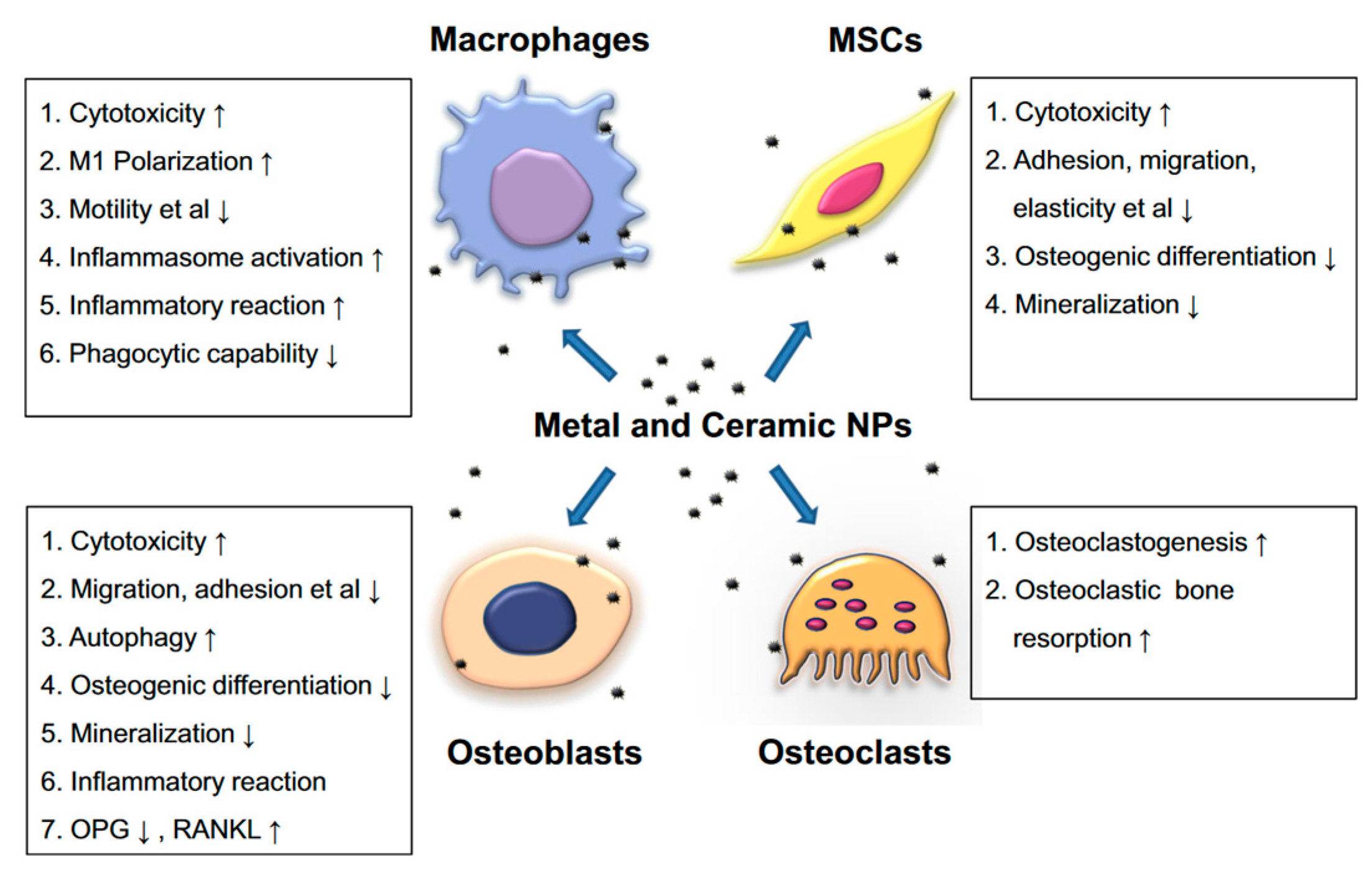
Figure 1. The particles’ drawbacks and negative effects on the cell functions at the implantation site [46].
Materials 15 07102 g001
Figure 2. The partition system proposed by Hood et al. [64] for the quantification of deterioration modes in the case of UHMWPE tibial insert of knee prostheses.
Materials 15 07102 g002
Figure 3. Macrophotographs of the UHMWPE tibial inserts retrieved from patients of Clinical Hospital Colentina. (a) IT1 sample—M2, M4, M5, M7 deterioration modes; (b) IT2 sample—M2, M3, M5, M7 deterioration modes; (c) IT3 sample—M1, M2, M3, M7 deterioration modes; (d) IT4 sample—M1, M2, M3, M4, M5, M7.
Materials 15 07102 g003
Figure 4. Stereomicroscopy images obtained in the case of IT1 sample.
Materials 15 07102 g004
Figure 5. Stereomicroscopy images obtained in the case of IT2 sample.
Materials 15 07102 g005
Figure 6. Stereomicroscopy images obtained in the case of IT3 sample.
Materials 15 07102 g006
Figure 7. Stereomicroscopy images obtained in the case of IT4 sample.
Materials 15 07102 g007
Figure 8. SEM images obtained for IT1 sample.
Materials 15 07102 g008
Figure 9. SEM images obtained for IT2 sample.
Materials 15 07102 g009
Figure 10. SEM images obtained for IT3 sample.
Materials 15 07102 g010
Figure 11. SEM images obtained for IT4 sample.
Materials 15 07102 g011
Figure 12. Spectra for the UHMWPE. (a) Standard ideal FTIR spectrum for unoxidized UHMWPE sample [92]; (b) FTIR spectra for the control and retrieved UHMWPE samples.
Materials 15 07102 g012
Figure 13. Small punch test load-displacement curves for the control and retrieved UHMWPE samples.
Materials 15 07102 g013
Table 1. Experimental samples and statistics data on patients with knee implant failure.
UHMWPE Tibial Insert DataPatient Clinical Data
Sample IDHospitalImplant Life Span [Years]ProducerPatient Age [Years]SexWeight [kg]/Height [m]KSPathologies
PreoperativePostoperative
IT1Colentina5Zimmer54M75/1.75585Diabetes, myelitis, vascular stroke, cervical spondylosis
IT2Colentina10Zimmer62F60/1.636390Diabetes, osteoporosis, cardiac insufficiency
IT3Colentina15Zimmer69M85/1.737194Schizophrenia
IT4Colentina10Zimmer65F70/1.67396Cervical spondylotic myelopathy, osteoporosis
M: male, F: female, KS: Knee Society Knee Score.
Table 2. Surface deterioration modes and Hood index computation for the retrieved UHMWPE tibial inserts.
Sample IDDeterioration ModeM1M2M3M4M5M6M7
IT1Zone-3, 7-60, 7, 8, 9-7
Grade01, 1011, 2, 1, 101
Hood score9
IT2Zone-1, 3, 8, 91, 6, 7-1-1
Grade01, 1, 1, 11, 1, 10101
Hood score9
IT3Zone53, 4, 5, 6, 73, 4---4
Grade11, 1, 1, 2, 21, 10001
Hood score10
IT4Zone2, 31, 91,922, 5, 6, 7, 9-4, 5, 6, 9
Grade1, 11, 12, 122, 3, 3, 3, 103, 3, 3, 2
Hood score32
Table 3. Wavenumbers associated with the peaks identified in the FTIR spectra for the five tested samples.
Sample IDC-H Asymmetric Stretch in CH2 [cm−1]C-H Symmetric Stretch in CH2 [cm−1]CH2 Bend [cm−1]CH2 Bend [cm−1]C=O Stretch [cm−1]C-O-C Stretch [cm−1]OH Stretch [cm−1]Reference of 1370 cm−1 Used for OI Computation [cm−1]
IT12914.82845.41465720.21738116633001369
IT22912.9284614657201720116733001365
IT3291328451464.77211728116633001369
IT42913.928441464.67201735116533001369.2
Control291428451463.772017381160-1366
Table 4. Oxidation index computation for retrieved and control samples characterized based on FTIR spectroscopy.
Sample IDAC=O [cm2]Aref [cm2]OI
IT163.4412.045.26
IT245.2194.6839.66
IT3118.07917.116.9
IT4185.9118.999.78
Control7.675.761.33
Table 5. Amorphous substance content in the case of the retrieved and control samples characterized based on FTIR spectroscopy.
Sample IDX [%]
IT156
IT262
IT367
IT415
Control-
Table 6. Mechanical properties of the retrieved and control samples after the small punch test.
Sample IDYoung’s Modulus [MPa]Initial Peak Load [N]Ultimate Load [N]Ultimate Displacement [mm]Work to Failure [mJ]
IT11100 ± 17568 ± 1.551 ± 2.63.8 ± 0.07188.92 ± 6
IT21267± 17060 ± 3.144 ± 4.33.7 ± 0.03163 ± 3
IT31160 ± 18063 ± 1.848 ± 2.93.77 ± 0.2178.4 ± 6
IT41300 ± 18058 ± 1040.5 ± 33.4 ± 0.04139.88 ± 2
Control987 ± 17575 ± 2.552 ± 1.84 ± 0.5206.06 ± 5
Publisher’s Note: MDPI stays neutral with regard to jurisdictional claims in published maps and institutional affiliations.