Review

Nanoparticles in Cancer Diagnosis and Treatment

1
DBT-ICGEB Centre for Advanced Bioenergy Research, International Centre for Genetic Engineering & Biotechnology, Aruna Asaf Ali Marg, New Delhi 110067, India
2
US India Business Council|US Chamber of Commerce, DLF Centre, Sansad Marg, New Delhi 110001, India
3
Department of Medical Sciences and Public Health, University of Cagliari, Monserrato, 09042 Cagliari, Italy
4
Department of Electrical and Electronic Engineering, University of Cagliari, Via Marengo 2, 09123 Cagliari, Italy
5
Department of Mechanical, Chemical and Material Engineering, University of Cagliari, Via Marengo 2, 09123 Cagliari, Italy
*
Authors to whom correspondence should be addressed.
Materials 2023, 16(15), 5354; https://doi.org/10.3390/ma16155354
Submission received: 30 May 2023 / Revised: 10 July 2023 / Accepted: 28 July 2023 / Published: 30 July 2023
(This article belongs to the Special Issue Nanomaterials for Medical Application (Second Volume))

Abstract

:
The use of tailored medication delivery in cancer treatment has the potential to increase efficacy while decreasing unfavourable side effects. For researchers looking to improve clinical outcomes, chemotherapy for cancer continues to be the most challenging topic. Cancer is one of the worst illnesses despite the limits of current cancer therapies. New anticancer medications are therefore required to treat cancer. Nanotechnology has revolutionized medical research with new and improved materials for biomedical applications, with a particular focus on therapy and diagnostics. In cancer research, the application of metal nanoparticles as substitute chemotherapy drugs is growing. Metals exhibit inherent or surface-induced anticancer properties, making metallic nanoparticles extremely useful. The development of metal nanoparticles is proceeding rapidly and in many directions, offering alternative therapeutic strategies and improving outcomes for many cancer treatments. This review aimed to present the most commonly used nanoparticles for cancer applications.

1. Introduction

Cancer, a multifaceted illness, is becoming a global health crisis and the leading cause of death and disability [1,2]. Around 9.6 million individuals died from cancer in 2018, affecting 18.1 million people worldwide. More than two-thirds of the world’s malignancies can be expected to be diagnosed by the year 2040. About 30% of early deaths in individuals aged 30–69 are brought on by cancer [3]. Utilization of biological agents such as terpenoids, plant alkaloids, anti-metabolites, and DNA-damaging alkylating chemicals are used to treat cancer. Unfortunately, contemporary chemotherapy has several drawbacks, most of which are attributable to the lack of target specificity and defects resulting in deficiencies and inconsistent clinical outcomes. Since normal cells also proliferate rapidly, chemotherapeutics are toxic to normal cells in the bone marrow, macrophages, digestive tract, and hair follicles [4,5]. This results in chronic toxicity, including myelosuppression, thrombocytopenia, anaemia, mucositis, organ malfunction, and alopecia. Due to this, physicians may choose to delay, halt, or modify the dosage of the prescribed treatment [6,7]. In addition to toxicity, chemotherapeutic resistance decreases the effectiveness of anticancer agents [8].
Surgical, chemotherapeutic, and radiotherapeutic cancer treatments are now accessible and widely utilized, as seen in Figure 1. However, their target is not just cancer but also healthy cells, which is the most significant problem of modern cancer treatment. Nanoparticles are nontoxic, stable, and biocompatible compounds found in nature, allowing them to be used as an effective drug delivery technique [9].
Metal nanoparticles (NPs) may overcome difficulties associated with traditional treatment. Reportedly, metal NPs perform a positive and potent function in cancer therapy by improving targeting, gene silencing, and medication delivery. Functionalized metal nanoparticles with targeted ligands enhance control over tumor energy deposition in tumors. In addition to their therapeutic use, metal NPs are employed to image cancer cells as a diagnostic tool. Not only do therapeutic systems based on metal NPs give simultaneous diagnosis and treatment, but they also permit regulated and targeted drug release, revolutionizing cancer treatment and control.
Recently, NPs and nanotechnology have drawn a lot of interest in cancer therapy. They receive a lot of attention in the field of cancer therapeutics because they may provide more effective and targeted drug delivery strategies to address the drawbacks of conventional chemotherapy [10,11]. Drug delivery at the target site is hampered by physical and metabolic obstacles [12]. Drug activity is hampered at the cancer level by cellular and non-cellular processes, which increases the risk of recurrence and mortality. Recent decades have seen a substantial increase in scientific interest in nanotechnology because of its distinct functional and physical characteristics [13]. Because of the unique physical and chemical characteristics of NPs, including their chemical composition, small size, vast surface area, and structure, all these applications are feasible and economical [14]. Applications of NPs may be advantageous in treating a number of diseases, including cancer [15]. Attractive potential for NP application includes diagnostics (nanoimaging), drug delivery systems (nanocarrier), and the medical use of NPs themselves [16,17,18].

Current State of Research on Metal Nanoparticles for Cancer Diagnosis and Treatment

The use of metal nanoparticles in the detection and treatment of cancer has been the focus of extensive investigation. Following is a summary of recent advancements in this area:
(1). Diagnosis: Scientists have continued to look at the use of metal nanoparticles as contrast agents for MRI, CT, and optical imaging. The use of lanthanide-doped upconversion nanoparticles, which may transform near-infrared light into visible light and enable deep-tissue imaging, is one recent advance [19]. Researchers have also looked into using metal nanoparticles to identify exosomes, tiny extracellular vesicles released by cancer cells, as a means of early cancer diagnosis. High-sensitivity exosome detection has been achieved by combining gold nanoparticles with surface-enhanced Raman scattering (SERS) [20].
(2). Treatment: Metal nanoparticles are still being researched as possible tools for photothermal treatment and localized drug delivery. Utilizing copper nanoparticles as possible therapies has been one recent discovery since it has been demonstrated that they cause cancer cells to die through oxidative stress [21]. Additionally, studies on the utilization of gold nanoparticles as delivery systems for other therapeutics, such as siRNA and chemotherapy medicines, have proceeded [22].
Nanotechnology’s increasingly popular applications in the treatment of cancer are largely due to its distinctively appealing properties for drug delivery, diagnosis, and imaging, the creation of synthetic vaccines, and the development of tiny medical devices, as well as the therapeutic properties of some nanomaterials themselves. Nanotherapies that incorporate some of these characteristics (such as enhanced circulation and reduced toxicity) are already in use, and others are showing great promise in clinical research, with definitive results anticipated in the near future. Several therapeutic nanoparticle (NP) platforms, such as liposomes, albumin NPs, and polymeric micelles, have been approved for the treatment of cancer, and many other nanotechnology-enabled therapeutic modalities, such as chemotherapy, hyperthermia, radiation therapy, gene or RNA interference (RNAi) therapy, and immunotherapy, are under clinical investigation. We have made significant advancements in the field of cancer nanomedicine, but we have also progressively come to understand the difficulties and possibilities that lay ahead. First and foremost, it is obvious that rigorous patient selection is necessary to determine which patients are most likely to benefit from a particular nanotherapy, given the complexity and variety of cancers [23]
Overall, the most recent studies in this area have concentrated on enhancing the specificity and effectiveness of metal nanoparticles for cancer detection and therapy, as well as discovering novel kinds of metal nanoparticles with special features for these purposes.

2. Role of Nanoparticles for Cancer, Biomedical Properties, and Therapeutics

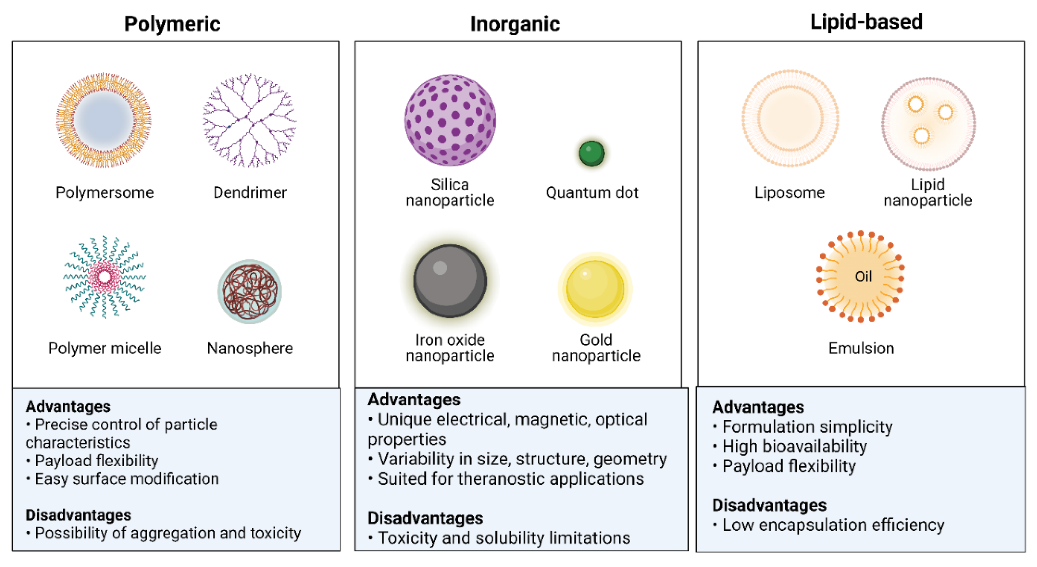
Nanotechnology has a growing potential for application in medical diagnosis and therapy. Nanotechnology advancements have developed novel and better nanomaterials for biomedical applications [24]. NPs are utilized in various applications due to their unique properties [25]. Multifunctional NPs can transport hydrophobic compounds, target disease cells both actively and passively, extend the time a drug is in the bloodstream, increase the entry and accumulation of pharmaceuticals at tumor sites, overcome drug resistance, increase the safety and tolerability of medications, and advance the development of other technologies [26,27]. NPs are used in medical applications because of their unique properties, such as quantum properties, a surface-to-mass ratio that is much higher than that of other particles, and the ability to absorb and transport other compounds, such as proteins and medicines. NPs can have variable compositions, as their beginning ingredients might be dextran, chitosan, biological lipids, phospholipids, lactic acid, or chemicals such as silica, metals, carbon, and other polymers [28,29,30,31,32]. Classes of NPs, along with their advantages and disadvantages, are shown in Figure 2.
NPs are the perfect theranostic tools for tumor tracking and therapy because of their size (1–100 nm) and the high surface-to-volume ratio [33]. Effective medicine delivery is made possible by directly coupling NPs to various biomolecules, which also permits anatomical and functional imaging. Three distinct processes are involved in the delivery of NPs: (a) systemic localization without RES sequestration; (b) extravasation from intratumoral capillaries; and (c) diffusion and penetration into cancerous cells. NPs are effective anticancer weapons due to the high accumulation levels in tumor cells [34]. Manufacturing NPs bigger than 50 nm prevents RES sequestration [35]. Since solid tumors have a unique microenvironment, NP is desirable for imaging and drug delivery. The tumor microenvironment comprises blood vessels, inflammatory cells, signalling molecules, extracellular matrix, and lymphocytes. Blood vessels, inflammatory cells, signalling molecules, extracellular matrix, and lymphocytes make up the tumor microenvironment [36]. Solid tumors differ from healthy tissue in permeable microvascular environments with capillary pores between 120 and 1200 nm in size, which makes it easier for NP to penetrate tumors [37].

3. Metal Nanoparticles and Their Application in Cancer

The use of nanotechnology as a novel technique for cancer diagnosis, monitoring, and treatment has recently attracted interest in the biomedical community. Nanomaterials vary in diameter size from 1 to 1000 nm and display several distinctive features that differ from those seen in tiny particles or bulk materials. Nanomaterials have the potential to be used in several biological applications because of their substantial specific surfaces, high surface activity, robust antioxidant properties, outstanding biocompatibility, and solubility for molecular modifications. Liposomes, carbon nanotubes, polymeric micelles, graphs, quantum dots, metallic NPs, and magnetic NPs are often employed in biomedical applications. Previously, it has been demonstrated that using them extensively can improve treatment results [38].

3.1. Gold-Based Nanoparticles

The biological application of metallic nanoparticles, particularly gold nanoparticles (Au NPs), has sparked attention among numerous nanomaterials primarily due to its apparent benefits. Various Au NP forms, including spherical, rod-like, cage-like, and others, in diameters ranging from 1 nm to more than 100 nm, can be prepared rapidly. Au NPs’ form and size significantly impact optical and electrical characteristics [39]. Moreover, Au NPs have a negative charge, and many biomolecules, including genes and targeting ligands, can readily functionalize them [40]. Au NPs are harmless and biocompatible [41]. Surface plasmon resonance (SPR) bands are present in Au NPs, which also have an ultra-small size, a macroscopic quantum tunnelling effect, and a distinct surface effect [42]. Due to these unique characteristics, Au NPs have emerged as the most promising material for various biological applications, such as drug delivery, molecular imaging, and biosensing.

Photoacoustic Imaging

Photoacoustic imaging (PAI) is a biomedical imaging technology that employs endogenous and exogenous contrasts and provides insightful data on the cellular and molecular properties of the tissue. Because of their inherent and geometrically induced optical components, Au NPs as exogenous contrast agents hold significant potential for PA imaging. The most popular Au NPs forms for PA imaging include shells, prisms, spheres, rods, cages, stars, and blisters [43].
Gold nanorods are frequently employed as PAI contrast agents. Using PAI, it has previously been reported that Au/Ag activatable nanoparticles react to reactive oxygen and nitrogen species (RONS). The gold core can be preserved, while just some of the shell can be removed using RONS. The PAI signal is reactivated as a result of the etching. Iodide-doping of silver improves RONS sensitivity, allowing the detection of physiologically relevant levels in a mouse model (Figure 3) [44].

3.2. Silver-Based Nanoparticles

Using silver (Ag) NPs as a safe and efficient method for treating cancer is challenging. Integrative studies involving physicists, chemists, engineers for creating NPs, and biologists who can evaluate the effects of Ag NPs in the cell systems they come in contact with, in aspects of cytotoxic effects and ability to damage malignant cells, are required [45]. Ag NPs are important and offer multiple advantages due to their size and shape-dependent features, such as optical, magnetic, chemical, and physical characteristics [46]. Ag NPs are included in numerous items, such as biosensors, composite fibers, antimicrobials, cosmetics, and electrical chemicals [47]. Additionally, Ag NPs can be utilized in cell electrodes, filters, nanocomposites, drug delivery, and medical imaging [47]. Ag is favoured over other NPs due to its superior light absorption, higher resolution, and stronger affinity for functionalization [48].
Like other noble metal NPs, Au NPs display exceptional SPR, making them ideal for usage in various fields, such as biosensing, catalysis, protein/gene transport, and photo-controlled delivery systems [49]. In addition to their antiproliferative effects on cancer cells, silver NPs can activate pathways that inhibit cell division and may be employed in the detection of many cancers like lung cancer, prostate cancer, hepatic cancer, cervical cancer, etc. Research on the antitumor effectiveness of Ag is still actively being conducted, despite technological advancements in its well-defined shapes and sizes and biocompatibility tests of both simple and coated Ag NPs.

3.3. Palladium-Based Nanoparticles

The development of palladium (Pd) based NPs is among the innovative nanomaterials that have significantly contributed to advancing the applications of noble metal nanomaterials in biomedicine. Pd-based nanomaterials have distinct advantages over other nanostructures made of noble metals. These advantages include biocompatibility and good photothermal stability. These characteristics make Pd-based nanomaterials stand out as exceptional and promising in biomedicine. After several years of research, Pd-based nanomaterials, such as Pd NSs, Pd@Au, Pd NPs, and Pt@Pt nanostructures, have been the subject of substantial investigation in the field of multimodal imaging-guided cancer treatment.
Pd-based nanomaterials have been noted to exhibit remarkable optical properties, high levels of biocompatibility, and high levels of stability in physiological conditions, all of which make them extremely promising for use in biomedical applications. Research on Pd-based nanomaterials started significantly later than that of other noble nanoparticles that have been extensively studied, such as gold (Au) and silver (Ag) nanomaterials. However, due to distinct qualities, including high photothermal conversion efficiency and photothermal stability, they have attracted a lot of attention in the nanomedicine sector. Pd-based nanomaterials display great near-infrared (NIR) absorption, a fast rate of photothermal conversion, outstanding biocompatibility, and exceptional photothermal stability. Pd nanosheets, porous/hollow Pd NPs, and Pd@M (M = Ag, Au, Pt, SiO2, ZIF-8) are examples of these nanomaterials. Pd-based nanoparticles have emerged as promising candidates for use as therapeutic agents and contrast agents in cancer imaging [50,51,52].
Pd nanosheets are typical examples of 2D nanomaterials with powerful NIR absorption, high photothermal conversion efficiency, outstanding photothermal stability, and a high level of biocompatibility. The diameters of Pd nanosheets (Pd NSs) can be easily altered to be anywhere between 5 and 120 nm while maintaining a consistent hexagonal shape [53,54]. The optical absorption peaks of Pd NSs shift depending on their size, but they are always found in the NIR range, thus offering exciting prospects for use in photothermal therapy (PTT). Table 1 summarises cancer therapies that utilize Pd-based nanoparticles, both as stand-alone PTT treatments and in combination with other therapeutic techniques [50].

3.4. Iron Oxide-Based Nanoparticles

Because of their exceptional magnetic properties, surface-to-volume ratio perfect for successful functionalization, and biocompatibility, iron oxide NPs are often used in biological applications. Because these nanoparticles are now utilized in the medical system as contrast agents and heating mediators, most of the attention is focused on their development for MRI or magnetic particle hyperthermia. As a result, it is essential to keep improving and making new materials that are better and more reliable [76] for molecular imaging and biosensing.

Super Paramagnetic Iron Oxide Nanoparticles for Cancer Treatment

Superparamagnetic iron oxide nanoparticles (SPIONs) have drawn more attention due to their superior superparamagnetism, magnetic heating abilities, and improved magnetic resonance imaging (MRI). In vivo imaging, magnetic thermotherapy, and simultaneous delivery of anticancer treatments are only a few benefits of conjugating SPIONs with medications to create delivery nanosystems. Additional targeting moieties such as transferrin, hyaluronic acid, antibodies, aptamers, folate, and targeting peptides are coated onto the surface of SPIONs to improve the targeting efficacy of pharmaceuticals delivered by a delivery nanosystem based on SPIONs [77]. SPIONs exceptional MRI enhancement capabilities can be employed as tracers to depict the location and status of illness in the body, in addition to being deposited in cancer cells through the EPR effect and under an external magnetic field. Furthermore, SPIONs have a lower potential for toxicity than other inorganic NPs like their carbon- and gold-based counterparts because they can biodegrade into ferric ions in the human body, particularly in cells with acidic conditions (such as the lysosome and endosome) [78]. Theranostic agents based on SPION are essential for the delivery of therapeutic payloads such chemotherapeutic drugs and genes and for the diagnosis of cancer.

3.5. Copper-Based Nanoparticles

For biomedical applications, copper-based NPs have gained more interest. When exposed to a near-infrared laser, copper chalcogenide NPs display excellent near-infrared absorption, exhibit effective light-to-heat transformation, and selectively thermally destroy the tumor. Smaller copper NPs demonstrate the fluorescence signal and optical imaging capabilities. Additionally, copper-based NPs provide a flexible means of drug administration and image-guided treatment. Current developments in the biological use of copper-based NPs with an emphasis on cancer imaging and therapy have been discussed by Zhou et al. [79].
Copper NPs have more uses than Au and Ag NPs due to their reduced price, greater cytotoxic action against cancer cells at low doses, and prolonged stability period [80]. Novel copper-containing NIR-absorbing nano-formulations, such as copper selenide (Cu2-xSe) nanocrystals, nanocubes, monodispersed CuTe nanorods, nanoplates, and copper bismuth sulfide (Cu3BiS3) nanostructures, have all been fabricated and further confirmed for PTT. All the nanomolecules of copper discussed above have strong anticancer potential and improved photothermal heating efficiency [81].

3.6. Selenium-Based Nanoparticles

The trace element selenium (Se) is essential. It is included as selenocysteine, the most significant component of the active centre of selenoproteins’ enzymatic activity. Numerous selenoproteins have oxidoreductase activity and hence control the redox balance in the body. Se has a small therapeutic window and very fragile toxicity margins, but Se nanoparticles (SeNPs) have remarkably lower toxicity. SeNPs have been investigated for their potential therapeutic effects in a number of oxidative stress and inflammation-induced diseases, including cancer, diabetes, nephropathy, and arthritis. SeNPs serve as a desirable drug delivery system for a variety of medications. The impact of nanosizing on Se’s pharmacological action has been covered in this article. Presently discussed is the function of SeNPs in the pharmacological defence against diverse inflammatory and oxidative stress-mediated situations. SeNPs’ potential impact on the pharmacokinetics and pharmacodynamics of selenoproteins, however, remains mainly unknown.
Human clinical trials validate the protective and curative functions of selenium in the initiation and progression of cancer. Diverse anticancer processes of selenium [82] can be divided into three major categories: thiol modification, chromatin binding and alteration, and reactive oxygen species (ROS) generation. Important factors affecting selenium’s biological activity, toxicity, and ability to prevent cancer are its amount and form [83]. Selenium’s anticancer activities have traditionally been linked to its organic form, particularly dietary selenium, and its antioxidant and pro-oxidant capabilities.
Studies on several malignancies, such as breast, lung, prostate, and colon cancers, demonstrate cancer-protective effects.
The most prevalent form of selenium in plants, seleniomethionine, was once believed to be the most chemopreventive and therapeutic form of selenium, but more recently, methylselenocysteine was discovered to have higher biological activity [84]. Since then, synthetic organoselenium species have been created that outperform their natural counterparts in terms of anticancer activity [85].
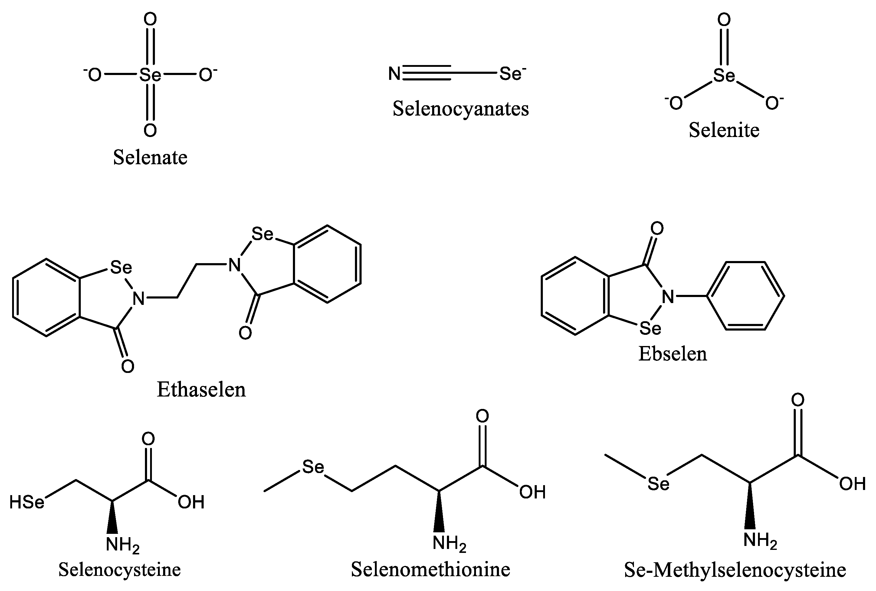
Despite their origins as an antioxidant, selenium-based medications like ebselen (Figure 4) have demonstrated potential anticancer action against breast, liver, and colon cancer cells. In Phase I clinical studies for the treatment of non-small cell lung cancer, ethaselen, a modified version of ebselen, has demonstrated better solubility [82,86]. According to studies, selenium can prevent cancer by preventing DNA damage brought on by the production of adducts caused by dimethylbenz(a) anthracene, which is a factor in the development of breast, colon, and liver cancers [87].

4. Nanoparticles for Medical Imaging

Medical imaging is commonly used to investigate biological processes, spot anomalies, and track the development of illnesses [88]. The clarity of medical pictures is continually being enhanced with the development of cutting-edge imaging technology. These methods include magnetic resonance imaging (MRI), computed tomography (CT), positron emission tomography (PET), optical fluorescence imaging, photoacoustic imaging (PA), single-photon emission computed tomography (SPECT), and ultrasound imaging (US) [89].
Imaging techniques serve a crucial function in the diagnosis and therapy of cancers. Many NPs, such as iron oxide NPs [90], can enhance imaging due to their magnetic, optical, acoustic, and structural properties [91]. Previous studies reported that injecting NPs into target tissues can improve image guidance and contrast for locating and removing tumors [92]. For instance, in cryosurgery, NPs can enhance the tumor’s and the ice ball’s imaging quality, enabling more accurate coverage and improved therapeutic effectiveness [93]. In addition, the majority of imaging NPs are composed of various metals. Table 2 provides examples of NPs derived from multiple materials and their application in medical imaging [94].
A non-invasive, micron-level resolution and biological imaging method is optical coherence tomography (OCT). Real-time diagnosis and surgical guiding are both facilitated by OCT. However, it cannot pick up inelastic dispersed light because it lacks coherence in the incident field [100]. Numerous studies have recently shown that the motion state of NPs may alter the amplitude of OCT, which could resolve this issue. Interfering with the movement of NPs in the magnetic field causes local changes in the dispersion of light. By adding magnetic NPs, which may alter optical scattering and make it possible to identify originally incoherent inelastic scattered light, one can regulate the movement of a magnetic field. The name of this cutting-edge imaging technique is magnetomotive optical coherence tomography (MMOCT) [96].
Magnetic resonance imaging (MRI) is one of the most effective non-invasive tumor detection methods [101]. However, the absence of an MRI signal comparison between normal and cancerous tissue hinders the clinical tumor diagnosis [102]. MRI is a scanning imaging technique that assesses the degree of hydrogen molecule magnetization inside water molecules. Due to the distinct variations in magnetization caused by each tissue’s protons, each anatomical structure shows a unique picture. Applying more excellent contrast agents can increase the visibility of images [103]. MRI scanning is a beneficial medical diagnostic technique for an accurate anatomical picture, and for increased diagnostic sensitivity, contrast agents are frequently utilized in MRI [104]. Using MRI, one can spot bodily tissues, organ states, blood flow, and physiochemical characteristics [105]. Chelate-based traditional contrast agents have limitations due to their biological stability and degree of toxicity when accumulated in cells [106]. For example, iodine is present in several contrast agents. The occurrence of incident hyperthyroidism has been associated with exposure to iodinated contrast media [107]. Instead of using contrast chemicals, which may be damaging to the body, alternatives have been developed to improve the scanning efficiency [108]. There are also metal NPs that can be combined with a material to work similarly to a contrast agent for MRI scanning [109].
Nanoparticles possess unique optical and magnetic properties that can be utilized to improve imaging techniques. For example, quantum dots are semiconductor nanoparticles that emit light of different wavelengths based on their size, making them ideal for fluorescent imaging. Iron oxide nanoparticles can be used as contrast agents in magnetic resonance imaging (MRI) due to their magnetic properties, providing better visualization of tumors. Gold nanoparticles, on the other hand, exhibit strong absorption and scattering of light, enabling enhanced photoacoustic imaging. These nanoparticles enhance the sensitivity, resolution, and specificity of imaging, allowing for early cancer detection, precise tumor localization, and monitoring of treatment response.

5. Targeting of Cancer Cells by Metallic Nanoparticles

An effective method for cancer diagnosis and treatment is to target cancer cells using metallic nanoparticles. Recently published research on the topic includes the following:
1. Copper nanoparticles: Copper nanoparticles have recently received interest as potential anticancer therapies due to their ability to induce oxidative stress in cancer cells. Reactive oxygen species (ROS), which lead to cell death, are produced by copper nanoparticles, which have been demonstrated to selectively target cancer cells while sparing healthy cells [110]. Additionally, it has been shown that copper nanoparticles increase the responsiveness of cancer cells to radiation therapy, promoting tumor remission [111].
2. Silver nanoparticles: Silver nanoparticles have also been investigated as a potential cancer therapy. By interacting with proteins on the cell surface and causing cell death by creating ROS, researchers have shown that silver nanoparticles may selectively target cancer cells [112]. Silver nanoparticles have also been studied as imaging agents for cancer diagnosis due to their powerful optical properties [113].
3. Zinc oxide nanoparticles: These particles have been studied as potential anticancer therapies since they may induce cancer cells to undergo apoptosis. The ability of zinc oxide nanoparticles to precisely target cancer cells and induce cell death by triggering caspases and suppressing anti-apoptotic proteins has been established [114]. Additionally, zinc oxide nanoparticles have been shown to overcome the drug resistance of cancer cells, improving the efficacy of chemotherapy treatments.
Recent research on metallic nanoparticles for cancer targeting has generally focused on improving the specificity and efficacy of existing agents as well as looking into novel kinds of nanoparticles with unique properties for cancer diagnosis and therapy.
A surplus of molecular information has resulted from the quick advancement in our understanding of the molecular pathophysiology of illnesses and the development of novel molecular biology tools. The speed of these advancements has led to the identification of numerous molecular targets for pharmacological action at a rate that much exceeds our current capacity to use this molecular knowledge [115,116]. Research and development efforts are currently being made to find and create medications that disrupt the signal transduction pathways used only by cancer cells. These medications will facilitate doctors to customize each patient’s care based on the specific molecular targets that their tumor produces. Drugs can be administered directly to the target organs where the tumor is located or to the cancer cell’s surface. They can also be administered using a drug delivery system. The primary components of such a targeted drug delivery are stated in the following:
  • The availability of specific targets first bullet;
  • Ligands for these targets;
  • Techniques for delivering the drug to its target via various delivery systems conjugated to the ligands.
Each form of cancer requires a distinct approach since they might be solid tumors or hematologic malignancies. Drug administration is made more difficult by solid tumors’ heterogeneous and dynamic biology, which constantly changes over time [117]. A complete understanding of the biology of tumor cells, their microenvironment, and their development patterns enables the creation of efficient, targeted drug delivery systems.
A drug can be targeted in two ways: by actively pursuing a drug carrier using target-specific ligands, or by using the distinct pathophysiological features of tumor tissue (Figure 5) [118].
Development or engineering of a medication or gene delivery system with an outstanding capacity to target tumor cells while preserving the normal healthy cells is vital for effective cancer therapy. It increases therapeutic effectiveness, protecting healthy cells from cytotoxicity. It can be done by strategically delivering NPs to the tumor microenvironment (TME) and then indirectly targeting the cancer cells there. These nano-formulations should be able to transit through a number of pharmacological and biological barriers. These barriers are intricate systems made up of cellular membranes, mechanical and physicochemical barriers, enzymatic barriers, and layers of epithelium, endothelium, and other tissue. To avoid non-specific targeting, these realities restrict the biocompatibility, surface chemistry, and size of NPs. However, merely internalizing an NP drug molecule in the cytosol does not guarantee that it has reached its subcellular target. To allow cellular or nuclear targeting, specific engineering and optimization are required [119].

5.1. Targeting Cancer Stem Cells with Nanomaterials

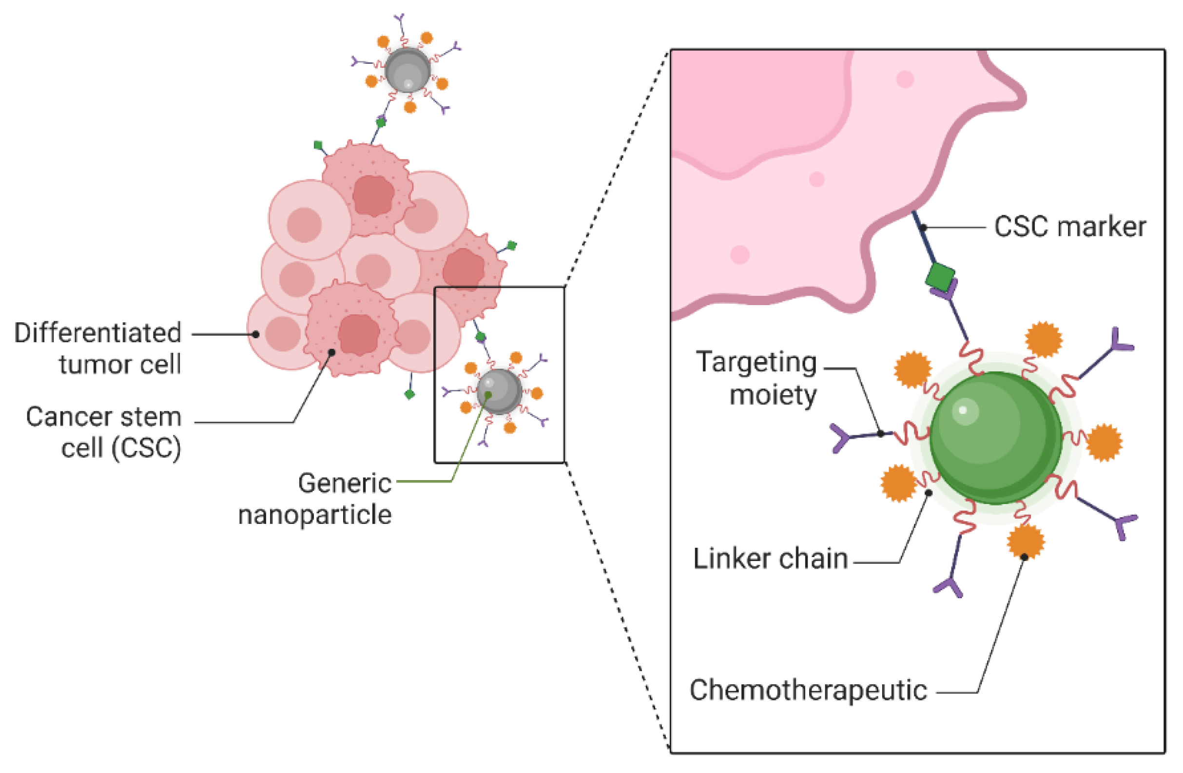
A collection of proliferating cells with high power and excellent resistance to medications, known as cancer stem cells (CSCs), is the primary cause of cancer. In the 1990s, CSCs were discovered and defined in the blood of leukaemia patients, and they appeared to play a significant role in malignancy. The CSCs promote tumor development, resist traditional treatments (such as chemotherapy and radiation), cause disease recurrence, and produce metastases. Consequently, there is a lot of interest in using nanoscale materials for CSC-directed cancer therapy. The NP surface has been engineered to precisely and successfully target the CSCs [120]. Potential therapeutic targets for eradicating cancer stem cells (CSCs) are surface indicators unique to CSCs. By joining targeting molecules to drug-delivery NPs, active targeting can be accomplished. These molecules can only attach to the markers present on CSCs. After binding, both internal and external events might cause drug release. These techniques have been used to create a variety of NP-based drug delivery platforms, as shown in Figure 6 [121].
Chemotherapy and radiation are two standard cancer therapies that CSCs notably resist. Therefore, to create effective medicines, knowing tumor biology is essential. New therapeutic approaches for cancer treatment include identifying and specifically targeting CSC-specific markers and signalling pathways [122,123,124]. Studies utilizing nanotechnology-based therapies with several surface markers and biochemical tests for identification have revealed the present effectiveness in the battle against CSCs [125].

5.2. Targeted Drug Delivery

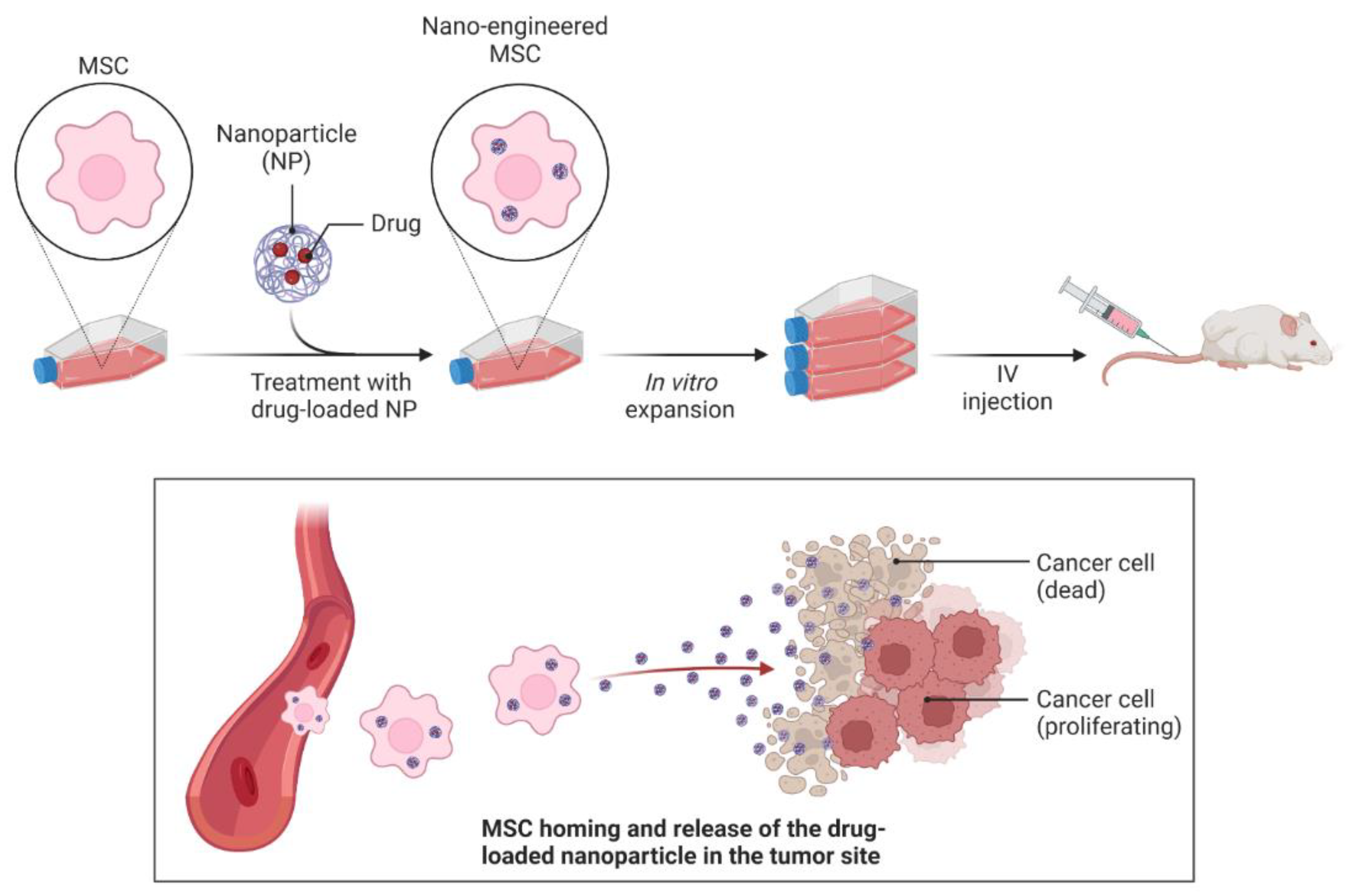
Despite being the most popular form of cancer treatment today, chemotherapy still suffers from the issues of inadequate target enrichment in regions of cancerous tumors and over-accumulation in healthy tissue [126]. Inhibiting cells that reproduce rapidly, such as hair follicles, bone marrow, gastrointestinal cells, and lymphocytes, may result in undesirable side effects like hair loss, mucositis, and even death. The use of inorganic nanoparticles in gene and drug therapy opens the door to targeted cancer detection and therapy with enhanced efficiency and better treatment outcome [85]. Compared to the standard of care, targeted medication delivery, which involves actively differentiating between healthy and cancer cells, is more effective and causes fewer side effects. Numerous studies have demonstrated that NPs may actively or passively target chemotherapy medicines to tumor cells [127]. Additionally, in several studies, NPs have been revealed to be crucial for the targeted administration of immunological medications [128]. The lack of cancer cell selectivity in chemotherapeutic drugs and the formation of many undesirable side effects prompted the development of innovative drug delivery techniques. The growth of “nano-engineered” mesenchymal stem cells (MSC) that can actively target the tumor site and shield the drug-loaded NP from vascular filtration and macrophage clearance is made possible by combining nanotechnology with cell therapy (Figure 7) [129,130].
Targeted medication delivery to tumors has the potential to lower peripheral/systemic toxicity, enhance the selectivity for killing cancer cells, and allow for dosage escalation. With improvements in identifying tumor-specific targets and developing several drug delivery methods for tumor targeting, hopes for developing an efficient, targeted drug delivery modality for cancer therapy have grown. Although the ultimate goal is to remove cancer from the patient altogether, more pragmatic aims to enhance the patient’s quality of life are near to being realized. In the subsequent years, the focus will be on creating systems that can effectively internalize into cancer cells while also being able to detect specific targets on cancer cells. Some of these issues could be solved by combining targeted strategies. Other promising ideas for medication targeting in cancer therapy include employing unique molecular addresses on the vascular endothelium and targeting using magnetic fields and ultrasound.
Nanoparticles can be engineered to selectively target cancer cells while sparing healthy tissues. This is achieved by attaching specific targeting ligands, such as antibodies or peptides, onto the nanoparticle surface. These ligands can recognize and bind to specific receptors or biomarkers overexpressed on cancer cells, facilitating the preferential accumulation of nanoparticles at the tumor site. By encapsulating or conjugating therapeutic agents, such as chemotherapy drugs, small interfering RNA (siRNA), or gene therapy vectors, nanoparticles can deliver these payloads directly to cancer cells. This targeted approach increases drug concentration within the tumor, reducing off-target effects and minimizing systemic toxicity.

5.3. Metal Nanoparticle Mediated Cryosurgery

Benefits of cryosurgery, the freezing of tumor tissue, include minimal cost, less invasiveness, decreased intraoperative bleeding, and fewer postoperative issues. Still, drawbacks include poor freezing efficiency and freezing damage to nearby tissues [93]. Antifreeze protein (AFP-1) has been used as a protective factor to aid with cold ablation, although the results are still not perfect [131]. As nanotechnology evolved, nano-cryosurgery arose as a concept. Nano-cryosurgery is predicated on the delivery of NPs with precise physical or chemical characteristics into tumor tissues. By utilizing the properties of NPs, it is able to manage range modification, ice ball formation direction, and freezing efficiency. As a result, nano-cryosurgery can destroy tumor tissue while shielding nearby healthy tissue from freezing simultaneously [132].
Intracellular ice creation during cryosurgery is essential for tumor cell destruction. Researchers have shown that NPs can successfully promote the development of intracellular ice in the interim [108]. NPs can cause heterogeneous nucleation by acting as outside particles. According to a recent study, NP-enriched tissues freeze more quickly than unenriched tissues and are more prone to heterogeneous nucleation. Moreover, NPs can considerably speed up and enhance the likelihood of ice production in cells, killing tumor cells more efficiently [133]. It has been demonstrated by the ease with which ice forms in tissue when the same freezing conditions are applied. Additionally, the thermal conductivity in tumor tissue would be greatly increased by NPs containing a metal oxide.
Since tumors are typically shaped irregularly, the ice crystals created by conventional cryosurgery only partially cover the tumor tissue. Nano-cryosurgery is more effective than traditional cryosurgery at solving this issue. Due to the fact that NPs may enter intracellular fluid and possess favourable physical characteristics, such as thermal conductivity, it is feasible to regulate the direction of ice ball development by scattering them [112]. Healthy tissue is more susceptible to injury. In recent years, cryosurgery utilizing phase change materials (PCMs) generated by nanoparticles has proven to have considerable promise for protecting the surrounding healthy tissues [134]. The use of NPs in cold ablation falls broadly into two categories: synergistic effect and protective effect, which differ in the NPs design specifications and in vivo dispersion. Future NPs may help nano-cryosurgery by being disseminated around the tumor while protective NPs are spread inside the cancer. In addition to overcoming the challenges of cold ablation of rare tumors, nano-cryosurgery technology can also improve the effectiveness of cold ablation and lessen the harm to good tissue. Table 3 lists a few recent instances of NPs being employed in cryosurgery [94].

6. Types and Biological Properties of Nanoparticle Delivery Platforms

Using molecular platforms created for drug transport and selective targeting could be more practical. Lipid nanocarriers (micelles, liposomes, and lipid nanoparticles), polymer conjugates, polymeric nanoparticles, and antibody-drug conjugates are a few examples of the proposed molecular platforms (ADCs). Selective targeting uses a platform’s inherent properties like size, charge, or the presence of a particular polymeric component (such as a polymer recognized by a specific receptor). It is also possible to directly functionalize a molecular platform’s surface with a targeting agent or moiety.
While natural and synthetic polymers have found use in various industries, from manufacturing materials to medicinal uses, recent decades have increased interest in using them as parts of complex drug delivery systems. Due to these advancements, a brand-new class of medications known as “polymer therapies” [139] emerged. Polymer-drug conjugates, polymer-DNA complexes (or polyplexes), polymer-protein conjugates, polymeric micelles, and polymer-aptamer conjugates, containing medicines covalently tethered to the polymer carrier, are some examples of these novel chemical entities [140,141].
Traditional micelles and liposomes, which depend on the physical encapsulation of medicines, can have problems, including the early release of the active ingredient, instability during storage and administration, and poor encapsulation. An early medication release impairs the effectiveness of treatment and causes systemic toxicity. Since pharmaceuticals are conjugated to the polymeric backbone utilizing cleavable linkers that only trigger drug release under specific circumstances, polymer treatments can address these problems [142]. A range of medication delivery methods for the treatment of cancer is shown in Figure 8 [143].

7. Current Limitations of Metal Nanoparticles and the Challenges

The use of metal nanoparticles in detecting and treating cancer comes with several limitations and challenges, despite the potential benefits. Following are some existing limitations and challenges mentioned in this section:
Biodistribution and toxicity: One of the main problems with using metal nanoparticles is their potential toxicity and biodistribution in the body. According to certain research, metal nanoparticles can accumulate in organs and tissues and cause toxicity and adverse effects. It is essential to accurately evaluate the toxicity and biodistribution of metal nanoparticles before using them in therapeutic contexts [144]. Due to their extremely small physical dimensions, nanoparticles are more hazardous than larger particles. Although fullerene and carbon nanotubes are highly hazardous when inhaled into the lungs, carbon black is not toxic. Similar to this, titanium oxide nanoparticles have been demonstrated to increase toxicity and cause oxidative stress in bacterial cells. Many non-toxic bulk substances turn poisonous when scaled down to the nanoscale. It is crucial to accurately assess the toxicity and biodistribution of metal nanoparticles before using them in therapeutic contexts.
Stability and aggregation: Metal NPs’ instability and susceptibility to aggregation may limit their effectiveness and selectivity for fighting cancer. Aggregation or agglomeration can take place in metal nanoparticles, which can result in modifications to the nanoparticles’ physicochemical characteristics and a decrease in their therapeutic efficacy. Several environmental conditions, such as changes in pH or the presence of biomolecules, can cause aggregation in biological systems. Metal nanoparticles must be stabilized and prevented from aggregating in order to be used in clinical settings with confidence. Researchers have recently devised unique techniques to maintain the stability of metal nanoparticles and prevent aggregation, including using surface coatings and conjugation with specific ligands [145].
Specificity and selectivity: In the process of using metal nanoparticles to target cancer, one of the challenges is attaining a specificity and selectivity adequate for cancer cells while sparing normal cells. Traditionally, cancer-treating chemotherapeutic agents are distributed non-specifically, damaging both healthy tissue and cancerous ones, resulting in low efficacy and high toxicity. Controlled drug delivery systems would be excellent carriers for chemotherapeutic agents, directing the chemotherapeutic agents to the tumor site, thereby increasing drug concentration in cancer cells and preventing toxicity to normal cells. Researchers are investigating new targeting ligands and techniques to boost the specificity and selectivity of metal nanoparticles for cancer cells [146].
Regulatory approval: The procedure for obtaining regulatory permission for the use of metal nanoparticles in therapeutic applications may be difficult and time-consuming. Extensive preclinical and clinical research is required in order to evaluate their safety, effectiveness, and impacts over the long term. In order to successfully incorporate metal nanoparticles into cancer diagnosis and therapy, it is vital to close the gap that exists between research conducted in the laboratory and its use in clinical settings. The clinical translation of metal nanoparticles for cancer diagnosis and treatment requires regulatory approval and clinical investigations. This strategy calls for substantial preclinical and clinical testing, which may be time-consuming and expensive. Still, it is necessary to guarantee that the treatment will be safe and effective.
To address these limitations and challenges, researchers, engineers, healthcare providers, and regulatory bodies must collaborate across disciplines. To overcome these obstacles and realize their full potential for cancer treatment and other biomedical applications, continued research and development in the field of metal nanoparticles is required.

8. Conclusions and Future Perspective

Biomedical applications of metal NPs have been the subject of many studies. They are becoming increasingly popular in the biomedical sector due to their high inertness and nanoscale architectures, which are equivalent in size to many biological molecules. Particle intrinsic features, such as electrical, physicochemical, and optical surface plasmon resonance, can be altered by modifying particle characteristics, including size, ease of synthesis, shape, environment, aspect ratio, and functionalization qualities. Numerous applications in a range of biomedical fields have resulted from this. Targeted medication delivery, sensing, imaging, photothermal and photodynamic therapy, and controlling two or three applications are a few of these applications.
Cancer is one of the diseases with the highest mortality rate in the world, even though there are already treatments available for cancer that have considerable drawbacks. Therefore, new anticancer strategies are once again necessary. One of the most promising treatments for cancer treatment is metal NPs. Iron, gold, silver, zinc, and titanium are examples of metals that may function as anticancer agents, naturally or by surface modification. Metal NPs are essential therapeutic and diagnostic tools in the fight against cancer.
According to researchers working to improve clinical outcomes, the greatest obstacle is chemotherapy for cancer. The therapeutic efficiency of cancer treatments may be enhanced with tailored medication administration, while unfavourable side effects may be decreased. Nanotechnology has caused a fundamental shift in medical science. One of the upcoming generation’s promising anticancer drugs is metal NPs. The extraordinary physical and chemical flexibility of metal nanoparticles has attracted a lot of research. Due to their distinctive plasmonic properties, noble metal NPs also offer a reliable way to monitor nano-complex drug carriers inside the body, making them a more effective treatment option with a reduced risk of adverse effects than conventional treatments. By using these NPs, practitioners may learn about and track therapeutic outcomes during therapy. Non-noble metal NPs are inexpensive and have distinct qualities, including magnetic and thermal capabilities. Metal nanoparticles have shown promise as a cancer treatment in several studies, and different compositions are now undergoing preclinical and clinical testing. With NP-based imaging, it may be possible to pinpoint the stage of a tumor, and approaches for treating it may be developed that reduce or eliminate the expected toxicity levels. Before conducting clinical research, however, it is necessary to analyze many production and utilization traits. Some measures include controlling preparation methods, reproducibility, stability, dose, the amount of accumulation at target and off-target areas, and, most crucially, toxicological concerns.
The future perspectives of nanoparticles in cancer diagnosis and treatment are highly promising. Following are some potential areas where nanoparticles will have a significant impact:
Early cancer detection: NPs can be engineered to specifically target biomarkers associated with cancer cells, allowing for highly sensitive and specific early detection. This could enable the diagnosis of cancer at its earliest stage, when treatment is most effective.
Improved imaging techniques: NPs can enhance existing imaging modalities such as MRI, CT, and optical imaging. By functionalizing nanoparticles with targeting ligands and contrast agents, they can improve the resolution, sensitivity, and specificity of cancer imaging, enabling accurate tumor localization and monitoring of treatment response.
Targeted drug delivery: NPs can serve as vehicles for targeted drug delivery, improving efficacy and reducing the side effects of chemotherapy. They can encapsulate anticancer drugs and deliver them directly to tumor sites, minimizing damage to healthy tissues. Additionally, stimuli-responsive nanoparticles can release drugs in response to specific triggers, such as pH, temperature, or enzyme activity within the tumor microenvironment.
Combination therapy: NPs offer opportunities for combining multiple therapeutic modalities into a single system. For example, nanoparticles can be loaded with chemotherapy drugs, immunotherapeutic agents, or gene therapies, allowing for synergistic effects and personalized treatment approaches. This approach holds promise for overcoming drug resistance and improving overall treatment outcomes.
Theranostics: NPs can integrate both diagnostic and therapeutic functionalities into a single system, known as theranostic nanoparticles. These multifunctional nanoparticles can simultaneously diagnose cancer, deliver therapy, and monitor treatment response. They have the potential to revolutionize personalized medicine and enable real-time monitoring of treatment efficacy.
Immunotherapy enhancement: NPs can play a crucial role in enhancing the efficacy of immunotherapies, such as immune checkpoint inhibitors or cancer vaccines. They can be used to deliver immunomodulatory agents, antigens, or adjuvants directly to immune cells or tumor sites, stimulating a robust immune response against cancer cells.
Microenvironment modulation: NPs can be designed to target the tumor microenvironment and modify its characteristics. They can normalize abnormal blood vessels, enhance drug penetration into tumors, modulate immune responses, or inhibit metastasis, thus improving the overall treatment outcomes.
Regulatory bodies play a crucial role in developing new clinical criteria for using metal NPs in cancer therapy and drug delivery, as well as new tools for evaluating the effectiveness and safety of such nanoparticles. Furthermore, scientists are striving to identify the best phytochemical conjugated metal NPs dosages for cancer patients and the most efficient way to give these doses. Once these issues are resolved, it is conceivable that metal NPs will become an indispensable therapeutic weapon in the fight against cancer.
There is an increasing demand for the practical translation of these technologies due to the continual advancement of research in nanoparticles and cancer. Researchers are attempting to create nanoparticle-based therapeutics that are both safe and effective so that they may be utilized in medical facilities to improve the results of patients who have cancer.

Author Contributions

Conceptualization, A.K. and B.B.; formal analysis, J.B. and A.K.; investigation, J.B. and A.D.P.; resources, G.G., A.K. and L.P.; data curation, J.B., B.B., L.P. and A.K.; writing—original draft preparation, J.B. and A.K.; writing—review and editing, J.B., B.B., A.D.P., G.G., L.P. and A.K.; supervision, A.K.; project administration, A.K. and L.P. All authors have read and agreed to the published version of the manuscript.

Funding

This research received no external funding.

Institutional Review Board Statement

Not applicable.

Informed Consent Statement

Not applicable.

Data Availability Statement

Not applicable.

Acknowledgments

The Università degli Studi di Cagliari is acknowledged by the authors.

Conflicts of Interest

The authors declare no conflict of interest.

References

  1. Vinardell, M.; Mitjans, M. Antitumor Activities of Metal Oxide Nanoparticles. Nanomaterials 2015, 5, 1004–1021. [Google Scholar] [CrossRef] [Green Version]
  2. Conde, J.; Doria, G.; Baptista, P. Noble Metal Nanoparticles Applications in Cancer. J. Drug Deliv. 2012, 2012, 751075. [Google Scholar] [CrossRef] [Green Version]
  3. World Health Organization. Global Centre for Traditional Medicine. Available online: https://www.who.int/initiatives/who-global-centre-for-traditional-medicine (accessed on 15 May 2022).
  4. Sutradhar, K.B.; Amin, M.L. Nanotechnology in Cancer Drug Delivery and Selective Targeting. ISRN Nanotechnol. 2014, 2014, 939378. [Google Scholar] [CrossRef] [Green Version]
  5. Zhao, G.; Rodriguez, L.B. Molecular targeting of liposomal nanoparticles to tumor microenvironment. Int. J. Nanomed. 2012, 8, 61–71. [Google Scholar] [CrossRef] [Green Version]
  6. Nguyen, K.T. Targeted Nanoparticles for Cancer Therapy: Promises and Challenges. J. Nanomed. Nanotechnol. 2011, 2, 1000103e. [Google Scholar] [CrossRef] [Green Version]
  7. Coates, A.; Abraham, S.; Kaye, S.B.; Sowerbutts, T.; Frewin, C.; Fox, R.M.; Tattersall, M.H.N. On the receiving end—Patient perception of the side-effects of cancer chemotherapy. Eur. J. Cancer Clin. Oncol. 1983, 19, 203–208. [Google Scholar] [CrossRef]
  8. Sharma, A.; Goyal, A.K.; Rath, G. Recent advances in metal nanoparticles in cancer therapy. J. Drug Target. 2017, 26, 617–632. [Google Scholar] [CrossRef]
  9. Ho, B.N.; Pfeffer, C.M.; Singh, A.T. Update on Nanotechnology-based Drug Delivery Systems in Cancer Treatment. Anticancer Res. 2017, 37, 5975–5981. [Google Scholar] [CrossRef] [Green Version]
  10. Prasad, R.; Pandey, R.; Varma, A.; Barman, I. Polymer-based nanoparticles for drug delivery systems and cancer therapeutics. In Natural Polymers for Drug Delivery; CABI: Wallingford, UK, 2017; pp. 53–70. [Google Scholar] [CrossRef]
  11. Ficai, D.; Ficai, A. New Challenges in Cancer Treatment, from Novel Agents to Innovative Administration. Anti-Cancer Agents Med. Chem. 2019, 19, 4–5. [Google Scholar] [CrossRef]
  12. Kumar, A.; Gatto, G.; Delogu, F.; Pilia, L. DFT study of [Pt(Cl)2L] complex (L = rubeanic acid) and its derived compounds with DNA purine bases. Chem. Phys. 2020, 530, 110646. [Google Scholar] [CrossRef]
  13. Sack, M.; Alili, L.; Karaman, E.; Das, S.; Gupta, A.; Seal, S.; Brenneisen, P. Combination of Conventional Chemotherapeutics with Redox-Active Cerium Oxide Nanoparticles—A Novel Aspect in Cancer Therapy. Mol. Cancer Ther. 2014, 13, 1740–1749. [Google Scholar] [CrossRef] [Green Version]
  14. Kumari, M.; Singh, S.P.; Chinde, S.; Rahman, M.F.; Mahboob, M.; Grover, P. Toxicity Study of Cerium Oxide Nanoparticles in Human Neuroblastoma Cells. Int. J. Toxicol. 2014, 33, 86–97. [Google Scholar] [CrossRef]
  15. Sreena, R.; Nathanael, A.J. Biodegradable Biopolymeric Nanoparticles for Biomedical Applications-Challenges and Future Outlook. Materials 2023, 16, 2364. [Google Scholar] [CrossRef]
  16. Sahoo, S.K.; Parveen, S.; Panda, J.J. The present and future of nanotechnology in human health care. Nanomed. Nanotechnol. Biol. Med. 2007, 3, 20–31. [Google Scholar] [CrossRef]
  17. Balogh, L.P. Nano-Enabled Medical Applications; CRC Press: Boca Raton, FL, USA, 2020. [Google Scholar] [CrossRef]
  18. Florence, A.T. “Targeting” nanoparticles: The constraints of physical laws and physical barriers. J. Control. Release 2012, 164, 115–124. [Google Scholar] [CrossRef]
  19. Du, K.; Feng, J.; Gao, X.; Zhang, H. Nanocomposites based on lanthanide-doped upconversion nanoparticles: Diverse designs and applications. Light Sci. Appl. 2022, 11, 222. [Google Scholar] [CrossRef]
  20. Yang, L.; Jia, J.; Li, S. Advances in the Application of Exosomes Identification Using Surface-Enhanced Raman Spectroscopy for the Early Detection of Cancers. Front. Bioeng. Biotechnol. 2022, 9, 808933. [Google Scholar] [CrossRef]
  21. He, H.; Zou, Z.; Wang, B.; Xu, G.; Chen, C.; Qin, X.; Yu, C.; Zhang, J. Copper Oxide Nanoparticles Induce Oxidative DNA Damage and Cell Death via Copper Ion-Mediated P38 MAPK Activation in Vascular Endothelial Cells. Int. J. Nanomed. 2020, 15, 3291–3302. [Google Scholar] [CrossRef]
  22. Kumar, S.; Mongia, A.; Gulati, S.; Singh, P.; Diwan, A.; Shukla, S. Emerging theranostic gold nanostructures to combat cancer: Novel probes for Combinatorial Immunotherapy and Photothermal Therapy. Cancer Treat. Res. Commun. 2020, 25, 100258. [Google Scholar] [CrossRef]
  23. Shi, J.; Kantoff, P.W.; Wooster, R.; Farokhzad, O.C. Cancer nanomedicine: Progress, challenges and opportunities. Nat. Rev. Cancer 2016, 17, 20–37. [Google Scholar] [CrossRef] [Green Version]
  24. Dessale, M.; Mengistu, G.; Mengist, H.M. Nanotechnology: A Promising Approach for Cancer Diagnosis, Therapeutics and Theragnosis. Int. J. Nanomed. 2022, 17, 3735–3749. [Google Scholar] [CrossRef]
  25. Machado, S.; Grosso, J.P.; Nouws, H.P.A.; Albergaria, J.T.; Delerue-Matos, C. Utilization of food industry wastes for the production of zero-valent iron nanoparticles. Sci. Total Environ. 2014, 496, 233–240. [Google Scholar] [CrossRef]
  26. Yang, Q.; Jones, S.W.; Parker, C.L.; Zamboni, W.C.; Bear, J.E.; Lai, S.K. Evading Immune Cell Uptake and Clearance Requires PEG Grafting at Densities Substantially Exceeding the Minimum for Brush Conformation. Mol. Pharm. 2014, 11, 1250–1258. [Google Scholar] [CrossRef]
  27. Zeineldin, R.; Syoufjy, J. Cancer Nanotechnology: Opportunities for Prevention, Diagnosis, and Therapy. In Cancer Nanotechnology: Methods and Protocols; Humana Press: New York, NY, USA, 2017; pp. 3–12. [Google Scholar] [CrossRef]
  28. Jani, P.; Subramanian, S.; Korde, A.; Rathod, L.; Sawant, K.K. Theranostic Nanocarriers in Cancer: Dual Capabilities on a Single Platform. In Functional Bionanomaterials: From Biomolecules to Nanoparticles; Springer: Cham, Switzerland, 2020; pp. 293–312. [Google Scholar] [CrossRef]
  29. Nikolova, M.; Slavchov, R.; Nikolova, G. Nanotechnology in Medicine. In Drug Discovery and Evaluation: Methods in Clinical Pharmacology; Springer: Cham, Switzerland, 2020; pp. 533–546. [Google Scholar] [CrossRef]
  30. Saxena, S.K.; Nyodu, R.; Kumar, S.; Maurya, V.K. Current Advances in Nanotechnology and Medicine. In NanoBioMedicine; Springer: Singapore, 2020; pp. 3–16. [Google Scholar] [CrossRef]
  31. Mukherjee, A.; Bhattacharyya, S. Nanotechnology in Medicine. In Biotechnology Business—Concept Delivery; Springer: Cham, Switzerland, 2020; pp. 57–64. [Google Scholar] [CrossRef]
  32. Guo, D.; Xie, G.; Luo, J. Mechanical properties of nanoparticles: Basics and applications. J. Phys. D Appl. Phys. 2014, 47, 013001. [Google Scholar] [CrossRef] [Green Version]
  33. Pedrosa, P.; Vinhas, R.; Fernandes, A.; Baptista, P. Gold Nanotheranostics: Proof-of-Concept or Clinical Tool? Nanomaterials 2015, 5, 1853–1879. [Google Scholar] [CrossRef] [Green Version]
  34. De, J. Drug delivery and nanoparticles: Applications and hazards. Int. J. Nanomed. 2008, 3, 133–149. [Google Scholar] [CrossRef] [Green Version]
  35. Ernsting, M.J.; Murakami, M.; Roy, A.; Li, S.-D. Factors controlling the pharmacokinetics, biodistribution and intratumoral penetration of nanoparticles. J. Control. Release 2013, 172, 782–794. [Google Scholar] [CrossRef] [Green Version]
  36. Barar, J.; Omidi, Y. Dysregulated pH in Tumor Microenvironment Checkmates Cancer Therapy. Bioimpacts 2013, 3, 149–162. [Google Scholar] [CrossRef]
  37. Omidi, Y.; Barar, J. Targeting tumor microenvironment: Crossing tumor interstitial fluid by multifunctional nanomedicines. Bioimpacts 2014, 4, 55–67. [Google Scholar] [CrossRef]
  38. Kong, F.-Y.; Zhang, J.-W.; Li, R.-F.; Wang, Z.-X.; Wang, W.-J.; Wang, W. Unique Roles of Gold Nanoparticles in Drug Delivery, Targeting and Imaging Applications. Molecules 2017, 22, 1445. [Google Scholar] [CrossRef] [Green Version]
  39. Verissimo, T.V.; Santos, N.T.; Silva, J.R.; Azevedo, R.B.; Gomes, A.J.; Lunardi, C.N. In vitro cytotoxicity and phototoxicity of surface-modified gold nanoparticles associated with neutral red as a potential drug delivery system in phototherapy. Mater. Sci. Eng. C 2016, 65, 199–204. [Google Scholar] [CrossRef] [PubMed]
  40. Fratoddi, I.; Venditti, I.; Cametti, C.; Russo, M.V. How toxic are gold nanoparticles? The state-of-the-art. Nano Res. 2015, 8, 1771–1799. [Google Scholar] [CrossRef]
  41. Hainfeld, J.F.; Slatkin, D.N.; Focella, T.M.; Smilowitz, H.M. Gold nanoparticles: A new X-ray contrast agent. Br. J. Radiol. 2006, 79, 248–253. [Google Scholar] [CrossRef]
  42. Kumar, A.; Zhang, X.; Liang, X.-J. Gold nanoparticles: Emerging paradigm for targeted drug delivery system. Biotechnol. Adv. 2013, 31, 593–606. [Google Scholar] [CrossRef] [PubMed]
  43. Li, W.; Chen, X. Gold nanoparticles for photoacoustic imaging. Nanomedicine 2015, 10, 299–320. [Google Scholar] [CrossRef] [Green Version]
  44. Mantri, Y.; Davidi, B.; Lemaster, J.E.; Hariri, A.; Jokerst, J.V. Iodide-doped precious metal nanoparticles: Measuring oxidative stress in vivo via photoacoustic imaging. Nanoscale 2020, 12, 10511–10520. [Google Scholar] [CrossRef]
  45. Vlăsceanu, G.M.; Marin, Ş.; Ţiplea, R.E.; Bucur, I.R.; Lemnaru, M.; Marin, M.M.; Grumezescu, A.M.; Andronescu, E. Silver nanoparticles in cancer therapy. In Nanobiomaterials in Cancer Therapy; William Andrew Publishing: Norwich, NY, USA, 2016; pp. 29–56. [Google Scholar] [CrossRef]
  46. Kent, R.D.; Vikesland, P.J. Controlled Evaluation of Silver Nanoparticle Dissolution Using Atomic Force Microscopy. Environ. Sci. Technol. 2012, 46, 6977–6984. [Google Scholar] [CrossRef] [Green Version]
  47. Abbasi, E.; Milani, M.; Fekri Aval, S.; Kouhi, M.; Akbarzadeh, A.; Tayefi Nasrabadi, H.; Nikasa, P.; Joo, S.W.; Hanifehpour, Y.; Nejati-Koshki, K.; et al. Silver nanoparticles: Synthesis methods, bio-applications and properties. Crit. Rev. Microbiol. 2014, 42, 173–180. [Google Scholar] [CrossRef]
  48. Nedelcu, I.-A.; Ficai, A.; Sonmez, M.; Ficai, D.; Oprea, O.; Andronescu, E. Silver Based Materials for Biomedical Applications. Curr. Org. Chem. 2014, 18, 173–184. [Google Scholar] [CrossRef]
  49. Qureshi, A.T.; Monroe, W.T.; Dasa, V.; Gimble, J.M.; Hayes, D.J. miR-148b–Nanoparticle conjugates for light mediated osteogenesis of human adipose stromal/stem cells. Biomaterials 2013, 34, 7799–7810. [Google Scholar] [CrossRef]
  50. Chen, A.; Ostrom, C. Palladium-Based Nanomaterials: Synthesis and Electrochemical Applications. Chem. Rev. 2015, 115, 11999–12044. [Google Scholar] [CrossRef] [PubMed] [Green Version]
  51. Chen, M.; Wu, B.; Yang, J.; Zheng, N. Small Adsorbate-Assisted Shape Control of Pd and Pt Nanocrystals. Adv. Mater. 2012, 24, 862–879. [Google Scholar] [CrossRef] [PubMed]
  52. Chen, X.; Shi, S.; Wei, J.; Chen, M.; Zheng, N. Two-dimensional Pd-based nanomaterials for bioapplications. Sci. Bull. 2017, 62, 579–588. [Google Scholar] [CrossRef]
  53. Huang, X.; Tang, S.; Mu, X.; Dai, Y.; Chen, G.; Zhou, Z.; Ruan, F.; Yang, Z.; Zheng, N. Freestanding palladium nanosheets with plasmonic and catalytic properties. Nat. Nanotechnol. 2010, 6, 28–32. [Google Scholar] [CrossRef] [PubMed]
  54. Chen, M.; Chen, S.; He, C.; Mo, S.; Wang, X.; Liu, G.; Zheng, N. Safety profile of two-dimensional Pd nanosheets for photothermal therapy and photoacoustic imaging. Nano Res. 2016, 10, 1234–1248. [Google Scholar] [CrossRef]
  55. Jain, P.K.; Huang, X.; El-Sayed, I.H.; El-Sayed, M.A. Noble Metals on the Nanoscale: Optical and Photothermal Properties and Some Applications in Imaging, Sensing, Biology, and Medicine. Acc. Chem. Res. 2008, 41, 1578–1586. [Google Scholar] [CrossRef]
  56. Fang, W.; Tang, S.; Liu, P.; Fang, X.; Gong, J.; Zheng, N. Pd Nanosheet-Covered Hollow Mesoporous Silica Nanoparticles as a Platform for the Chemo-Photothermal Treatment of Cancer Cells. Small 2012, 8, 3816–3822. [Google Scholar] [CrossRef]
  57. Huang, X.; Tang, S.; Yang, J.; Tan, Y.; Zheng, N. Etching Growth under Surface Confinement: An Effective Strategy to Prepare Mesocrystalline Pd Nanocorolla. J. Am. Chem. Soc. 2011, 133, 15946–15949. [Google Scholar] [CrossRef]
  58. Huang, X.; Tang, S.; Liu, B.; Ren, B.; Zheng, N. Enhancing the Photothermal Stability of Plasmonic Metal Nanoplates by a Core-Shell Architecture. Adv. Mater. 2011, 23, 3420–3425. [Google Scholar] [CrossRef] [Green Version]
  59. Chen, M.; Tang, S.; Guo, Z.; Wang, X.; Mo, S.; Huang, X.; Liu, G.; Zheng, N. Core-Shell Pd@Au Nanoplates as Theranostic Agents for In-Vivo Photoacoustic Imaging, CT Imaging, and Photothermal Therapy. Adv. Mater. 2014, 26, 8210–8216. [Google Scholar] [CrossRef]
  60. Tang, S.; Huang, X.; Zheng, N. Silica coating improves the efficacy of Pd nanosheets for photothermal therapy of cancer cells using near infrared laser. Chem. Commun. 2011, 47, 3948–3950. [Google Scholar] [CrossRef] [PubMed]
  61. Wei, J.; Li, J.; Sun, D.; Li, Q.; Ma, J.; Chen, X.; Zhu, X.; Zheng, N. A Novel Theranostic Nanoplatform Based on Pd@Pt-PEG-Ce6 for Enhanced Photodynamic Therapy by Modulating Tumor Hypoxia Microenvironment. Adv. Funct. Mater. 2018, 28, 1706310. [Google Scholar] [CrossRef]
  62. Shi, S.; Zhu, X.; Zhao, Z.; Fang, W.; Chen, M.; Huang, Y.; Chen, X. Photothermally enhanced photodynamic therapy based on mesoporous Pd@Ag@mSiO2 nanocarriers. J. Mater. Chem. B 2013, 1, 1133–1141. [Google Scholar] [CrossRef] [PubMed]
  63. Zhao, Z.; Shi, S.; Huang, Y.; Tang, S.; Chen, X. Simultaneous Photodynamic and Photothermal Therapy Using Photosensitizer-Functionalized Pd Nanosheets by Single Continuous Wave Laser. ACS Appl. Mater. Interfaces 2014, 6, 8878–8885. [Google Scholar] [CrossRef]
  64. Li, S.; Gu, K.; Wang, H.; Xu, B.; Li, H.; Shi, X.; Huang, Z.; Liu, H. Degradable Holey Palladium Nanosheets with Highly Active 1D Nanoholes for Synergetic Phototherapy of Hypoxic Tumors. J. Am. Chem. Soc. 2020, 142, 5649–5656. [Google Scholar] [CrossRef]
  65. Sun, D.; Huang, Y.; Zhang, X.; Peng, J.; Li, J.; Ming, J.; Wei, J.; Chen, X.; Zheng, N. A Pd corolla–human serum albumin–indocyanine green nanocomposite for photothermal/photodynamic combination therapy of cancer. J. Mater. Chem. B 2018, 6, 6969–6976. [Google Scholar] [CrossRef]
  66. Gil, Y.-G.; Kang, S.; Chae, A.; Kim, Y.-K.; Min, D.-H.; Jang, H. Synthesis of porous Pd nanoparticles by therapeutic chaga extract for highly efficient tri-modal cancer treatment. Nanoscale 2018, 10, 19810–19817. [Google Scholar] [CrossRef]
  67. Tang, S.; Chen, M.; Zheng, N. Multifunctional ultrasmall Pd nanosheets for enhanced near-infrared photothermal therapy and chemotherapy of cancer. Nano Res. 2014, 8, 165–174. [Google Scholar] [CrossRef]
  68. Shi, S.; Chen, X.; Wei, J.; Huang, Y.; Weng, J.; Zheng, N. Platinum(iv) prodrug conjugated Pd@Au nanoplates for chemotherapy and photothermal therapy. Nanoscale 2016, 8, 5706–5713. [Google Scholar] [CrossRef]
  69. Chen, X.; Zhu, X.; Xu, T.; Xu, M.; Wen, Y.; Liu, Y.; Liu, J.; Qin, X. Targeted hexagonal Pd nanosheet combination therapy for rheumatoid arthritis via the photothermal controlled release of MTX. J. Mater. Chem. B 2019, 7, 112–122. [Google Scholar] [CrossRef]
  70. Song, M.; Liu, N.; He, L.; Liu, G.; Ling, D.; Su, X.; Sun, X. Porous hollow palladium nanoplatform for imaging-guided trimodal chemo-, photothermal-, and radiotherapy. Nano Res. 2018, 11, 2796–2808. [Google Scholar] [CrossRef]
  71. Chen, M.; Guo, Z.; Chen, Q.; Wei, J.; Li, J.; Shi, C.; Xu, D.; Zhou, D.; Zhang, X.; Zheng, N. Pd nanosheets with their surface coordinated by radioactive iodide as a high-performance theranostic nanoagent for orthotopic hepatocellular carcinoma imaging and cancer therapy. Chem. Sci. 2018, 9, 4268–4274. [Google Scholar] [CrossRef] [PubMed] [Green Version]
  72. Guo, Z.; Chen, M.; Peng, C.; Mo, S.; Shi, C.; Fu, G.; Wen, X.; Zhuang, R.; Su, X.; Liu, T.; et al. pH-sensitive radiolabeled and superfluorinated ultra-small palladium nanosheet as a high-performance multimodal platform for tumor theranostics. Biomaterials 2018, 179, 134–143. [Google Scholar] [CrossRef] [PubMed]
  73. Hess, K.L.; Medintz, I.L.; Jewell, C.M. Designing inorganic nanomaterials for vaccines and immunotherapies. Nano Today 2019, 27, 73–98. [Google Scholar] [CrossRef]
  74. Zhao, P.; Jin, Z.; Chen, Q.; Yang, T.; Chen, D.; Meng, J.; Lu, X.; Gu, Z.; He, Q. Local generation of hydrogen for enhanced photothermal therapy. Nat. Commun. 2018, 9, 4241. [Google Scholar] [CrossRef] [Green Version]
  75. Zhou, G.; Wang, Y.S.; Jin, Z.; Zhao, P.; Zhang, H.; Wen, Y.; He, Q. Porphyrin–palladium hydride MOF nanoparticles for tumor-targeting photoacoustic imaging-guided hydrogenothermal cancer therapy. Nanoscale Horiz. 2019, 4, 1185–1193. [Google Scholar] [CrossRef]
  76. Blanco-Andujar, C.; Walter, A.; Cotin, G.; Bordeianu, C.; Mertz, D.; Felder-Flesch, D.; Begin-Colin, S. Design of iron oxide-based nanoparticles for MRI and magnetic hyperthermia. Nanomedicine 2016, 11, 1889–1910. [Google Scholar] [CrossRef]
  77. Zhi, D.; Yang, T.; Yang, J.; Fu, S.; Zhang, S. Targeting strategies for superparamagnetic iron oxide nanoparticles in cancer therapy. Acta Biomater. 2020, 102, 13–34. [Google Scholar] [CrossRef]
  78. Yu, M.K.; Park, J.; Jon, S. Targeting Strategies for Multifunctional Nanoparticles in Cancer Imaging and Therapy. Theranostics 2012, 2, 3–44. [Google Scholar] [CrossRef] [Green Version]
  79. Zhou, M.; Tian, M.; Li, C. Copper-Based Nanomaterials for Cancer Imaging and Therapy. Bioconjug. Chem. 2016, 27, 1188–1199. [Google Scholar] [CrossRef]
  80. Nagajyothi, P.C.; Muthuraman, P.; Sreekanth, T.V.M.; Kim, D.H.; Shim, J. Green synthesis: In-vitro anticancer activity of copper oxide nanoparticles against human cervical carcinoma cells. Arab. J. Chem. 2017, 10, 215–225. [Google Scholar] [CrossRef] [Green Version]
  81. Mandal, A. Copper Nanomaterials as Drug Delivery System against Infectious Agents and Cancerous Cells. J. Appl. Life Sci. Int. 2017, 15, 1–8. [Google Scholar] [CrossRef]
  82. Fernandes, A.P.; Gandin, V. Selenium compounds as therapeutic agents in cancer. Biochim. Biophys. Acta (BBA)-Gen. Subj. 2015, 1850, 1642–1660. [Google Scholar] [CrossRef] [PubMed]
  83. El-Bayoumy, K.; Sinha, R. Molecular chemoprevention by selenium: A genomic approach. Mutat. Res./Fundam. Mol. Mech. Mutagen. 2005, 591, 224–236. [Google Scholar] [CrossRef]
  84. Sanmartín, C.; Plano, D.; Sharma, A.K.; Palop, J.A. Selenium Compounds, Apoptosis and Other Types of Cell Death: An Overview for Cancer Therapy. Int. J. Mol. Sci. 2012, 13, 9649–9672. [Google Scholar] [CrossRef]
  85. Maiyo, F.; Singh, M. Selenium nanoparticles: Potential in cancer gene and drug delivery. Nanomedicine 2017, 12, 1075–1089. [Google Scholar] [CrossRef]
  86. Jing, F.; Fu, X.; Li, S.; Li, B.; Zhao, J.; Wang, X.; Liu, Y.; Chen, B. Synthesis and in Vitro Antiproliferative Evaluation of Novel Hybrids from 1,3,4-Thiadiazole and Benzisoselenazolone. Chem. Pharm. Bull. 2015, 63, 431–437. [Google Scholar] [CrossRef] [Green Version]
  87. El-Bayoumy, K. The protective role of selenium on genetic damage and on cancer. Mutat. Res./Fundam. Mol. Mech. Mutagen. 2001, 475, 123–139. [Google Scholar] [CrossRef]
  88. Massoud, T.F.; Gambhir, S.S. Molecular imaging in living subjects: Seeing fundamental biological processes in a new light. Gene Dev 2003, 17, 545–580. [Google Scholar] [CrossRef] [Green Version]
  89. European Society of Radiology. Medical imaging in personalised medicine: A white paper of the research committee of the European Society of Radiology (ESR). Insights Imaging 2011, 2, 621–630. [Google Scholar] [CrossRef] [Green Version]
  90. Baabu, P.R.S.; Kumar, H.K.; Gumpu, M.B.; Babu, K.J.; Kulandaisamy, A.J.; Rayappan, J.B.B. Iron Oxide Nanoparticles: A Review on the Province of Its Compounds, Properties and Biological Applications. Materials 2022, 16, 59. [Google Scholar] [CrossRef] [PubMed]
  91. Colombo, M.; Carregal-Romero, S.; Casula, M.F.; Gutiérrez, L.; Morales, M.P.; Böhm, I.B.; Heverhagen, J.T.; Prosperi, D.; Parak, W.J. Biological applications of magnetic nanoparticles. Chem. Soc. Rev. 2012, 41, 4306–4334. [Google Scholar] [CrossRef] [PubMed]
  92. Smith, B.R.; Gambhir, S.S. Nanomaterials for In Vivo Imaging. Chem. Rev. 2017, 117, 901–986. [Google Scholar] [CrossRef] [PubMed]
  93. Hou, Y.; Sun, Z.; Rao, W.; Liu, J. Nanoparticle-mediated cryosurgery for tumor therapy. Nanomed. Nanotechnol. Biol. Med. 2018, 14, 493–506. [Google Scholar] [CrossRef]
  94. Yu, Z.; Gao, L.; Chen, K.; Zhang, W.; Zhang, Q.; Li, Q.; Hu, K. Nanoparticles: A New Approach to Upgrade Cancer Diagnosis and Treatment. Nanoscale Res. Lett. 2021, 16, 88. [Google Scholar] [CrossRef]
  95. Huang, H.; Dong, Y.; Zhang, Y.; Ru, D.; Wu, Z.; Zhang, J.; Shen, M.; Duan, Y.; Sun, Y. GSH-sensitive Pt(IV) prodrug-loaded phase-transitional nanoparticles with a hybrid lipid-polymer shell for precise theranostics against ovarian cancer. Theranostics 2019, 9, 1047–1065. [Google Scholar] [CrossRef]
  96. Lai, J.; Wang, T.; Wang, H.; Shi, F.; Gu, W.; Ye, L. MnO nanoparticles with unique excitation-dependent fluorescence for multicolor cellular imaging and MR imaging of brain glioma. Microchim. Acta 2018, 185, 244. [Google Scholar] [CrossRef]
  97. Wang, D.; Lin, H.; Zhang, G.; Si, Y.; Yang, H.; Bai, G.; Yang, C.; Zhong, K.; Cai, D.; Wu, Z.; et al. Effective pH-Activated Theranostic Platform for Synchronous Magnetic Resonance Imaging Diagnosis and Chemotherapy. ACS Appl. Mater. Interfaces 2018, 10, 31114–31123. [Google Scholar] [CrossRef]
  98. Jiang, W.; Fang, H.; Liu, F.; Zhou, X.; Zhao, H.; He, X.; Guo, D. PEG-coated and Gd-loaded fluorescent silica nanoparticles for targeted prostate cancer magnetic resonance imaging and fluorescence imaging. Int. J. Nanomed. 2019, 14, 5611–5622. [Google Scholar] [CrossRef] [Green Version]
  99. Liu, Y.; Chen, S.; Sun, J.; Zhu, S.; Chen, C.; Xie, W.; Zheng, J.; Zhu, Y.; Xiao, L.; Hao, L.; et al. Folate-Targeted and Oxygen/Indocyanine Green-Loaded Lipid Nanoparticles for Dual-Mode Imaging and Photo-sonodynamic/Photothermal Therapy of Ovarian Cancer in Vitro and in Vivo. Mol. Pharm. 2019, 16, 4104–4120. [Google Scholar] [CrossRef]
  100. Huang, D.; Swanson, E.A.; Lin, C.P.; Schuman, J.S.; Stinson, W.G.; Chang, W.; Hee, M.R.; Flotte, T.; Gregory, K.; Puliafito, C.A.; et al. Optical Coherence Tomography. Science 1991, 254, 1178–1181. [Google Scholar] [CrossRef] [PubMed] [Green Version]
  101. Makela, A.V.; Gaudet, J.M.; Schott, M.A.; Sehl, O.C.; Contag, C.H.; Foster, P.J. Magnetic Particle Imaging of Macrophages Associated with Cancer: Filling the Voids Left by Iron-Based Magnetic Resonance Imaging. Mol. Imaging Biol. 2020, 22, 958–968. [Google Scholar] [CrossRef]
  102. Gao, Z.; Ma, T.; Zhao, E.; Docter, D.; Yang, W.; Stauber, R.H.; Gao, M. Small is Smarter: Nano MRI Contrast Agents—Advantages and Recent Achievements. Small 2016, 12, 556–576. [Google Scholar] [CrossRef]
  103. Busquets, M.A.; Estelrich, J.; Sánchez-Martín, M.J. Nanoparticles in magnetic resonance imaging: From simple to dual contrast agents. Int. J. Nanomed. 2015, 10, 1727–1741. [Google Scholar] [CrossRef] [Green Version]
  104. Boyle, G.E.; Ahern, M.; Cooke, J.; Sheehy, N.P.; Meaney, J.F. An Interactive Taxonomy of MR Imaging Sequences. RadioGraphics 2006, 26, e24. [Google Scholar] [CrossRef] [PubMed]
  105. Recommendations for Cross-Sectional Imaging in Cancer Management, Third Edition. Available online: https://www.rcr.ac.uk/publication/recommendations-cross-sectional-imaging-cancer-management-third-edition (accessed on 12 December 2022).
  106. Murphy, K.J.; Brunberg, J.A.; Cohan, R.H. Adverse reactions to gadolinium contrast media: A review of 36 cases. Am. J. Roentgenol. 1996, 167, 847–849. [Google Scholar] [CrossRef] [Green Version]
  107. Rhee, C.M. Association Between Iodinated Contrast Media Exposure and Incident Hyperthyroidism and Hypothyroidism. Arch. Intern. Med. 2012, 172, 153–159. [Google Scholar] [CrossRef] [Green Version]
  108. Lee, M.S.; Lee, G.-D.; Park, S.S.; Ju, C.-S.; Lim, K.T.; Hong, S.-S. Synthesis of TiO2/SiO2 nanoparticles in a water-in-carbon-dioxide microemulsion and their photocatalytic activity. Res. Chem. Intermed. 2005, 31, 379–389. [Google Scholar] [CrossRef]
  109. Blasiak, B.; van Veggel, F.C.J.M.; Tomanek, B. Applications of Nanoparticles for MRI Cancer Diagnosis and Therapy. J. Nanomater. 2013, 2013, 48578. [Google Scholar] [CrossRef] [Green Version]
  110. Das, M.; Duan, W.; Sahoo, S.K. Multifunctional nanoparticle–EpCAM aptamer bioconjugates: A paradigm for targeted drug delivery and imaging in cancer therapy. Nanomed. Nanotechnol. Biol. Med. 2015, 11, 379–389. [Google Scholar] [CrossRef]
  111. Jiang, Y.-W.; Gao, G.; Jia, H.-R.; Zhang, X.; Zhao, J.; Ma, N.; Liu, J.-B.; Liu, P.; Wu, F.-G. Copper Oxide Nanoparticles Induce Enhanced Radiosensitizing Effect via Destructive Autophagy. ACS Biomater. Sci. Eng. 2019, 5, 1569–1579. [Google Scholar] [CrossRef]
  112. Wan, C.; Tai, J.; Zhang, J.; Guo, Y.; Zhu, Q.; Ling, D.; Gu, F.; Gan, J.; Zhu, C.; Wang, Y.; et al. Silver nanoparticles selectively induce human oncogenic γ-herpesvirus-related cancer cell death through reactivating viral lytic replication. Cell Death Dis. 2019, 10, 932. [Google Scholar] [CrossRef] [Green Version]
  113. Lee, J.U.; Kim, W.H.; Lee, H.S.; Park, K.H.; Sim, S.J. Quantitative and Specific Detection of Exosomal miRNAs for Accurate Diagnosis of Breast Cancer Using a Surface-Enhanced Raman Scattering Sensor Based on Plasmonic Head-Flocked Gold Nanopillars. Small 2019, 15, e1804968. [Google Scholar] [CrossRef] [PubMed]
  114. Akhtar, M.J.; Ahamed, M.; Kumar, S.; Khan, M.M.; Ahmad, J.; Alrokayan, S.A. Zinc oxide nanoparticles selectively induce apoptosis in human cancer cells through reactive oxygen species. Int. J. Nanomed. 2012, 7, 845–857. [Google Scholar] [CrossRef] [Green Version]
  115. Pal, R.; Kumar, A.; Misra, G. Exploring TEAD2 as a drug target for therapeutic intervention of cancer: A multi-computational case study. Brief. Bioinf. 2021, 22, bbab007. [Google Scholar] [CrossRef]
  116. Ammarah, U.; Kumar, A.; Pal, R.; Bal, N.C.; Misra, G. Identification of new inhibitors against human Great wall kinase using in silico approaches. Sci. Rep. 2018, 8, 4894. [Google Scholar] [CrossRef] [Green Version]
  117. Au, J.L.S.; Jang, S.H.; Zheng, J.; Chen, C.T.; Song, S.; Hu, L.; Wientjes, M.G. Determinants of drug delivery and transport to solid tumors. J. Control. Release 2001, 74, 31–46. [Google Scholar] [CrossRef]
  118. Jain, K.K. Targeted Drug Delivery for Cancer. Technol. Cancer Res. Treat. 2016, 4, 311–313. [Google Scholar] [CrossRef] [PubMed] [Green Version]
  119. Gavas, S.; Quazi, S.; Karpiński, T.M. Nanoparticles for Cancer Therapy: Current Progress and Challenges. Nanoscale Res. Lett. 2021, 16, 173. [Google Scholar] [CrossRef]
  120. Lyakhovich, A.; Lleonart, M.E. Bypassing Mechanisms of Mitochondria-Mediated Cancer Stem Cells Resistance to Chemo- and Radiotherapy. Oxid. Med. Cell. Longev. 2016, 2016, 1716341. [Google Scholar] [CrossRef] [Green Version]
  121. Kumar, V.; Minocha, N.; Garg, V.; Dureja, H. Nanostructured materials used in drug delivery. Mater. Today Proc. 2022, 69, 174–180. [Google Scholar] [CrossRef]
  122. Roy, S.; Roy, S.; Kar, M.; Thakur, S.; Akhter, Y.; Kumar, A.; Delogu, F.; Padhi, S.; Saha, A.; Banerjee, B. p38 MAPK pathway and its interaction with TRF2 in cisplatin induced chemotherapeutic response in head and neck cancer. Oncogenesis 2018, 7, 53. [Google Scholar] [CrossRef] [PubMed] [Green Version]
  123. Roy, S.; Roy, S.; Kar, M.; Chakraborty, A.; Kumar, A.; Delogu, F.; Asthana, S.; Hande, M.P.; Banerjee, B. Combined treatment with cisplatin and the tankyrase inhibitor XAV-939 increases cytotoxicity, abrogates cancer-stem-like cell phenotype and increases chemosensitivity of head-and-neck squamous-cell carcinoma cells. Mutat. Res./Genet. Toxicol. Environ. Mutagen. 2019, 846, 503084. [Google Scholar] [CrossRef] [PubMed]
  124. Roy, S.; Kar, M.; Roy, S.; Padhi, S.; Kumar, A.; Thakur, S.; Akhter, Y.; Gatto, G.; Banerjee, B. Inhibition of CD44 sensitizes cisplatin-resistance and affects Wnt/β-catenin signaling in HNSCC cells. Int. J. Biol. Macromol. 2020, 149, 501–512. [Google Scholar] [CrossRef] [PubMed]
  125. Asghari, F.; Khademi, R.; Esmaeili Ranjbar, F.; Veisi Malekshahi, Z.; Faridi Majidi, R. Application of Nanotechnology in Targeting of Cancer Stem Cells: A Review. Int. J. Stem Cells 2019, 12, 227–239. [Google Scholar] [CrossRef]
  126. Bahrami, B.; Hojjat-Farsangi, M.; Mohammadi, H.; Anvari, E.; Ghalamfarsa, G.; Yousefi, M.; Jadidi-Niaragh, F. Nanoparticles and targeted drug delivery in cancer therapy. Immunol. Lett. 2017, 190, 64–83. [Google Scholar] [CrossRef]
  127. Zhang, J.; Sun, H.; Ma, P.X. Host−Guest Interaction Mediated Polymeric Assemblies: Multifunctional Nanoparticles for Drug and Gene Delivery. ACS Nano 2010, 4, 1049–1059. [Google Scholar] [CrossRef] [Green Version]
  128. Surendran, S.P.; Moon, M.J.; Park, R.; Jeong, Y.Y. Bioactive Nanoparticles for Cancer Immunotherapy. Int. J. Mol. Sci. 2018, 19, 3877. [Google Scholar] [CrossRef] [Green Version]
  129. Dadwal, A.; Baldi, A.; Kumar Narang, R. Nanoparticles as carriers for drug delivery in cancer. Artif. Cells Nanomed. Biotechnol. 2018, 46, 295–305. [Google Scholar] [CrossRef]
  130. Layek, B.; Sadhukha, T.; Panyam, J.; Prabha, S. Nano-Engineered Mesenchymal Stem Cells Increase Therapeutic Efficacy of Anticancer Drug through True Active Tumor Targeting. Mol. Cancer Ther. 2018, 17, 1196–1206. [Google Scholar] [CrossRef] [Green Version]
  131. Muldrew, K.; Rewcastle, J.; Donnelly, B.J.; Saliken, J.C.; Liang, S.; Goldie, S.; Olson, M.; Baissalov, R.; Sandison, G. Flounder Antifreeze Peptides Increase the Efficacy of Cryosurgery. Cryobiology 2001, 42, 182–189. [Google Scholar] [CrossRef]
  132. Liu, J.; Deng, Z.-S. Nano-Cryosurgery: Advances and Challenges. J. Nanosci. Nanotechnol. 2009, 9, 4521–4542. [Google Scholar] [CrossRef]
  133. Di, D.-R.; He, Z.-Z.; Sun, Z.-Q.; Liu, J. A new nano-cryosurgical modality for tumor treatment using biodegradable MgO nanoparticles. Nanomed. Nanotechnol. Biol. Med. 2012, 8, 1233–1241. [Google Scholar] [CrossRef]
  134. Chua, K.J.; Chou, S.K.; Ho, J.C. An analytical study on the thermal effects of cryosurgery on selective cell destruction. J. Biomech. 2007, 40, 100–116. [Google Scholar] [CrossRef]
  135. Ye, P.; Kong, Y.; Chen, X.; Li, W.; Liu, D.; Xie, Y.; Zhou, Y.; Zou, H.; Chang, Z.; Dai, H.; et al. Fe3O4 nanoparticles and cryoablation enhance ice crystal formation to improve the efficiency of killing breast cancer cells. Oncotarget 2016, 8, 11389–11399. [Google Scholar] [CrossRef] [PubMed] [Green Version]
  136. Wang, H.; Agarwal, P.; Liang, Y.; Xu, J.; Zhao, G.; Tkaczuk, K.H.R.; Lu, X.; He, X. Enhanced cancer therapy with cold-controlled drug release and photothermal warming enabled by one nanoplatform. Biomaterials 2018, 180, 265–278. [Google Scholar] [CrossRef] [PubMed]
  137. Lv, Y.; Zou, Y.; Yang, L. Uncertainty and sensitivity analysis of properties of phase change micro/nanoparticles for thermal protection during cryosurgery. Forsch. Ingenieurwes. 2012, 76, 41–50. [Google Scholar] [CrossRef]
  138. Jelveh, S.; Chithrani, D.B. Gold Nanostructures as a Platform for Combinational Therapy in Future Cancer Therapeutics. Cancers 2011, 3, 1081–1110. [Google Scholar] [CrossRef] [PubMed]
  139. Duncan, R. The dawning era of polymer therapeutics. Nat. Rev. Drug Discov. 2003, 2, 347–360. [Google Scholar] [CrossRef]
  140. Duncan, R. Polymer therapeutics: Top 10 selling pharmaceuticals—What next? J. Control. Release 2014, 190, 371–380. [Google Scholar] [CrossRef] [PubMed]
  141. Duncan, R. Polymer therapeutics at a crossroads? Finding the path for improved translation in the twenty-first century. J. Drug Target. 2017, 25, 759–780. [Google Scholar] [CrossRef]
  142. Maeda, H.; Nakamura, H.; Fang, J. The EPR effect for macromolecular drug delivery to solid tumors: Improvement of tumor uptake, lowering of systemic toxicity, and distinct tumor imaging in vivo. Adv. Drug Deliv. Rev. 2013, 65, 71–79. [Google Scholar] [CrossRef]
  143. Maso, K.; Grigoletto, A.; Vicent, M.J.; Pasut, G. Molecular Platforms for Targeted Drug Delivery. Int. Rev. Cell Mol. Biol. 2019, 346, 1–50. [Google Scholar] [CrossRef] [PubMed]
  144. Chen, Y.-S.; Hung, Y.-C.; Liau, I.; Huang, G.S. Assessment of the In Vivo Toxicity of Gold Nanoparticles. Nanoscale Res. Lett. 2009, 4, 858–864. [Google Scholar] [CrossRef] [PubMed] [Green Version]
  145. Xie, J.; Lee, S.; Chen, X. Nanoparticle-based theranostic agents. Adv. Drug Deliv. Rev. 2010, 62, 1064–1079. [Google Scholar] [CrossRef] [PubMed] [Green Version]
  146. Dang, Y.; Guan, J. Nanoparticle-based drug delivery systems for cancer therapy. Smart Mater. Med. 2020, 1, 10–19. [Google Scholar] [CrossRef] [PubMed]
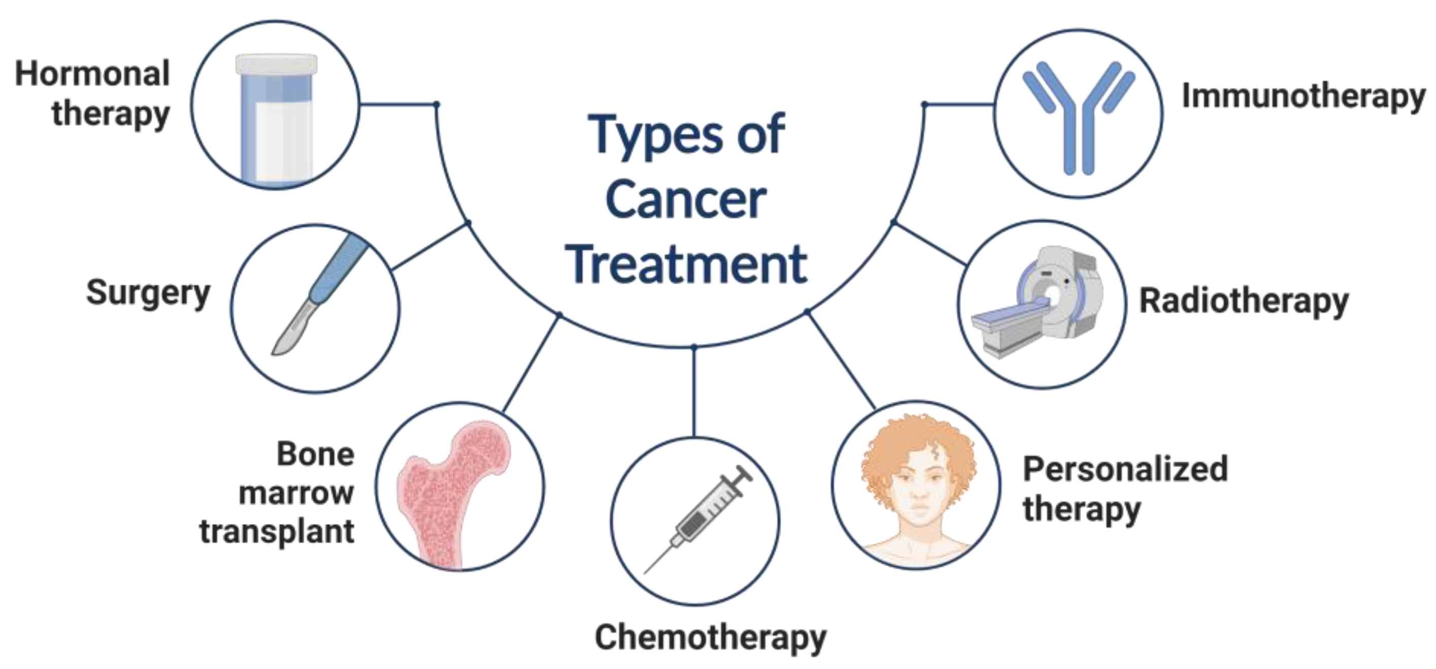
Figure 1. Types of cancer treatment.
Materials 16 05354 g001
Figure 2. Classification of nanoparticles based on their physicochemical properties.
Materials 16 05354 g002
Figure 3. Using iodide-doped gold nanoparticles and photoacoustic imaging to measure oxidative stress (adapted from reference [44]).
Materials 16 05354 g003
Figure 4. Selenium species with anticancer properties.
Materials 16 05354 g004
Figure 5. Several drug-targeting techniques for the tumor.
Materials 16 05354 g005
Figure 6. Active targeting of cancer stem cells with nanoparticles.
Materials 16 05354 g006
Figure 7. Nano-engineered MSCs as active targeting drug delivery vehicles.
Materials 16 05354 g007
Figure 8. Drug delivery platforms for cancer treatment. (A) Gold nanospheres, (B) Gold nanorods, (C) Carbon nanotubes, (D) Nanogels, (E) Polymeric NPs, (F) Polymeric micelles, and (G) Liposomes.
Materials 16 05354 g008
Table 1. Summary of Pd-based nanoparticles for cancer treatment.
Type of TherapyMaterialReference
Photothermal therapy (PTT)Pd NSs [55,56]
Pd collora[57]
Pd@Ag nanoplates[58]
Pd@Au nanoplates[59]
Pd NS-CO Pd-TAT[60]
PTT and photodynamic therapy (PDT)Pd@Pt-PEG-Ce6[61]
Pd@Ag@mSiO2-Ce6[62]
Pd-PEI-Ce6[63]
H-Pd NSs[64]
PLCs-HSA-ICG[65]
PTT and chemotherapyDox-loaded 8dc-Pd NPs[66]
SPNS-DOX[67]
Pd@Au-PEG-Pt[68]
HMSS-NH2/DOX@Pd[69]
PTT and radiation therapy[131I]PHPdNPs-DOX[70]
131I-Pd-PEG[71,72]
PTT and immunotherapyPd-CpG[73]
PTT and hydrogen therapyPdH0.2 nanocubes[74]
PdH-MOF[75]
Table 2. Different materials make typical NPs platforms for medical imaging.
NanoparticlesSize (nm)Targeting
Material
Cell LineImaging
Technology
Ref.
PLGA-mPEG151.1 ± 1.3cRGDSKOV-3 cellsUltrasound[95]
MnO-TETT6.7 ± 1.2NoneC6 glioma cellsFluorescence/T1-MRI 1[96]
Ultra small MnO@mesoporous silica30–50DoxHeLa cellsMRI-guided chemotherapy[97]
PEG–coated and Gdloaded flourescent
silica
125.5 ± 9.9YPSMA-1LNCaP and PC3 prostate cancer cellsMRI/fluorescence imaging[98]
Oxygen/indocyanine green-loaded (OINPs)300FolateSKOV3 ovarian cancer cellsUltrasound/
photoacoustic
[99]
1 Magnetic resonance imaging.
Table 3. Common nanoparticle platforms utilized in cryosurgery.
NPsSize (nm)Thermal Conductivity (W/m K)Heat
Capacity
(J/m3 K)
BenefitsRef.
Fe3O48–147.13.2 × 106More intracellular ice formation, high thermal conductivity[135]
MgO5034.33.2 × 106Nontoxic, biodegradable, and few side-effects[133]
HCPN-CG103.9 ± 1.5NoneNoneCold-responsive NP for control drug release and NIR-induced photothermal effect.[136]
PCM10–200.352.56 × 106Health tissue protection[137]
Au3297.72.2 × 106Good biological compatibility, high thermal conductivity[138]
Disclaimer/Publisher’s Note: The statements, opinions and data contained in all publications are solely those of the individual author(s) and contributor(s) and not of MDPI and/or the editor(s). MDPI and/or the editor(s) disclaim responsibility for any injury to people or property resulting from any ideas, methods, instructions or products referred to in the content.