Article

A pH-Responsive Polycaprolactone–Copper Peroxide Composite Coating Fabricated via Suspension Flame Spraying for Antimicrobial Applications

1
Cixi Biomedical Research Institute, Wenzhou Medical University, Wenzhou 325035, China
2
Institute of Biomedical Engineering, Ningbo Institute of Materials Technology and Engineering, Chinese Academy of Sciences, Ningbo 315201, China
3
Graduate School of Engineering, Tohoku University, Sendai 980-8577, Japan
4
Zhejiang-Japan Joint Laboratory for Antibacterial and Antifouling Technology, Ningbo Cixi Institute of Biomedical Engineering, Ningbo 315201, China
*
Authors to whom correspondence should be addressed.
Materials 2024, 17(11), 2666; https://doi.org/10.3390/ma17112666
Submission received: 24 April 2024 / Revised: 17 May 2024 / Accepted: 28 May 2024 / Published: 1 June 2024
(This article belongs to the Special Issue New Advances in Functionalization of Metal Materials)

Abstract

:
In this study, a pH-responsive polycaprolactone (PCL)–copper peroxide (CuO2) composite antibacterial coating was developed by suspension flame spraying. The successful synthesis of CuO2 nanoparticles and fabrication of the PCL-CuO2 composite coatings were confirmed by microstructural and chemical analysis. The composite coatings were structurally homogeneous, with the chemical properties of PCL well maintained. The acidic environment was found to effectively accelerate the dissociation of CuO2, allowing the simultaneous release of Cu2+ and H2O2. Antimicrobial tests clearly revealed the enhanced antibacterial properties of the PCL-CuO2 composite coating against both Escherichia coli and Staphylococcus aureus under acidic conditions, with a bactericidal effect of over 99.99%. This study presents a promising approach for constructing pH-responsive antimicrobial coatings for biomedical applications.

1. Introduction

In recent years, bacterial resistance infections have emerged as a significant global health challenge. Biomaterial-associated infections pose a serious threat to global human health [1]. Bacterial resistance to antibiotics can be acquired through mutations in the chromosomal genes or the horizontal transfer of resistance genes, resulting in infections that are difficult to treat [2]. In the medical field, the development of chronic wounds occurs when the healing process of hemostasis, inflammation, hyperplasia, and re-epithelialization is not completed promptly following a skin injury [3,4]. Bacterial infection poses a significant challenge in the management of chronic, non-healing wounds [5]. The interplay between the extended healing time of a wound, which heightens the risk of bacterial infection, and the presence of bacterial infection, particularly drug-resistant strains, which in turn delays wound healing, constitutes a challenging aspect of the management of chronic, hard-to-heal wounds. Therefore, there is a long-standing need for the development of innovative antibacterial materials [6]. These materials may include surface coatings, nanoparticles, or hydrogels designed to overcome the resistance of these microorganisms or enhance the efficacy of antibiotic therapy when used in combination. Metal nanoparticles are currently under investigation for their antimicrobial properties and have shown promise as effective antibacterial agents. Balcucho et al. utilized copper oxide (CuO) metal nanoparticles to fabricate composites capable of releasing Cu2+ ions, which showed remarkable growth inhibition of methicillin-resistant Staphylococcus aureus, exceeding four logarithms [7].
Among the various strategies explored, the catalytic treatment of metal peroxide nanoparticles based on the in situ Fenton reaction has garnered considerable attention as a promising antibacterial approach [8,9,10]. The mechanism underlying the Fenton reaction involves the conversion of hydrogen peroxide (H2O2) to highly reactive hydroxyl radicals (•OH). It can result in oxidation damage to the membrane and the cell wall and display high and broad-spectrum antibacterial activity compared with traditional antibiotics [11]. Several metal peroxide nanoparticles, such as zinc peroxide (ZnO2) and calcium peroxide (CaO2), have been constructed as Fenton reaction–based chemodynamic therapy (CDT) agents [12,13]. Recently, Lin et al. first reported the successful synthesis of CuO2 nanodots, which could self-supply H2O2 in the acidic environment and produce highly toxic •OH via the Fenton reaction between Cu2+ and H2O2 [14]. CuO2 is a copper oxide with a unique structure that contains Cu2⁺ and O22− ions in its molecular structure. It has a bent, end-on structure with inequivalent oxygens and peroxide-like O—O distances, typically 1.4–1.55 Å. It maintains the same spin multiplicity as Cu and CuO and presents a controversial ground state in the neutral 3d-metal dioxide series [15]. CuO2 is synthesized from H2O2 and Cu2+ under alkaline conditions. Under weak acid conditions, CuO2 could reversibly decompose into Cu2+ and H2O2, leading to a Fenton-like reaction between these decomposition products, which in turn generates reactive oxygen species [14]. Both Cu2+ and H2O2 are well-known antimicrobial agents and have been extensively studied for bacterial infection control. Unlike antibiotics, Cu2+ and H2O2 are not susceptible to bacterial resistance [16]. CuO2 demonstrated strong antibacterial effects for biofilm treatment and wound healing [17,18]. The initial environment at the site of bacterial infection is weakly acidic [19], which favors the dissociation of CuO2 and enables the application of the pH-responsiveness of CuO2.
However, the inherent instability of CuO2 under neutral conditions significantly limits its practical application [20]. Additionally, the indiscriminate nature of hydroxyl radicals produced via the Fenton reaction may pose risks of off-target cytotoxicity and tissue damage, highlighting the need for targeted delivery and controlled release strategies to minimize adverse effects. Studies have shown that encapsulation can significantly improve the stability of CuO2 [18,20]. Compared to other drug delivery systems (such as nanoparticles, electrostatic spinning [21,22], hydrogels [23], gelatin sponges [24,25], etc.), coatings have higher drug-carrying abilities, are easy to store, and can be used to treat large bacterial infections. The thermal spray processes for polymer coating production include flame spraying, high-velocity oxygen fuel (HVOF)/high-velocity air fuel (HVAF), plasma spraying, and cold spraying [26]. Coating biodegradable polymers by flame spraying is a widely used method, with advantages including low cost, simplicity, and environmental friendliness [27].
Polycaprolactone (PCL) is a hydrophobic polyester that has garnered considerable attention in various biomedical applications. This is primarily due to its exceptional biocompatibility, ability to blend with other polymers, distinctive rheological properties, and controlled release of active compounds. These characteristics are closely tied to its biodegradability [7,28]. Therefore, PCL presents itself as a promising choice for integration as a structural component of dressings that come into direct contact with living tissue. The incorporation of CuO2 nanoparticles into a PCL matrix not only preserves the intrinsic properties of the nanoparticles but also extends their stability and facilitates their controlled release [29].
This study focuses on developing a biodegradable and biocompatible material for antimicrobial applications in the weakly acidic microenvironment. An innovative approach using the suspension flame spraying method was employed to fabricate pH-responsive antimicrobial coatings with different contents of CuO2 nanoparticles in the PCL matrix. Various analyses were conducted to confirm the successful incorporation of CuO2. Their release mechanisms and antimicrobial properties were also examined under different pH conditions. The study offers a universal fabrication method for pH-responsive CuO2-loaded composite coatings for effectively combating biomaterial-associated infections.

2. Materials and Methods

2.1. Materials and Reagents

Copper (II) chloride dihydrate (CuCl2·2H2O), polyvinylpyrrolidone (PVP, MW of ~10,000), hydrogen peroxide (H2O2, 30%), sodium hydroxide (NaOH), sulfuric acid (H2SO4), and potassium permanganate (KMnO4) were purchased from Sinopharm Group Co., Ltd., Shanghai, China. PCL powders (200 mesh, MW of ~80,000) were provided by Nature Works, Minneapolis, MN, USA.

2.2. Sample Preparation

CuO2 was synthesized according to Lin et al. [14] with slight modifications. To begin, 5 g PVP was added into 50 mL of aqueous solution containing 0.05 M CuCl2. After that, 50 mL of 0.10 M NaOH and 5 mL of 30% H2O2 were sequentially incorporated into the above mixture solution. After stirring for 30 min, the resulting nanoparticles were separated and purified to obtain the CuO2 powder after freeze-drying.
PCL powders were suspended in 50:50 (v/v) ethanol/water, and then 0.1%, 0.3%, and 0.6% (wt/wt) CuO2 powders relative to PCL powders were added to prepare the PCL-CuO2 coatings. The coatings were prepared by flame spraying (CDS 8000, Castolin, Kriftel, Germany) on 316 L stainless-steel plates with dimensions of 25 × 20 × 2.0 mm [30]. The suspension was injected into the flame using a homemade spray atomizer. Acetylene was used as the fuel gas, with a flow rate of 1.5 Nm3/h and a working pressure of 0.1 MPa. Oxygen was used as the combustion gas, with a flow rate of 2.5 Nm3/h and a working pressure of 0.5 MPa. The spray distance was 200 mm. The thicknesses of PCL and PCL-CuO2 coatings were about 320–380 μm, measured by the coating thickness gauge. A schematic diagram of the preparation of the coating by liquid flame spraying is shown in Figure 1.

2.3. Sample Characterization

The microstructures of the powders and coatings were examined using a field emission scanning electron microscope (SEM, S4800, Hitachi, Tokyo, Japan). The cross sections of the coatings were characterized using an energy dispersive X-ray detector (EDX, XFlash 6–100, Bruker, Ettlingen, Germany). The particle size and zeta potential of CuO2 were measured by nanometer particle size analyzer (Litesizer 500, Anton Paar, Graz, Austria). Chemical composition was characterized using X-ray photoelectron spectroscopy (XPS, AXIS ULTRA DLD, Shimadzu, Kyoto, Japan). The X-ray diffraction (XRD) patterns were obtained on a D8-Advance X-ray diffractometer (Bruker, Germany), with Cu Kα radiation at a voltage of 40 kV and a tube current of 40 mA. Chemistry of the powders and coatings was detected by Fourier transform infrared spectroscopy (FT-IR, Nicolet iS50, Thermo Scientific, Waltham, MA, USA), operated at a spectral resolution of 4 cm−1 with a scan range of 4000~400 cm−1.

2.4. Colorimetric Determination of Peroxo Groups

KMnO4 solution is a strong purplish-red oxidizer that can oxidize H2O2, thus causing the purplish-red color to fade [14]. KMnO4 was dissolved in 0.1 M aqueous H2SO4 solution to obtain a concentration of 50 μg/mL, and then the acidic KMnO4 solution was treated with H2O (control), H2O2, Cu(OH)2, freshly synthesized CuO2, and the CuO2 suspension placed at room temperature for a short period, consecutively. After 10 min of incubation, photographs were taken to record the fading, and UV-vis spectra were examined at 400–650 nm.

2.5. pH-Responsive Release of Copper Ions

The areas surrounding the coating and the back of the substrate were sealed with epoxy resin, ensuring that only the coating surface remained exposed. For the release analysis, three parallel samples were used for each group of PCL-CuO2 composite coatings and each pH condition. Generally, coating samples were individually immersed in 15 mL PBS buffer at pH 7.4 or pH 5.5 in 50-mL falcon tubes with gentle shaking at 37 °C. At predetermined time intervals, 6 mL solution was collected with the addition of 6 mL fresh PBS. The content of copper ions was determined using inductively coupled plasma optical emission spectroscopy (ICP-OES; SPECTRO ARCOS, SPECTRO Analytical Instruments, Kleve, Germany).

2.6. In Vitro Antibacterial Effect of PCL-CuO2 Coatings

Escherichia coli (E. coli, ATCC25922) and Staphylococcus aureus (S. aureus, ATCC6538) were used to evaluate the antimicrobial effect of the coatings. We performed the tests according to the Japanese Industrial Standard (JIS) Z 2801 (ISO 22196:2011 [31], measurement of antibacterial activity on plastics and other nonporous surfaces) with some modifications [7]. Three parallel samples of each group of coatings were placed in sterile 6-well plates, and the bacteria cultured to logarithmic growth phase were diluted to about 1×10⁶ CFU/mL by gradient dilution in PBS at pH 7.4 or pH 5.5. Then, 10 μL bacterial suspension was dropped on the coating surface and covered with a sterile polyethylene film (5 × 5 mm) and then incubated at 37 °C with a relative humidity of at least 95% for 2 h. Afterward, bacteria were collected by washing with 1 mL PBS buffer and used for determination of survival rate by standard plate counting method. LB medium was used to grow E. coli, and TSB was used to grow S. aureus. The bactericidal effect can be calculated using the formula: R = (N0 − N1)/N0 × 100%, where N0 represents the number of bacterial colonies in the control group and N1 represents the number of bacterial colonies in the experimental group.

3. Results and Discussion

3.1. Morphological Characterization of PCL and CuO2 Powders

The morphology of commercially available PCL powders and synthesized nanosized CuO2 particles was examined by SEM analysis. The PCL powders showed irregular morphology and a wide size range, roughly from 10 to 60 μm (Figure 2a). The synthesized CuO2 particles were formed by aggregation of low nanosized particles and showed irregular shape and good dispersion (Figure 2b).

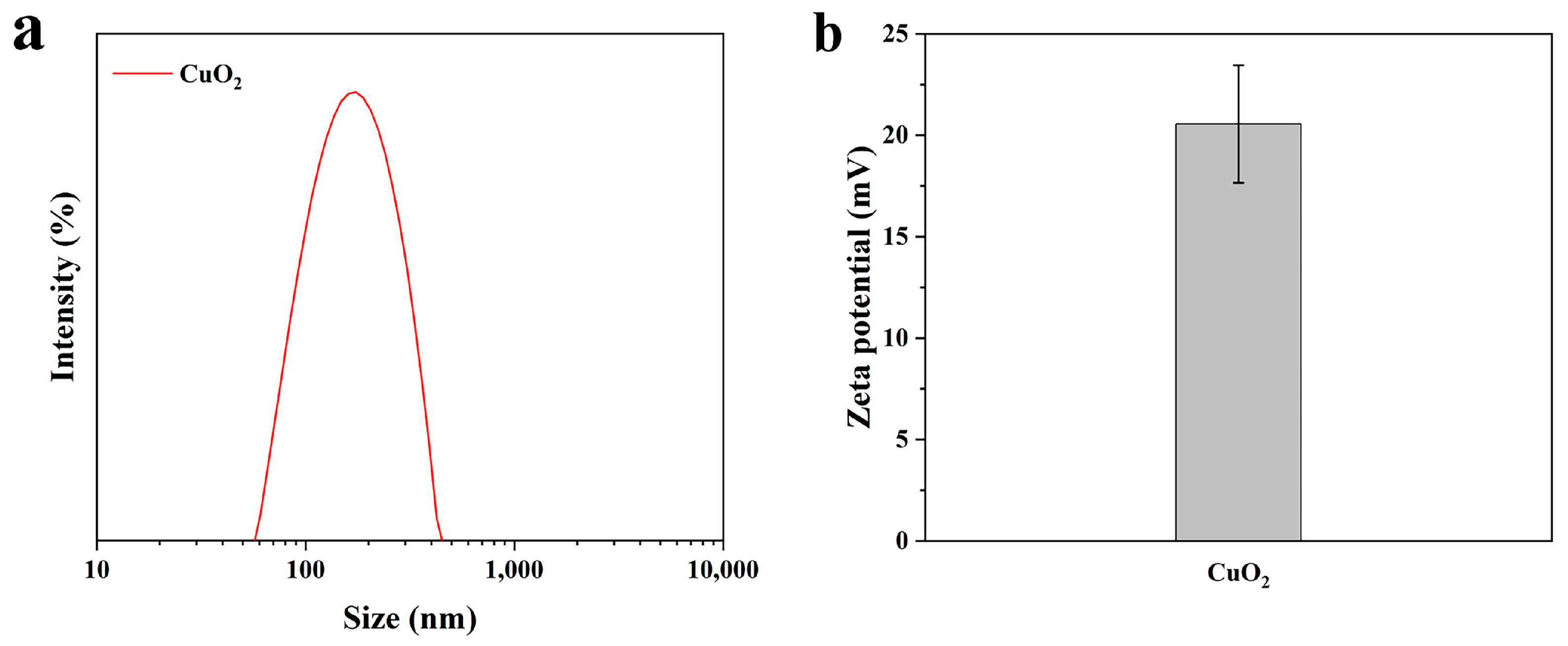
3.2. The Particle size and Zeta Potential of CuO2

The particle size analysis showed that the average hydrodynamic diameter of the CuO2 was 163 ± 1.80 nm (Figure 3a). The results are consistent with the SEM results described above. Zeta potential is a good indicator of the magnitude of electrostatic interactions between dispersed particles and can be used as a reference for the stability of nanoparticle dispersions [32]. The average zeta potential carried by CuO2 was 20.6 ± 2.9 mV (Figure 3b), indicating that it has good dispersibility.

3.3. XPS Analysis of CuO2 Powders

The X-ray photoelectron (XPS) spectrum of the fully scanned region of CuO2 powders exhibited characteristic peaks of C 1s, N 1s, O 1s, and Cu 2p (Figure 4a). The peaks of C 1s and N 1s indicated the presence of PVP. The XPS spectrum of Cu 2p displayed characteristic peaks at 953.9 eV and 933.6 eV, respectively, accompanied by two satellite peaks at 962.1 eV and 942.1 eV, respectively, indicating that the valence state of copper in CuO2 is +2 [14] (Figure 4b). Furthermore, the O 1s XPS spectrum showed three distinct peaks at 529.5, 531.5, and 533.0 eV, ascribed to Cu-O, C=O, and O-O bonds, respectively [33]. The presence of peroxo groups in the synthesized CuO2 powder was confirmed by the presence of the O-O bond (Figure 4c). The XPS spectrum of C 1s showed three characteristic peaks at 284.8, 286.3, and 288.3 eV, which were assigned to C-C, C-N, and C=O, respectively [34] (Figure 4d). The above indicated the successful preparation of CuO2 nanoparticles.

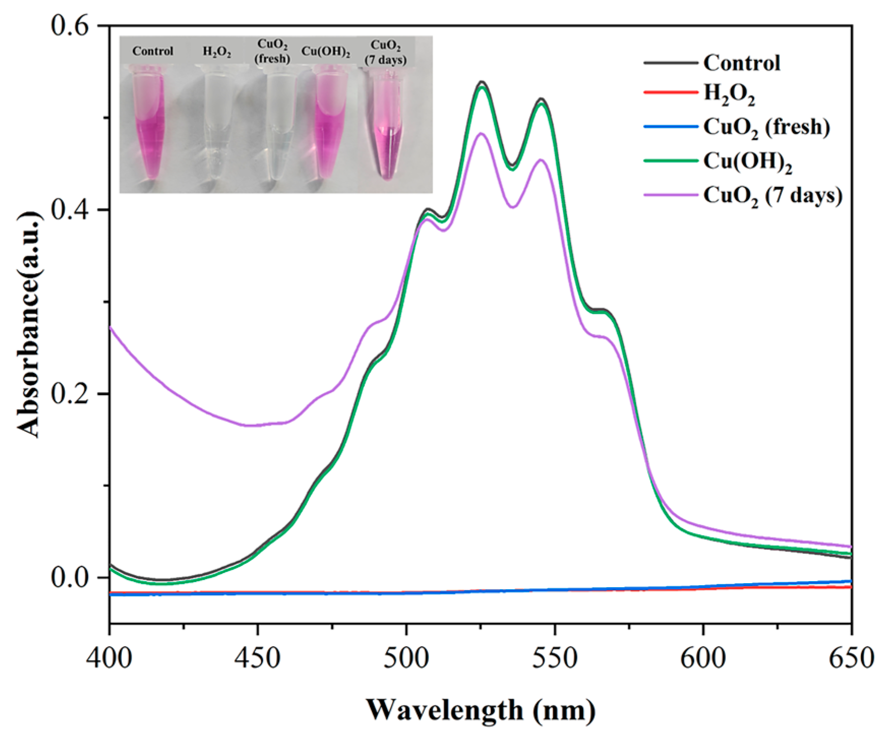
3.4. Potassium Permanganate Colorimetric Analysis of Synthesized CuO2

Furthermore, a KMnO4-based colorimetric method was used to examine the synthesized CuO2 powders. The absorption peaks of MnO4 disappeared when mixed with H2O2 or the synthesized CuO2 powder but remained when mixed with H2O or Cu(OH)2 (Figure 5). It also suggests the presence of peroxo groups in the synthesized CuO2 powders, which is consistent with the above XPS results. However, CuO2 in water is unstable and easily decomposed [20]. As shown in Figure 5, the CuO2 suspension almost completely lost the ability to decolorize potassium permanganate after sitting at room temperature for 7 days, indicating the fast decomposition of CuO2.

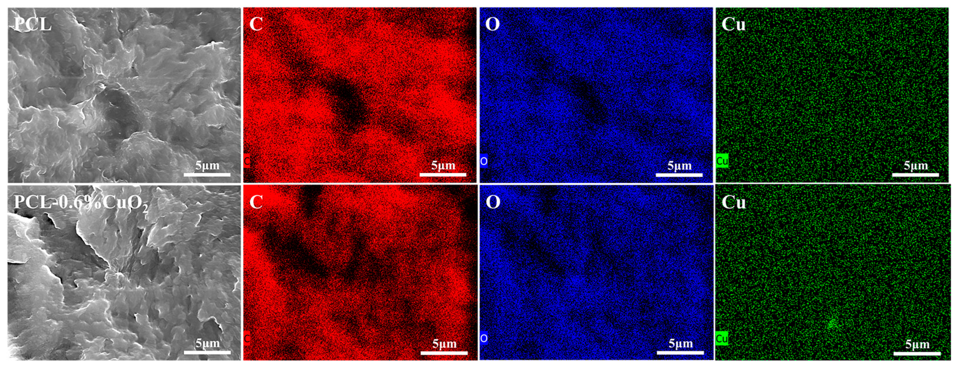
3.5. SEM and EDX Analysis of the PCL and PCL-CuO2 Coatings

Figure 6a–d demonstrated that PCL and PCL-CuO2 coatings had smooth surfaces with visible pores. This might result from the fast evaporation of deionized water or ethanol during the manufacturing process [35]. There were no visible CuO2 particles on the surface or cross-section, which might be due to the homogeneous entrapment of PCL [36], as shown in Figure 6(a–d,a-1–d-1). These results demonstrated that the addition of CuO2 has no significant impact on either the surface or internal structure of the PCL coating. Furthermore, an enrichment of Cu was shown in the PCL-0.6% CuO2 coating in Figure 7, indicating the incorporation of CuO2 nanoparticles within the PCL matrix.

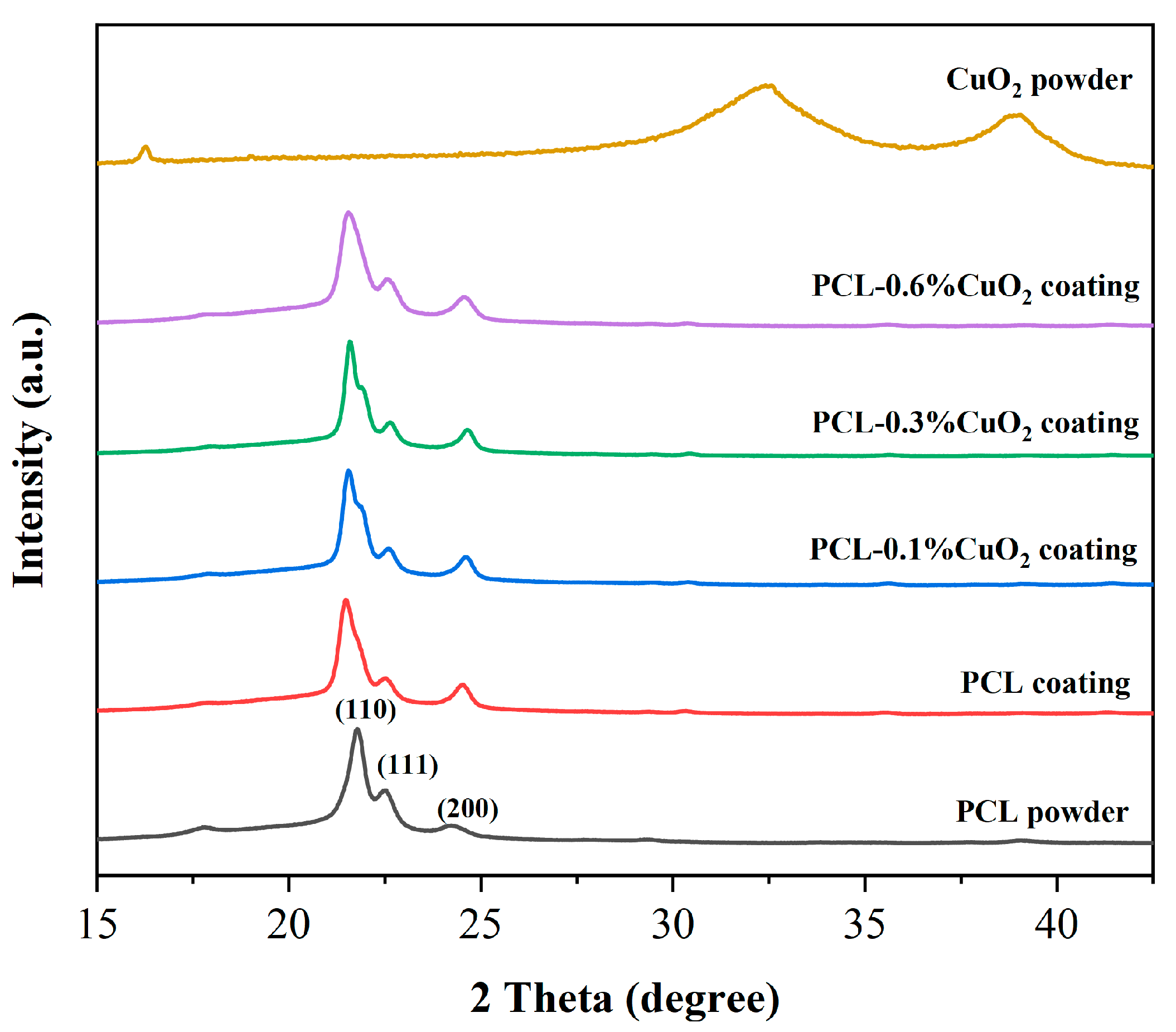
3.6. XRD Analysis of the Powders and Coatings

In Figure 8, the XRD pattern analysis showed that the PCL powder exhibited intense diffraction peaks at 21.8°, 22.5°, and 24.2°, which are assigned to the planes (110), (111), and (200) of PCL, respectively [37]. The XRD pattern of the synthesized CuO2 nanoparticles was consistent with that reported in the literature, with two envelope peaks at 32.3° and 38.8° [33], indicating poor crystallization of the synthesized CuO2 [38]. These two envelope peaks were not observed in the PCL-CuO2 composite coating, which might be due to the low content of CuO2. These results indicated the successful synthesis of CuO2 nanoparticles and fabrication of PCL-CuO2 coatings.

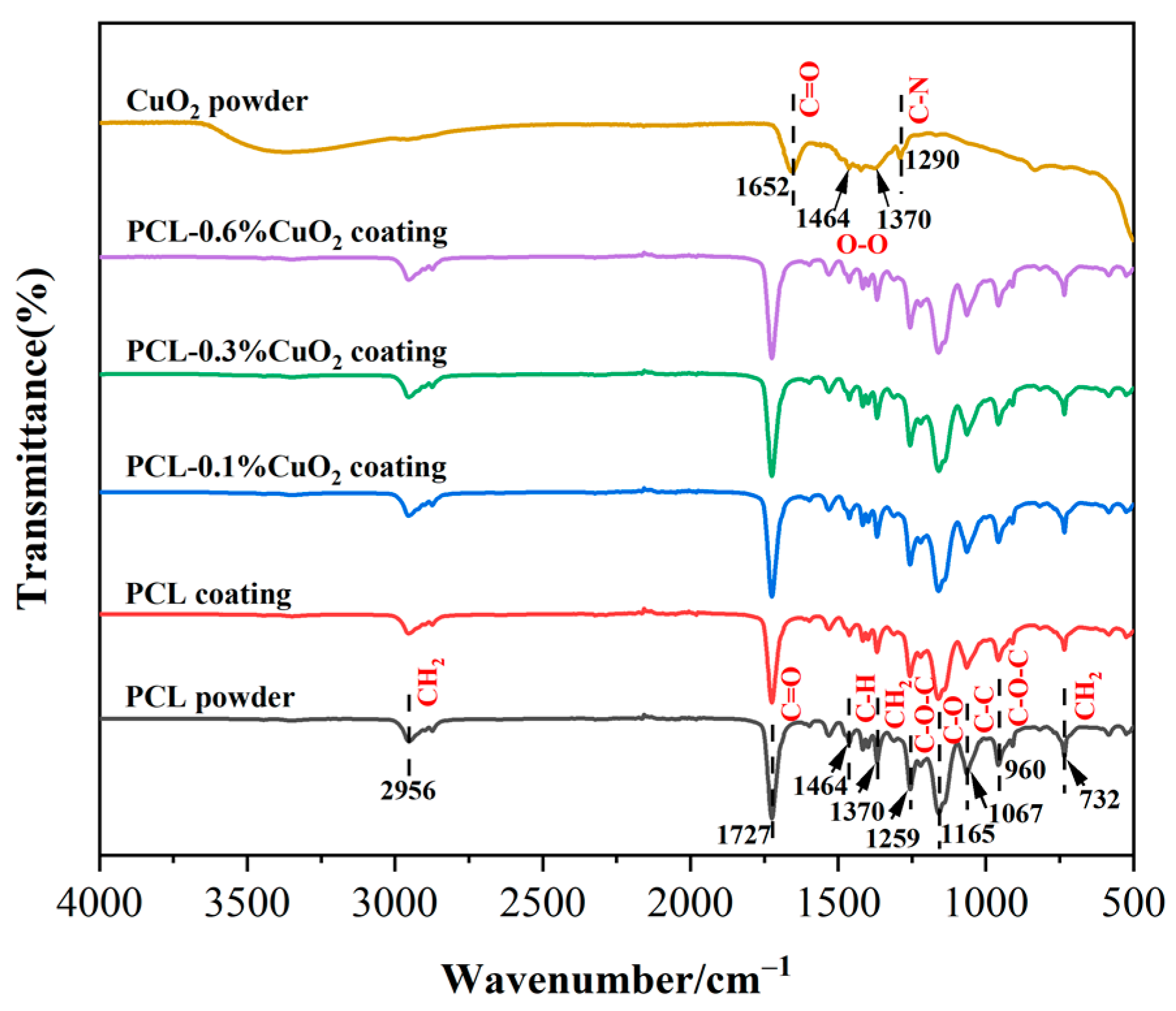
3.7. FT-IR Analysis of the Powders and Coatings

The FT-IR spectra of PCL powder, CuO2 powder, PCL coating, and PCL-CuO2 composite coatings were analyzed, as shown in Figure 9. For PCL powder, the peak at 2956 cm−1 was the asymmetric stretching vibration of CH2, and the characteristic peak at 1727 cm−1 was the stretching vibration of the C=O group. The peak of stretching vibration of C-H at 1464 cm−1, 1370 cm−1 belongs to CH2, and the peak of asymmetric stretching vibration of C-O-C group at 1259 cm−1, 1165 cm−1 belongs to C-O. The peaks at 1067 cm−1, 960 cm−1, and 732 cm−1 represented the C-C, C-O-C, and CH2 vibration peaks, respectively [39]. There was no significant difference in the position of the infrared absorption peak between the PCL powder and the prepared coating, indicating that the chemical composition of the PCL coatings prepared by suspension flame spraying was not changed. The characteristic peaks at 1652 cm−1 and 1290 cm−1 in the CuO2 powder are attributed to the tensile vibration between C=O and C-N in PVP [40], and the two small peaks displayed at 1464 cm−1 and 1370 cm−1 are the characteristic peaks of peroxide group (O-O) [41]. The above characteristic peaks of the CuO2 powder were not detected in the PCL-CuO2 coatings, which might be due to the low content of CuO2 in the composite coatings.

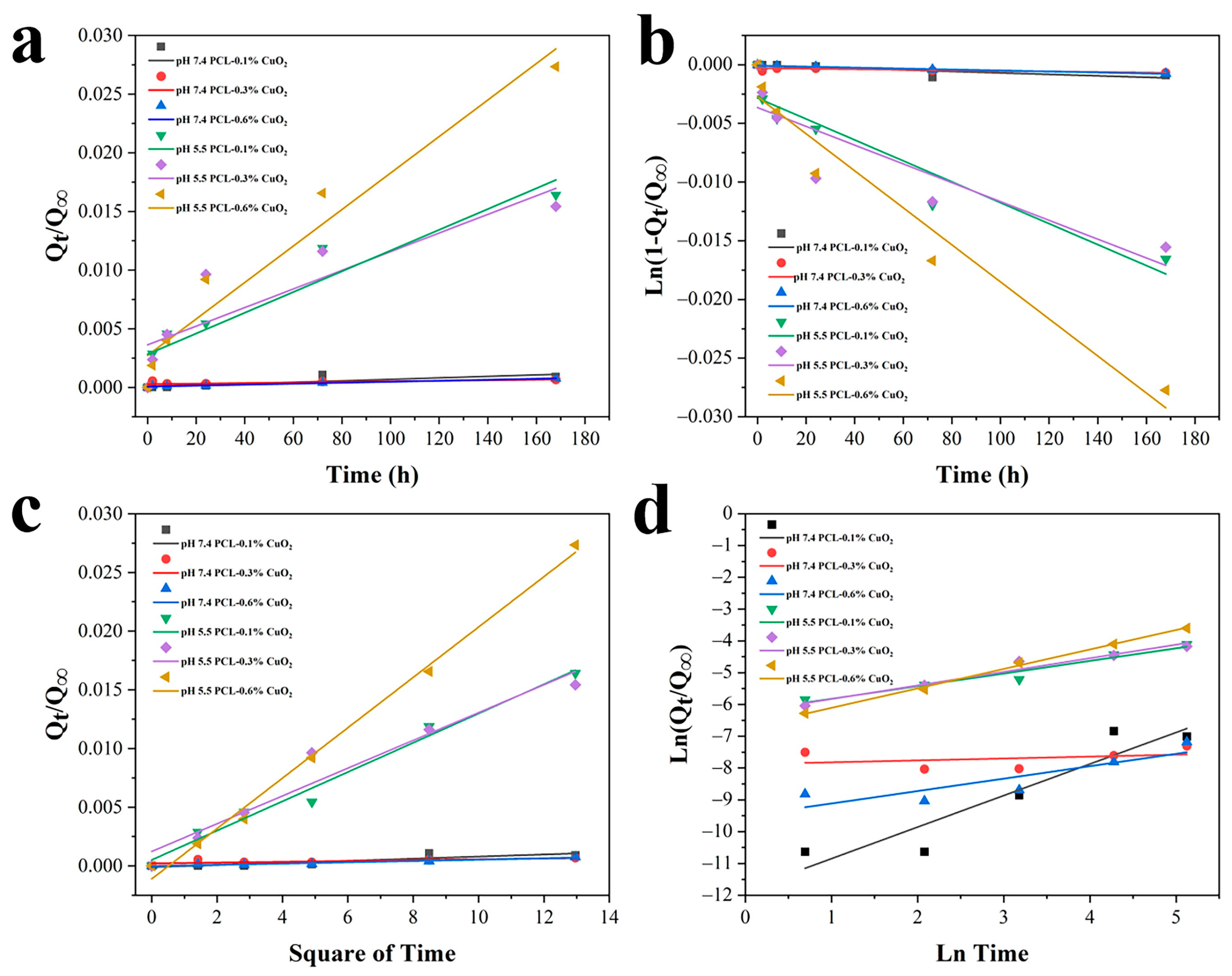
3.8. pH-Responsive Release of PCL-CuO2 Coatings

Under weakly acidic conditions, CuO2 decomposes to produce Cu2+ and H2O2, which further produces reactive oxygen species via the Fenton reaction [14]. The release of Cu2+ from the PCL-CuO2 coatings was examined under different pH conditions (Figure 10). As expected, increasing Cu2+ release with the content of CuO2 was clearly observed at pH 5.5, while the release at pH 7.4 was negligible. At pH 5.5, the copper ion release from PCL-0.1% CuO2 coating for 7 days was 0.15 mg/L; from PCL-0.3% CuO2 coating, it was 0.41 mg/L; and from PCL-0.6% CuO2 coating, it was 1.45 mg/L. However, the release of copper ions from PCL-0.1% CuO2, PCL-0.3% CuO2, and PCL-0.6% CuO2 coatings at pH 7.4 for 7 days were only 0.01, 0.02, and 0.04 mg/L, respectively. The above indicated that the PCL-CuO2 coatings showed sustained release in an acid-responsive and dose-dependent manner. A previous study reported that a concentration of Cu2+ of no more than 9 ppm showed no significant effect on the growth of cells compared with normal conditions [42]. In this study, the release of Cu2+ was very slow, and the concentration of Cu2+ after 7 days continuous release was significantly lower than 9 ppm, suggesting high biocompatibility of the PCL-CuO2 coatings.
Furthermore, the data were fitted with four commonly used drug release models (Figure 11a–d) and listed in Table 1. It was noted that, under pH 5.5, the release for the PCL-0.3% CuO2 and PCL-0.6% CuO2 coatings fitted the Korsmeyer–Peppas model, with the highest linearity correlation coefficient (R2 = 0.964, 0.997). The release exponent n for PCL-0.3% CuO2 was ≤0.45, indicating the drug release mechanism follows Fick’s laws of diffusion. On the contrary, the release exponent n for PCL-0.6% CuO2 was higher than 0.45, suggesting a combined erosion and diffusion release mechanism, named non-Fickian transport. The release of the PCL-0.1% CuO2 coating was best interpreted by the Higuchi equation (R2 = 0.984), indicating a relatively slower diffusion from the PCL matrix. However, it was difficult to define the release models of the PCL-CuO2 coatings under pH 7.4, which generally showed an R2 value lower than those under pH 5.5. This might result from the extremely slower release of the composite coatings under pH 7.4. The complexity of the release mechanisms for these coatings might be due to the combined impact of CuO2 diffusion from the coating and the decomposition reaction of CuO2.

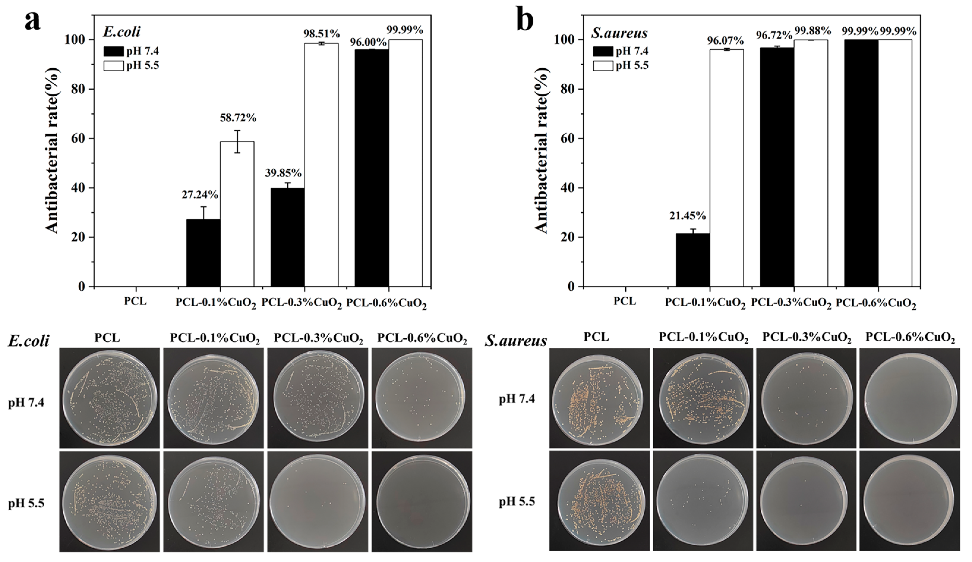
3.9. In Vitro Antibacterial Properties of PCL-CuO2 Coatings

The decomposition of CuO2 displays a pH-responsive manner and can continuously release copper ions and H2O2 under weakly acidic conditions. Therefore, the antibacterial effect of the PCL-CuO2 composite coating was examined at pH 5.5 and 7.4 using E. coli and S. aureus. It was found that the composite coatings exhibited a significantly higher antibacterial effect against both E. coli and S. aureus at pH 5.5 compared to pH 7.4 (Figure 12a,b). Furthermore, the antibacterial effect of the composite coatings was dose-dependent and reached over 99.99% killing efficacy against both E. coli and S. aureus for 0.6% CuO2 content at pH 5.5. Moreover, the composite coatings displayed superior killing efficacy for S. aureus compared to E. coli under both pH conditions, which might be due to there being a difference in the antimicrobial activity of metal nanoparticles depending on the bacterial wall structure [7]. Gram-positive bacteria with higher peptidoglycan and cell wall protein content are more sensitive to copper [16,43]. Excitingly, there is no significant change in the antimicrobial effect of these coatings after 10 months (Figure S1), suggesting the excellent stability of CuO2 within the PCL-CuO2 coatings.

4. Conclusions

In this research, PCL-CuO2 composite coatings with pH-responsive antimicrobial properties were fabricated using a suspension flame spraying technique. Morphology and chemical characterization confirmed the well-maintained PCL matrix and the successful incorporation of CuO2 nanoparticles in the composite coatings. The release study found that the PCL-CuO2 coatings showed sustained release in an acid-responsive and dose-dependent manner. The composite coating with 0.6% (w/w) CuO2 exhibited over 99.99% antibacterial effect against E. coli and S. aureus under a mildly acidic (pH 5.5) condition. The CuO2 nanoparticles in the PCL-CuO2 composite coatings showed significantly enhanced stability in comparison with fast decomposition in an aqueous solution and still exhibited potent antimicrobial efficacy after 10 months storage. Our cost-effective fabrication method of pH-responsive antimicrobial coatings provides a new solution for the development of biomedical materials for various applications.

Supplementary Materials

The following supporting information can be downloaded at: https://www.mdpi.com/article/10.3390/ma17112666/s1, Figure S1. In vitro antibacterial activity test of PCL-CuO2 coatings at pH 7.4 and pH 5.5 after 10 months of storage at room temperature. Antimicrobial effect of (a) E. coli and (b) S. aureus after 2 h incubation on the PCL-CuO2 composite coatings.

Author Contributions

Conceptualization, B.Z. and T.C.; investigation, T.C., D.Z., Y.Z., D.K., Z.W., Z.H., M.S. and X.A.; formal analysis, T.C., D.Z., Y.Z. and D.K.; visualization, D.Z., Y.Z., Z.H. and Y.D.; resources, Z.W.; validation, Z.W., Z.H., M.S. and X.A.; methodology, Y.D.; writing—original draft preparation, T.C. and B.Z.; writing—review and editing, B.Z. and H.L.; supervision, B.Z.; funding acquisition, B.Z. and H.L. All authors have read and agreed to the published version of the manuscript.

Funding

This research was supported by Zhejiang Basic Public Welfare Research Program of China (LGF20C100001), National Natural Science Foundation of China (41706076), Key Research and Development Program of Ningbo (2023Z195), and K.C. Wong Education Foundation (GJTD-2019-13).

Institutional Review Board Statement

Not applicable.

Informed Consent Statement

Not applicable.

Data Availability Statement

There is no data used in the study.

Conflicts of Interest

The authors declare no conflict of interest.

References

  1. Khameneh, B.; Diab, R.; Ghazvini, K.; Fazly Bazzaz, B.S. Breakthroughs in bacterial resistance mechanisms and the potential ways to combat them. Microb. Pathog. 2016, 95, 32–42. [Google Scholar] [CrossRef] [PubMed]
  2. Bakkeren, E.; Diard, M.; Hardt, W.D. Evolutionary causes and consequences of bacterial antibiotic persistence. Nat. Rev. Microbiol. 2020, 18, 479–490. [Google Scholar] [CrossRef] [PubMed]
  3. Rodrigues, M.; Kosaric, N.; Bonham, C.A.; Gurtner, G.C. Wound Healing: A Cellular Perspective. Physiol. Rev. 2019, 99, 665–706. [Google Scholar] [CrossRef] [PubMed]
  4. Olsson, M.; Järbrink, K.; Divakar, U.; Bajpai, R.; Upton, Z.; Schmidtchen, A.; Car, J. The humanistic and economic burden of chronic wounds: A systematic review. Wound Repair Regen. 2019, 27, 114–125. [Google Scholar] [CrossRef] [PubMed]
  5. Yang, H.; Wang, W.-S.; Tan, Y.; Zhang, D.-J.; Wu, J.-J.; Lei, X. Investigation and analysis of the characteristics and drug sensitivity of bacteria in skin ulcer infections. Chin. J. Traumatol. 2017, 20, 194–197. [Google Scholar] [CrossRef]
  6. Nudelman, R.; Gavriely, S.; Bychenko, D.; Barzilay, M.; Gulakhmedova, T.; Gazit, E.; Richter, S. Bio-assisted synthesis of bimetallic nanoparticles featuring antibacterial and photothermal properties for the removal of biofilms. J. Nanobiotechnol. 2021, 19, 452. [Google Scholar] [CrossRef] [PubMed]
  7. Balcucho, J.; Narvaez, D.M.; Castro-Mayorga, J.L. Antimicrobial and Biocompatible Polycaprolactone and Copper Oxide Nanoparticle Wound Dressings against Methicillin-Resistant Staphylococcus aureus. Nanomaterials 2020, 10, 1692. [Google Scholar] [CrossRef] [PubMed]
  8. Xie, X.; Wang, R.; Zhang, X.; Ren, Y.; Du, T.; Ni, Y.; Yan, H.; Zhang, L.; Sun, J.; Zhang, W.; et al. A photothermal and self-induced Fenton dual-modal antibacterial platform for synergistic enhanced bacterial elimination. Appl. Catal. B 2021, 295, 120315. [Google Scholar] [CrossRef]
  9. Park, S.C.; Kim, N.H.; Yang, W.; Nah, J.W.; Jang, M.K.; Lee, D. Polymeric micellar nanoplatforms for Fenton reaction as a new class of antibacterial agents. J. Control. Release 2016, 221, 37–47. [Google Scholar] [CrossRef]
  10. Li, F.; Huang, K.; Chang, H.; Liang, Y.; Zhao, J.; Yang, S.; Liu, F. A polydopamine coated nanoscale FeS theranostic platform for the elimination of drug-resistant bacteria via photothermal-enhanced Fenton reaction. Acta Biomater. 2022, 150, 380–390. [Google Scholar] [CrossRef]
  11. Liu, Y.; Guo, Z.; Li, F.; Xiao, Y.; Zhang, Y.; Bu, T.; Jia, P.; Zhe, T.; Wang, L. Multifunctional Magnetic Copper Ferrite Nanoparticles as Fenton-like Reaction and Near-Infrared Photothermal Agents for Synergetic Antibacterial Therapy. ACS Appl. Mater. Interfaces 2019, 11, 31649–31660. [Google Scholar] [CrossRef]
  12. Bergs, C.; Brück, L.; Rosencrantz, R.R.; Conrads, G.; Elling, L.; Pich, A. Biofunctionalized zinc peroxide (ZnO2) nanoparticles as active oxygen sources and antibacterial agents. RSC Adv. 2017, 7, 38998–39010. [Google Scholar] [CrossRef]
  13. Thi, P.L.; Lee, Y.; Tran, D.L.; Hoang Thi, T.T.; Park, K.M.; Park, K.D. Calcium peroxide-mediated in situ formation of multifunctional hydrogels with enhanced mesenchymal stem cell behaviors and antibacterial properties. J. Mater. Chem. B 2020, 8, 11033–11043. [Google Scholar] [CrossRef] [PubMed]
  14. Lin, L.S.; Huang, T.; Song, J.; Ou, X.Y.; Wang, Z.; Deng, H.; Tian, R.; Liu, Y.; Wang, J.F.; Liu, Y.; et al. Synthesis of Copper Peroxide Nanodots for H2O2 Self-Supplying Chemodynamic Therapy. J. Am. Chem. Soc. 2019, 141, 9937–9945. [Google Scholar] [CrossRef]
  15. Gutsev, G.L.; Rao, B.K.; Jena, P. Systematic Study of Oxo, Peroxo, and Superoxo Isomers of 3d-Metal Dioxides and Their Anions. J. Phys. Chem. A 2000, 104, 11961–11971. [Google Scholar] [CrossRef]
  16. Zhang, R.; Jiang, G.; Gao, Q.; Wang, X.; Wang, Y.; Xu, X.; Yan, W.; Shen, H. Sprayed copper peroxide nanodots for accelerating wound healing in a multidrug-resistant bacteria infected diabetic ulcer. Nanoscale 2021, 13, 15937–15951. [Google Scholar] [CrossRef]
  17. Li, M.; Lan, X.; Han, X.; Shi, S.; Sun, H.; Kang, Y.; Dan, J.; Sun, J.; Zhang, W.; Wang, J. Acid-Induced Self-Catalyzing Platform Based on Dextran-Coated Copper Peroxide Nanoaggregates for Biofilm Treatment. ACS Appl. Mater. Interfaces 2021, 13, 29269–29280. [Google Scholar] [CrossRef]
  18. Cui, H.; Liu, M.; Yu, W.; Cao, Y.; Zhou, H.; Yin, J.; Liu, H.; Que, S.; Wang, J.; Huang, C.; et al. Copper Peroxide-Loaded Gelatin Sponges for Wound Dressings with Antimicrobial and Accelerating Healing Properties. ACS Appl. Mater. Interfaces 2021, 13, 26800–26807. [Google Scholar] [CrossRef] [PubMed]
  19. Jones, E.M.; Cochrane, C.A.; Percival, S.L. The Effect of pH on the Extracellular Matrix and Biofilms. Adv. Wound Care 2015, 4, 431–439. [Google Scholar] [CrossRef]
  20. Zu, Y.; Wang, Y.; Yao, H.; Yan, L.; Yin, W.; Gu, Z. A Copper Peroxide Fenton Nanoagent-Hydrogel as an In Situ pH-Responsive Wound Dressing for Effectively Trapping and Eliminating Bacteria. ACS Appl. Bio Mater. 2022, 5, 1779–1793. [Google Scholar] [CrossRef]
  21. Yang, S.B.; Rabbani, M.M.; Ji, B.C.; Han, D.W.; Lee, J.S.; Kim, J.W.; Yeum, J.H. Optimum Conditions for the Fabrication of Zein/Ag Composite Nanoparticles from Ethanol/H2O Co-Solvents Using Electrospinning. Nanomaterials 2016, 6, 230. [Google Scholar] [CrossRef] [PubMed]
  22. Li, H.; Chen, X.; Lu, W.; Wang, J.; Xu, Y.; Guo, Y. Application of Electrospinning in Antibacterial Field. Nanomaterials 2021, 11, 1822. [Google Scholar] [CrossRef] [PubMed]
  23. Varaprasad, K.; Mohan, Y.M.; Vimala, K.; Raju, K.M. Synthesis and Characterization of Hydrogel-Silver Nanoparticle-Curcumin Composites for Wound Dressing and Antibacterial Application. J. Appl. Polym. Sci. 2011, 121, 784–796. [Google Scholar] [CrossRef]
  24. Ulubayram, K.; Cakar, A.N.; Korkusuz, P.; Ertan, C.; Hasirci, N. EGF containing gelatin-based wound dressings. Biomaterials 2001, 22, 1345–1356. [Google Scholar] [CrossRef] [PubMed]
  25. Bates, M.G.; Risselada, M.; Peña-Hernandez, D.C.; Hendrix, K.; Moore, G.E. Antibacterial activity of silver nanoparticles against Escherichia coli and methicillin-resistant Staphylococcus pseudintermedius is affected by incorporation into carriers for sustained release. Am. J. Vet. Res. 2024, 85, 1–11. [Google Scholar] [CrossRef]
  26. Koivuluoto, H. A Review of Thermally Sprayed Polymer Coatings. J. Therm. Spray Technol. 2022, 31, 1750–1764. [Google Scholar] [CrossRef]
  27. Pan, Y.; Zhou, D.; Cui, T.; Zhang, Y.; Ye, L.; Tian, Y.; Zhou, P.; Liu, Y.; Saitoh, H.; Zhang, B.; et al. A facile strategy for fine-tuning the drug release efficacy of poly-L-lactic acid-polycaprolactone coatings by liquid flame spray. Prog. Org. Coat. 2023, 183, 107807. [Google Scholar] [CrossRef]
  28. Zhou, J.; Yang, Y.; Detsch, R.; Boccaccini, A.R.; Virtanen, S. Iron surface functionalization system—Iron oxide nanostructured arrays with polycaprolactone coatings: Biodegradation, cytocompatibility, and drug release behavior. Appl. Surf. Sci. 2019, 492, 669–682. [Google Scholar] [CrossRef]
  29. Zhuo, X.; Lei, T.; Miao, L.; Chu, W.; Li, X.; Luo, L.; Gou, J.; Zhang, Y.; Yin, T.; He, H.; et al. Disulfiram-loaded mixed nanoparticles with high drug-loading and plasma stability by reducing the core crystallinity for intravenous delivery. J. Colloid Interface Sci. 2018, 529, 34–43. [Google Scholar] [CrossRef]
  30. Pan, Y.; Ye, L.; Zhou, P.; Feng, X.; Liu, Y.; Zhang, B.; Li, H. Facile suspension flame spray fabrication of polylactic acid-povidone-iodine coatings for antimicrobial applications. Mater. Lett. 2023, 341, 134293. [Google Scholar] [CrossRef]
  31. ISO 22196:2011; Measurement of Antibacterial Activity on Plastics and Other Non-Porous Surfaces. ISO: Geneva, Switzerland, 2011.
  32. Pochapski, D.J.; Carvalho dos Santos, C.; Leite, G.W.; Pulcinelli, S.H.; Santilli, C.V. Zeta Potential and Colloidal Stability Predictions for Inorganic Nanoparticle Dispersions: Effects of Experimental Conditions and Electrokinetic Models on the Interpretation of Results. Langmuir 2021, 37, 13379–13389. [Google Scholar] [CrossRef]
  33. Yang, D.; Huo, J.; Zhang, Z.; An, Z.; Dong, H.; Wang, Y.; Duan, W.; Chen, L.; He, M.; Gao, S.; et al. Citric acid modified ultrasmall copper peroxide nanozyme for in situ remediation of environmental sulfonylurea herbicide contamination. J. Hazard. Mater. 2023, 443 Pt B, 130265. [Google Scholar] [CrossRef]
  34. Atif, M.; Chen, C.; Irfan, M.; Mumtaz, F.; He, K.; Zhang, M.; Chen, L.; Wang, Y. Poly(2-methyl-2-oxazoline) and poly(4-vinyl pyridine) based mixed brushes with switchable ability toward protein adsorption. Eur. Polym. J. 2019, 120, 109199. [Google Scholar] [CrossRef]
  35. Qi, Z.; Yu, H.; Chen, Y.; Zhu, M. Highly porous fibers prepared by electrospinning a ternary system of nonsolvent/solvent/poly(l-lactic acid). Mater. Lett. 2009, 63, 415–418. [Google Scholar] [CrossRef]
  36. Ebrahimifar, M.; Taherimehr, M. Evaluation of in-vitro drug release of polyvinylcyclohexane carbonate as a CO2-derived degradable polymer blended with PLA and PCL as drug carriers. J. Drug Delivery Sci. Technol. 2021, 63, 102491. [Google Scholar] [CrossRef]
  37. Yakub, G.; Ignatova, M.; Manolova, N.; Rashkov, I.; Toshkova, R.; Georgieva, A.; Markova, N. Chitosan/ferulic acid-coated poly(epsilon-caprolactone) electrospun materials with antioxidant, antibacterial and antitumor properties. Int. J. Biol. Macromol. 2018, 107 Pt A, 689–702. [Google Scholar] [CrossRef]
  38. Cheng, P.; Luo, Z.; Che, Y.; Liu, Y.; Ke, F.; Cheng, G. Preparation and Study of Copper Peroxide and Copper Oxide for Electrochemical Reduction of CO2: Recommending a Comprehensive Research Inorganic Chemistry Experiment. Univ. Chem. 2022, 37, 2207146. [Google Scholar] [CrossRef]
  39. Pekdemir, M.E.; Öner, E.; Kök, M.; Qader, I.N. Thermal behavior and shape memory properties of PCL blends film with PVC and PMMA polymers. Iran. Polym. J. 2021, 30, 633–641. [Google Scholar] [CrossRef]
  40. Wei, D.; Xiong, D.; Zhu, N.; Wang, Y.; Hu, X.; Zhao, B.; Zhou, J.; Yin, D.; Zhang, Z. Copper Peroxide Nanodots Encapsulated in a Metal-Organic Framework for Self-Supplying Hydrogen Peroxide and Signal Amplification of the Dual-Mode Immunoassay. Anal. Chem. 2022, 94, 12981–12989. [Google Scholar] [CrossRef]
  41. Jannesari, M.; Akhavan, O.; Madaah Hosseini, H.R.; Bakhshi, B. Graphene/CuO2 Nanoshuttles with Controllable Release of Oxygen Nanobubbles Promoting Interruption of Bacterial Respiration. ACS Appl. Mater. Interfaces 2020, 12, 35813–35825. [Google Scholar] [CrossRef]
  42. Fowler, L.; Engqvist, H.; Ohman-Magi, C. Effect of Copper Ion Concentration on Bacteria and Cells. Materials 2019, 12, 3798. [Google Scholar] [CrossRef]
  43. Tiwari, M.; Narayanan, K.; Thakar, M.B.; Jagani, H.V.; Venkata Rao, J. Biosynthesis and wound healing activity of copper nanoparticles. IET Nanobiotechnol. 2014, 8, 230–237. [Google Scholar] [CrossRef] [PubMed]
Figure 1. Schematic diagram of coatings prepared by liquid flame spraying.
Materials 17 02666 g001
Figure 2. SEM images of (a) PCL powders and (b) CuO2 powders.
Materials 17 02666 g002
Figure 3. The particle size distribution (a) and zeta potential (b) of synthesized CuO2.
Materials 17 02666 g003
Figure 4. (a) XPS spectrum of synthesized CuO2 powders. (bd) XPS expanded patterns of Cu 2p, O 1s, and C 1s.
Materials 17 02666 g004
Figure 5. Colorimetric analysis demonstrating the presence of peroxo groups in CuO2 and its stability. CuO2 (fresh), freshly synthesized CuO2. CuO2 (7 days), CuO2 suspension left at room temperature for 7 days.
Materials 17 02666 g005
Figure 6. SEM images of (a) the PCL coating, (b) the PCL-0.1% CuO2 coating, (c) the PCL-0.3% CuO2 coating, and (d) the PCL-0.6% CuO2 coating. (a-1d-1) The fracture surfaces morphology of the corresponding coatings.
Materials 17 02666 g006
Figure 7. EDX results of the PCL coating and the PCL-0.6% CuO2 coating.
Materials 17 02666 g007
Figure 8. XRD patterns of PCL powders, synthesized CuO2 powders, the PCL coating, and PCL-CuO2 composite coatings.
Materials 17 02666 g008
Figure 9. FT-IR spectra of PCL powders, CuO2 powders, the PCL coating, and PCL-CuO2 composite coatings.
Materials 17 02666 g009
Figure 10. Cumulative release of Cu2+ from PCL-CuO2 coatings.
Materials 17 02666 g010
Figure 11. Characteristics of the Cu2+ release behaviors of the PCL-CuO2 coatings by applying the zero-order model (a), the first-order model (b), the Higuchi model (c), and the Korsmeyer–Peppas model (d).
Materials 17 02666 g011
Figure 12. In vitro antibacterial activity test of PCL-CuO2 coatings at pH 7.4 and pH 5.5. Antimicrobial effect of (a) E. coli and (b) S. aureus after 2 h incubation on the PCL-CuO2 composite coatings.
Materials 17 02666 g012
Table 1. The variables calculated from the release kinetics of Cu2+.
Sample7.4 0.1%7.4 0.3%7.4 0.6%5.5 0.1%5.5 0.3%5.5 0.6%
Zero-order
model
K0 (×10−5)0.620.240.428.847.9415.55
R20.6910.4490.9710.9020.7740.953
First-order
model
K1 (×10−5)−0.62−0.24−0.42−8.92−8.01−15.78
R20.6910.4490.9710.9040.7760.955
Higuchi
model
KHI (×10−5)0.900.350.5512.511.821.5
R20.7860.5210.9440.9840.9430.995
Korsmeyer–Peppas
model
KKP (×10−5)0.083.710.7520.2918.3612.31
R20.8790.1080.7640.9570.9640.997
n0.990.060.390.400.430.61
Disclaimer/Publisher’s Note: The statements, opinions and data contained in all publications are solely those of the individual author(s) and contributor(s) and not of MDPI and/or the editor(s). MDPI and/or the editor(s) disclaim responsibility for any injury to people or property resulting from any ideas, methods, instructions or products referred to in the content.

