Article

Early Diagnosis of Pine Wood Nematode Disease Based on Chlorophyll Fluorescence Parameters and Organic Acids

Co-Innovation Center for Sustainable Forestry in Southern China, College of Forestry and Grassland, Nanjing Forestry University, Nanjing 210037, China
*
Author to whom correspondence should be addressed.
Forests 2023, 14(8), 1582; https://doi.org/10.3390/f14081582
Submission received: 2 July 2023 / Revised: 29 July 2023 / Accepted: 1 August 2023 / Published: 3 August 2023
(This article belongs to the Special Issue Advance in Pine Wilt Disease)

Abstract

:
Pine wood nematode disease is a destructive disease to the ecological environment and forest resources. The disease is produced by Bursaphelenchus xylophilus, and the onset rate is extremely fast and the pathogenesis of the disease is not clear. Prevention of this disease is still important in production, so it is of great significance to explore its early diagnosis. In this study, the changes of chlorophyll fluorescence dynamic induction parameters, non-photochemical quenching parameters and organic acid content in needles of 7-year-old-infected Pinus hwangshanensis were studied by chlorophyll fluorescence technology and organic acid mass combined technology. The results showed that in the chlorophyll fluorescence dynamic induction group, Fm/Fo, Fv/Fo, Fm, Psi_o and Pi_Abs showed a significant downward trend, while Phi_Do, ABS/RC, TRO/RC and DIO/RC showed a significant uptrend. In non-photochemical quenching, Fm showed a downward trend, while Qp_L1, Qp_L2, QY_L1, QY_L2 and QY_Lss showed a significant upward trend. In the change of organic acid content, ferulic acid, syringic acid, gallic acid and jasmonic acid showed a significant increasing trend, while benzoic acid and salicylic acid showed a first increasing trend and then decreasing trend. Experimental results show that pine wood nematode had significant effects on photosynthesis and the organic acid content of pine before it showed symptoms of the disease. After comprehensive analysis, chlorophyll fluorescence dynamic induction parameters Pi_Abs, Fm, Phi_Do, TRO/RC, Psi_o, non-photochemical quenching parameters QY_L2, Qp_L1, QY_Lss, QY_L1 and organic acids ferulic acid, syringic acid, benzoic acid and gallic acid contents are ideal indicators for early diagnosis of pine wood nematodiasis.

1. Introduction

The pine wood nematode (Bursapherenchus xylophilus, PWN) is an important plant pathogen with a complex life history and the ability to parasitize plants independently [1]. Pine wilt disease (PWD) originated in the United States [1]. It has caused great damage to the forest resources and ecosystems of countries around the world and is also known as the cancer of pine [2]. PWNs were first reported in Nanjing, China, in 1982 [3] and in Busan, South Korea, in 1998 [4,5]. At present, the main control methods of PWD are physical control, chemical control and biological control, but PWD is still in the state of spreading and developing [6]. It is difficult to diagnose PWD in the early stage, and it is difficult to take timely measures and cure infected pine trees. Therefore, early detection of PWD, timely detection of susceptible PWD and control of the spread of the epidemic is an important means to control the disease.
Chlorophyll fluorescence refers to the fluorescence emitted by the chlorophyll of the plant after receiving the excitation light and can also be understood as a small part of the light absorbed by the plant is re-emitted in the form of light. Plants grow and develop through photosynthesis, which is a complex photophysical, photochemical and biochemical transformation process [7]. Chlorophyll molecules of plants absorb light quantums to excite electrons from the ground state to the excited state and then return to the ground state in different ways, such as photochemical reactions, fluorescence scattering and heat dissipation [8]. Since there is competition for energy use among photochemical reactions, fluorescence scattering and heat dissipation, changes in fluorescence yield can reflect changes in photochemical efficiency and heat dissipation of plants [8]. The phenomenon of chlorophyll fluorescence means that the energy level of chlorophyll molecules changes from the ground state to the excited state after absorbing photons. When the molecules change from the excited state back to the ground state, photons will be released, which is chlorophyll fluorescence. Chlorophyll fluorescence is closely related to photosynthesis under natural conditions [9]. The chlorophyll fluorescence technique can be used to obtain various parameters of the photochemical reaction of plant photosystem II (PSII) by measuring the initial fluorescence, immediate fluorescence and maximum fluorescence [9]. After infection by PWNs, host photosynthesis and other physiological functions will be affected to some extent [10]. Early diagnosis during the asymptomatic phase after infection minimizes damage to the host and controls the progression of the disease [11,12]. Therefore, the best control of PWNs is to use early diagnosis to determine whether a pine tree is infected before it shows symptoms and to treat the infected pine tree [13,14]. Chlorophyll fluorescence is a noninvasive technique [15]. Fluorescence and fluorescence yields measured by chlorophyll fluorescence techniques can provide information on the photosynthetic potential of victim pines [16,17]. The photosynthesis of pine trees infected with PWD will be affected to a certain extent so that the light energy absorbed by pine trees will not be effectively utilized but will be rapidly re-emitted in the form of heat, which can be found through the change in fluorescence yield [18]. Using chlorophyll as a probe, chlorophyll fluorescence technology can quickly reflect the change in the light energy absorption ability of plant leaves under stress, which is an effective method to analyze the functional mechanism of photosynthesis of plant leaves under stress [19,20]. It has been demonstrated in previous studies that when pine trees are invaded by PWNs for a period, substantial changes in their variable chlorophyll fluorescence content occur [21]. The use of chlorophyll fluorescence for the early diagnosis of PWD is, therefore, of great importance for its control.
It was found that the infected thin-walled and lipid-secreting pine wood cells were diseased and died before the nematode reached the infected site, suggesting that the pathogenesis of PWN may involve plant defence against toxins [22]. Mamiya suggests that the main reason for the degeneration and death of affected thin-walled tissue and lipid-secreting pine cells before the arrival of the PWN is not the nematode itself but the involvement of some type of chemically toxic substance in the pathogenic process [23]. There are two guesses about the toxin theory in the pathogenesis of PWD: one is that the associated bacteria of the PWN produce toxins that cause the death of pine trees, and the other is that when the PWN invades the pine, the victim pine will resist the invasion of the PWN by producing abnormal metabolites, but it will also lead to the death of the victim pine. Therefore, it is presumed that this abnormal metabolic substance is toxic [24]. Oku extracted toxins from pine trees infected with PWNs and found that when they were applied to healthy pine trees, wilting occurred in the healthy pine trees, whereas no wilting occurred when extracts from healthy pine trees were applied [22]. Zhang analyzed the metabolites of resistant and susceptible black pines after inoculation with PWNs and found that benzoic acid levels increased rapidly with increasing PWN populations in susceptible black pines but were consistently lower in resistant black pines [25]. Toxins such as benzoic acid (BA), catechol (CA), 8-hydroxycarvone (8-HCA), dihydrocabinol (DCA), 10-hydroxyverbenone (10-HV) and phenylacetic acid (PA) were isolated from pine trees infected with PWNs by several researchers, and a direct relationship was found between these toxins and pine tree wilt. These toxic substances are some of the secondary metabolites of the affected pine trees in the process of resisting the invasion of PWNs [26]. This may suggest that PWN invasion can induce the production of phytotoxins in pine trees to defend against them, but that excessive accumulation of toxins can also cause damage to the tree’s own cellular tissues. The metabolic pathway of toxin production in pine trees may be an important pathway for pine resistance, but it is still unclear which toxin plays the dominant role in the pathogenesis of PWD and the mechanisms involved [27]. An in-depth study of toxins is, therefore, of great importance for the control of PWNs.
In the preliminary experiment, Liu used 4-year-old Pinus thunbergii as the research material and the changes in chlorophyll fluorescence parameters in pine needles infected with PWD were studied [28]. It was found that the parameters of Fv/Fo, Phi_Do, DIO/RC and ABS/RC of the OJIP curve could reflect the effects of nematode infestation on light energy utilization and absorption capacity of Pinus thunbergii needles [28]. In this experiment, the activity of the PSII reaction center and the energy distribution coefficient of photosynthesis were studied by chlorophyll fluorescence technique using 7-year-old Pinus hwangshanensis as an inoculation material. In order to provide a reference for early diagnosis of PWD from chlorophyll fluorescence and organic acid, the change of acid content was measured by liquid–mass combination.

2. Experimental Materials and Methods

2.1. Material Preparation

The test PWN AMA3 was provided by Nanjing Forestry University. AMA3 was separated using the Baerman funnel method [29]. After rinsing, 20,000 pieces /mL of suspension will be prepared for use. The 7-year-old Pinus hwangshanensis was purchased from Songmiao family farm in Yuexi County, Anqing City, Anhui Province and cultivated at Nanjing Forestry University.

2.2. PWN Inoculation and Disease Observation

The test was conducted on 31 August 2022. There were one treatment group and two control groups. The inoculation method is cutting-tubbing [30].The treatment group (AMA3 group) consisted of 8 trees and each tree was inoculated with 20,000 AMA3. Control group 1 (the water group) was inoculated with 1 mL of sterile water for 6 plants. Control group 2 (the band girdle group) was treated with a band girdle for 6 plants, and the bark was peeled 15 cm from the base 30 cm away from the pine. The incidence was observed at 1, 5, 9, 13, 17 and 21 days after inoculation, and the disease index was recorded.
The disease index of Pinus hwangshanensis was judged according to the method proposed by Tan [31]: grade 0 is normal, leaves are green, representing a value of 0; grade 1 is leaves faded green 1/2 or less, yellowish 1/4 or less, representing a value of 1; grade 2 is 1/2 and above leaves faded green, 1/4 to 3/4 leaves are yellowish, representing a value of 2; grade 3 is 3/4 and above leaves are yellowish, 1/2 and below leaves are reddish, representing a value of 3; grade 4 is 1/2 or above, leaves are reddish, representing a value of 4. The method is as follows:
D i s e a s e   i n d e x = n u m b e r   o f   p l a n t s   o f   e a c h   d i s e a s e   g r a d e × d i s e a s e   g r a d e T o t a l   n u m b e r   o f   p l a n t s × t h e   h i g h e s t   d i s e a s e   g r a d e × 100 .

2.3. Determination of Chlorophyll Fluorescence Parameters of Pinus hwangshanensis Needles

Needles near the inoculation site were selected for each pine tree, and chlorophyll fluorescence parameters were measured at 1, 5, 9, 13, 17 and 21 d. The measurements were carried out from 9:00 am to 12:00 pm. Determination of chlorophyll fluorescence induction curve (OJIP) and nonphotochemical quenching (NPQ) parameters in P. hwangshanensis needles was conducted using a small excitation FluorPen FP 110 chlorophyll fluorometer. The needles were subjected to 30 min of dark acclimatization before measurement.
Both OJIP and NPQ were measured using 80% saturated pulsed light. The measured OJIP parameters were analyzed to obtain the following variables: electron transfer state of photosystem II (Fm/Fo), maximum fluorescence of photosystem II under dark adaptation (Fm), the potential activity of PSII (Fv/Fo), the performance index of photosystem II (PI_Abs), electron transport yield of captured excitons (Psi_O), Absorbed photonic flux per unit reaction center (Phi_Do), Absorbed light energy per unit reaction center (ABS/RC), Initial captured photonic flux per unit reaction centre (TRO/RC) and dissipation flux (DIO/RC) of each reaction center. And, the measured NPQ parameters were analyzed to obtain the following variables: chemical quenching coefficients of photosystem II (Qp_L1, Qp_L2), effective quantum efficiency of photosystem II (Qy_L1, Qy_L2, Qy_Lss) and maximum fluorescence under dark adaptation of photosystem II (Fm).

2.4. Determination of the Organic Acid Content of Pinus hwangshanensis Needles

Several needles were collected at 1, 5, 9, 13 and 17 d after inoculation from locations near the inoculation site of P. hwangshanensis, and the needles were snap frozen in liquid nitrogen and stored in a −80 °C refrigerator for the determination of six organic acids, including benzoic, salicylic, ferulic, gallic, jasmonic and syringic acids, in the needles. The assay was performed using liquid mass spectrometry (HPLC-MS /MS: Shimadzu LC-30AD, Kyoto, Japan) by Nanjing Intech Biotechnology Co. Each treatment was replicated 3 times.
(1) Sample preparation: the sample was accurately weighed to 0.4 g, 1 mL of 50% methanol solution was added, refluxed for 2.5 h, centrifuged at 10,000× g for 5 min, the supernatant was removed, the sample was passed through a 0.22 μm filter membrane and placed in a refrigerator at −20 °C for measurement.
(2) Chromatographic conditions: column: ACQUITY UPLC HSS T3 100 × 2.1 mm 1.8 μm; column temperature: 30 °C; flow rate: 0.2 mL/min. Flow stage A:B = (0.1% formic acid):(methanol). The gradient elution was as follows: 0–6 min, 95% A, 5% B; 6–15 min, 40% A, 60% B; 15–20 min, 5% A, 95% B; 20–25 min, 5% A, 95% B; 25–25.1 min, 95% A, 5% B; 25.1–35 min, 95% A, 5% B; injection volume: 1 µL.

2.5. Data Analysis

Single-factor ANOVA and Duncan analysis were performed on the experimental data using IBM SPSS Statistics 25. Origin 2018 64 Bit was used to build the graphs.

3. Results

3.1. Observation of the Condition of Pinus hwangshanensis

At 17 d after inoculation, the needles in the band girdle group became discolored, while there was no change in the needles in the AMA3 and water groups. At 21 d after inoculation, one of the pine trees in the AMA3 group showed a greenish discoloration of the needles, with a disease index of 12.5. At 120 d after inoculation, four of the pine trees showed wilting and reddening of the needles, at which point, there was no more sap in the pine and the whole plant died. The remaining pines all showed needle wilt, with one showing more than 3/4 of the needles yellowing and three showing more than 1/2 of the needles fading to green, with a condition index of 78.13. During the experiment, the P. hwangshanensis in the water group remained healthy with green needles (Table 1). PWNs were isolated from all inoculated groups, while no PWNs were isolated from the two control groups.

3.2. Effect of PWNs on Chlorophyll Fluorescence Parameters of Pinus hwangshanensis Needles

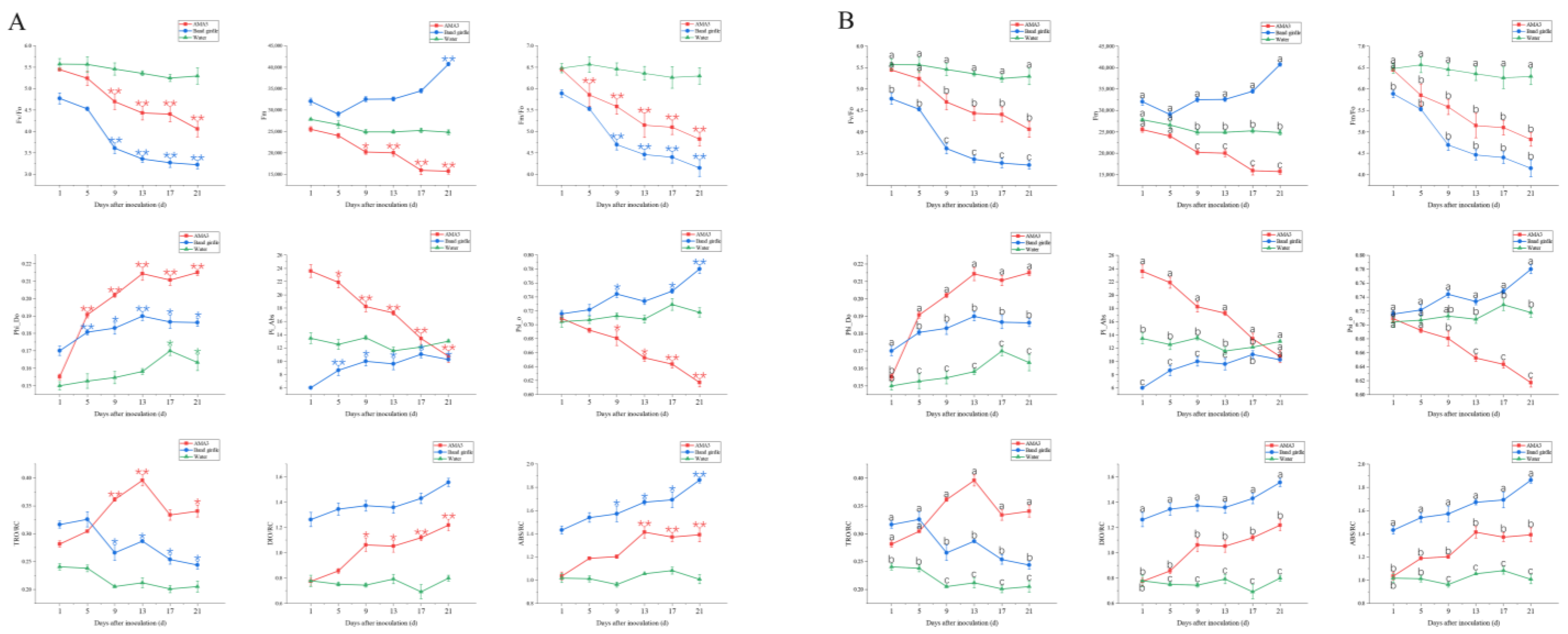
3.2.1. Variation in the OJIP Parameters of Pinus hwangshanensis Needles

Figure 1A shows the variation in the performance of each group at different time points. Figure 1B shows the variation in the performance of different groups on the same day. It can be seen from Figure 1A,B that PWNs have a significant effect on the light energy absorption of conifer leaves. Fm/Fo, Fv/Fo, Fm, Psi_o and Pi_Abs all showed a significant downward trend, while Phi_Do, ABS/RC, TRO/RC and DIO/RC showed a significant upward trend in the AMA3 group compared to the water group. At 1 d after inoculation, Pi_Abs showed a significant upwards trend compared with the water group. At 5 d after inoculation, Fm/Fo, Phi_Do and DIO/RC showed a significant downward trend compared with the water group. At 9 d after inoculation, Fm, Fv/Fo, ABS/RC and TRO/RC showed a significant downward trend compared with the water group. At 13 d after inoculation, Psi_o showed a significant downward trend compared with the water group. At 21 d after inoculation, the needles showed symptoms of chlorosis. As shown in Figure 1, Fm, Psi_o and Pi_Abs in the AMA3 group showed a significant decline after inoculation, while Fm, Psi_o and Pi_Abs in the band girdle group showed a significant increase. At 1 d after inoculation, Pi_Abs in the AMA3 group was much higher than that in the band girdle group, showing a significant difference. TRO/RC in the AMA3 group showed an overall upward trend after inoculation, while that in the band girdle group showed a downward trend. At 9 d after inoculation, TRO/RC in the AMA3 group was significantly higher than that in the band girdle group. Compared with the water group, Phi_Do in both the inoculation group and the band girdle group showed a significant increasing trend, but Phi_Do in the AMA3 group was significantly higher than that in the band girdle group from 5 d after inoculation.

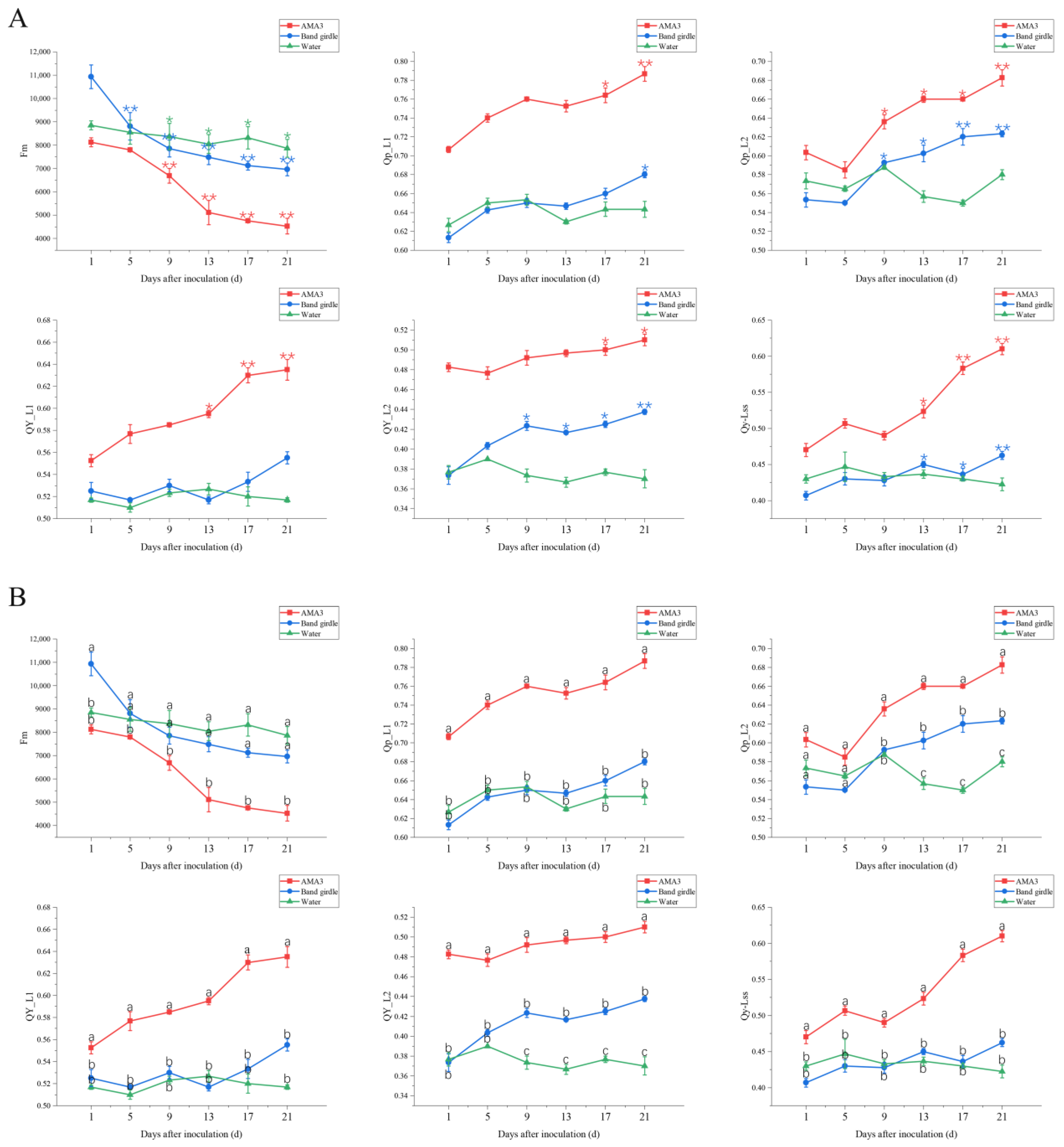
3.2.2. Variation in the NPQ Parameters of Pinus hwangshanensis Needles

Figure 2A shows the variation in the performance of each group at different time points. Figure 2B shows the variation in the performance of different groups on the same day. It can be seen from Figure 2A,B that PWNs has a significant impact on the heat dissipation ability of conifer leaves. The experiment showed that Fm had a significant downward trend and Qp_L1, Qp_L2, QY_L1, QY_L2 and QY_Lss showed a significant upward trend in the AMA3 group compared to the water and band girdle groups. On day 1 after inoculation, the parameters of Qp_L1, QY_L1 and QY_L2 in AMA3 group were significantly different from those in the two control groups and were always higher than those in the two control groups. On day 5 after inoculation, QY_Lss parameters were significantly higher and Fm parameters were much lower in the AMA3 group than in the two control groups. At 9 d after inoculation, Qp_L2 parameters in AMA3 group showed an obvious upward trend and showed significant difference compared with the two control groups. From 13 to 21 d after inoculation, the Qp_L1, Qp_L2, QY_L1, QY_L2 and QY_Lss parameters were always significantly higher and Fm parameters were always significantly lower than those of the two controls.

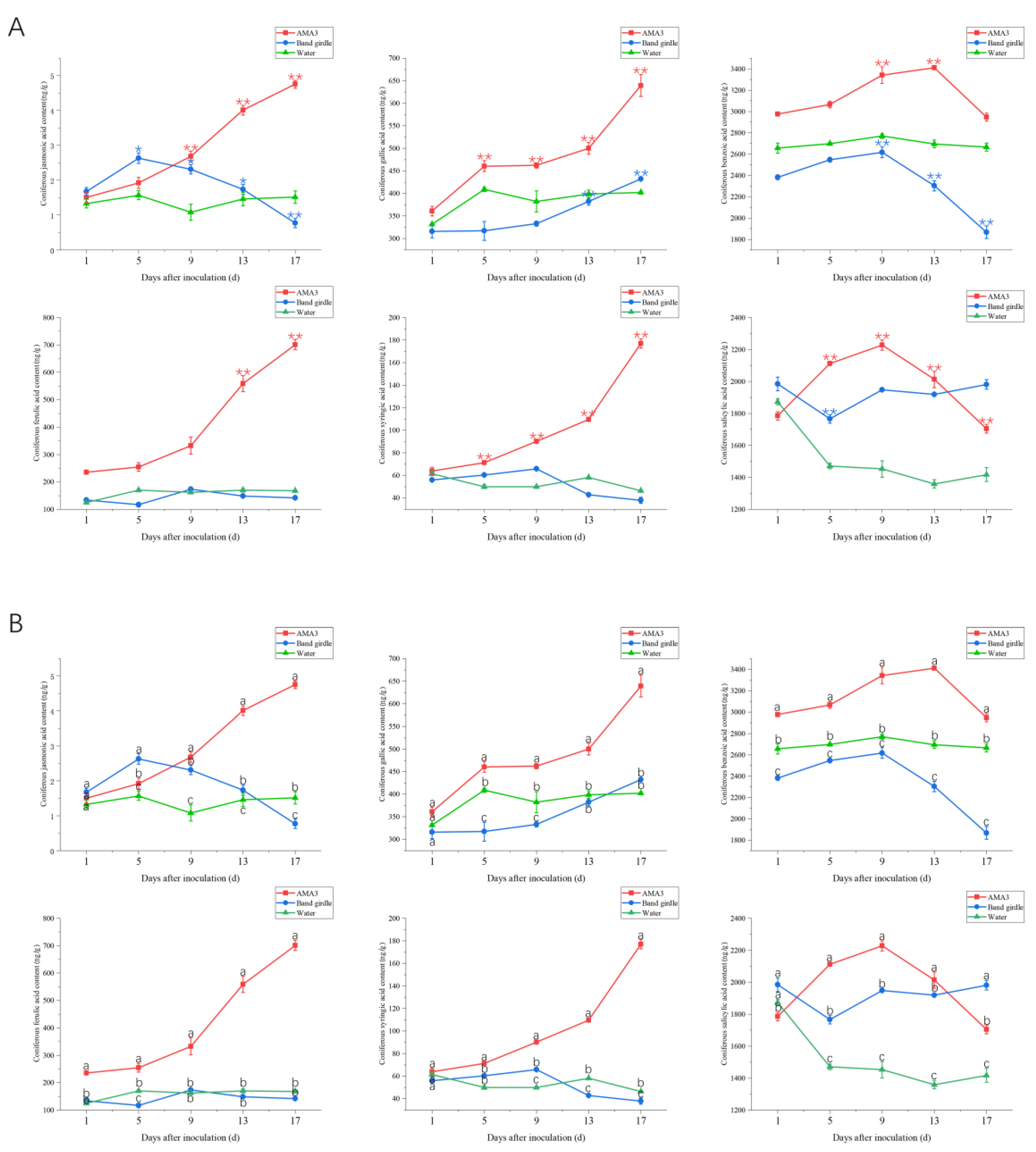
3.3. Effect of PWNs on the Organic Acid Content of the Needles of Pinus hwangshanensis

Figure 3A and Figure 3B, respectively show the changes in the performance of each group at different time points and the changes in the performance of different groups on the same day. As can be seen from Figure 3A,B, the organic acid content in pine trees will also be affected to a certain extent when pine trees are infected by PWNs. The experiments showed that the ferulic, gallic, syringic and jasmonic acid contents in the AMA3 group showed a significant upwards trend, while the benzoic and salicylic acid contents showed an upward and then downwars trend but were always higher than those in the AMA3 group compared to the water and band girdle groups. At 1 d after inoculation, the ferulic and benzoic acid levels were significantly higher in the AMA3 group than in the two controls. At 5 d after inoculation, gallic acid and syringic acid were higher in the AMA3 group than in the two controls, but gallic acid was more significant. At 13 d after inoculation, the content of jasmonic acid in AMA3 group showed an obvious increasing trend was higher than that in control groups. At 17 d after inoculation, the salicylic acid content of the AMA3 group dropped below that of the band girdle group but was still higher than that of the water group.

4. Discussion

OJIP curves can be used to observe transient changes in fluorescence rise in light-induced darkness and are extremely sensitive to changes in plant photosystem II (PSII) efficiency [18]. NPQ can consume the excess absorbed light in the plant as heat dissipation, preventing the reaction center from being damaged by light and is one of the important photoprotection mechanisms in higher plants [32,33]. It was found that compared with the water group, parameters Fm, Psi_o and Pi_Abs in the AMA3 group showed a significant downward trend, and there was a significant difference. Phi_o, TRO/RC, DIO/RC, ABS/RC, Qp_L1, Qp_L2, QY_L1, QY_L2 and QY_Lss showed an upward trend with significant differences. The invasion of PWN after inoculation may lead to thermal dissipation of photosynthetic system energy by increasing the number of QA non-reducing reaction centers (RCS). During the determination of OJIP and NPQ, the Fm parameters showed a decreasing trend, which may be caused by the decrease in light absorption due to the influence of PSII electron transfer in the infected pine. The decrease of Psi_o indicates that the opening of PSIIactive reaction center and the quantum yield of electron transfer flux may be decreased due to the invasion of PWN. The decrease of Fm and Psi_o parameters indicates that the reactivity openness and electron transfer flux quantum yield of Pinus hwangshanensis are reduced due to PWN, which is the same as the results of Liu Fei’s study on the early diagnosis of black pine by chlorophyll fluorescence [28], and the same results have also been obtained in the study of other plant diseases [18,34]. Pi_Abs showed an obvious downward trend after inoculation with PWNs, which was also shown in citrus greening disease [34], which may be caused by the negative impact on the electron transport efficiency in the plant after infection. These results indicated that the oxygen-releasing complex (OEC) on the PSII electron donor side of Pinus hwangshanensis Pinus hwangshanensis needles was damaged [35]. After inoculation with PWN, the TRo/RC parameters of infected pine showed an increasing trend, which may be caused by the decrease in the number of active centers in the PSII reaction center due to the increase in the number of PWNs. The increase of ABS/Rc and DIo/RC parameters may be due to the inactivation of the PSII reaction center of Pinus hwangshanensis needles caused by the invasion of PWNs, and the increase of Phi_Do parameter indicates the increase of non-photochemical burst quantum yield of infected Pinus hwangshanensis. The parameters QY_L1, QY_L2 and QY_Lss can show the effective quantum efficiency of the PSII system in pine trees. The increase of these parameters indicates that the effective quantum efficiency of PSII in pine trees may be increased by PWN. Qp represents the proportion of open centers ready to accept electrons in the PSII reaction [16]. Qp_L1 and Qp_L2 indicate the chemical quenching coefficient of PSII. Qp_L1 and Qp_L2 showed an increasing trend, indicating that the energy level transmitted to the reaction center was also increasing [16,36], which was contrary to the experimental results of the black spot of kale [37]. Studies have shown that a reduction in the rate of electron transfer is associated with a rise in NPQ [38], a phenomenon that is also present in virus-infected plants [39]. This increase is probably due to the PSII protection process and the destruction of the optical system. However, not all nonphotochemical quenching processes result in an increase in NPQ [37].
In the present study, it was found that the toxin had a certain effect on the occurrence of PWD. The results showed that the AMA3 group’s ferulic acid, syringic acid, gallic acid and jasmonic acid showed a significant upwards trend, and benzoic acid and salicylic acid showed an upward and then a downward trend compared to the two control groups. Ferulic acid is involved in the biosynthesis of lignin and is a binding and stabilizing agent for cell walls [40]. Syringic acid is a phenolic derivative of lignin [41]. Lignin is rapidly deposited under biological stress [42]. The increasing number of PWNs in pine trees leads to the deposition of lignin and, therefore, to the increasing content of ferulic and syringic acids. Gallic acid can cause adverse allergic reactions in affected plants. The gallic acid content of the P. hwangshanensis needles in the inoculated group showed an increase in the early stages of the disease and rose sharply before showing symptoms. This suggests that as the PWN population increases, the affected pine trees resist their invasion by increasing gallic acid. Jasmonic acid and salicylic acid are important signal transduction factors for disease resistance in plants and changes in their levels are associated with plant defense responses [43]. Plants respond to biological or abiotic stimuli by increasing the content of jasmonic acid and salicylic acid in an attempt to defend themselves by inducing their own internal production of secondary metabolic substances. Salicylic acid plays an important defense role when pine trees are infested with PWNs [44], and jasmonic acid acts as a downstream signaling molecule for salicylic acid, which together with salicylic acid stimulates resistance responses in affected plants [45]. Previous studies have shown that benzoic acid wilts and kill the host pine tree by accumulating its own content and poisoning plant tissue cells [46]. The results of the experiment showed that the jasmonic acid content of the AMA3 group increased before the symptoms manifested, which was the same as the change in jasmonic acid content in the body of Pinus radiata after inoculation with PWN [47]. This suggests that the affected plants stimulate a resistance response to PWN invasion by increasing jasmonic acid content. The salicylic acid content of the needles of P. hwangshanensis in the AMA3 group showed a decreasing trend on day 9 after inoculation, and the benzoic acid content showed a decreasing trend on day 13 after inoculation, which is the same result as Tan Jiajin’s research [48]. The similarity of content dynamics of salicylic acid and benzoic acid content further suggests that a close relationship between the two is maintained within plant tissues [48]. This indicated that the invasion of PWN would stimulate the victim pine to produce a large amount of salicylic acid and benzoic acid to resist the invasion of PWN. However, due to the rapid spread of PWN, the victim pine could not resist the invasion of nematode, resulting in the decline of salicylic acid and benzoic acid.

5. Conclusions

In this paper, to explore the potential of using chlorophyll fluorescence techniques and organic acid detection in the early diagnosis of PWD in P. hwangshanensis, the effects of PWNs on the function of PSII in the photosynthetic mechanism and changes in organic acid content within the needles of P. hwangshanensis were investigated using healthy and band girdle P. hwangshanensis as controls. The results of this study showed that the chlorophyll fluorescence parameters Pi_Abs, Fm, Phi_Do, TRO/RC, Psi_o, Qp_L1, Qp_L2, QY_L1, QY_L2, QY_Lss and ferulic acid, syringic acid, benzoic acid and gallic acid were better candidates for the early diagnosis of PWD in P. hwangshanensis. As this experiment was conducted in a greenhouse, further verification of how these indicators perform on P. hwangshanensis in the forest is needed.

Author Contributions

Conceptualization, L.S. experimental, L.S.; methodology, L.S.; software, L.S.; validation, L.S., X.L., F.L. and Y.H.; formal analysis, L.S.; resources, L.S.; data curation, L.S.; writing—original draft preparation, L.S.; writing—review and editing, J.T. and J.Y. All authors have read and agreed to the published version of the manuscript.

Funding

This study was funded by the China National Key Research and Development Program (2021YFD1400900).

Informed Consent Statement

All participants in the study received informed consent.

Data Availability Statement

The data presented in this study are available on request from the corresponding author.

Conflicts of Interest

The authors declare no conflict of interest.

References

  1. Kikuchi, T.; Cotton, J.A.; Dalzell, J.J.; Hasegawa, K.; Kanzaki, N.; McVeigh, P.; Takanashi, T.; Tsai, I.J.; Assefa, S.A.; Cock, P.J.; et al. Genomic insights into the origin of parasitism in the emerging plant pathogen Bursaphelenchus xylophilus. PLoS Pathog. 2011, 7, e1002219. [Google Scholar] [CrossRef] [Green Version]
  2. Miao, L.; Wang, Y.; Li, R.H.; Liu, C.F.; Fan, Y.S. Damage, transmission route and control measures pf pine wood nematode disease. J. Jilin For. Sci. Technol. 2023, 52, 44–45. [Google Scholar]
  3. Ye, J.R.; Wu, X.Q. Research progress of pine wood nematode disease. For. Dis. Insects China 2022, 41, 1–10. [Google Scholar]
  4. Lim, E.T.; Do, M.S. Pine wilt disease detection based on deep learning using an unmanned aerial vehicle. KSCE J. Civ. Environ. Eng. Res. 2021, 41, 317–325. [Google Scholar]
  5. Lee, S.; Park, S.J.; Baek, G.; Kim, H.; Lee, C.W. Detection of Damaged Pine Tree by the Pine Wilt Disease Using UAV Image. Korean J. Remote Sens. 2019, 35, 359–373. [Google Scholar]
  6. Jiang, X.F.; Li, M.; Zhang, L.P.; Zhang, Y.; Wu, F.; Tong, Y.X.; Chen, F.M. Control strategies and measures of pine wood nematode disease. Sci. Biodisaster 2023, 46, 136–140. (In Chinese) [Google Scholar]
  7. Mota, M.; Vieira, P. (Eds.) Discovery of pine wood nematode in portugal and in Europe. In Proceedings of the International Workshop on Pine Wood Nematode, Bursaphelenchus xylophilus, Evora, Portugal, 20–22 August 2001; Volume 8, pp. 20–22. [Google Scholar]
  8. Zhang, X.R.; Guo, W.H.; Wang, R.Q.; Si, J.Y. Comparative study on chlorophyll fluorescence characteristics of three pine species. Shandong For. Sci. Technol. 2010, 40, 1–5. [Google Scholar]
  9. Li, X.; Feng, W.; Zeng, X.C. Advances in chlorophyll fluorescence analysis and its application. Acta Bot. Boreali-Occident. Sin. 2006, 26, 11. [Google Scholar]
  10. Berger, S.; Sinha, A.K.; Roitsch, T. Plant physiology meets phytopathology: Plant primary metabolism and plant-pathogen interactions. J. Exp. Bot. 2007, 58, 4019–4026. [Google Scholar] [CrossRef]
  11. Strange, R.N.; Scott, P.R. Plant disease: A threat to global food security. Annu. Rev. Phytopathol. 2005, 43, 83–116. [Google Scholar] [CrossRef]
  12. Lemanczyk, G.; Kwasna, H. Effects of sharp eyespot (Rhizoctonia cerealis) on yield and grain quality of winter wheat. Eur. J. Plant Pathol. 2013, 135, 187–200. [Google Scholar] [CrossRef] [Green Version]
  13. Fofana, B.; Banks, T.W.; McCallum, B.; Strelkov, S.E.; Cloutier, S. Temporal gene expression profiling of the wheat leaf rust pathosystem using cDNA microarray reveals differences in compatible and incompatible defence pathways. Int. J. Plant Genom. 2007, 2007, 17542. [Google Scholar] [CrossRef] [PubMed] [Green Version]
  14. Kocal, N.; Sonnewald, U.; Sonnewald, S. Cell Wall-Bound Invertase Limits Sucrose Export and Is Involved in Symptom Development and Inhibition of Photosynthesis during Compatible Interaction between Tomato and Xanthomonas campestris pv vesicatoria. Plant Physiol. 2008, 148, 1523–1536. [Google Scholar] [CrossRef] [Green Version]
  15. Christen, D.; Schönmann, S.; Jermini, M.; Strasser, R.J.; Défago, G. Characterization and early detection of grapevine (Vitis vinifera) stress responses to esca disease by in situ chlorophyll fluorescence and comparison with drought stress. Environ. Exp. Bot. 2007, 60, 504–514. [Google Scholar] [CrossRef]
  16. Maxwell, K.; Johnson, G.N. Chlorophyll fluorescence-a practical guide. J. Exp. Bot. 2000, 51, 659–668. [Google Scholar] [CrossRef] [PubMed]
  17. Murchie, E.H.; Lawson, T. Chlorophyll fluorescence analysis: A guide to good practice and understanding some new applications. J. Exp. Bot. 2013, 64, 3983–3998. [Google Scholar] [CrossRef] [Green Version]
  18. Ajigboye, O.O.; Bousquet, L.; Murchie, E.H.; Ray, R.V. Chlorophyll fluorescence parameters allow the rapid detection and differentiation of plant responses in three different wheat pathosystems. Funct. Plant Biol. 2016, 43, 356–369. [Google Scholar] [CrossRef]
  19. Linn, A.I.; Zeller, A.K.; Pfündel, E.E.; Gerhards, R. Features and applications of a field imaging chlorophyll fluorometer to measure stress in agricultural plants. Precis. Agric. 2021, 22, 947–963. [Google Scholar] [CrossRef]
  20. Recchia, I.; Sparla, F.; Pupillo, P. Photosynthetic properties of spring geophytes assessed by chlorophyll fluorescence analysis. Plant Physiol. Biochem. 2017, 118, 510–518. [Google Scholar] [CrossRef]
  21. Melakeberhan, H.; Toivonen, P.M.A.; Vidaver, W.E.; Webster, J.M.; Dube, S.L. Effect of Bursaphelenchus xylophilus on the water potential and water-splitting complex of photosystem II of Pinus sylvestris seedlings. Physiol. Mol. Plant Pathol. 1991, 38, 83–91. [Google Scholar] [CrossRef]
  22. Oku, H.S.T.; Kurozumi, S. Participation of toxin in wilting of Japanese pines caused by a nematode. Naturwissenschaften 1979, 66, 211. [Google Scholar] [CrossRef]
  23. Mamiya, Y. Inoculation of the first vear pine(Pinus densiflora) seedlings with Bursaphelenchns lignicolns and the histopathology of diseased seedlinns. J. Jpn. For. Soc. 1980, 62, 176–183. [Google Scholar]
  24. Li, Y.X.; Zhang, X.Y. Advances in Research on Pathogenic Mechanism of Pine Wood Nematode Disease. Chin. J. Environ. Entomol. 2018, 40, 11. [Google Scholar]
  25. Zhang, F.; Kajiwara, J.; Mori, Y.; Ohira, M.; Tsutsumi, Y.; Kondo, R. Metabolites from resistant and susceptible Pinus thunbergii after inoculation with pine wood nematode. Am. J. Plant Sci. 2013, 4, 512–518. [Google Scholar] [CrossRef] [Green Version]
  26. Kawatsu; Kazuyoshi. Pathogenic Toxin of Pine Wood Disease. Kagaku Sebutsu 1998, 36, 120–124. [Google Scholar] [CrossRef]
  27. Hu, L.J.; Wu, X.Q. Research progress on the mechanism of resistance to pine wood nematode disease. Life Sci. 2018, 30, 659–666. [Google Scholar] [CrossRef]
  28. Liu, F.; Zhang, M.; Hu, J.; Pan, M.; Shen, L.; Ye, J.; Tan, J. Early diagnosis of pine wilt disease in Pinus thunbergii based on chlorophyll fluorescence parameters. Forests 2023, 14, 154. [Google Scholar] [CrossRef]
  29. Viglierchio, D.R.; Schmitt, R.V. On the methodology of nematode extraction from field samples: Baermann funnel modifications. J. Nematol. 1983, 15, 438–444. [Google Scholar]
  30. Xie, L.Q.; Ju, Y.W.; Yang, Z.D.; Zhao, B.G. Changes of the number of bacteria and nematodes in the inoculated branches of Pinus thunbergii. J. Zhejiang AF Univ. 2005, 22, 310–314. [Google Scholar]
  31. Tan, J.J.; Wu, H.P. Effects of Pine Wood Nematode on Free Proline Content in Leaves of Pine. J. South China Agric. Univ. 2000, 21, 1. [Google Scholar]
  32. Ruiz-sola, M.A.; Petroutsos, D. A Toolkit for the characterization of the photoprotective capacity of green algae. Methods Mol. Biol. 2018, 1829, 315–323. [Google Scholar]
  33. Mou, S.; Zhang, X.; Ye, N.; Miao, J.; Cao, S.; Xu, D.; Fan, X.; An, M. Analysis of Delta pH and the xanthophyll cycle in NPQ of the Antarctic sea ice alga Chlamydomonas sp. ICE-L. Extremophiles 2013, 17, 477–484. [Google Scholar] [CrossRef] [PubMed]
  34. Weng, H.; Liu, Y.; Captoline, I.; Li, X.; Ye, D.; Wu, R. Citrus Huanglongbing detection based on polyphasic chlorophyll a fluorescence coupled with machine learning and model transfer in two citrus cultivars. Comput. Electron. Agric. 2021, 187, 106289. [Google Scholar] [CrossRef]
  35. Zhang, Z.; Wu, J.; Xi, Y.; Zhang, L.; Gao, Q.; Wang-Pruski, G. Effects of autotoxicity on seed germination, gas exchange attributes and chlorophyll fluorescence in melon seedlings. J. Plant Growth Regul. 2022, 41, 993–1003. [Google Scholar] [CrossRef]
  36. Nosek, M.; Kornaś, A.; Kuźniak, E.; Miszalski, Z. Plastoquinone redox state modifies plant response to pathogen. Plant Physiol. Biochem. 2015, 96, 163–170. [Google Scholar] [CrossRef] [PubMed]
  37. Macioszek, V.K.; Gapińska, M.; Zmienko, A.; Sobczak, M.; Skoczowski, A.; Oliwa, J.; Kononowicz, A.K. Complexity of Brassica oleracea-Alternaria brassicicola susceptible interaction reveals downregulation of photosynthesis at ultrastructural, transcriptional, andphysiological levels. Cells 2020, 9, 2329. [Google Scholar] [CrossRef]
  38. Yang, Z.X.; Yang, Y.F.; Yu, S.Z.; Wang, R.G.; Wang, Y.; Chen, H.L. Photosynthetic, photochemical and osmotic regulation changes in tobacco resistant and susceptible to Alternaria alternata. Trop. Plant Pathol. 2018, 43, 413–421. [Google Scholar] [CrossRef]
  39. Rys, M.; Juhász, C.; Surówka, E.; Janeczko, A.; Saja, D.; Tóbiás, I.; Skoczowski, A.; Barna, B.; Gullner, G. Comparison of a compatible and an incompatible pepper-tobamovirus interaction by biochemical and non-invasive techniques: Chlorophyll a fluorescence, isothermal calorimetry and FT-Raman spectroscopy. Plant Physiol. Biochem. 2014, 83, 267–278. [Google Scholar] [CrossRef] [Green Version]
  40. Baucher, M. Lignin biosynthesis. Annu. Rev. Plant Biol. 2003, 54, 519–546. [Google Scholar]
  41. Liu, S.Y.; Minard, R.D.; Bollag, J.M. Oligomerization of Syringic Acid, a Lignin Derivative, by a Phenoloxidase. Soil Sci. Soc. Am. J. 1981, 45, 1100–1105. [Google Scholar] [CrossRef]
  42. Caño-Delgado, A.; Penfield, S.; Smith, C.; Catley, M.; Bevan, M. Reduced cellulose synthesis invokes lignification and defense responses in Arabidopsis thaliana. Plant J. 2003, 34, 351–362. [Google Scholar] [CrossRef]
  43. Wang, Y.; Wen, T.; Hu, J.; Han, R.; Zhu, Y.; Guan, Y.; Zhu, S. Relationship between endogenous salicylic acid and antioxidant enzyme activities in maize seedlings under cold stress. Exp. Agric. 2013, 49, 295–308. [Google Scholar] [CrossRef]
  44. Lee, I.H.; Kim, J.; Woo, K.S.; Jang, K.H.; Kim, Y.H.; Shim, D. De novo assembly and transcriptome analysis of the Pinus densiflora response to pine wilt disease in nature. Plant Biotechnol. Rep. 2018, 12, 229–236. [Google Scholar] [CrossRef]
  45. Truman, W.; Bennett, M.H.; Kubigsteltig, I.; Turnbull, C.; Grant, M. Arabidopsis systemic immunity uses conserved defense signaling pathways and is mediated by jasmonates. Proc. Natl. Acad. Sci. USA 2007, 104, 1075–1080. [Google Scholar] [CrossRef]
  46. Qian, W.Q.; Xiang, F.Q.; Wang, M.; Wang, S.L. Effects of Pine Wood Nematode Infection on Organic Acids and Enzymes in Masson Pine. Hunan For. Sci. Technol. 2009, 36, 3. [Google Scholar]
  47. Qin, S.J.; Qi, J.Y.; Liu, R.J.; Chen, F.M.; Hao, D.J.; Li, Z.X.; Sun, S.R. Physiological Response of Pinus tabulaeformis after Infection with Pine Wood Nematodes in Natural State. J. Shenyang Agric. Univ. 2021, 52, 625–632. [Google Scholar]
  48. Tan, J.J.; Ye, J.R.; Hao, D.J. Changes of organic acid contents of Pinus thunbergii after infection with pine wood nematode. Plant Pathol. Bull. 2008, 38, 3. [Google Scholar]
Figure 1. (A) Changes in parameters of the chlorophyll fluorescence induction curve of Pinus hwangshanensis needles under PWN stress. Note: * < 0.05, ** < 0.01. (B) Changes in parameters of the chlorophyll fluorescence induction curve of Pinus hwangshanensis needles under PWN stress. Note: a, b, c are significantly different and represents the difference between treatments on the same day after inoculation, p < 0.05.
Forests 14 01582 g001
Figure 2. (A) Changes in nonphotochemical quenching parameters of Pinus hwangshanensis needles under PWN stress. Note: * < 0.05, ** < 0.01. (B) Changes in nonphotochemical quenching parameters of Pinus hwangshanensis needles under PWN stress. Note: a, b, c are significantly different and represents the difference between treatments on the same day after inoculation, p < 0.05.
Forests 14 01582 g002
Figure 3. (A) Changes in organic acid content in needle leaves of Pinus hwangshanensis under PWN stress. Note: * < 0.05, ** < 0.01. (B) Changes in organic acid content in needle leaves of Pinus hwangshanensis under PWN stress. Note: a, b, c are significantly different and represents the difference between treatments on the same day after inoculation, p < 0.05.
Forests 14 01582 g003
Table 1. Disease index of Pinus hwangshanensis inoculated with PWN.
TreatmentDisease Index
0 d5 d9 d13 d17 d21 d38 d55 d120 d
AMA30000012.537.559.478.13
Band girdle000016.744.454.283.3100
Water000000000
Disclaimer/Publisher’s Note: The statements, opinions and data contained in all publications are solely those of the individual author(s) and contributor(s) and not of MDPI and/or the editor(s). MDPI and/or the editor(s) disclaim responsibility for any injury to people or property resulting from any ideas, methods, instructions or products referred to in the content.

