Article

Transcriptome Analysis Reveals the Impact of Arbuscular Mycorrhizal Symbiosis on Toona ciliata var. pubescens Seedlings

Jiangxi Provincial Key Laboratory of Conservation Biology, Jiangxi Provincial Key Laboratory of Subtropical Forest Resources Cultivation, College of Forestry/College of Art and Landscape, Jiangxi Agricultural University, Nanchang 330045, China
*
Author to whom correspondence should be addressed.
Forests 2024, 15(4), 673; https://doi.org/10.3390/f15040673
Submission received: 28 February 2024 / Revised: 23 March 2024 / Accepted: 4 April 2024 / Published: 8 April 2024
(This article belongs to the Special Issue Adaptive Mechanisms of Tree Seedlings to Adapt to Stress)

Abstract

:
Toona ciliata var. pubescens, known as “Chinese mahogany”, has high commercial value and is classified as a level II priority protected wild plant in China. However, due to overexploitation and its poor natural regeneration capacity, natural T. ciliata var. pubescens forests show varying degrees of decline in habitat adaptability. Arbuscular mycorrhizal fungi (AMF) symbiosis presents a potential strategy to enhance its regeneration. In this study, T. ciliata var. pubescens seedlings were inoculated with Septoglomus viscosum, followed by RNA-Seq analysis to compare gene expression differences between AMF-inoculated (AMI) and non-mycorrhizal (NM) treatments three months post-inoculation. A total of 16,163 differentially expressed genes (DEGs) were upregulated by AMF colonization, constituting 96.46% of the total DEGs. Specifically, 14,420 DEGs were exclusively expressed in the AMI treatment, while 35 DEGs were completely silenced. Most of the upregulated DEGs were located on the cell membrane, nucleus, and cytoskeleton and functioned in protein binding, S-adenosylmethionine-dependent methyltransferase activity, and lipid binding during cellular/macromolecule/protein localization, intracellular/protein transport, the cell cycle, and signal transduction. Additionally, lots of key genes related to oxidative stress responses, nutrient transport, and small GTPase-mediated signal transduction were found to be upregulated. These results suggest that AMF inoculation may enhance root cell growth by activating genes involved in nutrient uptake, stress responses, signal transduction, and substance transportation. This study elucidates the molecular mechanisms underlying the growth promotion of T. ciliata var. pubescens through AMF symbiosis, laying a foundation for the future application of AMF in its natural forest regeneration.

1. Introduction

Toona ciliata. var. pubescens (Franch.) Hand.-Mazz., a fast-growing commercial tree species, is classified as a level II priority protected wild plant in China. Commonly referred to as “Chinese mahogany”, it is highly valued commercially for its red timber, which boasts an appealing aesthetic texture and moderate density. In addition, it also has high ornamental value and can be grown in gardens. Despite these attributes, overexploitation and its limited natural regeneration capacity have led to a decline in habitat adaptability among natural T. ciliata var. pubescens forests, with their distribution increasingly confined to areas like valleys and streams [1,2]. Research indicates that the primary factors inhibiting natural regeneration include soil moisture, nutrients, pathogens, and surface litter [1,3,4]. T. ciliate var. pubescens is naturally distributed in the red soil regions of South–Central China, where the soil is characterized by a poor structure and nutrient deficiency, particularly in phosphorus (P) [5]. Furthermore, soilborne pathogens significantly contribute to seed decay and the unsuccessful natural regeneration of this species [1,3].
Arbuscular mycorrhizal fungi (AMF) establish symbiotic relationships with nearly 90% of plant species, including flowering plants, bryophytes, and ferns [6]. Given that AMF biotrophically colonize the root cortex of plants and develop an extraradical mycelium extending beyond the nutrient-depleted zone surrounding plant roots, the application of AMF significantly enhances the uptake and translocation of mineral nutrients, especially phosphate [7,8]. This process notably improves the nutrient status of plants. Beyond phosphate, AMF can also enhance the nitrate uptake capacity of plants. Compared with nonmycorrhizal poplar plants, mycorrhizal plants have a higher maximum uptake rate (V-max) value of NO3 [9]. Furthermore, a feedback mechanism exists whereby the colonization of roots by AMF and AMF’s beneficial effects are enhanced under conditions of limited phosphate or nitrogen, indicating an adaptive response to nutrient scarcity in plants [10].
In addition, AMF influence the development and abiotic stress tolerance of plants by interfering with the phytohormone and redox balance of their host plants [11]. AMF colonization generally promotes plant lateral root formation by increasing the phosphate, auxin, and cytokinin content and decreasing ethylene and strigolactone levels [12]. Fenugreek plants inoculated with AMF exhibit reduced levels of oxidative damage in comparison to non-mycorrhizal counterparts [13]. Moreover, the efficacy of AMF in ameliorating oxidative stress is amplified with the increasing intensity of salt stress. Furthermore, AMF also play roles in ecosystems by improving ecosystem nitrogen cycling [14], soil quality [15], and the abundance and diversity of rhizosphere microorganisms [16]. Therefore, the optimized application of AMF in sustainable forestry and agriculture will be crucial for responding to climate change and the increasing human population [15,17].
Pan et al. [2] confirmed that the natural forest community of T. ciliate var. pubescens live in symbiosis with AMF, underscoring the ecological importance of AMF in the succession and stability of plant communities [18]. AMF may facilitate the natural regeneration of T. ciliata var. pubescens through various mechanisms: (1) enhancing soil aggregate structure and improving soil physical conditions [19], (2) boosting effective nutrient absorption and transportation in plants [7], (3) increasing plant resilience to drought and other stressors [11], and (4) combating soil-borne pathogens to prevent seed decay [20]. Nevertheless, research into the growth adaptability of AMF on T. ciliata var. pubescens and the potential benefits of exogenous AMF supplementation, including the underlying molecular mechanisms, remains unexplored.
Fu [21] investigated the AMF community composition in the rhizosphere soil of natural T. ciliata var. pubescens forests in the Guanshan National Nature Reserve, Jiangxi, finding that the genus Glomus dominated the community, comprising 68.18% of its total, though without identifying specific species. Septoglomus viscosum (T. H. Nicolson) C. Walker et al. and Glomus viscosum T.H. Nicolson, which are synonymous and fall under the genus Glomus [22], have been widely used to promote plant growth [23,24]. Preliminary studies indicated that S. viscosum application could enhance phosphorus levels in the phosphorus-deficient soils of China’s subtropical climate zone [25], where natural T. ciliata var. pubescens forests were found, implying potential growth benefits for T. ciliata var. pubescens. Consequently, this experiment intended to employ S. viscosum as an inoculant to investigate its impact on the growth and nutrient uptake of T. ciliata var. pubescens seedlings. During the past several years, transcriptome sequencing has become the main technical means to study the molecular mechanism of biological processes such as signal transduction, metabolism, and protein synthesis in plant–AMF symbiosis. Accordingly, the present study aimed to explore the effects of AMF inoculation on the growth and transcriptome responses in the roots of potted T. ciliata var. pubescens seedings, using RNA-seq technology. This research is expected to shed light on the molecular basis of the interaction between T. ciliata var. pubescens and its principal AMF symbionts, focusing on aspects such as nutrient absorption and resistance. Furthermore, it aims to provide a theoretical basis for the further study of the effect of AMF on the regeneration of natural T. ciliate var. pubescens forests.

2. Materials and Methods

2.1. Experimental Material and Experimental Design

The preparation of the S. viscosum inoculum followed the methodology outlined by Liu et al. [26]. Corn was selected as the host plant, and a nutrient-rich soil was homogeneously mixed with sieved river sand in a 3:1 volume ratio to serve as the growth medium, which was subsequently sterilized by high-pressure steam sterilization. The culture containers were disinfected using 2% sodium hypochlorite solution, followed by the addition of the prepared culture medium. The S. viscosum strains were evenly spread on the medium surface, covered with a layer of 2 cm of medium, and irrigated. After 24 h, 30 disinfected corn seeds per pot (immersed in a 2% sodium hypochlorite solution for 5 min) were placed and covered with a 0.5 cm layer of substrate. Four months later, the inoculum was harvested, comprising propagation matrix soil, extraradical mycelium, intraradical mycelium, and colonized root segments, with 20 spores per gram.
The seeds of T. ciliata var. pubescens from the same strain were collected at the Jiangxi Guanshan Nature Reserve, China. Seeds of T. ciliata var. pubescens were surface-sterilized with 10% H2O2 for 5 min, rinsed with deionized water, and then soaked in deionized water in the dark at 25 °C for 12 h. The pregerminated seeds were planted in two trays filled with autoclaved sand (121 °C, 2 h) and placed in an incubator for germination with a 12 h photoperiod and temperatures of 25 °C during the day and 17 °C at night. Upon reaching two true leaves, seedlings were transplanted into pots with 11 cm in bottom diameter, 16 cm in upper outer diameter, and 12.5 cm in height. The culture medium was a mixture of autoclaved (121 °C, 2 h) garden soil, turf, and sand (3:1:1, v:v:v). Each pot was filled with 600 g of medium, onto which 30 g of the AMF inoculum was evenly spread, followed by the planting of 2 seedlings and covering with 150 g medium. This procedure constituted the AMF inoculation treatment, designated as AMI. Additionally, 30 g of inoculum sterilized by high-pressure steam sterilization was added as a control, denoted as NM. The pots were randomly placed in the growth room seedbed of the laboratory, supplemented with an agricultural sodium lamp. The lighting time was approximately 16 h per day, the temperature was controlled at 26 °C, and the humidity was controlled at approximately 60%. Water was regularly administered every week to keep the soil moist. There were three replicates (two pots for each replicate) for each treatment and two seedlings for each pot.

2.2. Harvest

According to the method of Chen et al. [27], plants were harvested three months post-transplantation. At the time of harvest, metrics such as plant height, number of branches, and root growth for each seedling were recorded. The roots of the T. ciliata var. pubescens seedlings were thoroughly rinsed with distilled water. Subsequently, roots gathered from two pots were segmented into small pieces, uniformly mixed, and then bifurcated: one portion was preserved in liquid nitrogen for RNA extraction, while the other was stored at −20 °C to facilitate quantification of AM colonization.

2.3. Quantification of AMF Colonization

AMF colonization was assessed using the modified acetic-acid–ink technique [28]. Root fragments underwent a clearance process in 10% (w/v) KOH at 90 °C for 3–4 h. Upon cooling, these fragments were washed with distilled water and subjected to bleaching in 1% (w/v) H2O2-KOH alkaline solution for 30 min. Subsequently, the root fragments were acidified in a 5% (v/v) acetic acid solution for roughly 30 min, stained with 5% (v/v) acetic-acid–ink solution at 75 °C for 15 min, and then decolorized in distilled water for over 20 h. Around six root segments were then mounted on a slide using a polyvinyl-alcohol–lactic-acid–glycerol solution and observed under an Olympus light microscope. The extent of AMF root colonization was quantified using the gridline intersection method [29].

2.4. RNA Extraction and Sequencing

Total RNA from three biological replicates of the two treatments was extracted using TRIzol (Invitrogen, Carlsbad, CA, USA). The quality of RNA, including degradation and contamination, was assessed on 1% agarose gels. RNA purity and integrity were evaluated using a NanoPhotometer® spectrophotometer (IMPLEN, Los Angeles, CA, USA) and an RNA Nano 6000 Assay Kit for the Agilent Bioanalyzer 2100 system (Agilent Technologies, Santa Clara, CA, USA), respectively. RNA samples were sent to Novogene Bioinformatics Technology Co., Ltd., (Tianjing, China), for sequencing. Sequencing libraries were generated using the NEBNext® Ultra™ RNA Library Prep Kit for Illumina® (NEB, Ipswich, MA, USA), following the manufacturer’s recommendations. Index-coded samples were clustered on a cBot Cluster Generation System with the TruSeq PE Cluster Kit v3-cBot-HS (Illumina, San Diego, CA, USA), following the manufacturer’s instructions. After cluster generation, the library preparations were sequenced on an Illumina HiSeq platform, yielding paired-end reads of 150 bp.

2.5. Assembly and Annotation

To prepare the clean reads, adapters sequences, reads containing poly-N, and low-quality reads were removed from the raw data. All downstream analyses utilized these high-quality clean reads. Transcriptome assembly was performed using Trinity [30], with the min_kmer_cov set to 2 by default and all other parameters at their default settings. The longest transcript for each gene was designated as the unigene. Counts of all transcripts and unigenes formed the basis for subsequent bioinformatics analysis. Additionally, all unigenes were annotated against the following public databases: Nr (NCBI nonredundant protein sequences), Nt (NCBI nonredundant nucleotide sequences), KOG/COG (Clusters of Orthologous Groups of proteins), and GO (Gene Ontology).

2.6. Differential Expression Analysis

The transcriptome assembled by Trinity was utilized as the reference sequence (Ref). Clean reads from each sample were aligned to the Ref by using RSEM, followed by the application of the FPKM (fragments per kilobase of transcript per million mapped reads) method to normalize gene expression levels [31]. The DESeq2 R package was employed for the analysis of differentially expressed genes (DEGs) between the two treatments [32]. DESeq2 employs statistical routines based on the negative binomial distribution model to detect differential expression in digital gene expression data. The p-values were adjusted using the Benjamini and Hochberg method to control the false discovery rate (FDR), referred to as adjusted p-values (padj) [33]. Genes with a padj < 0.05 and an absolute log2 (fold change, FC) > 1 identified by DESeq2 were deemed as significantly differentially expressed [34].

2.7. GO (Gene Ontology) and KEGG (Kyoto Encyclopedia of Genes and Genomes) Enrichment Analysis

GO enrichment analysis for DEGs was conducted using the GOseq R package (version 1.40.0), which is based on the Wallenius noncentral hypergeometric distribution. This method adjusts for gene length bias in DEGs [35]. For KEGG pathway enrichment analysis, KOBAS 3.0 software [36] was used to statistically evaluate the enrichment of DEGs in KEGG pathways, leveraging the comprehensive data available through the Kyoto Encyclopedia of Genes and Genomes (KEGG, http://www.genome.jp/kegg/, accessed on 15 March 2020).

2.8. Statistical Analyses

The influence of AMF inoculation on plant height and branch number was analyzed by t-test with IBM SPSS v.21 statistical software (SPSS Inc., Chicago, IL, USA).

2.9. Data Availability

The raw sequencing data were deposited in NCBI SRA database, and the accession number was PRJNA688375.

3. Results

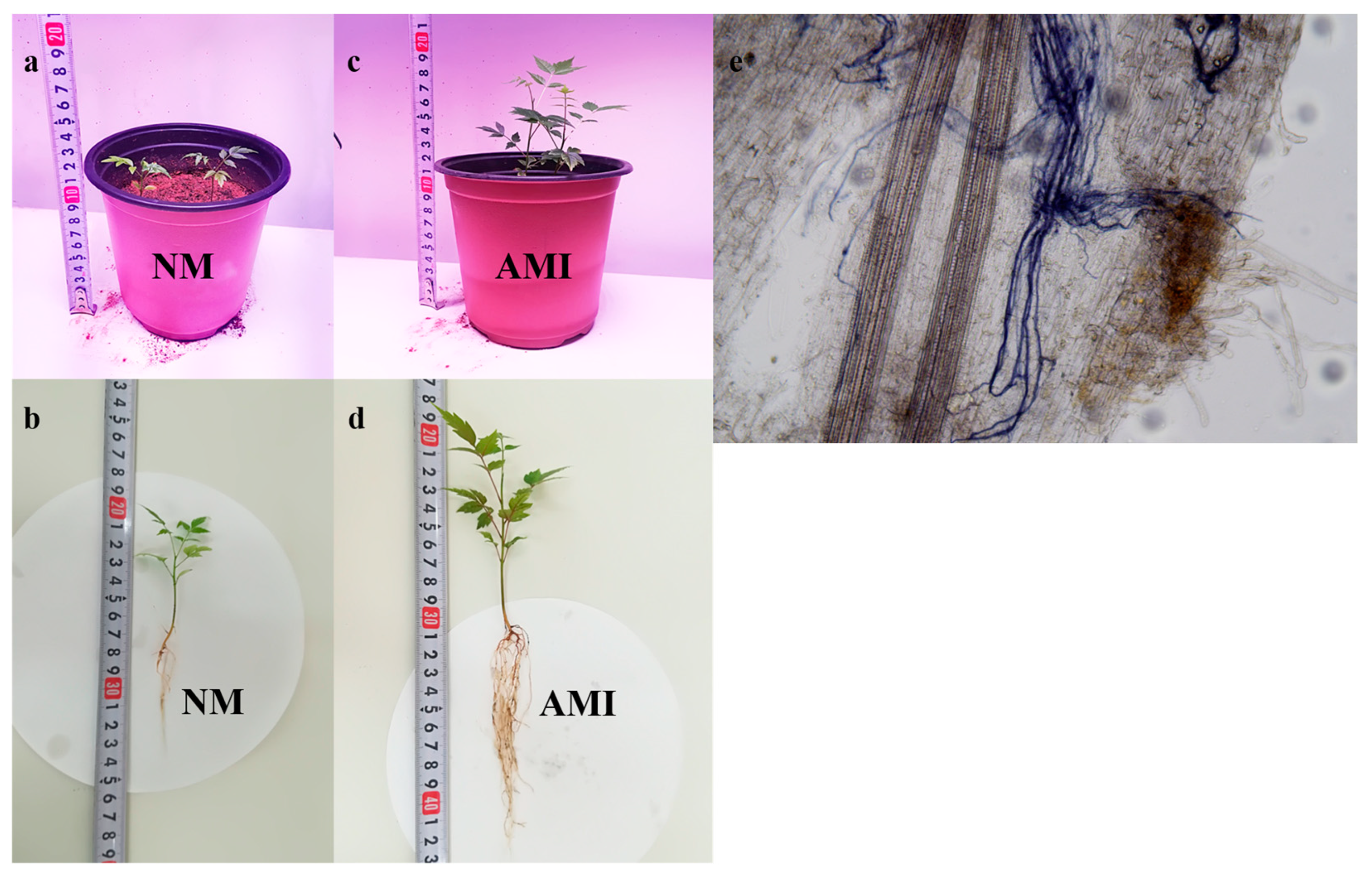
3.1. AM Colonization and Plant Growth

The mycorrhizal colonization rate was 24.63 ± 6.70% (mean ± SD) in the inoculated roots of T. ciliata var. pubescens seedlings (AMI treatment), while the non-inoculated roots (NM treatment) did not show any mycorrhizal structures (Figure 1e; Table 1). Compared with those in the NM treatment, the seedlings inoculated with AMF grew rapidly, and their plant height and branch number were significantly higher (Figure 1a–d; Table 1). The root systems in the AMI treatment were also stronger and more vigorous than the root systems in the NM treatment (Figure 1b,d; Table 1).

3.2. Sequencing Data Quality

The constructed library was sequenced by Illumina HiSeq TM 2000, and the sequencing quality list was obtained (Table 2). After trimming and applying the quality filter, there were 523,854,904 clean reads (high-quality reads) in total. The clean read number of the individually sequenced libraries ranged from 79,156,164 to 91,312,188, accounting for 93.50% to 95.16% of the corresponding raw reads. The Phred quality score (Q score) is commonly utilized to assess the accuracy of sequencing platform. In this study, the minimum percentage of bases with Q scores exceeding 20 (Q ≥ 20) and 30 (Q ≥ 30) in the individually sequenced libraries was 98.29% and 94.73%, respectively.

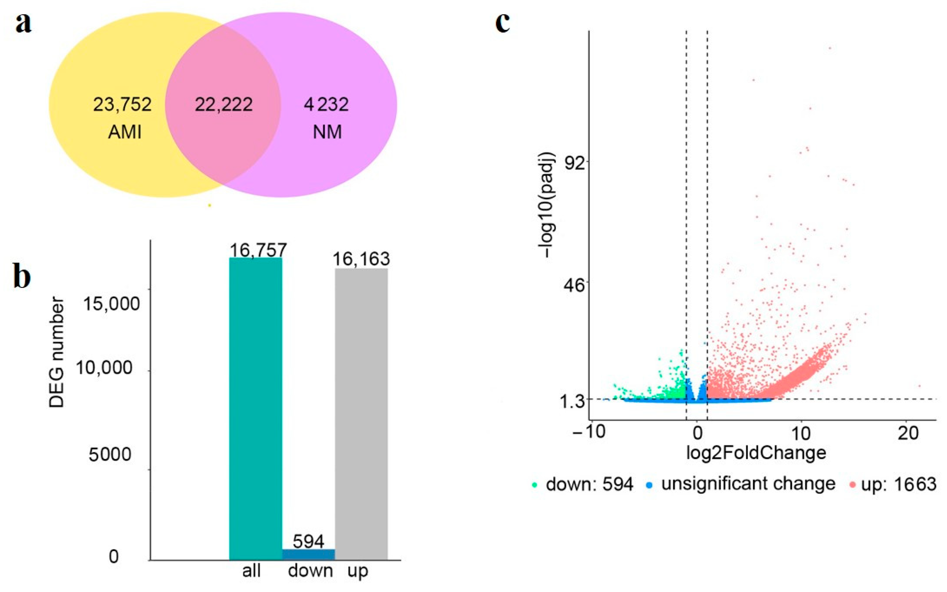
3.3. Analysis of DEGs

In the present study, a total of 50,206 genes were identified (Figure 2a). Among these, 23,752 genes were only expressed in the AMI treatment, accounting for 47.31% of the total identified genes, and 4232 genes were only expressed in the NM treatment. There were 22,222 genes co-expressed in both treatments, accounting for 44.26% of the total identified genes.
In the comparison between AMI and NM, a total of 16,757 DEGs were identified, including 16,163 upregulated DEGs and 594 downregulated DEGs, which accounted for 96.46% and 3.54% of the total number of DEGs (Figure 2b,c). In addition, among the above upregulated and downregulated DEGs, there were 14,420 DEGs that were only expressed in the AMI treatment, whose read count value was zero in the NM treatment, and 35 DEGs that were totally silenced in the AMI treatment, whose read count value was zero in the AMI treatment.

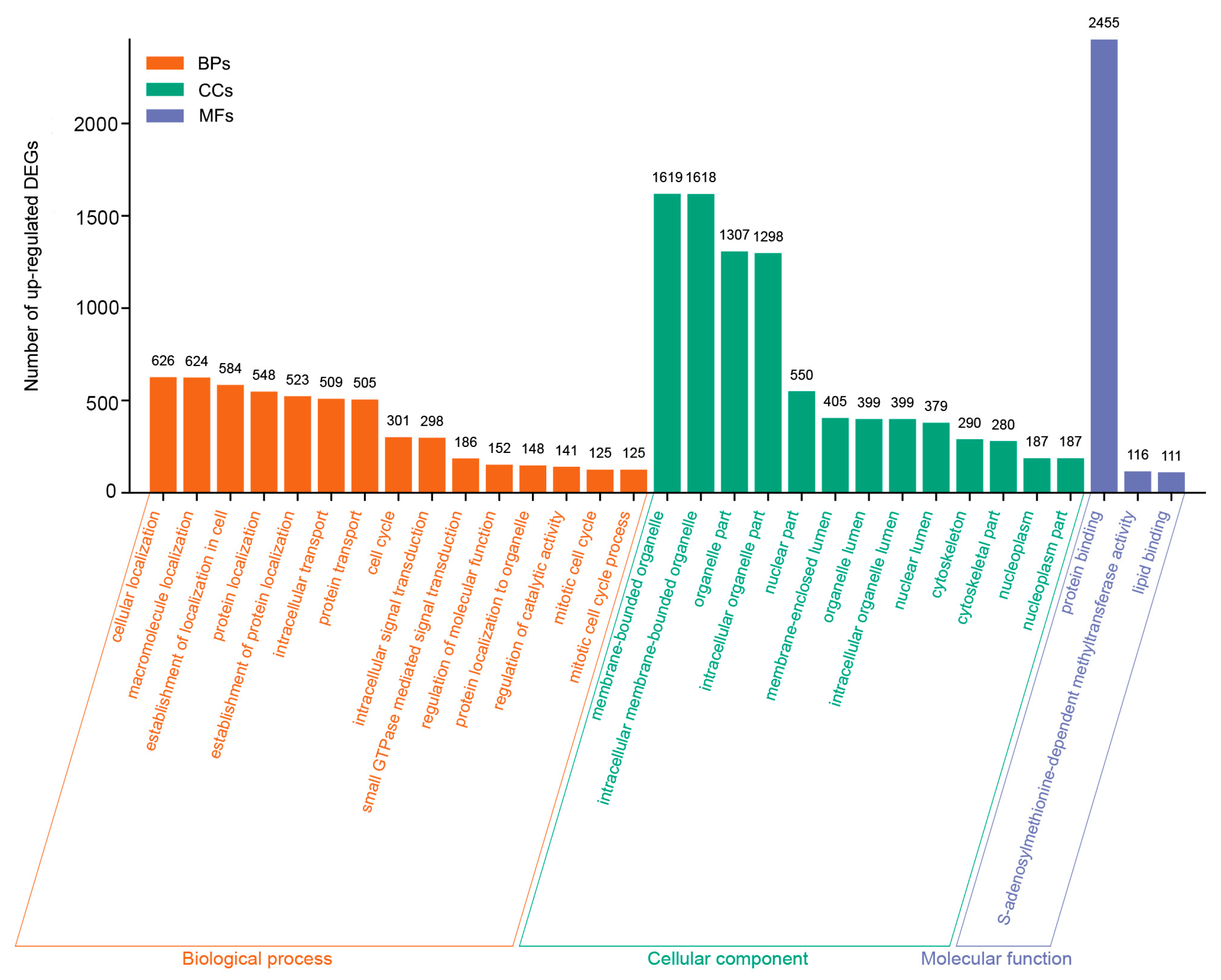
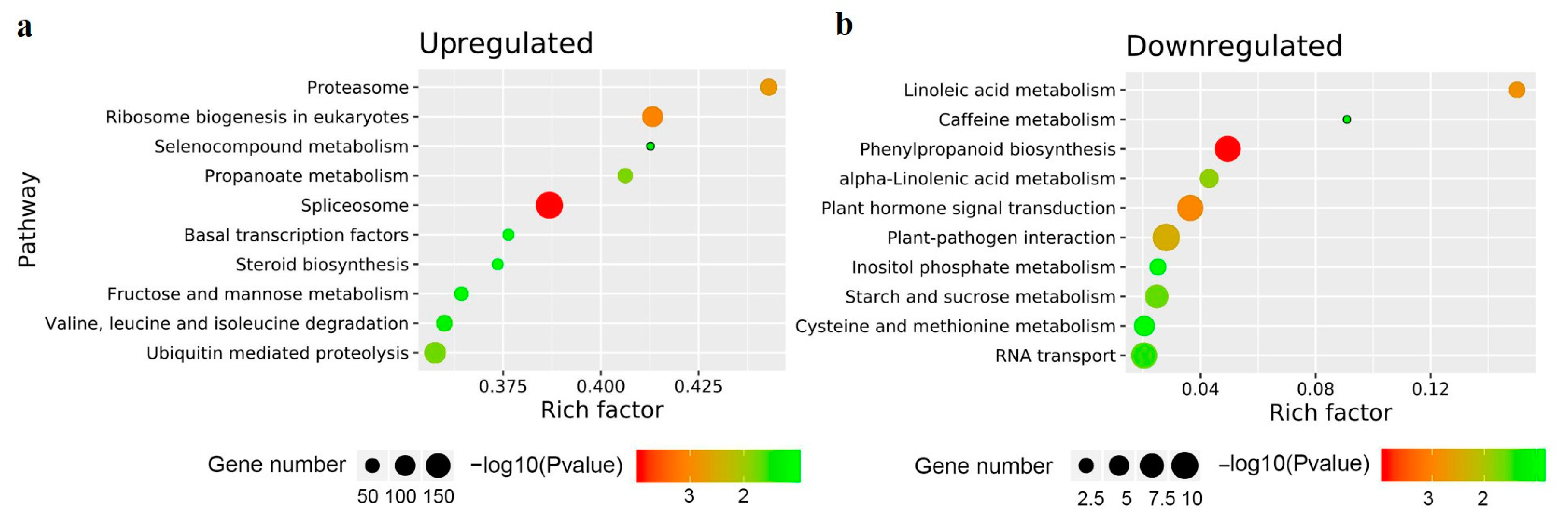
3.4. GO and KEGG Enrichment Analysis of the DEGs

To obtain a biological view of the DEGs in the roots of T. ciliata var. pubescens seedlings in the AMI and NM treatments, a GO enrichment analysis was conducted. The upregulated DEGs were significantly enriched in 20, 13, and 21 GO terms (padj < 0.05) from the ranges of molecular functions, cellular components, and biological processes, respectively (Supplementary Table S1). However, no significant GO terms were enriched in the downregulated DEGs.
The GO term “protein binding” (2455) included the highest number of upregulated DEGs from the aspects of molecular function (Figure 3). Other upregulated DEGs were mainly classified as “S-adenosylmethionine-dependent methyltransferase activity” (116) and “lipid binding” (111). For the cellular component category, most of the upregulated DEGs were located on the cell membrane, nuclear, and cytoskeleton, and thus they were mainly enriched in “membrane-bounded organelle” (1619), “organelle part” (1307), “nuclear part” (550), “cytoskeletal part” (280), and “nucleoplasm part” (187), as well as other similar terms (Figure 3). In the category of biological processes, the GO terms “cellular localization” (626), “macromolecule localization” (624), “establishment of localization in cell” (584), “protein localization” (548), “establishment of protein localization” (523), “intracellular transport” (509), and “protein transport” (505) included at least 500 upregulated DEGs. In addition, other upregulated DEGs were mainly classified as “cell cycle” (301), “intracellular signal transduction” (298), “small GTPase mediated signal transduction” (186), and “regulation of catalytic activity” (141) (Figure 3). The KEGG enrichment analysis showed that upregulated DEGs were mainly involved in “spliceosome” (188 DEGs) (Figure 4), and the downregulated DEGs were significantly enriched in the “phenylpropanoid biosynthesis” pathway (9 DEGs) (Figure 4).

3.5. Upregulated DEGs Related to the Oxidative Stress Response and Nutrient Transport

We focused on the upregulated DEGs that were related to oxidative stress response and nutrient transport. Of the DEGs related to the oxidative stress response (Table 3), 17 upregulated DEGs were annotated as “glutathione S-transferase (GST)”, 11 of which were only expressed in the AMI treatment. Furthermore, three, two, and two genes only expressed in the AMI treatment were annotated as “glutathione peroxidase (GPX)”, “glutathione reductase (GR/NADPH)”, and “glutathione synthase (GS)”, respectively. A total of eight genes only expressed in the AMI treatment were involved in “superoxide dismutase” (SOD). Two upregulated DEGs had the function of “peroxidase (POD)”, and the expression levels of these two DEGs were changed from 4- to 8-fold (2 < Log2FC < 5) by AM colonization. In addition to DEGs related to antioxidant enzymes, DEGs involved in “stress-induced phosphoprotein (STIP1)”, “heat shock 70 kDa protein1/8 (HSP 70 1/8)”, “heat shock 70 kDa protein 4 (HSP 70 4)”, and “heat shock 70 kDa protein 5 (HSP 70 5)” were also upregulated.
For the DEGs related to nutrient transport (Table 4), a total of 12 upregulated DEGs (11 genes were only expressed in the AMI treatment) were related to zinc transporters, and a total of 11 upregulated DEGs (9 genes were only expressed in the AMI treatment) were related to phosphate transporters, including “MFS transporter, PHS family, inorganic phosphate transporter” (5); “sodium-dependent phosphate transporter” (3); “phosphate transporter” (2); and “MFS transporter, ACS family, solute carrier family 17 (sodium-dependent inorganic phosphate cotransporter), other” (1). Eleven upregulated DEGs were involved in the transport of nitrogen, including inorganic nitrogen and organic nitrogen, such as ammonium transporters, nitrate transporters, and cationic amino acid transporters. Among the five DEGs annotated as “ammonium transporter, Amt family”, one gene was only expressed in the AMI treatment, and the expression of the four other genes changed from 4- to 64-fold (2 < Log2FC < 6). In addition, one upregulated DEG had the function “sulfate transporter” (Table 3). Furthermore, 19, 4, 6, and 17 upregulated DEGs were related to “V-type H+-transporting ATPase”, “H+-transporting ATPase”, “mitochondrial ABC transporter ATM”, and “Ca2+-transporting ATPase”.

4. Discussion

Fu [21] found that the root mycorrhizal colonization rate for T. ciliata var. pubescens was approximately 24.1% in the natural forest community, which was lower than that for the associated trees in the community, such as Melliodendron xylocarpum Hand.-Mazz., Castanopsis tibetana Hance, and Choerospondias axillaris (Roxb.) B. L. Burtt & A. W. Hill, but higher than that of Alniphyllum fortunei (Hemsl.) Makino and Mallotus japonicus (L. f.) Müll. Arg. In this paper, T. ciliata var. pubescens seedlings were artificially inoculated with S. viscosum, and the mycorrhizal colonization rate after 3 months was 24.63 ± 6.70% (Table 1), which is in line with the previous study.
In our investigation, T. ciliata var. pubescens seedlings inoculated with AMF exhibited a more robust root system, increased plant height, and a greater number of branches compared to their noninoculated counterparts (Figure 1, Table 1). This was consistent with numerous studies. The application of AMF biofertilizers was shown to mitigate the growth inhibition resulting from continuous cropping in Panax quinquefolius L. [16]. In the case of the woody plant Poncirus trifoliata L. Raf., AMF inoculation significantly enhanced the formation and growth of seedling lateral roots [27]. Seedling establishment was identified as a critical factor in the natural population regeneration of T. ciliata var. pubescens. In natural forests, barriers such as litter or understory weeds could impede seed germination by preventing the radicle from reaching the soil. Seedlings that managed to germinate often struggled to withstand adverse external conditions, resulting in a high rate of natural mortality [1]. Consequently, we speculated that symbiosis with AMF might help the seedlings of T. ciliata var. pubescens overcome unfavorable conditions for germination. However, the mechanisms of the symbiosis between T. ciliata var. pubescens and AMF have not been studied. Utilizing RNA-Seq technology, we discovered that AM colonization upregulated the expression of 16,163 genes (Figure 2a,b) in the roots of T. ciliata var. pubescens, a number significantly higher than the numbers reported for other plants, like P. trifoliata [27], Helianthus annuus L. [37], and wheat [38]. This remarkable increase confirms that biological processes within the roots of T. ciliata var. pubescens are profoundly activated following AMF inoculation.
AM symbiosis and mycorrhizal formation necessitate the involvement of various signal-transduction pathways between plants and AMF. Small GTP-binding proteins, as a highly conserved signaling module across eukaryotes, play crucial roles in numerous biological processes, including signal transduction, cell proliferation and differentiation, cytoskeletal organization, intracellular membrane trafficking, and nuclear transport [39,40,41]. In our study, pathways related to small GTPase-mediated signal transduction were enriched by 186 upregulated DEGs (Figure 3). These findings suggest that small GTPase-mediated signal transduction in the roots of T. ciliata var. pubescens inoculated with AMF is notably active. This activity is beneficial for transmitting developmental or environmental signals and coordinating cell-to-cell communication, thereby facilitating the establishment of symbiosis between AMF and T. ciliata var. pubescens.
Reactive oxygen species (ROS) are by-products of various metabolic pathways, and their excessive accumulation can lead to cell death [42,43]. AMF symbiosis can upregulate antioxidant enzyme activity to remove excess ROS, thus improving plant tolerance to stress [44]. For instance, the activities of SOD, CAT, and GST were increased in colonized wheat under arsenic stress [45]. At the molecular level, differentially expressed GST and POD proteins were identified in both mycorrhizal Elaeagnus angustifolia L. [46] and Medicago sativa L. [47]. Transcriptome analyses in the present study showed that many DEGs related to antioxidant enzymes, such as SOD, GST, GPX, GR, POD, and GS, were upregulated by AM colonization in T. ciliata var. pubescens seedlings (Table 3), consistent with previous research. Notably, GST, GPX, GR, and GS are involved in the glutathione peroxidase cycle, which is responsible for converting hydrogen peroxide (H2O2) into water [42]. This suggests that the H2O2 scavenging mechanism centered on glutathione was activated in mycorrhizal T. ciliata var. pubescens seedlings. Moreover, besides DEGs involved in antioxidant enzymes, DEGs related to heat shock proteins, particularly HSP70s, were also found to be upregulated in mycorrhizal T. ciliata var. pubescens seedlings (Table 3). This finding aligns with previous studies demonstrating activation of the heat shock protein gene in mycorrhizal roots of Pisum sativum L. [48]. Notably, plant Hsp70s are known to be involved in the response to abiotic stress [49]. Taken together, these findings indicate that AMF symbiosis enhances resistance to adverse conditions by increasing the expression of genes encoding antioxidant enzymes and HSP70s in T. ciliata var. pubescens seedlings.
Nutrient exchange is the main purpose of symbiosis between AMF and most terrestrial plants [50]. Recent studies have highlighted the pivotal role of specialized transporters in facilitating nutrient transport across cellular membranes, essential for the uptake and exchange of nutrients in AM symbiosis [51]. Plants absorb nitrogen in two forms: inorganic forms, such as nitrate (NO3) and ammonium (NH4+); and organic forms, such as amino acids [52]. Nitrogen transporters such as ammonium transporters (AMTs), nitrite transporters (NRTs), and amino acid transporters are crucial in managing nitrogen flux, thereby supporting normal plant metabolism and promoting both vegetative and reproductive growth [53]. Similarly, phosphate transporters (PTs) play a fundamental role in phosphorus absorption and transport within plants, optimizing phosphorus uptake and utilization efficiency [54]. Moreover, the essential micronutrients zinc and iron rely on zinc iron permease (ZIP) proteins for their absorption, transport, and distribution, ensuring the maintenance of Zn and Fe homeostasis in plants [55].
Our transcriptome analysis revealed a significant upregulation of DEGs related to ZIPs, PTs, AMTs, NRTs, cationic amino acid transporters, and sulfate transporters in T. ciliata var. pubescens seedlings following AM colonization (Table 4). This finding aligns with observations from other plant species, such as Hordeum vulgare L. [56], M. truncatula [57,58], and P. trichocarpa [51]. Beyond these specialized transporters, our study also identified the activation of numerous active transport pumps, including “V-type H+-transporting ATPase”, “H+-transporting ATPase”, “mitochondrial ABC transporter ATM”, and “Ca2+-transporting ATPase” (Table 4). These pumps are instrumental in moving compounds across cellular membranes against a chemical gradient, a process powered by the energy derived from ATP hydrolysis [59,60]. For example, the translocation of phosphate from the soil solution into the cytosol is expedited by H+-ATPases and Na+-ATPases. Similarly, nitrogen and phosphorus can be transported across the cell membrane when bound to substrate-binding proteins or specific chelating peptides, facilitated by ABC transporters for subsequent cellular absorption. These insights underscore that AM symbiosis significantly enhances nutrient uptake in T. ciliata var. pubescens seedlings, emphasizing the intricate and efficient nutrient acquisition strategies enabled by this symbiotic interaction.

5. Conclusions

In this study, we performed a comprehensive transcriptome analysis on the roots of T. ciliata var. pubescens seedlings that were inoculated with AMF or noninoculated. AMF inoculation significantly enhanced both root formation and aboveground growth. Through an RNA sequencing analysis, we identified 16,163 DEGs in response to AMF inoculation. Predominantly, the upregulated DEGs were localized to the cell membrane, nucleus, and cytoskeleton, playing critical roles in protein binding, S-adenosylmethionine-dependent methyltransferase activity, and lipid binding. These activities were involved in essential processes such as cellular and macromolecule localization, intracellular protein transport, the cell cycle, and signal transduction. Notably, the upregulation of genes linked to nutrient uptake, stress response, signal transduction, and substance transportation in T. ciliata var. pubescens augments its ability to effectively utilize soil nutrients and enhances its resilience under adverse environmental conditions. This finding indirectly highlights AMF’s potential to improve the natural regeneration of T. ciliata var. pubescens. Further investigations should focus on isolating and studying naturally occurring AMF from T. ciliata var. pubescens roots to explore their potential in subsequent studies.

Supplementary Materials

The following are available online at https://www.mdpi.com/article/10.3390/f15040673/s1, Table S1: Go enrichment analysis of upregulated DEGs (adjusted p-value < 0.05).

Author Contributions

Conceptualization, X.-R.J., W.L. and L.Z.; methodology, X.-R.J., J.-F.P., M.Z. and W.L.; validation, X.-R.J., W.L. and L.Z.; resources, X.-R.J., J.-F.P., M.Z., X.-Y.G. and Q.W.; writing—original draft preparation, X.-R.J.; writing—review and editing, W.L. and L.Z.; visualization, X.-R.J.; project administration, W.L.; funding acquisition, W.L. All authors have read and agreed to the published version of the manuscript.

Funding

This work was supported by the Jiangxi Forestry Science and Technology Innovation Foundation (Innovation special (2019) No. 25), Jiangxi Provincial Natural Science Foundation (20212BAB215014), and the Central Finance Forestry Science and Technology Promotion Demonstration Project (JXTG[2024]25).

Data Availability Statement

The data presented in this study are available upon request from the corresponding author.

Acknowledgments

We express our gratitude to Xiaohua Yao for assistance with sampling. Special thanks are extended to the people of the Administration of Guanshan National Natural Reserve for their support in facilitating sample collection.

Conflicts of Interest

The authors declare no conflict of interest.

References

  1. Huang, H.L.; Zhang, L.; Liao, C.K. Seed rain, soil seed bank, and natural regeneration of natural Toona ciliata var. pubescens forest. J. Appl. Ecol. 2012, 23, 972–978. [Google Scholar]
  2. Pan, J.; Wang, Q.; Guo, X.; Jiang, X.; Cheng, Q.; Fu, L.; Liu, W.; Zhang, L. Local patterns of arbuscular mycorrhizal fungal diversity and community structure in a natural Toona ciliata var. pubescens forest in South Central China. PeerJ 2021, 9, e11331. [Google Scholar] [CrossRef] [PubMed]
  3. Guo, X.Y.; Fu, L.; Zhang, L.; Su, H.; Liang, Y.L. Effects of forest soil and soil-borne fungi on seed germination and seedling survival of Toona ciliate var. pubescens. For. Res. 2017, 30, 871–877. [Google Scholar]
  4. Liu, W.; Jin, J.; Li, Y.; Tan, Z.; Jiang, J.; Liu, J. Effects of waters, fertilization with different nitrogen and phosphorus ratios on the growth, nutrient distribution and chlorophyll fluorescence characteristics of Toona ciliate var. pubescens seedlings. J. West China For. Sci. 2021, 50, 83–90. [Google Scholar]
  5. Sun, B.; Zhang, T.; Zhao, Q. Comprehensive evaluation of soil fertility in the hilly and mountainous region of Southeastern China. Acta Pedol. Sin. 1995, 32, 362–369. [Google Scholar]
  6. Brundrett, M.C.; Tedersoo, L. Evolutionary history of mycorrhizal symbioses and global host plant diversity. New Phytol. 2018, 220, 1108–1115. [Google Scholar] [CrossRef] [PubMed]
  7. Ezawa, T.; Saito, K. How do arbuscular mycorrhizal fungi handle phosphate? New insight into fine-tuning of phosphate metabolism. New Phytol. 2018, 220, 1116–1121. [Google Scholar] [CrossRef] [PubMed]
  8. Ferrol, N.; Azcón-Aguilar, C.; Pérez-Tienda, J. Review: Arbuscular mycorrhizas as key players in sustainable plant phosphorus acquisition: An overview on the mechanisms involved. Plant Sci. 2019, 280, 441–447. [Google Scholar] [CrossRef] [PubMed]
  9. Wu, F.; Fang, F.; Wu, N.; Li, L.; Tang, M. Nitrate transporter gene expression and kinetics of nitrate uptake by Populus × canadensis ‘Neva’ in relation to arbuscular mycorrhizal fungi and nitrogen availability. Front. Microbiol. 2020, 11, 176. [Google Scholar] [CrossRef]
  10. Bennett, A.E.; Groten, K. The costs and benefits of plant-arbuscular mycorrhizal fungal interactions. Annu. Rev. Plant Biol. 2022, 73, 649–672. [Google Scholar] [CrossRef]
  11. Begum, N.; Cheng, Q.; Ahanger, M.A.; Raza, S.; Khan, M.I.; Ashraf, M.; Ahmed, N.; Zhang, L. Role of arbuscular mycorrhizal fungi in plant growth regulation: Implications in abiotic stress tolerance. Front. Plant Sci. 2019, 10, 1068. [Google Scholar] [CrossRef]
  12. Fusconi, A. Regulation of root morphogenesis in arbuscular mycorrhizae: What role do fungal exudates, phosphate, sugars and hormones play in lateral root formation? Annu. Rev. 2014, 113, 19–33. [Google Scholar] [CrossRef] [PubMed]
  13. Evelin, H.; Kapoor, R. Arbuscular mycorrhizal symbiosis modulates antioxidant response in salt-stressed Trigonella foenum-graecum plants. Mycorrhiza 2014, 24, 197–208. [Google Scholar] [CrossRef] [PubMed]
  14. Zhai, S.; Wu, Y.; Xu, C.; Chen, W.; Feng, J.; Zheng, Q.; Meng, Y.; Yang, H. Symbiotic soil fungi suppress N2O emissions but facilitate nitrogen remobilization to grains in sandy but not clay soils under organic amendments. Appl. Soil Ecol. 2021, 167, 104012. [Google Scholar] [CrossRef]
  15. Thirkell, T.J.; Charters, M.D.; Elliott, A.J.; Sait, S.M.; Field, K.J. Are mycorrhizal fungi our sustainable saviours? Considerations for achieving food security. J. Ecol. 2017, 105, 921–929. [Google Scholar] [CrossRef]
  16. Liu, N.; Shao, C.; Sun, H.; Liu, Z.; Guan, Y.; Wu, L.; Zhang, L.; Pan, X.; Zhang, Z.; Zhang, Y.; et al. Arbuscular mycorrhizal fungi biofertilizer improves American ginseng (Panax quinquefolius L.) growth under the continuous cropping regime. Geoderma 2020, 363, 114155. [Google Scholar] [CrossRef]
  17. Loo, W.T.; Chua, K.O.; Mazumdar, P.; Cheng, A.; Osman, N.; Harikrishna, J.A. Arbuscular mycorrhizal symbiosis: A strategy for mitigating the impacts of climate change on tropical legume crops. Plants 2022, 11, 2875. [Google Scholar] [CrossRef] [PubMed]
  18. Boller, T.; Wiemken, A.; Sanders, I.R. Different arbuscular mycorrhizal fungal species are potential determinants of plant community structure. Ecology 1998, 79, 2082–2091. [Google Scholar]
  19. Gillespie, A.W.; Farrell, R.E.; Walley, F.L.; Ross, A.R.; Leinweber, P.; Eckhardt, K.U.; Regier, T.Z.; Blyth, R.I. Glomalin-related soil protein contains non-mycorrhizal-related heat-stable proteins, lipids and humic materials. Soil Biol. Biochem. 2011, 43, 766–777. [Google Scholar] [CrossRef]
  20. Huang, Y.; Jiang, Y.; Wang, Z.; Song, F.; He, L.; Tian, R.; Wu, L. The suppression effect and mechanism of arbuscular mycorrhizal fungi against plant root rot. J. Appl. Ecol. 2021, 32, 1890–1902. [Google Scholar]
  21. Fu, L. Study on the community characteristics of arbuscular mycorrhizal fungi in soil of the community of natural forest of Toon ciliata. Master’s Thesis, Jiangxi Agricultural University, Nanchang, China, 2018. [Google Scholar]
  22. Redecker, D.; Schüßler, A.; Stockinger, H.; Stürmer, S.L.; Morton, J.B.; Walker, C. An evidence-based consensus for the classification of arbuscular mycorrhizal fungi (Glomeromycota). Mycorrhiza 2013, 23, 515–531. [Google Scholar] [CrossRef]
  23. Tedone, L.; Ruta, C.; De Cillis, F.; De Mastro, G. Effects of Septoglomus viscosum inoculation on biomass yield and steviol glycoside concentration of some Stevia rebaudiana chemotypes. Sci. Hortic. 2020, 262, 109026. [Google Scholar] [CrossRef]
  24. Tarraf, W.; Ruta, C.; Tagarelli, A.; De Cillis, F.; De Mastro, G. Influence of arbuscular mycorrhizae on plant growth, essential oil production and phosphorus uptake of Salvia officinalis L. Ind. Crop. Prod. 2017, 102, 144–153. [Google Scholar] [CrossRef]
  25. Li, J.; Wang, Q.; Wang, Y.; Zhang, Q.; Luo, J.; Jiang, X.; Liu, W.; Zhao, X. Effects of arbuscular mycorrhizal fungi on improvement of degraded landscape soil in an ionized rare earth mining area, subtropical China. Soil Sci. Soc. Am. J. 2022, 86, 275–292. [Google Scholar] [CrossRef]
  26. Liu, W.; Zhao, X.; Zhang, Q.; Li, J.; Xue, H.; Zhang, J.; Wu, J.; Deng, G.; Zhang, W.; Li, X.; et al. A Strain of Septoglomus viscosum, including Its Microbial Agent and Application. Chinese Patent CN110819542B, 9 February 2021. [Google Scholar]
  27. Chen, W.; Li, J.; Zhu, H.; Xu, P.; Chen, J.; Yao, Q. Arbuscular mycorrhizal fungus enhances lateral root formation in Poncirus trifoliata (L.) as revealed by RNA-Seq analysis. Front Plant Sci. 2017, 8, 2039. [Google Scholar] [CrossRef]
  28. Vierheilig, H.; Coughlan, A.P.; Wyss, U.; Piché, Y. Ink and vinegar., a simple staining technique for arbuscular-mycorrhizal fungi. Appl. Environ. Microbiol. 1998, 64, 5004–5007. [Google Scholar] [CrossRef] [PubMed]
  29. McGonigle, T.P.; Miller, M.H.; Evans, D.G.; Fairchild, G.L.; Swan, J.A. A new method which gives an objective measure of colonization of roots by vesicular-arbuscular mycorrhizal fungi. New Phytol. 1990, 115, 495–501. [Google Scholar] [CrossRef] [PubMed]
  30. Grabherr, M.G.; Haas, B.J.; Yassour, M.; Levin, J.Z.; Thompson, D.A.; Amit, I.; Adiconis, X.; Fan, L.; Raychowdhury, R.; Zeng, Q.; et al. Full-length transcriptome assembly from RNA-Seq data without a reference genome. Nat Biotechnol. 2011, 29, 644–652. [Google Scholar] [CrossRef]
  31. Li, B.; Dewey, C. RSEM: Accurate transcript quantification from RNA-Seq data with or without a reference genome. BMC Bioinform. 2011, 12, 123. [Google Scholar] [CrossRef]
  32. Wang, L.; Feng, Z.; Wang, X.; Wang, X.; Zhang, X. DEGseq: An R package for identifying differentially expressed genes from RNA-seq data. Bioinformatics 2010, 26, 136–138. [Google Scholar] [CrossRef]
  33. Benjamini, Y.; Hochberg, Y. Controlling the false discovery rate: A practical and powerful approach to multiple testing. J. R. Stat. Soc. Ser. B Methodol. 1995, 57, 289–300. [Google Scholar] [CrossRef]
  34. Anders, S.; Huber, W. Differential Expression of RNA-Seq Data at the Gene Level—The DESeq Package; European Molecular Biology Laboratory (EMBL): Heidelberg, Germany, 2012. [Google Scholar]
  35. Young, M.D.; Wakefield, M.J.; Smyth, G.K.; Oshlack, A. Gene ontology analysis for RNA-seq: Accounting for selection bias. Genome Biol. 2010, 11, R14. [Google Scholar] [CrossRef] [PubMed]
  36. Mao, X.; Cai, T.; Olyarchuk, J.G.; Wei, L. Automated genome annotation and pathway identification using the KEGG orthology (KO) as a controlled vocabulary. Bioinformatics 2005, 21, 3787–3793. [Google Scholar] [CrossRef] [PubMed]
  37. Vangelisti, A.; Natali, L.; Bernardi, R.; Sbrana, C.; Turrini, A.; Hassani-Pak, K.; Hughes, D.; Cavallini, A.; Giovannetti, M.; Giordani, T. Transcriptome changes induced by arbuscular mycorrhizal fungi in sunflower (Helianthus annuus L.) roots. Sci. Rep. 2018, 8, 4–14. [Google Scholar] [CrossRef] [PubMed]
  38. Li, M.; Wang, R.; Tian, H.; Gao, Y. Transcriptome responses in wheat roots to colonization by the arbuscular mycorrhizal fungus Rhizophagus irregularis. Mycorrhiza 2018, 28, 747–759. [Google Scholar] [CrossRef] [PubMed]
  39. Pandey, G.K.; Sharma, M.; Pandey, A.; Shanmugam, T. Overview of small GTPase signaling proteins in plants. In GTPases; Springer Briefs in Plant Science; Springer: Cham, Switzerland, 2015; pp. 9–14. [Google Scholar]
  40. Yuksel, B.; Memon, A.R. Legume small GTPases and their role in the establishment of symbiotic associations with Rhizobium spp. Plant Signal. Behav. 2009, 4, 257–260. [Google Scholar] [CrossRef] [PubMed]
  41. Nielsen, E. The Small GTPase Superfamily in Plants: A conserved regulatory module with novel functions. Annu. Rev. Plant Biol. 2020, 71, 247–272. [Google Scholar] [CrossRef] [PubMed]
  42. Mittler, R. Oxidative stress., antioxidants and stress tolerance. Trends Plant Sci. 2002, 7, 405–410. [Google Scholar] [CrossRef] [PubMed]
  43. Apel, K.; Hirt, H. Reactive oxygen species: Metabolism, oxidative stress, and signal transduction. Annu. Rev. Plant Biol. 2004, 55, 373–399. [Google Scholar] [CrossRef]
  44. Jajoo, A.; Mathur, S. Role of arbuscular mycorrhizal fungi as an underground savior for protecting plants from abiotic stresses. Physiol. Mol. Biol. Plants 2021, 27, 2589–2603. [Google Scholar] [CrossRef]
  45. Sharma, S.; Anand, G.; Singh, N.; Kapoor, R. Arbuscular mycorrhiza augments arsenic tolerance in wheat (Triticum aestivum L.) by strengthening antioxidant defense system and thiol metabolism. Front. Plant Sci. 2017, 8, 906. [Google Scholar] [CrossRef]
  46. Jia, T.; Wang, J.; Chang, W.; Fan, X.; Sui, X.; Song, F. Proteomics analysis of Echinacea angustifolia seedlings inoculated with arbuscular mycorrhizal fungi under salt stress. Int. J. Mol. Sci. 2019, 20, 788. [Google Scholar] [CrossRef] [PubMed]
  47. Sui, X.; Wu, Q.; Chang, W.; Fan, X.; Song, F. Proteomic analysis of the response of Funnelifor mismosseae/Medicago sativa to atrazine stress. BMC Plant Biol. 2018, 18, 289. [Google Scholar] [CrossRef]
  48. Rivera-Becerril, F.; van Tuinen, D.; Martin-Laurent, F.; Metwally, A.; Dietz, K.J.; Gianinazzi, S.; Gianinazzi-Pearson, V. Molecular changes in Pisum sativum L. roots during arbuscular mycorrhiza buffering of cadmium stress. Mycorrhiza 2005, 16, 51–60. [Google Scholar] [CrossRef]
  49. Usman, M.G.; Rafii, M.Y.; Martini, M.Y.; Yusuff, O.A.; Ismail, M.R.; Miah, G. Molecular analysis of Hsp70 mechanisms in plants and their function in response to stress. Biotechnol. Genet. Eng. Rev. 2017, 33, 26–39. [Google Scholar] [CrossRef] [PubMed]
  50. Rui, W.; Mao, Z.; Li, Z. The roles of phosphorus and nitrogen nutrient transporters in the arbuscular mycorrhizal symbiosis. Int. J. Mol. Sci. 2022, 23, 11027. [Google Scholar] [CrossRef]
  51. Silvia, C.; Loic, C.; Alexis, S.; Annette, N.; Alexander, E.; Daphnée, B.; Ghislaine, R.; Daniel, W.; Christophe, R.; Joachim, K.; et al. Imbalanced regulation of fungal nutrient transports according to phosphate availability in a symbiocosm formed by poplar, sorghum, and Rhizophagus irregularis. Front. Plant Sci. 2019, 10, 1617. [Google Scholar]
  52. Bücking, H.; Kafle, A. Role of arbuscular mycorrhizal fungi in the nitrogen uptake of plants: Current knowledge and research Gaps. Agronomy 2015, 5, 587–612. [Google Scholar] [CrossRef]
  53. Tegeder, M.; Masclaux-Daubresse, C. Source and sink mechanisms of nitrogen transport and use. New Phytol. 2018, 217, 35–53. [Google Scholar] [CrossRef]
  54. Daram, P.; Brunner, S.; Rausch, C.; Steiner, C.; Amrhein, N.; Bucher, M. Pht2;1 Encodes a low-affinity phosphate transporter from Arabidopsis. Plant Cell 1999, 11, 2153–2166. [Google Scholar] [CrossRef]
  55. Guerinot, M.L. The ZIP family of metal transporters. Biochim. Biophys. Acta (BBA) Biomembr. 2000, 1465, 190–198. [Google Scholar] [CrossRef]
  56. Watts-Williams, S.J.; Cavagnaro, T.R. Arbuscular mycorrhizal fungi increase grain zinc concentration and modify the expression of root ZIP transporter genes in a modern barley (Hordeum vulgare) cultivar. Plant Sci. 2018, 274, 163–170. [Google Scholar] [CrossRef] [PubMed]
  57. Watts-Williams, S.J.; Tyerman, S.D.; Cavagnaro, T.R. The dual benefit of arbuscular mycorrhizal fungi under soil zinc deficiency and toxicity: Linking plant physiology and gene expression. Plant Soil 2017, 420, 375–388. [Google Scholar] [CrossRef]
  58. Zhu, J.N.; Huang, H.; Du, Y.; Tang, J.J.; Chen, X. The transgenerational effect of arbuscular mycorrhizal fungi on root phosphatase activity of host plant Medicago truncatula. Chin. J. Ecol. 2022, 41, 912–918. [Google Scholar]
  59. Wilkens, S. Structure and mechanism of ABC transporters. F1000Prime Rep. 2015, 7, 14. [Google Scholar] [CrossRef]
  60. Lane, T.S.; Rempe, C.S.; Davitt, J.; Staton, M.E.; Peng, Y.; Soltis, D.E.; Melkonian, M.; Deyholos, M.; Leebens-Mack, J.H.; Chase, M.; et al. Diversity of ABC transporter genes across the plant kingdom and their potential utility in biotechnology. BMC Biotechnol. 2016, 16, 47. [Google Scholar] [CrossRef]
Figure 1. Above- and belowground growth of T. ciliata var. pubescens seedlings non-affected and affected by AMF inoculation. (a,c) The above growth. (b,d) The belowground growth. (e) Hyphae of AMF in the roots of AMI treatment.
Forests 15 00673 g001
Figure 2. Gene analysis between the nonmycorrhizal and AMF inoculation treatments. (a) Venn diagram of co-expressed genes in the comparison group. (b,c) Number of upregulated DEGs and downregulated DEGs.
Forests 15 00673 g002
Figure 3. Dominant GO terms with at least 100 upregulated DEGs (adjusted p-value < 0.05). BPs, biological processes; CCs, cellular components; MFs, molecular functions.
Forests 15 00673 g003
Figure 4. Top 10 KEGG pathways with upregulated DEGs (a) and downregulated DEGs (b).
Forests 15 00673 g004
Table 1. The effects of AMF on the plant height and branch number of T. ciliata var. pubescens seedlings and on mycorrhizal colonization.
TreatmentPlant Height (cm)Branch Number Mycorrhizal Colonization (%)
NM6.21 ± 0.563.28 ± 0.480.00 ± 0.00
AMI15.07 ± 1.09 ***5.28 ± 0.47 ***24.63 ± 6.70 ***
Notes: NM, nonmycorrhizal treatment; AMI, AM inoculation treatment. The values in the table are mean ± standard deviation (SD). *** A significant difference at p < 0.001, as determined by t-test with IBM SPSS v.21 statistical software.
Table 2. Summary of Illumina transcriptome sequencing.
SampleRaw ReadsClean ReadsClean BasesError RateQ ≥ 20Q ≥ 30GC pct
NM_190483268860625886.45G0.0298.3695.0942.54
NM_295951276913121886.85G0.0298.4395.143.14
NM_396105596910357566.83G0.0298.4695.1643.19
AMI_184660684791561645.94G0.0298.4895.3441.29
AMI_290570192859019126.44G0.0298.2994.7342.66
AMI_396210968903862966.78G0.0298.3894.9642.63
Notes: NM, nonmycorrhizal treatment; AMI, AM inoculation treatment; Q ≥ 20, percentage of bases with a Phred quality score (Q score) greater than 20; Q ≥ 30, percentage of bases with a Phred quality score greater than 30; GC pct, the percentage of G and C bases in all clean reads.
Table 3. Upregulated DEGs involved in the oxidative stress response.
Gene Function Annotated by KONumberlog2FC
Glutathione S-transferase 17 (11)1.35–16.22
Heat shock 70 kDa protein 1/89 (9)5.31–10.53
Heat shock transcription factor, other eukaryote7 (7)5.68–10.03
Stress-induced phosphoprotein 15 (5)5.71–10.39
Heat shock 70 kDa protein 44 (3)1.05–9.01
Heat shock 70 kDa protein 54 (4)6.06–11.06
Superoxide dismutase, Fe-Mn family4 (4)5.22–8.54
Superoxide dismutase, Cu-Zn family4 (4)6.8–10.72
Glutathione peroxidase 3 (3)7.14–11.63
Glutathione reductase (NADPH) 2 (2)8.65–8.98
Glutathione gamma-glutamylcysteinyltransferase 2 (1)1.76–5.9
Peroxidase 2 (0)2.58–4.36
Glutathione reductase (NADPH) 2 (2)8.65–8.98
Glutathione synthase 2 (2)6.37–7.41
Heat shock protein 90 kDa beta2 (2)6.85–8.46
HSP20 family protein1 (1)5.63
Notes: KO, KEGG Orthology annotation; Log2FC, Log2FoldChange. Data in the brackets are the number of genes only expressed in the AMI treatment.
Table 4. Upregulated DEGs involved in the inorganic nutrient transport.
Gene Function Annotated by KONumberlog2FC
Solute carrier family 30 (zinc transporter), member 26 (6)5.07–9.37
Zinc transporter, ZIP family4 (4)6.41–8.11
Solute carrier family 39 (zinc transporter), member 1/2/32 (1)4.41–5.38
MFS transporter, PHS family, inorganic phosphate transporter5 (4)1.28–15.42
Solute carrier family 20 (sodium-dependent phosphate transporter)3 (2)5.03–12.21
Phosphate transporter2 (2)8.57–8.98
MFS transporter, ACS family, solute carrier family 17 (sodium-dependent inorganic phosphate cotransporter), other1 (1)8.65
Ammonium transporter, Amt family5 (1)2.52–14.69
MFS transporter, NNP family, nitrate/nitrite transporter4 (2)1.8–9.54
Solute carrier family 7 (cationic amino acid transporter), member 1111.59
Sulfate transporter 1, high affinity1 (1)6.82
V-type H+-transporting ATPase19 (19)6.28–10.7
H+-transporting ATPase 4 (3)2.52–12.71
Mitochondrial ABC transporter ATM6 (6)5.18–9.21
Ca2+-transporting ATPase17 (16)2.41–10.52
Notes: KO, KEGG Orthology annotation; Log2FC, Log2FoldChange. Data in the brackets are the number of genes only expressed in the AMI treatment.
Disclaimer/Publisher’s Note: The statements, opinions and data contained in all publications are solely those of the individual author(s) and contributor(s) and not of MDPI and/or the editor(s). MDPI and/or the editor(s) disclaim responsibility for any injury to people or property resulting from any ideas, methods, instructions or products referred to in the content.

