Brief Report

Characterization of Mycoviruses in Armillaria ostoyae and A. cepistipes in the Czech Republic

1
Department of Genetics and Agricultural Biotechnology, Faculty of Agriculture and Technology, University of South Bohemia in České Budějovice, Na Sádkách 1780, 370 05 České Budějovice, Czech Republic
2
Department of Forest Protection and Wildlife Management, Faculty of Forestry and Wood Technology, Mendel University in Brno, Zemědělská 1, 613 00 Brno, Czech Republic
3
Instituto de Biología Molecular y Celular de Plantas, Universidad Politécnica de Valencia-CSIC, 46022 Valencia, Spain
*
Author to whom correspondence should be addressed.
Viruses 2024, 16(4), 610; https://doi.org/10.3390/v16040610
Submission received: 18 March 2024 / Revised: 8 April 2024 / Accepted: 11 April 2024 / Published: 15 April 2024
(This article belongs to the Collection Mycoviruses)

Abstract

:
Members of the genus Armillaria are widespread forest pathogens against which effective protection has not yet been developed. Due to their longevity and the creation of large-scale cloning of Armillaria individuals, the use of mycoviruses as biocontrol agents (BCAs) against these pathogens could be an effective alternative. This work describes the detection and characterization of viruses in Armillaria spp. collected in the Czech Republic through the application of stranded total RNA sequencing. A total of five single-stranded RNA viruses were detected in Armillaria ostoyae and A. cepistipes, including viruses of the family Tymoviridae and four viruses belonging to the recently described “ambivirus” group with a circular ambisense genome arrangement. Both hammerhead (HHRz) and hairpin (HpRz) ribozymes were detected in all the ambiviricot sequences. Armillaria viruses were compared through phylogenetic analysis and confirmed their specific host by direct RT-PCR. One virus appears to infect both Armillaria species, suggesting the occurrence of interspecies transmission in nature.

1. Introduction

Fungal pathogens are important members in forest ecosystems. They affect the diversity, structure and dynamics of forest communities substantially [1,2]. The fungal genus Armillaria includes more than 40 described species [3], which are causal agents of root rot in a wide variety of trees, shrubs and some herbs [4], including economically important conifers (e.g., Abies, Picea, Pinus) and agricultural crops (e.g., Citrus, Juglans, Malus, Prunus, Vitis) [5]. Norway spruce (Picea abies) is a major host of Armillaria in the Czech Republic. Over the last few years, Czech forests of Norway spruce seem to be suffering a generalized decline [6,7], which is enhanced by pathogenic fungi like Armillaria [8,9]. The decline is often caused by a combination of abiotic and biotic factors that limit tree growth, reduce foliage quality, and weaken root systems. These interactions can kill individual trees and entire stands [10,11]. Although there are relatively efficient methods for the protection of agricultural crops against Armillaria root rot, none of the established methods can completely eliminate the mycelium from the contaminated site [12,13]. Therefore, other strategies have to be pushed towards the development of environmentally friendly control approaches [14].
Fungal viruses (mycoviruses) of plant-interacting fungi are particularly significant for crop protection because they can influence the phenotype of their host. An increasing number of mycoviruses with the ability to induce hypovirulence in their host have been reported [15,16,17]. There are important forest pathogenic fungi, such as Cryphonectria parasitica, Phytophthora spp., Ophiostoma spp., Gremmeniella abietina, Hymenoscyphus fraxineus, Heterobasidion annosum and Armillaria spp., which host mycoviruses with diverse fungus-virus relationships [18,19,20]. In Armillaria, early studies by Blattny (1973) [21] and Reaves et al. (1988) [22] described the presence of virus-like particles in A. mellea and A. ostoyae, but it was not until 2021 that viruses were confirmed by molecular characterization [13,23,24].
Previous research on Armillaria root rot fungi has identified several RNA viruses. These studies have primarily aimed to understand the diversity and impact of these viruses on Armillaria species, focusing on their ecological roles and potential effects on fungal virulence. To date, the virome of the genus Armillaria has been molecularly investigated in samples from Finland, Russia (Siberia), South Africa and Switzerland. Several positive- and negative-stranded ssRNA viruses and even two dsRNA mycoviruses have been described. Mitoviruses, ourmia-like viruses, tymoviruses, phlegiviruses, ambi-like viruses, several previously unclassified (+)ssRNA and two unclassified dsRNA viruses have been described in five different Armillaria species (A. mellea, A. borealis, A. cepistipes, A. ostoyae, A. gallica) [13,23,24].
The main objectives of this study were (i) to confirm the occurrence of mycoviruses in a collection of isolates of the genus Armillaria in Central Europe, and (ii) to describe their genomic features and phylogenetic relationships.

2. Materials and Methods

2.1. Sampling

Armillaria isolates were collected in the years 2019–2020 in the Czech Republic, mainly in northeastern Moravia, the Moravian Karst and southeastern Bohemia (Table 1). Spores from fruiting bodies or rhizomorphs were transferred and cultured on ME agar (7.5 g bacteriological agar, 3.5 g corn agar, 10 g malt extract, 10 g glucose/0.5 L; pH 5.5) at 25 °C for approximately four weeks before being harvested. Subsequently, samples on the same ME agar were stored at 4 °C. A total of 13 samples, including Armillaria ostoyae and A. cepistipes, were cultivated and analyzed.

2.2. Isolation of Double-Stranded (ds) RNA

The samples were first examined for the presence of potential viral dsRNA molecules. Mycelium from one Petri dish was lyophilized and collected in a 50 mL Falcon tube. Two stainless steel beads with a diameter of 10 mm and liquid nitrogen were added to the Falcon tube and mycelium was homogenized by vortexing at maximum speed (3400 RPM/approx. 2 min). After homogenization, dsRNA was extracted using protocol Morris and Dodds (1979) [25] with some modifications, as described in the work of Tonka et al. (2021) [26]. Previously confirmed dsRNA-hosting isolates of Gremmeniella [27] and Phytophthora [28] were used as the positive control of each isolation.

2.3. Isolation of Total RNA

Thirteen samples were selected for stranded total RNA sequencing analysis (Table 1). Approximately 50–100 mg of fresh mycelium was collected from the agar of each sample and transferred to the 2 mL tubes with steel beads. The tubes were immersed in liquid nitrogen and vortexed (using Labnet vortex mixer VX-200, Labnet, Edion, NJ, USA) until the mycelium was ground to a fine powder. Isolation of total RNA was performed using a SPLIT RNA Extraction Kit (Lexogen, Vienna, Austria) following the protocol provided by the manufacturer, eluted in 30 µL of EB buffer. Total RNA was visualized on an agarose gel and stored at −80 °C.

2.4. Stranded Total RNA Sequencing

The isolated total RNA of 13 Armillaria samples was pooled into one mixed sample and treated with TURBO DNA-free TM Kit (InvitrogenTM (Waltham, MA, USA), Thermo Fisher Scientific (Waltham, MA, USA)). RNA quantity was measured by Qubit® Fluorometer (Life Technologies (Carlsbad, CA, USA), Thermo Fisher Scientific). Total RNA was sent to SEQme s.r.o. (Dobris, Czech Republic) for RNA library construction and RNA sequencing. Prior to the library preparation, ribosomal RNA (rRNA) was depleted using the NEBNext rRNA Depletion Kit (Human/Mouse/Rat). The library was prepared using an NEBNext Ultra II Directional RNA Library Prep Kit for Illumina. The quality of the prepared library was checked using an Agilent Bioanalyzer 2100 High sensitivity DNA Kit, Invitrogen Collibri Library Quantification Kit and Qubit 1X dsDNA High-Sensitivity Assay Kit. A KAPA Library Quantification Kit for Illumina platform was used for absolute qPCR-based quantification of the Illumina libraries flanked by the P5 and P7 flow cell oligo sequences. Libraries underwent paired-end (PE) (2 × 150 nt) sequencing on a NovaSeq6000 (DS-150) (Illumina, San Diego, CA, USA) using a NovaSeq S4 v1.5 reagent kit. An “in-lane” PhiX control spike was included in each lane of the flow cell.

2.5. Bioinformatics

2.5.1. Data Preprocessing

The raw sequencing data were downloaded from the data repository of the sequencing company SEQme s. r. o. (Dobris, Czech Republic). The processing was carried out on the local server of the University of South Bohemia. In the first step, data quality was assessed using the FASTQC v.0.11.9 program [29]. This control step revealed the presence of a small number of adapter sequences. The nucleotide sequences of adapters were obtained according to the used library kit and sequencer machine mentioned earlier [30]. Adapter and quality trimming were performed using the program Cutadapt v3.4 [31] with requirements for a Phred score higher than 30 and a minimum length of the truncated sequence of 50 bases. The software is available at the code depository GitHub [32]. Subsequently, data quality of trimmed reads was again assessed in FASTQC program mentioned previously.

2.5.2. Host Reads’ Removal

After preprocessing the data, it was necessary to remove reads belonging to host fungi. The STAR v2.7.9a program was used for this purpose [33]. Assembly GCA_900157425.1 version 2 of strain C18/9 of A. ostoyae was used as a reference sequence. After mapping the reads to the Armillaria genome, only unmapped reads were left for further processing.

2.5.3. Discovery of Known Virus

To reveal already known viruses, the Viral NCBI database [34] was used for this purpose. We downloaded only complete viral RefSeq nucleotide sequences. Our reads were aligned to this reference database by BWA v0.7.17.-r1188 program package [35]. In the next step, we calculated the coverage for each viral genome using Samtools kit v1.16.1 [32]. If the coverage was higher than 80 percent, we visualized the alignment for particular viral genome with IGV program v2.16.1 [36] and manually confirmed or denied the presence of the published virus.

2.5.4. Discovery of Novel Viruses

In the first step, de novo assembly was performed with unmapped reads from STAR aligner. The toolkit SPAdes version 3.15.3 [37] with default settings for metagenomics was used for this purpose. Assembled contigs shorter than 1000 bp were discarded. In the next step, the rest of contigs were compared to several databases: Viral UniProt KB [38], Viral NCBI [34], RVDB [39], Virus-Host DB [40]. Each of the databases was downloaded to a local server and was used to search for similarities between database sequences and assembled contigs. For this purpose, the program BLASTx and BLASTn v2.12.0+ was used. The threshold for the E-value was set to 1 × 10⁻³. All contigs with at least one hit were further used to search for similarity with the NCBI database nr (RefSeq non-redundant proteins) or nt (RefSeq Nucleotide) specifying the Armillaria taxon. If a positive hit was found, the E-value values were subsequently compared. If this value was higher for the results from viral databases, the contig was removed. In the last step, the remaining contigs were searched with the entire nr and nt databases and the threshold for the E-value was set as 1 × 10⁻⁵.
Potential protein-encoding segments were detected with a coding open reading frame (ORF) finder using Geneious® v.-8.1.9. Depth of coverage: for the calculation of the coverage depth, we used the following formula: (Total reads mapped to the final identified virus * average read length)/virus genome or contig length.

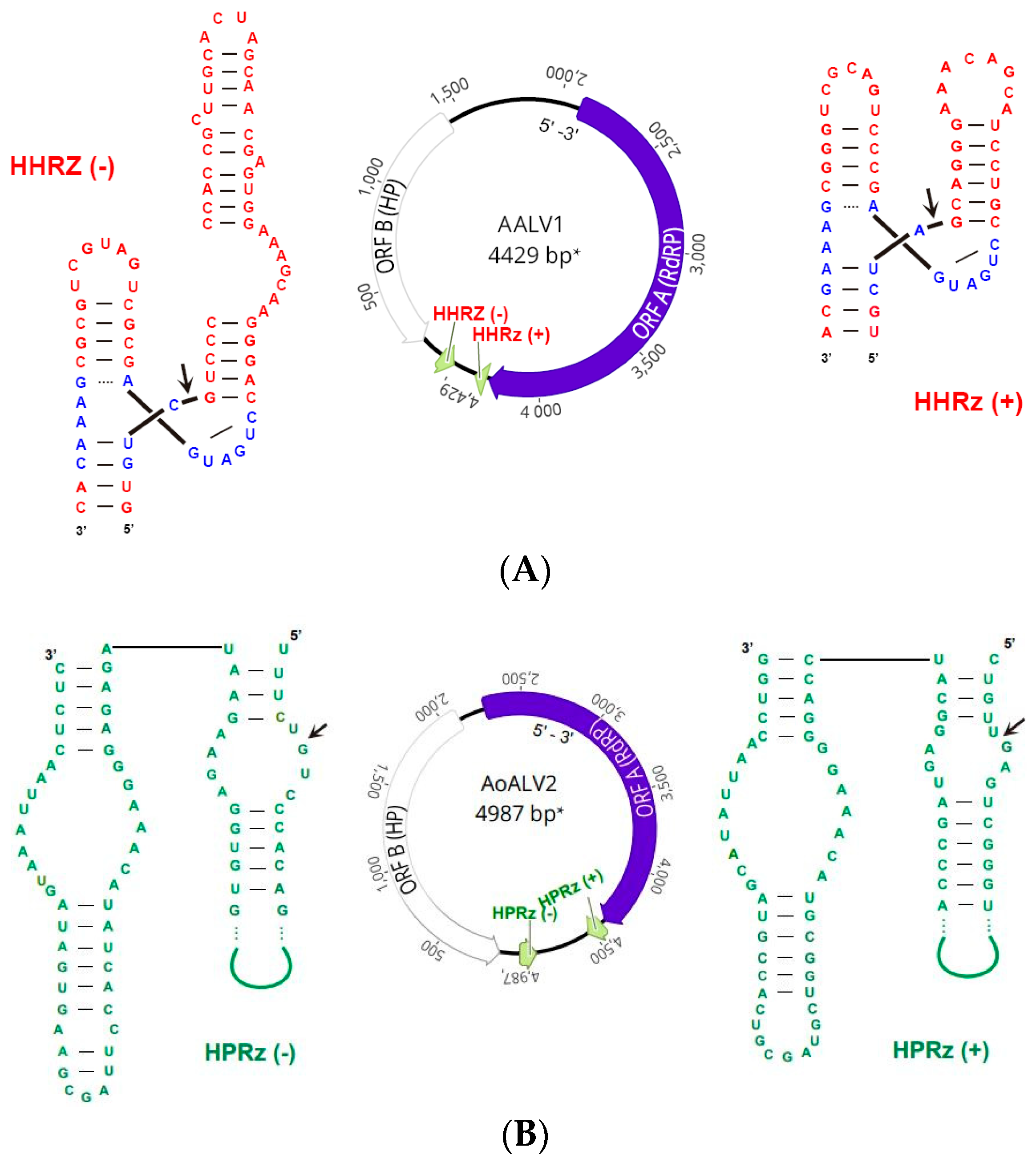
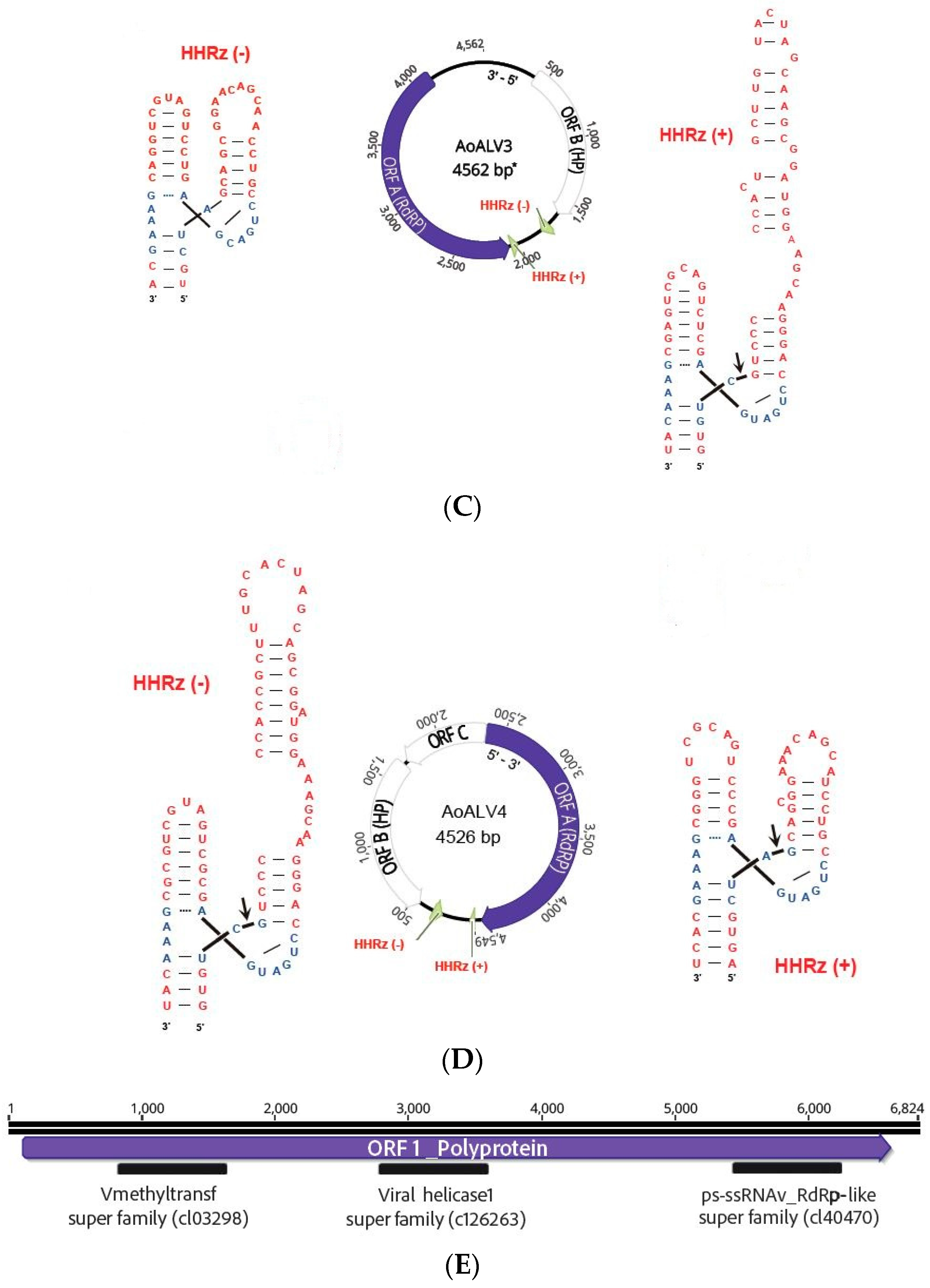
2.5.5. Ribozymes’ Detection

The identification of potential ribozymes followed a customized protocol adapted from the viroid-like sequence search pipeline outlined by Lee et al. in 2023 [41]. The process began with the recognition of covalently closed circular RNAs (cccRNAs) using a modified version of the reference-free CIRIT algorithm, as introduced by Qin et al. in 2020 [42]. This algorithm entails the exploration of overlapping regions between the starting and ending points of contigs to pinpoint cccRNAs. Unlike the original algorithm, our modified version repeatedly attempted to dissect potential cccRNAs into individual units, with the condition of maintaining a minimum 95% similarity within repeated regions. Subsequently, the identified cccRNAs underwent a search for known self-cleaving ribozymes, employing Infernal, a tool introduced by Nawrocki and Eddy in 2013 [43]. Ribozymes surpassing Rfam’s curated gathering threshold or exhibiting E-values below 0.1 were identified in each polarity. In addition to this, a subset of sequences underwent further scrutiny using an RNA motif, a tool developed by Macke et al. in 2001 [44]. This supplementary analysis aimed to uncover more diverse ribozymes that may not have been initially detected by Infernal.

2.6. Retro Transcription Polymerase Chain Reaction (RT-PCR) and Sanger Sequencing

Approximately 50–100 mg of fresh mycelium was collected and crushed in a mortar. Crushed mycelium was mixed with 600 µL of lysis buffer (LB; included in the PureLinkTM RNA Mini Kit) and 6 µL 2-mercaptoethanol and transferred to the 1.5 mL sterile tube. Isolation of total RNA was carried out using PureLinkTM RNA Mini Kit (Invitrogen, USA). The RNA was eluted in 30 μL of elution buffer and 1 µL of RNase inhibitor was added. The quality of total RNA was assessed by gel electrophoresis in 1.5% agarose gel and total RNA was stored at −80 °C. LunaScript® RT SuperMix Kit (New England Biolabs, Hitchin, UK) was used for the synthesis of cDNA, as reported by Tonka (2022) [45]. The success of cDNA synthesis was verified by amplification of the eukaryotic region translation elongation factor 1-alpha-eEF1A (tefa) and checked by gel electrophoresis in 1.5% agarose gel [46]. If the amplification was successful, the cDNA was used in a PCR reaction with virus-specific primers. The virus-specific primers were designed to partially amplify the ORFs of putative viruses based on sequencing data from RNA sequencing using program Geneious v.-8.1.9. PCR amplification was performed with 12 μL PPP Master Mix (Top-Bio, Vestec, Czech Republic) 1 μL of each 10 mM primer (Table S1), 4 μL of cDNA and PCR-grade water in a total volume of 25 μL. PCR conditions were as follows: 94 °C 2 min, 25× (94 °C 1 min, 60 °C 1 min, 72 °C 2 min) 72 °C 5 min. Due to the low concentration of putative viruses, the PCR amplification was repeated with fresh chemicals and 4 μL of resulting product from the first PCR amplification was used as a template; then, PCR products were visualized by gel electrophoresis in 1.5% agarose gel. All resulting fragments with the appropriate size were cut from the gel, cleaned using NucleoSpin® Gel and PCR Clean-up kit-Macherey-Nagel (BioTech a.s, Prague, Czech Republic), and ExoSAP-IT™ PCR Product Cleanup Reagent (Thermo Fisher Scientific, USA), and then fragments were sent to SEQme (Dobříš, Czech Republic) for Sanger sequencing.

2.7. Conserved Domains

NCBI CD-search tool [47] was used to search for putative conserved domains in predicted amino acid (aa) sequences. All sequences were then aligned to aa sequences of related viruses retrieved from the GenBank using MUSCLE v3.8.425 in Geneious v.-8.1.9.

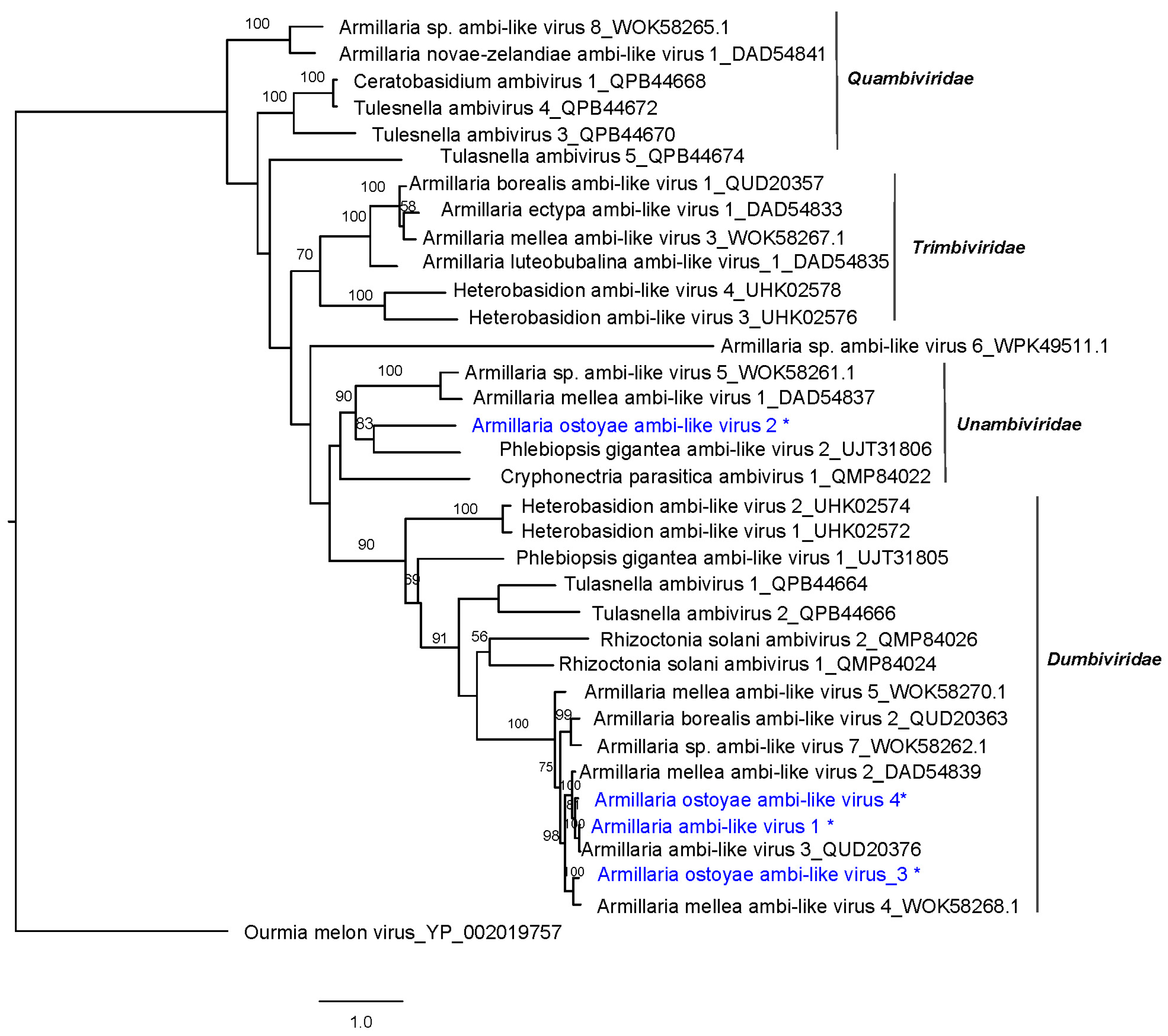
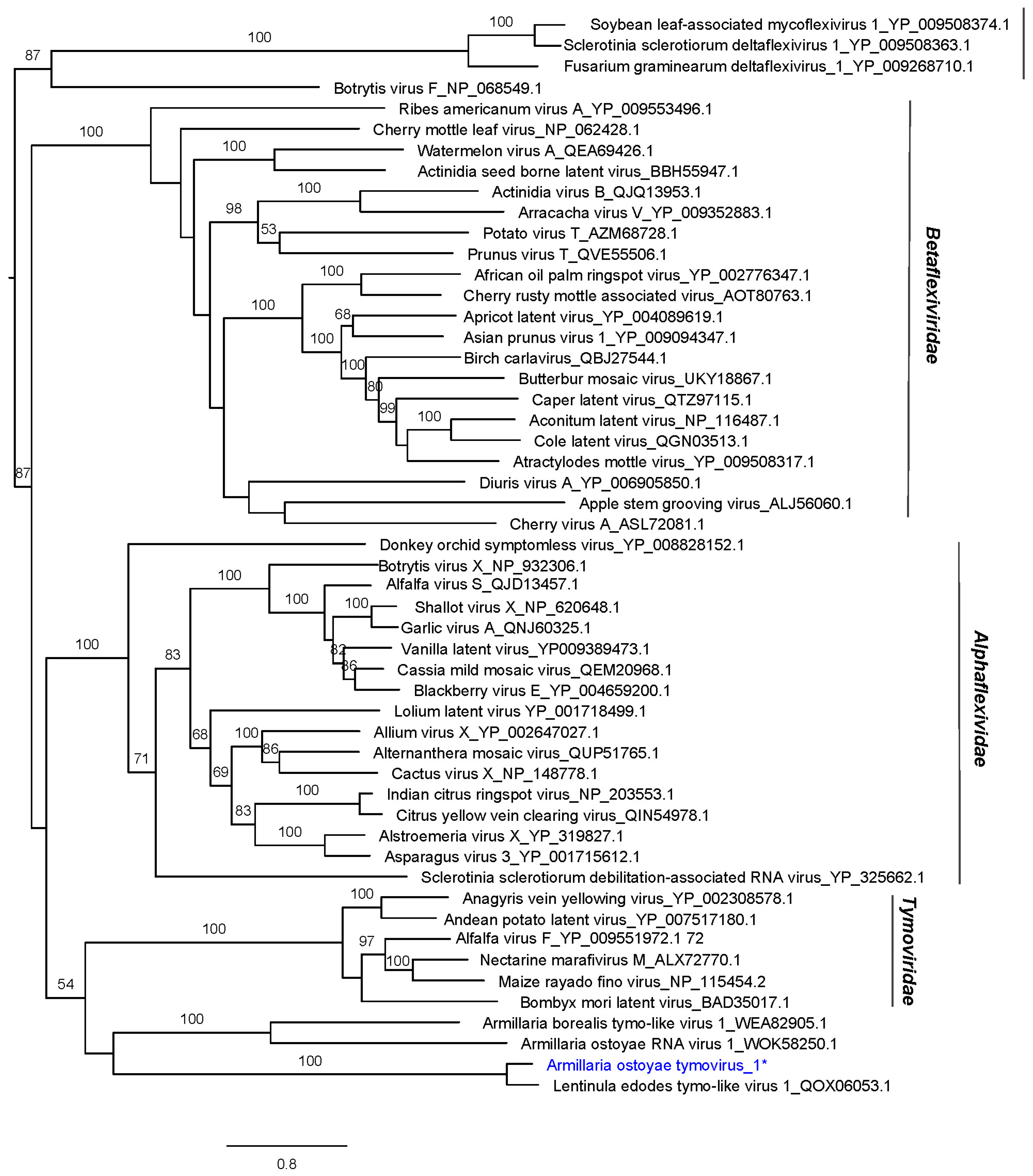
2.8. Phylogenetic Analyses

Amino acid (aa) sequences of RdRP regions of each virus were included in phylogenetic analyses. Sequences were aligned in Geneious v. 8.1.9 by MUSCLE v3.8.425 [48] together with known aa sequences of viruses considered to be related. Phylogenetic trees were built using the maximum likelihood method [49] in RAxML-HPC v.8 on XSEDE running in the CIPRES Science Gateway web portal [50]. Bootstrapping was performed by using the recommended parameters provided by the CIPRES Science Gateway portal. The trees were visualized in FIGTREE (V1.4.4).

3. Results and Discussion

In this study, we described the genomes of five putative single-stranded (ss) RNA viruses hosted by pathogenic fungi belonging to the genus Armillaria (specifically A. ostoyae and A. cepistipes) from the Czech Republic. These new viruses include a member of the family Tymoviridae and members of recently described virus group, tentatively named “Ambiviruses” [51,52,53,54].

3.1. dsRNA Screening

No dsRNA elements were detected in Czech isolates of Armillaria spp. As later demonstrated by high-throughput sequencing (HTS) of total RNA, no dsRNA viruses appear to occur in our collection of isolates. Some ssRNA viruses can produce dsRNA intermediates during their replication. However, not all ssRNA viruses generate dsRNA, and the amount produced varies among viral species. While dsRNA extraction methods can capture dsRNA, they may not effectively detect all ssRNA viruses, especially those with low dsRNA abundance. Our result agrees with that of the study of Linnakoski et al. (2021) [13], which dealt with the detection of mycoviruses in isolates of fungi of the genus Armillaria from Finland, Russia, and North Africa, and it also corresponds to the results of Dvořák’s work (2008) [55], which did not detect dsRNA viruses in Armillaria from the Czech Republic. However, Shamsi et al. (2024), in their work, report two detected mycoviruses with a dsRNA genome (a partitivirus and a phlegivirus) in samples originating from Switzerland [24]. Dvořák, in 2008 [55], and Blattný et al., in 1973 [21], studied the Armillaria fungi virome from the Czech Republic in their works. In his research, Dvořák (2008) [55] detected possible double-stranded molecules in forty samples from the Czech Republic, but they were not confirmed to be viral, which coincides with the results of this study. Likewise, Blattný et al. (1973) [21] and Reaves et al. (1988) [22] detected virus-like particles in their works on Armillaria mellea and Armillaria ostoyae, but they were not further verified molecularly. Blattný et al. (1973) [21] described these particles as rod-shaped (22–28 × 119 nm) or isometric (30 nm).

3.2. Identification of ssRNA Viruses

Total RNA sequencing generated 113 million paired-end (PE) reads. After quality trimming and de novo assembly, a total of 8809 contigs longer than 1 kb were obtained. BLASTX comparison of the contigs revealed four viral contigs with sequence similarities to members of the recently described circular RNA virus group “Ambiviruses” and one related to the members of the order Tymovirales, in particular, the family Tymoviridae (Table 2).
Regarding ambi-like viruses, Sutela et al.’s work in 2020 [51] was the first study to describe “ambiviruses” in the endomycorrhizal fungi Ceratobasidium sp. and Tulasnella sp. Then, they were discovered in Cryphonectria parasitica [52] and many agaricomycetes including Armillaria spp. [13,24], Heterobasidion spp. [20], Rhizoctonia spp. [13], and Phlebiopsis gigantea [56]. As in these studies, the genomic sequences of the putative ambi-like viruses detected in Czech isolates of Armillaria spp. were approx. 4.5 kb long and contained typical RdRP conserved domains including the GDD motif (Figure S1A). Based on the pairwise sequence comparison (PASC) percentages (Table S2A,B), four different viruses were identified and considered different species following the same criteria as those of Sutela et al. in 2020 [51], Forgia at al. in 2021 [52] and Turina et al. in 2023 [53]
They were tentatively named considering their host identity (Table 2). AALV1, AoALV2 and AoALV3 contained two and AoALV4 contained three non-overlapping ambisense ORFs. AALV1 encodes a first positive (+) sense ORF (ORF A) corresponding to the RdRP (713 aa long) and a second negative (−) sense ORF (ORF B) corresponding to a hypothetical protein (HP) 410 aa long. AoALV2 encodes the (−) sense ORF B-HP 655 aa long, and the (+) sense ORF A-RdRP 710 aa long. AoALV3 contains first (+) sense ORF corresponding to a hypothetical protein (ORF B) 400 aa long and a second negative sense ORF (ORF A) corresponding to RdRP 682 aa long. This arrangement shows that the contig was assembled in the 3′ to 5′ orientation. AoALV4 encodes a first (−) sense ORF B-HP 405 aa long, a second negative ORF 220 aa long, and a third (+) sense ORF A-RdRP 713 aa long.
Ambivirus genomes have the unique feature of having circular genomes encoding RdRP and divergent ribozymes in various combinations in both sense and antisense orientations [54]. In our analyses, several hammerhead (HHRz) and hairpin (HpRz) ribozymes were detected. This is in agreement with the findings of Forgia et al. (2023) [54], who describe the HHRz and HpRz motifs and predicted cleavage sites in both known fungal ambiviruses and ambiviricot RdRP palmprints of 439 distinct species-like operational taxonomic units (sOTUs) found in GenBank. Although HHRz typically consists of 30 to 40 nucleotides (nts), in Armillaria ambiviruses, some of them seem to be slightly longer, ranging from 55 to 78 nts. The representation of Armillaria ambiviruses and their ribozymes is illustrated in Figure 1A–D, and the positions in the particular ambivirus contigs are shown in Supplementary Table S3.
The phylogenetic relationships of Czech A. ostoyae ambiviruses with other “ambiviruses” (Figure 2) show that they group in two separated virus clusters, both of them including viruses from A. mellea and A. borealis. This result suggests a monophyletic origin of the ambiviricot sequences in Armillaria spp.
An Armillaria tymovirus 1. A viral contig resembling features of those from the genus Tymoviridae was detected in Armillaria ostoyae. Armillaria ostoyae tymovirus 1 is approximately 6.8 kb length and encodes one (+) sense ORF with 2172 aa (Figure 1E). Based on the sequence analysis of tymoviruses, conserved sequences were revealed (Figure S1B), including conserved regions belonging to pfam01660 super family Vmethyltransf cl03298 in region 143–314 (E-value 6.72 × 10⁻⁹). This methyltransferase domain has been detected in a wide range of viruses and is involved in mRNA capping. Furthermore, conserved sequences were detected belonging to pfam01443 superfamily Viral_helicase1 cl26263 located 1113–1322 (E-value 2.66 × 10⁻¹⁴) and ps-ssRNAv_RdRp-like super family cl40470 located 1600–1799 (E-value 2.73 × 10⁻²⁵) [57].
The order Tymovirales was first described in 2004 and currently comprises five families: Alphaflexiviridae, Betaflexiviridae, Gammaflexiviridae, Deltaflexiviridae, and Tymoviridae [58]. Members of the order Tymovirales have a 5.9 to 9.0 kb (+) ssRNA genome that is often polyadenylated. The largest protein, the replication-associated polyprotein (RP), is encoded by all members of the Tymovirales order. Tymovirales RP usually contains sets of conserved functional domains [59].
Viruses belonging to this order usually have a wide range of host organisms. Betaflexiviridae and Tymoviridae are usually considered plant viruses [59], a single member of Gammaflexiviridae has been detected in a filamentous fungus [58,60], and Alphaflexiviridae have been detected both in plants and fungi [61]. Although most viruses in the order Tymovirales are plant viruses, several members of this order are known to infect plant pathogenic fungi, including Botrytis cinerea, Fusarium boothii, Fusarium graminearum, R. solani, and S. sclerotiorum [62,63,64,65,66]. Armillaria ostoyae tymovirus 1 is the second tymovirus described within the genus Armillaria after the study of Shami et al. (2004) [24].
The phylogenetic relationships with other members from the order Tymoviriales from GenBank are shown in Figure 3. The phylogenetically closest virus to the virus AoTV1 is Lentinula edodes tymo-like virus 1, which was detected in a Chinese sample of the fungus Lentinula edodes.

3.3. RT-PCR Screening

Some of the viruses described in Czech isolates of Armillaria (Figure 1) are hosted by more than one isolate, and even by different species, suggesting interspecies transmission. AALV1 is present in isolate 1 (Armillaria cepistipes) and in isolate 7 (A. ostoyae), AoALV2 was detected just in isolate 7 (A. ostoyae), AoALV3 is present in isolates 7 and 13 (A. ostoyae), AoALV4 in isolates 6, 7, 8 and 13 (A. ostoyae), and AoTV1 was detected just in isolate 13 (A. ostoyae; Figure S2). Interestingly, isolates 1 and 7, between which virus interspecies transmission has occurred, were collected at localities approximately 50 km apart. These results suggest that ambiviruses are transmitted efficiently in Armillaria in the Czech Republic, as seems to happen in other regions of the Northern hemisphere [13,24], where ambi-like viruses have been found to be very common in Armillaria isolates and occur often in A. borealis and A. mellea from Finland, Siberia, and Switzerland. This efficiency could also be related to the fact that members of the genus Armillaria, as well as other fungi causing tree root rot, such as Heterobasidion spp. or Rosellinia necatrix, usually form large clonal individuals that grow for decades. It can therefore be considered that virus accumulation and interspecies transmission of mycoviruses are considered rather rare. However, laboratory experiments have shown that these are possible within the fungal genera Aspergillus [67], Sclerotinia [68] and Cryphonectria [69]. In vitro studies have shown mycovirus transmissions between somatically incompatible fungal strains of Heterobasidion [70,71,72], being relatively common in both the laboratory and in nature.

4. Conclusions

Our study confirms the presence of one tymovirus and five “ambiviruses” infecting Czech populations of A. ostoyae and A. cepistipes. Their potential effect on infected Armillaria hosts and their ability to be transmitted intra- and interspecies should be further investigated.

Supplementary Materials

The following supporting information can be downloaded at: https://www.mdpi.com/1999-4915/16/4/610/s1, Figure S1. A, Amino acid alignment of ambi-like viruses described in this study and related members. B, Amino acid aligment of Armillaria tymovirus described in this study and related members. Figure S2. Resulting RT-PCR gels. Table S1: List of primers used for direct virus RT-PCR detection. Table S2. A, pairwise sequence comparison (PASC) percentages (%) based on nucleotide sequence of ambi-like viruses hosted by Armillaria cepistipes and A. ostoyae. B, PASC % based on RdRP amino acid (aa) sequence of ambi-like viruses hosted by Armillaria cepistipes and A. ostoyae. Table S3: Types and genomic position of the ribozymes detected in Armillaria ambiviruses.

Author Contributions

Conceptualization, L.B. and T.T.; methodology, L.W., L.B. and T.T.; software, O.H.; validation and analysis, L.B., T.T., L.W. and M.d.l.P.; writing and editing, L.W., L.B., O.H. and T.T., visualization, L.B., L.W. and M.d.l.P.; project administration, T.T.; funding acquisition, L.B., T.T., V.Č. All authors have read and agreed to the published version of the manuscript.

Funding

This study was supported by the Ministry of Agriculture of the Czech Republic, project QK1920412 and University of South Bohemia, project GAJU 080/2022/Z.

Institutional Review Board Statement

Not applicable.

Informed Consent Statement

Not applicable.

Data Availability Statement

All data generated or analyzed during this study are included in this published article and its Supplementary Information files.

Acknowledgments

This work was supported by the Ministry of Agriculture of the Czech Republic with the project QK1920412. Computational resources were supplied by the project “e-InfrastrukturaCZ” (e-INFRA CZ LM2018140) supported by the Ministry of Education, Youth and Sports of the Czech Republic.

Conflicts of Interest

The authors declare that they have no competing interests.

Correction Statement

This article has been republished with a minor correction to resolve spelling and grammatical errors. This change does not affect the scientific content of the article.

References

  1. Hansen, E.M.; Goheen, E.M. Phellinus weirii and other native root pathogens as determinants of forest structure and process in Western North America. Annu. Rev. Phytopathol. 2000, 38, 515–539. [Google Scholar] [CrossRef] [PubMed]
  2. Gilbert, G.S. Evolutionary ecology of plant diseases in natural ecosystems. Annu. Rev. Phytopathol. 2002, 40, 13–43. [Google Scholar] [CrossRef] [PubMed]
  3. Coetzee, M.; Wingfield, B.D.; Wingfield, M.J. Armillaria root-rot pathogens: Species boundaries and global distribution. Pathogens 2018, 7, 83. [Google Scholar] [CrossRef] [PubMed]
  4. Williams, R.E.; Shaw, C.G.; Wargo, P.M.; Sites, W.H. Armillaria Root Disease. In Forest Insect and Disease Leaflet 78; U.S. Department of Agriculture Forest Service: Radnor, PA, USA, 1986. [Google Scholar]
  5. Baumgartner, K.; Coetzee, M.; Hoffmeister, D. Secrets of the subterranean pathosystem of Armillaria. Mol. Plant Pathol. 2011, 12, 515–534. [Google Scholar] [CrossRef] [PubMed]
  6. Holuša, J.; Lubojacký, J.; Čurn, V.; Tonka, T.; Resnerová, K.; Horák, J. Combined effects of drought stress and Armillaria infection on tree mortality in Norway spruce plantations. For. Ecol. Manag. 2018, 427, 434–445. [Google Scholar] [CrossRef]
  7. Cienciala, E.; Tumajer, J.; Zatloukal, V.; Beranová, J.; Holá, Š.; Hůnová, I.; Russ, R. Recent spruce decline with biotic pathogen infestation as a result of interacting climate, deposition and soil variables. Eur. J. Forest. Res. 2017, 136, 307–317. [Google Scholar] [CrossRef]
  8. Turčáni, M. Podiel podkorneho hmyzu na hynutí smrekových porastov postihnutých žltnutím. In Aktuálne Problémy v Ochrane Lesa; Zúbrik, M., Ed.; Lesnický Výskumný Ústav: Banská Štiavnica, Slovakia, 2001; pp. 56–60. [Google Scholar]
  9. Jankovský, L. Role houbových patogenů v chřadnutí smrku. In Collection of Lectures of the Professional Seminar Budišov Nad Budišovkou; Research Institute of Forestry and Hunting: Budišov and Budišovkou, Czech Republic, 2014; pp. 20–30. [Google Scholar]
  10. Garcia, O. A simple and effective forest stand mortality model. Math. Comput. For. Nat.-Resour. Sci. 2009, 1, 1–9. [Google Scholar]
  11. Van Mantgem, P.J.; Stephenson, N.L.; Byrne, J.C.; Daniels, L.D.; Franklin, J.F.; Fulé, P.Z.; Harmon, M.E.; Larson, A.J.; Smith, J.M.; Taylor, A.H.; et al. Widespread increase of tree mortality rates in the western United States. Science 2009, 323, 521–524. [Google Scholar] [CrossRef] [PubMed]
  12. Cleary, M.R.; Arhipova, N.; Morrison, D.J.; Thomsen, I.M.; Sturrock, R.N.; Vasaitis, R.; Gaitnieks, T.; Stenlid, J. Stump removal to control root disease in Canada and Scandinavia: A synthesis of results from long-term trial. For. Ecol. Manag. 2013, 290, 5–14. [Google Scholar] [CrossRef]
  13. Linnakoski, R.; Sutela, S.; Coetzee, M.P.A.; Duong, T.A.; Pavlov, I.N.; Litovka, Y.A. Armillaria root rot fungi host single-stranded RNA viruses. Sci. Rep. 2021, 11, 7336. [Google Scholar] [CrossRef]
  14. Prospero, S.; Botella, L.; Santini, A.; Robin, C. Biological control of emerging forest diseases: How can we move from dreams to reality? For. Ecol. Manag. 2021, 496, 119377. [Google Scholar] [CrossRef]
  15. Nuss, D.L. Hypovirulence: Mycoviruses at the fungal-plant interface. Nat. Rev. Microbiol. 2005, 3, 632–642. [Google Scholar] [CrossRef] [PubMed]
  16. García-Pedrajas, M.D.; Cañizares, M.C.; Sarmiento-Villamil, J.L.; Jacquat, A.G.; Dambolena, J.S. Mycoviruses in Biological Control: From Basic Research to Field Implementation. Phytopathology 2019, 109, 1828–1839. [Google Scholar] [CrossRef]
  17. Tonka, T.; Walterova, L.; Curn, V. Biological control of pathogenic fungi: Can mycoviruses play an important role? J. Cent. Eur. Agric. 2022, 23, 540–551. [Google Scholar] [CrossRef]
  18. Muñoz-Adalia, E.J.; Fernández, M.M.; Diez, J.J. The use of mycoviruses in the control of forest diseases. Biocontrol Sci. Technol. 2016, 26, 577–604. [Google Scholar] [CrossRef]
  19. Vainio, E.J.; Jurvansuu, J.; Hyder, R.; Kashif, M.; Piri, T.; Tuomivirta, T.; Poimala, A.; Xu, P.; Mäkelä, S.; Nitisa, D.; et al. Heterobasidion partitivirus 13 mediates severe growth debilitation and major alterations in the gene expression of a fungal forest pathogen. J. Virol. 2018, 92, 1744. [Google Scholar] [CrossRef]
  20. Sutela, S.; Piri, T.; Vainio, E.J. Discovery and community dynamics of novel ssRNA mycoviruses in the conifer pathogen Heterobasidion parviporum. Front. Microbiol. 2021, 12, 770787. [Google Scholar] [CrossRef] [PubMed]
  21. Blattny, C.; Kralik, O.; Veselsky, J.; Kasala, B.; Herzova, H. Particles resembling virions accompanying the proliferation of Agaric mushrooms. Česká Mykol. 1973, 27, 1–5. [Google Scholar]
  22. Reaves, J.L.; Allen, T.C.; Shaw, C.G.; Dashek, W.V.; Mayfield, J.E. Occurrence of virus like particles in isolates of Armillaria. J. Ultrastruct. Mol. Struct. Res. 1988, 98, 217–221. [Google Scholar] [CrossRef]
  23. Tonka, T.; Walterová, L.; Hejna, O.; Čurn, V. Molecular characterization of a ssRNA mycovirus isolated from the forest pathogenic fungus Armillaria ostoyae. Acta Virol. 2022, 66, 290–294. [Google Scholar] [CrossRef]
  24. Shamsi, W.; Heinzelmann, R.; Ulrich, S.; Kondo, H.; Cornejo, C. Decoding the RNA virome of the tree parasite Armillaria provides new insights into the viral community of soil-borne fungi. Environ. Microbiol. 2024, 26, e16583. [Google Scholar] [CrossRef] [PubMed]
  25. Morris, T.J.; Dodds, J.A. Isolation and analysis of double-stranded RNA from virus-infected plant and fungal tissue. Phytopathology 1979, 69, 854. [Google Scholar] [CrossRef]
  26. Tonka, T.; Walterová, L.; Hejna, O.; Čurn, V. Metodika Identifikace, Determinace a Přenosu Mykovirů u Hub Rodu Armillaria; University of South Bohemia in České Budějovice: České Budějovice, Czech Republic, 2021; ISBN 978-80-7394-891-7. [Google Scholar]
  27. Botella, L.; Dvořák, M.; Capretti, P.; Luchi, N. Effect of temperature on GaRV6 accumulation and its fungal host, the conifer pathogen Gremmeniella abietina. For. Pathol. 2017, 47, 12291. [Google Scholar] [CrossRef]
  28. Botella, L.; Jung, T. Multiple viral infections detected in Phytophthora condilina by total and small RNA sequencing. Viruses 2021, 13, 620. [Google Scholar] [CrossRef] [PubMed]
  29. Andrews, S. Babraham Bioinformatics—FastQC. A Quality Control Tool for High Throughput Sequence Data. Soil. 2010. Available online: https://www.bioinformatics.babraham.ac.uk/projects/fastqc (accessed on 26 May 2023).
  30. Illumina Adapter Sequences. Available online: https://emea.support.illumina.com/downloads/illumina-adapter-sequences-document-1000000002694.html (accessed on 26 May 2023).
  31. Martin, M. Cutadapt Removes Adapter Sequences from High-Throughput Sequencing Reads. 2012. Available online: https://cutadapt.readthedocs.io/en/stable (accessed on 26 May 2023).
  32. GitHub. Available online: https://github.com/marcelm/cutadapt (accessed on 27 May 2023).
  33. Dobin, A.; Davies, C.A.; Schlesinger, F.; Drenkow, J.; Zaleski, C.; Sonali, J.; Philippe, B.; Chaisson, M.; Gingeras, T.R. STAR: Ultrafast universal RNA-seq aligner. Bioinformatics 2013, 29, 15–21. [Google Scholar] [CrossRef] [PubMed]
  34. Viral NCBI. Available online: https://www.ncbi.nlm.nih.gov/labs/virus/vssi/#/ (accessed on 29 May 2023).
  35. Burrows-Wheeler Alingner. Available online: https://bio-bwa.sourceforge.net/ (accessed on 27 May 2023).
  36. IGV. Available online: https://software.broadinstitute.org/software/igv/ (accessed on 27 May 2023).
  37. Bankevich, A.; Nurk, S.; Antipov, D.; Gurevich, A.A.; Dvorkin, M.; Kulikov, A.S.; Lesin, V.M.; Nikolenko, S.I.; Pham, S.; Prjibelski, A.D.; et al. SPAdes: A New Genome Assembly Algorithm and Its Applications to Single-Cell Sequencing. J. Comput. Biol. 2012, 19, 455–477. [Google Scholar] [CrossRef] [PubMed]
  38. Viral UniProtKB. Available online: https://www.uniprot.org/uniprotkb?query=10239 (accessed on 29 May 2023).
  39. RVDB. Available online: https://rvdb.dbi.udel.edu/ (accessed on 29 May 2023).
  40. Virus-Host DB. Available online: https://www.genome.jp/virushostdb/ (accessed on 29 May 2023).
  41. Lee, B.D.; Neri, U.; Roux, S.; Wolf, Y.I.; Camargo, A.P.; Krupovic, M.; Simmonds, P.; Kyrpides, N.; Gophna, U.; Dolja, V.V.; et al. Mining metatranscriptomes reveals a vast world of viroid-like circular RNAs. Cell 2023, 186, 646–661. [Google Scholar] [CrossRef] [PubMed]
  42. Qin, Y.; Xu, T.; Lin, W.; Jia, Q.; He, Q.; Liu, K.; Du, J.; Chen, L.; Yang, X.; Du, F.; et al. Reference-free and de novo Identification of Circular RNAs. bioRxiv 2020. [Google Scholar] [CrossRef]
  43. Nawrocki, E.P.; Eddy, S.R. Infernal 1.1: 100-fold faster RNA homology searches. Bioinformatics 2013, 29, 2933–2935. [Google Scholar] [CrossRef]
  44. Macke, T.J.; Ecker, D.J.; Gutell, R.R.; Gautheret, D.; Case, D.A.; Sampath, R. RNAMotif, an RNA secondary structure definition and search algorithm. Nucleic Acids Res. 2001, 29, 4724–4735. [Google Scholar] [CrossRef]
  45. Tonka, T.; Walterová, L.; Čurn, V. Development of RT-PCR for rapid detection of ssRNA ambi-like mycovirus in a root rot fungi (Armillaria spp.). Acta Virol. 2022, 66, 287–289. [Google Scholar] [CrossRef]
  46. Čurn, V.; Tonka, T.; Křížová, L.; Jozová, E. Metodika Izolace DNA Analýzy Molekulárních Markerů u Hub; University of South Bohemia in České Budějovice: České Budějovice, Czech Republic, 2019; ISBN 978-80-7394-781-1. [Google Scholar]
  47. NCBI CD-Search Tool. Available online: https://www.ncbi.nlm.nih.gov/Structure/cdd/wrpsb.cgi (accessed on 27 May 2023).
  48. Edgar, R.C. MUSCLE: Multiple sequence alignment with high accuracy and high throughput. Nucleic Acids Res. 2004, 32, 1792–1797. [Google Scholar] [CrossRef] [PubMed]
  49. Stamatakis, A.; Hoover, P.; Rougemont, J.; Diego, S.; Jolla, L. A rapid bootstrap algorithm for the RAxML web servers. Syst. Biol. 2008, 57, 758–771. [Google Scholar] [CrossRef]
  50. Miller, M.A.; Pfeiffer, W.; Schwartz, T. Creating the CIPRES Science Gateway for inference of large phylogenetic trees. In Proceedings of the 2010 Gateway Computing Environments Workshop, New Orleans, LA, USA, 14 November 2010. [Google Scholar] [CrossRef]
  51. Sutela, S.; Forgia, M.; Vainio, E.J.; Chiapello, M.; Daghino, S.; Vallino, M.; Martino, E.; Girlanda, M.; Perotto, S.; Turina, M. The virome from a collection of endomycorrhizal fungi reveals new viral taxa with unprecedented genome organization. Virus Evol. 2020, 6, 76. [Google Scholar] [CrossRef]
  52. Forgia, M.; Isgandarli, E.; Aghayeva, D.N.; Huseynova, I.; Turina, M. Virome characterization of Cryphonectria parasitica isolates from Azerbaijan unveiled a new mymonavirus and a putative new RNA virus unrelated to described viral sequences. Virology 2021, 553, 51–61. [Google Scholar] [CrossRef]
  53. Turina, M.; Lee, B.D.; Sabanadzovic, S.; Vainio, E.J.; Navarro, B.; Simmonds, P. Create One New Phylum, Ambiviricota, Including One New Class, One New Order, Four New Families, Four New Genera, and 20 New Species, in Kingdom Orthornavirae (Realm Riboviria). ICTV TaxoProp 2023.007F. 2023. Available online: https://ictv.global/files/proposals/pending?fid=11741#block-teamplus-page-title (accessed on 27 May 2023).
  54. Forgia, M.; Navarro, B.; Daghino, S.; Cervera, A.; Gisel, A.; Perotto, S.; Aghayeva, D.N.; Akinyuwa, M.F.; Gobbi, E.; Zheludev, I.N.; et al. Hybrids of RNA viruses and viroid-like elements replicate in fungi. Nat. Commun. 2023, 14, 2591. [Google Scholar] [CrossRef]
  55. Dvořák, J. Výskyt RNA Elementů u Rodu Armillaria. Master’s Thesis, Charles University, Prague, Czech Republic, 2008. [Google Scholar]
  56. Drenkhan, T.; Sutela, S.; Veeväli, V.; Vainio, E.J. Phlebiopsis gigantea strains from Estonia show potential as native biocontrol agents against Heterobasidion root rot and contain diverse dsRNA and ssRNA viruses. Biol. Control 2022, 167, 104837. [Google Scholar] [CrossRef]
  57. Lu, S.; Wang, J.; Chitsaz, F.; Derbyshire, M.K.; Geer, R.C.; Gonzales, N.R.; Gwadz, M.; Hurwitz, D.I.; Marchler, G.H.; Song, J.S.; et al. CDD/SPARCLE: The conserved domain database in 2020. Nucleic Acids Res. 2020, 48, D265–D268. [Google Scholar] [CrossRef] [PubMed]
  58. Adams, M.J.; Kreuze, J.F.; Martelli, G.P. Tymovirales. In Virus Taxonomy: Classification and Nomenclature of Viruses; Elsevier: Waltham, MA, USA, 2011; pp. 901–903. [Google Scholar]
  59. King, A.M.Q.; Michael, J.; Carstens, E.B.; Lefkowitz, E.J. Virus Taxonomy: Classification and Nomenclature of Viruses; American Press/Elsevier: Amsterdam, The Netherlands, 2012. [Google Scholar]
  60. Howitt, R.L.; Beever, R.E.; Pearson, M.N.; Forster, R.L. Genome characterization of botrytis virus F, aflexuous rod-shaped mycovirus resembling plant ‘potex-like’ viruses. J. Gen. Virol. 2001, 82, 67–78. [Google Scholar] [CrossRef]
  61. Adams, M.J.; Candresse, T.; Hammond, J.; Kreuze, J.F.; Martelli, G.P.; Mamba, S.; Pearson, M.N.; Ryu, K.H.; Vaira, A.M. Alphaflexiviridae. In Virus Taxonomy: Classification and Nomenclature of Viruses; Elsevier: Waltham, MA, USA, 2011; pp. 904–919. [Google Scholar] [CrossRef]
  62. Howitt, R.L.; Beever, R.E.; Pearson, M.N.; Forster, R.L. Genome characterization of a flexuous rod-shaped mycovirus, botrytis virus X, reveals high amino acid identity to genes from plant ‘potex-like’ viruses. Arch. Virol. 2006, 151, 563–579. [Google Scholar] [CrossRef]
  63. Li, K.; Zheng, D.; Cheng, J.; Chen, T.; Fu, Y.; Jiang, D.; Xie, J. Characterization of a novel Sclerotinia sclerotiorum RNA virus as the prototype of a new proposed family within the order Tymovirales. Virus Res. 2016, 219, 92–99. [Google Scholar] [CrossRef] [PubMed]
  64. Li, P.; Lin, Y.; Zhang, H.; Wang, S.; Qiu, D.; Guo, L. Molecular characterization of a novel mycovirus of the family Tymoviridae isolated from the plant pathogenic fungus Fusarium graminearum. Virology 2016, 489, 86–94. [Google Scholar] [CrossRef] [PubMed]
  65. Bartholomaus, A.; Wibberg, D.; Winkler, A.; Puhler, A.; Schluter, A.; Varrelmann, M. Identification of a novel mycovirus isolated from Rhizoctonia solani (AG 2-2 IV) provides further information about genome plasticity within the order Tymovirales. Arch. Virol. 2017, 162, 555–559. [Google Scholar] [CrossRef] [PubMed]
  66. Mizutani, Y.; Abraham, A.; Uesaka, K.; Kondo, H.; Suga, H.; Suzuki, N.; Chiba, S. Novel mitoviruses and a unique tymo-like virus in hypovirulent and virulent strains of the Fusarium head blight fungus, Fusarium boothii. Viruses 2018, 10, 584. [Google Scholar] [CrossRef] [PubMed]
  67. Coenen, A.; Kevei, F.; Hoekstra, R.F. Factors affecting the spread of double-stranded RNA viruses in Aspergillus nidulans. Genet. Res. 1997, 69, 1–10. [Google Scholar] [CrossRef] [PubMed]
  68. Melzer, M.S.; Ikeda, S.S.; Boland, G.J. Interspecific transmission of double-stranded RNA and hypovirulence from Sclerotinia sclerotiorum to S. minor. Phytopathology 2002, 92, 780–784. [Google Scholar] [CrossRef] [PubMed]
  69. Liu, Y.; Linder-Basso, D.; Hillman, B.; Kaneko, S.; Milgroom, M.G. Evidence for interspecies transmission of viruses in natural populations of filamentous fungi in the genus Cryphonectria. Mol. Ecol. 2003, 12, 1619–1628. [Google Scholar] [CrossRef]
  70. Ihrmark, K.; Johannesson, H.; Stenström, E.; Stenlid, J. Transmission of double-stranded RNA in Heterobasidion annosum. Fungal Genet. Biol. 2002, 36, 147–154. [Google Scholar] [CrossRef]
  71. Vainio, E.J.; Korhonen, K.; Tuomivirta, T.T.; Hantula, J. A novel putative partitivirus of the saprotrophic fungus Heterobasidion ecrustosum infects pathogenic species of the Heterobasidion annosum complex. Fungal Biol. 2010, 114, 955–965. [Google Scholar] [CrossRef]
  72. Vainio, E.J.; Hakanpää, J.; Dai, Y.C.; Hansen, E.; Korhonen, K.; Hantula, J. Species of Heterobasidion host a diverse pool of partitiviruses with global distribution and interspecies transmission. Fungal Biol. 2011, 115, 1234–1243. [Google Scholar] [CrossRef]
Figure 1. Schematic presentation of AALV1 (A), AoALV2 (B), AoALV3 (C) and AoALV4 (D) genomes with predicted ORFs and ribozymes (rbz) secondary structures drawn as inlay; black arrows show the potential self-cleavage site at the predicted HHRz and HPRz motifs. Conserved residues are highlighted in blue; (E) schematic representation of AoTV1 genome with predicted ORF and its conserved motifs. The rbz (+) polarity is defined as the RNA strand coding for the polymerase (ORF A). * nt positions of the region spanning the ribozyme in the original virus contig. For more information of the rbz position refer to Table S3.
Viruses 16 00610 g001aViruses 16 00610 g001b
Figure 2. RAxML phylogenetic tree based on the predicted RdRP of representative ambiviruses. Nodes are labeled with bootstrap support values ≥50%. Branch lengths are scaled to the expected underlying number of amino acid substitutions per site. Tree is rooted in the midpoint and uses Ourmia melon virus as an outgroup. Czech Armillaria viruses are written in blue and indicated with an asterisk.
Viruses 16 00610 g002
Figure 3. RAxML phylogenetic tree based on the predicted RdRP of Armillaria ostoyae tymovirus 1 (blue letters and *) and other members of the order Tymovirales. Nodes are labeled with bootstrap support values ≥50%. Branch lengths are scaled to the expected underlying number of amino acid substitutions per site.
Viruses 16 00610 g003
Table 1. Data collection of the examined Armillaria isolates in this study.
Isolate CodeFungal
Species
Site CoordinatesCountryTree HostFungal MaterialCollection Date
(Day.Month.Year)
1A. cepistipes49.5147885 N, 17.5546327 ECzech RepublicPicea abiesRhizomorphs29.9.2020
2A. ostoyae48.9813906 N, 14.4205031 ECzech RepublicPicea abiesFruiting body31.10.2019
3A. ostoyae49.81582 N, 17.34951 ECzech RepublicPicea abiesFruiting body27.10.2019
4A. ostoyae48.6232622 N, 14.6442581 ECzech RepublicPicea abiesFruiting body16.10.2019
5A. ostoyae50.10238 N, 16.0683 ECzech RepublicPicea abiesFruiting body23.10.2019
6A. ostoyae49.5226228 N, 17.5717912 ECzech RepublicPicea abiesRhizomorphs5.9.2019
7A. ostoyae49.8084936 N, 17.4939458 ECzech RepublicPicea abiesRhizomorphs4.8.2020
8A. ostoyae49.31372 N, 16.77221 ECzech RepublicPicea abiesFruiting body23.10.2019
9A. ostoyae48.6078636 N, 14.6688486 ECzech RepublicPicea abiesFruiting body21.10.2019
10A. ostoyae49.8084936 N, 17.4939458 ECzech RepublicPicea abiesRhizomorphs4.8.2020
11A. ostoyae49.32247 N, 16.78645 ECzech RepublicPicea abiesFruiting body23.10.2019
12A. ostoyae48.9815597 N, 14.4162544 ECzech RepublicPicea abiesFruiting body4.11.2020
13A. ostoyae49.5419285 N, 17.3919515 ECzech RepublicPicea abiesRhizomorphs14.8.2019
Table 2. Mycoviruses detected in Armillaria samples in this study.
Virus NameAcronymLAccession Number aMost Similar Virus bE ValueQ (%)I (%)Mapped ReadsDepth of Coverage
Armillaria
ambi-like virus 1
AALV14663ON380550Armillaria spp. ambi-like virus 30.09696.3124,902780.91
Armillaria
ostoyae ambi-like virus 2
AoALV24541ON380551Phlebiopsis gigantea ambi-like virus 22 × 10−1034136.6527,239863.10
Armillaria
ostoyae ambi-like virus 3
AoALV34562ON380552Armillaria mellea ambi like virus 40.05680.9421,362678.90
Armillaria
ostoyae ambi-like virus 4
AoALV44549ON380553Armillaria ambi-like virus 30.04691.576169196.26
Armillaria
ostoyae tymovirus 1
AoTV16824ON380554Lentinula edodes tymo-like virus 10.09468.164959106.22
a Accession number in GenBank. b Most similar viruses in GenBank (BLASTX) accession numbers: MW423812.1 (Armillaria ambi-like virus 3), MZ448625.1 (Phlebiopsis gigantea ambi-like virus 2), BK014421.1 (Armillaria mellea ambi-like virus 2), MW423813.1 (Armillaria ambi-like virus 3), MN744726.1 (Lentinula edodes tymo-like virus 1); Q, query cover; I, Identity; L, virus sequence length; depth of coverage was calculated by following formula: (Total reads mapped to the final identified virus * average read length)/virus genome or contig length.
Disclaimer/Publisher’s Note: The statements, opinions and data contained in all publications are solely those of the individual author(s) and contributor(s) and not of MDPI and/or the editor(s). MDPI and/or the editor(s) disclaim responsibility for any injury to people or property resulting from any ideas, methods, instructions or products referred to in the content.

