Enhancement in Figure:
In the original publication [1], the figures were not sharp enough. The enhanced Figure 1, Figure 3, Figure 4, Figure 5, and Figure 7 appear below. The authors state that the scientific conclusions are unaffected. This correction was approved by the Academic Editor. The original publication has also been updated.
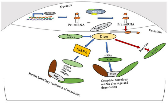
Figure 1.
Schematic representation of mechanism of gene silencing by miRNAs and siRNAs. Adapted and modified from [12] under the Creative Commons license.
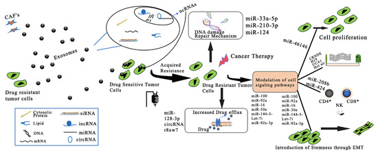
Figure 3.
Exosomal ncRNA-related mechanisms implicated in CRC drug resistance. CAFs: cancer-associated fibroblasts; ncRNAs: noncoding RNAs; miRNA: microRNA; circRNA: circular RNA; lncRNA: long noncoding RNA. Adapted and modified from [75] under the Creative Commons license.
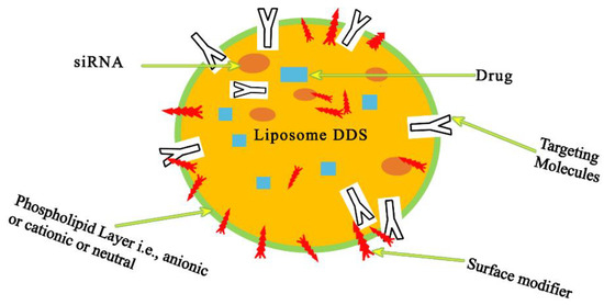
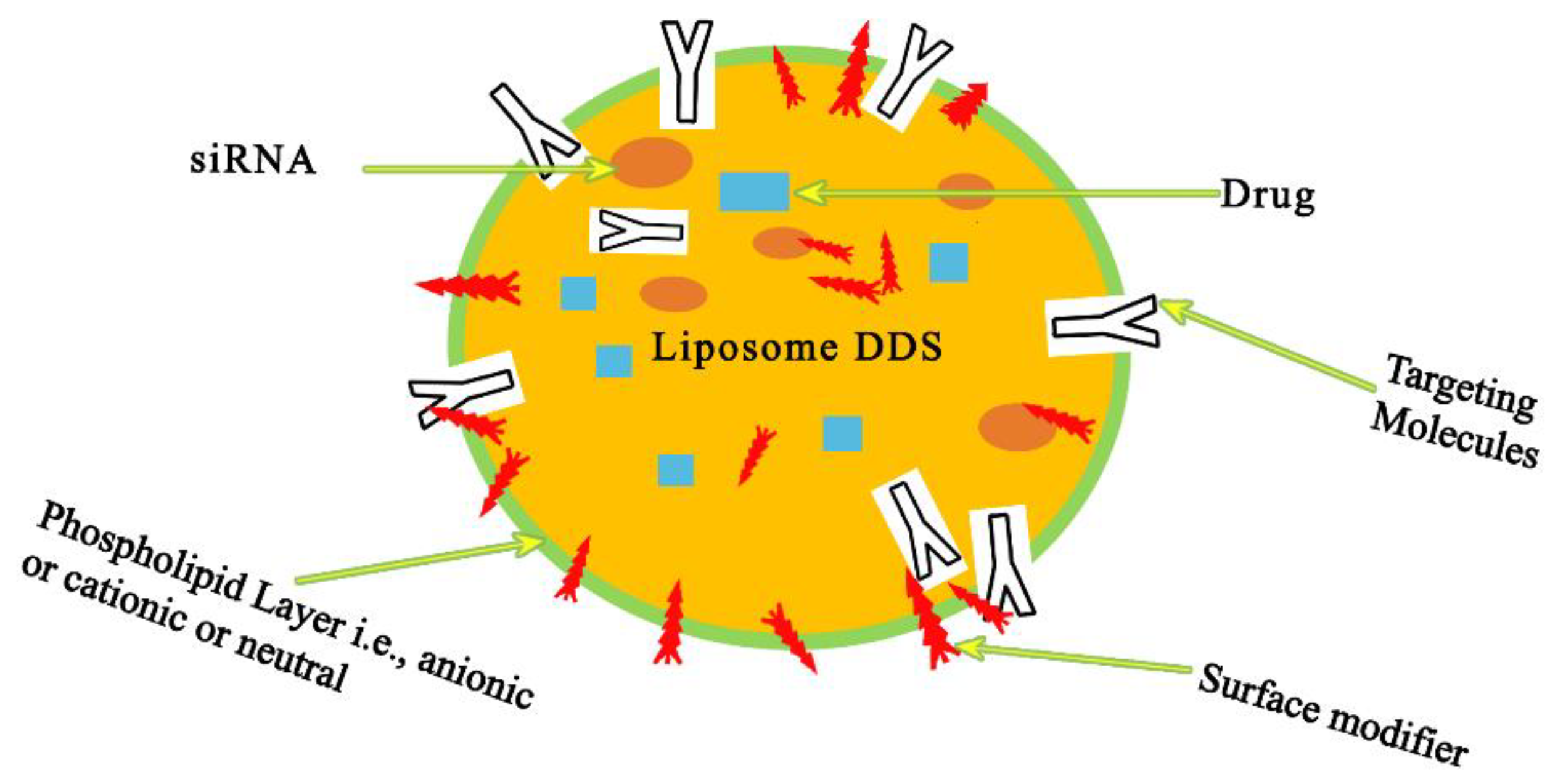
Figure 4.
Liposome-based carriers are sphere-shaped vesicles made of synthetic or natural phospholipids. Surface-modifier and -targeting groups can be conjugated to the outer surface. Phospholipids naturally form a bilayer upon aqueous dispersion, with the non-polar tails facing one other and the polar heads facing towards the aqueous phase. Hydrophilic molecules and RNAs are incorporated into the resulting inner core, while hydrophobic molecules are encapsulated in the lipid bilayer. Adapted and modified from [81] under the Creative Commons license.

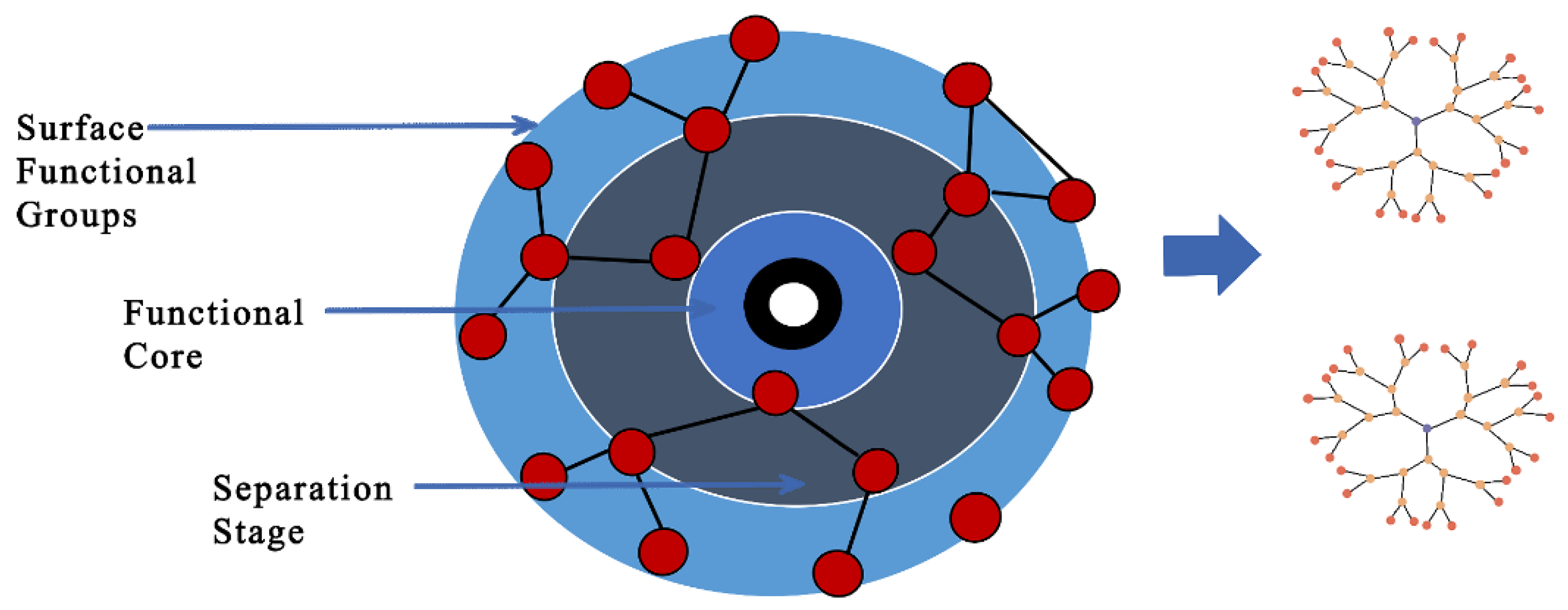
Figure 5.
Illustration of dendritic molecular structures with a central core, repeating branches, and terminal reactive functional groups. They can be classified as polymers and hyperbranched polymers with convergent and divergent architectures, depending on their molecular nature weight. Their nanostructure provides dendrimer–drug conjugation via different interactions such as electrostatic and hydrophobic/hydrogen bonds or the capacity for drug encapsulation within the central cavity and/or between the dendrons (branches). Adapted and modified from [87] under the Creative Commons license.

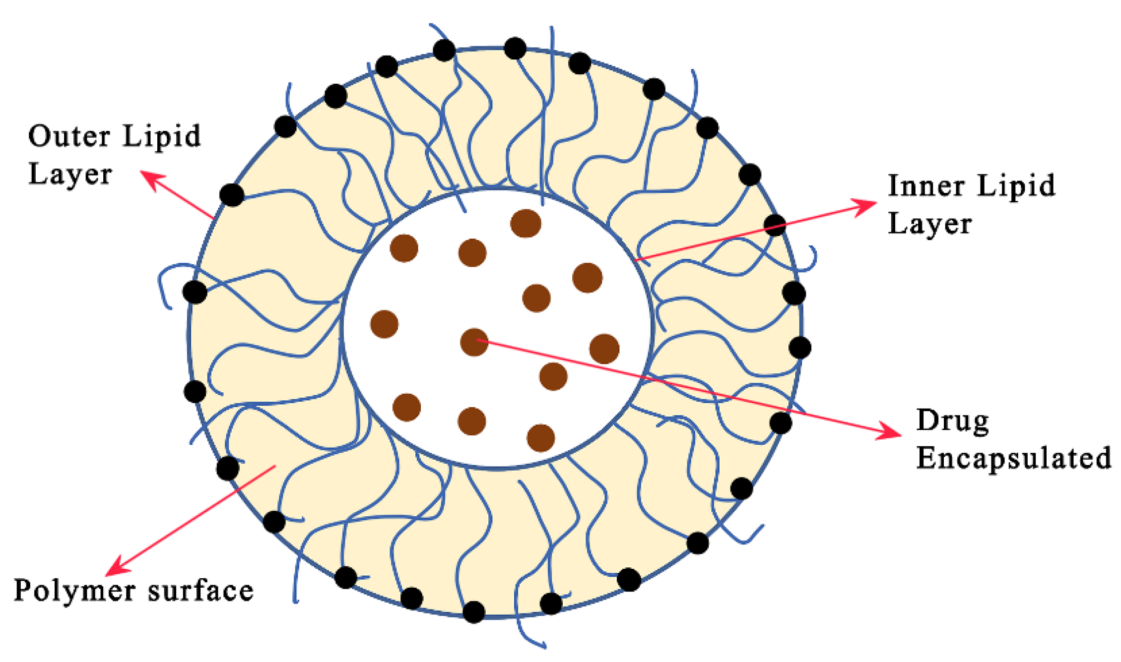
Figure 7.
Illustration of a bilayer lipid-based nanocarrier with encapsulated drugs in the core; the self-assembled supramolecular architecture is shown. The solid lipid matrix encapsulates bioactive components, particularly lipophilic molecules, and releases them gradually over time. Lipid polymer nanoparticles typically have spherical particles and sizes in the range of 10 to 1000 nm. There are several forms of lipid-based nanocarriers (liposomes and niosomes) reported in reference [81] for drug delivery.
Reference
- Zare, M.; Pemmada, R.; Madhavan, M.; Shailaja, A.; Ramakrishna, S.; Kandiyil, S.P.; Donahue, J.M.; Thomas, V. Encapsulation of miRNA and siRNA into Nanomaterials for Cancer Therapeutics. Pharmaceutics 2022, 14, 1620. [Google Scholar] [CrossRef]

Disclaimer/Publisher’s Note: The statements, opinions and data contained in all publications are solely those of the individual author(s) and contributor(s) and not of MDPI and/or the editor(s). MDPI and/or the editor(s) disclaim responsibility for any injury to people or property resulting from any ideas, methods, instructions or products referred to in the content. |
© 2023 by the authors. Licensee MDPI, Basel, Switzerland. This article is an open access article distributed under the terms and conditions of the Creative Commons Attribution (CC BY) license (https://creativecommons.org/licenses/by/4.0/).