Article

Porphyrin as a Cryoprotectant for Graphene Oxide-Coated Gold Nanorods to Produce Conjugated Product with Improved Stability and Opto-Phototherapeutic Properties

by
Thabang Calvin Lebepe
1,2,
Rodney Maluleke
1,2,
Nande Mgedle
1,2 and
Oluwatobi Samuel Oluwafemi
1,2,*
1
Centre for Nanomaterials Science Research, University of Johannesburg, Johannesburg 2028, South Africa
2
Department of Chemical Sciences, University of Johannesburg, Johannesburg 2028, South Africa
*
Author to whom correspondence should be addressed.
Pharmaceutics 2023, 15(11), 2538; https://doi.org/10.3390/pharmaceutics15112538
Submission received: 29 August 2023 / Revised: 22 October 2023 / Accepted: 24 October 2023 / Published: 27 October 2023

Abstract

:
Graphene oxide (GO) as a coating material for gold nanorods (AuNRs) has gained interest in reducing toxicity and improving the photothermal profiling of AuNRs. However, there is still a challenge regarding the storage of colloidal suspensions of GO-coated AuNRs (GO@AuNRs). Hence, the conjugation of GO@AuNRs to meso-tetra-(4-sulfonatophenyl)porphyrin (TPPS4), an anionic water-soluble porphyrin, has been reported to enhance their re-dispensability and improve their phototherapeutic properties. The AuNRs and GO were synthesised using seed-mediated and Hummers’ methods, respectively. The GO@AuNRs were conjugated to TPPS4 and characterised using ultraviolet–visible–near-infrared (UV-Vis-NIR) spectroscopy, zeta analyser, dynamic light scattering (DLS), photoluminescence spectroscopy (PL), x-ray diffraction (XRD), high-resolution transmission electron microscopy (HRTEM) and Fourier-transform infrared spectroscopy (FTIR) before freeze-drying. The results showed that the AuNRs were sandwiched between GO and TPPS4. After freeze-drying, the freeze-dried conjugate was dispensed in deionised water without adding cryoprotectants and its properties were compared to those of the unfreeze-dried conjugate. The results showed that the freeze-dried conjugate contained similar optical properties to the unfreeze-dried conjugate. However, the bare GO@AuNRs showed a change in the optical properties after freeze-drying. These results revealed that porphyrin is an excellent additive to reduce the freeze-drying stress tolerance of GO@AuNRs. The freeze-dried conjugate also showed both singlet oxygen and photothermal properties of GO@AuNRs and porphyrin. These results indicated that the freeze-dried conjugate is a promising dual photodynamic and photothermal agent, and porphyrin can act as a cryoprotectant.

1. Introduction

Numerous attempts have been made to improve the application of AuNRs by applying different functionalisation strategies such as ligand exchange and single or multi-layer encapsulation approaches using polymers, graphene-based materials, proteins, and metal oxides [1,2,3,4]. However, some coatings limit the biomedical application of AuNRs because they lead to thermal instability, lack of versatility, and more toxicity risk of AuNRs. Current research has shown that conjugating, coating, or functionalising AuNRs with graphene oxide (GO) increases the surface area of the AuNRs. This modification advances the application of AuNRs as drug carriers [5,6] and as biocompatible photothermal stable material [2,5,7,8]. Furthermore, it has been shown that the cytotoxicity of AuNRs can be lowered by removing CTAB and encapsulating them with GO [8].
On the other hand, AuNRs have been functionalised with photosensitisers for different applications such as synergistic phototherapy [9,10,11,12,13] and as a sensor [14,15]. However, nanoparticle colloidal preservation in suspensions is still a major challenge. Many polymers have been used to improve their colloidal stability [16,17,18,19]. Freeze-drying, also known as lyophilisation or cryodesiccation technique, is also used to maintain the nanomaterials’ colloidal stability by converting them into a solid mass. This technique has been used widely to preserve constantly changing biological systems such as proteins, antibodies, plasma constituents, and others in the pharmaceutical, food, and agricultural industries for safe shipping and storage [20]. During lyophilisation, cryoprotectants such as mono- and disaccharides, surfactants, amino acids, and polymers are usually used to reduce stresses [20,21,22,23].
Nonetheless, when GO is used to coat the AuNRs, the samples cannot be freeze-dried. This is because during the lyophilisation, the oxygen-containing functional groups are removed from GO, exposing AuNRs to lower temperatures and thus changing their shape. In addition, the sample cannot be redispersed in water because the oxygen-containing functional groups on the graphene are the reason for the easy dispensability of GO in water [24]. In this study, the GO-coated AuNRs were conjugated to meso-tetra-(4-sulfonatophenyl)porphyrin (TPPS4), a water-soluble porphyrin to enhance their re-dispensability. Water-soluble porphyrins like TPPS4 have been used to improve the solubility of nanomaterials [25,26], and because they are used for photodynamic applications, they can enhance the nanomaterials’ therapeutic applications for cancer and antimicrobial applications [27]. However, to the best of our knowledge, the effect of conjugating porphyrin to improve the GO-coated AuNRs (GO@AuNRs) as cryoprotectants has not been investigated. Unlike when polymers are used as cryoprotectants, conjugation with porphyrin will reduce stresses during lyophilisation and improve the therapeutic effect of GO@AuNRs. The as-synthesised conjugate (GO@AuNRs-TPPS4) was found to be easily re-dispersible in water after freeze-drying compared to GO@AuNRs. The conjugate photo-decay time was found to be quicker compared to TPPS4, even though the quantum yield was lower in the conjugate because of the GO@AuNRs. The singlet oxygen generation kinetics were found to be slower, and the conjugation did not affect the photothermal properties of GO@AuNRs. These results show that porphyrin can be used as a cryoprotectant, and the conjugate can be used as a dual phototherapeutic agent (Scheme 1).

2. Materials and Methods

2.1. Materials

Hydrogen tetra-chloroauric hydrate (HAuCl4.xH2O, 99.9%), sodium borohydride (NaBH4, 99%), silver nitrate (AgNO3, 99%), cetyltrimethylammonium bromide (CTAB, ≥99%), hydroquinone (HQ, %), sodium oleate (NaOL, ≥99%), hydrochloric acid (HCl, 12.1 M), graphite powder, sulfuric acid (H2SO4), sodium nitrate (NaNO3), potassium permanganate (KMnO4), peroxide (H2O2), sodium hydroxide (NaOH), polyvinylpyrrolidone (PVP), and methanol were purchased from Sigma-Aldrich, Kempton Park, South Africa. All solutions of gold salt, AgNO3, and NaBH4 were freshly prepared. All glasswares used in the experiments were cleaned and washed thoroughly with MilliQ water (15.0 MΩ cm @ 25 °C) and dried before use.

2.2. Characterisation Techniques

UV-Vis-NIR JASCO V-770 spectrophotometer (JASCO Corp., Tokyo, Japan) was used to measure the UV-Vis-NIR absorption spectra of the samples. The surface chemistry was investigated using Spectrum Two UATR spectrometer, Perkin Elmer(Beaconsfield, UK). The morphologies of the samples were captured by high-resolution transmission electron microscopy (HRTEM, JEOL 2010,200 KV, Tokyo, Japan). The sizes of the AuNRs and composites were measured from TEM images using ImageJ software (version 1.8.0). Zeta sizer (Microtrac Nanotrac Wave II, Microtrac MRB, Duesseldorf, Germany) was used to measure the surface charge of the materials. The fluorescence spectra of the samples were measured using Shimadzu spectrophotometer (RF-6000, Kyoto, Japan). For NIR irradiation, a dst11-LUMICS-808 nm 27W continuous Nd: YV04 air-cooled laser system (OsTech e. K., Berlin, Germany) with an optical fibre to deliver an 8 mm beam diameter was used.

2.3. Synthesis of Graphene Oxide

GO was prepared using a modified Hummers’ method [28]. Briefly, ice-cold concentrated H2SO4 (69 mL) was added to a mixture of graphite flakes (3.0031 g) and NaNO3 (1.5062 g) under continuous stirring in an ice bath. This was followed by the slow addition of KMnO4 (~10 g) and warmed to 35 °C while stirring for 30 min. Next, 138 mL of distilled water was added in portions, the temperature was increased to 95 °C, and it was stirred for 30 min. The solution was then cooled to room temperature, and the reaction was terminated by adding H2O2 (30 mL). The solution was left to settle, and the suspension was discarded, followed by pH neutralisation with distilled water and allowed to dry at room temperature. The obtained graphite oxide powder was exfoliated into GO sheets using ultrasonication.

2.4. Synthesis of Gold Nanorods

The AuNRs were prepared using the seed-mediated method [29]. CTAB (3.46 g) was added to 95 mL of warm deionised water (DW) and stirred to dissolve the powder completely. After cooling the CTAB solution to room temperature, 5 mL of the HAuCl4·xH2O (0.01 M) solution was added and mixed gently for 15 min, followed by the addition of 4.6 mL freshly prepared ice-cold NaBH4 (0.1 M) in NaOH (0.1 M). The growth solution was prepared by mixing the binary surfactant solution (CTAB 0.037 M and NaOL 0.23 M in 400 mL of warm DW) with 100 mL of 0.01 M HAuCl4·xH2O, followed by 2 mL of AgNO3 (0.1 M) and 1.8 mL HCl (12.1 M) under gentle stirring. Finally, 25 mL HQ (0.1 M) and a 100 mL seed solution were added under continuous stirring. The solution was left uninterrupted for 16−20 h at room temperature, followed by purification and characterisation.

2.5. Synthesis of Graphene Oxide-Coated Gold Nanorods

To fabricate the GO@AuNRs nanocomposite, PVP (16 mg) was added to GO dispersion (4 mL, 1 mg/10mL in H2O), followed by stirring for 30 min. Then, 2 mL of AuNRs dispersion was added to the PVP-stabilised GO solution under vigorous stirring for 24 h at room temperature. The resulting dispersion was washed and centrifuged three times to remove excess PVP and AuNRs. The final pellet was dissolved in 2 mL of deionised water.

2.6. Synthesis of Meso-Tetra-(4-sulfonatophenyl)porphyrin (TPPS4)

The meso-tetra-(4-sulfonatophenyl)porphyrin (TPPS4) was synthesised using our previously reported method [30], by firstly preparing TPPH2 as an intermediate porphyrin (UV-Vis spectrum λ: Soret band (410 nm) and Q-bands (512 nm, 549 nm, 590 nm, and 648 nm); PL emission λ: 650 nm and 730 nm with excited wavelength 512 nm) and it was then sulphonated using H2SO4 to produce TPPS4.

2.7. Conjugation of TPPS4 to GO@AuNRs

The formation of GO@AuNRs nanocomposite-porphyrin (GO@AuNRs-TPPS4) conjugate was carried out using our previous method [12] with modifications. Briefly, 0.2 mL of TPPS4 (0.1 mg/mL) was added to 2 mL of GO@AuNRs and stirred for 1 h at room temperature. The solution was characterised then freeze-dried and stored for further analysis.

2.8. Freeze-Drying of AuNRs, GO@AuNRs, and GO@AuNRs-TPPS4

The AuNRs, GO@AuNRs, and GO@AuNRs-TPPS4 were frozen by adding 15 mL of each sample in 50 mL centrifuge tubes. They were frozen for 24 h at −80 °C in an ultra-low temperature deep freezer (Meling −86 °C Ultra-low Temperature Freezer DW-HL50, Zhongke Meiling Cryogenics Co., Ltd., Anhui, China) before the freeze-drying process, which lasted for 48 h. The freeze dryer (MRC, FDL-10-50-Bench top freeze dryer, Wirsam Scientific and Precision Equipment (Pty) Ltd., Johannesburg, South Africa) was set at −50 °C with a pressure of 0.1 mbar. The freeze-dried samples were then characterised. Triplicates were performed for all samples in this work.

2.9. Photothermal Evaluation

The temperature changes were measured using an RS-1384 PRO thermocouple. The photothermal efficiency of water, GO@AuNRs, and GO@AuNRs-TPPS4 was measured by placing 3 mL of water, GO@AuNRs solution, and GO@AuNRs-TPPS4 solution separately in a UV-Vis quartz cuvette followed by irradiation under a continuous 808 laser with a power density of 1.27 W·cm−2. The temperature changes were measured using the RS-1384 PRO thermocouple. The photothermal conversion efficiency (PCE) was evaluated by irradiating 3 mL GO@AuNRs or GO@AuNRs-TPPS4 with 808 nm power density of 1.27 W·cm−2 until a constant temperature was reached, and then the laser system was switched off. The temperature of the dispersions was recorded every 30 s using a thermocouple. The PCE was calculated following Li et al.’s calculation with modifications [31].

2.10. Fluorescence Quantum Yield Measurements

TPPS4 and GO@AuNRs-TPPS4 were dispersed in distilled water to have an absorbance close to 0.1 at 560 nm, while methylene blue (MB) was dispersed in water to have an absorbance close to 0.1 at 560 nm. The samples were placed in a 3 mL pre-cleaned quartz cuvette with 1 cm optical path. The entire spectrum was scanned against the background spectrum of water. The emission spectra and quantum yield (QY) were obtained at ambient conditions using a Shimadzu RF-6000 (Japan) spectrophotometer. The QY was calculated using the following Equation (1):
Q Y s = A s A M B × A b s M B A b s s n M B 2 n s 2 × Q Y M B ,
where QYS and QYMB are the quantum yields of the sample and reference, AS and AMB are the area under the high-intensity peaks of the sample and reference, AbsS and AbsMB are the absorbances at the excitation wavelength of the sample and reference, and nS and nMB are the refractive indexes of the sample and reference, respectively. The reference standard used was the MB solution in water with QY of 0.52.

2.11. Singlet Oxygen Quantum Yield Evaluation

Singlet oxygen quantum yield (SOQY) was determined following Tsolekile et al. [30]. Briefly, the standard of 1,3-diphenyl benzofuran (DPBF) solution (32 mM in DMSO), MB solution (20 mM in DMSO), TPPS4 solution (10 mM in water), and GO solution (0.1 mg/mL in water) were prepared. Each sample was separately mixed in the ratio of 1:1 with DPBF and the mixtures were irradiated using photoluminescence spectrophotometry laser light at 535 nm for 6 min with a 1.5 min time interval. The DPBF intensity decrease was monitored at 471 nm peak position. The SOQY was calculated against an aqueous solution of methylene blue (20 mM) as a standard (SOQY = 0.52) using Equation (2):
S O Q Y = I s I M B M s M M B A b s s A b s M B S O Q Y M B ,
where SOQY and SOQYMB are the SOQY of the samples (TPPS4, conjugate) and the reference (MB, 0.52), Mc and MMB are the slopes of the samples and reference, Abss and AbsMB are the absorbance of the samples and the reference at irradiation wavelength, and IS and IMB are the refractive indexes of the sample and reference, respectively.

3. Results

3.1. Characterisation of Gold Nanorods, Graphene Oxide, and Graphene Oxide-Anchored Gold Nanorods Conjugated to TPPS4

GO was synthesised using the Hummers’ method, and the aqueous dispersibility was improved by modifying it with PVP [12,32,33]. The UV spectra of the as-synthesised GO (Figure 1A) showed small shoulder peaks corresponding to π-π* carbon–carbon and n-π* oxygen–carbon transitions. When GO was modified with PVP, the π-π* and n-π* of GO disappeared, indicating the interaction of PVP with GO. The FTIR analyses of GO, PVP, and GO-PVP were performed to confirm the modification. The inset TEM image of GO-PVP in Figure 1A shows a sheet with slight folding. The AuNRs with the size range of 40.70 ± 9.1 nm × 7.31 ± 1.6 nm (Figure 1B) and absorption at 820 nm were synthesised using the seed-mediated method previously reported [29]. The AuNRs were anchored on the modified GO via electrostatic interaction. The UV-Vis-NIR of the GO-anchored AuNRs did not show changes in the transverse surface plasmon resonance (TSPR) or LSPR peak wavelength position. However, the absorbance decreased (Figure 1C). The GO-PVP peak is also seen in the GO@AuNRs UV-Vis-NIR spectrum, confirming the bend of the AuNRs and GO-PVP. The zeta potential of the AuNRs decreased from 30 mV to 12 mV after the formation of GO@AuNRs.
The GO@AuNRs were conjugated to TPPS4 via electrostatic stacking. The UV-Vis-NIR spectrum of GO@AuNRs-TPPS4 shows the TSPR of AuNRs merged with the Q-bands of porphyrin. In addition, a slightly red-shifted longitudinal surface plasmon resonance (LSPR) of AuNRs as well as the Soret peak of TPPS4 can be observed (Figure 1D). Unlike our previous paper on 5,10,15,20-tetrakis (3-methyl pyridyl) porphyrin conjugated to GO@AuNRs [12], the Soret band was slightly red shifted and according to the literature, this red shift is due to the H-aggregation due to the electrostatic interaction of TPPS4 with the AuNRs and GO [34,35]. The PL results were similar to graphene–porphyrin conjugates reported [36,37,38,39,40,41,42,43]. However, there was a drastic emission reduction because of the red shift in the UV-Vis spectra. This could be attributed to the weak interaction of TPPS4 with GO@AuNRs surface via the coordination interaction between gold or graphene and pyrrole nitrogen atoms [15]. The GO@AuNRs-TPPS4 sample emits pink-red under UV light (Figure 1D). In addition, the zeta potential of the GO@AuNRs-TPPS4 was negatively charged (−18.0 mV) due to the negatively charged TPPS4 (−22.9 mV) anchored on the surface of AuNRs (Figure 1E). The FTIR spectrum of GO@AuNRs-TPPS4 (Figure 1F) exhibits most of the GO@AuNRs bands, such as alkyl C-H bands, OH broadband, C=O, and C-O stretching vibration at ~2850 and ~2918 cm−1, ~3424 cm−1, ~1667 cm−1, and ~1240 cm−1, respectively. The OH band in GO@AuNRs-TPPS4 was more stretched. However, the TPPS4 vibration peaks of SO32– at 1124 cm−1 and 1198 cm−1 in GO@AuNRs-TPPS4 are not visible because they are covered by GO. The XRD of the bare-AuNRs shows diffraction peaks at 17.9°, 21.4°, 24.8°, 28.2°, and 31.6° corresponding to the (002), (102), (210), (301), and (310) for CTAB [44], whereas, the diffraction peaks at 38.8°, 45.8°, 65.0°, 77.5°, and 81.9° are attributed to (111), (200), (220), (311), and (222) of Au (JCPDS file: 04-0784) (Figure 1G(i)). The AuNRs diffraction peaks in the GO@AuNRs are almost covered—only a few peaks can be seen (Figure 1G(ii)). However, after conjugation they completely disappeared. These results suggest that the AuNRs are completely sandwiched between GO and porphyrin; thus, the XRD spectrum shows only the strong broad diffraction peak at 23.4°, which corresponds to (002), the characteristic peak of amorphous carbon (Figure 1G(iii)).
The TEM of GO@AuNRs-TPPS4 in Figure 1G shows a GO sheet decorated with AuNRs. The DLS of AuNRs, GO, GO@AuNRs, and GO@AuNRs-TPPS4 were evaluated. Figure 2 shows similar AuNRs hydrodynamic size (Figure 2A) in both the GO@AuNRs (Figure 2C) and GO@AuNRs-TPPS4 (Figure 2D) in the range of 49.9 ± 9.2 nm. The difference between these and AuNRs is that in both the GO@AuNRs and GO@AuNRs-TPPS4, another peak is seen at ~1000 nm, which is attributed to GO (Figure 2B). This might be the reason behind the similar TEM micrographs observed for both GO@AuNRs-TPPS4 and GO@AuNRs.

3.2. Lyophilisation of Nanocomposite Conjugated to Porphyrin

The colloidal stability of GO@AuNRs-TPPS4 upon free drying was tested and compared to the AuNRs and GO@AuNRs. The AuNRs synthesised were freeze-dried to maintain their colloidal stability. We noticed during the course of the study that AuNRs were not stable in water with time. However, the AuNRs lost their shape after freeze-drying, as shown by the UV-Vis-NIR spectrum in Figure 3A. After freeze-drying, the AuNRs LSPR disappeared as the only visible peak was the TSPR, which was broad at 585 nm; similar results were observed by Khlebtsov et al. [22]. The AuNRs were then coated with GO at a 1:2 ratio; however, the solid mass obtained was not dispersible in deionised water, making it difficult to capture its UV-Vis as the solids were just settling down in the deionised water. According to Ham et al. [24], the oxygen-containing functional groups are removed during the freeze-drying process. Studies have shown that the oxygen-containing functional groups on the graphene are the reason for the easy dispersibility of GO in water [24]. In contrast to both the AuNRs and mGO@AuNRs, the mGO@AuNRs-TPPS4 was soluble in water as TPPS4 after freeze-drying. TPPS4 has been reported to improve the solubility of nanomaterials such as quantum dots [30]. The UV-Vis-NIR spectrum of freeze-dried GO@AuNRs-TPPS4 shows all the peaks of both the TPPS4 and AuNRs, the Soret, Q band, and LSPR with no shift compared to the GO@AuNRs-TPPS4 before freeze-drying (Figure 3B). These results confirm that there was no aggregation of the AuNRs and TPPS4. The FTIR of the freeze-dried GO@AuNRs-TPPS4 in Figure 3C displayed all the GO@AuNRs-TPPS4 peaks seen before freeze-drying. However, the OH peak was less pronounced in the freeze-dried GO@AuNRs-TPPS4. The PL of the freeze-dried GO@AuNRs-TPPS4 was performed, which also shows similar peaks as the unfreeze-dried GO@AuNRs-TPPS4 with the Q(0,0) and Q(0,1) (Figure 3D). This result shows that GO@AuNRs-TPPS4 can be lyophilised without losing its characteristics and was due to the TPPS4. Similar studies have shown that porphyrins can improve solubility of graphene materials [25].

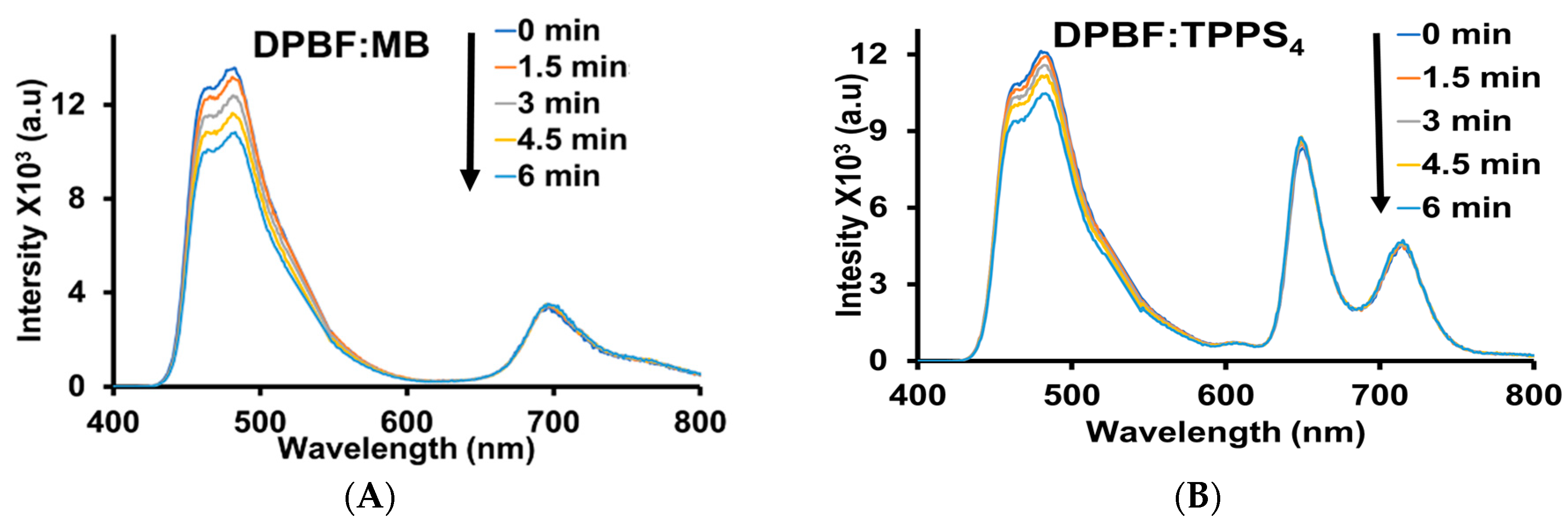
3.3. Lifetime, Quantum Yield, and Singlet Oxygen Generation Profiling

The relative fluorescence quantum yield of TPPS4 and GO@AuNRs-TPPS4 were analysed using PL and UV-Vis-NIR spectroscopy. The UV-Vis-NIR spectroscopy was used to obtain the absorbance lower than 0.1 a.u or close to 0.1 a.u for all the samples at 560 nm, which is the excitation wavelength. All the samples were excited at 560 nm, and methylene blue (MB) was used as the reference. The area under the curve of MB, TPPS4, and GO@AuNRs-TPPS4 was obtained from the PL emission spectrum when all the samples were excited at 560 nm. The relative fluorescence quantum yield of TPPS4 and GO@AuNRs-TPPS4 were 1.14 and 0.32, respectively. The relative fluorescence quantum yield was reduced by 72% for TPPS4 after the composite formation. The 72% reduction in relative fluorescence quantum yield observed in TPPS4 might be due to the strong interaction of TPPS4 with AuNRs, which caused the TPPS4 to H-aggregate. The lifetimes of the PL of TPPS4 and GO@AuNRs-TPPS4 were calculated using a time-correlated single-photon counting (TCSPC) technique with a pulsed LED at 450 nm. The lifetime of the PL intensity decays substantially as a bi-exponential function with the characteristic decay time average (τavg) of 6.11 ns and 6.75 ns (Figure 4, Table 1), respectively. The results show an increase in the decay time of GO@AuNRs-TPPS4 compared to TPPS4. This can be attributed to the electron absorption of AuNRs, which increases the electron transfer in both states and can be assigned to efficient back electron transfer and recovery of the ground state conjugate [14,43,45]. These results verify the efficient emission quenching of TPPS4 by the GO@AuNRs in the GO@AuNRs-TPPS4, as observed in the fluorescence intensity quenching in the PL spectra (Figure 1D).
The photochemical method was used to detect the singlet oxygen emission quantum yield (SOQY) of TPPS4 and GO@AuNRs-TPPS4 using DPBF as a singlet oxygen scavenger and MB as the reference. The singlet oxygen generation kinetics of the mixed samples with DPBF were acquired by irradiation at 535 nm at an interval of 1.5 min for 6 min. The PL spectrum changes of the DPBF peak at 471 nm were monitored (Figure 5A–C). The singlet oxygen generation reaction rate constants were found to be 0.013 and 0.13 min−1 for TPPS4 and GO@AuNRs-TPPS4, respectively, as calculated from first-order integrated rate reaction Equation (3):
ln i n t t = l n i n t 0 k t
where [int]t is the intensity of DPBF (471 nm) at a particular irradiation time (t), while [int]0 is the initial intensity at the beginning of the reaction, and k is the reaction rate constant, which is the slope of the graph.
The slopes of the graphs in Figure 5D and, together with Equation (2), were used to calculate the SOQY of TPPS4 and GO@AuNRs-TPPS4. The SOQY of GO@AuNRs-TPPS4 was more dramatically higher than the bare TPPS4, respectively. The SOQY of TPPS4 (0.276) almost doubled after the formation of GO@AuNRs-TPPS4 (0.5056).

3.4. Photothermal Profiling Characterisation

Photothermal evaluation of water, GO@AuNRs, and GO@AuNRs-TPPS4 was performed using an 808 nm laser with a power density of 1.27 W.cm−2 for 6 min (Figure 6). The results show an increase in temperature change when irradiated with time. The water temperature increased to only 9.6 °C, while the GO@AuNRs and GO@AuNRs-TPPS4 increased to 38.5 and 41.0 °C, respectively.

4. Conclusions

In summary, the GO@AuNRs-TPPS4 was successfully synthesised and could tolerate freeze-drying stress without adding cryoprotectant during the freeze-drying process. The conjugate exhibited photothermal and photodynamic properties upon irradiation. The GO was synthesised using Hummers’ method and modified with PVP before being decorated with 40.70 ± 9.1 nm × 7.31 ± 1.6 nm AuNRs absorbing at 820 nm, which was synthesised via the seed-mediated method. The optical and structural characterisation methods were used to confirm the formation of GO@AuNRs. The GO@AuNRs was then conjugated to TPPS4. The conjugation was confirmed by optical and structural characterisation method, revealing that the AuNRs were sandwiched between the GO and TPPS4. The AuNRs, GO@AuNRs, and conjugate were freeze-dried. Among the freeze-dried materials, the conjugate was easily dispersible in water and kept its optical properties compared to the original sample. The freeze-dried conjugate showed an increase in the decay time compared to TPPS4, attributed to the electron absorption of AuNRs’ increasing electron transfer in both states. The SOQY of TPPS4 (0.276) increased by 55% after the formation of GO@AuNRs-TPPS4 (0.5056). The photothermal profiling showed that GO@AuNRs-TPPS4 produced more heat than the bare GO@AuNRs, which is high enough for it to be used as a photothermal agent. The results showed that GO@AuNRs-TPPS4 is an excellent opto-phototherapeutic agent with good dispersion after freeze-drying.

Author Contributions

Conceptualisation, T.C.L. and O.S.O.; methodology, T.C.L.; software, T.C.L.; validation, T.C.L. and O.S.O.; formal analysis, R.M. and N.M.; investigation, T.C.L.; resources, O.S.O.; data curation, T.C.L.; writing—original draft preparation, T.C.L.; writing—review and editing, O.S.O.; visualisation, T.C.L.; supervision, O.S.O.; project administration, O.S.O.; funding acquisition, O.S.O. All authors have read and agreed to the published version of the manuscript.

Funding

This work was supported by the National Research Foundation (NRF), South Africa, under the South Africa/Japan bilateral program (Grant number 108669), Equipment-Related Travel and Training grants (Grant number 118666), Competitive Program for Rated Researchers (Grant numbers 106060 and 129290), and Freestanding Doctoral Scholarship (Grant numbers 112867 and 131237).

Institutional Review Board Statement

Not applicable.

Informed Consent Statement

Not applicable.

Data Availability Statement

The data presented in this study are available on request from the corresponding author.

Acknowledgments

The authors would like to thank the University of Johannesburg, South Africa, the research committee (URC), and the Faculty of Science Research Committee (FRC) for financial support.

Conflicts of Interest

The authors declare no conflict of interest.

References

  1. Shi, X.; Perry, H.L.; Wilton-Ely, J.D. Strategies for the functionalisation of gold nanorods to reduce toxicity and aid clinical translation. Nanotheranostics 2021, 5, 155–165. [Google Scholar] [CrossRef] [PubMed]
  2. Qi, Z.; Shi, J.; Zhu, B.; Li, J.; Cao, S. Gold nanorods/graphene oxide nanosheets immobilized by polydopamine for efficient remotely triggered drug delivery. J. Mater. Sci. 2020, 55, 14530–14543. [Google Scholar] [CrossRef]
  3. Requejo, K.I.; Liopo, A.; Zubarev, E.R. Gold Nanorods Synthesis with Small Thiolated Molecules. Langmuir 2020, 36, 3758–3769. [Google Scholar] [CrossRef]
  4. Vines, J.B.; Yoon, J.-H.; Ryu, N.-E.; Lim, D.-J.; Park, H. Gold Nanoparticles for Photothermal Cancer Therapy. Front. Chem. 2019, 7, 167. [Google Scholar] [CrossRef] [PubMed]
  5. Sun, B.; Wu, J.; Cui, S.; Zhu, H.; An, W.; Fu, Q.; Shao, C.; Yao, A.; Chen, B.; Shi, D. In situ synthesis of graphene oxide/gold nanorods theranostic hybrids for efficient tumor computed tomography imaging and photothermal therapy. Nano Res. 2017, 10, 37–48. [Google Scholar] [CrossRef]
  6. Khan, M.S.; Pandey, S.; Bhaisare, M.L.; Gedda, G.; Talib, A.; Wu, H.-F. Graphene oxide@ gold nanorods for chemo-photothermal treatment and controlled release of doxorubicin in mice Tumor. Colloids Surf. B Biointerfaces 2017, 160, 543–552. [Google Scholar] [CrossRef] [PubMed]
  7. Wei, Q.; Ni, H.; Jin, X.; Yuan, J. Graphene oxide wrapped gold nanorods for enhanced photo-thermal stability. RSC Adv. 2015, 5, 54971–54977. [Google Scholar] [CrossRef]
  8. Dembereldorj, U.; Choi, S.Y.; Ganbold, E.O.; Song, N.W.; Kim, D.; Choo, J.; Lee, S.Y.; Kim, S.; Joo, S.W. Gold Nanorod-Assembled PEGylated Graphene-Oxide Nanocomposites for Photothermal Cancer Therapy. Photochem. Photobiol. 2014, 90, 659–666. [Google Scholar] [CrossRef]
  9. Duman, F.D.; Sebek, M.; Thanh, N.T.; Loizidou, M.; Shakib, K.; MacRobert, A.J. Enhanced photodynamic therapy and fluorescence imaging using gold nanorods for porphyrin delivery in a novel in vitro squamous cell carcinoma 3D model. J. Mater. Chem. B 2020, 8, 5131–5142. [Google Scholar] [CrossRef]
  10. Ferreira, D.C.; Monteiro, C.S.; Chaves, C.R.; Sáfar, G.A.M.; Moreira, R.L.; Pinheiro, M.V.B.; Martins, D.C.S.; Ladeira, L.O.; Krambrock, K. Hybrid systems based on gold nanostructures and porphyrins as promising photosensitizers for photodynamic therapy. Colloids Surf. B Biointerfaces 2017, 150, 297–307. [Google Scholar] [CrossRef]
  11. Hlapisi, N.; Motaung, T.E.; Linganiso, L.Z.; Oluwafemi, O.S.; Songca, S.P. Encapsulation of gold nanorods with porphyrins for the potential treatment of cancer and bacterial diseases: A critical review. Bioinorg. Chem. Appl. 2019, 2019, 7147128. [Google Scholar] [CrossRef]
  12. Lebepe, T.C.; Parani, S.; Ncapayi, V.; Maluleke, R.; Mbaz, G.I.M.; Fanoro, O.T.; Varghese, J.R.; Komiya, A.; Kodama, T.; Oluwafemi, O.S. Graphene Oxide-Gold Nanorods Nanocomposite-Porphyrin Conjugate as Promising Tool for Cancer Phototherapy Performance. Pharmaceuticals 2021, 14, 1295. [Google Scholar] [CrossRef] [PubMed]
  13. Zhao, T.; Wu, H.; Yao, S.Q.; Xu, Q.-H.; Xu, G.Q. Nanocomposites Containing Gold Nanorods and Porphyrin-Doped Mesoporous Silica with Dual Capability of Two-Photon Imaging and Photosensitization. Langmuir 2010, 26, 14937–14942. [Google Scholar] [CrossRef] [PubMed]
  14. Liu, J.J.; Yuan, D.; Zhang, H.Z.; Lu, Y.D.; Wang, N.; Zou, H.Y.; Wang, J. Rapid detection of heparin by gold nanorods and near-infrared fluorophore ensemble based platform via nanometal surface energy transfer. Sens. Actuators B Chem. 2018, 274, 318–323. [Google Scholar] [CrossRef]
  15. Yuan, D.; Liu, J.J.; Zhang, H.Z.; Wang, N.; Zou, H.Y.; Huang, C.Z.; Wang, J. Highly selective detection of spermine in human urine via a nanometal surface energy transfer platform. Talanta 2018, 188, 218–224. [Google Scholar] [CrossRef]
  16. Liu, K.; Zheng, Y.; Lu, X.; Thai, T.; Lee, N.A.; Bach, U.; Gooding, J.J. Biocompatible gold nanorods: One-step surface functionalization, highly colloidal stability, and low cytotoxicity. Langmuir 2015, 31, 4973–4980. [Google Scholar] [CrossRef]
  17. Moore, T.L.; Rodriguez-Lorenzo, L.; Hirsch, V.; Balog, S.; Urban, D.; Jud, C.; Rothen-Rutishauser, B.; Lattuada, M.; Petri-Fink, A. Nanoparticle colloidal stability in cell culture media and impact on cellular interactions. Chem. Soc. Rev. 2015, 44, 6287–6305. [Google Scholar] [CrossRef]
  18. Schubert, J.; Chanana, M. Coating Matters: Review on Colloidal Stability of Nanoparticles with Biocompatible Coatings in Biological Media, Living Cells and Organisms. Curr. Med. Chem. 2018, 25, 4553–4586. [Google Scholar] [CrossRef]
  19. Mahmoud, N.N.; Aqabani, H.; Hikmat, S.; Abu-Dahab, R. Colloidal Stability and Cytotoxicity of Polydopamine-Conjugated Gold Nanorods against Prostate Cancer Cell Lines. Molecules 2021, 26, 1299. [Google Scholar] [CrossRef]
  20. Hamaly, M.A.; Abulateefeh, S.R.; Al-Qaoud, K.M.; Alkilany, A.M. Freeze-drying of monoclonal antibody-conjugated gold nanorods: Colloidal stability and biological activity. Int. J. Pharm. 2018, 550, 269–277. [Google Scholar] [CrossRef]
  21. Alkilany, A.M.; Abulateefeh, S.R.; Mills, K.K.; Bani Yaseen, A.I.; Hamaly, M.A.; Alkhatib, H.S.; Aiedeh, K.M.; Stone, J.W. Colloidal Stability of Citrate and Mercaptoacetic Acid Capped Gold Nanoparticles upon Lyophilization: Effect of Capping Ligand Attachment and Type of Cryoprotectants. Langmuir 2014, 30, 13799–13808. [Google Scholar] [CrossRef]
  22. Khlebtsov, B.N.; Khanadeev, V.A.; Panfilova, E.V.; Pylaev, T.E.; Bibikova, O.A.; Staroverov, S.A.; Bogatyrev, V.A.; Dykman, L.A.; Khlebtsov, N.G. New types of nanomaterials: Powders of gold nanospheres, nanorods, nanostars, and gold-silver nanocages. Nanotechnol. Russ. 2013, 8, 209–219. [Google Scholar] [CrossRef]
  23. Zagami, R.; Franco, D.; Pipkin, J.D.; Antle, V.; De Plano, L.; Patanè, S.; Guglielmino, S.; Monsù Scolaro, L.; Mazzaglia, A. Sulfobutylether-β-cyclodextrin/5,10,15,20-tetrakis(1-methylpyridinium-4-yl)porphine nanoassemblies with sustained antimicrobial phototherapeutic action. Int. J. Pharm. 2020, 585, 119487. [Google Scholar] [CrossRef]
  24. Ham, H.; Khai, T.V.; Park, N.H.; So, D.S.; Lee, J.W.; Na, H.G.; Kwon, Y.J.; Cho, H.Y.; Kim, H.W. Freeze-drying-induced changes in the properties of graphene oxides. Nanotechnology 2014, 25, 235601. [Google Scholar] [CrossRef] [PubMed]
  25. Cheng, F.; Adronov, A. Noncovalent functionalization and solubilization of carbon nanotubes by using a conjugated Zn–porphyrin polymer. Chem. Eur. J. 2006, 12, 5053–5059. [Google Scholar] [CrossRef] [PubMed]
  26. Tsolekile, N.; Ncapayi, V.; Parani, S.; Sakho, E.H.M.; Matoetoe, M.C.; Songca, S.P.; Oluwafemi, O.S. Synthesis of fluorescent CuInS2/ZnS quantum dots—Porphyrin conjugates for photodynamic therapy. MRS Commun. 2018, 8, 398–403. [Google Scholar] [CrossRef]
  27. Amos-Tautua, B.M.; Songca, S.P.; Oluwafemi, O.S. Application of Porphyrins in Antibacterial Photodynamic Therapy. Molecules 2019, 24, 2456. [Google Scholar] [CrossRef] [PubMed]
  28. Sakho, E.H.M.; Oluwafemi, O.S.; Thomas, S.; Kalarikkal, N. Dynamic energy transfer in non-covalently functionalized reduced graphene oxide/silver nanoparticle hybrid (NF-RGO/Ag) with NF-RGO as the donor material. J. Mater. Sci. Mater. Electron. 2017, 28, 2651–2659. [Google Scholar] [CrossRef]
  29. Lebepe, T.C.; Oluwafemi, O.S. Photothermal Conversion Profiling of Large-Scaled Synthesized Gold Nanorods Using Binary Surfactant with Hydroquinone as a Reducing Agent. Nanomaterials 2022, 12, 1723. [Google Scholar] [CrossRef]
  30. Tsolekile, N.; Ncapayi, V.; Obiyenwa, G.K.; Matoetoe, M.; Songca, S.; Oluwafemi, O.S. Synthesis of meso-tetra-(4-sulfonatophenyl) porphyrin (TPPS4)–CuInS/ZnS quantum dots conjugate as an improved photosensitizer. Int. J. Nanomed. 2019, 14, 7065. [Google Scholar] [CrossRef]
  31. Li, X.; Zhou, J.; Dong, X.; Cheng, W.-Y.; Duan, H.; Cheung, P.C.K. In vitro and in vivo photothermal cancer therapeutic effects of gold nanorods modified with mushroom β-Glucan. J. Agric. Food Chem. 2018, 66, 4091–4098. [Google Scholar] [CrossRef]
  32. Huang, P.; Wang, S.; Wang, X.; Shen, G.; Lin, J.; Wang, Z.; Guo, S.; Cui, D.; Yang, M.; Chen, X. Surface Functionalization of Chemically Reduced Graphene Oxide for Targeted Photodynamic Therapy. J. Biomed. Nanotechnol. 2015, 11, 117–125. [Google Scholar] [CrossRef] [PubMed]
  33. Wu, X.; Field, R.; Wu, J.J.; Zhang, K. Polyvinylpyrrolidone modified graphene oxide as a modifier for thin film composite forward osmosis membranes. J. Membr. Sci. 2017, 540, 251–260. [Google Scholar] [CrossRef]
  34. Wu, L.; Feng, L.; Ren, J.; Qu, X. Electrochemical detection of dopamine using porphyrin-functionalized graphene. Biosens. Bioelectron. 2012, 34, 57–62. [Google Scholar] [CrossRef] [PubMed]
  35. Trapani, M.; De Luca, G.; Romeo, A.; Castriciano, M.A.; Scolaro, L.M. Spectroscopic investigation on porphyrins nano-assemblies onto gold nanorods. Spectrochim. Acta Part A Mol. Biomol. Spectrosc. 2017, 173, 343–349. [Google Scholar] [CrossRef] [PubMed]
  36. Banerjee, I.; Mondal, D.; Martin, J.; Kane, R.S. Photoactivated antimicrobial activity of carbon nanotube−porphyrin conjugates. Langmuir 2010, 26, 17369–17374. [Google Scholar] [CrossRef]
  37. Geng, J.; Jung, H.-T. Porphyrin functionalized graphene sheets in aqueous suspensions: From the preparation of graphene sheets to highly conductive graphene films. J. Phys. Chem. C 2010, 114, 8227–8234. [Google Scholar] [CrossRef]
  38. Geng, J.; Kong, B.-S.; Yang, S.B.; Jung, H.-T. Preparation of graphene relying on porphyrin exfoliation of graphite. Chem. Commun. 2010, 46, 5091–5093. [Google Scholar] [CrossRef]
  39. Wang, A.; Yu, W.; Xiao, Z.; Song, Y.; Long, L.; Cifuentes, M.P.; Humphrey, M.G.; Zhang, C. A 1, 3-dipolar cycloaddition protocol to porphyrin-functionalized reduced graphene oxide with a push-pull motif. Nano Res. 2015, 8, 870–886. [Google Scholar] [CrossRef]
  40. Wang, A.; Yu, W.; Huang, Z.; Zhou, F.; Song, J.; Song, Y.; Long, L.; Cifuentes, M.P.; Humphrey, M.G.; Zhang, L. Covalent functionalization of reduced graphene oxide with porphyrin by means of diazonium chemistry for nonlinear optical performance. Sci. Rep. 2016, 6, 23325. [Google Scholar] [CrossRef]
  41. Chen, Q.; Brambilla, L.; Daukiya, L.; Mali, K.S.; De Feyter, S.; Tommasini, M.; Müllen, K.; Narita, A. Synthesis of Triply Fused Porphyrin-Nanographene Conjugates. Angew. Chem. Int. Ed. 2018, 57, 11233–11237. [Google Scholar] [CrossRef]
  42. Wang, J.; Wei, J.; Su, S.; Qiu, J.; Hu, Z.; Hasan, M.; Vargas, E.; Pantoya, M.; Wang, S. Thermal-Recoverable Tough Hydrogels Enhanced by Porphyrin Decorated Graphene Oxide. Nanomaterials 2019, 9, 1487. [Google Scholar] [CrossRef] [PubMed]
  43. Gacka, E.; Burdzinski, G.; Marciniak, B.; Kubas, A.; Lewandowska-Andralojc, A. Interaction of light with a non-covalent zinc porphyrin–graphene oxide nanohybrid. Phys. Chem. Chem. Phys. 2020, 22, 13456–13466. [Google Scholar] [CrossRef] [PubMed]
  44. Abraham, P.; Renjini, S.; Nancy, T.M.; Kumary, V.A. Electrochemical synthesis of thin-layered graphene oxide-poly (CTAB) composite for detection of morphine. J. Appl. Electrochem. 2020, 50, 41–50. [Google Scholar] [CrossRef]
  45. Mao, B.; Calatayud, D.G.; Mirabello, V.; Hodges, B.J.; Martins, J.A.R.; Botchway, S.W.; Mitchels, J.M.; Pascu, S.I. Interactions between an Aryl Thioacetate-Functionalized Zn(II) Porphyrin and Graphene Oxide. Adv. Funct. Mater. 2016, 26, 687–697. [Google Scholar] [CrossRef]
Scheme 1. Schematic diagram of water-soluble porphyrin to enhance photo properties of GO-coated AuNRs as well as re-dispensability after freeze-drying.
Pharmaceutics 15 02538 sch001
Figure 1. (A) UV-Vis spectra of GO and GO-PVP (inset: TEM image of GO-PVP, scale: 200 nm). (B) Bare BS-HQ-AuNRs TEM image (scale: 50 nm; inset: size distribution histogram). (C) UV-Vis-NIR spectra of AuNRs and GO@AuNRs. (D) UV-Vis-NIR and PL spectra of TPPS4 and GO@AuNRs-TPPS4 (excited at 415 nm; inset: photographic images of GO@AuNRs-TPPS4 under normal light (a) and UV-light irradiation (b)). (E) Zeta potential of GO, PVP, GO-PVP, AuNRs, GO@AuNRs, TPPS4, and GO@AuNRs-TPPS4. (F) FTIR spectra of TPPS4, GO@AuNRs, and GO@AuNRs-TPPS4. (G) XRD pattern of GO@AuNRs-TPPS4 (i), GO@AuNRs (ii), and AuNRs (iii). (H) TEM image of GO@AuNRs-TPPS4.
Pharmaceutics 15 02538 g001
Figure 2. DLS hydrodynamic size of (A) AuNRs, (B) GO, (C) GO@AuNRs, and (D) GO@AuNRs-TPPS4.
Pharmaceutics 15 02538 g002
Figure 3. (A) UV-Vis-NIR spectra of AuNRs before and after freeze-drying. (B) UV-Vis-NIR spectra of GO@AuNRs-TPPS4 before and after freeze-drying. (C) FTIR spectra of GO@AuNRs-TPPS4. (D) PL of GO@AuNRs-TPPS4 before and after freeze-drying (excited at 560 nm).
Pharmaceutics 15 02538 g003
Figure 4. PL lifetime of TPPS4 and GO@AuNRs-TPPS4.
Pharmaceutics 15 02538 g004
Figure 5. PL emission spectra of (A) DPBF: MB, (B) DPBF: TPPS4, and (C) DPBF: GO@AuNRs-TPPS4 excited at 560 nm for 6 min at the intervals of 1.5 min. (D) Integrated emission intensity time graph of DPBF at 471 nm after mixing with MB, TPPS4, and GO@AuNRs-TPPS4 and irradiated at 560 nm for different time intervals of 6 min.
Pharmaceutics 15 02538 g005aPharmaceutics 15 02538 g005b
Figure 6. The photothermal profiling of water, GO@AuNRs, and GO@AuNRs-TPPS4 irradiated with 808 lasers (a power density of 1.27 W.cm−2).
Pharmaceutics 15 02538 g006
Table 1. The time-resolved fluorescence decay of TPPS4 and GO@AuNRs-TPPS4.
Sample PLQY (%)τ1 (ns)τ2 (ns)χ2τavg (ns)
TPPS41.142.36 (4.86%)9.85 (95.14%)1.286.11
GO@AuNRs-TPPS40.314.86 (1.89%)8.63 (98.11%)1.306.75
Disclaimer/Publisher’s Note: The statements, opinions and data contained in all publications are solely those of the individual author(s) and contributor(s) and not of MDPI and/or the editor(s). MDPI and/or the editor(s) disclaim responsibility for any injury to people or property resulting from any ideas, methods, instructions or products referred to in the content.

