Highlights
What are the main findings?
- Extrapolation from animal skin models poorly predicts clinical outcomes.
- A spectrum of in vitro human skin models, built from defined cell types and sources, reproduces key in vivo characteristics and supports longer cultures.
What is the implication of the main finding?
- Validated in vitro skin models can replace animal testing while improving translational relevance and ethics.
- Emerging biofabrication and microphysiological technologies enable more complex, human-relevant systems.
Abstract
Research on in vitro skin models has advanced remarkably, driven by a better understanding of the skin and the search for more ethical and efficient methods. The development of these models was initially motivated by the need for reduced animal testing and a faster and more ethical approach for the safety evaluation of cosmetic and pharmaceutical products. Stricter regulations and growing ethical awareness have driven further evolution, resulting in more refined and reliable methods. Diversity of cell types is crucial to replicating the complexity of human skin, including epithelial, dendritic, endothelial, and adipose cells, providing environments that closely mimic the physiological skin environment. This allows for more precise studies on skin interactions with cosmetic, dermatological, and pharmaceutical products. In vitro skin models have applications in toxicity testing, dermatological product evaluation, skin ageing studies, and drug research, reducing dependence on animal testing. This review presents a look at the different types of in vitro skin models developed for various applications, with a brief look at their strengths and drawbacks. Models developed for disease-specific applications are also covered. Techniques such as bioprinting and organ-on-a-chip have revolutionised the manufacturing of these models. Challenges persist, such as the need to improve vascularisation and faithfully replicate skin architecture. The promising future of these models points to an exciting path forward for dermatological research and the cosmetic industry. This review addresses the history and regulations of skin models, explores various skin models, and highlights the most recent advances, outlining future perspectives and offering a comprehensive overview.
1. Introduction
The skin is the largest organ in the body and performs several vital functions. One of its main tasks is to protect against external insults, including physical, chemical, or biological threats. This is the first line of defence of the body and plays a crucial role in the immune system. Furthermore, the skin actively contributes to temperature regulation, preventing excessive water loss from the body [1].
Skin exhibits an intricate structure and is composed of two fundamental layers, the dermis and the epidermis. The epidermis, the outermost layer, is a non-vascularized stratified epithelium, predominantly made up of ectoderm-derived keratinocytes, that undergoes continuous cell renewal [2]. These cells play a crucial role in the defence functions of the skin, controlling inflammation through the release of pro-inflammatory cytokines essential for modulating skin irritation and sensitisation [3,4]. Additionally, the appendages (hair follicles, sebaceous glands, and sweat glands) are located in this layer. Moreover, the keratinisation process, which involves cell differentiation into several layers (basal layer, stratum spinosum, stratum granulosum, stratum lucidum, and stratum corneum), is crucial in constituting an essential barrier against the penetration of chemical substances and microorganisms [2,5]. The dermis is connected to the epidermis through the basement membrane, constituting connective tissue. This stratum contains sweat glands, hair follicles, muscles, sensory neurons, and blood vessels, with fibroblasts being the main cell type identified [2,6] (Figure 1).
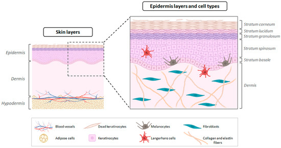
Figure 1.
Schematic representation of skin layers (epidermis, dermis and hypodermis) with particular focus on the epidermis: layers (stratum corneum, stratum lucidum, stratum granulosum, stratum spinosum, stratum basale) and most prominent cell types (keratinocytes at different stages, melanocytes, Langerhans cells). Fibroblasts are the primary cell type identified in the dermis.
As skin comes in direct contact with the environment, it is exposed to various chemical and physical agents capable of causing damage or triggering adverse effects [7,8]. According to Pesonen et al. (2021), hairdressers and cosmetologists have a high rate of occupational skin diseases due to frequent contact with products used in their professions, leading to issues such as skin irritation and dermatitis, often affecting their hands [9]. Allergic contact dermatitis is more prevalent than irritant contact dermatitis, being associated with exposure to hair care products, including detergents, additives, permanent wave solutions, bleaching agents, fragrances, dyes, nail acrylates, and nickel sulphate (NiSO4) in cosmetology equipment [9,10]. Therefore, it is crucial to identify these agents in terms of their skin irritation potential, considering the different skin types present in individuals, including those with and without dermatological conditions [9,10].
Experiments involving animals have been recorded as early as the 5th century BC; however, their use has increased significantly since the 19th century [11]. Several animal species have been widely used in research aimed at the development and risk assessment of medicines, cosmetic products, chemical substances, medical devices, and vaccines, consolidating themselves as important models for understanding the effects resulting from the use of or exposure to these products [12,13]. It is estimated that approximately 200 million animals are currently used annually in scientific research, giving rise to ethical and scientific concerns [14]. The main limitations of these models include the difficulty in extrapolating the data obtained to humans owing to the unique physiological characteristics and biological complexity of different species, which can compromise the predictability of toxic effects and pathological conditions that may occur in humans [15,16].
Therefore, several alternatives to the use of animals in experiments have been proposed, especially in drug and chemical testing. These methods offer advantages such as greater time efficiency and cost effectiveness and lower labour requirement, in addition to being aligned with the ethical principles of scientific research [12,17].
Two-dimensional models, characterised by monolayer growth, are widely used in various assays to understand biological processes. However, these models have limitations as they lose essential phenotypic and functional characteristics, such as the cell differentiation in the epidermis, because of dissimilarities with the in vivo environment. In contrast, 3D culture models have emerged as complex systems that reproduce the morphological and functional characteristics of in vivo tissues and can be monotypic (using a single cell type) or multicellular (co-culture) [18,19].
Three-dimensional (3D) skin models outperform 2D models by preserving the tissue architecture, nutrient/oxygen gradients, and cell–cell/matrix signalling that regulate epidermal differentiation, stratum corneum formation, and inflammatory responses. These aspects are lost in 2D monolayers and result in less predictable gene expression and pharmacodynamic profiles in humans [18,20]. This can be seen in reconstructed human epidermis (RHE) and human skin equivalents (HSE), which better reproduce the skin barrier and allow functional readings such as transepithelial/epidermal electrical resistance (TEER) and transepidermal water loss (TEWL). These characteristics allow for skin-representative topical assays, in accordance with the RHE method for skin irritation (OECD TG 439) [21].
Platforms such as skin-on-a-chip (SoC) add controlled perfusion and shear, facilitate co-cultures with endothelial and immune components, and extend the experimental windows for permeation, inflammation, and metabolism, bringing the microenvironment closer to in vivo conditions and strengthening in vitro–in vivo correlation (IVIVC) in cutaneous pharmacology [22,23].
In 2024, the European Medicines Agency (EMA) updated its guideline on quality and equivalence for locally acting dermal products, emphasising in vitro assays such as the in vitro release test (IVRT) and the in vitro permeation test (IVPT), in addition to skin integrity criteria. This framework consolidates RHE, HSE, and SoC platforms as new approach methodologies, or NAMs, for the development and evaluation of dermal and transdermal products [24,25]. In 2022, the Food and Drug Administration (FDA), established these platforms as support for the screening, classification, and IVPT of formulations, with barrier and integrity readouts such as TEER, TEWL, and tritiated water (HTO) [26].
This review presents a comprehensive analysis of current in vitro skin models, focusing on several areas of study, including dermatology, cosmetics, pharmaceuticals, and toxicology. A detailed understanding of the structure and functions of the skin is essential for significant advances in these areas. This review critically addresses regulations and the types and sources of cells used, as well as explores models that replicate specific pathological conditions. It is important to develop a thorough understanding of these models, highlighting their applicability and innovative potential in research and development in health sciences and dermatology.
For in vitro skin permeation studies with human skin, the European Medicines Agency (EMA) guideline for equivalence of locally acting topical products in the European Union recommends including at least 12 skin donors, with 2 or more replicates per donor, totalling at least 24 experiments per product. Equivalence should be demonstrated by 90% confidence intervals within 80–125% for both peak permeation flux and total permeate quantity. In cases of high variability, clinically justified widening may be applied, accepting ranges of 69.84–143.19% [24]. In parallel, the Organisation for Economic Co-operation and Development (OECD) Test Guideline TG 439 provides a validated RHE method for skin irritation hazard identification that underpins non-animal safety assessment [21]. While these tools can support in vitro–in vivo correlation (IVIVC) for selected products, ongoing efforts to improve HSE barrier properties and to integrate dynamic perfusion in SoC aim to further narrow gaps to native skin and to strengthen decision-making in topical and transdermal development [23].
Methods of the Narrative Review
This is a narrative (non-systematic) review. We searched PubMed, Scopus and Web of Science (January 2018–August 2025) using predefined terms related to in vitro skin models (RHE/HSE, immunocompetent, vascularised/adipose-integrated, appendages, skin-on-a-chip, bioprinting) and pharmaceutical endpoints (barrier, TEER, permeation, irritation/sensitisation). We prioritised peer-reviewed articles from the last seven years, adding seminal/regulatory sources (e.g., OECD) as needed. Inclusion: human cell-based models (primary, immortalised, iPSC) and ex vivo explants relevant to dermal/transdermal drug development and safety. Exclusion: animal-only models and sources lacking methodological detail. We extracted model attributes (cell types/sources, structure, immune/endothelial/adipose/appendage components, culture platform), functional readouts, validation/regulatory status and applications. No protocol registration, risk-of-bias assessment or meta-analysis was performed.
This manuscript uses ChatGPT version 5 Pro (OpenAI) to improve the clarity, cohesion, and grammar of the text. The tool was used to clarify linguistic and stylistic adjustments, without making changes to the scientific content or conclusions. All final decisions regarding the writing and structure of the manuscript were made entirely by the authors.
2. History and Regulation
The use of animals in scientific and medical research has been a common practice throughout history and has contributed to significant advances in various fields. However, there is growing concern among researchers that animal experimentation may not always be based on scientifically sound premises, and its continued acceptability is a result of the lack of better alternatives [15].
Although there are notable genetic, physiological, cellular, and molecular similarities between several animal models and humans, it is crucial to recognise the limitations inherent to the use of animals, and the extrapolation of results from models to humans must be carried out with caution. Therefore, increasingly, scientific organisations and government regulatory agencies recognise that alternative methods have the potential to replace animal testing, contributing to improved efficiency and safety in the development of new treatments for humans [15,27].
The practice of testing pharmaceutical products on animals began in 1937 in the United States following a tragic incident that resulted in the death of 107 people, including adults and children [15]. This event led to the establishment of the U.S. Federal Food, Drug, and Cosmetic Act in 1938, which made toxicity testing on animals mandatory [28,29,30]. Subsequently, in 1946, the requirement was incorporated into the Nuremberg Code (National Institutes of Health, 1949) and later into the Declaration of Helsinki in 1964 (World Medical Assembly Declaration of Helsinki, 1964), establishing that clinical trials involving human participants should be planned based on results obtained through prior animal experimentation [15].
In 1959, Russell and Burch introduced the 3Rs principle—Replace, Reduce, and Refine—which aimed to promote the ethical use of animals by encouraging alternative in vitro methods for testing products with toxic potential [31].
In Europe, the regulation of cosmetic products began in 1976 when member countries of the European Economic Community (EEC) adopted Directive 76/768/EEC. This directive aimed to harmonise cosmetic regulations by establishing common criteria for safety, labelling, and packaging [32,33].
In 1997, the United States took additional steps toward reducing animal testing with the creation of the Interagency Coordinating Committee for the Validation of Alternative Methods (ICCVAM) and the National Toxicology Program Interagency Center for the Evaluation of Alternative Toxicological Methods. These agencies were tasked with coordinating the development, validation, and promotion of alternative toxicology testing methods across U.S. government institutions [34]. The FDA emphasised that, for cosmetic testing purposes, alternative scientifically valid methods should be considered before resorting to whole-animal testing [35].
In the early 2000s, significant progress was made toward alternative testing approaches. In 2000, a pre-validation study of the first in vitro skin corrosion model using 3D reconstructed human skin models was published, offering an alternative to animal testing [36,37]. By 2002, this method had been validated by the Organization for Economic Co-operation and Development (OECD) as the first official alternative for cosmetic product safety assessment [38].
Building on these efforts, Directive 2003/15/EC (the Seventh Amendment to Directive 76/768/EEC) scheduled a ban on animal testing for finished cosmetic products starting in 2004. This ban was widened in 2009 to include cosmetic ingredients. However, products tested on animals outside of Europe could still be marketed within the European Union. A full ban on animal testing for cosmetic purposes came into effect only in 2013, following a 20-year process supported by extensive investment [33].
Regulation of the use of non-animal skin models varies from country to country regarding the scope of accepted methods, the role of defined approaches, and the pace of updating technical standards. In some countries, these methods are already a starting point for safety assessment, but in others, their adoption is gradual and conditional on product categories or international standards. In the European Union, in vitro RHE methods for skin irritation and corrosion are widely accepted, in particular OECD TG 439 and related standards. The European Medicines Agency (EMA) has begun to emphasise the in vitro release test (IVRT) and the in vitro permeation test (IVPT) in its quality and equivalence guidelines for local skin products, which has reinforced the adoption of these models in regulatory dossiers. In parallel, the European Chemicals Agency (ECHA), within the scope of REACH, advises registrants on the use of OECD in chemico and in vitro guidelines and defined approaches (DAs) for skin sensitization. In the case of medical devices, the guidelines are being updated, keeping biocompatibility assessment aligned with revisions to the ISO 10993 series [39,40,41,42].
In the United States, the Food and Drug Administration published specific guidance for IVPT testing in abbreviated new drug applications (ANDAs) for topical drugs, consolidating the routine use of in vitro human skin and barrier integrity readings in equivalence strategies [26,43,44]. In the United Kingdom, the government confirmed in 2023 that it will no longer issue licences for animal testing of substances used exclusively as cosmetic ingredients, in alignment with validated and internationally accepted alternatives. Under the UK REACH, the Health and Safety Executive (HSE) clarifies that the defined approaches (DAs) of the OECD TG 497 guideline can fully meet the skin sensitization requirements, including potency estimation [45,46].
In Canada, the ban on animal testing for cosmetics came into effect on 2023, with the publication of implementation guidelines for industry, prompting a migration to alternative methods such as reconstructed skin models [47,48,49].
Since July 2016, in Brazil, the National Health Surveillance Agency (ANVISA) has established standards to reduce the use of animals in safety and efficacy testing of medicines, cosmetics, health products, sanitizers, and related products, mandating the priority adoption of alternative methods recognised by the National Council for the Control of Animal Experimentation (CONCEA), whenever available [50]. In addition to the formal recognition of methods by CONCEA and the creation of BRACVAM, there was regulatory progress with Normative Resolution CONCEA 58 of 2023, which prohibits the use of animals when safety and efficacy have already been demonstrated by validated methods. Recent legislative initiatives aimed at a broader prohibition in the cosmetics sector has accelerated the adoption of RHE and related testing in regulatory practice [50,51,52].
In China, since 2021, certain general-use cosmetics can obtain exemptions from animal testing based on regulatory criteria, paving the way for greater use of in vitro methods, although additional requirements remain for special categories. In 2024, the NIFDC published technical guidelines that include integrated strategies for skin sensitization testing and assessment, expanding the regulatory use of ADs [53].
The ban on animal testing for cosmetics and the importation of cosmetics tested on animals consolidated the transition to alternatives, including non-animal skin models was achieved in India in 2013 [54].
Regulatory reforms in South Korea culminated in measures that penalise the use of animal testing for cosmetics and promote its replacement with internationally accepted alternative methods [55,56].
The Japanese regulatory agency (JaCVAM) supported the national adoption of OECD guidelines, including TG 439, which allows the regulatory use of reconstructed epidermis models in skin irritation and corrosion assessments, and a 2025 statement recognising the regulatory utility of a DA for skin sensitization, in line with the national adoption of OECD methods [57].
Regulations on cosmetic testing are well established and harmonised internationally, with widespread acceptance of non-animal methods and defined approaches for skin irritation, corrosion, and sensitization. In the case of pharmaceutical products, especially systemic ones, and even topical ones, the regulatory landscape remains heterogeneous, requirements vary between agencies, the validation of alternative methodologies is progressing unevenly, and many decisions are still based on clinical data and, in some cases, traditional preclinical evidence [58,59].
Despite regulatory progress, many experiments involving animals are still conducted today, particularly for immunotoxicity testing. These studies face numerous challenges, including high costs, ethical concerns, and questionable relevance for human risk assessment. Therefore, researchers continue to investigate and validate alternative in vitro methods capable of predicting the toxic potential of cosmetic ingredients [60,61]. However, comprehensive strategies to assess the predictability and reproducibility of testing approaches for specific regulatory needs are yet to be developed. This constitutes a major obstacle to their regulatory approval and implementation of novel in vitro methods, ultimately leading to limited confidence in their application [60,62].
The FDA and U.S. lawmakers have acknowledged the inherent limitations of animal testing and, by passing the FDA Modernization Act 2.0 in December 2022 [43,44], authorised the use of more accurate and human-relevant alternative methods for preclinical drug evaluation. These alternatives include technologies such as induced pluripotent stem cells (iPSCs), organoids, organs-on-chips (OoC), and advanced artificial intelligence tools, including generative adversarial networks (GANs), large language models, and synthetic digital twins [63,64]. By integrating multiomics and structural biology data, these cutting-edge approaches offer enhanced predictive capabilities for assessing toxicity, metabolism, and therapeutic efficacy in humans [65]. This legislation marks a significant paradigm shift, steering preclinical research away from outdated animal models and toward innovative, humane, and scientifically advanced methodologies [66].
Updates over the last three years on in vitro skin models, organised by regulatory agencies in each country, are presented in Table 1, which indicates the accepted method or model, the year and the intended purpose (Table 1).

Table 1.
Regulatory acceptance of in vitro skin models and methods in the last three years.
3. Cell Types Used in In Vitro Skin Models
Many of the skin models reported in the literature are constructed with primary cells collected from surgically discarded tissue. This strategy ensures a source of quality cells while minimising the need for invasive collection [69]. However, the importance of ethical and regulatory considerations in the use of these tissues is worth highlighting, wherein the use of any material of biological origin from donors is a process subject to review and approval by a Research Ethics Committee. Additionally, obtaining informed consent from the donor while fully adhering to ethical principles and relevant regulations is essential. These measures guarantee ethical and legal compliance in the conduct of research, ensuring protection of the rights and well-being of donors [70,71].
One of the main objectives of developing alternative methods to animal models is to mimic human morphology, physiology, and function, thereby creating a setting comparable to the in vivo environment to obtain authentic responses in experiments [17]. Primary cells are favoured for their superior ability to preserve the characteristics of the organism in vivo compared to established cell lines. These cells maintain their phenotypic and functional characteristics, providing a more accurate representation of the biological complexity of the original tissue [72].
Primary cells provide crucial authenticity in studies aiming to replicate the physiological conditions of the skin, particularly in the dermatology, toxicology, and cosmetics fields. However, using primary cells poses challenges because of high variability among donors, which makes reproducibility between experiments difficult. Moreover, these cells can only be used for a few passages, necessitating frequent collection, which further presents a challenge, given the limited availability of donor tissue [72,73,74].
Models prepared using commercially available lineage cells offer a viable alternative, resembling the morphology and function of skin in vivo without involving ethical or logistical concerns. Furthermore, these cell lines can be maintained in culture for multiple passages, eliminating variability between donors and providing a longer-lasting source of cells, allowing numerous tests to be conducted with the same cell line [75].
However, it is important to highlight that, despite their advantages in terms of durability and practicality, immortalised cells cannot replicate all in vivo physiological characteristics. This limitation may make them less reliable for certain experiments, especially those that require a more accurate representation of the physiological conditions of the skin [76].
Models built with human pluripotent stem cells (hPSC), which include human embryonic stem cells (hESCs) and human-induced pluripotent stem cells (hiPSCs), can potentially overcome challenges associated with models constructed using cell lines and primary cells, especially in studies of the pathophysiology of human skin diseases. hPSCs exhibit a remarkable capacity for self-renewal and differentiation into all cell types derived from the three embryonic germ layers, making it possible to build several models from a single donor, thereby offering a virtually unlimited resource and the ability to generate genetically identical skin models, reproducing skin morphology, physiology, and function with low genetic variation [77,78,79,80,81].
It is important to highlight that 3D skin cultivation using hPSC also presents challenges, especially related to handling and the cost of the technology. The time required for maintenance and differentiation of hPSCs can be prolonged, requiring specialised handling skills and significant investments compared with conventional cell sources. However, these complexities are largely related to the initial establishment of the hPSC line. Once established, handling and cost are similar to those of other cell types. Additionally, the existence of cell banks that offer hPSCs and iPSCs can facilitate the development of these models, especially in areas such as drug development and investigation of new therapies [81].
The choice between primary cells, established cell lines, or hPSC to construct skin models will depend on the specific goals of the experiment as well as the desired characteristics of the in vitro model, including the required cell types. Each approach has its advantages and limitations, highlighting the importance of a careful choice to ensure the relevance and authenticity of the model developed [82,83].
Interestingly, 3D hPSC skin models have been developed and subsequently undergone genome editing to express specific functional, biochemical, or pathological characteristics. This has been achieved through gene editing techniques, such as the CRISPR/Cas9 system, or by deriving skin lines that exhibit mosaicism. These approaches have been explored by several authors, as highlighted by Marinova et al. (2021) [84], Dabelsteen et al. (2020) [85], and Enjalbert et al. (2020) [86]. Marinova et al. (2021) [84] utilised the CRISPR-Cas9 system to edit the gene POFUT1, which is involved in glycosylation. The primary goal was to generate 3D organotypic skin models using genetically edited human keratinocytes (N/TERT-1 line) to study the role of glycans in epithelial tissue formation and homeostasis. Although this research does not address a specific disease directly, these models have potential applications in understanding diseases related to glycosylation disorders. Similarly, Dabelsteen et al. (2020) [85] developed a human organotypic platform based on genetically edited cells using CRISPR-Cas9. Their aim was the systematic and detailed analysis of specific glycan structures in tissue development and maintenance. Although not directly linked to a specific disease, the findings provide groundwork for future studies on conditions involving altered glycosylation, such as congenital disorders of glycosylation. Enjalbert et al. (2020) [86] employed CRISPR/Cas9 gene editing to generate a knockout (KO) cell line for the ABCA12 gene to investigate the pathological mechanisms of Harlequin Ichthyosis (HI), a severe skin disease caused by loss-of-function mutations in ABCA12. This study identified dysregulated pathways, including increased inflammatory cytokines IL-36α, IL-36γ, STAT1, and NOS2, as well as decreased levels of the immune inhibitor IL-37. Inhibition of these pathways significantly improved the lipid barrier in the HI 3D skin model, highlighting potential therapeutic targets [86].
These studies demonstrate the ability to genetically manipulate 3D skin models to investigate various conditions and phenotypes relevant to dermatological and biomedical research [81,84,85,86]. This approach offers a versatile and powerful platform to explore the mechanisms underlying cutaneous diseases, as well as to evaluate potential therapies and intervention strategies.
3.1. Keratinocytes, Fibroblasts, and Melanocytes
Keratinocytes, fibroblasts, and melanocytes used in the construction of skin models are often primary cells obtained from discarded surgical tissues. These tissues are largely obtained from surgical procedures such as phimosis surgery, generally in neonates [87,88], or from tissues resulting from plastic surgery (e.g., abdominoplasty), often from women [89,90]. Furthermore, some of these primary cells are commercially available in biobanks, meaning that in-house isolation is not necessary [91].
Researchers have also used models composed of immortalised cells, as exemplified in the study by Şenkal et al. (2022), where the use of the Immortalized Human Keratinocyte (HaCaT) cell line in reconstructed human epidermis (RHE) cultures demonstrated the ability to reproduce a phenotype and gene expression profile similar to those of native skin [92]. Furthermore, this model was effective for the evaluation of cell viability and chemical toxicity through skin irritation tests [92]. Mini et al. (2021) also presented consistent results when they used the HaCaT cell line to construct an RHE model [93]. Similarly, Szymański et al. (2020) obtained promising results in constructing a complete 3D human skin equivalent model using conditionally immortalised keratinocytes (Ker-CT), epidermal keratinocytes immortalised by human telomerase and CDK4, BJ-5ta human foreskin fibroblasts immortalised with human telomerase reverse transcriptase (hTERT), and melanocytes [75]. This model effectively evaluated the cytotoxicity of genipin, a collagen cross-linking agent, demonstrating non-toxicity at concentrations up to 10 μM for keratinocytes and 150 μM for fibroblasts. Additionally, the model facilitated the analysis of cytokine secretion, including GM-CSF, IL-10, IL-15, IL-1α, IL-6, IL-7, IL-8, and MCP-1, highlighting its utility in investigating skin inflammation and immune responses. These specific applications underscore the potential of this model for drug delivery studies, cytotoxicity evaluations, irritation, and sensitisation assessments, providing a valuable and ethical alternative to animal models in dermatological and toxicological research. This model demonstrated potential for use in comprehensive studies including drug delivery, cytotoxicity, irritation, and sensitisation [75].
Skin models that use stem cells have been extensively investigated, with emphasis on the use of iPSCs, as they can be obtained from a wider and more ethically acceptable source and can be reprogrammed from somatic cells. hESCs are derived from the inner cell mass (ICM) of blastocysts obtained through voluntary donations resulting from in vitro fertilisation (IVF) treatments and are strictly regulated by ethical and legal standards in several countries [94].
The differentiation of hESCs into fibroblasts and keratinocytes has been achieved through the autogenic food-free system. Fu et al. described successful differentiation into fibroblasts [95], whereas Kidwai et al. applied the same principle to obtain keratinocytes [96]. Cherbuin et al. corroborated these findings [95,96,97]. More recently, Luo et al. employed iPSC technology to differentiate them into keratinocytes (iPSC-KCs) and fibroblasts (iPSC-FBs), constructing a 3D skin model using organ-on-a-chip technology. In this model, iPSC-KCs and iPSC-FBs were cultured on a Matrigel® coating, and the model presented morphology, physiology, and biochemistry similar to those of native skin [98]. The development of the skin-on-a-chip (SoC) by Luo et al. is a significant advance, standing out as a potentially applicable approach in clinical settings as well as in the cosmetic and pharmaceutical industries [98]. The SoC is a miniaturised model that combines human skin cells with microfluidic technology to simulate the structure and function of real skin, enabling a more accurate and ethical assessment of responses to chemical substances, representing a promising alternative to animal testing [98].
Skin pigmentation studies are crucial for treating pigmentary disorders. Melanocyte skin models play an important role in advancing the understanding of these phenomena. However, there are challenges in the isolation and culture of melanocytes, especially those from adult skin. Cohen et al. overcame this difficulty by isolating donor fibroblasts and reprogramming them into iPSCs, which were subsequently differentiated into melanocytes [99]. This resulted in a highly pure population of melanocytes with genomic stability and proliferative capacity. These melanocytes were used to construct a 3D skin model incorporating normal keratinocytes and fibroblasts [99]. The use of iPSC technology made it possible to overcome the challenges associated with obtaining melanocytes, resulting in an effective, reproducible model with reduced genetic variability. This model has demonstrated robust melanogenic responsiveness, providing a valuable tool for skin pigmentation studies [99]. In a subsequent study, the integration of dermal stem cell spheroids into reconstructed skin equivalents showcased the capacity of these cells to integrate into the basal layer of the epidermis and differentiate into melanocytes. These findings have prompted inquiry into whether the initial epigenetic or genetic alterations responsible for transformation occur within the dermis rather than the epidermis [100,101].
The versatility of these approaches, as demonstrated, exhibits significant potential for the development of in vitro skin models that authentically mimic the morphology and function of human skin. These models highlight the advantages and consistency of this approach in investigations that require detailed assessments of cellular phenotypes and responses in in vitro skin models. This panorama of cell procurement options, from primary cells to iPSCs, provides a robust basis for studies in diverse areas, including dermatology, toxicology, cosmetics, and pharmacology [102,103]. The variety of models available opens doors to a deeper understanding of cutaneous biological processes, which can drive significant advances in research and practical applications.
3.2. Dendritic Cells of the Skin
Antigen-presenting cells (APCs), including macrophages, monocytes, and dendritic cells (DCs), orchestrate immune responses in the skin; these cells reside among keratinocytes [104]. Langerhans cells (LCs) stand out as DCs of the skin that present antigens to T lymphocytes, playing a crucial role in the pathogenesis of several diseases. LCs are present in small quantities in the epidermis (2–3%) and go through different stages of maturation in the epidermis (immature), with final maturation taking place in lymph nodes (mature) [105,106,107].
Zaba et al. identified these dendritic cells by their low CD14 expression and high CD11c expression [108,109]. The activity of these cells is highly influenced by the stimulus received. For example, keratinocytes respond to inflammatory stimuli, such as interleukin (IL)-1β, lipopolysaccharide (LPS), or sensitising agents, such as chloro-2,4-dinitrobenzene (DNCB) or NiSO4, secreting several cytokines, such as IL-1, tumour necrosis factor alpha (TNF-α), or IL-18 [4,110,111,112]. Cutaneous DCs, including LCs and dermal DCs, are activated by this process, triggering hapten phagocytosis, followed by cell maturation and upregulation of adhesion molecules [113,114,115]. These events are crucial for cutaneous immune responses and indicate the dynamic complexity of interactions between keratinocytes and DCs in the skin.
The use of primary cutaneous DCs is challenging because of their low abundance in the epidermis and the difficulties associated with extraction and in vitro propagation, which is limited to a few passages [116]. However, some cell lines derived from human myeloid leukaemia exhibit phenotypic characteristics similar to those of DCs. It is important to note that although these cell lines possess characteristics similar to those of DCs, their responsiveness to cytokines, as mentioned previously, is generally more modest compared to that of native DCs in the skin. However, the CD34+ human acute myeloid leukaemia cell line (MUTZ-3) responds positively to granulocyte–macrophage colony-stimulating factor (GM-CSF), interleukin 4 (IL-4), and TNF-α. These cytokines are known to be fundamental both in vivo and in vitro for the generation of DCs from monocytes and CD34+ stem cells. CD34+ MUTZ-3 behaves like an immortalised equivalent of the CD34+ precursors of DCs and can acquire a phenotype similar to that of interstitial or Langerhans-type DCs. This includes the expression of a wide range of functional antigen processing and presentation pathways, especially following stimulation with specific cytokines [117,118].
Immunocompetent skin models described in the literature have focused on the incorporation of LC substitutes from the MUTZ-3 cell line [119,120,121,122]. These substitutes can originate from the CD34+ hematopoietic progenitor, obtained from umbilical cord blood [123], or be generated from CD14+ peripheral blood mononuclear cells [121,124]. This model is valuable for performing irritation tests, skin sensitisation, and assessments of allergies to substances. Its versatility allows its application in RHE models, full-thickness skin models, and SoC platforms, validating several proposed objectives [119,120,121,122].
For instance, Ouwehand et al. developed a full-thickness skin equivalent incorporating MUTZ-3-derived LCs, which, upon exposure to allergens like nickel sulphate and resorcinol, exhibited maturation and migration responses akin to native LCs, indicating the model’s utility in studying allergen-induced immune responses [120]. Similarly, Laubach et al. integrated MUTZ-3-derived LCs into a 3D full-thickness skin model, demonstrating the cells’ phenotypic plasticity and their potential in investigating cutaneous immune responses [119]. These specific applications underscore the practical relevance of MUTZ-3-based immunocompetent skin models in dermatological and toxicological research [119,120,121,122].
Another widely used cell line is the human monocytic leukaemia cell line (THP-1), which can be differentiated into immature dendritic cells (iDCs). These iDCs exhibit robust capacity and sensitivity to identify sensitising and model-classified chemicals such as DNCB and NiSO4 in vitro [125]. Furthermore, owing to their characteristics as APCs, these cells can phagocytose pathogen-derived membrane components and activate T cells through upregulation of IL-12p40 following sensitiser treatment [125]. Skin models using THP-1 cells have also been successfully demonstrated, highlighting their equivalence and functionality in relation to native skin in the context of sensitisation to substances [125].
Other cell lines of DCs have also been described that offer ease of cultivation and reproducible results. Among them are the human histiocytic lymphoma cell line (U-937), the human bone-derived acute myeloid leukaemia cell line (KG-1), the human promyelocytic leukaemia cell line (HL-60), and the human erythroleukaemia cell line (K562) [126,127,128]. However, these cell lines are associated with challenges such as genomic and phenotypic drift, especially after numerous passages, in addition to metabolic deficiencies and dysregulation of signalling mechanisms [126,127,128,129].
Another strategy involves the use of pluripotent stem cells, which are capable of maintaining the characteristics of primary cells along with the potential for unlimited growth and differentiation into a variety of cell types. The use of iPSCs is particularly relevant for deriving DCs, offering potential use in clinical applications and suitability for large-scale production [130,131]. However, to date, none of the epidermal or full-thickness skin models developed with iPSCs have incorporated immune cells; only 3D skin models with immortalised MUTZ-3 and THP-1 cells have been successfully constructed [119,125].
3.3. Endothelial Cells
Several complex skin models have been developed in recent years, and the inclusion of endothelial cells in skin models represents a significant advance, especially in simulating the in vivo vascular network [132]. The presence of these cells contributes to an authentic blood–epidermal barrier formation, resulting in models that more accurately reflect the interface between the bloodstream and the skin [132]. This allows studies on the skin permeability of chemical substances and medicines, providing valuable insights for the development of pharmaceutical and cosmetic products. Moreover, such models express immunological and inflammatory responses with greater complexity [23].
Skin microcirculation plays a fundamental role in homeostasis, thermoregulation, blood pressure, inflammatory responses, and nutrient supply. Therefore, it is crucial to incorporate vascular structures into in vitro models [133,134]. Costa-Almeida et al. performed a promising study in regenerative medicine, evaluating the ability of different types of fibroblasts to stimulate the formation of capillary-like structures when co-cultured with endothelial cells [135]. Human dermal fibroblasts (HDFs), including those from human neonatal foreskin and juvenile HDF, were co-cultured with outgrowth endothelial cells (OECs) obtained from umbilical cord blood or with human umbilical vein endothelial cells (HUVECs) [135,136]. DF, especially juvenile HDF, showed excellent efficacy in promoting the formation of capillary-like structures, regardless of the type of endothelial cells used in co-culture. Differential marker analysis indicated that specific characteristics of HDF played a significant role in this effect [135].
Another innovative approach to constructing vascularized skin models involves the use of human microvascular endothelial cells [137,138]. An equivalent vascularized skin model was developed using a vascularized biological scaffold called BioVaSc [138]. This scaffold is derived from a decellularised segment of porcine jejunum and is considered similar to a custom bioreactor system [139,140,141]. BioVaSc, implanted with human fibroblasts, keratinocytes, and human microvascular endothelial cells, presented a morphology that mimics native human skin [138]. Furthermore, it demonstrated a robust and effective barrier function, with endothelial cells lining the walls of the formed vessels, allowing perfusion with a physiological volume flow. This skin equivalent, with a vasculature similar to that seen in vivo, stands out as a sophisticated in vitro model for dermatological research, suggesting its potential for skin grafting and as an optimised alternative to animal models in pre-clinical studies [138].
Skin models using stem cells derived from human adipose tissue also represent an important platform for reconstructing hair structures in vitro [100,132]. Reconstructed tissue models, which include all three skin compartments in vivo, have been developed for efficacy testing and chemical compound mode-of-action prediction. As already mentioned, microcirculation in the skin plays a crucial role in regulating homeostasis, thermoregulation, and blood pressure, as well as directing inflammatory responses and providing nutrients and other systemic factors [142]. This vascular network, composed mainly of microvascular endothelial cells, performs important functions, such as the synthesis and secretion of chemokines and cytokines [143]. These substances activate the immune system, promoting the migration of leukocytes to inflamed areas and participating in the formation of the extracellular matrix (ECM) [144]. Therefore, the incorporation of vascular structures in in vitro models becomes crucial.
Tissue vascularisation is recognised as one of the main challenges in tissue engineering, especially in the context of reconstructed skin equivalents. This challenge has been addressed through the introduction of vascular cells, the provision of angiogenic factors, and the use of techniques such as microfluidics, bioprinting, and self-assembly [145,146,147]. However, a crucial issue related to the long-term maintenance of vascularized tissues in in vitro environments has often been overlooked. The complex interplay between growth factors and cytokines, particularly in tissues composed of more than two cell types, makes the maintenance of these tissues a challenging task. Despite this limitation, such models provide a valuable platform for studying skin diseases and testing new therapies over extended periods [23].
Although notable advances have been made, there are still challenges, such as the lack of SoC models with a simulated immune microenvironment. Current models lack LCs, macrophages, and other immune cells in the vascular system, which prevents the study of cell migration through the skin and microbiome layers, affecting permeability and other such mechanisms. Continued development of these models is essential to improving their accuracy and applicability in dermatological research [23,148].
3.4. Adipose Cells
Although early reconstructed skin models revolutionised animal-free testing, they were designed primarily for acute irritation and phototoxicity assays and therefore contained only epidermal keratinocytes or, at most, an epidermis-dermis construct, leaving the metabolically active adipose layer out of the equation. Over the past decade, however, evidence has shown that subcutaneous adipocytes secrete adipokines, cytokines and antimicrobial peptides that modulate inflammation, drug absorption, wound healing and barrier homeostasis. Tri-layer models that integrate vascularized adipose tissue now display gene-expression profiles, lipid metabolism and immune responses much closer to native skin, improving the prediction of chronic toxicity and transdermal pharmacokinetics. Adipose tissue cells can be acquired via abdominoplasty liposuction [149] or through mesenchymal stem cells (MSCs), which are a type of adult stem cell capable of differentiating into various cell types, such as adipocytes, osteocytes, and chondrocytes [150].
The construction of models with adipose tissue allows a comprehensive analysis of the systemic action of drugs, enabling proper study of the factors affecting the effective transfer of the compound to blood vessels and adipose tissue. The evaluation of parameters such as penetration, metabolism, and action of the test compound becomes crucial in this context [150].
The integration of adipose tissue into these models not only enhances the understanding of the action of specific compounds but also promotes advances in the search for effective therapeutic solutions [100,150].
Jäger et al. developed a complete 3D skin model with three layers. The model presented morphology and physiology similar to those of native skin in vivo, with dermal and epidermal layers, as well as the synthesis of leptin, a marker of adipose metabolism. This model with three layers was developed to overcome fibroblast-mediated contraction, which is one of the challenges encountered in 3D models that incorporate both dermis and epidermis [151].
Additional studies involving adipose tissue-derived stem cells (ADSCs) observed that these cells had the capacity for angiogenesis, migration, differentiation into fibroblasts, and positive regulation of macrophage chemotaxis, processes that are extremely important for healing. Furthermore, the production of growth factors associated with these processes has been noted in several studies [152,153,154,155]. Additionally, the paracrine function of ADSCs in promoting angiogenesis and generating growth factors that stimulate neovascularisation has been reported [153,156,157,158], and the combined release of vascular growth factors and fibroblast growth factors favours dermal fibroblast reconstitution, promoting wound healing [159,160].
3.5. Cutaneous Appendages
Cutaneous appendages, such as sweat glands (SGs) and hair follicles (HFs), play essential roles in maintaining skin homeostasis, including thermal regulation, immune defense, secretion of bioactive factors, and support for tissue regeneration. The interaction between these appendages has been directly implicated in wound healing and cutaneous re-epithelialization but is still incompletely understood due to the scarcity of in vitro models that comprehensively recapitulate their morphogenesis and function [133]. Recent advances in tissue engineering have enabled the development of more complex 3D skin models, which not only include the multilayered epidermis and a living dermis rich in fibroblasts and extracellular matrix but also incorporate functional appendages, such as HFs and SGs. This incorporation significantly expands the applicability of these models in toxicological, pharmacological, cosmetic, and regenerative medicine testing [161].
In the case of hair follicles, in vitro reconstruction involves cocultures of primary keratinocytes or induced pluripotent stem cell (hiPSC)-derived cells with dermal papilla cells or crest-like neural cranial mesenchyme. This arrangement reconstitutes the essential structures of the HF, including the bulb (ALP+/SOX2+), proliferative matrix (Ki-67+), and epithelial sheaths, and can produce keratinised shafts that enter a growth cycle after grafting [162,163]. More recent work, such as that by Motter et al., has demonstrated the feasibility of bioprinting follicular units using cellular spheroids, establishing a three-dimensional architecture similar to human skin with integrated functional HFs, which represents a significant advance towards more realistic regenerative models [164].
Complementarily, eccrine SGs have been regenerated from specific precursor lines, such as the EC23 line, or derived from hiPSCs, forming duct-secretory spirals that express markers such as CK8/CK18, AQP5, and CFTR, in addition to myoepithelial α-SMA+ cells. These structures are capable of secreting functional sweat in response to cholinergic stimuli, demonstrating their functionality in vitro [165,166]. The integration of these glands into cutaneous models represents an important milestone in the development of more physiologically relevant equivalents, since sweating is directly linked to thermoregulation, cutaneous defence, and the cellular microenvironment.
Recent studies have focused on the interaction between these two appendages. Zhang et al. (2021) [133] for example, developed a model combining bioprinted scaffolds containing mesenchymal stem cells and dermal homogenate with trichogenic spheroids of keratinocytes and fibroblasts, obtaining a tissue that simultaneously reproduced functional HFs and SGs. After 7 to 14 days of culture, the construct expressed follicular (KRT17, ALP, and CDH3) and glandular (KRT18/19, AQP5, and α-SMA) markers, demonstrating the maturation of both appendages. Functional analyses showed that HFs positively influenced SG differentiation, while the glandular microenvironment primarily favoured sweat maturation. These findings highlight the crucial role of specific epithelial–mesenchymal interactions and the microenvironment in the coordinated formation of cutaneous appendages [133].
Thus, the integrated and functional presence of HFs and SGs within cutaneous models with organised epidermis and dermis represents the contemporary criterion for defining an “advanced skin model.” Such models offer versatile platforms to simultaneously study barrier, secretion, wound healing, immune response, and thermoregulatory physiology, expanding their translational potential for clinical and industrial applications [161,167].
4. Applications of In Vitro Skin Models
Several laboratories around the world have attempted to construct in vitro skin models, targeting various applications [161,168]. These models include several types such as RHE, skin equivalent, single-cell, or multi-cell models, as well as models that simulate both healthy conditions and specific dermatological diseases (e.g., Atopic dermatitis, psoriasis) [169]. These structures can be constructed manually or using advanced techniques, such as bioprinting, and can be implemented in different formats, such as culture plates or microfluidics. Each model is designed with a specific purpose, highlighting the diversity of approaches and applications to meet varying research and development needs (Table 2).
Three-dimensional culture models are complex systems that mimic the morphological and functional characteristics of in vivo tissues and can be monotypic (models that use a single cell type) or multitypic (co-culture of different cells) [170]. In vitro RHE is a 3D culture model that mimics the human epidermis and can vary in its cellular composition. Most RHE models found in the literature are monotypic models built exclusively with keratinocytes [89,90,171,172,173,174]; however, there are also multitypic models (co-culture with melanocytes for pigmented models or with LCs for immunological models) [175,176,177,178].
Although the thickness of the skin in vitro (in micrometres and/or by number of layers) is informative for comparing models with human skin, these data are reported heterogeneously. In commercial RHEs (EpiSkin, SkinEthic, EpiDerm), the literature tends to emphasise histological similarity and the presence of a functional stratum corneum, but without standardising numerical thickness values, as these vary from batch to batch [179]. In full-thickness skin models, some articles describe layers/architecture with scale bars (for example, T-Skin™ reports stratified epidermis with ~8–12 living layers under the stratum corneum, with 100 µm bar figures), but do not always provide the average thickness in µm of the entire tissue [180]. In skin-on-a-chip, studies highlight improved morphogenesis and barrier function under perfusion, but often without quantifying the absolute thickness of the epidermis/dermis in the main text [181]. In 3D bioprinting, there are detailed descriptions of architecture (e.g., inclusion of rete ridges), but thickness values vary depending to printing parameters and construct design, reinforcing the need to report protocol and scale along with the values when available [182].
Table 2.
Comparison of in vitro human skin models: cell type employed, structural classification, validation status (OECD) and application.
Table 2.
Comparison of in vitro human skin models: cell type employed, structural classification, validation status (OECD) and application.
| In Vitro Skin Model | Cell Types | Skin Model Type | Model Validated by the OECD | Model Application |
|---|---|---|---|---|
| KeraSkin™ [176,183,184,185,186] | Primary human keratinocytes | Reconstructed epidermis | Yes | Irritation test (TG 439), phototoxicity, Genotoxicity |
| Reconstructed Human Epidermis [187] | Primary human keratinocytes | Reconstructed epidermis | No | Skin barrier function and hydration, Skin irritation, Skin corrosion, UV exposure, DNA damage, Bacterial adhesion, Omics, Permeability, and disease studies |
| SkinEthic™ RHE [174,188,189,190,191,192] | Primary human keratinocytes | Reconstructed epidermis | Yes | Skin irritation, Skin corrosion, Medical devices, UV Exposure, DNA Damage, Bacterial adhesion, Omics, Permeability, |
| EpiSkin™ Human Epidermis [175,179,193,194,195,196] | Primary human keratinocytes | Reconstructed epidermis | Yes | Skin irritation, Skin corrosion, UV Exposure, DNA Damage, Bacterial adhesion, Omics, Permeability |
| LabCyte EPI-MODEL [197,198,199,200,201] | Primary human keratinocytes | Reconstructed epidermis | Yes | Skin irritation, Skin corrosion, Skin sensitisation, Molecular analyses, Evaluation of biological processes |
| EpiDerm™ [202,203,204,205] | Primary human keratinocytes | Reconstructed epidermis | Yes | Skin irritation, skin corrosion, Skin Hydration, Dermal Drug Delivery, Phototoxicity, Dermal Genotoxicity, Epidermal Differentiation |
| Straticell RHE [206,207] | Primary human keratinocytes | Reconstructed epidermis | No | Safety and efficacy testing of topical products, skin penetration studies, and interactions with skin microorganisms |
| In house reconstructed human epidermis (ALI) [88,89,208,209,210] | Primary human keratinocytes | Reconstructed epidermis | No | Analyses of toxicological studies, assessment of skin phototoxicity, biochemical studies, and disease studies |
| Straticell RHE-MEL [211] | Primary human keratinocytes and melanocytes | Reconstructed epidermis (pigmented) | No | Evaluation of depigmenting ingredients and investigation of pigmentation disorders |
| KeraSkin-M™ [176,177,178,183,184,212] | Primary human keratinocytes and melanocytes | Reconstructed epidermis (pigmented) | No | Irritation test (TG 439), Regulation of skin pigmentation |
| SkinEthic™ RHPE [213,214,215,216] | Primary human keratinocytes and melanocytes | Reconstructed epidermis (pigmented) | No | Pigmentation, Depigmentation, Omics, UV Exposure |
| MelanoDerm™ [217,218,219,220] | Primary human keratinocytes and melanocytes | Reconstructed epidermis (pigmented) | No | Skin Brightening, Pigmentation Studies |
| SkinEthic™ RHE-LC [120,216,221,222] | Primary human keratinocytes and Langerhans cells | Reconstructed epidermis & Langerhans | No | Skin Immune Response, UV Exposure, Bacterial adhesion, Omics, Permeability |
| Straticell RHE-huSN [223] | Primary human keratinocytes and iPSC-derived sensory neurons | Reconstructed epidermis & sensory neurons | No | Neurocutaneous interactions, assessment of soothing properties of dermocosmetic ingredients, and investigation of sensory mechanisms |
| KeraSkin-FT™ [183,224,225] | Primary human keratinocytes and fibroblasts | Full thickness skin (epidermis & dermis) | No | Irritation test (TG 439), Microbiome, ageing |
| Full Thickness Skin Model [226] | Primary human keratinocytes and fibroblasts | Full thickness reconstructed skin tissue (epidermis & dermis) | No | Skin irritation, skin sensitisation, genotoxicity, Pigmentation, skin barrier function and moisturising, anti-ageing, stress/inflammation, UV protection |
| T-Skin™ [180,188,227,228,229] | Primary human keratinocytes and fibroblasts | Full thickness reconstructed skin tissue (epidermis & dermis) | No | UV Exposure, DNA Damage, Bacterial adhesion, Omics, Permeability |
| EpiDerm™ FT [230,231,232,233] | Primary human keratinocytes and fibroblasts | Full thickness reconstructed skin tissue (epidermis & dermis) | No | Anti-ageing, Wound Healing, Skin Hydration, UV Protection |
| Phenion® FT Skin Model [234,235,236] | Primary human keratinocytes and fibroblasts | Full thickness reconstructed skin tissue (epidermis & dermis) | No | Used for basic and clinical dermatological research, cosmetic claim support, drug delivery and penetration studies, wound healing, toxicity testing, and evaluating environmental effects on skin physiology |
| Phenion® FT AGED [234,237,238] | Primary human keratinocytes and fibroblasts | Full thickness reconstructed skin tissue (epidermis & dermis)—senescent | No | Skin ageing and testing the efficacy of anti-aging products. |
| Phenion® FT LONG-LIFE [234,239] | Primary human keratinocytes and fibroblasts | Full thickness reconstructed skin tissue (epidermis & dermis)—aged | No | long-term studies, enabling evaluation of delayed effects, repeated substance exposure, skin regeneration, and in vitro research on skin tumour development and treatment. |
| Skimune 3D® [240,241] | Primary human keratinocytes and fibroblasts | Full thickness reconstructed skin tissue (epidermis & dermis) | No | Assessment of toxicity and safety of drugs, cosmetics, chemicals, and biologics. It evaluates immune responses, including dose toxicity, cell viability, apoptosis, necrosis, and effects on cytokines, growth factors, and chemokines, helping to identify immune reactions missed by simpler assays. |
| Pigmented Skin Model [99,242,243,244] | Primary human keratinocytes, fibroblasts and melanocytes | Full thickness reconstructed skin tissue (epidermis & dermis, pigmented) | No | Skin pigmentation, Pigmentation disorder, Skin ageing |
| Neurodermatology Cell Model [245] | Primary human keratinocytes, sensory neurons | Full thickness reconstructed skin tissue (epidermis & dermis) incl. sensory neurons | No | Skin ageing, Inflammatory skin disorders, Soothing, Skin pigmentation Photo-aging, Warming sensation, Microcirculation, Hair growth |
| Vascularised Human Skin Equivalent [224] | Primary human keratinocytes, dermal fibroblasts, HUVECs, preadipocytes, mature adipocytes | Bioprinting vascularised human skin equivalent | No | Development of cosmetics, topical treatments for skin rejuvenation, and therapies to improve skin health and appearance in ageing populations |
| In house full-thickness skin [91] | Primary human keratinocytes, diabetic dermal fibroblasts, diabetic adipocytes | Full-thickness skin with vascularised hypodermis | No | Diabetic skin model, Insulin resistance, Inflammation, Skin disease modelling, Drug testing |
| Vascularised Human Skin Equivalent [246] | Primary human keratinocytes, dermal fibroblasts, cord blood-derived endothelial cells, placental pericytes | Bioprinting vascularised human skin equivalent | No | Regenerative medicine (skin grafts) |
| Skimune® [241,247] | Full thickness skin tissue (explant) incl. Peripheral Blood Mononuclear Cells | Full thickness reconstructed skin tissue (explant)—dermis, epidermis; incl. Blood samples | No | Assessment of immunotoxicity and hypersensitivity in response to chemicals, drugs, biologics, and cell or gene therapies, using autologous skin and blood to predict human immune responses. |
| Ex vivo explants [248] | Full thickness skin tissue (explant) | Full thickness skin tissue (explant)—dermis, epidermis | No | Anti Aging Pigmentation Stress/Inflammation UV Protection |
| HypoSkin® [249,250] | Full thickness skin tissue (explant) | Full thickness skin tissue (explant)—dermis, epidermis & hypodermis (adipose) | No | Evaluation of local injection reactions, testing injectable formulations’ safety, investigating immune mechanisms, and conducting molecular analyses to understand skin responses |
| NativeSkin® [251,252,253,254] | Full thickness skin tissue (explant) | Full thickness skin tissue (explant)—dermis, epidermis | No | Cosmetic claims, Skin delivery, Toxicity, Wound healing, Skin microbiome |
| Full-thickness skin equivalent with integrated hair follicle [164] | Primary human keratinocytes, dermal fibroblasts, dermal papilla cells (DPCs), human umbilical vein endothelial cells (HUVECs), melanocytes | Full-thickness skin equivalent with integrated hair follicle-like structures via 3D bioprinting | No | Hair follicle formation, Skin appendage modelling, Drug testing, Cosmetic testing, Regenerative medicine |
| Full-thickness human skin-on-a-chip with enhanced epidermal morphogenesis and barrier function [181] | Primary human fibroblasts and N/TERT human keratinocytes | Full-thickness human skin equivalent on chip, with fibrin-based dermal matrix and dynamic perfusion. | No | In vitro tool for basic research and pharmaceutical, toxicological, and cosmetic sectors |
Although keratinocytes secrete the basal lamina and remain anchored to it, the absence of cellular interaction with fibroblasts hinders the construction of an RHE. Fibroblasts significantly influence epidermal structuring in vivo by promoting keratinocyte differentiation [255]. Therefore, constructing an RHE in vitro requires the use of a culture medium supplemented with various additives that promote keratinocyte proliferation and differentiation, in combination with traditional culture systems and an air–liquid interface. This condition facilitates cellular differentiation, enabling the development of an epidermal model containing all four in vivo epidermal layers: the basal layer, spinous layer, granular layer, and stratum corneum [255]. Such an in vitro RHE represents the function of the epidermis more accurately than 2D keratinocyte cultures, as it allows for the topical application of chemical substances to the outermost layer of the skin, the stratum corneum [132,256,257].
The RHE replicates the structure and complexity of the in vivo epidermis and can also demonstrate several measurable epidermal functions, such as water loss and transepithelial electrical resistance (TEER). Therefore, this model can be used for various studies, mainly those analysing dermal exposure, such as the evaluation of irritants [88,208,209,258,259] and skin sensitisers [260]. Moreover, it can also be used for metabolic analyses of products [210], analyses of cutaneous immunotoxicological responses [209] and toxicological studies [174], assessments of skin phototoxicity [261], and biochemical [210] and disease studies [262].
To date, seven validated RHEs have been recognised by the OECD and are widely marketed in Europe, North America, and Asia. These are EpiDerm® (MatTek Corporation) [21,204], EpiSkin® (L’OREAL) [21,175], SkinEthic® (L’OREAL) [21,191], LabCyte EPI-MODEL (Japan Tissue Engineering Co., Ltd., Aichi, Japan) [21,198], epiCS® (CellSystems®) [21,263], Skin+® (Sterlab) [21], and KeraSkinTM SIT (BioSolution) [21,176,183,184,212].
Further, there are more elaborate skin models, such as human skin equivalents (HSE), in which different cell types, such as keratinocytes and fibroblasts, are incorporated to investigate additional functions, especially for dermatological medicines, cosmetics, and pharmaceuticals [256]. These models include other cells such as melanocytes, responsible for skin pigmentation [99,243,244], LCs, which are epithelial DCs fundamental to the dermal immune system [245,264], endothelial cells, which contribute to the formation of blood capillaries [224,246], adipose cells [91], and hair follicles [164].
Pharmaceutical Applications
For topical and transdermal products, IVPT remains a cornerstone to quantify drug delivery from semisolids and patches and to compare formulations under controlled conditions. Robust study design (diffusion-cell geometry, receptor-phase composition, sampling/analytics) and skin integrity testing (TEWL, electrical resistance, or 3H2O) are critical to generate meaningful performance data and support rational IVIVC. The TEWL varies widely with anatomical site, instrument, and environment; therefore, there is no single “standard” value for intact human skin, although practical thresholds in IVPT typically adopt cutoffs of up to approximately 15 g m−2 h−1, depending on the method. In RHE models, TEWL is useful as a barrier indicator, but values depend on the model and protocol, always requiring reporting of the device and measurement conditions [265,266,267]. Recent best-practice papers emphasise pre-defined integrity criteria, careful handling of flux/lag-time calculations, and transparent reporting to improve cross-lab reproducibility [265,268,269].
In vitro–in vivo correlation (IVIVC) in topical products aims to demonstrate that metrics obtained in in vitro assays predict performance in the body, allowing for the replacement or reduction in clinical equivalence studies when scientifically justifiable. For cutaneous products, IVIVC typically integrates in vitro release (IVRT) and in vitro permeation through human skin (IVPT) data with local in vivo readings such as tape stripping, TS, drug-specific pharmacodynamics, and, increasingly, physiology-based pharmacokinetic models (PBPK) to translate fluxes and quantities measured in ex vivo skin into local and systemic profiles in humans. The actual regulatory guidelines for local cutaneous products in Europe formalise the role of IVRT and IVPT in demonstrating equivalence, offering a structured pathway for constructing IVIVC when critical quality characteristics and experimental design are rigorously controlled [24]
The choices of skin, diffusion cell, dose, and receptor conditions impact the predictive capacity of IVRT [265,268]. Furthermore, some authors demonstrate that using dermal PBPK to link IVRT and IVRT to human outcomes reduces uncertainties by incorporating vehicle and active ingredient properties, partitioning, and diffusion through skin layers. These demonstrates that PBPK are capable of translating in vitro data to in vivo scenarios with minimal calibration and better predictive reliability [270,271]. Another important point to consider is anchoring the correlation in sensitive in vivo endpoints, for example, TS in volunteers, which has already been shown to be associated with IVRT in metronidazole creams, serving as an experimental bridge between the bench and the clinic [272]. Together, these elements strengthen IVIVC for topics and are aligned with the regulatory movement that prioritises integrated batteries of new approach methods in cutaneous products [24].
Microphysiological (“skin-on-a-chip”) models add perfusion and shear to conventional 3D skin equivalents, enabling dynamic exposure scenarios, co-culture with endothelial/immune components, and longer-term readouts of barrier, permeation and inflammation. State-of-the-art reviews in Pharmaceutics (2022), Lab on a Chip (2023) and JID (2024) outline how SoC may de-risk formulation decisions and provide animal-free dermal absorption platforms complementary to human/porcine skin and RHE/HSE models [23,273,274].
Skin-on-chip validation criteria depend on the context of use and target metrics with acceptance criteria, as recommended in microphysiology standardisation initiatives and regulatory workshops [275,276]. Device and perfusion conditions must be verified, as well as barrier confirmation using complementary indicators such as real-time for electrical integrity, permeability assays with hydrophilic or lipophilic tracers, histology, and immunostaining of epidermal differentiation.
Transepithelial electrical resistance (TEER) of excised human skin used in IVPT typically adopts integrity thresholds of ≥10 kΩ cm2, but the FDA itself emphasises that absolute values depend on the electrodes, frequency, and effective area, recommending method-dependent criteria. For other species, comparative studies indicate integrity references of approximately 5 kΩ cm2 for mice and guinea pigs, 4 kΩ cm2 for swine, 3 kΩ cm2 for rats, and 0.8 kΩ cm2 for rabbits [277]. In contrast, intact human skin in vivo can exceed 50 kΩ cm2. In reconstructed models, mature RHE values typically range from 1 to 3 kΩ cm2, with averages reported around 1.6 to 1.7 kΩ cm2 for models comparable to those validated by the OECD, while full-thickness equivalents tend to have lower values, around 0.7 to 0.9 kΩ cm2. When RHE is performed with immortalised lines, values can drop substantially; for example, KerCT may not reach ranges considered adequate even when presenting claudins. Interlaboratory heterogeneity reinforces the need to standardise protocols and report measurement conditions, including electrode type, frequency, temperature, and area [181,278,279].
These three axes TEER, permeability, and labelling are recognised as central tools for assessing barriers in organ-on-chip systems [280,281]. For calibration and quality control, reference compounds and formulations, including classic markers such as tritiated water, are used, and permeability databases help quantify intra- and interindividual variability and establish acceptance ranges prior to comparative studies [265]. And when the goal is clinical predictability, evidence is integrated with physiologically-based pharmacokinetic models (PBPK) to translate in vitro measured fluxes and quantities into human local or systemic profiles [270,282].
Despite the widespread use of TEER as a readout of integrity, numerically matching RHE or RHS to excised human skin is challenging for methodological and biological reasons. The absolute value of TEER depends on experimental factors such as temperature, geometry and effective membrane area, electrode positioning, and measurement frequency, which introduces significant heterogeneity between laboratories and hinders direct comparisons with native skin [283,284,285].
Furthermore, structural and compositional differences between reconstructed models and ex vivo skin affect electrical conduction and response to perturbations, so that functional barrier equivalence does not always translate into TEER equivalence [280,286].
From a regulatory perspective and in toxicological and cosmetic practice, internationally accepted RHE irritation methods base their acceptance on performance standards and primary functional readings. Barrier and irritation assessment in reconstructed epidermal models typically uses viability by MTT and time to toxicity (ET50) with controls such as Triton X as primary endpoints. X-100, often complemented by histology and differentiation immunostains. This framework is reflected in the guidelines and performance standards associated with OECD TG 439 and in widely used validated protocols rather than in universal TEER thresholds, which reinforces the use of TEER as a complementary and context-dependent metric [21].
The regulatory landscape increasingly references these in vitro approaches: the FDA (2022) [35] provides principles for IVPT in ANDAs, while the EMA (2024) [24] details quality/equivalence expectations for locally applied, locally acting cutaneous products (including IVRT/IVPT, tape-stripping and cutaneous PK) [24,26]. In parallel, OECD TG 439 and performance-standard updates strengthen the acceptance of RHE-based irritation models within integrated strategies, reinforcing the role of in vitro systems as NAMs in pharmaceutical development.
5. In Vitro Models to Study Skin Diseases
In vitro skin models have become powerful platforms for investigating various dermatological conditions, including inflammatory disorders such as psoriasis, atopic dermatitis (AD), and vitiligo [287], various forms of skin damage, such as wounds, scars, burns, and photodamage [256], as well as neoplastic transformations such as melanoma [283]. These models offer a physiologically relevant simulation of human skin pathologies, supporting more specific, ethical, and reproducible approaches in dermatological, pharmaceutical, and toxicological research [288].
A comprehensive review by Sarama et al. (2022) [289] highlights how both psoriasis (PS) and atopic dermatitis (AD) have been effectively modelled using different in vitro strategies, including 2D cocultures, full-thickness human skin equivalents (HSEs), and advanced skin-on-a-chip microphysiology platforms. These systems successfully recapitulate hallmark pathological features such as keratinocyte hyperproliferation, parakeratosis, Th17/Th2-mediated inflammation, barrier dysfunction, and immune cell infiltration. Furthermore, they allow detailed histological evaluation, molecular profiling of gene and protein expression (e.g., IL-17A, S100A7, and filaggrin), and functional permeability assays, providing a robust translational alternative to animal testing [289].
Furthermore, Jang et al. (2023) reviewed recent progress in 3D in vitro models for AD that capture key features of the disease—such as a Th2-skewed cytokine environment, reduced filaggrin expression, T-cell infiltration, and dysbiosis—in platforms such as skin organoids and skin-on-a-chip microfluidic systems [290]. These complex models enable mechanistic studies and preclinical screening of topical therapies, employing histological staining, ELISA, immunofluorescence, and transcriptomic analyses to assess the efficacy and mechanistic pathways of candidate treatments.
Together, these studies reinforce the growing relevance of in vitro skin disease models as alternatives to in vivo experimentation, offering accurate and human-relevant systems for studying disease mechanisms and testing therapeutic strategies.
6. Advances in Skin Models
As the limitations of extrapolating data from animal models to humans owing to phylogenetic differences are increasingly recognised, the demand for alternative testing methods has grown among researchers [23,291,292,293,294,295]. However, aspects such as reliability, reproducibility, and translational potential must be considered in the development of alternative models. Therefore, new methodologies were developed that aimed to achieve a high similarity level between the model and the native organ, in addition to addressing issues such as scalability and controlled manipulation. In this context, a wide variety of techniques have been developed to cultivate cells in 3D structures and have been excellently described in several articles [23,291,292,293,295]. These techniques include bioprinting and microfluidics platforms, which have been useful tools to reduce the dependence on animal models in research. These promising approaches offer significant advantages, allowing the simulation of physiological conditions that bear greater resemblance to the human environment and facilitating the performance of more accurate and clinically relevant studies. Furthermore, the use of these technologies can contribute to a significant reduction in the use of animals in experiments, aligning with ethical principles and promoting faster advances in research in different areas [183].
Three-dimensional (3D) printing has significantly impacted diverse fields such as engineering, manufacturing, and medicine, including the advancement of alternative pre-clinical testing methods. Within this context, the development of biocompatible 3D printing systems has shown promise for tissue engineering applications. Specifically, 3D bioprinting, a technique involving the precise layer-by-layer deposition of bioinks comprising living cells, extracellular matrix analogues, and bioactive molecules, stands out as a powerful tool in tissue engineering. It allows cells to grow and mature within biocompatible, controlled environments, forming functional tissues that accurately recapitulate the complex spatial relationships present in native biological structures [292].
This technology has revolutionised the production of skin models, making it possible to scale up and automate their fabrication [296,297]. Compared to traditional, manually produced skin equivalents, 3D bioprinted models offer benefits, as to employ chemically defined bioinks. These chemically defined materials eliminate variability typically associated with animal-derived components and substantially reduce the risk of pathogen transmission, enhancing the overall safety and consistency of the models [298].
Despite technological advances, several challenges in 3D bioprinting still need to be addressed. Among these are the complex preparatory conditions required for bioprinted skin constructs and the difficulties associated with achieving sufficient stability and robustness of biofabricated skin, which currently limit wider application and consistent results [292].
Another advancement is the culturing of cells on microfluidic chips, often referred to as ‘organs on a chip’ (OoC), which represent dynamic models that offer a controllable environment. The SoC technologies, which are nothing more than a skin built on a microfluidic system, can control parameters such as topography, fluidic shear stress, and culture perfusion [299]. These scaffold-free microfluidic models significantly improve the mimicry of organs present in living organisms, as they receive a continuous supply of nutrients and oxygen while also removing metabolic waste, creating an artificial network. This approach enables the production of bulky tissues and the assembly of multiple organoids or spheroids, allowing the in vitro creation of complete systems [300].
In addition to conventional OoC, 4D skin models are emerging, constructions that incorporate stimulus-responsive biomaterials and bioinks (e.g., shape-memory polymers and programmable hydrogels) capable of changing shape, properties, or function over time under the influence of light, temperature, pH, enzymes, or external fields. These systems have been explored for wound healing, infection control, and local factor delivery, as well as for dynamic barrier models when integrated with microfluidics (4D SoC). However, their commercial availability is still in its infancy compared to RHEs and traditional full-thickness models, most examples remain on research platforms, facing challenges of standardisation, durability, long-term biocompatibility, and regulatory validation. The Literature consolidates the concept and repertoire of smart materials for 4D skin, while work with full-thickness SoC demonstrates how perfusion and precise control of the microenvironment improve epidermal morphogenesis and barrier function, characteristics that 4D constructs seek to make adaptable and programmable [301,302,303].
The advancement of SoC technology has enabled the transition from static 3D cultures to dynamic models that more closely resemble human physiology [23]. Among the advantages of the microfluidics system, its ability to provide precise culture conditions and improved cell monitoring stands out. Furthermore, this system requires a few cells for cultivation, which minimises reagent consumption. The ability to maintain highly controlled conditions, along with the automation capability, significantly reduces the time needed to perform experiments, ensuring high throughput and the ability to perform real-time analyses. However, it is important to highlight some disadvantages, such as the challenges faced in analytical chemistry and the high costs associated with maintaining and running the system [23,304].
There is a continuous need for innovation and improvement in the area of 3D skin models, aiming to overcome challenges and maximise their potential in several areas, including tissue engineering, biomedicine, toxicology, and the cosmetic and pharmaceutical industries. A report by Grand View Research, published in 2016, analysed the global 3D bioprinting crop market, estimating its value at US$558.0 million in 2016 with a compound annual growth rate (CAGR) of 14.8%, predicting that it would reach US$2.6 billion in 2024. However, more recent reports indicate a much more significant growth than initially estimated [304]. In 2023, the 3D bioprinting market was valued at US$20.37 billion, with a CAGR of 23.3% predicted for the 2023–2030 period (Grand View Research, 2022) [305]. These data reflect the growing demand for prototyping applications in various industry sectors, with an emphasis on the healthcare sector.
Despite representing a noteworthy advance in science, in vitro skin model constructs still present significant challenges that need to be overcome. Although these models enable cellular interactions and mimic some properties of human skin, such as its permeability, they still cannot adequately reproduce the natural dynamics of skin. Other important limitations include the lack of a functional immune system and the absence of blood vessels, crucial elements for maintaining adequate physiological conditions. However, the incorporation of vascularisation may offer a promising solution to expand the viability of these skin models. It is also important to highlight that these models (e.g., 3D bioprinting and SoC) have higher costs than manual methods [306,307].
7. Challenges and Conclusions
Three-dimensional skin models have vast potential to accurately reproduce the morphology, physiology, and biochemistry of human skin and are essential for in vitro investigation of cellular mechanisms, safety assessments, studies on skin diseases, and drug testing. With the evolution of technologies such as bioprinting and OoC, these models have become even more complex and comprehensive, incorporating multiple cell types in dynamic and variable environments. These advances make it possible to observe the interaction between cells, more faithfully replicating the natural events that occur in human skin [308].
Current in vitro methods have different degrees of regulatory maturity, which directly influences their experimental selection. RHE models are the most established for regulatory purposes, with specific OECD guidelines for skin irritation (TG 439) and corrosion (TG 431), widely accepted for hazard classification according to the GHS, which promotes reproducibility, interlaboratory comparability, and routine use in the chemical and cosmetic industries [21,309].
Full-thickness skin models expand barrier physiology and allow for additional purposes, such as genotoxicity in the 3D Skin Comet with Phenion FT, already followed by EURL ECVAM, but still with a more restricted validation scope than RHE and, therefore, presenting less trans-regulatory standardisation [310].
Currently, approaches to skin sensitization do not have a single, individually validated 3D model. Regulatory acceptance falls to Defined Approaches and IATA, which combine in chemico and in vitro methods according to APO for sensitization, according to OECD GD 497 and related documents, providing robust decision-making but requiring integration of multiple sources and expertise for interpretation [311,312,313].
Systems-on-chip (SoC) and bioprinting offer dynamic microenvironments, perfusion, and architectural control, with the potential to address vascularization and immunocompetence gaps. However, they still lack harmonised protocols, multi-site evidence, and specific OECD guidelines, limiting their immediate regulatory use. Dermal absorption assays in diffusion cells are covered by OECD TG 428, demonstrating the acceptance of in vitro methodologies for transdermal kinetics, although extrapolation to advanced 3D models still requires further standardisation [314].
Indeed, RHE is the most validated option and the one with the most regulatory readiness in terms of irritation and corrosion, full-thickness expansion mechanisms, and endpoints but with less standardisation, while SoC and bioprinting are advancing in physiological relevance, although they are still in the formal validation phase.
In order to accelerate the adoption and standardisation of advanced skin models, researchers should follow good in vitro method practices (GIVIMP) for the design, quality control, and detailed reporting of critical variables such as TEER, thickness, and viability, favouring reproducibility and interlaboratory studies. Industry should prioritise RHE in already validated scenarios for irritation and corrosion, as per OECD TG 439 and OECD TG 431, and integrate dermal absorption by diffusion cells, as per OECD TG 428, when assessing exposure. Regulators should expand guidelines for Defined Approaches in sensitization (OECD GD 497) and promote specific roadmaps for SoC and bioprinting aligned with GIVIMP, accelerating the transition from proof of concept to regulatory acceptance [315].
Despite notable advances in the development of in vitro skin models, challenges remain in achieving complete equivalence with in vivo models, especially in capturing complex systemic responses. Additionally, we face the significant challenge of seeking alternative methods that eliminate the need for animals and animal-derived reagents in skin models and other cultured organs [316]. These reagents, used in several stages, from supplementation of the culture medium to the outcome of the assay, include components of animal origin, such as serum, Matrigel™, ECM proteins, and antibodies, among others. The use of these products presents notable challenges, such as immunogenic potential and biological variability, owing to their animal origin, compromising the replicability and validity of the results, in addition to raising ethical considerations related to animal welfare. Furthermore, these reagents may contain pathogens and exhibit batch-to-batch variability, impacting the reproducibility of the data obtained [295].
Ethical considerations regarding the use of these reagents reflect the growing concern for animal welfare, driving the continued need to develop more ethical and sustainable alternatives for the cultivation of in vitro models. These challenges highlight the crucial importance of exploring alternatives free of animal-derived components, aiming to improve the reliability, consistency, and integrity of the in vitro models developed [295].
In this context, in silico methods play an important role in the analysis of chemical substances, candidate medicines, and biological interactions, significantly contributing to reducing the use of in vitro models and, consequently, the dependence on animal-derived products. Although in vitro and in vivo assays are still required to generate and validate in silico data, this approach enables time and cost savings by facilitating the development of chemically defined media [295].
Several chemical substitutes for animal-derived reagents have been studied, with some showing promising results, such as the protein sericin, a byproduct of the silk industry, as a substitute for foetal bovine serum (FBS), and recombinant antibodies that can be produced without an animal host [317,318]. However, it is imperative to conduct more studies on these alternative methodologies with robust validations to gain acceptance among researchers and the scientific community at large.
Although there are effective methods that meet specific objectives, especially in the area of risk and safety assessment, it is still common to observe a gap between scientific advances and the updating of legislation in several countries. In addition, many researchers are still resistant to adopting animal-free approaches, which constitutes a major challenge for the consolidation of ethical and innovative scientific practices. Overcoming this barrier requires not only greater awareness but also the generation of robust scientific evidence that strengthens the confidence of professionals in the field and supports the formulation of public and regulatory policies favourable to the use of alternative methods [316].
Policy momentum now favours NAMs in drug development. The FDA Modernization Act 2.0 (2022) authorises non-animal alternatives for IND/BLA submissions, and the FDA’s 2025 Roadmap charts pathways to reduce animal use in preclinical safety. For skin models, the near-term priorities are (i) standardised IVPT/IVRT protocols with shared integrity thresholds and reporting templates; (ii) benchmarking HSE/SoC barrier and metabolism versus native skin; (iii) integrating immune/vascular components under microfluidic perfusion to capture disease-relevant endpoints. Together with evolving EMA/FDA guidance, these advances should accelerate adoption of in vitro skin NAMs for topical bioequivalence, risk/safety and efficacy screening [20,24,245].
In this context, it is crucial to invest in developing more advanced and representative experimental models capable of simulating the skin both morphologically and functionally, while incorporating elements of other systems such as the nervous, immune, and circulatory systems. This complexity is vital for achieving functional equivalence that can build enough confidence to significantly reduce, or even replace, the use of animal models. In addition, it is crucial to improve in vitro cell durability, allowing long-term tests to be performed, such as chronic or repeated-dose tests. With these advances, it will be possible to promote a more modern, precise science free from animal experimentation.
Author Contributions
Conceptualization, V.S.C.G., B.A. and L.F.; Methodology, V.S.C.G. and R.C.; Investigation, V.S.C.G., R.C., B.C.B., M.L.F.d.S. and I.L.; Resources, B.A. and L.F.; Data Curation, V.S.C.G.; Writing—Original Draft, V.S.C.G., R.C., B.C.B., M.L.F.d.S., I.L., B.A. and L.F.; Writing—Review and Editing, V.S.C.G. and L.F.; visualisation, R.C.; Supervision, B.A. and L.F.; Project Administration, V.S.C.G. and L.F.; Funding Acquisition, B.A. and L.F. All authors have read and agreed to the published version of the manuscript.
Funding
This work was supported by the Coordination for the Improvement of Higher Education Personnel (CAPES) through a doctoral scholarship and the PDSE program (Programa de Doutorado Sanduíche no Exterior); by the INOVA Program—Innovative Ideas, through projects PEP-ICC-008-FIO-21-2-39 and PEP-ICC-008-FIO-21-2-36; and by the AESI 2020 initiative (grant PI20CIII/00029). The study was also funded by grant PID2021-126715OB-I00, awarded by MCIN/AEI/10.13039/501100011033 and the ERDF—A Way of Making Europe, and by grant PI22CIII/00055, awarded by the Instituto de Salud Carlos III (ISCIII).
Institutional Review Board Statement
Not applicable.
Informed Consent Statement
Not applicable.
Data Availability Statement
No new data were created or analyzed in this study. Data sharing is not applicable to this article.
Acknowledgments
The authors would like to acknowledge the support provided by their respective institutions throughout the writing process. During the preparation of this manuscript/study, the authors used ChatGPT version 5 Pro (OpenAI, San Francisco, CA, USA) to improve the clarity, grammar, and overall readability of the manuscript. The authors have reviewed and edited the output and take full responsibility for the content of this publication.
Conflicts of Interest
The authors declare no conflicts of interest.
References
- Lee, H.J.; Kim, M. Skin Barrier Function and the Microbiome. Int. J. Mol. Sci. 2022, 23, 13071. [Google Scholar] [CrossRef]
- Yousef, H.; Alhajj, M.; Sharma, S. Anatomy, Skin (Integument), Epidermis; StatPearls Publishing: Treasure Island, FL, USA, 2023. [Google Scholar]
- Bernard, F.-X.; Morel, F.; Camus, M.; Pedretti, N.; Barrault, C.; Garnier, J.; Lecron, J.-C. Keratinocytes under Fire of Proinflammatory Cytokines: Bona Fide Innate Immune Cells Involved in the Physiopathology of Chronic Atopic Dermatitis and Psoriasis. J. Allergy 2012, 2012, 718725. [Google Scholar] [CrossRef]
- Das, P.; Mounika, P.; Yellurkar, M.L.; Prasanna, V.S.; Sarkar, S.; Velayutham, R.; Arumugam, S. Keratinocytes: An Enigmatic Factor in Atopic Dermatitis. Cells 2022, 11, 1683. [Google Scholar] [CrossRef]
- Lefèvre-Utile, A.; Braun, C.; Haftek, M.; Aubin, F. Five Functional Aspects of the Epidermal Barrier. Int. J. Mol. Sci. 2021, 22, 11676. [Google Scholar] [CrossRef]
- Brown, T.M.; Krishnamurthy, K. Histology, Dermis; StatPearls Publishing: Treasure Island, FL, USA, 2023. [Google Scholar]
- Olusegun, O.A.; Martincigh, B.S. Allergic Contact Dermatitis: A Significant Environmental and Occupational Skin Disease. Int. J. Dermatol. 2021, 60, 1082–1091. [Google Scholar] [CrossRef]
- Uter, W.; Aalto-Korte, K.; Agner, T.; Andersen, K.E.; Bircher, A.J.; Brans, R.; Bruze, M.; Diepgen, T.L.; Foti, C.; Giménez Arnau, A.; et al. The Epidemic of Methylisothiazolinone Contact Allergy in Europe: Follow-up on Changing Exposures. J. Eur. Acad. Dermatol. Venereol. 2020, 34, 333–339. [Google Scholar] [CrossRef]
- Pesonen, M.; Koskela, K.; Aalto-Korte, K. Hairdressers’ Occupational Skin Diseases in the Finnish Register of Occupational Diseases in a Period of 14 Years. Contact Dermat. 2021, 84, 236–239. [Google Scholar] [CrossRef] [PubMed]
- Karagounis, T.K.; Cohen, D.E. Occupational Hand Dermatitis. Curr. Allergy Asthma Rep. 2023, 23, 201–212. [Google Scholar] [CrossRef] [PubMed]
- Kiani, A.K.; Pheby, D.; Henehan, G.; Brown, R.; Sieving, P.; Sykora, P.; Marks, R.; Falsini, B.; Capodicasa, N.; Miertus, S.; et al. Ethical Considerations Regarding Animal Experimentation. J. Prev. Med. Hyg. 2022, 63, E255–E266. [Google Scholar] [CrossRef] [PubMed]
- Doke, S.K.; Dhawale, S.C. Alternatives to Animal Testing: A Review. Saudi Pharm. J. 2015, 23, 223–229. [Google Scholar] [CrossRef]
- Kinter, L.B.; Dehaven, R.; Johnson, D.K.; Degeorge, J.J. A Brief History of Use of Animals in Biomedical Research and Perspective on Non-Animal Alternatives. ILAR J. 2021, 62, 7–16. [Google Scholar] [CrossRef]
- Taylor, K.; Alvarez, L.R. An Estimate of the Number of Animals Used for Scientific Purposes Worldwide in 2015. Altern. Lab. Anim. 2019, 47, 196–213. [Google Scholar] [CrossRef]
- Van Norman, G.A. Limitations of Animal Studies for Predicting Toxicity in Clinical Trials: Is It Time to Rethink Our Current Approach? JACC Basic Transl. Sci. 2019, 4, 845–854. [Google Scholar] [CrossRef] [PubMed]
- Ram, R. Extrapolation of Animal Research Data to Humans: An Analysis of the Evidence. In Animal Experimentation: Working Towards a Paradigm Change; Brill: Leiden, The Netherlands, 2019; pp. 341–375. ISBN 9789004391192. [Google Scholar] [CrossRef]
- Husain, A.; Meenakshi, D.U.; Ahmad, A.; Shrivastava, N.; Khan, S.A. A Review on Alternative Methods to Experimental Animals in Biological Testing: Recent Advancement and Current Strategies. J. Pharm. Bioallied Sci. 2023, 15, 165–171. [Google Scholar] [CrossRef]
- Duval, K.; Grover, H.; Han, L.H.; Mou, Y.; Pegoraro, A.F.; Fredberg, J.; Chen, Z. Modeling Physiological Events in 2D vs. 3D Cell Culture. Physiology 2017, 32, 266–277. [Google Scholar] [CrossRef] [PubMed]
- Sun, M.; Liu, A.; Yang, X.; Gong, J.; Yu, M.; Yao, X.; Wang, H.; He, Y. 3D Cell Culture—Can It Be as Popular as 2D Cell Culture? Adv. NanoBiomed Res. 2021, 1, 2000066. [Google Scholar] [CrossRef]
- Fontoura, J.C.; Viezzer, C.; dos Santos, F.G.; Ligabue, R.A.; Weinlich, R.; Puga, R.D.; Antonow, D.; Severino, P.; Bonorino, C. Comparison of 2D and 3D Cell Culture Models for Cell Growth, Gene Expression and Drug Resistance. Mater. Sci. Eng. C 2020, 107, 110264. [Google Scholar] [CrossRef] [PubMed]
- OECD. Test No. 439: In Vitro Skin Irritation—Reconstructed Human Epidermis Test Method; TG 439 OECD Guidelines for the Testing of Chemicals; OECD: Paris, France, 2013. [Google Scholar] [CrossRef]
- Costa, S.; Vilas-Boas, V.; Lebre, F.; Granjeiro, J.M.; Catarino, C.M.; Moreira Teixeira, L.; Loskill, P.; Alfaro-Moreno, E.; Ribeiro, A.R. Microfluidic-Based Skin-on-Chip Systems for Safety Assessment of Nanomaterials. Trends Biotechnol. 2023, 41, 1282–1298. [Google Scholar] [CrossRef]
- Fernandez-Carro, E.; Angenent, M.; Gracia-Cazaña, T.; Gilaberte, Y.; Alcaine, C.; Ciriza, J. Modeling an Optimal 3D Skin-on-Chip within Microfluidic Devices for Pharmacological Studies. Pharmaceutics 2022, 14, 1417. [Google Scholar] [CrossRef]
- EMA. Guideline on Quality and Equivalence of Locally Applied, Locally Acting Cutaneous Products; European Medicines Agency: Amsterdam, The Netherlands, 2024. [Google Scholar]
- Eichner, A.; Mrestani, Y.; Hukauf, M.; Wohlrab, J. Discussion of EMA Draft Guideline on Quality and Equivalence of Topical Products Based on Comparison of Approved Mometasone Furoate Drugs. Dermatol. Ther. 2024, 14, 2153–2169. [Google Scholar] [CrossRef]
- Center for Drug Evaluation and Research (CDER), FDA. Cder In Vitro Permeation Test Studies for Topical Drug Products Submitted in ANDAs Guidance for Industry; DRAFT GUIDANCE; Center for Drug Evaluation and Research (CDER), FDA: Silver Spring, MD, USA, 2022. [Google Scholar]
- Frühwein, H.; Paul, N.W. “Lost in Translation?” Animal Research in the Era of Precision Medicine. J. Transl. Med. 2025, 23, 152. [Google Scholar] [CrossRef]
- Wax, P.M. Elixirs, Diluents, and the Passage of the 1938 Federal Food, Drug and Cosmetic Act. Ann. Intern. Med. 1995, 122, 456–461. [Google Scholar] [CrossRef]
- Greek, R.; Pippus, A.; Hansen, L.A. The Nuremberg Code Subverts Human Health and Safety by Requiring Animal Modeling. BMC Med. Ethics 2012, 13, 16. [Google Scholar] [CrossRef]
- Swaters, D.; van Veen, A.; van Meurs, W.; Turner, J.E.; Ritskes-Hoitinga, M. A History of Regulatory Animal Testing: What Can We Learn? Altern. Lab. Anim. 2022, 50, 322–329. [Google Scholar] [CrossRef]
- Tannenbaum, J.; Bennett, T. Russell and Burch’s 3Rs Then and Now: The for Clarity in Definition and Purpose. J. Am. Assoc. Lab. Anim. Sci. 2015, 54, 120–132. [Google Scholar]
- Pedrosa, T.N. Desenvolvimento de Epiderme Humana Reconstruída (RHE) Como Plataforma de Testes In Vitro Para Irritação, Sensibilização, Dermatite Atópica e Fotoimunossupressão. Ph.D. Thesis, Universidade de São Paulo, São Paulo, Brazil, 2016. [Google Scholar] [CrossRef]
- Pauwels, M.; Rogiers, V. Human Health Safety Evaluation of Cosmetics in the EU: A Legally Imposed Challenge to Science. Toxicol. Appl. Pharmacol. 2010, 243, 260–274. [Google Scholar] [CrossRef]
- ICCVAM. Interagency Coordinating Committee on Validation of Alternative Methods; National Institutes of Health (NIH): Bethesda, MD, USA, 2019. [Google Scholar]
- FDA—U.S. Food & Drug Administration. Animal Testing & Cosmetics. Available online: https://www.fda.gov/cosmetics/product-testing-cosmetics/animal-testing-cosmetics (accessed on 3 October 2025).
- Liebsch, M.; Traue, D.; Barrabas, C.; Spielmann, H.; Uphill, P.; Wilkins, S.; Mcpherson, J.P.; Wiemann, C.; Kaufmann, T.; Remmele, M.; et al. The ECVAM Prevalidation Study on the Use of EpiDerm for Skin Corrosivity Testing. Altern. Lab. Anim. 2000, 28, 371–401. [Google Scholar] [CrossRef]
- Catarino, C.M.; do Nascimento Pedrosa, T.; Pennacchi, P.C.; de Assis, S.R.; Gimenes, F.; Consolaro, M.E.L.; de Moraes Barros, S.B.; Maria-Enlger, S.S. Skin Corrosion Test: A Comparison between Reconstructed Human Epidermis and Full Thickness Skin Models. Eur. J. Pharm. Biopharm. 2018, 125, 51–57. [Google Scholar] [CrossRef] [PubMed]
- Fentem, J.H.; Botham, P.A. ECVAM’s Activities in Validating Alternative Tests for Skin and Irritation. Altern. Lab. Anim. 2002, 30, 61–67. [Google Scholar] [CrossRef] [PubMed]
- ECHA. How to Use New or Revised in Vitro Test Methods to address Skin Corrosion/Irritation; European Chemicals Agency: Helsinki, Finland, 2017. [Google Scholar]
- ECHA. Skin Sensitisation; European Chemicals Agency: Helsinki, Finland, 2021. [Google Scholar]
- Non-Animal Methods in Science and Regulation; EURL ECVAM Status Report 2024; Publications Office of the European Union: Luxembourg, 2025.
- European Commission. Medical Devices. Available online: https://health.ec.europa.eu/medical-devices-sector_en (accessed on 3 October 2025).
- Han, J.J. FDA Modernization Act 2.0 Allows for Alternatives to Animal Testing. Artif. Organs 2023, 47, 449–450. [Google Scholar] [CrossRef] [PubMed]
- Adashi, E.Y.; O’Mahony, D.P.; Cohen, I.G. The FDA Modernization Act 2.0: Drug Testing in Animals Is Rendered Optional. Am. J. Med. 2023, 136, 853–854. [Google Scholar] [CrossRef]
- Department for Environment, Food & Rural Affairs (DEFRA). Promoting Alternative Methods to Animal Testing. Available online: https://www.gov.uk/government/publications/reviews-and-reports-on-uk-reach/promoting-alternative-methods-to-animal-testing (accessed on 3 October 2025).
- HSE. Minimisation of Animal Testing. Available online: https://www.hse.gov.uk/reach/animal-testing/index.htm (accessed on 3 October 2025).
- Silva, R.J.; Tamburic, S. A State-of-the-Art Review on the Alternatives to Animal Testing for the Safety Assessment of Cosmetics. Cosmetics 2022, 9, 90. [Google Scholar] [CrossRef]
- Government of Canada. Animal Testing Ban on Cosmetics. Available online: https://www.canada.ca/en/health-canada/services/cosmetics/animal-testing-ban.html (accessed on 3 October 2025).
- Government of Canada. Guidance Document: Animal Testing Ban on Cosmetics. Available online: https://www.canada.ca/en/health-canada/services/cosmetics/animal-testing-ban/guidance-document.html (accessed on 3 October 2025).
- Brazil—Ministry of Health. Métodos Alternativos Ao Uso de Animais São Aprovados. Available online: https://www.gov.br/anvisa/pt-br/assuntos/noticias-anvisa/2016/metodos-alternativos-ao-uso-de-animais-sao-aprovados (accessed on 3 October 2025).
- Ministério da Ciência, Tecnologia e Inovação; Conselho Nacional de Controle de Experimentação Animal. RESOLUÇÃO No 58, DE 24 DE FEVEREIRO DE 2023. Available online: http://www.abmes.org.br/public/index.php/legislacoes/detalhe/4219/resolucao-concea-n-58 (accessed on 3 October 2025).
- Regina de Oliveira, N.; Borges Gouveia Barroso, W.; Fernandes Delgado, I. Organ-on-a-Chip Technology: Assessing the Global of Applicability in the Regulatory Context of pharmaceutical Products. Vigilância Sanitária em Debate 2025, e02311. [Google Scholar] [CrossRef]
- REACH24. China NIFDC Introduces Technical Guidelines for Cosmetics Safety Assessment. Available online: https://cosmetic.chemlinked.com/news/cosmetic-news/china-nifdc-introduces-technical-guidelines-for-cosmetics-safety-assessment (accessed on 3 October 2025).
- PETA. A Big Win for Compassion: India Ends Mandatory Animal Testing in Drug Development. Available online: https://www.petaindia.com/blog/a-big-win-for-compassion-india-ends-mandatory-animal-testing-in-drug-development/ (accessed on 3 October 2025).
- Ministry of food and Drug Safety. MFDS-Developed Skin Irritation Test Method Approved as OECD Test Guideline. Available online: https://mfds.go.kr/eng/brd/m_61/view.do?seq=114&srchFr=&srchTo=&srchWord=&srchTp=&itm_seq_1=0&itm_seq_2=0&multi_itm_seq=0&company_cd=&company_nm=&page=5 (accessed on 3 October 2025).
- Kim, J.; Kang, N.-H.; Kim, J.S.; Kim, I.S.; Kim, Y. Round Robin Study for Evaluation an in Vitro Skin Irritation Test for Medical Device Extracts Using KeraSkinTM in Korea. Food Chem. Toxicol. 2024, 192, 114942. [Google Scholar] [CrossRef]
- JaCVAM. OECD Test Guidelines Validated by JaCVAM. Available online: https://www.jacvam.go.jp/en/international-acceptance-test-methods.html (accessed on 4 October 2025).
- Edwards, M.; Blanquie, O.; Moriarty, O.; Beken, S.; Ehmann, F. New Approach Methodologies: EU Regulatory Horizons. Nat. Rev. Drug Discov. 2025, 24, 571–572. [Google Scholar] [CrossRef] [PubMed]
- Forlini, C.; Koraichi-Emeriau, F.; Schmitt, B.G.; Koch, W.; Seidel, S.; Gura, E.; Haack, M.; Eigler, D. Applicability of the in Vitro Skin Irritation Methods (EpiSkinTM, EpiDermTM SIT) to Organosilicon-Based Substances. Regul. Toxicol. Pharmacol. 2025, 157, 105778. [Google Scholar] [CrossRef] [PubMed]
- Bas, A.; Burns, N.; Gulotta, A.; Junker, J.; Drasler, B.; Lehner, R.; Aicher, L.; Constant, S.; Petri-Fink, A.; Rothen-Rutishauser, B. Understanding the Development, Standardization, and Validation Process of Alternative In Vitro Test Methods for Regulatory Approval from a Researcher Perspective. Small 2021, 17, e2006027. [Google Scholar] [CrossRef] [PubMed]
- Corsini, E.; Roggen, E.L. Immunotoxicology: Opportunities for Non-Animal Test Development. Altern. Lab. Anim. 2009, 37, 387–397. [Google Scholar] [CrossRef]
- Piersma, A.H.; Burgdorf, T.; Louekari, K.; Desprez, B.; Taalman, R.; Landsiedel, R.; Barroso, J.; Rogiers, V.; Eskes, C.; Oelgeschläger, M.; et al. Workshop on Acceleration of the Validation and Regulatory Acceptance of Alternative Methods and Implementation of Testing Strategies. Toxicol. In Vitro 2018, 50, 62–74. [Google Scholar] [CrossRef]
- Zhang, A.; Xing, L.; Zou, J.; Wu, J.C. Shifting Machine Learning for Healthcare from Development to Deployment and from Models to Data. Nat. Biomed. Eng. 2022, 6, 1330–1345. [Google Scholar] [CrossRef]
- Arora, A.; Arora, A. Generative Adversarial Networks and Synthetic Patient Data: Current Challenges and Future Perspectives. Future Healthc. J. 2022, 9, 190–193. [Google Scholar] [CrossRef]
- Rao, M.S.; Gupta, R.; Liguori, M.J.; Hu, M.; Huang, X.; Mantena, S.R.; Mittelstadt, S.W.; Blomme, E.A.G.; Van Vleet, T.R. Novel Computational Approach to Predict Off-Target Interactions for Small Molecules. Front. Big Data 2019, 2, 25. [Google Scholar] [CrossRef]
- Zushin, P.J.H.; Mukherjee, S.; Wu, J.C. FDA Modernization Act 2.0: Transitioning beyond Animal Models with Human Cells, Organoids, and AI/ML-Based Approaches. J. Clin. Investig. 2023, 133, e175824. [Google Scholar] [CrossRef] [PubMed]
- REACH24. Technical Guidelines for IATA. Available online: https://cosmetic.chemlinked.com/database/view/1808 (accessed on 4 October 2025).
- AICIS. AICIS Categorisation Guidelines 24 September 2024. 2024. Available online: https://www.industrialchemicals.gov.au/sites/default/files/2024-09/Categorisation%20Guidelines%2024%20September%202024.pdf (accessed on 4 October 2025).
- Choi, M.; Park, M.; Lee, S.; Lee, J.W.; Cho, M.C.; Noh, M.; Lee, C. Establishment of Immortalized Primary Human Foreskin Keratinocytes and Their Application to Toxicity Assessment and Three Dimensional Skin Culture Construction. Biomol. Ther. 2017, 25, 296–307. [Google Scholar] [CrossRef] [PubMed]
- Sugarman, J. Ethics Considerations Regarding Donors’ and Patients’ Consent. In Brain Organoids in Research and Therapy; Springer: Cham, Switzerland, 2022; pp. 121–130. [Google Scholar]
- Ellul, B.; Galea, G. Ethical and Regulatory Aspects of Cell and Tissue Banking. In Essentials of Tissue and Cells Banking; Springer: Cham, Switzerland, 2021; pp. 241–271. [Google Scholar]
- Pan, C.; Kumar, C.; Bohl, S.; Klingmueller, U.; Mann, M. Comparative Proteomic Phenotyping of Cell Lines and Primary Cells to Assess Preservation of Cell Type-Specific Functions. Mol. Cell. Proteom. 2009, 8, 443–450. [Google Scholar] [CrossRef] [PubMed]
- Supp, D.M.; Hahn, J.M.; Combs, K.A.; McFarland, K.L.; Powell, H.M. Isolation and Feeder-Free Primary Culture of Four Cell Types from a Single Human Skin Sample. STAR Protoc. 2022, 3, 101172. [Google Scholar] [CrossRef] [PubMed]
- Kabacik, S.; Lowe, D.; Cohen, H.; Felton, S.; Spitzer, J.; Raj, K. Isolation of Five Different Primary Cell Types from a Single Sample of Human Skin. STAR Protoc. 2022, 3, 101378. [Google Scholar] [CrossRef]
- Szymański, Ł.; Jęderka, K.; Cios, A.; Ciepelak, M.; Lewicka, A.; Stankiewicz, W.; Lewicki, S. A Simple Method for the Production of Human Skin Equivalent in 3D, Multi-Cell Culture. Int. J. Mol. Sci. 2020, 21, 4644. [Google Scholar] [CrossRef]
- Madelaire, C.B.; Klink, A.C.; Israelsen, W.J.; Hindle, A.G. Fibroblasts as an Experimental Model System for the Study of Comparative Physiology. Comp. Biochem. Physiol. B Biochem. Mol. Biol. 2022, 260, 110735. [Google Scholar] [CrossRef]
- Takahashi, K.; Tanabe, K.; Ohnuki, M.; Narita, M.; Ichisaka, T.; Tomoda, K.; Yamanaka, S. Induction of Pluripotent Stem Cells from Adult Human Fibroblasts by Defined Factors. Cell 2007, 131, 861–872. [Google Scholar] [CrossRef]
- Yu, J.; Vodyanik, M.A.; Smuga-Otto, K.; Antosiewicz-Bourget, J.; Frane, J.L.; Tian, S.; Nie, J.; Jonsdottir, G.A.; Ruotti, V.; Stewart, R.; et al. Induced Pluripotent Stem Cell Lines Derived from Human Somatic Cells. Sci. Rep. 2007, 318, 1917–1920. [Google Scholar] [CrossRef] [PubMed]
- Zhu, Z.; Huangfu, D. Human Pluripotent Stem Cells: An Emerging Model in Developmental Biology. Development 2013, 140, 705–717. [Google Scholar] [CrossRef]
- Avior, Y.; Sagi, I.; Benvenisty, N. Pluripotent Stem Cells in Disease Modelling and Drug Discovery. Nat. Rev. Mol. Cell Biol. 2016, 17, 170–182. [Google Scholar] [CrossRef]
- Khurana, P.; Kolundzic, N.; Flohr, C.; Ilic, D. Human Pluripotent Stem Cells: An Alternative for 3D in Vitro Modelling of Skin Disease. Exp. Dermatol. 2021, 30, 1572–1587. [Google Scholar] [CrossRef]
- Oceguera-Yanez, F.; Avila-Robinson, A.; Woltjen, K. Differentiation of Pluripotent Stem Cells for Modeling Human Skin Development and Potential Applications. Front. Cell Dev. Biol. 2022, 10, 1030339. [Google Scholar] [CrossRef]
- Fritsche, E.; Haarmann-Stemmann, T.; Kapr, J.; Galanjuk, S.; Hartmann, J.; Mertens, P.R.; Kämpfer, A.A.M.; Schins, R.P.F.; Tigges, J.; Koch, K. Stem Cells for Next Level Toxicity Testing in the 21st Century. Small 2021, 17, e2006252. [Google Scholar] [CrossRef]
- Marinova, I.N.; Wandall, H.H.; Dabelsteen, S. Protocol for CRISPR-Cas9 Modification of Glycosylation in 3D Organotypic Skin Models. STAR Protoc. 2021, 2, 100668. [Google Scholar] [CrossRef]
- Dabelsteen, S.; Pallesen, E.M.H.; Marinova, I.N.; Nielsen, M.I.; Adamopoulou, M.; Rømer, T.B.; Levann, A.; Andersen, M.M.; Ye, Z.; Thein, D.; et al. Essential Functions of Glycans in Human Epithelia Dissected by a CRISPR-Cas9-Engineered Human Organotypic Skin Model. Dev. Cell 2020, 54, 669–684.e7. [Google Scholar] [CrossRef] [PubMed]
- Enjalbert, F.; Dewan, P.; Caley, M.P.; Jones, E.M.; Morse, M.A.; Kelsell, D.P.; Enright, A.J.; O’Toole, E.A. 3D Model of Harlequin Ichthyosis Reveals Inflammatory Therapeutic Targets. J. Clin. Investig. 2020, 130, 4798–4810. [Google Scholar] [CrossRef]
- Aasen, T.; Belmonte, J.C.I. Isolation and Cultivation of Human Keratinocytes from Skin or Plucked Hair for the Generation of Induced Pluripotent Stem Cells. Nat. Protoc. 2010, 5, 371–382. [Google Scholar] [CrossRef] [PubMed]
- Gagosian, V.S.C.; Claro, F.C.; Schwarzer, A.C.d.A.P.; Cruz, J.V.; Thá, E.L.; Trindade, E.d.S.; Magalhães, W.L.E.; Pestana, C.B.; Leme, D.M. The Potential Use of Kraft Lignins as Natural Ingredients for Cosmetics: Evaluating Their Photoprotective Activity and Skin Irritation Potential. Int. J. Biol. Macromol. 2022, 222, 2535–2544. [Google Scholar] [CrossRef]
- Capallere, C.; Plaza, C.; Meyrignac, C.; Arcioni, M.; Brulas, M.; Busuttil, V.; Garcia, I.; Bauza, É.; Botto, J.M. Property Characterization of Reconstructed Human Epidermis Equivalents, and Performance as a Skin Irritation Model. Toxicol. In Vitro 2018, 53, 45–56. [Google Scholar] [CrossRef]
- Botchkareva, N.V.; Westgate, G.E. (Eds.) Molecular Dermatology: Methods and Protocols (Methods in Molecular Biology); Humana: Totowa, NJ, USA, 2020. [Google Scholar]
- Kim, B.S.; Ahn, M.; Cho, W.W.; Gao, G.; Jang, J.; Cho, D.W. Engineering of Diseased Human Skin Equivalent Using 3D Cell Printing for Representing Pathophysiological Hallmarks of Type 2 Diabetes in Vitro. Biomaterials 2021, 272, 120776. [Google Scholar] [CrossRef]
- Şenkal, S.; Burukçu, D.; Hayal, T.B.; Kiratli, B.; Şişli, H.B.; Sağraç, D.; Asutay, B.; Sümer, E.; Şahin, F.; Doğan, A. 3D Culture of HaCaT Keratinocyte Cell Line as an in vitro Toxicity Model. Trak. Univ. J. Nat. Sci. 2022, 23, 211–220. [Google Scholar] [CrossRef]
- Mini, C.A.; Dreossi, S.A.C.; Abe, F.R.; Maria-Engler, S.S.; Oliveira, D.P. Immortalized Keratinocytes Cells Generates an Effective Model of Epidermal Human Equivalent for Irritation and Corrosion Tests. Toxicol. In Vitro 2021, 71, 105069. [Google Scholar] [CrossRef] [PubMed]
- Matthews, K.R.W.; Moralí, D. National Human Embryo and Embryoid Research Policies: A Survey of 22 Top Research-Intensive Countries. Regen. Med. 2020, 15, 1905–1917. [Google Scholar] [CrossRef] [PubMed]
- Fu, X.; Toh, W.S.; Liu, H.; Lu, K.; Li, M.; Hande, M.P.; Cao, T. Autologous Feeder Cells from Embryoid Body Outgrowth Support the Long-Term Growth of Human Embryonic Stem Cells More Effectively than Those from Direct Differentiation. Tissue Eng. Part C 2010, 16, 719–733. [Google Scholar] [CrossRef] [PubMed]
- Kidwai, F.K.; Liu, H.; Toh, W.S.; Fu, X.; Jokhun, D.S.; Movahednia, M.M.; Li, M.; Zou, Y.; Squier, C.A.; Phan, T.T.; et al. Differentiation of Human Embryonic Stem Cells into Clinically Amenable Keratinocytes in an Autogenic Environment. J. Investig. Dermatol. 2013, 133, 618–628. [Google Scholar] [CrossRef]
- Cherbuin, T.; Movahednia, M.M.; Toh, W.S.; Cao, T. Investigation of Human Embryonic Stem Cell-Derived Keratinocytes as an In Vitro Research Model for Mechanical Stress Dynamic Response. Stem Cell Rev. Rep. 2015, 11, 460–473. [Google Scholar] [CrossRef]
- Luo, Z.; Gong, C.; Mao, X.; Wang, Z.; Liu, Z.; Ben, Y.; Zhang, W. Integrated Organ-on-a-Chip with Human-Induced Pluripotent Stem Cells Directional Differentiation for 3D Skin Model Generation. J. Wuhan Univ. Technol. Mater. Sci. Ed. 2022, 37, 1271–1278. [Google Scholar] [CrossRef]
- Cohen, C.; Flouret, V.; Herlyn, M.; Fukunaga-Kalabis, M.; Li, L.; Bernerd, F. Induced Pluripotent Stem Cells Reprogramming Overcomes Technical Limitations for Highly Pigmented Adult Melanocyte Amplification and Integration in 3D Skin Model. Pigment. Cell Melanoma Res. 2023, 36, 232–245. [Google Scholar] [CrossRef]
- Mathes, S.H.; Ruffner, H.; Graf-Hausner, U. The Use of Skin Models in Drug Development. Adv. Drug Deliv. Rev. 2014, 69–70, 81–102. [Google Scholar] [CrossRef]
- Nissan, X.; Larribere, L.; Saidani, M.; Hurbain, I.; Delevoye, C.; Feteira, J.; Lemaitre, G.; Peschanski, M.; Baldeschi, C. Functional Melanocytes Derived from Human Pluripotent Stem Cells Engraft into Pluristratified Epidermis. Proc. Natl. Acad. Sci. USA 2011, 108, 14861–14866. [Google Scholar] [CrossRef]
- Dinella, J.; Koster, M.I.; Koch, P.J. Use of Induced Pluripotent Stem Cells in Dermatological Research. J. Investig. Dermatol. 2014, 134, 1–5. [Google Scholar] [CrossRef] [PubMed]
- Naito, C.; Yamaguchi, T.; Katsumi, H.; Kimura, S.; Kamei, S.; Morishita, M.; Sakane, T.; Kawabata, K.; Yamamoto, A. Utility of Three-Dimensional Skin from Human-Induced Pluripotent Stem Cells as a Tool to Evaluate Transdermal Drug Permeation. J. Pharm. Sci. 2019, 108, 3524–3527. [Google Scholar] [CrossRef] [PubMed]
- Kabashima, K.; Honda, T.; Ginhoux, F.; Egawa, G. The Immunological Anatomy of the Skin. Nat. Rev. Immunol. 2019, 19, 19–30. [Google Scholar] [CrossRef]
- Fransson, J.; Heffler, L.C.; Tengvall Linder, M.; Scheynius, A. Culture of Human Epidermal Langerhans Cells in a Skin Equivalent. Br. J. Dermatol. 1998, 139, 598–604. [Google Scholar] [CrossRef]
- Romani, N.; Clausen, B.E.; Stoitzner, P. Langerhans Cells and More: Langerin-Expressing Dendritic Cell Subsets in the Skin. Immunol. Rev. 2010, 234, 120–141. [Google Scholar] [CrossRef] [PubMed]
- Pogorzelska-Dyrbuś, J.; Szepietowski, J.C. Density of Langerhans Cells in Nonmelanoma Skin Cancers: A Systematic Review. Mediat. Inflamm. 2020, 2020, 8745863. [Google Scholar] [CrossRef]
- Zaba, L.C.; Krueger, J.G.; Lowes, M.A. Resident and “Inflammatory” Dendritic Cells in Human Skin. J. Investig. Dermatol. 2009, 129, 302–308. [Google Scholar] [CrossRef]
- Zaba, L.C.; Fuentes-Duculan, J.; Steinman, R.M.; Krueger, J.G.; Lowes, M.A. Normal Human Dermis Contains Distinct Populations of CD11c+BDCA-1+ Dendritic Cells and CD163+FXIIIA+ Macrophages. J. Clin. Investig. 2007, 117, 2517–2525. [Google Scholar] [CrossRef]
- Ni, X.; Lai, Y. Crosstalk between Keratinocytes and Immune Cells in Inflammatory Skin Diseases. Explor. Immunol. 2021, 1, 418–431. [Google Scholar] [CrossRef]
- Chieosilapatham, P.; Kiatsurayanon, C.; Umehara, Y.; Trujillo-Paez, J.V.; Peng, G.; Yue, H.; Nguyen, L.T.H.; Niyonsaba, F. Keratinocytes: Innate Immune Cells in Atopic Dermatitis. Clin. Exp. Immunol. 2021, 204, 296–309. [Google Scholar] [CrossRef]
- Morizane, S.; Mukai, T.; Sunagawa, K.; Tachibana, K.; Kawakami, Y.; Ouchida, M. “Input/Output Cytokines” in Epidermal Keratinocytes and the Involvement in Inflammatory Skin Diseases. Front. Immunol. 2023, 14, 123959. [Google Scholar] [CrossRef]
- Xu, H.; Guan, H.; Zu, G.; Bullard, D.; Hanson, J.; Slater, M.; Elmets, C.A. The Role of ICAM-1 Molecule in the Migration of Langerhans Cells in the Skin and Regional Lymph Node. Eur. J. Immunol. 2001, 10, 3085–3093. [Google Scholar] [CrossRef]
- Scheib, N.; Tiemann, J.; Becker, C.; Probst, H.C.; Raker, V.K.; Steinbrink, K. The Dendritic Cell Dilemma in the Skin: Between Tolerance and Immunity. Front. Immunol. 2022, 13, 929000. [Google Scholar] [CrossRef] [PubMed]
- Pan, Y.; Hochgerner, M.; Cichoń, M.A.; Benezeder, T.; Bieber, T.; Wolf, P. Langerhans Cells: Central Players in the Pathophysiology of Atopic Dermatitis. J. Eur. Acad. Dermatol. Venereol. 2024, 39, 278–289. [Google Scholar] [CrossRef] [PubMed]
- Du, W.; Lenz, D.; Köhler, R.; Zhang, E.; Cendon, C.; Li, J.; Massoud, M.; Wachtlin, J.; Bodo, J.; Hauser, A.E.; et al. Rapid Isolation of Functional Ex Vivo Human Skin Tissue-Resident Memory T Lymphocytes. Front. Immunol. 2021, 12, 624013. [Google Scholar] [CrossRef]
- Masterson, A.J.; Sombroek, C.C.; De Gruijl, T.D.; Graus, Y.M.F.; Van Der Vliet, H.J.J.; Lougheed, S.M.; Van Den Eertwegh, A.J.M.; Pinedo, H.M.; Scheper, R.J. MUTZ-3, a Human Cell Line Model for the Cytokine-Induced Differentiation of Dendritic Cells from CD34+ Precursors. Blood 2002, 100, 701–703. [Google Scholar] [CrossRef]
- Sakamoto, T.; Koya, T.; Togi, M.; Yoshida, K.; Kato, T.; Ishigaki, Y.; Shimodaira, S. Different In Vitro-Generated MUTZ-3-Derived Dendritic Cell Types Secrete Dexosomes with Distinct Phenotypes and Antigen Presentation Potencies. Int. J. Mol. Sci. 2022, 23, 8362. [Google Scholar] [CrossRef]
- Laubach, V.; Zöller, N.; Rossberg, M.; Görg, K.; Kippenberger, S.; Bereiter-Hahn, J.; Kaufmann, R.; Bernd, A. Integration of Langerhans-like Cells into a Human Skin Equivalent. Arch. Dermatol. Res. 2011, 303, 135–139. [Google Scholar] [CrossRef] [PubMed]
- Ouwehand, K.; Teissier, S.M.; Gibbs, S.; McLeod, J.; Roggen, E.L. The Impact of Sensitizing Compounds on the Interactions between Epithelial Cells and dendritic Cells. In Progress Towards Novel Testing Strategies for in Vitro Assessment of Allergens; Transworld Research Network: Kerala, India, 2011; pp. 55–65. ISBN 9788178955193. [Google Scholar]
- Bock, S.; Said, A.; Müller, G.; Schäfer-Korting, M.; Zoschke, C.; Weindl, G. Characterization of Reconstructed Human Skin Containing Langerhans Cells to Monitor Molecular Events in Skin Sensitization. Toxicol. In Vitro 2018, 46, 77–85. [Google Scholar] [CrossRef] [PubMed]
- Rodrigues Neves, C.T.; Spiekstra, S.W.; de Graaf, N.P.J.; Rustemeyer, T.; Feilzer, A.J.; Kleverlaan, C.J.; Gibbs, S. Titanium Salts Tested in Reconstructed Human Skin with Integrated MUTZ-3-Derived Langerhans Cells Show an Irritant Rather than a Sensitizing Potential. Contact Dermat. 2020, 83, 337–346. [Google Scholar] [CrossRef]
- Regnier, M.; Patwardhan, I.A.; Scheynius, A.; Schmidt, R. Reconstructed Human Epidermis Composed of Keratinocytes, Melanocytes and Langerhans Cells. Med. Biol. Eng. Comput. 1998, 36, 821–824. [Google Scholar] [CrossRef]
- Bechetoille, N.; Dezutter-Dambuyant, C.; Damour, O.; André, V.; Orly, I.; Perrier, E. Effects of Solar Ultraviolet Radiation on Engineered Human Skin Equivalent Containing Both Langerhans Cells and Dermal Dendritic Cells. Tissue Eng. 2007, 13, 2667–2679. [Google Scholar] [CrossRef]
- Hölken, J.M.; Teusch, N. The Monocytic Cell Line THP-1 as a Validated and Robust Surrogate Model for Human Dendritic Cells. Int. J. Mol. Sci. 2023, 24, 1452. [Google Scholar] [CrossRef]
- dos Santos, G.G.; Reinders, J.; Ouwehand, K.; Rustemeyer, T.; Scheper, R.J.; Gibbs, S. Progress on the Development of Human in Vitro Dendritic Cell Based Assays for Assessment of the Sensitizing Potential of a Compound. Toxicol. Appl. Pharmacol. 2009, 236, 372–382. [Google Scholar] [CrossRef] [PubMed]
- van den Broek, L.J.; Bergers, L.I.J.C.; Reijnders, C.M.A.; Gibbs, S. Progress and Future Prospectives in Skin-on-Chip Development with Emphasis on the Use of Different Cell Types and Technical Challenges. Stem Cell Rev. Rep. 2017, 13, 418–429. [Google Scholar] [CrossRef]
- Thélu, A.; Catoire, S.; Kerdine-Römer, S. Immune-Competent in Vitro Co-Culture Models as an Approach for Skin Sensitisation Assessment. Toxicol. In Vitro 2020, 62, 104691. [Google Scholar] [CrossRef]
- Frombach, J.; Sonnenburg, A.; Krapohl, B.D.; Zuberbier, T.; Peiser, M.; Stahlmann, R.; Schreiner, M. Lymphocyte Surface Markers and Cytokines Are Suitable for Detection and Potency Assessment of Skin-Sensitizing Chemicals in an in Vitro Model of Allergic Contact Dermatitis: The LCSA-Ly. Arch. Toxicol. 2018, 92, 1495–1505. [Google Scholar] [CrossRef]
- Monkley, S.; Krishnaswamy, J.K.; Göransson, M.; Clausen, M.; Meuller, J.; Thörn, K.; Hicks, R.; Delaney, S.; Stjernborg, L. Optimised Generation of IPSC-Derived Macrophages and Dendritic Cells That Are Functionally and Transcriptionally Similar to Their Primary Counterparts. PLoS ONE 2020, 15, e0243807. [Google Scholar] [CrossRef] [PubMed]
- Elahi, Z.; Jameson, V.; Sakkas, M.; Butcher, S.K.; Mintern, J.D.; Radford, K.J.; Wells, C.A. Efficient Generation of Human Dendritic Cells from Induced Pluripotent Stem Cell by Introducing a Feeder-Free Expansion Step for Hematopoietic Progenitors. J. Leukoc. Biol. 2025, 117, qiaf045. [Google Scholar] [CrossRef]
- Hofmann, E.; Schwarz, A.; Fink, J.; Kamolz, L.P.; Kotzbeck, P. Modelling the Complexity of Human Skin In Vitro. Biomedicines 2023, 11, 794. [Google Scholar] [CrossRef]
- Zhang, S.; Wan, Z.; Kamm, R.D. Vascularized Organoids on a Chip: Strategies for Engineering Organoids with Functional Vasculature. Lab Chip 2021, 21, 473–488. [Google Scholar] [CrossRef]
- Dellaquila, A.; Le Bao, C.; Letourneur, D.; Simon-Yarza, T. In Vitro Strategies to Vascularize 3D Physiologically Relevant Models. Adv. Sci. 2021, 8, 2100798. [Google Scholar] [CrossRef]
- Costa-Almeida, R.; Gomez-Lazaro, M.; Ramalho, C.; Granja, P.L.; Soares, R.; Guerreiro, S.G. Fibroblast-Endothelial Partners for Vascularization Strategies in Tissue Engineering. Tissue Eng. Part A 2015, 21, 1055–1065. [Google Scholar] [CrossRef]
- Lu, C.; Jin, A.; Liu, H.; Gao, C.; Sun, W.; Zhang, Y.; Dai, Q.; Liu, Y. Advancing Tissue Engineering through Vascularized Cell Spheroids: Building Blocks of the Future. Biomater. Sci. 2025, 13, 1901–1922. [Google Scholar] [CrossRef]
- Gao, C.; Lu, C.; Qiao, H.; Zhang, Y.; Liu, H.; Jian, Z.; Guo, Z.; Liu, Y. Strategies for Vascularized Skin Models in Vitro. Biomater. Sci. 2022, 10, 4724–4739. [Google Scholar] [CrossRef]
- Rimal, R.; Muduli, S.; Desai, P.; Marquez, A.B.; Möller, M.; Platzman, I.; Spatz, J.; Singh, S. Vascularized 3D Human Skin Models in the Forefront of Dermatological Research. Adv. Healthc. Mater. 2024, 13, e2303351. [Google Scholar] [CrossRef] [PubMed]
- Nyström, A.; Bruckner-Tuderman, L. Matrix Molecules and Skin Biology. Semin. Cell Dev. Biol. 2019, 89, 136–146. [Google Scholar] [CrossRef] [PubMed]
- Khoshnood, N.; Zamanian, A. Decellularized Extracellular Matrix Bioinks and Their Application in Skin Tissue Engineering. Bioprinting 2020, 20, e00095. [Google Scholar] [CrossRef]
- Footner, E.; Firipis, K.; Liu, E.; Baker, C.; Foley, P.; Kapsa, R.M.I.; Pirogova, E.; O’Connell, C.; Quigley, A. Layer-by-Layer Analysis of In Vitro Skin Models. ACS Biomater. Sci. Eng. 2023, 9, 5933–5952. [Google Scholar] [CrossRef]
- Sanchez, B.; Li, L.; Dulong, J.; Aimond, G.; Lamartine, J.; Liu, G.; Sigaudo-Roussel, D. Impact of Human Dermal Microvascular Endothelial Cells on Primary Dermal Fibroblasts in Response to Inflammatory Stress. Front. Cell Dev. Biol. 2019, 7, 44. [Google Scholar] [CrossRef]
- Sanchez, M.C.; Lancel, S.; Boulanger, E.; Neviere, R. Targeting Oxidative Stress and Mitochondrial Dysfunction in the Treatment of Impaired Wound Healing: A Systematic Review. Antioxidants 2018, 7, 98. [Google Scholar] [CrossRef] [PubMed]
- Wong, R.S.Y.; Tan, T.; Pang, A.S.R.; Srinivasan, D.K. The Role of Cytokines in Wound Healing: From Mechanistic Insights to Therapeutic Applications. Explor. Immunol. 2025, 5, 1003183. [Google Scholar] [CrossRef]
- Amirsadeghi, A.; Jafari, A.; Eggermont, L.J.; Hashemi, S.S.; Bencherif, S.A.; Khorram, M. Vascularization Strategies for Skin Tissue Engineering. Biomater. Sci. 2020, 8, 4052–4073. [Google Scholar] [CrossRef] [PubMed]
- Tavakoli, S.; Klar, A.S. Bioengineered Skin Substitutes: Advances and Future Trends. Appl. Sci. 2021, 11, 1493. [Google Scholar] [CrossRef]
- Ponmozhi, J.; Dhinakaran, S.; Varga-medveczky, Z.; Fónagy, K.; Bors, L.A.; Iván, K.; Erdő, F. Development of Skin-on-a-chip Platforms for Different Utilizations: Factors to Be Considered. Micromachines 2021, 12, 294. [Google Scholar] [CrossRef] [PubMed]
- Bouwstra, J.A.; Helder, R.W.J.; El Ghalbzouri, A. Human Skin Equivalents: Impaired Barrier Function in Relation to the Lipid and Protein Properties of the Stratum Corneum. Adv. Drug Deliv. Rev. 2021, 175, 113802. [Google Scholar] [CrossRef]
- Lightfoot Vidal, S.E.; Tamamoto, K.A.; Nguyen, H.; Abbott, R.D.; Cairns, D.M.; Kaplan, D.L. 3D Biomaterial Matrix to Support Long Term, Full Thickness, Immuno-Competent Human Skin Equivalents with Nervous System Components. Biomaterials 2019, 198, 194–203. [Google Scholar] [CrossRef]
- Huayllani, M.T.; Sarabia-Estrada, R.; Restrepo, D.J.; Boczar, D.; Sisti, A.; Nguyen, J.H.; Rinker, B.D.; Moran, S.L.; Quiñones-Hinojosa, A.; Forte, A.J. Adipose-Derived Stem Cells in Wound Healing of Full-Thickness Skin Defects: A Review of the Literature. J. Plast. Surg. Hand Surg. 2020, 54, 263–279. [Google Scholar] [CrossRef]
- Jäger, J.; Vahav, I.; Thon, M.; Waaijman, T.; Spanhaak, B.; de Kok, M.; Bhogal, R.K.; Gibbs, S.; Koning, J.J. Reconstructed Human Skin with Hypodermis Shows Essential Role of Adipose Tissue in Skin Metabolism. Tissue Eng. Regen. Med. 2024, 21, 499–511. [Google Scholar] [CrossRef]
- Zografou, A.; Tsigris, C.; Papadopoulos, O.; Kavantzas, N.; Patsouris, E.; Donta, I.; Perrea, D. Improvement of Skin-Graft Survival after Autologous Transplantation of Adipose-Derived Stem Cells in Rats. J. Plast. Reconstr. Aesthetic Surg. 2011, 64, 1647–1656. [Google Scholar] [CrossRef]
- Vidor, S.B.; Terraciano, P.B.; Valente, F.S.; Rolim, V.M.; Kuhl, C.P.; Ayres, L.S.; Garcez, T.N.A.; Lemos, N.E.; Kipper, C.E.; Pizzato, S.B.; et al. Adipose-Derived Stem Cells Improve Full-Thickness Skin Grafts in a Rat Model. Res. Vet. Sci. 2018, 118, 336–344. [Google Scholar] [CrossRef] [PubMed]
- Ren, S.; Chen, J.; Duscher, D.; Liu, Y.; Guo, G.; Kang, Y.; Xiong, H.; Zhan, P.; Wang, Y.; Wang, C.; et al. Microvesicles from Human Adipose Stem Cells Promote Wound Healing by Optimizing Cellular Functions via AKT and ERK Signaling Pathways. Stem Cell Res. Ther. 2019, 10, 47. [Google Scholar] [CrossRef] [PubMed]
- Samberg, M.; Stone, R.; Natesan, S.; Kowalczewski, A.; Becerra, S.; Wrice, N.; Cap, A.; Christy, R. Platelet Rich Plasma Hydrogels Promote in Vitro and in Vivo Angiogenic Potential of Adipose-Derived Stem Cells. Acta Biomater. 2019, 87, 76–87. [Google Scholar] [CrossRef]
- Lin, Y.C.; Grahovac, T.; Oh, S.J.; Ieraci, M.; Rubin, J.P.; Marra, K.G. Evaluation of a Multi-Layer Adipose-Derived Stem Cell Sheet in a Full-Thickness Wound Healing Model. Acta Biomater. 2013, 9, 5243–5250. [Google Scholar] [CrossRef]
- Sun, B.; Guo, S.; Xu, F.; Wang, B.; Liu, X.; Zhang, Y.; Xu, Y. Concentrated Hypoxia-Preconditioned Adipose Mesenchymal Stem Cell-Conditioned Medium Improves Wounds Healing in Full-Thickness Skin Defect Model. Int. Sch. Res. Not. 2014, 2014, 652713. [Google Scholar] [CrossRef]
- Ozpur, M.A.; Guneren, E.; Canter, H.I.; Karaaltin, M.V.; Ovali, E.; Yogun, F.N.; Baygol, E.G.; Kaplan, S. Generation of Skin Tissue Using Adipose Tissue-Derived Stem Cells. Plast. Reconstr. Surg. 2016, 137, 134–143. [Google Scholar] [CrossRef] [PubMed]
- Yu, J.; Wang, M.Y.; Tai, H.C.; Cheng, N.C. Cell Sheet Composed of Adipose-Derived Stem Cells Demonstrates Enhanced Skin Wound Healing with Reduced Scar Formation. Acta Biomater. 2018, 77, 191–200. [Google Scholar] [CrossRef]
- Hsu, L.C.; Peng, B.Y.; Chen, M.S.; Thalib, B.; Ruslin, M.; Tung, T.D.X.; Chou, H.H.; Ou, K.L. The Potential of the Stem Cells Composite Hydrogel Wound Dressings for Promoting Wound Healing and Skin Regeneration: In Vitro and in Vivo Evaluation. J. Biomed. Mater. Res. B Appl. Biomater. 2019, 107, 278–285. [Google Scholar] [CrossRef]
- Quílez, C.; Bebiano, L.B.; Jones, E.; Maver, U.; Meesters, L.; Parzymies, P.; Petiot, E.; Rikken, G.; Risueño, I.; Zaidi, H.; et al. Targeting the Complexity of In Vitro Skin Models: A Review of Cutting-Edge Developments. J. Investig. Dermatol. 2024, 144, 2650–2670. [Google Scholar] [CrossRef]
- Lee, J.; Rabbani, C.C.; Gao, H.; Steinhart, M.R.; Woodruff, B.M.; Pflum, Z.E.; Kim, A.; Heller, S.; Liu, Y.; Shipchandler, T.Z.; et al. Hair-Bearing Human Skin Generated Entirely from Pluripotent Stem Cells. Nature 2020, 582, 399–404. [Google Scholar] [CrossRef]
- Ahmed, I.; Sun, J.; Brown, J.; Khosrotehrani, K.; Shafiee, A. An Optimized Protocol for Generating Appendage-Bearing Skin Organoids from Human-Induced Pluripotent Stem Cells. Biol. Methods Protoc. 2024, 9, bpae019. [Google Scholar] [CrossRef]
- Catarino, C.M.; Schuck, D.C.; Dechiario, L.; Karande, P. Incorporation of Hair Follicles in 3D Bioprinted Models of Human Skin. Sci. Adv. Appl. Sci. Eng. 2023, 9, eadg0297. [Google Scholar]
- Robles-Munoz, V.D. The Development of an in Vitro 3D Histotypic Model of The Human Eccrine Sweat Gland. Ph.D. Thesis, Queen Mary University, London, UK, 2014. [Google Scholar]
- Dingwall, H.L.; Tomizawa, R.R.; Aharoni, A.; Hu, P.; Qiu, Q.; Kokalari, B.; Martinez, S.M.; Donahue, J.C.; Aldea, D.; Mendoza, M.; et al. Sweat Gland Development Requires an Eccrine Dermal Niche and Couples Two Epidermal Programs. Dev. Cell 2024, 59, 20–32.e6. [Google Scholar] [CrossRef] [PubMed]
- Lee, J.; van der Valk, W.H.; Serdy, S.A.; Deakin, C.C.; Kim, J.; Le, A.P.; Koehler, K.R. Generation and Characterization of Hair-Bearing Skin Organoids from Human Pluripotent Stem Cells. Nat. Protoc. 2022, 17, 1266–1305. [Google Scholar] [CrossRef] [PubMed]
- Hong, Z.X.; Zhu, S.T.; Li, H.; Luo, J.Z.; Yang, Y.; An, Y.; Wang, X.; Wang, K. Bioengineered Skin Organoids: From Development to Applications. Mil. Med. Res. 2023, 10, 40. [Google Scholar] [CrossRef]
- Morgner, B.; Tittelbach, J.; Wiegand, C. Induction of Psoriasis- and Atopic Dermatitis-like Phenotypes in 3D Skin Equivalents with a Fibroblast-Derived Matrix. Sci. Rep. 2023, 13, 1807. [Google Scholar] [CrossRef] [PubMed]
- Cacciamali, A.; Villa, R.; Dotti, S. 3D Cell Cultures: Evolution of an Ancient Tool for New Applications. Front. Physiol. 2022, 13, 836480. [Google Scholar] [CrossRef]
- Roguet, R.; Cohen, C.; Dossou, K.G.; Rougier, A. Episkin, a Reconstituted Human Epidermis for Assessing in Vitro the Irritancy of Topically Applied Compounds. Toxicol. In Vitro 1994, 8, 283–291. [Google Scholar] [CrossRef]
- Cannon, C.L.; Neal, P.J.; Southee, J.A.; Kubilus, J.; Klausner, M. New Epidermal Model for Dermal Irritancy Testing. Toxicol. In Vitro 1994, 8, 889–891. [Google Scholar] [CrossRef] [PubMed]
- Poumay, Y.; Coquette, A. Modelling the Human Epidermis in Vitro: Tools for Basic and Applied Research. Arch. Dermatol. Res. 2007, 298, 361–369. [Google Scholar] [CrossRef] [PubMed]
- Pedrosa, A.F.; Lisboa, C.; Branco, J.; Pellevoisin, C.; Miranda, I.M.; Rodrigues, A.G. Malassezia Interaction with a Reconstructed Human Epidermis: Keratinocyte Immune Response. Mycoses 2019, 62, 932–936. [Google Scholar] [CrossRef]
- Alépée, N.; Grandidier, M.H.; Cotovio, J. Sub-Categorisation of Skin Corrosive Chemicals by the EpiSkinTM Reconstructed Human Epidermis Skin Corrosion Test Method According to UN GHS: Revision of OECD Test Guideline 431. Toxicol. In Vitro 2014, 28, 131–145. [Google Scholar] [CrossRef]
- Han, J.; Kim, S.; Lee, S.H.; Kim, J.S.; Chang, Y.J.; Jeong, T.C.; Kang, M.J.; Kim, T.S.; Yoon, H.S.; Lee, G.Y.; et al. Me-Too Validation Study for in Vitro Skin Irritation Test with a Reconstructed Human Epidermis Model, KeraSkinTM for OECD Test Guideline 439. Regul. Toxicol. Pharmacol. 2020, 117, 104725. [Google Scholar] [CrossRef]
- Kim, S.; Lee, G.; Ji Gwak, E.; Ji Kim, S.; Hyon Lee, S.; Lim, K.-M. Development of an In Vitro Pigmented Skin Model to Evaluate the Effectiveness of Whitening Functional Cosmetic Ingredients. J. Soc. Cosmet. Sci. Korea 2021, 47, 297–304. [Google Scholar] [CrossRef]
- Yoon, J.H.; Jo, C.S.; Hwang, J.S. Comprehensive Analysis of Exosomal MicroRNAs Derived from UVB-Irradiated Keratinocytes as Potential Melanogenesis Regulators. Int. J. Mol. Sci. 2024, 25, 3095. [Google Scholar] [CrossRef]
- Netzlaff, F.; Lehr, C.M.; Wertz, P.W.; Schaefer, U.F. The Human Epidermis Models EpiSkin®, SkinEthic® and EpiDerm®: An Evaluation of Morphology and Their Suitability for Testing Phototoxicity, Irritancy, Corrosivity, and Substance Transport. Eur. J. Pharm. Biopharm. 2005, 60, 167–178. [Google Scholar] [CrossRef]
- Bataillon, M.; Lelièvre, D.; Chapuis, A.; Thillou, F.; Autourde, J.B.; Durand, S.; Boyera, N.; Rigaudeau, A.S.; Besné, I.; Pellevoisin, C. Characterization of a New Reconstructed Full Thickness Skin Model, t-SkinTM, and Its Application for Investigations of Anti-Aging Compounds. Int. J. Mol. Sci. 2019, 20, 2240. [Google Scholar] [CrossRef]
- Sriram, G.; Alberti, M.; Dancik, Y.; Wu, B.; Wu, R.; Feng, Z.; Ramasamy, S.; Bigliardi, P.L.; Bigliardi-Qi, M.; Wang, Z. Full-Thickness Human Skin-on-Chip with Enhanced Epidermal Morphogenesis and Barrier Function. Mater. Today 2018, 21, 326–340. [Google Scholar] [CrossRef]
- Jang, I.; Kim, J.H.; Kim, M.; Marinho, P.A.; Nho, Y.; Kang, S.; Yun, W.S.; Shim, J.H.; Jin, S. 3D Bioprinting of in Vitro Full-Thickness Skin Model with a Rete Ridge Structure. Int. J. Bioprinting 2024, 10, 487–501. [Google Scholar] [CrossRef]
- KeraSkin 3D Skin Model. Available online: https://keraskin.cafe24.com/shop2/skin-skin2/index2.html#anchor2 (accessed on 8 October 2025).
- Jung, K.M.; Lee, S.H.; Jang, W.H.; Jung, H.S.; Heo, Y.; Park, Y.H.; Bae, S.J.; Lim, K.M.; Seok, S.H. KeraSkinTM-VM: A Novel Reconstructed Human Epidermis Model for Skin Irritation Tests. Toxicol. In Vitro 2014, 28, 742–750. [Google Scholar] [CrossRef]
- Lee, S.-H.; Jung, H.-S.; Kim, S.-Y.; Kim, H.S.; Lim, K.-M.; Chung, Y.-S.; Choe, T.-B. Evaluating the Micronucleus Induction Potential for the Genotoxicity Assay Using the Human Skin Model, KeraSkinTM. J. Soc. Cosmet. Sci. Korea 2016, 42, 211–216. [Google Scholar] [CrossRef][Green Version]
- Kang, N.H.; Kim, S.H.; Kim, J. KoCVAM-Led Development of Phototoxicity Alternative Test Method Using Reconstructed Human Epidermis Model (KeraSkinTM). Food Chem. Toxicol. 2024, 188, 114698. [Google Scholar] [CrossRef]
- Creative Bioarray. In Vitro Reconstructed Human Epidermis (RHE). Available online: https://www.creative-bioarray.com/in-vitro-reconstructed-human-epidermis-rhe.htm (accessed on 26 May 2025).
- EPISKIN. Episkin. Available online: https://www.episkin.com/ (accessed on 25 May 2025).
- Kandárová, H.; Hayden, P.; Klausner, M.; Kubilus, J.; Sheasgreen, J. An in Vitro Skin Irritation Test (SIT) Using the EpiDerm Reconstructed Human Epidermal (RHE) Model. J. Vis. Exp. 2009, 29, 1366. [Google Scholar] [CrossRef]
- Alépée, N.; Tornier, C.; Robert, C.; Amsellem, C.; Roux, M.H.; Doucet, O.; Pachot, J.; Méloni, M.; de Brugerolle de Fraissinette, A. A Catch-up Validation Study on Reconstructed Human Epidermis (SkinEthicTM RHE) for Full Replacement of the Draize Skin Irritation Test. Toxicol. In Vitro 2010, 24, 257–266. [Google Scholar] [CrossRef] [PubMed]
- Alépée, N.; Grandidier, M.H.; Tornier, C.; Cotovio, J. An Integrated Testing Strategy for in Vitro Skin Corrosion and Irritation Assessment Using SkinEthicTM Reconstructed Human Epidermis. Toxicol. In Vitro 2015, 29, 1779–1792. [Google Scholar] [CrossRef]
- Gautherot, J.; Loiola, R.A.; Barcham, R.; Dini, C.; Alépée, N.; Videau, C. SkinEthicTM Is a Good Alternative for Testing Phototoxicity in Vitro Using Reconstructed Human Epidermis. Toxicol. Lett. 2024, 399, S381. [Google Scholar] [CrossRef]
- EPISKIN. Skin Irritation Protocol SkinEthic (Validated). Available online: https://www.episkin.com/News/SkinEthic-RHE-and-HCE-validated-protocols (accessed on 25 May 2025).
- Flamand, N.; Marrot, L.; Belaidi, J.P.; Bourouf, L.; Dourille, E.; Feltes, M.; Meunier, J.R. Development of Genotoxicity Test Procedures with Episkin®, a Reconstructed Human Skin Model: Towards New Tools for in Vitro Risk Assessment of Dermally Applied Compounds? Mutat. Res. Genet. Toxicol. Environ. Mutagen. 2006, 606, 39–51. [Google Scholar] [CrossRef] [PubMed]
- Netzlaff, F.; Kaca, M.; Bock, U.; Haltner-Ukomadu, E.; Meiers, P.; Lehr, C.M.; Schaefer, U.F. Permeability of the Reconstructed Human Epidermis Model Episkin® in Comparison to Various Human Skin Preparations. Eur. J. Pharm. Biopharm. 2007, 66, 127–134. [Google Scholar] [CrossRef] [PubMed]
- Alépée, N.; Grandidier, M.H.; Cotovio, J. The EpiSkinTM Human Epidermis Model for In Vitro Skin Corrosion of Test Chemicals. In Alternatives for Dermal Toxicity Testing; Springer International Publishing: Cham, Switzerland, 2017; pp. 107–125. [Google Scholar]
- Japan Tissue Engineering. LabCyte Epi-Model. Available online: https://www.jpte.co.jp/business/LabCyte/epi-model/index.html (accessed on 27 May 2025).
- Katoh, M.; Hamajima, F.; Ogasawara, T.; Hata, K.I. Assessment of Human Epidermal Model LabCyte EPI-MODEL for in Vitro Skin Irritation Testing According to European Centre for the Validation of Alternative Methods (ECVAM)-Validated Protocol. J. Toxicol. Sci. 2009, 34, 327–334. [Google Scholar] [CrossRef]
- Katoh, M.; Hamajima, F.; Ogasawara, T.; Hata, K. Assessment of the Human Epidermal Model LabCyte-MODEL for In Vitro Skin Corrosion Testing According to the OECD Test Guideline 431. J. Toxicol. Sci. 2010, 35, 411–417. [Google Scholar] [CrossRef][Green Version]
- Hikima, T.; Kaneda, N.; Matsuo, K.; Tojo, K. Prediction of Percutaneous Absorption in Human Using Three-Dimensional Human Cultured Epidermis LabCyte EPI-MODEL. Biol. Pharm. Bull. 2012, 35, 362–368. [Google Scholar] [CrossRef][Green Version]
- Kashiwazaki, J.; Nakamura, K.; Hara, Y.; Harada, R.; Wada, I.; Kanemitsu, K. Evaluation of the Cytotoxicity of Various Hand Disinfectants and Ozonated Water to Human Keratinocytes in a Cultured Epidermal Model. Adv. Ski. Wound Care 2020, 33, 313–318. [Google Scholar] [CrossRef] [PubMed]
- MatTek. EpiDermTM. Available online: https://www.mattek.com/mattek-product/epiderm/ (accessed on 25 May 2025).
- Park, Y.H.; Jeong, S.H.; Yi, S.M.; Choi, B.H.; Kim, Y.R.; Kim, I.K.; Kim, M.K.; Son, A.S.W. Assessment of Phototoxicity, Skin Irritation, and Sensitization Potential of Polystyrene and TiO2 Nanoparticles. J. Phys. Conf. Ser. 2011, 304, 012050. [Google Scholar] [CrossRef]
- Chapman, K.E.; Thomas, A.D.; Wills, J.W.; Pfuhler, S.; Doak, S.H.; Jenkins, G.J.S. Automation and Validation of Micronucleus Detection in the 3D EpiDermTM Human Reconstructed Skin Assay and Correlation with 2D Dose Responses. Mutagenesis 2014, 29, 165–175. [Google Scholar] [CrossRef]
- Hayden, P.J.; Bachelor, M.; Ayehunie, S.; Letasiova, S.; Kaluzhny, Y.; Klausner, M.; Kandárová, H. Application of MatTek In Vitro Reconstructed Human Skin Models for Safety, Efficacy Screening, and Basic Preclinical Research. Appl. In Vitro Toxicol. 2015, 1, 226–233. [Google Scholar] [CrossRef]
- Straticell. Epidermis. Available online: https://straticell.com/reconstructed-human-epidermis/ (accessed on 27 May 2025).
- Andres, E.; Molinari, J.; Remoué, N.; Sá-Rocha, V.M.; Barrichello, C.; Hurtado, S.P. Successful Micronucleus Testing with the EPI/001 3D Reconstructed Epidermis Model: Preliminary Findings. Mutat. Res. Genet. Toxicol. Environ. Mutagen. 2012, 743, 36–41. [Google Scholar] [CrossRef]
- Groeber, F.; Schober, L.; Schmid, F.F.; Traube, A.; Kolbus-Hernandez, S.; Daton, K.; Hoffmann, S.; Petersohn, D.; Schäfer-Korting, M.; Walles, H.; et al. Catch-up Validation Study of an In Vitro Skin Irritation Test Method Based on an Open Source Reconstructed Epidermis (Phase II). Toxicol. In Vitro 2016, 36, 254–261. [Google Scholar] [CrossRef] [PubMed]
- Pedrosa, T.N.; Catarino, C.M.; Pennacchi, P.C.; Assis, S.R.; Gimenes, F.; Consolaro, M.E.L.; Barros, S.B.M.; Maria-Engler, S.S. A New Reconstructed Human Epidermis for in Vitro Skin Irritation Testing. Toxicol. In Vitro 2017, 42, 31–37. [Google Scholar] [CrossRef] [PubMed]
- Jia, T.; Qiao, W.; Yao, Q.; Wu, W.; Kaku, K. Treatment with Docosahexaenoic Acid Improves Epidermal Keratinocyte Differentiation and Ameliorates Inflammation in Human Keratinocytes and Reconstructed Human Epidermis Models. Molecules 2019, 24, 3156. [Google Scholar] [CrossRef]
- Straticell. Pigmentation. Available online: https://straticell.com/pigmentation/ (accessed on 27 May 2025).
- Biosolution Co. Protocol for In Vitro Skin Irritation Test with a 3D-Reconstructed Human Skin Model, KeraSkinTM-VM; Version 1.5. 2019. Available online: https://drive.google.com/file/d/1Q-DDwx4Oe14YTkAoXRWey7YSc0NUMQ4F/view?usp=sharing (accessed on 26 May 2025).
- EPISKIN. RHPE Pigmented Epidermis. Available online: https://www.episkin.com/RHPE-Pigmented-Epidermis (accessed on 28 May 2025).
- Bissett, D.L.; Farmer, T.; Mcphail, S.; Reichling, T.; Tiesman, J.P.; Juhlin, K.D.; Hurley, G.J.; Robinson, M.K. Genomic Expression Changes Induced by Topical N-Acetyl Glucosamine in Skin Equivalent Cultures in Vitro. J. Cosmet. Dermatol. 2007, 6, 232–238. [Google Scholar] [CrossRef]
- Zeitoun, H.; Michael-Jubeli, R.; El Khoury, R.; Baillet-Guffroy, A.; Tfayli, A.; Salameh, D.; Lteif, R. Skin Lightening Effect of Natural Extracts Coming from Senegal Botanical Biodiversity. Int. J. Dermatol. 2020, 59, 178–183. [Google Scholar] [CrossRef]
- Eilstein, J.; Léreaux, G.; Arbey, E.; Daronnat, E.; Wilkinson, S.; Duché, D. Xenobiotic Metabolizing Enzymes in Human Skin and SkinEthic Reconstructed Human Skin Models. Exp. Dermatol. 2015, 24, 547–549. [Google Scholar] [CrossRef]
- MatTek. MelanoDermTM. Available online: https://www.mattek.com/mattek-product/melanoderm/ (accessed on 25 May 2025).
- Coelho, S.G.; Koo, E.; Hearing, V.J. Standardization of in Vitro Macrophotography for Assessment of Cutaneous Responses. Photochem. Photobiol. 2009, 85, 1032–1037. [Google Scholar] [CrossRef]
- Lee, J.H.; Lee, E.-S.; Bae, I.-H.; Hwang, J.-A.; Kim, S.-H.; Kim, D.-Y.; Park, N.-H.; Rho, H.S.; Kim, Y.J.; Oh, S.-G.; et al. Antimelanogenic Efficacy of Melasolv (3,4,5-Trimethoxycinnamate Thymol Ester) in Melanocytes and Three-Dimensional Human Skin Equivalent. Skin. Pharmacol. Physiol. 2017, 30, 190–196. [Google Scholar] [CrossRef]
- Nomura, N.; Sawamura, A.; Miyadai, N. Cooperation of Cysteine, Ascorbic Acid, Pyridoxine, and a-Lipoic Acid in Antioxidant Effects on Human Keratinocytes and 3D Human Skin Models. J. Nutr. Sci. Vitaminol. 2024, 70, 454–461. [Google Scholar] [CrossRef] [PubMed]
- EPISKIN. RHE-LC. Available online: https://www.episkin.com/RHE-LC (accessed on 28 May 2025).
- Facy, V.; Flouret, V.; Régnier, M.; Schmidt, R. Reactivity of Langerhans Cells in Human Reconstructed Epidermis to Known Allergens and UV Radiation. Toxicol. In Vitro 2005, 19, 787–795. [Google Scholar] [CrossRef]
- Straticell. Neurocosmetics. Available online: https://straticell.com/neurocosmetics/ (accessed on 27 May 2025).
- Kim, B.S.; Gao, G.; Kim, J.Y.; Cho, D.W. 3D Cell Printing of Perfusable Vascularized Human Skin Equivalent Composed of Epidermis, Dermis, and Hypodermis for Better Structural Recapitulation of Native Skin. Adv. Healthc. Mater. 2019, 8, 1801019. [Google Scholar] [CrossRef] [PubMed]
- Lee, K.S.; Lee, S.; Wang, H.; Lee, G.; Kim, S.; Ryu, Y.H.; Chang, N.H.; Kang, Y.W. Analysis of Skin Regeneration and Barrier-Improvement Efficacy of Polydeoxyribonucleotide Isolated from Panax ginseng (C.A. Mey.) Adventitious Root. Molecules 2023, 28, 7240. [Google Scholar] [CrossRef]
- Creative Bioarray. In Vitro Full Thickness Skin Model. Available online: https://www.creative-bioarray.com/in-vitro-full-thickness-skin-model.htm (accessed on 26 May 2025).
- EPISKIN. T-Skin. Available online: https://www.episkin.com/T-Skin (accessed on 28 May 2025).
- Lelièvre, D.; Canivet, F.; Thillou, F.; Tricaud, C.; Le Floc’h, C.; Bernerd, F. Use of Reconstructed Skin Model to Assess the Photoprotection Afforded by Three Sunscreen Products Having Different SPF Values against DNA Lesions and Cellular Alterations. J. Photochem. Photobiol. 2024, 19, 100213. [Google Scholar] [CrossRef]
- Pannakal, S.T.; Durand, S.; Gizard, J.; Sextius, P.; Planel, E.; Warrick, E.; Lelievre, D.; Lelievre, C.; Eilstein, J.; Beaumard, F.; et al. A Proprietary Punica Granatum Pericarp Extract, Its Antioxidant Properties Using Multi-Radical Assays and Protection Against UVA-Induced Damages in a Reconstructed Human Skin Model. Antioxidants 2025, 14, 301. [Google Scholar] [CrossRef]
- MatTek. EpiDermFTTM. Available online: https://www.mattek.com/mattek-product/epidermft/ (accessed on 25 May 2025).
- Afaq, F.; Zaid, M.A.; Khan, N.; Dreher, M.; Mukhtar, H. Protective Effect of Pomegranate-Derived Products on UVB-Mediated Damage in Human Reconstituted Skin. Exp. Dermatol. 2009, 18, 553–561. [Google Scholar] [CrossRef]
- Titova, L.V.; Ayesheshim, A.K.; Golubov, A.; Fogen, D.; Rodriguez-Juarez, R.; Hegmann, F.A.; Kovalchuk, O. Intense THz Pulses Cause H2AX phosphorylation and Activate DNA Damage Response in Human Tissue. Biomed. Opt. Express 2013, 4, 559–568. [Google Scholar] [CrossRef]
- Mei Xiong, Z.; O’Donovan, M.; Sun, L.; Young Choi, J.; Ren, M.; Cao, K. Anti-Aging Potentials of Methylene Blue for Human Skin Longevity. Sci. Rep. 2017, 7, 2475. [Google Scholar] [CrossRef]
- Phenion. Reconstructed Tissues. Available online: https://www.phenion.com/products/reconstructed-tissues (accessed on 29 May 2025).
- Ackermann, K.; Lombardi Borgia, S.; Korting, H.C.; Mewes, K.R.; Schäfer-Korting, M. The Phenion® Full-Thickness Skin Model for Percutaneous Absorption Testing. Ski. Pharmacol. Physiol. 2010, 23, 105–112. [Google Scholar] [CrossRef] [PubMed]
- Page, K.; Westerink, W.; Sullivan, K.; McDonald, T.; Roper, C. Assessment of the Utility of the Novel Phenion® Full Thickness Human Skin Model for Detecting the Skin Irritation Potential of Antimicrobial Cleaning Products. Toxicol. In Vitro 2024, 94, 105726. [Google Scholar] [CrossRef]
- Pinto, D.; Trink, A.; Giuliani, G.; Rinaldi, F. Protective Effects of Sunscreen (50+) and Octatrienoic Acid 0.1% in Actinic Keratosis and UV Damages. J. Investig. Med. 2022, 70, 92–98. [Google Scholar] [CrossRef] [PubMed]
- Roy, B.; Pekec, T.; Yuan, L.; Shivashankar, G.V. Implanting Mechanically Reprogrammed Fibroblasts for Aged Tissue Regeneration and Wound Healing. Aging Cell 2024, 23, e14032. [Google Scholar] [CrossRef]
- Akh, L.A.; Ishak, M.O.; Harris, J.F.; Glaros, T.G.; Sasiene, Z.J.; Mach, P.M.; Lilley, L.M.; McBride, E.M. -Omics Potential of in Vitro Skin Models for Radiation Exposure. Cell. Mol. Life Sci. 2022, 79, 390. [Google Scholar] [CrossRef]
- Alcyomics. 3D Innervated Skin Model. Available online: https://alcyomics.com/special_programs/3d-innervated-skin-model/ (accessed on 26 May 2025).
- Ahmed, M.M. Bioprinting of Skin Equivalents for Immunotoxicity Testing. Ph.D. Thesis, Newcastle University, Newcastle upon Tyne, UK, 2020. [Google Scholar]
- Creative Bioarray. In Vitro Pigmented Skin Model. Available online: https://www.creative-bioarray.com/in-vitro-pigmented-skin-model.htm (accessed on 26 May 2025).
- Zoio, P.; Ventura, S.; Leite, M.; Oliva, A. Pigmented Full-Thickness Human Skin Model Based on a Fibroblast-Derived Matrix for Long-Term Studies. Tissue Eng. Part C Methods 2021, 27, 433–443. [Google Scholar] [CrossRef]
- Hall, M.J.; Lopes-Ventura, S.; Neto, M.V.; Charneca, J.; Zoio, P.; Seabra, M.C.; Oliva, A.; Barral, D.C. Reconstructed Human Pigmented Skin/Epidermis Models Achieve Epidermal Pigmentation through Melanocore Transfer. Pigment. Cell Melanoma Res. 2022, 35, 425–435. [Google Scholar] [CrossRef]
- Creative Bioarray. In Vitro Neurodermatology Cell Model. Available online: https://www.creative-bioarray.com/in-vitro-neurodermatology-cell-model.htm (accessed on 26 May 2025).
- Baltazar, T.; Merola, J.; Catarino, C.; Xie, C.B.; Kirkiles-Smith, N.C.; Lee, V.; Hotta, S.; Dai, G.; Xu, X.; Ferreira, F.C.; et al. Three Dimensional Bioprinting of a Vascularized and Perfusable Skin Graft Using Human Keratinocytes, Fibroblasts, Pericytes, and Endothelial Cells. Tissue Eng. Part A 2020, 26, 227–238. [Google Scholar] [CrossRef]
- ReproCELL & Alcyomics. Skimune® 3D: Human Skin Equivalent Model for Immunotoxicity Testing. Available online: https://alcyomics.com/special_programs/skimune-3d/ (accessed on 29 May 2025).
- Creative Bioarray. Ex Vivo Skin Explants. Available online: https://www.creative-bioarray.com/ex-vivo-skin-explants.htm (accessed on 26 May 2025).
- Genoskin. Skin Model for Subcutaneous Injections. Available online: https://genoskin.com/tissue-samples/skin-model-subcutaneous-injections/ (accessed on 27 May 2025).
- Scholaert, M.; Peries, M.; Braun, E.; Martin, J.; Serhan, N.; Loste, A.; Bruner, A.; Basso, L.; Merle, E.; Descargues, P.; et al. High-Dimensional Profiling of Immune Response to Vaccines Using Bio-Stabilized and Injectable Natural Human Skin. Presented at the Joint Conference of the Société Française d’Immunologie (SFI) and the Deutsche Gesellschaft für Immunologie (DGfI), Soc Francaise dImmunologie, Strasbourg, France, 26–29 September 2023. [Google Scholar]
- Genoskin. NativeSkin®—Human Skin Model. Available online: https://genoskin.com/tissue-samples/human-skin-models-nativeskin/ (accessed on 27 May 2025).
- De Wever, B.; Kurdykowski, S.; Descargues, P. Human Skin Models for Research Applications in Pharmacology and Toxicology: Introducing NativeSkin ®, the “Missing Link” Bridging Cell Culture and/or Reconstructed Skin Models and Human Clinical Testing. Appl. In Vitro Toxicol. 2015, 1, 26–32. [Google Scholar] [CrossRef]
- Dezest, M.; Le Bechec, M.; Chavatte, L.; Desauziers, V.; Chaput, B.; Grolleau, J.L.; Descargues, P.; Nizard, C.; Schnebert, S.; Lacombe, S.; et al. Oxidative Damage and Impairment of Protein Quality Control Systems in Keratinocytes Exposed to a Volatile Organic Compounds Cocktail. Sci. Rep. 2017, 7, 10707. [Google Scholar] [CrossRef]
- Poursharifi, N.; Hassanpouramiri, M.; Zink, A.; Ucuncu, M.; Parlak, O. Transdermal Sensing of Enzyme Biomarker Enabled by Chemo-Responsive Probe-Modified Epidermal Microneedle Patch in Human Skin Tissue. Adv. Mater. 2024, 36, e2403758. [Google Scholar] [CrossRef] [PubMed]
- Frankart, A.; Malaisse, J.; De Vuyst, E.; Minner, F.; Rouvroit, C.L.; Poumay, Y. Epidermal Morphogenesis during Progressive in Vitro 3D Reconstruction at the Air—Liquid Interface. Exp. Dermatol. 2012, 21, 871–875. [Google Scholar] [CrossRef] [PubMed]
- Hofmann, E.; Fink, J.; Pignet, A.L.; Schwarz, A.; Schellnegger, M.; Nischwitz, S.P.; Holzer-Geissler, J.C.J.; Kamolz, L.P.; Kotzbeck, P. Human In Vitro Skin Models for Wound Healing and Wound Healing Disorders. Biomedicines 2023, 11, 1056. [Google Scholar] [CrossRef]
- Suthar, D.; Raut, R.; Bajaj, A. Advances in Skin-Mimetic Platforms: A Comprehensive Review of Drug Permeation Models. J. Drug Deliv. Sci. Technol. 2024, 98, 105887. [Google Scholar] [CrossRef]
- Fusco, L.; Garrido, M.; Martín, C.; Sosa, S.; Ponti, C.; Centeno, A.; Alonso, B.; Zurutuza, A.; Vázquez, E.; Tubaro, A.; et al. Skin Irritation Potential of Graphene-Based Materials Using a Non-Animal Test. Nanoscale 2020, 12, 610–622. [Google Scholar] [CrossRef]
- Schmidt, S.; Moser, M.; Sperandio, M. The Molecular Basis of Leukocyte Recruitment and Its Deficiencies. Mol. Immunol. 2013, 55, 49–58. [Google Scholar] [CrossRef]
- Basketter, D.A.; Gerberick, G.F. Skin Sensitization Testing: The Ascendancy of Non-Animal Methods. Cosmetics 2022, 9, 38. [Google Scholar] [CrossRef]
- Líšková, A.; Letašiová, S.; Jantová, S.; Brezová, V.; Kandarova, H. Evaluation of Phototoxic and Cytotoxic Potential of TiO2 Nanosheets in a 3D Reconstructed Human Skin Model. ALTEX 2020, 37, 441–450. [Google Scholar] [CrossRef]
- Klicks, J.; von Molitor, E.; Ertongur-Fauth, T.; Rudolf, R.; Hafner, M. In Vitro Skin Three-Dimensional Models and Their Applications. J. Cell Biotechnol. 2017, 3, 21–39. [Google Scholar] [CrossRef]
- ESAC. ESAC Statement on the Scientific Validity of an In-Vitro Test Method for Skin Corrosivity Testing. 2009. Available online: https://tsar.jrc.ec.europa.eu/system/files/Published/ESAC30_skincorrosion_revised%20_20100921.pdf (accessed on 11 December 2024).
- Zhu, R.; Yao, X.; Li, W. Langerhans Cells and Skin Immune Diseases. Eur. J. Immunol. 2024, 54, e2250280. [Google Scholar] [CrossRef]
- Lane, M.E. In Vitro Permeation Testing for the Evaluation of Drug Delivery to the Skin. Eur. J. Pharm. Sci. 2024, 201, 106873. [Google Scholar] [CrossRef] [PubMed]
- White, E.A.; Horne, A.; Runciman, J.; Orazem, M.E.; Navidi, W.C.; Roper, C.S.; Bunge, A.L. On the Correlation between Single-Frequency Impedance Measurements and Human Skin Permeability to Water. Toxicol. In Vitro 2011, 25, 2095–2104. [Google Scholar] [CrossRef] [PubMed]
- Kundu, D.; Jayaraman, A.; Sen, C.K. Clinical Measurement of Transepidermal Water Loss. Adv. Wound Care 2025. [Google Scholar] [CrossRef] [PubMed]
- Nair, R.S.; Billa, N.; Morris, A.P. Optimizing In Vitro Skin Permeation Studies to Obtain Meaningful Data in Topical and Transdermal Drug Delivery. AAPS PharmSciTech 2025, 26, 147. [Google Scholar] [CrossRef]
- Lehman, P.A.; Franz, T.J. Observations on the Tritiated Water and TEWL Skin Integrity Tests: Relevance to In Vitro Permeation Testing (IVPT). Pharm. Res. 2024, 41, 1149–1161. [Google Scholar] [CrossRef]
- Zhang, Y.; Clarke, J.F.; Dancik, Y. The Impact of Using Measured In Vitro Data to Develop Physiologically Based Pharmacokinetic Models of Dermal Absorption: An IVIVE Case Study. AAPS J. 2025, 27, 135. [Google Scholar] [CrossRef] [PubMed]
- Dancik, Y.; Zhang, Y.; Telaprolu, K.C.; Polak, S. Physiologically Based Pharmacokinetic Modelling of in Vitro Skin Permeation of Sunscreen Actives under Various Experimental Conditions. Int. J. Pharm. 2025, 682, 125977. [Google Scholar] [CrossRef]
- Rath, S.; Kanfer, I. In Vitro–In Vivo Correlations (IVIVC) for Predicting the Clinical Performance of Metronidazole Topical Creams Intended for Local Action. Pharmaceutics 2023, 15, 268. [Google Scholar] [CrossRef] [PubMed]
- Govey-Scotland, J.; Johnstone, L.; Myant, C.; Friddin, M.S. Towards Skin-on-a-Chip for Screening the Dermal Absorption of Cosmetics. Lab Chip 2023, 23, 5068–5080. [Google Scholar] [CrossRef]
- Cho, S.W.; Malick, H.; Kim, S.J.; Grattoni, A. Advances in Skin-on-a-Chip Technologies for Dermatological Disease Modeling. J. Investig. Dermatol. 2024, 144, 1707–1715. [Google Scholar] [CrossRef]
- Tomlinson, L.; Ramsden, D.; Leite, S.B.; Beken, S.; Bonzo, J.A.; Brown, P.; Candarlioglu, P.L.; Chan, T.S.; Chen, E.; Choi, C.K.; et al. Considerations from an International Regulatory and Pharmaceutical Industry (IQ MPS Affiliate) Workshop on the Standardization of Complex In Vitro Models in Drug Development. Adv. Biol. 2024, 8. [Google Scholar] [CrossRef]
- CEN/CENELEC FGOoC. Focus Group Organ-on-Chip Standardization Roadmap. 2024. Available online: https://www.cencenelec.eu/media/publication-july-2024-fg-ooc-roadmap.pdf (accessed on 3 October 2025).
- Davies, D.J.; Ward, R.J.; Heylings, J.R. Multi-Species Assessment of Electrical Resistance as a Skin Integrity Marker for in Vitro Percutaneous Absorption Studies. Toxicol. In Vitro 2004, 18, 351–358. [Google Scholar] [CrossRef] [PubMed]
- FDA. Skin Integrity Testing. Available online: https://www.fda.gov/media/138052/download (accessed on 4 October 2025).
- Sanches, P.L.; Vieira Carias, R.B.; Alves, G.G.; Catarino, C.M.; Bosquetti, B.; De Castilho Costa, M.C.; Di Pietro Micali, A.; Schuck, D.C.; Granjeiro, J.M.; Ribeiro, A.R. Pre-Validation of a Novel Reconstructed Skin Equivalent Model for Skin Irritation and Nanoparticle Risk Assessment. Nanoscale Adv. 2025, 7, 1353–1367. [Google Scholar] [CrossRef]
- Malik, M.; Steele, S.A.; Mitra, D.; Long, C.J.; Hickman, J.J. Trans-Epithelial/Endothelial Electrical Resistance (TEER): Current State of Integrated TEER Measurements in Organ-on-a-Chip Devices. Curr. Opin. Biomed. Eng. 2025, 34, 100588. [Google Scholar] [CrossRef]
- Holzreuter, M.A.; Segerink, L.I. Innovative Electrode and Chip Designs for Transendothelial Electrical Resistance Measurements in Organs-on-Chips. Lab Chip 2024, 24, 1121–1134. [Google Scholar] [CrossRef]
- Krumpholz, L.; Polak, S.; Wiśniowska, B. Towards the Understanding of the IVPT Results Variability—Development, Verification and Validation of the PBPK Model of Caffeine in Vitro Human Skin Permeation. Eur. J. Pharm. Sci. 2025, 204, 106943. [Google Scholar] [CrossRef]
- Srinivasan, B.; Kolli, A.R.; Esch, M.B.; Abaci, H.E.; Shuler, M.L.; Hickman, J.J. TEER Measurement Techniques for In Vitro Barrier Model Systems. J. Lab Autom. 2015, 20, 107–126. [Google Scholar] [CrossRef]
- CellQart. What Do You Know About… …TEER? 2021. Available online: https://www.sterlitech.com/media/pdf_downloads/What_do_you_know_about_TEER-050621.pdf?srsltid=AfmBOoo6vqt07TdyTsQcx1C9rVwcbVFal2YR43X08qAON1rCTtzQB8lb (accessed on 4 October 2025).
- Felix, K.; Tobias, S.; Jan, H.; Nicolas, S.; Michael, M. Measurements of Transepithelial Electrical Resistance (TEER) Are Affected by Junctional Length in Immature Epithelial Monolayers. Histochem. Cell Biol. 2021, 156, 609–616. [Google Scholar] [CrossRef]
- Gelker, M.; Müller-Goymann, C.C.; Viöl, W. Plasma Permeabilization of Human Excised Full-Thickness Skin by Μs-and Ns-Pulsed DBD. Ski. Pharmacol. Physiol. 2020, 33, 69–76. [Google Scholar] [CrossRef] [PubMed]
- Chaturvedi, D.; Paranjape, S.; Jain, R.; Dandekar, P. Disease-Related Biomarkers as Experimental Endpoints in 3D Skin Culture Models. Cytotechnology 2023, 75, 165–193. [Google Scholar] [CrossRef]
- Michielon, E.; López González, M.; Burm, J.L.A.; Waaijman, T.; Jordanova, E.S.; de Gruijl, T.D.; Gibbs, S. Micro-Environmental Cross-Talk in an Organotypic Human Melanoma-in-Skin Model Directs M2-like Monocyte Differentiation via IL-10. Cancer Immunol. Immunother. 2020, 69, 2319–2331. [Google Scholar] [CrossRef] [PubMed]
- Sarama, R.; Matharu, P.K.; Abduldaiem, Y.; Corrêa, M.P.; Gil, C.D.; Greco, K.V. In Vitro Disease Models for Understanding Psoriasis and Atopic Dermatitis. Front. Bioeng. Biotechnol. 2022, 10, 803218. [Google Scholar] [CrossRef]
- Jang, H.J.; Lee, J.B.; Yoon, J.K. Advanced In Vitro Three-Dimensional Skin Models of Atopic Dermatitis. Tissue Eng. Regen. Med. 2023, 20, 539–552. [Google Scholar] [CrossRef]
- Cai, R.; Gimenez-Camino, N.; Xiao, M.; Bi, S.; Divito, K.A. Technological Advances in Three-Dimensional Skin Tissue Engineering. Rev. Adv. Mater. Sci. 2023, 62, 1–22. [Google Scholar] [CrossRef]
- Liu, J.; Zhou, Z.; Zhang, M.; Song, F.; Feng, C.; Liu, H. Simple and Robust 3D Bioprinting of Full-Thickness Human Skin Tissue. Bioengineered 2022, 13, 10087–10097. [Google Scholar] [CrossRef]
- Jin, R.; Cui, Y.; Chen, H.; Zhang, Z.; Weng, T.; Xia, S.; Yu, M.; Zhang, W.; Shao, J.; Yang, M.; et al. Three-Dimensional Bioprinting of a Full-Thickness Functional Skin Model Using Acellular Dermal Matrix and Gelatin Methacrylamide Bioink. Acta Biomater. 2021, 131, 248–261. [Google Scholar] [CrossRef] [PubMed]
- Cui, M.; Wiraja, C.; Zheng, M.; Singh, G.; Yong, K.T.; Xu, C. Recent Progress in Skin-on-a-Chip Platforms. Adv. Ther. 2022, 5, 2100138. [Google Scholar] [CrossRef]
- Mohamadali, M.; Ghiaseddin, A.; Irani, S.; Amirkhani, M.A.; Dahmardehei, M. Design and Evaluation of a Skin-on-a-Chip Pumpless Microfluidic Device. Sci. Rep. 2023, 13, 8861. [Google Scholar] [CrossRef] [PubMed]
- Jorgensen, A.M.; Gorkun, A.; Mahajan, N.; Willson, K.; Clouse, C.; Jeong, C.G.; Varkey, M.; Wu, M.; Walker, S.J.; Molnar, J.A.; et al. Multicellular Bioprinted Skin Facilitates Human-like Skin Architecture in Vivo. Sci. Transl. Med. 2023, 15, eadf7547. [Google Scholar] [CrossRef]
- Zhang, T.; Sheng, S.; Cai, W.; Yang, H.; Li, J.; Niu, L.; Chen, W.; Zhang, X.; Zhou, Q.; Gao, C.; et al. 3-D Bioprinted Human-Derived Skin Organoids Accelerate Full-Thickness Skin Defects Repair. Bioact. Mater. 2024, 42, 257–269. [Google Scholar] [CrossRef]
- Gudapati, H.; Torigoe, R.M.S.; Tahmasebifar, A.; Purushothaman, K.R.; Wyles, S. First-in-Kind 3D Bioprinted Human Skin Model Using Recombinant Human Collagen. Arch. Dermatol. Res. 2025, 317, 704. [Google Scholar] [CrossRef]
- Schimek, K.; Hsu, H.H.; Boehme, M.; Kornet, J.J.; Marx, U.; Lauster, R.; Pörtner, R.; Lindner, G. Bioengineering of a Full-Thickness Skin Equivalent in a 96-Well Insert Format for Substance Permeation Studies and Organ-on-a-Chip Applications. Bioengineering 2018, 5, 43. [Google Scholar] [CrossRef]
- Kucinska, M.; Murias, M.; Nowak-Sliwinska, P. Beyond Mouse Cancer Models: Three-Dimensional Human-Relevant in Vitro and Non-Mammalian in Vivo Models for Photodynamic Therapy. Mutat. Res. Rev. Mutat. Res. 2017, 773, 242–262. [Google Scholar] [CrossRef]
- Damiati, L.A.; Alsudir, S.A.; Mohammed, R.Y.; Majrashi, M.A.; Albrahim, S.H.; Algethami, A.; Alghamdi, F.O.; Alamari, H.A.; Alzaydi, M.M. 4D Printing in Skin Tissue Engineering: A Revolutionary Approach to Enhance Wound Healing and Combat Infections. Bioprinting 2025, 45, e00386. [Google Scholar] [CrossRef]
- Alanazi, B.N.; Ahmed, H.A.; Alharbi, N.S.; Ebrahim, N.A.A.; Soliman, S.M.A. Exploring 4D Printing of Smart Materials for Regenerative Medicine Applications. RSC Adv. 2025, 15, 32155–32171. [Google Scholar] [CrossRef]
- Wang, Z.; Ma, D.; Liu, J.; Xu, S.; Qiu, F.; Hu, L.; Liu, Y.; Ke, C.; Ruan, C. 4D Printing Polymeric Biomaterials for Adaptive Tissue Regeneration. Bioact. Mater. 2025, 48, 370–399. [Google Scholar] [CrossRef] [PubMed]
- Katt, M.E.; Placone, A.L.; Wong, A.D.; Xu, Z.S.; Searson, P.C. In Vitro Tumor Models: Advantages, Disadvantages, Variables, and Selecting the Right Platform. Front. Bioeng. Biotechnol. 2016, 4, 12. [Google Scholar] [CrossRef] [PubMed]
- Grand View Research. 3D Bioprinting Market Size & Share Analysis Report, 2030. Available online: https://www.grandviewresearch.com/industry-analysis/3d-bioprinting-market (accessed on 20 May 2025).
- Risueño, I.; Valencia, L.; Jorcano, J.L.; Velasco, D. Skin-on-a-Chip Models: General Overview and Future Perspectives. APL Bioeng. 2021, 5, 030901. [Google Scholar] [CrossRef]
- Lee, M.; Hwang, J.H.; Lim, K.M. Alternatives to in Vivo Draize Rabbit Eye and Skin Irritation Tests with a Focus on 3D Reconstructed Human Cornea-like Epithelium and Epidermis Models. Toxicol. Res. 2017, 33, 191–203. [Google Scholar] [CrossRef]
- Jang, J.; Yi, H.G.; Cho, D.W. 3D Printed Tissue Models: Present and Future. ACS Biomater. Sci. Eng. 2016, 2, 1722–1731. [Google Scholar] [CrossRef] [PubMed]
- OECD. Test Guideline No 431. 2025. Available online: https://www.oecd.org/en/publications/2019/06/test-no-431-in-vitro-skin-corrosion-reconstructed-human-epidermis-rhe-test-method_g1g6ed0a.html (accessed on 6 October 2025).
- EURL. ECVAM—3D Reconstructed Human Skin Comet Assay. Available online: https://tsar.jrc.ec.europa.eu/test-method/tm2020-01 (accessed on 6 October 2025).
- OECD. Guideline No. 497 Defined Approaches on Skin Sensitisation Section 4 Health Effects. 2025. Available online: https://www.oecd.org/en/publications/2023/07/guideline-no-497-defined-approaches-on-skin-sensitisation_30b1718f.html (accessed on 3 October 2025).
- OECD. Joint Meeting of the Chemicals Committee and the Working Party on Chemicals, Pesticides and Biotechnology: New Guidance Document on an Integrated Approach on Testing and Assessment (IATA) for Skin Corrosion and Irritation; Series on Testing and Assessment. 2014. Available online: https://www.oecd.org/content/dam/oecd/en/publications/reports/2017/04/guidance-document-on-an-integrated-approach-on-testing-and-assessment-iata-for-skin-corrosion-and-irritation_g1g79138/9789264274693-en.pdf (accessed on 6 October 2025).
- OECD. Joint Meeting of the Chemicals Committee and the Working Party on Chemicals, Pesticides and Biotechnology: Guidance Document on the Reporting of Defined Approaches and Individual Information Sources to Be Used Within Integrated Approaches to Testing and Assessment (IATA) for Skin Sensitisation; Series on Testing & Assessment No. 256. 2016. Available online: https://www.oecd.org/content/dam/oecd/en/publications/reports/2017/07/guidance-document-on-the-reporting-of-defined-approaches-and-individual-information-sources-to-be-used-within-integrated-approaches-to-testing-and-assessment-iata-for-skin-sensitisation_g1g7e262/9789264279285-en.pdf (accessed on 6 October 2025).
- OECD. OECD/OCDE 428 OECD Guideline for the Testing of Chemicals. Available online: https://www.oecd.org/en/publications/2004/11/test-no-428-skin-absorption-in-vitro-method_g1gh4b52.html (accessed on 6 October 2025).
- OECD. Guidance Document on Good In Vitro Method Practices (GIVIMP); OECD Series on Testing and Assessment; OECD: Paris, France, 2018; ISBN 9789264311008. [Google Scholar]
- Duarte, A.C.; Costa, E.C.; Filipe, H.A.L.; Saraiva, S.M.; Jacinto, T.; Miguel, S.P.; Ribeiro, M.P.; Coutinho, P. Animal-Derived Products in Science and Current Alternatives. Biomater. Adv. 2023, 151, 213428. [Google Scholar] [CrossRef]
- Lee, J.H.; Kim, T.M.; Choi, I.G.; Choi, J.W. Phenolic Hydroxyl Groups in the Lignin Polymer Affect the Formation of Lignin Nanoparticles. Nanomaterials 2021, 11, 1790. [Google Scholar] [CrossRef]
- Liu, L.; Wang, J.; Duan, S.; Chen, L.; Xiang, H.; Dong, Y.; Wang, W. Systematic Evaluation of Sericin Protein as a Substitute for Fetal Bovine Serum in Cell Culture. Sci. Rep. 2016, 6, 31516. [Google Scholar] [CrossRef]

Disclaimer/Publisher’s Note: The statements, opinions and data contained in all publications are solely those of the individual author(s) and contributor(s) and not of MDPI and/or the editor(s). MDPI and/or the editor(s) disclaim responsibility for any injury to people or property resulting from any ideas, methods, instructions or products referred to in the content. |
© 2025 by the authors. Licensee MDPI, Basel, Switzerland. This article is an open access article distributed under the terms and conditions of the Creative Commons Attribution (CC BY) license (https://creativecommons.org/licenses/by/4.0/).